THALAMIC BURSTING IN RATS DURING DIFFERENT AWAKE ...baccala/pdc/papers/Fanselow et al 5-31-01...
30
1 THALAMIC BURSTING IN RATS DURING DIFFERENT AWAKE BEHAVIORAL STATES Erika E. Fanselow 1 , Koichi Sameshima 2 , Luiz A. Baccala 3 , Miguel A.L. Nicolelis 1 ; 1 Department of Neurobiology, Duke University Medical Center, Durham, NC; 2 Department of Medical Informatics and Functional Neurosurgery Laboratory, School of Medicine, 3 Department of Telecommunications and Control Engineering, University of Sao Paulo, Brazil Classification: Biology, Neurobiology Corresponding Author: Erika E. Fanselow Department of Neurobiology, rm. #333 Duke University Medical Center Durham, NC 27710 Phone: (919)684-4581 Fax: (919)684-4431 e-mail: [email protected] Number of text pages: 23 Number of figures: 7 Number of tables: 0 Number of words in abstract: 248 Number of characters in paper: 35,049 Abbreviations: VPM, ventroposterior medial nucleus of the thalamus; SI, primary somatosensory cortex; PDC, partial directed coherence Acknowledgements: This work was supported by National Institute of Dental Research grants DE11121-01 and DE13810-01 to M.A.L.N., a pre-doctoral NRSA grant (MH- 12316-01A1) to E.E.F., The Research Support Foundation of the State of São Paulo grants (FAPESP 96/12118-9) to K.S. and (FAPESP 99/07641-2) to L.A.B.
Transcript of THALAMIC BURSTING IN RATS DURING DIFFERENT AWAKE ...baccala/pdc/papers/Fanselow et al 5-31-01...
1
THALAMIC BURSTING IN RATS DURING DIFFERENT AWAKE BEHAVIORAL STATES
Erika E. Fanselow1, Koichi Sameshima2, Luiz A. Baccala3, Miguel A.L. Nicolelis1; 1Department of Neurobiology, Duke University Medical Center, Durham, NC;
2Department of Medical Informatics and Functional Neurosurgery Laboratory, School of Medicine, 3Department of Telecommunications and Control Engineering, University of
Sao Paulo, Brazil Classification: Biology, Neurobiology Corresponding Author: Erika E. Fanselow Department of Neurobiology, rm. #333 Duke University Medical Center Durham, NC 27710 Phone: (919)684-4581 Fax: (919)684-4431 e-mail: [email protected] Number of text pages: 23 Number of figures: 7 Number of tables: 0 Number of words in abstract: 248 Number of characters in paper: 35,049 Abbreviations: VPM, ventroposterior medial nucleus of the thalamus; SI, primary somatosensory cortex; PDC, partial directed coherence Acknowledgements: This work was supported by National Institute of Dental Research grants DE11121-01 and DE13810-01 to M.A.L.N., a pre-doctoral NRSA grant (MH-12316-01A1) to E.E.F., The Research Support Foundation of the State of São Paulo grants (FAPESP 96/12118-9) to K.S. and (FAPESP 99/07641-2) to L.A.B.

2
ABSTRACT
Thalamic neurons have two firing modes: tonic and bursting. It was originally
suggested that bursting only occurs during states such as slow-wave sleep, when there is
little or no information relayed through the thalamus. However, bursting occurs during
wakefulness in both the visual and somatosensory thalamus, and could theoretically
influence sensory processing. Here, we used chronically-implanted electrodes to record
from the ventroposterior medial nucleus of the thalamus (VPM) and primary
somatosensory cortex (SI) of awake, freely moving rats during different behaviors. These
behaviors included quiet immobility, exploratory whisking, and whisker twitching
(small-amplitude 7-12Hz whisker movements). We demonstrated that thalamic bursting
appeared during the oscillatory activity occurring prior to whisker twitching movements,
and continued throughout the whisker twitching. In addition, thalamic bursting occurred
during whisker twitching substantially more often than during the other behaviors.
However, when SI was inactivated by muscimol infusion, whisker twitching was never
observed. In addition, cortical responses to infraorbital nerve stimulation were relayed by
the thalamus during whisker twitching and the amount of cortical area activated was
similar to that during whisking. Finally, by employing a statistical technique called
Partial Directed Coherence to identify the direction of influence of neural activity
between VPM and SI, we observed that there was more directional coherence from SI to
VPM during whisker twitching than during the other behaviors. Based on these findings,
we propose that during whisker twitching, a descending signal from SI triggers thalamic
bursting that primes the thalamocortical loop for enhanced signal detection during
whisking behavior.

3
INTRODUCTION
It has been demonstrated that thalamic relay neurons have two distinct modes of
firing. These are the tonic firing mode, in which neurons fire single action potentials at a
time, and the bursting mode, in which cells fire bursts of 2-7 action potentials (1, 2).
During the tonic firing mode, cells respond to stimuli with individual spikes that can
closely follow incoming activity (6-8). In contrast, during the burst mode, cells respond
to incoming stimuli with bursts that do not directly resemble the afferent activity since
the bursts are all-or-nothing events and there is a long refractory period between them (7,
8).
It was originally thought that the tonic mode was associated with behavioral states
during which afferent stimuli are readily relayed by the thalamus (6, 9-11). These states
include wakefulness and REM sleep. In contrast, the bursting mode was thought to only
occur during times when no afferent information was, in theory, transmitted by the
thalamus (1, 12, 13), [for review, see McCormick (14)], such as during slow-wave sleep,
barbiturate anesthesia, and pathological conditions such as seizure activity. However, it
has recently been demonstrated that thalamic bursting can occur during awake states as
well, in multiple species [cats (15-17), guinea pigs (18), rabbits (19) and monkeys (20,
21)], and that sensory information can be conveyed to the cortex during the burst activity
(19).
Because the bursting mode can occur during wakefulness, it is possible that
instead of corresponding to little or no thalamic relay of afferent activity, this mode of
firing could involve the transmission of incoming activity and could actually play a role
4
in sensory processing (15, 16, 22). In previous studies of the rat vibrissal system, our
laboratory has demonstrated that responses to afferent stimuli differ in multiple ways
depending on the behavioral state of an animal (23). We conducted the present study to
determine whether thalamic bursting occurs during any of these behavioral states and, if
so, what it’s role may be. In particular, we focused on a behavioral state called ‘whisker
twitching’, during which rats twitch their whiskers in small-amplitude, rhythmic
movements at 7-12Hz (24-26). During the whisker twitching, there are very robust,
coherent oscillations of neural activity in the vibrissal areas of the brainstem, thalamus
and cortex (24, 26). These oscillations begin ~550ms prior to the onset of the whisker
twitching movements, and are similar to the µ-rhythm described in humans (27), and the
somatosensory-motor rhythm in cats (28). Since this activity is a prime candidate to lead
to burst firing, we investigated whether the whisker twitching behavior could involve
thalamic burst firing. Further, we propose that the whisker twitching behavior may be
designed for very specific types of enhanced stimulus detection and sensory processing.
METHODS
Implantation of recording electrodes: Neural activity was recorded using stainless-steel
microwire electrodes implanted into the vibrissal regions of VPM and SI of three female
Long-Evans hooded rats. The electrodes and implantation procedures have been
described elsewhere (29). Briefly, under pentobarbital anesthesia, craniotomies were
made above VPM (-3.0 RC, 2.8mm ML, relative to bregma) and SI (-3.0 RC, 5.5 ML),
coordinates from Paxinos and Watson (30). Electrode bundles (for VPM) or arrays (for
SI) each containing 16 microwires were slowly lowered to the appropriate depth (VPM,
5
~5.5mm; SI, ~1.1mm) and their locations were verified by evoking responses to manual
stimulation of the vibrissae. When the electrodes were in the correct location, they were
cemented to the skull with dental acrylic. Rats were allowed to recover for 1 week before
recording sessions began.
Inactivation of SI activity by muscimol infusion: A cannula was attached to the array of
electrodes implanted in SI that allowed controlled delivery of muscimol to that area. The
cannula tip was positioned such that the muscimol was infused directly next to the
microwires. For each experiment, 500nl of muscimol in sterile saline (1ng/nl) were
slowly infused into SI (31, 32). Inactivation of SI was verified in two ways: 1) action
potentials recorded by the electrodes implanted in SI were completely eliminated by 30
minutes after infusion and remained inactivated for ~8-9 hours, and 2) there was a
consistent decrement in the overall level of activity in VPM following the infusion, due
to the inactivation of cortico-thalamic inputs, that lasted throughout the time action
potentials in SI were blocked.
Construction and implantation of nerve cuff electrode: Chronically-implanted nerve cuff
electrodes were used to stimulate the infraorbital nerve. The construction and
implantation of these electrodes have been described previously (23). Briefly, nerve cuff
electrodes consisted of two bands of platinum (Goodfellow, Berwyn, PA) 0.5mm wide,
embedded in Sylgard (Factor II, Lakeside, AZ). The platinum bands were constructed to
run around the circumference of the nerve and current was passed between the bands to
activate axons in the nerve. Lead wires were run beneath the skin to a connector that was
attached to the skull with dental acrylic.
6
Responses in VPM and SI to nerve cuff stimulation were similar to those evoked
by manual whisker stimulation (23). Each stimulus was 100µsec in duration and the
current level was set individually for each rat (range: 5-9mA, see ref. (23) for details).
Recording neural activity: Neural activity was recorded from the implanted microwires
and processed using a Multi-neuron Acquisition Processor (Plexon, Dallas, TX). Voltage-
time threshold windows were used to identify waveforms of single units (29). From
these, spike times were recorded, and these data were processed as described below.
Recordings were made both of single-unit activity and of the activity of multi-unit
clusters. A total of 63 single-unit recordings in VPM and 58 single-unit recordings in SI
were made during these experiments. Recording sessions were each ~2 hours long.
Behavioral analyses: During recording sessions, rats were put into a recording chamber
(60cm x 30cm) and allowed to move freely. Signals from the electrodes were transmitted
to the recording device using headstage wires that did not impair the rat’s normal activity.
High-resolution video recordings were made during the recording sessions. The video
tapes were analyzed in order to identify the behavioral states throughout each recording
session. Four behavioral states were defined for these experiments: 1) quiet immobility:
rat awake, standing or sitting, displaying no voluntary movement; 2) active: involves any
form of motor activity other than that of the whiskers; 3) whisking: large-amplitude
whisker movements associated with exploratory behavior; and 4) whisker twitching:
small-amplitude movements of the whiskers at a frequency of 7-12Hz, accompanied by
robust oscillatory neural activity in the vibrissal representations in the brainstem nuclei,
VPM and SI (25, 26).
7
Data analysis: The time periods during which each behavior occurred were determined
by video analysis. In addition, periods of whisker twitching were verified by finding the
first principal component (PC1) of all the neural responses within a given area (26). The
oscillatory activity during the whisker twitching state is clearly evident in PC1 as high-
amplitude modulations of activity at 7-12Hz (e.g fig 1a,c, 6c).
We assessed the amount of bursting activity of single units during different
periods of our recordings using software developed for such analyses (Nex Software,
Plexon, Dallas TX). A burst was defined as having a minimum of 2 spikes, a maximum
interspike interval of 10ms, and had to be separated from other bursts by more than 10ms.
Burst activity is reported in bursts per second.
To identify the direction of activity between VPM and SI during different
behavioral states, we employed the recently-introduced method of partial directed
coherence (PDC) (33-35). This technique has been described in detail elsewhere (33-35).
Briefly, PDC is a frequency domain representation of the key concept of Granger
causality, which states that an observed time-series x(n) Granger-causes another series
y(n), if knowledge of x(n)’s past significantly improves prediction of y(n). This relation
between time-series is not reciprocal, i.e. x(n) may Granger-cause y(n) without y(n)
necessarily Granger-causing x(n). Nullity of PDC between two structures at a given
frequency suggests the lack of direct link between those structures. Since existing direct
feedback relationships between each pair of channels are explicitly exposed, PDC allows
the uncovering of co-activations among multichannel neuronal recordings by highlighting
neuronal groups that possibly drive other neuronal groups.
8
To compute PDC multivariate autoregressive modeling of the data, in the context
of signals derived from neuronal spiking, one must first convolve the spike impulse
trains, ∑ −=k ki ttts )()( δ , with a suitable kernel such as )/()/sin()( ww TtTtth ππ= , and
sample the resulting signal at the appropriate rate (33, 36). One also needs to subtract the
DC component of each signal. Formally, this leads to discrete time signals )(nxi ,
,1 Ni ≤≤ one for each structure under scrutiny whose mutual relations can be described
by a set of p (model order) matrices rA of the autoregressive model. For
)()( ff AIA −= with fjez
p
rr
r zf π21)( −==
−∑= AA and )( faij being A ’s i,j-th element, it
follows 0)( =faij for all f, whenever the past of )(nx j implies no prediction
improvement upon )(nxi ’s present (i. e. equivalent to lack of Granger causality because
the condition 0)( =raij is required.). In this paper we used the following normalized
definition for 'partial directed coherence':
.)()(
)()(
1
*∑=
∆=
N
iijij
ijij
fafa
fafπ
Note: the name 'partial directed coherence' follows from its relationship to partial
coherence (35). The classical coherence functions, which measure the degree of
simultaneous activation between two areas (37), were calculated in the framework of
multivariate autoregressive model as explained elsewhere (35).
For the PDC analysis, we used the PC1 of the activity collected from all the
electrodes in a given brain area (VPM or SI). The levels of coherence were quantified by
finding the mean value of coherence across 1-50Hz for each 2 second analysis time
9
window throughout the recording session. These values were calculated for classical
coherence, directed coherence from VPM to SI and directed coherence from SI to VPM.
They were then correlated with the behavioral states described above, as determined by
video tape analysis. Results for each coherence type and behavior were normalized to the
classical coherence value for the quiet state for a given experiment.
ANOVA’s were performed where appropriate to compare results. When post-hoc
tests were indicated, ‘Tukey’s honestly significant difference’ test was used. The alpha
level in all cases was 0.05.
RESULTS
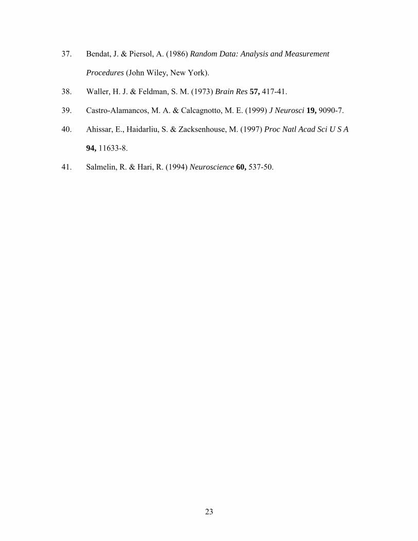
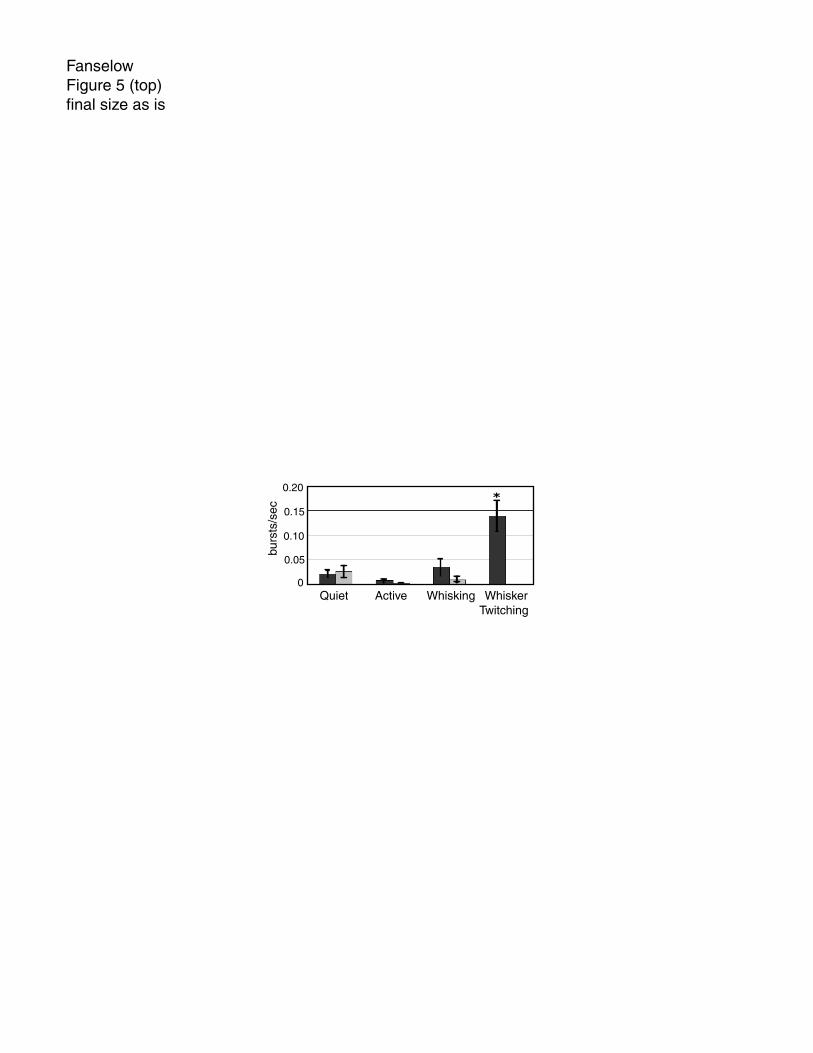
Bursting during different behavioral states: During the whisker twitching behavior robust
oscillations of neuronal activity were observed in VPM and SI. During this oscillatory
activity neurons in VPM and SI frequently fired bursts of action potentials that were
synchronized with the oscillatory activity as well as with the whisker twitching
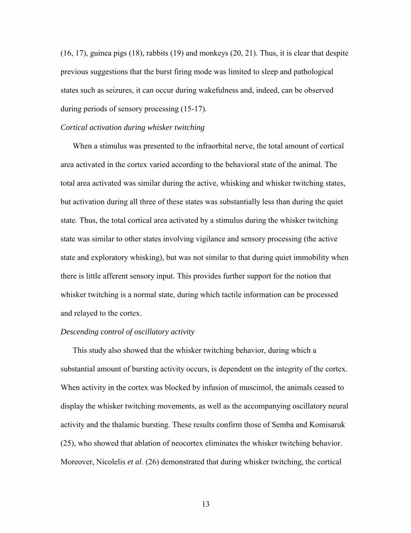
movements (fig. 1a). Note that not every neuron fired with every oscillatory cycle
(fig.1c). In contrast, rhythmic burst activity was not observed during periods when no
whisker twitching and oscillatory activity were present (fig. 1b). We quantified the burst
rate during the four behavioral states we analyzed: quiet immobility, active, exploratory
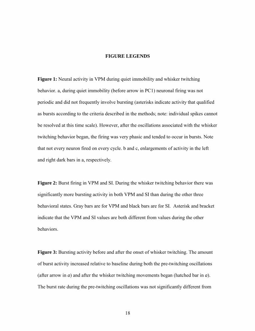
whisking and whisker twitching. The results show that in both VPM and SI, there was
substantially more bursting activity during the whisker twitching behavior (VPM,
0.139±0.032 bursts/sec±SEM; SI, 0.174±0.071; fig. 2) than during any of the other three
10
behaviors (VPM: quiet, 0.022±0.007; active, 0.008±0.03; whisking, 0.035±0.018; SI:
quiet, 0.040±0.018; active, 0.011±0.006; whisking, 0.013±0.006; fig. 2, p < 0.0001).
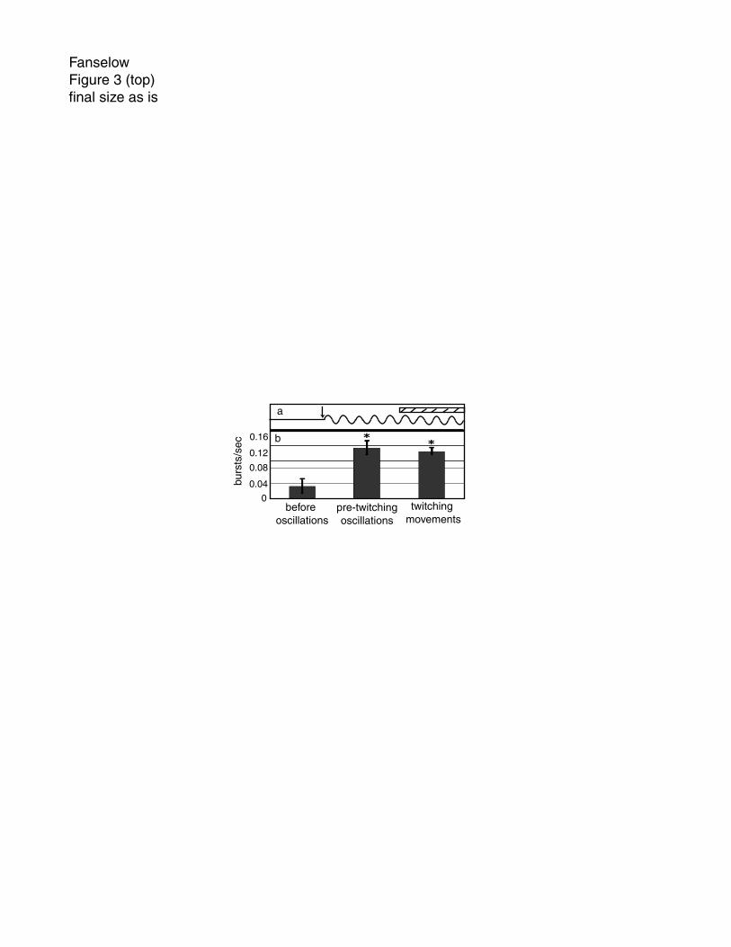
Bursting during the pre-twitching oscillation period: Oscillatory neural activity in VPM
preceded the onset of the small-amplitude whisker twitching movements by an average of
576 ±28ms. We quantified the amount of burst activity during this period, termed the pre-
twitching oscillation period, and found that there was no statistical difference between the
rate of bursting during this period and that during the whisker twitching movements
themselves, during which the oscillatory activity continued (0.134±0.018 bursts/sec,
0.124±0.010, respectively; fig 3b). However, the amount of bursting activity in both of
these cases was significantly different from the level of bursting during baseline periods
when there was no oscillatory activity or whisker twitching movements (0.032±0.019;
p<0.01).
Cumulative cortical area activated: Responses in SI to infraorbital nerve stimulation
differed in two ways according to behavioral state. First, the total amount of cortical area
activated by a single stimulus was much larger during the quiet state than during any of
the other three states (all values normalized to total cumulative area activated for quiet
state for each experiment, i.e. quiet = 100% (active, 53.21±6.94%; whisking,
34.23±10.27%; whisker twitching, 50.79±13.79% ; fig. 4). This activity developed over
the course of ~28ms after the stimulus was presented. Second, the rate at which the
cortical area was activated was fastest during the quiet state (18.60±0.90% of quiet state
max area/sec), intermediate during the active state (12.36±1.12) and slowest during the
whisking and whisker twitching states (5.22±1.81, 6.88±4.24, respectively).
11
Burst activity during inactivation of SI: After SI was inactivated by muscimol infusion,
animals never exhibited the whisker twitching behavior and neither the thalamic
oscillatory activity nor the accompanying burst activity were observed in VPM.
However, during SI inactivation the amount of bursting activity was not altered in VPM
during the quiet, active and whisking behaviors (fig. 5). The whisker twitching behavior
did fully return after the muscimol dose wore off (~8-9 hours).
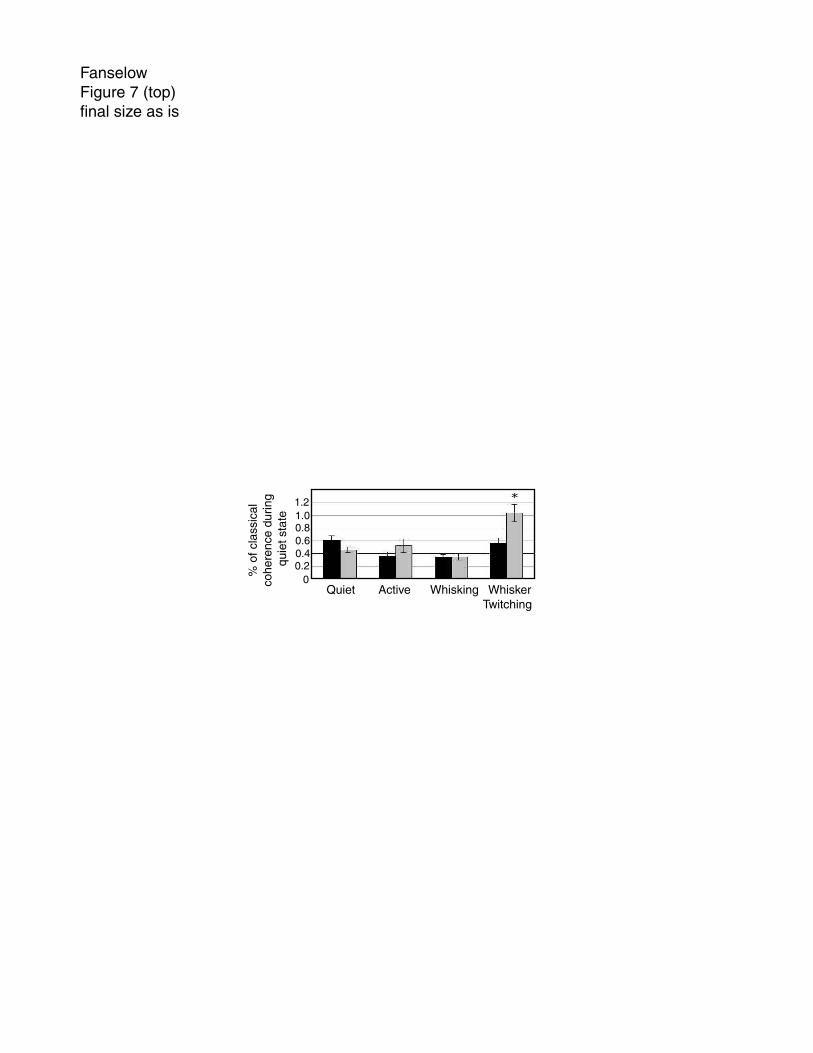
Direction of information flow: We used PDC analysis to determine the direction of
information flow from one region (VPM or SI) to another during the behavioral states we
studied (fig. 6). During the quiet, active and whisking states, there was no statistically
significant difference in the amount of directed coherence from VPM to SI compared to
that from SI to VPM (quiet, 61.18±6.71 % of quiet classical coherence value,
45.92±4.20; active, 52.41±10.44, 35.51±7.53; whisking, 35.06±5.52, 34.95±4.12,
respectively, fig. 7). In addition, there was no statistically significant difference in the
amount of directed coherence between these three behaviors in either direction. In
contrast, during the whisker twitching behavior there was a significantly larger amount of
directed coherence from SI to VPM than from VPM to SI (103.25±13.45, 56.36±8.3
respectively; p<0.03). The amount of directed coherence from VPM to SI during whisker
twitching was indistinguishable from that during the other behaviors.
DISCUSSION
The results of this study demonstrate that thalamocortical neurons can fire in the
bursting mode during the whisker twitching behavior of awake rats. Contrary to what has
12
typically been believed, we showed that the SI cortex does respond to stimuli during this
behavior, demonstrating that the bursting mode is not limited to states in which there is
no relay of sensory information by the thalamus to the cortex. In addition, the whisker
twitching behavior is dependent upon, and is likely initiated by, the cortex. The finding
that there is more descending influence from SI to VPM, as measured by the PDC
technique, during whisker twitching compared to the other behavioral states, as well as
the advantages that burst activity can have for particular types of sensory transmission (7,
8, 17, 38, 39), suggest to us that thalamic bursting activity during the whisker twitching
behavior in awake rats may serve to optimize the detection of afferent stimuli.
The burst firing mode is not limited to sleep and pathological states
In this paper we demonstrated that thalamocortical cells are capable of firing high-
frequency bursts of action potentials during whisker twitching, a normal, waking
behavioral state in the rat. During this state, which is accompanied by µ-oscillations
throughout the brainstem, thalamic and cortical levels of the vibrissal system (26), bursts
occur substantially more frequently than during any other behavioral state analyzed.
Previously, Fanselow and Nicolelis (23) showed that there are responses to tactile stimuli
during the whisker twitching behavior in both VPM and SI. Therefore, our results
provide a further challenge to the notion that thalamic bursting activity is associated only
with states during which there is no relay of sensory information by the thalamus to the
cortex.
The bursts of action potentials described here were similar to those observed both in
vitro (4, 14); [for review see Steriade et al. 1990 (5)] and in vivo (1, 3). These results
corroborate other studies that have shown bursting activity during wakefulness in cats ,
13
(16, 17), guinea pigs (18), rabbits (19) and monkeys (20, 21). Thus, it is clear that despite
previous suggestions that the burst firing mode was limited to sleep and pathological
states such as seizures, it can occur during wakefulness and, indeed, can be observed
during periods of sensory processing (15-17).
Cortical activation during whisker twitching
When a stimulus was presented to the infraorbital nerve, the total amount of cortical
area activated in the cortex varied according to the behavioral state of the animal. The
total area activated was similar during the active, whisking and whisker twitching states,
but activation during all three of these states was substantially less than during the quiet
state. Thus, the total cortical area activated by a stimulus during the whisker twitching
state was similar to other states involving vigilance and sensory processing (the active
state and exploratory whisking), but was not similar to that during quiet immobility when
there is little afferent sensory input. This provides further support for the notion that
whisker twitching is a normal state, during which tactile information can be processed
and relayed to the cortex.
Descending control of oscillatory activity
This study also showed that the whisker twitching behavior, during which a
substantial amount of bursting activity occurs, is dependent on the integrity of the cortex.
When activity in the cortex was blocked by infusion of muscimol, the animals ceased to
display the whisker twitching movements, as well as the accompanying oscillatory neural
activity and the thalamic bursting. These results confirm those of Semba and Komisaruk
(25), who showed that ablation of neocortex eliminates the whisker twitching behavior.
Moreover, Nicolelis et al. (26) demonstrated that during whisker twitching, the cortical

14
oscillations phase-led those of the thalamus by an average of 9.19 ms. Furthermore, in
that study, the oscillatory activity in the cortex began prior to that in the thalamus in
~80% of whisker twitching episodes.
In addition, by using the statistical technique of PDC, we demonstrated here that
during the whisker twitching behavior there was ~2 times the amount of directed
coherence from SI to VPM than in any other behavioral state analyzed. The implication
of the PDC analysis technique is that when there is partial directed coherence from one
structure to another, activity in the first structure is likely to be influencing activity in the
second structure. Thus, our results suggest that during the whisker twitching state, there is
a large degree of descending influence from SI to VPM, compared with the other
behavioral states. Taken together, these findings suggest that the cortex plays a much
bigger role in driving activity in the thalamus during whisker twitching than it does
during other sates.
Function of the bursting mode and whisker-twitching behavior
Based on the results presented in this paper and other studies discussed here, we
postulate that the thalamic bursting mode is not associated only with situations during
which there is little relay of sensory information through the thalamus. Instead, we
propose that during the whisker twitching mode, the vibrissal system is actually primed to
detect incoming stimuli and that the thalamus is quite capable of transmitting sensory
information during this time. In the rat whisker system this “priming” signal would occur
at approximately the same frequency of the main exploratory behavior, exploratory
whisking, used by the animal to sample tactile information in the surrounding
environment.
15
There are several reasons why bursting activity could be advantageous for signal
detection. First, it has been demonstrated that the probability of firing of VP neurons in
the cat in response to stimulation of the medial lemniscus differs depending on the phase
of the 10-15Hz wave activity recorded in the thalamus (38). Thus, one function of the
rhythmic bursts could be to provide cyclic periods of heightened sensitivity to incoming
stimulation (24). This is particularly relevant to the rat vibrissal system in which the rate
of whisker movements during exploratory whisking can range from 4-10Hz. According
to this view, the thalamocortical loop may be tuned to process afferent activity at
approximately this rate . In support of this hypothesis, Ahissar and colleagues have
demonstrated that cortical neurons in the rat somatosensory system can entrain their
oscillatory firing to the frequency of peripheral whisker stimulation (40). In this way, the
whisker twitching state may function to prepare the vibrissal system to optimally detect
and process incoming tactile information that is sampled in a rhythmic fashion, such as
occurs during whisking. Thus, the finding that thalamic bursting does occur during
epochs of whisker twitching movements suggests that the thalamocortical loop is primed
prior to the initiation of whisker movements for better detection of potential incoming
whisker stimulation.
Second, in the rat thalamocortical loop, 10Hz stimulation has been shown to elicit
long-term potentiation in corticothalamic synapses (39). Thus, oscillatory activity
originating in the SI cortex and whose frequency range is centered near 10Hz, such as
that during the whisker twitching behavior, could enhance the ability of VPM neurons to
respond to trains of tactile stimuli conveyed by the ascending trigeminal lemniscal
pathways.
16
Finally, a burst of action potentials is very effective at activating neurons and
therefore provides a very robust signal to its downstream target (17). In this way, while
trains of single action potentials may be capable of closely following the details of a
stimulus in a linear fashion, the neuronal bursts may be an effective way to alert the
thalamocortical loop to the presence of a previously undetected or unattended stimulus
(8, 17). For example, Guido and Weyand (17) showed in the lateral geniculate nucleus
(LGN) of awake, behaving cats that bursting activity increased during the first cycle of
stimulation with a drifting grating, but then shifted into a tonic firing mode as
presentation of the stimulus continued. This suggests that initial detection of the stimulus
involved burst activity, whereas subsequent processing utilized the more detail-sensitive
tonic firing. In addition, Sherman and Guillery (7) reported that responses of cat LGN
cells to a drifting grating vary depending on the level of depolarization of a cell, and thus,
its firing mode. When a cell was sufficiently depolarized (-65mV), it fired in tonic mode
and its output closely followed the contour of the grating. However, if the cell was
sufficiently hyperpolarized such that It was inactivated (-75mV), the cell fired in burst
mode and gave strong, non-linear responses to the grating. Because these bursts are
superimposed on the relatively low level of background activity that is characteristic of
the bursting mode, they result in a higher signal-to-noise ratio than during the tonic mode
(7). Thus, bursting activity may serve to optimize the initial detection of a stimulus. This
effect may be even stronger in the somatosensory thalamus, where the bursting activity
has been shown to be higher than occurs in the LGN (21).
For the reasons outlined above, we believe that thalamic burst firing may endow
the rat vibrissal system with a very effective signal detection mode that allows it to detect

17
weak, new, and potentially important, tactile information. It has been suggested
previously that both bursting activity in the visual system (7, 8, 17) and the µ-oscillations
observed in humans (41) and rats (24) serve as preparatory states, priming a given
sensory system for signal detection. The results presented in the current paper corroborate
this idea. Taken together, these findings support the postulate that the nervous system is
not a passive detector of afferent stimuli, but that instead it plays an active role in
optimizing the detection of potential incoming sensory information. Thus, by utilizing
thalamic bursting, the rat vibrissal system can prepare itself, prior to the activation of any
peripheral receptors, to rapidly detect the onset of tactile input, which it can subsequently
process in more detail using the tonic firing mode by shifting into an active exploratory
behavior.
18
FIGURE LEGENDS
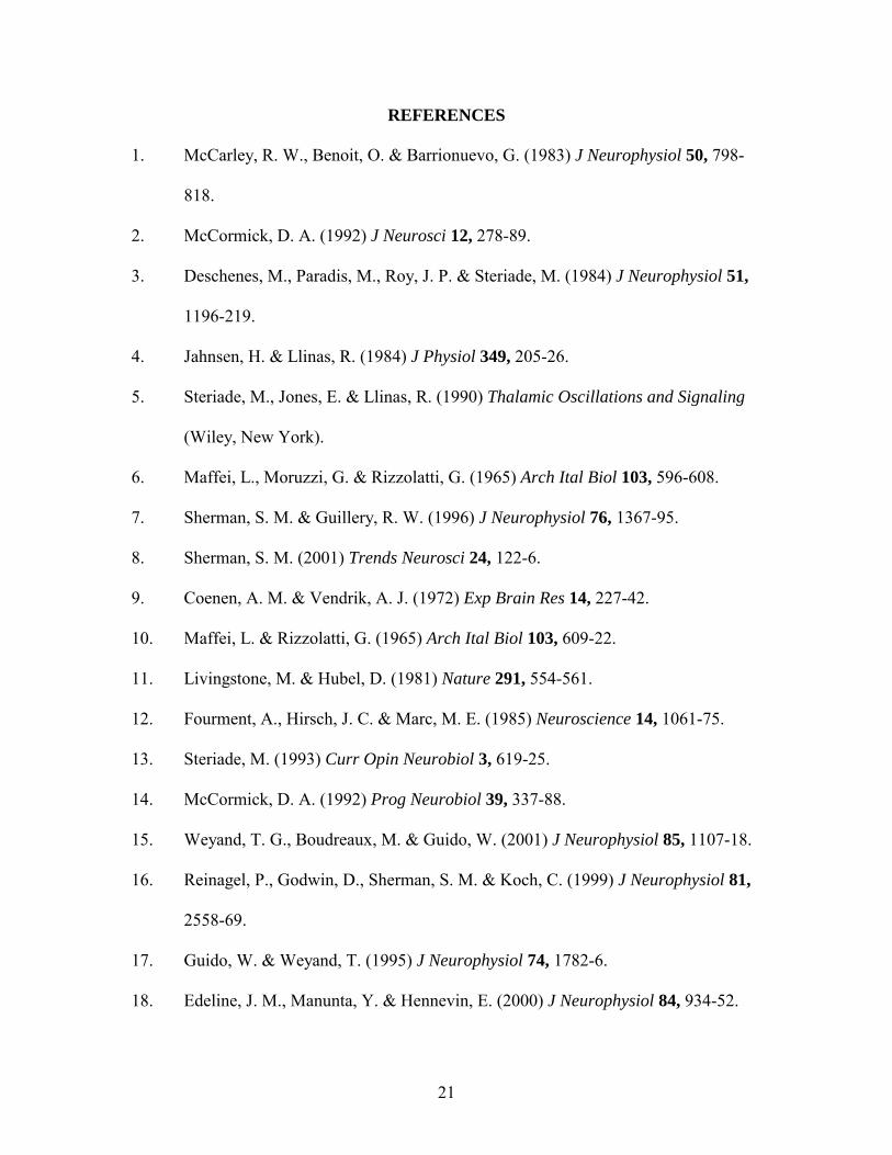
Figure 1: Neural activity in VPM during quiet immobility and whisker twitching
behavior. a, during quiet immobility (before arrow in PC1) neuronal firing was not
periodic and did not frequently involve bursting (asterisks indicate activity that qualified
as bursts according to the criteria described in the methods; note: individual spikes cannot
be resolved at this time scale). However, after the oscillations associated with the whisker
twitching behavior began, the firing was very phasic and tended to occur in bursts. Note
that not every neuron fired on every cycle. b and c, enlargements of activity in the left
and right dark bars in a, respectively.
Figure 2: Burst firing in VPM and SI. During the whisker twitching behavior there was
significantly more bursting activity in both VPM and SI than during the other three
behavioral states. Gray bars are for VPM and black bars are for SI. Asterisk and bracket
indicate that the VPM and SI values are both different from values during the other
behaviors.
Figure 3: Bursting activity before and after the onset of whisker twitching. The amount
of burst activity increased relative to baseline during both the pre-twitching oscillations
(after arrow in a) and after the whisker twitching movements began (hatched bar in a).
The burst rate during the pre-twitching oscillations was not significantly different from

19
that after the whisker twitching movements began. Asterisks in b indicate values
significantly different from the ‘before oscillations’ condition.
Figure 4: Cumulative sum of activated cortical area. The total amount of cortical area
activated in the first 28ms following stimulation of the infraorbital nerve was
substantially larger during the quiet behavior than during the other three behavioral
states. In addition, during the quiet behavior, the amount of cortical area activated
increased at a higher rate. Values are presented as a percent of cumulative area activated
during the quiet behavior.
Figure 5: Burst firing in VPM before and after inactivation of SI. When SI was intact,
there was a large amount of bursting during the whisker twitching state. However, after
SI was inactivated by muscimol infusion, the animal did not display any whisker
twitching behavior. Bursting activity during the other three behavioral states was not
affected by muscimol infusion. Black bars show pre-muscimol and gray bars show post-
muscimol values. Asterisk indicates value is significantly different from all others.
Figure 6: Partial Directed Coherence during different behaviors. The traces in this figure
are from 250sec of a recording that includes periods of whisker twitching behavior. a,
Bars indicate times when the rat was engaged in the whisker twitching behavior. b, Raster
plots showing spike times for three single-neuron recordings. c, PC1 of activity in VPM.
d, PDC from SI to VPM. e, PDC from VPM to SI. f, classical coherence between VPM

20
and SI. Grayscale bar indicates fraction of total power at each frequency for a given
source (VPM or SI).
Figure 7: During the whisker twitching state there was a significantly larger amount of
directed coherence from SI to VPM than from VPM to SI (asterisk). In addition, the
amount of directed coherence between SI and VPM during whisker twitching was larger
than during any other behavioral state. Black bars show directed coherence from VPM to
SI, and gray bars show directed coherence from SI to VPM. Values were normalized to
the amount of classical coherence for each measure.
21
REFERENCES
1. McCarley, R. W., Benoit, O. & Barrionuevo, G. (1983) J Neurophysiol 50, 798-
818.
2. McCormick, D. A. (1992) J Neurosci 12, 278-89.
3. Deschenes, M., Paradis, M., Roy, J. P. & Steriade, M. (1984) J Neurophysiol 51,
1196-219.
4. Jahnsen, H. & Llinas, R. (1984) J Physiol 349, 205-26.
5. Steriade, M., Jones, E. & Llinas, R. (1990) Thalamic Oscillations and Signaling
(Wiley, New York).
6. Maffei, L., Moruzzi, G. & Rizzolatti, G. (1965) Arch Ital Biol 103, 596-608.
7. Sherman, S. M. & Guillery, R. W. (1996) J Neurophysiol 76, 1367-95.
8. Sherman, S. M. (2001) Trends Neurosci 24, 122-6.
9. Coenen, A. M. & Vendrik, A. J. (1972) Exp Brain Res 14, 227-42.
10. Maffei, L. & Rizzolatti, G. (1965) Arch Ital Biol 103, 609-22.
11. Livingstone, M. & Hubel, D. (1981) Nature 291, 554-561.
12. Fourment, A., Hirsch, J. C. & Marc, M. E. (1985) Neuroscience 14, 1061-75.
13. Steriade, M. (1993) Curr Opin Neurobiol 3, 619-25.
14. McCormick, D. A. (1992) Prog Neurobiol 39, 337-88.
15. Weyand, T. G., Boudreaux, M. & Guido, W. (2001) J Neurophysiol 85, 1107-18.
16. Reinagel, P., Godwin, D., Sherman, S. M. & Koch, C. (1999) J Neurophysiol 81,
2558-69.
17. Guido, W. & Weyand, T. (1995) J Neurophysiol 74, 1782-6.
18. Edeline, J. M., Manunta, Y. & Hennevin, E. (2000) J Neurophysiol 84, 934-52.

22
19. Swadlow, H. A. & Gusev, A. G. (2001) Nat Neurosci 4, 402-8.
20. Ramcharan, E. J., Cox, C. L., Zhan, X. J., Sherman, S. M. & Gnadt, J. W. (2000)
J Neurophysiol 84, 1982-7.
21. Ramcharan, E. J., Gnadt, J. W. & Sherman, S. M. (2000) Vis Neurosci 17, 55-62.
22. Guido, W., Lu, S. M. & Sherman, S. M. (1992) J Neurophysiol 68, 2199-211.
23. Fanselow, E. E. & Nicolelis, M. A. (1999) J Neurosci 19, 7603-16.
24. Semba, K., Szechtman, H. & Komisaruk, B. R. (1980) Brain Res 195, 281-98.
25. Semba, K. & Komisaruk, B. R. (1984) Neuroscience 12, 761-74.
26. Nicolelis, M. A., Baccala, L. A., Lin, R. C. & Chapin, J. K. (1995) Science 268,
1353-8.
27. Gastout, H. (1952) Rev. Neurol. 87, 176-182.
28. Braizer, M. (1963) EEG and Clin. Neurophys. 15, 287-298.
29. Nicolelis, M. A., Ghazanfar, A. A., Faggin, B. M., Votaw, S. & Oliveira, L. M.
(1997) Neuron 18, 529-37.
30. Paxinos, G. & Watson, C. (1986) The Rat Brain (Academic Press, Harcourt,
Brace, Jovanovich, San Diego).
31. Martin, J. H. (1991) Neurosci Lett 127, 160-4.
32. Krupa, D. J. & Thompson, R. F. (1997) Learn Mem 3, 545-56.
33. Sameshima, K. & Baccala, L. A. (1999) J Neurosci Methods 94, 93-103.
34. Baccala, L. & Sameshima, K. (2001) Progress in Brain Research 130.
35. Baccala, L. & Sameshima, K. (2001) Biol. Cybernetics 84, 463-474.
36. French, A. S. & Holden, A. V. (1971) Kybernetik 8, 165-71.
23
37. Bendat, J. & Piersol, A. (1986) Random Data: Analysis and Measurement
Procedures (John Wiley, New York).
38. Waller, H. J. & Feldman, S. M. (1973) Brain Res 57, 417-41.
39. Castro-Alamancos, M. A. & Calcagnotto, M. E. (1999) J Neurosci 19, 9090-7.
40. Ahissar, E., Haidarliu, S. & Zacksenhouse, M. (1997) Proc Natl Acad Sci U S A
94, 11633-8.
41. Salmelin, R. & Hari, R. (1994) Neuroscience 60, 537-50.
PC1
VP
M n
euro
ns
1
2
3
4
5
a
b
PC1
VP
M n
euro
ns 12
345
c
PC1
VP
M n
euro
ns 12
345
500ms
* * ** * ** ** * * * ** **
* * * ** ** * * * ** **** *** * ** ** ***
* ** ***** ** *
* * * * * * * ** * * * * *
*******************
**
* * *
* * * ** * *
* * * * * **
0 2 4 6 8 10 sec
500ms
Fanselow Figure 1 (top)final size as is
0.25
0
0.05
0.10
0.15
0.20
Quiet Active Whisking Whisker Twitching
* }
burs
ts/s
ec
Fanselow Figure 2 (top)final size as is
before oscillations
pre-twitchingoscillations
twitching movements
0
0.04
0.08
0.12
0.16 **
burs
ts/s
ec
a
b
Fanselow Figure 3 (top)final size as is

bin in ms
activ
ated
are
a/m
ax a
rea
durin
g qu
iet b
ehav
ior
2 6 10 14 18 22 26 0
20
40
60
80
100 quietactive
whiskingwhiskertwitching
Fanselow Figure 4 (top)final size as is
Quiet Active Whisking Whisker Twitching
0
0.05
0.10
0.15
0.20*
burs
ts/s
ec
Fanselow Figure 5 (top)final size as is

20 sec
a
b
c
d
e
f
whisker twitching behavior
single neuronrasters
PC1
SI to VPM
VPM to SI
classicalcoherence
50
0Hz
50
0
Hz
50
0
Hz
0
1.0
Fanselow Figure 6 (top)final size as is
Quiet Active Whisking Whisker Twitching
% o
f cla
ssic
al
cohe
renc
e du
ring
quie
t sta
te 1.01.2
0.80.60.40.2
0
*
Fanselow Figure 7 (top)final size as is