TGF beta in murine morphogenetic processes: the early embryo
14
Development 108, 645-656 (1990) Printed in Great Britain © T h e Company of Biologists Limited 1990 645 TGF beta in murine morphogenetic processes: the early embryo and cardiogenesis ROSEMARY J. AKHURST 1 *, SIGRID A. LEHNERT 1 , ANDREAS FAISSNER 2 and ELIZABETH DUFFIE 1 1 Duncan Guthrie Institute of Medical Genetics, University of Glasgow, Yorkhill, Glasgow G3 8SJ, UK 2 Department of Neurobiology, Im Neuenheimer Feld 364, D-6900 Heidelberg, FRG *To whom correspondence should be addressed Summary The tissue distribution of TGF beta-1 RNA was exam- ined within whole mouse embryos from implantation to 10.5 days gestational age and, in the developing heart, up to 8 days postpartum. The earliest high level ex- pression of TGF beta-1 RNA is at 7.0 days postcoUum ip.c.) in the cardiac mesoderm. At 8.0 days gestational age, cardiac TGF beta-1 RNA expression is limited to endocardia! cells. By 9.5 days p.c, this expression pattern becomes regionalised to those cells that overlie cardiac cushion tissue. High TGF beta-1 RNA levels continue to persist in endothelial cells of the heart valves until approximately one week postpartum. The TGF beta-1 RNA distribution was compared with the extracellular distributions of polypeptides for TGF beta and Jl/tenascin. As previously reported, endo- thelial expression of TGF beta-1 RNA is correlated with mesenchymal expression of TGF beta polypeptide, suggesting a paracrine mode of action for this growth factor in cardiac development. Minor discrepancies in the distributions of TGF beta-1 RNA and the extracellu- lar form of the TGF beta polypeptide suggest that translational or post-translational control of protein levels occurs and/or the possibility that the antibody used may also recognise other members of the TGF beta polypeptide family. A correlation between endothelial TGF beta-1 ex- pression and distribution of Jl/tenascin in the mesen- chyme gives further support to the proposition that the biological effects of TGF beta-1 may, in part, be mediated by Jl/tenascin. Key words: TGF beta-1, Jl/tenascin, in situ hybridisation, epithelial-mesenchymal interaction, epithelial-mesenchymal transformation, mouse embryo, cardiogenesis. Introduction It is becoming increasingly clear that polypeptide growth factors play a central role in many embryonic processes. Not only are these molecules and their receptors present within the early embryo in vivo (Twardzik et al. 1982; Heine et al. 1987; Rappolee et al. 1988), but they initiate and/or modulate a variety of cellular events which are essential to the development of the organism (Sporn et al. 1986; Sporn and Roberts, 1988; Roberts and Sporn, 1989). This is exemplified well by the family of growth factors related to trans- forming growth factor beta-1, which itself has pre- viously been termed a 'panregulin' by Sporn et al. (1986). Studies of TGF beta action on cells in vitro have been invaluable to our understanding of the role that these molecules might play in biological processes in vivo, though the interpretation of results has been con- founded by the multifunctional nature of this growth factor (Sporn et al. 1986; Roberts and Sporn, 1989) and the multiplicity of closely related molecules within this family (ten-Dijke et al. 1988; Jakowlew et al. 1988a, b; Kondaiah et al. 1989). It is clear that the biological effects of TGF beta are dependent on the presence of other growth factors, either acting synergystically (Kimelman and Kirschner, 1987) or antagonistically (Baird and Durkin, 1986; Jennings et al. 1988); they are also dependent on the cell type and the nature of the culture system. This is particularly well demonstrated by the effects of TGF beta-1 on endothelial cells in vitro, which can modulate cellular growth, movement, adhesiveness, invasiveness and/or three-dimensional morphology, dependent on the endothelial cell source and the exact culture system (Heimark et al. 1986; Sato and Rifkin, 1989; Gamble and Vadas, 1988; Potts and Runyan, 1989; Madri et al. 1988). For a full understanding of in vivo function, it is also necessary to determine the sites of synthesis and action of individual growth factors during embryological de-
Transcript of TGF beta in murine morphogenetic processes: the early embryo

Development 108, 645-656 (1990)Printed in Great Britain © T h e Company of Biologists Limited 1990
645
TGF beta in murine morphogenetic processes: the early embryo and
cardiogenesis
ROSEMARY J. AKHURST1*, SIGRID A. LEHNERT1, ANDREAS FAISSNER2 and ELIZABETH
DUFFIE1
1 Duncan Guthrie Institute of Medical Genetics, University of Glasgow, Yorkhill, Glasgow G3 8SJ, UK2Department of Neurobiology, Im Neuenheimer Feld 364, D-6900 Heidelberg, FRG
*To whom correspondence should be addressed
Summary
The tissue distribution of TGF beta-1 RNA was exam-ined within whole mouse embryos from implantation to10.5 days gestational age and, in the developing heart,up to 8 days postpartum. The earliest high level ex-pression of TGF beta-1 RNA is at 7.0 days postcoUumip.c.) in the cardiac mesoderm. At 8.0 days gestationalage, cardiac TGF beta-1 RNA expression is limited toendocardia! cells. By 9.5 days p.c, this expressionpattern becomes regionalised to those cells that overliecardiac cushion tissue. High TGF beta-1 RNA levelscontinue to persist in endothelial cells of the heart valvesuntil approximately one week postpartum.
The TGF beta-1 RNA distribution was compared withthe extracellular distributions of polypeptides for TGFbeta and Jl/tenascin. As previously reported, endo-thelial expression of TGF beta-1 RNA is correlated withmesenchymal expression of TGF beta polypeptide,suggesting a paracrine mode of action for this growth
factor in cardiac development. Minor discrepancies inthe distributions of TGF beta-1 RNA and the extracellu-lar form of the TGF beta polypeptide suggest thattranslational or post-translational control of proteinlevels occurs and/or the possibility that the antibodyused may also recognise other members of the TGF betapolypeptide family.
A correlation between endothelial TGF beta-1 ex-pression and distribution of Jl/tenascin in the mesen-chyme gives further support to the proposition that thebiological effects of TGF beta-1 may, in part, bemediated by Jl/tenascin.
Key words: TGF beta-1, Jl/tenascin, in situ hybridisation,epithelial-mesenchymal interaction,epithelial-mesenchymal transformation, mouse embryo,cardiogenesis.
Introduction
It is becoming increasingly clear that polypeptidegrowth factors play a central role in many embryonicprocesses. Not only are these molecules and theirreceptors present within the early embryo in vivo(Twardzik et al. 1982; Heine et al. 1987; Rappolee et al.1988), but they initiate and/or modulate a variety ofcellular events which are essential to the developmentof the organism (Sporn et al. 1986; Sporn and Roberts,1988; Roberts and Sporn, 1989). This is exemplifiedwell by the family of growth factors related to trans-forming growth factor beta-1, which itself has pre-viously been termed a 'panregulin' by Sporn et al.(1986).
Studies of TGF beta action on cells in vitro have beeninvaluable to our understanding of the role that thesemolecules might play in biological processes in vivo,though the interpretation of results has been con-founded by the multifunctional nature of this growth
factor (Sporn et al. 1986; Roberts and Sporn, 1989) andthe multiplicity of closely related molecules within thisfamily (ten-Dijke et al. 1988; Jakowlew et al. 1988a,b;Kondaiah et al. 1989). It is clear that the biologicaleffects of TGF beta are dependent on the presence ofother growth factors, either acting synergystically(Kimelman and Kirschner, 1987) or antagonistically(Baird and Durkin, 1986; Jennings et al. 1988); they arealso dependent on the cell type and the nature of theculture system. This is particularly well demonstratedby the effects of TGF beta-1 on endothelial cells invitro, which can modulate cellular growth, movement,adhesiveness, invasiveness and/or three-dimensionalmorphology, dependent on the endothelial cell sourceand the exact culture system (Heimark et al. 1986; Satoand Rifkin, 1989; Gamble and Vadas, 1988; Potts andRunyan, 1989; Madri et al. 1988).
For a full understanding of in vivo function, it is alsonecessary to determine the sites of synthesis and actionof individual growth factors during embryological de-

646 R. J. Akhurst and others
velopment. TGF beta-1 and beta-2, for example, areinterchangeable in most biological activity assays,though they clearly have different temporal and spatialprofiles of gene expression during murine embryogen-esis (compare Lehnert and Akhurst, (1988) and Peltonet al. (1989)), suggesting different in vivo functions.Recent reports confirm that, indeed, TGF beta-1 and -2do show some specificity in biological action (Jenningsetal. 1988; Ohta et al. 1987).
In an earlier study, we examined the global ex-pression pattern of the gene encoding TGF beta-1 in themid-gestation murine embryo (Lehnert and Akhurst,1988). A novel finding of this study was that the TGFbeta-1 gene is transiently expressed in epithelial cellswhich are involved in morphogenetic interactions incases where TGF beta protein is localised in theunderlying mesenchyme (Heine et al. 1987). A reason-able inference from this observation is that a majorfunction of epitheliaUy derived TGF beta-1 is as aparacrine modulator of extracellular matrix (ECM)deposition in the underlying mesenchyme. This growthfactor is known to increase accumulation of ECMmaterial by increasing production of both matrix pro-teins and inhibitors of extracellular proteases, whichprevent ECM degradation. It also inhibits the pro-duction of matrix-degradation enzymes (Roberts et al.1986; Ignotz and Massague, 1986) and the responsive-ness of cells to the ECM is increased by stimulation ofthe production of cell surface receptors for matrixmolecules (Ignotz and Massague, 1987).
Extracellular proteins that have elevated synthesisrates include not only collagen I and fibronectin butmore specialised molecules such as thrombospondin(Penttinen etal. 1988), osteopontin (Noda etal. 1988),tenascin (Pearson et al. 1988), osteonectin/SPARC(Noda and Rodan, 1987) and chondroitin/dermatansulphate proteoglycans (Hiraki et al. 1988). The embry-onic distribution of tenascin is of particular significancein this context since this is relatively restricted in theembryo compared with other ECM molecules and itsdistribution is almost completely correlated with thepresence of TGF beta-1 RNA (Chiquet-Ehrismann etal. 1986; Lehnert and Akhurst, 1988). Furthermore,TGF beta is known to transcriptionally activate thetenascin gene (Pearson et al. 1988) and, in an in vitromodel, epitheliaUy derived TGF beta-1 can inducesynthesis of tenascin by the underlying stroma (Chi-quet-Ehrismann et al. 1989). Tenascin could thereforemediate some of the effects of TGF beta in embryogen-esis.
In our previous report, TGF beta-1 RNA was foundspecifically in the epithelial component of the heartvalves at 12.5 days post coitum (p.c.) suggesting thatTGF beta-1 is a modulator of septation and valveformation (Lehnert and Akhurst, 1988). In this paper,we have extended our study of embryonic TGF beta-1gene expression to include stages from implantation to10.5 days gestation. We have also specifically examinedlater stages of cardiac development, in particular withrespect to accumulation of tenascin in the extracellularmatrix.
Progenitor cells of the mammalian heart, the cardiacmesoderm cells, appear very early in embryogenesis,around gastrulation. These progenitors are thought toarise through induction by the underlying pharyngealendoderm (Jacobson and Sater, 1988). Splanchno-pleuric mesoderm gives rise to two primitive tubes, theendocardium and the myocardial mantle, the latterencapsulating the former. Initially, the endotheliumand myocardium are separated by an extensive acellularbasement membrane, the cardiac jelly. Early eventsleading to septation and formation of prevalvular mes-enchyme include a regional differentiation of the endo-cardium. Endothelial cells within the region of theatrioventricular (AV) canal and outflow tract becomecapable of mesenchymal transformation and migrateinto the underlying cardiac jelly populating the mesen-chyme of the cardiac cushion tissue (Manasek, 1976).
The process of epithelial-mesenchymal transform-ation of endocardial cells has been extensively studiedby Markwald and others in an in vitro collagen gelsystem. It involves an inductive event emanating fromthe myocardium. There is both regional specificity inthe ability of the myocardium to produce such a signaland in the competence of the endocardium to respond(Krug et al. 1985; Mjaatvedt et al. 1987). Mjaatvedt andMarkwald (1989) have recently isolated a particulatefraction that is capable of 'activating' the AV endo-thelium to undergo this transformation event in vitro.
The possibility that TGF beta may contribute toepithelial-mesenchymal cell transformation wasdemonstrated by Potts and Runyan (1989) who showedthat, in combination with chick ventricular myocar-dium, TGF beta could initiate this event in atrioven-tricular endothelium. Furthermore, in vitro this processwas inhibited by antibodies that block TGF beta ac-tivity. We will discuss our results on the distribution ofTGF beta-1 and tenascin in the light of these obser-vations.
Materials and methods
Mouse stocksAll embryos and neonates were obtained from Parkes femalesmated with NIH males (Olac). Noon on the day of thecopulation plug was 0.5 days post coitum (p.c). All tissue wasfixed overnight in ice-cold 4% paraformaldehyde in phos-phate-buffered saline, then dehydrated and embedded inparaffin.
Probe synthesisA full-length cDNA encoding murine TGF beta-1 (Derynck etal. 1986) was kindly provided by Dr R. Derynck (Genentech).To eliminate the possibility that this probe could cross-reactwith other members of the TGF beta gene family, a 600nucleotide Apal-Kpnl fragment corresponding to the precur-sor region of the TGF beta-1 protein (amino acid residues68-268) was subcloned into the riboprobe vector Bluescribe(Vector Cloning Systems). This removes the DNA codingsequences encoding the region of amino acid terminal hom-ology seen between TGF beta proteins (amino acids 1-30)and the conserved mature 112 amino acid portion of the TGFbeta-1 polypeptide. The resultant probe shows greater than

TGF beta in murine morphogenesis
50% nucleotide sequence divergence from other TGF betagenes.
35S-labelled sense and antisense riboprobes were generatedto a specific activity of 109disintsmin~' jtg~' using T3 or T7polymerase. Probes were digested to an average length of 100nucleotides by controlled alkaline hydrolysis (Cox et al. 1984)and used at a final concentration of 30pg^l~' in hybridis-ations.
In situ hybridisationIn situ hybridisation to 7/im tissue sections was performedessentially as described by Wilkinson et al. (1987) using 60%formamide in the hybridisation mix and a hybridisationtemperature of 52°C. Exposure times (llford K5 emulsion)were between 3 and 15 days. After development, slides werestained in haemotoxylin and mounted. Photomicrography wasperformed using an Olympus BH-2 microscope and eitherKodak Panatomic-X or Ektachrome EPY-50 film.
ImmunohistochemistryPolyclonal rabbit antibodies included anti-mouse fibronectin(Biogenesis Ltd); anti-alpha 1 chymotrypsin (Dakopatts);anti-TGF beta, CC, kindly supplied by Dr K. Flanders (Heineetal. 1987); and anti Jl-tenascin, KAF9 and KAF10 (Steindleretal. 1989).
Antigens were localised in 7jim tissue sections using anavidin-biotin-peroxidase system (ABC System, Dakopatts).For TGF beta detection, deparaffinised sections were pre-fixed in Bouin's fixative for 30min at room temperature,followed by extensive washing in 70% ethanol. (This was notnecessary for unmasking the epitopes of the other antigens).After blocking endogenous peroxidase in hydrogen peroxide/methanol, sections were permeabilised with O.lmgml"1
hyaluronidase in 0.1 M sodium acetate, 0.15 M sodium chloridepH5.5 for 30min at 37°C. Non-specific antibody binding wasblocked with donkey serum (0.3%), mouse serum (0.3%),gelatin (0.1%) and BSA (0.1%) in phosphate-bufferedsaline.
Primary antibody was applied at 20^gml~' in the blockingcocktail, overnight at 4°C. The secondary antibody wasbiotinylated donkey anti-rabbit (Amersham) and the Dako-patts ABC system protocol was followed using diamino-benzidine as the chromogen. Sections were lightly stained inhaemotoxylin before examination using an Olympus BH-2microscope.
In situ hybridisation and immunohistochemistry were per-formed on adjacent serial sections in order to compare therelative distribution of TGF beta-1 transcripts with that ofTGF beta and tenascin polypeptides.
Results
In our previous in situ hybridisation study of TGF beta-1 RNA distribution within the midgestation murineembryo, a full-length cDNA probe against TGF beta-1was employed (Lehnert and Akhurst, 1988). This intro-duced the possibility that our data may not representTGF beta-1 transcript distribution specifically but cross-reaction to RNA of other TGF beta family members.We therefore subcloned a 600 nucleotide fragment thatwas known to be specific to the TGF beta-1 gene.Repetition of the original in situ hybridisation studyusing this probe produced identical results (data notshown) confirming that our original report representedhybridisation to TGF beta-1 transcripts.
Extraembryonic expression of TGF beta-1The decidual swellings of pregnant mice were dissectedfrom the uterus at 6.0 to 9.5 days p. c. 7 jan sections cuttransversely with respect to the uterus were subjected toin situ hybridisation using the anti-sense and sense TGFbeta-1 specific probes. The maternal deciduum con-tained abundant RNA transcripts encoding TGF beta-1. This was most clear in the decidua capsularis at 7.5days p.c. with a gradient of TGF beta-1 RNA concen-tration across the deciduum, being highest near theuterine wall (Fig. 1A,B). By 9.5 days p .c , TGF beta-1RNA was seen in the decidua basalis, which hasdeveloped large lacunae and sinusoids filled with ma-ternal blood. Hybridisation was most intense in the cellslining the lacunae. The chorion also showed a signal(Fig. 1D,E).
The first appearance of detectable TGF beta-1 RNAin tissues derived from the zygote appears in theextraembryonic blood islands of the yolk sac at 7.0 daysp.c. These cell clusters arise from mesodermal hae-mangioblasts, the progenitors of both haemopoieticstem cells and endothelial cells, which contribute to theextraembryonic vasculature (Pardanaud et al. 1989). At7.5 days p.c., the in situ hybridisation signal was evenlydistributed over all cells of the blood island(Fig. 1G,H). The resolution of the technique did notallow us to determine whether the signal emanatedfrom the haemopoietic or endothelial cells. However,since in later development, both haemopoietic cells andembryonic endothelial cells express TGF beta-1, wewould predict that both cell types express this gene inthe blood islands.
By 9.5 days p.c, clusters of cells that appear pro-erythroid in morphology appear in the blood islands asintense foci of hybridisation superimposed upon thehybridisation signal seen at 7.5 days (Fig. II). Thiswould agree with the observations of Wilcox andDerynck (1988) who identified these TGF beta-1-expressing cells as proerythroid in nature. We have alsoobserved hybridisation to erythroid progenitors withinthe fetal liver at 12.5 daysp.c. (data not shown), thoughby 14.5 days, the predominant liver cell type expressingabundant TGF beta-1 transcripts is the megakaryocyte(Lehnert and Akhurst, 1988).
At 7 to 8.5 days p.c, the allantois has high levels ofTGF beta-1 RNA. The expression in this tissue isprobably related to active vascularisation.
Early embryonic expression of TGF beta-1 occursduring vascularisation and angiogenesisThe cardiac mesoderm cells within the head region ofthe 7.0 day p.c. embryo were the first cell type of theembryo per se to express TGF beta-1 RNA. These cellswere identified by their most ventral and anteriorposition within the head mesoderm (Fig. 2D,E). At thisstage, it is impossible to discern whether these hybridis-ing cells are endocardial precursors or progenitors ofthe myoepicardium. Several embryos at this stage andearlier were examined in detail by serial sectioning butno other embryonic tissue type, either ectodermal,

>l•»»
T.
.. •
G
S3
a.o3-
* i
Fig. 1. TGF beta-1 expression in extraembryonic structures. (A,D,G,I) Bright-field and (B,C,E,F,H) corresponding dark-field photomicrographs showing in situhybridisation to TGF beta-1-specific gene probe. (A-C) Deciduum capsularis of 7.5 dayp.c. embryo: (A, B) antisense probe; (C) sense probe. (D-F) Chorion anddeciduum basalis of 9.5 day p.c. embryo: (D, E) antisense probe; (F), sense probe. (G, H) Yolk sac blood islands of 7.5 day p.c. embryo with antisense probe.(I) yolk sac blood islands of 9.5 day p.c. embryo showing strong hybridisation to proerthyroid progenitor (arrow), y, yolk sac endoderm; c, chorion; e, embryo;1, lacunae; b, blood island. Scale bar in A-F=200^m. Scale bar in G-I=50/im.

TGF beta in murine morphogenesis 649
n
Fig. 2. TGF beta expression in the post-gastrula stage embryo. (A-C) Sagittal section through a 7.5 day p.c. embryo(commencement of somitogenesis). (A, D) Bright-field and (B,C,E,F) corresponding dark-field images. D-F is a high powerphotomicrograph of the head region of the embryo shown in A-C. (B,E) TGF beta-1 antisense probe; (C,F) sense probe.c, chorion; a, amnion; b, blood islands; n, neuroepithelium; h, heart mesoderm. Scale bar in A-C=200/<m. Scale bar inD-F=50j/m.
endodermal or mesodermal appeared to contain signifi-cant levels of TGF beta-1 RNA.
Serial sections adjacent to those subjected to in situhybridisation were also examined by immunocytochem-istry for TGF beta protein, as indicated by staining withthe CC antibody described by Flanders etal. (1989). Noreactivity with this antibody was observed in the heartmesoderm, despite positive staining seen in the ma-ternal mesometrium of the same sections (data notshown). In fact, no CC immunoreactivity in the regionof the heart could be detected until at least 9.5 daysp.c.. Even then the staining was very faint compared topositive control tissues (data not shown).
By 8.0 days p.c. (5-7 somite stage), TGF beta-1RNA expression in the heart is clearly limited to the
endocardium. At this stage, the two cardiac cell layersare distinctly separated by the acellular cardiac jelly. Allendocardial cells showed similar high levels of hybridis-ation whereas the myocardium was negative(Fig. 3A,B). Twelve hours later (approximately 8- to12-somite stage), before regional transformation ofendocardial cells into the cushion tissue has com-menced, all endocardial cells still showed high levels ofTGF beta-1 RNA expression. It was, however, notedthat some endocardial cells produced a higher auto-radiographic signal than others, though this was notobviously correlated with relative position within theprimitive heart tube (Fig. 3C,D,F,G).
Also, at 8.5 days p . c , isolated cells within the well-developed head mesenchyme showed hybridisation to

650 R. J. Akhurst and others
£ • '
- • •
V•*• "%
• • . r \
%
, ;>"
>v
;•\ •
I '. . -
r• •'
• <
•.' 4
H ••
^» •• • •
• r >
. -
i-.
i
r•
•
1
•
. »
.< ..
• . • ' * '
Fig. 3. TGF beta-1 in early endocardial cells. (A,C,F) Bright-field and (B,D,E,G) corresponding dark-fieldphotomicrograph showing in situ hybridisation to (A,B,C,D,F,G) antisense TGF beta-1 probe, and (E) sense probe.(A,B) Transverse section through a 8.0 day p.c. embryo (5-7 somites). (C,D,E) Section through a 8.5 day p.c. embryo at astage when the embryo is turning (7-12 somites). Boxed area in C is shown magnified in F-G. e, endocardium (positive);m, myocardium (negative); n, neuroepithelium of head; h, allantoic outgrowth. Scale bars: (A,B,F,G)=25/jm;(C,D,E)=100/tm.
the TGF beta-1 antisense probe. Similarly, by 9.5 daysp.c, a striking pattern of positive hybridisation is seenin cells immediately adjacent to the neural tube, alongthe entire axis of the embryo (data not shown). Webelieve that these are capillary endothelial cells under-going angiogenesis to form the plexus of blood vesselsaround the neural tube (Poole and Coffin, 1989).
Endothelial cells of the major blood vessels also showhybridisation to the TGF beta-1 probe at early stages ofembryogenesis. These include the anterior cardinalveins, caudal veins and arteries and internal carotidartery (Fig. 4). By 11.0 days p.c, endothelial ex-pression of TGF beta-1 RNA within these major vesselsis extinguished though the major vessels proximal to the

A
v , c
.-.•^w*'
M
Fig. 5. Correlation between endothelial expression of TGF beta-1 and mesenchymal expression of Jl/tenascin.(A,D,G,J,M) Bright-field photomicrographs showing hybridisation to anti-sense TGF beta-1 probe, (B,E,H,K,N)corresponding dark-field photomicrographs. (C,F,I,L,O) Adjacent sections showing immunoreactivity with the KAF 9antibody. (A-C) Transverse section through 9.5 day p.c. heart at the level of the atria and outflow tract; (D-F) Transversesection through the ventricular region of a 9.5 day p.c. heart showing the newly formed cushion tissue. Boxed area in F isshown magnified in G-I. (J-L) Sagittal section through 10.5 day p.c. heart showing atrioventricular valve region.(M-O) Sagittal section through 10.5 day p.c. heart showing ventricle and outflow tract, o, outflow tract; 1, liver; c, cushiontissue; a, atrium; v, ventricle; e, endothelium; m, myocardium. Note some of the circulating blood cells give a positive signaland the liver shows intense hybridisation in D,E,J,K,M and N. Scale bar (A,B,C,D,E,F,J,K,M,N,O)=200/nn,(G,H,I,L)=50^m.

Fig. 6. Immunocytochemical localisation of TGF beta and tenascin polypeptide in developing heart valves.(A-C) Atrioventricular valves of a 12.5 day p.c. embryo showing staining with (A) CC antibody; (B) KAF-9 antibody;(C) anti-alpha chymotrypsin antibody. (D-F) Section through a 16.5 day p.c. heart showing the AV valve region and thesemilunar valves of the pulmonary artery, stained for (D) CC antibody; (E) KAF 9; (F) anti-fibronectin antibody.(G) Section through the aorta of a newborn mouse heart stained with KAF 9. (H) KAF staining in the region of theforamen ovale of a newborn mouse. (I) KAF 9 staining in the region of the AV valves and semilunar valves of thepulmonary artery of an 8 day postpartum mouse. (J-L) Sections through the semilunar valves of the adult aorta showingstaining with (J) CC antibody; (K) KAF 9 antibody; (L) anti-alpha chymotrypsin antibody, m, mesenchyme; e,endocardium; AV, atrioventricular valve region; s, semilunar valves; a, lumen of aorta; v, ventricular cavity. Scale bar inA-C=50/im. Scale bar in D-L=200/an.

TGF beta in murine morphogenesis 651
Fig. 4. TGF beta-1 expression in the endothelial cells of themajor blood vessels. (A) Bright-field and (B,C)corresponding dark-field photomicrographs.(A-C) Transverse section through 9.5 day p.c. embryoshowing in situ hybridisation with (A,B) antisense TGFbeta-1 probe and (C) sense probe, c, cardinal vein; a, dorsalaorta. Scale bar=200^tm.
heart, including the truncus venosus and the hepatocar-diac channel, showed more persistent TGF beta-1 geneexpression to late fetal stages, possibly being correlatedwith later morphogenetic processes.
TGF beta-1 RNA expression in endocardial cushiontissue is correlated with mesenchymal expression ofJl/tenascinThe cushion tissue of the heart develops by epithelial—mesenchymal transformation of endocardial cells asthese cells migrate into the underlying cardiac jellywhere they proliferate to form dense mesenchymalmasses. Fusion of cushion tissue plays a major role in
septation of the heart and a portion of the cushion tissuecontributes to valve formation (Manasek, 1976). By 9.5days p.c., transformation of endothelial cells into mes-enchymal cushion tissue is commencing in the AV canalregion and in the outflow tract. By this time, endocar-dial expression of TGF beta-1 has terminated in thecells overlying the ventricular and atrial myocardium.High TGF beta-1 RNA levels, however, still persistspecifically in the endothelium of the AV region and inthe outflow tract (Fig. 5A,B,D,E). This differentialendothelial gene expression is apparent before themorphological appearance of mesenchymal cells (datanot shown).
In all situations where we previously detected highlevels of TGF beta-1 gene expression in epithelial cells(Lehnert and Akhurst, 1988), it has been reported thatthe ECM protein, Jl/tenascin, accumulates in theopposed mesenchymal tissue (Chiquet-Ehrismann et al.1986) implying that TGF beta-1 may mediate some of itseffects via this molecule (Chiquet-Ehrismann et al.1989). To further examine this correlation, serial sec-tions adjacent to those used for in situ hybridisationstudies to the TGF beta-1 probe were subjected toimmunohistochemistry using KAF9 and KAF10 poly-clonal antibodies raised against Jl/tenascin (Steindleretal. 1989).
At early embryonic stages (7.0 days p .c) , very weaktenascin staining was observed in the anterior meso-derm compared to posterior regions of the embryo.Prior to the appearance of mesenchymal cells in thecushion tissue, at 9.5 days p . c , staining for Jl/tenascinwas also observed in the cardiac jelly of the outflowtract (data not shown). As cushion tissue develops, thisstaining intensified in the ECM surrounding mesenchy-mal cells. The localisation of Jl/tenascin staining in thistissue is clearly correlated with regions of the endo-thelium expressing high levels of TGF beta-1 RNA(Fig. 5).
TGF beta-1 and Jl/tenascin expression duringseptation and valvular maturationA generalised observation was that throughout embryo-genesis TGF beta-1 RNA expression was observed inendothelia of the heart and major blood vessels inregions where morphogenetic processes involving mes-enchymal cell growth, cell migration and tissue fusiontake place. In these areas, staining for Jl/tenascin andTGF beta protein was observed in the mesenchyme.This included formation of the spiral aorticopulmonaryseptum, formation of the interventricular septum, theatrioventricular valves and the semilunar valves of thepulmonary and aortic arteries. During septation of theatria, which does not involve cushion tissue, no endo-thelial TGF beta-1 expression was observed, or stainingwith the Jl/tenascin antibodies (Fig. 5A,B,C).
By 15.5 days, septation of the heart and the outflowtract is complete (Fananapazir and Kaufman, 1988) andthe cardiac valves are easily identified as such by theircharacteristic shape. However, endothelial expressionof TGF beta-1 RNA persists at high levels in all heartvalves even to birth. By following TGF beta-1 RNA

652 R. J. Akhurst and others
Fig. 7. TGF beta-1 RNA distribution in postpartum hearts.(A-C) Longitudinal section through the atrioventricularvalves of a newborn mouse heart. Bright-field (A) and dark-field (B,C) images, probed with antisense (A,B) and sense(C) TGF beta-1 probes. (D,E) Section through the semilunarvalves of the aorta of an adult (6 weeks of age) mouse heartprobed with anti-sense TGF beta-1. Scale bar represents200nm.
expression at 48h intervals postpartum, it was foundthat valvular TGF beta-1 RNA expression finallyceased 8 days postpartum (Fig. 7).
At later stages of cardiogenesis, there is little corre-lation between the sites and stages of TGF beta-1 RNAexpression and of its encoded polypeptide, as assessedimmunohistochemically. Also, tenascin distribution be-comes gradually more distally located to the TGF beta-1 producing cells (compare Figs 6 and 7). At 12.5 daysp.c., staining for Jl/tenascin is most intense in themesenchyme immediately adjacent to the TGF beta-1producing endothelium (Fig. 6B) whereas CC antibodystaining for extracellular TGF beta polypeptide is moreprevalent in the deeper layers of the mesenchyme(Fig. 6A). By 16.5 days p.c, this differential distri-bution is reversed. Jl/tenascin staining is located at thebase of the valve cusps whereas CC antibody staining ispredominantly in the valve leaflets (Fig. 6D,E,J,K).
Postpartum, Jl/tenascin staining decreases both in
area of distribution and in intensity. This is correlatedwith the decrease in TGF beta-1 gene expression. Atbirth, tenascin is seen in a wide area at the base of thevalves, around the foramen orale and in the tunicamedia of the aorta (Fig. 6G,H). By eight days postpar-tum, this area of staining is reduced in size, and in theadult, tenascin staining is weak and limited to a smallregion at the base of the valves (Fig. 6I,K). In general,the intensity of Jl/tenascin staining correlates with thelevel of TGF beta-1 gene expression, though, at laterstages, this extracellular matrix molecule is locatedsomewhat distally from the endothelial cells whichexpress TGF beta-1 RNA.
Conversely, the CC antibody showed an increasingintensity of staining as valvular maturation proceeded.Staining in the adult heart was intense and restricted tothe mesenchymal portion of the valve leaflets and thebase of the cusps, as previously demonstrated byThompson et al. (1989) (Fig. 6J). At this stage, how-
TGF beta in murine morphogenesis 653
ever, TGF beta-1 gene expression in the valvularendothelium has already been down-regulated for atleast five weeks (Fig. 7).
Discussion
TGF beta-1 in vasculogenesis and angiogenesisThe first evidence of TGF beta-1 gene expression in theconceptus, as detected by in situ hybridisation, appearsin the extraembryonic blood islands of the yolk sac.Rappolee et al. (1988) have shown that this gene isactivated very soon after fertilisation, though the levelsof expression detected in their study using the polym-erase chain reaction are below the sensitivity of in situhybridisation used here. Our findings are entirely con-sistent with those of Wilcox and Derynck (1988), whoidentified the TGF beta-1 expressing haemopoietic cellsseen at 9.5 days p.c. as proerythroid in nature. We havefurther shown that the gene is activated, albeit at alower level, in haemangioblasts prior to the emergenceof overt erythyroid precursors. Expression in theseearly progenitor cells would be consistent with TGFbeta-1 acting as an autocrine negative regulator ofhaematopoiesis (Keller et al. 1989).
Since members of the TGF beta gene superfamilyhave been reported to have mesoderm-inducing activityin an amphibian system (Kimelman and Kirschner,1987; Rosa et al. 1988; Smith, 1989), we examinedpregastrula- and gastrula-stage embryos for expressionof TGF beta-1. In the heterologous Xenopus system,mammalian TGF beta-2 can induce the production ofmesoderm from ectoderm (Rosa et al. 1988), whereasTGF beta-1 can only potentiate the mesoderm-inducingactivity of fibroblast growth factor (Kimelman andKirschner, 1987). If TGF beta-1 is an endogenousmammalian mesoderm-inducing factor (or cofactor),one might expect elevated expression of this RNA ineither extraembryonic or primitive endoderm. This wasnot observed, making it unlikely that TGF beta-1 playssuch a role. The possibility that TGF beta-1 is producedat low levels from these early embryonic cells cannot beentirely ruled out, since the sensitivity of the in situhybridisation technique may be limiting.
The first embryonic expression of TGF beta-1 occursin the cardiogenic plate, making this molecule an earlyembryonic marker of cardiac mesoderm. AlthoughTGF beta-1 is clearly not an inducer of cardiac meso-derm itself, it remains a possibility that this moleculecontributes to other inductive events elicited by thecardiogenic plate at later stages (Jacobson and Sater,1988).
Due to the lack of molecular markers that differen-tiate endocardial from epimyocardial cells, and becauseof the poor resolution of the in situ hybridisationtechnique, it is impossible to say with which cell typeTGF beta-1 expression is associated at this early stage.However, by the time that endocardial and myocardialcells are morphologically distinguishable, TGF beta-1expression is clearly limited to the endothelial cell type.Early expression of TGF beta-1 in cardiac mesoderm
might therefore simply reflect the vasculogenic capacityof this mesenchymal tissue.
Vascular development in vertebrates occurs by twodistinct processes, 'vasculogenesis', the laying down ofnew blood vessels in situ, and 'angiogenesis', theoutgrowth of vessels from pre-existing ones (Poole andCoffin, 1989; Risau et al. 1988). We have observed TGFbeta-1 gene expression both in endothelial cells liningthe major blood vessels at early stages and in capillaryendothelial cells of the head and trunk region. This hasalso been previously demonstrated by Wilcox andDerynck (1988). It is clear, therefore, that TGF beta-1is associated with both of these mechanisms.
In vitro, TGF beta has a number of biological effectson endothelial cells all of which are relevant to thegrowth and morphogenesis of vascular structures. Itinduces the elaboration of ECM, which in turn affectsendothelial growth (Madri etal. 1988). It is also a potentinhibitor of endothelial cell growth (Takehara et al.1987; Heimark et al. 1986), though this latter effect canbe modulated, for example, by fibroblast growth factor(Baird and Durkin, 1986; Jennings etal. 1988). In vivo,TGF beta-1 may thus act as an autocrine negativeregulator of endothelial cell growth. In this respect, it isinteresting to note that, at times of rapid cell division,this gene is activated in several cell types which areknown to be negatively regulated by TGF beta-1. Inaddition to endothelial cells, this includes haemopoi-etic, epithelial and lymphopoietic cells, all of which arepositively regulated by other growth factors actingantagonistically to TGF beta-1 (Akhurst et al. 1988;Wilcox and Derynck, 1988; Lehnert and Akhurst, 1988;Akhurst etal. 1989). It has been suggested in these casesthat the differentiated cell types are the source of TGFbeta-l,which then modulates growth of the stem cells ina paracrine manner (Akhurst etal. 1988; Parkinson andBalmain, 1989). This type of growth regulation wouldobviously be very important in embryonic processes.
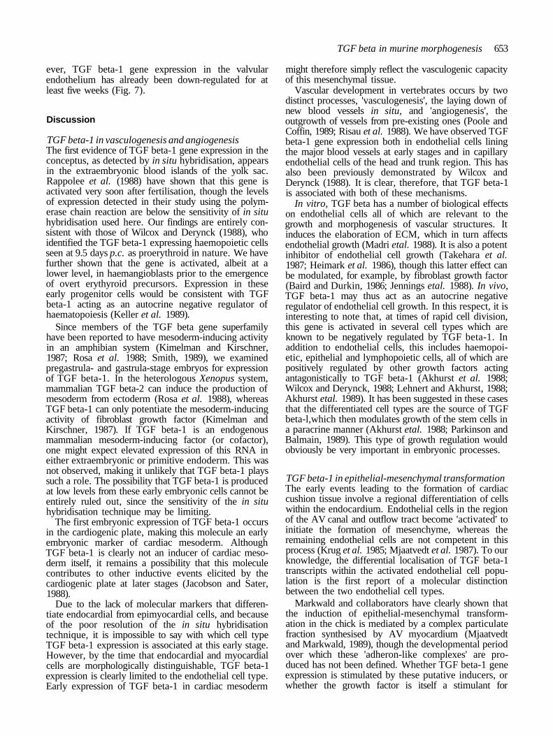
TGF beta-1 in epithelial-mesenchymal transformationThe early events leading to the formation of cardiaccushion tissue involve a regional differentiation of cellswithin the endocardium. Endothelial cells in the regionof the AV canal and outflow tract become 'activated' toinitiate the formation of mesenchyme, whereas theremaining endothelial cells are not competent in thisprocess (Krug et al. 1985; Mjaatvedt et al. 1987). To ourknowledge, the differential localisation of TGF beta-1transcripts within the activated endothelial cell popu-lation is the first report of a molecular distinctionbetween the two endothelial cell types.
Markwald and collaborators have clearly shown thatthe induction of epithelial-mesenchymal transform-ation in the chick is mediated by a complex particulatefraction synthesised by AV myocardium (Mjaatvedtand Markwald, 1989), though the developmental periodover which these 'adheron-like complexes' are pro-duced has not been defined. Whether TGF beta-1 geneexpression is stimulated by these putative inducers, orwhether the growth factor is itself a stimulant for

654 R. J. Akhurst and others
production of myocardial inducers, remains to betested.
The fact that TGF beta-1 is expressed ubiquitously inendocardial cells at early stages would argue against adirect role in generating the regional induction signal. Itis, however, possible that the AV myocardium has anactive role in sustaining endothelial TGF beta geneexpression specifically in the region of cushion tissueformation. TGF beta would subsequently contribute insome way to the epithelial-mesenchymal transition. Inthis respect, it is interesting to note that we have alsoobserved high levels of TGF beta gene expressionduring the epithelial-mesenchymal transformation thatoccurs during growth and fusion of the secondary palate(Fitzpatrick and Akhurst unpublished). Conversely,regionalised TGF beta-1 gene expression may merelybe a consequence of endogenous stimulation in re-sponse to the increased endothelial mitotic activityrequired to regenerate the endocardium as cells are lostto the mesenchymal layer (Fitzharris, 1981).
Recent experiments performed by Potts and Runyonsuggest that TGF beta-1 or -2, in combination withother factors, can mediate the epithelial-mesenchymaltransition (Potts and Runyan, 1989). They showed thatchick ventricular myocardium (not normally competentto induce transformation), when supplemented withTGF beta, can mediate mesenchyme formation fromAV endocardium and that this process is blocked byantibodies against TGF beta. They suggested that thesource of TGF beta was the myocardium.
In the present study, it is clearly shown that theendocardium is a more plentiful source of TGF beta-1RNA than the myocardium. In addition, our resultswould predict that the AV endocardium should alreadybe producing TGF beta-1 RNA at the stage equivalentto that examined by Potts and Runyan. One would nottherefore expect additional TGF beta to have any effecton the ability of AV endothelial cells to transform tomesenchyme in the in vitro system used by Potts andRunyan. Our data could be reconciled with theirs if oneassumes that one major function of the AV myocardiumis in maintaining this elevated TGF beta-1 gene ex-pression in the overlying endocardium, and that thisexpression is then required for later events in thecellular transformation process, as discussed above.
A number of issues need to be resolved for both themouse and chick systems before definitive conclusionsmay be made. First, it is now clear that there are at leastfive forms of vertebrate TGF beta encoded by differentgenes (ten-Dijke et al. 1988; Jakowlew et al. 1988a,b;Kondaiah et al. 1989). Potts and Runyon did notdistinguish between these different forms in the chickwhereas we have only, so far, examined the expressionof one gene in the mouse. Second, there could bespecies differences in production and utilisation of thedifferent molecular forms of TGF beta. Sato and Rifkin(1989) have shown, for example, that bovine aorticendothelial cells are inhibited in cell movement by TGFbeta produced from bovine smooth muscle cells but notthat produced from rat smooth muscle cells. Untilcomparative analyses on these species are performed
using molecular probes to each polypeptide form, thesepoints will remain unresolved.
TGF beta-1 in cardiac morphogenesisBased on the data presented in this paper and on ourearlier study of embryonic gene expression of TGFbeta-1, we would suggest that a major function of TGFbeta-1 in endocardial cushion tissue is not in the initial'activation' of endothelial cells in the epithelial-mesenchymal transition per se, but in modulation of theactivity of the underlying mesenchymal cells, particu-larly with respect to elaboration of the ECM. Thealtered composition of the ECM might then affectendothelial and mesenchymal cell phenotype. Thisproposition is based entirely on circumstantial evi-dence.
We previously showed that the TGF beta-1 gene wasactivated in embryonic epithelia which are undergoingspecific morphogenetic events involving localised epi-thelial and mesenchymal cell growth and migration(Lehnert and Akhurst, 1988). We suggested that thisgrowth factor was mainly acting via a paracrine mech-anism on the underlying mesenchymal tissue, modulat-ing growth, differentiation and ECM deposition. In allcases where we observed epithelial expression of TGFbeta-1 RNA, there is mesenchymal localisation of theECM molecule, tenascin (Chiquet-Ehrismann et al.1986). Furthermore, TGF beta-1 is known to stimulatetranscription of genes encoding ECM proteins includingthat for tenascin (Pearson et al. 1988).
We were particularly interested in comparing thedistribution of tenascin with that of TGF beta-1 RNA,since this molecule has a more restricted distributionthan that of other ECM molecules and might be a morespecific marker for TGF beta-1-induced modulation ofthe matrix. A correlation between epithelial expressionof TGF beta-1 RNA and mesenchymal localisation oftenascin polypeptide is seen during chondrogenesis andosteogenesis and during the development of the kidney,gut, tooth, hair follicle and salivary gland (Mackie etal.1987; Aufderheide et al. 1987; Aufderheide andEkblom, 1988; Chiquet-Ehrismann etal. 1986; Lehnertand Akhurst, 1988).
We have now extended this correlation to includemorphogenesis of the heart. Indeed, we have shownthat the mesenchymal distribution of tenascin polypep-tide during morphogenesis of the heart valves andseptae is more tightly correlated to endothelial TGFbeta-1 RNA than are the distributions of collagen(Colvee and Hurle, 1981) or fibronectin (Icardo, 1985).During early cardiac valve morphogenesis tenascinlocalisation is immediately adjacent to the TGF beta-1producing endothelial cells, whereas during later stagesof development, and in the adult, tenascin is localisedmore distally from the endothelium. Clearly, manyfactors must contribute to induction of tenascin ex-pression. At later stages in cardiogenesis TGF beta-1may not play such a major role. Furthermore, morpho-genetic movements may relocate extracellular mol-ecules, such as tenascin, to sites distant from their siteof synthesis.

TGF beta in murine morphogenesis 655
Tenascin is known to stimulate growth of certainepithelial cells (Chiquet-Ehrismann etal. 1986), thoughno mitogenic effects on endothelial cells have beendocumented. It disrupts cell-substratum and cell-cellcontacts and promotes cell mobility by interfering withthe adhesive action of fibronectin (Chiquet-Ehrismannet al. 1988, 1989; Spring et al. 1989). These changes incellular phenotype are all obligatory for transformationof endothelial cells into mesenchymal cushion tissue(Krug et al. 1985; Icardo, 1989). The effects of TGFbeta-1 on endocardia! cushion formation and sub-sequent morphogenesis could clearly be mediated, inpart, by tenascin.
High levels of both TGF beta RNA and tenascinprotein in the cardiac valves are correlated with times ofmesenchymal and endothelial cell growth and tissuemovement. In contrast, the intense staining with the CCantibody against TGF beta polypeptide does not appearuntil fairly late stages of embryogenesis and continuesto increase early postpartum. It therefore appears thatthere is lack of correlation between TGF beta-1 RNAlevels, indicative of active gene expression, and TGFbeta polypeptide levels. It is possible that in the adult,very low transcription levels may be translated to givemuch higher levels of protein. Many examples of post-transcriptional control of TGF beta have been cited(Assoian et al. 1987; Kehrl et al. 1986). In addition, theCC antibody may recognise protein products of otherTGF beta genes. The antibody is known not to cross-react with TGF beta-2 polypeptide (Ellingsworth et al.1986), but there have been no reports on its degree ofcross-reactivity with TGF beta-3 or the putative mam-malian TGF betas -4 and -5. This last point will only beresolved when gene probes and antibodies are availablewhich show complete specificity of reactivity to indi-vidual gene products.
Thanks to Dr R. Derynck for supplying the murine TGFbeta-1 cDNA probe and to Dr K. Flanders for supplying theCC antibody. Thanks also to Marion Hoggan for typing themanuscript. This work was supported in part by Birthright/RCOG and by the MRC.
References
AKHURST, R. J., FEE, F. AND BALMAIN, A. (1988). Localizedproduction of TGF-beta mRNA in tumour promoter-stimulatedmouse epidermis. Nature, Lond. 331, 363-365.
AKHURST, R. J., LEHNERT, S. A., GATHERER, D. AND DUFFIE, E.
(1990). The role of TGF beta in mouse development. Ann. N.Y.Acad. Sci. (In Press).
ASSOIAN, R. K., FLEURDELYS, B. E., STEVENSON, H. C, MILLER, P.
J., MADTES, D. K., RAINES, E. W., ROSS, R. AND SPORN, M. B.
(1987). Expression and secretion of type beta transforminggrowth factor by activated human macrophages. Proc. naln.Acad. Sci. U.S.A. 84, 6020-6024.
AUFDERHE1DE, E . , CHIQUET-EHRISMANN, R. AND EKBLOM, P.
(1987). Epithelial-mesenchymal interactions in the developingkidney lead to expression of tenascin in the mesenchyme. J. CellBiol. 105, 599-608.
AUFDERHEIDE, E. AND EKBLOM, P. (1988). Tenascin during gutdevelopment: Appearance in the mesenchyme, shift in molecularforms, and dependence on epithelial-mesenchymal interactions.J. Cell Biol. 107, 2341-2349.
BAIRD, A. AND DURKIN, T. (1986). Inhibition of endothelial cell
proliferation by beta-type transforming growth factor:interactions with acidic and basic fibroblast growth factors.Biochem. Biophys. Res. Comm. 138, 476-482.
CHIQUET-EHRISMANN, R., KALLA, P. AND PEARSON, C. A. (1989).
Participation of tenascin and transforming growth factor beta inreciprocal epithelial-mesenchymal interactions of MCF7 cells andfibroblasts. Cancer Res. 49, 4322-4325.
CHIQUET-EHRISMANN, R., KALLA, P., PEARSON, C. A., BECK, K.
AND CHIQUET, M. (1988). Tenascin interferes with fibronectinaction. Cell 53, 383-390.
CHIQUET-EHRISMANN, R., MACKIE, E. J., PEARSON, C. A. AND
SAKAKURA, T. (1986). Tenascin: an extracellular matrix proteininvolved in tissue interactions during fetal development andoncogenesis. Cell 47, 131-139.
COLVEE, E. AND HURLE, J. M. (1981). Maturation of theextracellular material of the semilunar heart valves in the mouse.Anal. Embryol. 162, 343-352.
Cox, K. H., DELEON, D. V., ANGERER, L. M. AND ANGERER, R. C.
(1984). Detection of mRNAs in sea urchin embryos by in situhybridisation using asymmetric RNA probes. Devi Biol. 101,485-502.
DERYNCK, R., JARRETT, J. A., CHEN, E. Y. AND GOEDDEL, D. V.
(1986). The murine transforming growth factor-beta precursor. J.biol. Chem. 261, 4377-4379.
ELLINGSWORTH, L. R., BRENNAN, J. E., FOK, K., ROSEN, D. M.,
BENTZ, H., PIEZ, K. A. AND SEYEDIN, S. M. (1986). Antibodies
to the N-terminal portion of cartilage-inducing factor A andtransforming growth factor beta. Immunohistochemicallocalization and association with differentiating cells. J. biol.Chem. 261, 12362-12367.
FANANAPAZIR, K. AND KAUFMAN, M. H. (1988). Observation on thedevelopment of the aortico-pulmonary spiral septum of themouse. J. Anat. u58, 157-172.
FITZHARRJS, T. P. (1981). Origin and migration of cushion tissue inthe developing heart. SEM 254(11), 255-260.
FLANDERS, K. C, THOMPSON, N. L., CISSEL, D. S., VAN
OBBERGHEN, SCHILLING, E., BAKER, C. C, KASS, M. E.,
ELLINGSWORTH, L. R., ROBERTS, A. B. AND SPORN, M. B. (1989).
Transforming growth factor-beta 1: histochemical localizationwith antibodies to different epitopes. J. Cell Biol. 108, 653-660.
GAMBLE, J. R. AND VADAS, M. A. (1988). Endothelial adhesivenessfor blood neutrophils is inhibited by transforming growth factor-beta. Science 242, 97-99.
HEIMARK, R. L., TWARDZIK, D. R. AND SCHWARTZ, S. M. (1986).
Inhibition of endothelial regeneration by type-beta transforminggrowth factor from platelets. Science 233, 1078-1080.
HEINE, U., MUNOZ, E. F., FLANDERS, K. C, ELLINGSWORTH, L.
R., LAM, H. Y., THOMPSON, N. L., ROBERTS, A. B. AND SPORN,
M. B. (1987). Role of transforming growth factor-beta in thedevelopment of the mouse embryo. J. Cell Biol. 105, 2861-2876.
HIRAKI, Y., INOUE, H., HIRAI, R., KATO, Y. AND SUZUKI, F.
(1988). Effect of transforming growth factor beta on cellproliferation and glycosaminoglycan synthesis by rabbit growth-plate chondrocytes in culture. Biochim. biophys. Acta 969,91-99.
ICARDO, J. M. (1985). Distribution of fibronectin during themorphogenesis of the truncus. Anat. Embryol. 171, 193-200.
ICARDO, J. M. (1989). Changes in endocardia! cell morphologyduring development of the endocardial cushions. Anat. Embryol.179, 443-448.
IGNOTZ, R. A. AND MASSAGUE, J. (1986). Transforming growthfactor-beta stimulates the expression of fibronectin and collagenand their incorporation into the extracellular matrix. J. biol.Chem. 261, 4337-4345.
IGNOTZ, R. A. AND MASSAGUE, J. (1987). Cell adhesion proteinreceptors as targets for transforming growth factor-beta action.Cell 51, 189-197.
JACOBSON, A. G. AND SATER, A. K. (1988). Features of embryonicinduction. Development 104, 341-359.
JAKOWLEW, S. B., DILLARD, P. J., KONDAIAH, P., SPORN, M. B.
AND ROBERTS, A. B. (1988a). Complementary deoxyribonucleicacid cloning of a novel transforming growth factor-betamessenger ribonucleic acid from chick embryo chondrocytes.Mol. Endocrinol. 2, 747-755.
J A K O W L E W , S . B . , K O N D A I A H , P . , D I L L A R D , P . J . , SPORN, M. B.

656 R. J. Akhurst and others
AND ROBERTS, A. B. (1988fc). A novel low molecular weightribonucleic acid (RNA) related to transforming growth factorbeta messenger RNA. Mol. Endocrinol. 2, 1056-1063.
JENNINGS, J. C, MOHAN, S., LJNKHART, T. A., WIDSTROM, R. ANDBAYUNK, D. J. (1988). Comparison of the biological actions ofTGF beta-1 and TGF beta-2: differential activity in endothelialcells. J. cell. Physiol. 137, 167-172.
KEHRL, J. H., ROBERTS, A. B., WAKEFIELD, L. M., JAKOWLEW, S.AND SPORN, M. B. (1986). Transforming growth factor beta is animportant immunomodulatory protein for human B lymphocytes.J. Immunol. 137, 3855-3860.
KELLER, J. R., SING, G. K., ELLINGSWORTH, L. R. AND RUSCETTI,
F. W. (1989). Transforming growth factor beta: possible roles inthe regulation of normal and leukemic hematopoietic cellgrowth. J. Cell Biochem. 39, 175-184.
KIMELMAN, D. AND KIRSCHNER, M. (1987). Synergistic induction ofmesoderm by FGF and TGF-beta and the identification of anmRNA coding for FGF in the early Xenopus embryo. Cell 51,869-877.
KONDAIAH, P., SANDS, M. S., SMITH, J. M., FIELDS, A., ROBERTS,A. B., SPORN, M. B. AND MELTON, D. A. (1990). Identificationof a novel transforming growth factor beta (TGF beta 5) mRNAin Xenopus laevis. J. biol. Chem. (In Press).
KRUG, E. L., RUNYAN, R. B. AND MARKWALD, R. R. (1985).Protein extracts from early embryonic hearts initiate cardiacendothelial cytodifferentiation. Devi Biol. 112, 414-426.
LEHNERT, S. A. AND AKHURST, R. J. (1988). Embryonic expressionpattern of TGF beta type-1 RNA suggests both paracrine andautocrine mechanisms of action. Development 104, 263-273.
MACKIE, E. J., THESLEFF, I. AND CHIQUET-EHRISMANN, R. (1987).Tenascin is associated with chondrogeneic and osteogenicdifferentiation in vivo and promotes chondrogenesis in vitro. J.Cell Biol. 105, 2569-2579.
MADRI, J. A., PRATT, B. M. AND TUCKER, A. M. (1988).Phenotypic modulation of endothelial cells by transforminggrowth factor-beta depends upon the composition andorganization of the extracellular matrix. J. Cell Biol. 106,1375-13S4.
MANASEK, F. J. (1976). Heart development: Interactions involvedin cardiac morphogenesis. In The Cell Surface in AnimalEmbryogenesis and Development (edited by Poste, G. andNichloson, G. L.) North Holland, Amsterdam: Elsevier,p. 545-598.
MJAATVEDT, C. H., LEPERA, R. C. AND MARKWALD, R. R. (1987).Myocardial specificity for initiating endothelial-mesenchymal celltransition in embryonic chick heart correlates with a paniculatedistribution of fibronectin. Devi Biol. 119, 59-67.
MJAATVEDT, C. H. AND MARKWALD, R. R. (1989). Induction of anepithelial-mesenchymal transition by an in vivo adheron-likecomplex. Devi Biol. 13<i, 118-128.
NODA, M. AND RODAN, G. A. (1987). Type beta transforminggrowth factor (TGF beta) regulation of alkaline phosphataseexpression and other phenotype-related mRNAs in osteoblasticrat osteosarcoma cells. J. Cell Physiol. 133, 426—437.
NODA, M., YOON, K., PRINCE, C. W., BUTLER, W. T. AND RODAN,G. A. (1988). Transcriptional regulation of osteopontinproduction in rat osteosarcoma cells by type beta transforminggrowth factor. J. biol. Chem. 263, 13916-13921.
OHTA, M., GREENBERGER, J. S., ANKLESARIA, P., BASSOLS, A. ANDMASSAGUE, J. (1987). Two forms of transforming growth factor-beta distinguished by multipotenrial haematopoietic progenitorcells. Nature 329, 539-541.
PARDANAUD, L., YASSINE, F. AND DIETERLEN-LIEVRE, F. (1989).Relationship between vasculogenesis, angiogenesis andhaemopoiesis during avian ontogeny. Development 105, 473-485.
PARKINSON, K. AND BALMAIN, A. (1990). Chalones revisited-Apossible role for TGF beta in tumour promotion. Carcinogenesis(In Press).
PEARSON, C. A., PEARSON, D., SHIBAHARA, S. AND HOFSTEENGE, J.(1988). Tenascin: cDNA cloning and induction by TGF-beta.EMBO J. 7, 2977-2982.
PELTON, R. W., NOMURA, S., MOSES, H. L. AND HOGAN, B. L. M.(1989). Expression of transforming growth factor beta-2 RNAduring murine embryogenesis. Development 106, 759-767.
PENTTINEN, R. P., KOBAYASHI, S. AND BORNSTEIN, P. (1988).
Transforming growth factor beta increases mRNA for matrixproteins both in the presence and in the absence of changes inmRNA stability. Proc. natn. Acad. Sci. U.S.A. 85, 1105-1108.
POOLE, T. J. AND COFFIN, J. D. (1989). Vasculogenesis andangiogenesis: Two distinct morphogenetic mechanisms establishembryonic vascular pattern. J. exp. Zool. 251, 224-231.
POTTS, J. D. AND RUNYAN, R. B. (1989). Epithelial-mesenchymalcell transformation in the embryonic heart can be mediated, inpart, by transforming growth factor beta. Devi Biol. 134,392-401.
RAPPOLEE, D. A., BRENNER, C. A., SCHULTZ, R., MARK, D. ANDWERB, Z. (1988). Developmental expression of PDGF, TGF-alpha, and TGF-beta genes in preimplantation mouse embryos.Science 241, 1823-1825.
RJSAU, W., SARIOLA, H., ZERWES, H-G., SASSE, J., EKBLOM, P.,KEMLER, R. AND DOETSCHMAN, T. (1988). Vasculogenesis andangiogenesis in embryonic-stem-cell-derived embryoid bodies.Development 102, 471-478.
ROBERTS, A. B. AND SPORN, M. B. (1989). The transforming growthfactor betas. Handbook of Experimental Pharmacology, (InPress).
ROBERTS, A. B., SPORN, M. B., ASSOIAN, R. K., SMITH, J. M.,ROCHE, N. S., WAKEFIELD, L. M., HEINE, U. I., LIOTTA, L. A.,FALANGA, V., KEHRL, J. H. AND FAUCI, A. S. (1986).Transforming growth factor type beta: rapid induction of fibrosisand angiogenesis in vivo and stimulation of collagen formation invitro. Proc. natn. Acad. Sci. U.S.A. 83, 4167-4171.
ROSA, F., ROBERTS, A. B., DANIELPOUR, D., DART, L. L., SPORN,M. B. AND DAWID, I. B. (1988). Mesoderm induction inamphibians: the role of TGF beta 2-like factors. Science 239,783-785.
SATO, Y. AND RIFKIN, D. B. (1989). Inhibition of endothelial cellmovement by pericytes and smooth muscle cells: activation of alatent transforming growth factor-beta 1-like molecule by plasminduring co-culture. J. Cell Biol. 109, 309-315.
SMITH, J. C. (1989). Mesoderm induction and mesoderm-inducingfactors in early amphibian development. Development 105,665-677.
SPORN, M. B. AND ROBERTS, A. B. (1988). Peptide growth factorsare multifunctional. Nature 332, 217-219.
SPORN, M. B., ROBERTS, A. B., WAKEFIELD, L. M. AND ASSOIAN,R. K. (1986). Transforming growth factor-beta: biologicalfunction and chemical structure. Science 233, 532-534.
SPRING, J., BECK, K. AND CHIQUET-EHRISMANN, R. (1989). Twocontrary functions of tenascin: Dissection of active sites byrecombinant tenascin fragments. Cell 59, 325-334.
STEINDLER, D. A., COOPER, N. G. F., FAISSNER, A. ANDSCHACHNER, M. (1989). Boundaries defined by adhesionmolecules during development of the cerebral cortex: TheJl/tenascin glycoprotein in the mouse somatosensory corticalbarrel field. Devi Biol. 131, 243-260.
TAKEHARA, K., LEROY, E. C. AND GROTENDORST, G. R. (1987).TGF-beta inhibition of endothelial cell proliferation: alteration ofEGF binding and EGF-induced growth-regulatory (competence)gene expression. Cell 49, 415-422.
TEN-DUKE, P., HANSEN, P., IWATA, K. K., PIELER, C. ANDFOULKES, J. G. (1988). Identification of another member of thetransforming growth factor type beta gene family. Proc. natn.Acad. Sci. U.S.A. 85, 4715-4719.
THOMPSON, N. L., FLANDERS, K. C, SMITH, J. M.. ELUNGSWORTH,L. R., ROBERTS, A. B. AND SPORN, M. B. (1989). Expression oftransforming growth factor-beta 1 in specific cells and tissues ofadult and neonatal mice. J. Cell Biol. 108, 661-669.
TWARDZIK, D. R., RANCHALIS, J. R. AND TODARO, G. J. (1982).Mouse embryos contain transforming growth factors related tothose isolated from tumor cells. Cancer Res. 45, 5413-5416.
WILCOX, J. N. AND DERYNCK, R. (1988). Developmental expressionof transforming growth factors alpha and beta in the mousefetus. Mol. Cell Biol. 8, 3415-3422.
WILKINSON, D. G., BAILES, J. A. AND MCMAHON, A. P. (1987).Expression of the proto-oncogene int-1 is restricted to specificneural cells in the developing mouse embryo. Cell 50, 79-88.
(Accepted 29 January 1990)