Textbook of remedial massage Grace 9780729539692 Grace ... · TEXTBOOK MassageOF REMEDIAL Sandra...
34
Transcript of Textbook of remedial massage Grace 9780729539692 Grace ... · TEXTBOOK MassageOF REMEDIAL Sandra...

TEXTBOOK OF REMEDIAL
MassageSandra Grace PhD, MSc(Chiro), Grad Cert Sports Chiro, Cert Clinical Chiro Paediatrics, Dip Acup, DBM, DO, DC, Dip Ed, BA
Senior Lecturer in Osteopathic Medicine, Southern Cross University Adjunct Research Associate, The Education for Practice Institute, Charles Sturt University
Dr. Mark Philip Deal B.Sc.(Syd.), Grad. D.C., D.O., Dip Ed. (Technical)
Chiropractor, Osteopath, Acupuncturist and Educator Independent Contractor to Noosa Parade Medical Centre

v
Preface 000Acknowledgements 000Foreword 000Contributors 000Reviewers 000
1. Remedial massage in Australian healthcare 000Defining remedial massage 000The context of remedial massage: use of complementary and alternative medicine in Australian healthcare 000The remedial massage workforce in Australia 000Attitudes towards CAM by medical and other healthcare practitioners 000Evidence for remedial massage practice 000
Evidence for the effectiveness of massage therapy 000
Future directions 000Appendix 1.1: Massage therapy: systematic reviews 000References 000
2. Assessment procedures for remedial massage: an evidence-based approach 000Principles of assessment in remedial massage practice 000Purpose of assessments in remedial massage 000
Demonstrating treatment effectiveness 000Remedial massage assessment procedures 000
Client history 000Outcome measures 000Postural observation 000Gait analysis 000Functional tests 000Palpation 000
Legal and ethical requirements for remedial massage assessment 000Key messages 000Review questions 000Appendix 2.1: Outcome measures 000Appendix 2.2: Skin cancer 000References 000
Contents
3. Planning remedial massage therapy 000Principles of treatment 000Prioritising client safety 000
Swedish massage before remedial massage 000Active movements before passive 000Treating within pain tolerance 000
Overview of a remedial massage treatment approach 000
Short- and long-term treatment plan 000Client education and self-help 000Clients’ preferences 000
Treatment protocols in remedial massage practice 000
Aligning treatment to assessment findings 000Monitoring and reviewing treatment plans 000Recording remedial massage treatments 000Key messages 000Review questions 000References 000
4. Remedial massage techniques for muscles 000Assessing muscle function 000
Functional deficit demonstration 000Postural assessment 000Testing muscles for length 000Testing for strength 000Palpation 000
Remedial massage techniques for muscles 000Deep gliding (with-fibre) frictions and cross-fibre frictions 000Deep transverse friction (DTF) 000Soft tissue release (STR) 000
Key messages 000Review questions 000References 000
5. Muscle stretching 000Introduction 000Functional anatomy 000
Tissue range of motion 000Muscle functions 000Types of muscle contractions 000

vi
C O N T E N T S
Physical characteristics and neurological reflexes associated with stretching 000
Active and passive insufficiency 000Neurological events that mediate stretching 000
Types of muscle stretching 0001. Passive stretch 0002. Active, active-assisted stretch 0003. Muscle energy technique 0004. Active isolated stretch 0005. Ballistic stretch 0006. Dynamic stretch 0007. Static stretch 0008. Proprioceptive Neuromuscular Facilitation 000
Stretching treatment goals 000Assessing clients’ suitability for muscle stretching 000
History 000Postural observation/analysis 000Gait disturbance 000Functional tests 000Palpation 000
Evidence for the effectiveness of muscle stretching 000Contraindications and precautions 000Applying muscle stretching in remedial massage 000
Adapting stretches for individual clients 000Therapist’s position 000Recommendations 000
Review questions 000References 000
6. Myofascial trigger points 000Introduction 000What is a trigger point? 000Locating trigger points 000Classifying trigger points 000
Active and latent trigger points 000What causes trigger points? 000Identifying trigger points 000
Characteristics of trigger points 000Treating trigger points 000
Manual techniques 000Evidence-base of treatments for trigger points 000Contraindications and precautions 000Key messages 000
Review questions 000Appendix 6.1 Some key trigger points 000References 000
7. Joint articulation 000Introduction 000What is joint articulation? 000Evidence for the effectiveness of joint articulation and mobilisation 000Assessing clients for treatment with joint articulation 000
History 000Functional tests 000Palpation 000
Treating clients with joint articulation 000Guidelines for joint articulation 000Joint articulation techniques 000Contraindications 000
Key messages 000Review questions 000References 000
8. Myofascial release 000Introduction 000Functional anatomy review 000What is myofascial release? 000Assessing clients for myofascial restrictions 000
History 000Postural analysis 000Functional tests 000Palpation 000
Treating clients with myofascial releasing techniques 000
Evidence for the effectiveness of fascial releasing techniques 000Applying the techniques 000
Integrating myofascial releasing techniques with remedial massage 000
Contraindications and precautions 000Key messages 000Review questions 000References 000
9. Lymphatic drainage massage 000Introduction 000Evidence for the effectiveness of lymphatic drainage massage 000Functional anatomy review 000

vii
C O N T E N T S
Function of the lymphatic system 000Oedema 000
Assessing clients for lymphatic drainage massage 000
History 000Red flag conditions for oedema 000Postural observation 000Functional tests 000Palpation 000Differential diagnosis 000
Treating clients with lymphatic drainage massage 000
Basic principles of lymphatic drainage massage 000Length of treatment 000Explanation of treatment 000Regional lymphatic drainage massage 000
Contraindications and precautions 000Absolute contraindications 000Relative contraindications 000Local contraindications 000
Review questions 000References 000
10. The low back and pelvis 000Introduction 000Functional anatomy review 000Assessment of the lumbar and sacroiliac region 000
Case history 000Outcome measures 000Postural analysis 000Gait assessment 000Functional tests 000Palpation 000
Remedial massage treatment for the low back and pelvis 000
Muscle strain 000Non-specific low back pain 000Degenerative joint disease (Osteoarthritis/Degenerative arthritis) 000Lower crossed syndrome 000Lumbar disc syndrome 000Spondylolisthesis 000Sacroiliac syndrome 000
Key messages 000Review questions 000Appendix 10.1 000References 000
11. The thoracic region 000Introduction 000Functional anatomy review 000
Typical vertebrae – T2–T8 000Special features of T1 and T9–12 000
Assessment of the thoracic region 000Case history 000Outcome measures 000Postural analysis 000Gait analysis 000Functional tests 000Palpation 000
Remedial massage treatment for the thoracic region 000
Muscle strain 000Thoracic facet syndrome 000Degenerative joint disease (Osteoarthritis/Degenerative arthritis) 000Hyperkyphosis of the upper thoracic spine (dowager’s hump) 000Scheuermann’s disease (Vertebral epiphysitis) 000Osteoporosis 000Scoliosis 000Thoracic outlet syndrome (Anterior scalene syndrome) 000
Key messages 000Review questions 000References 000
12. The cervical region 000Introduction 000Functional anatomy review 000Assessment of the cervical region 000
Case history 000Outcome measures 000Postural analysis 000Functional tests 000Palpation 000
Remedial massage treatment for the cervical region 000
Muscle strain 000Non-specific neck pain (including cervical sprain, strain and vertebral subluxation) 000Degenerative joint disease (Osteoarthritis/Degenerative arthritis) 000Upper crossed syndrome 000Mechanical/acquired torticollis 000Cervical disc syndrome 000

viii
C O N T E N T S
Whiplash (acceleration/deceleration) associated disorders 000
Key messages 000Review questions 000References 000
13. The head and face 000Introduction 000Headache 000
Assessment of the headache client 000Remedial massage treatment for headache 000
Sinusitis 000Assessment of the client with sinus pain 000Remedial massage treatment for sinusitis 000
Temporomandibular joint disorders 000Disorders of the temporomandibular joint 000Assessment of the client with TMJ disorder 000
Key messages 000Headache 000Sinusitis 000Temporomandibular joint 000
Review questions 000Appendix 13.1 000References 000
14. The chest 000Introduction 000Functional anatomy review 000Assessment of the chest 000
Case history 000Outcome measures 000Postural analysis 000Functional tests 000Palpation 000
Remedial massage treatment for the chest region 000
Muscle strain 000Rib subluxation/fracture 000Respiratory conditions 000
Key messages 000Review questions 000References 000
15. The shoulder region 000Introduction 000Functional anatomy 000Assessing the shoulder region 000
Case history 000
Outcome measures 000Postural analysis 000Functional tests 000Palpation 000
Remedial massage treatment for the shoulder region 000
Overview of remedial massage treatment approach for the shoulder region 000Muscle strain 000Shoulder impingement syndrome 000Degenerative joint disease (DJD, degenerative arthritis, osteoarthritis) 000Adhesive capsulitis (frozen shoulder) 000
Acromioclavicular sprains, clavicle fractures and glenohumeral dislocations 000Key messages 000Review questions 000Appendix 15.1: Shoulder Pain and Disability Index 000References 000
16. The elbow region 000Introduction 000Functional anatomy 000Assessing the elbow and forearm 000
Case history 000Outcome measures 000Postural analysis 000Functional tests 000Palpation 000
Remedial massage treatment for the elbow region 000
Overview of remedial massage treatment approach for the elbow region 000Muscle strain 000Olecranon bursitis 000Elbow sprain 000Fractures and dislocations 000
Key messages 000Review questions 000References 000
17. The wrist and hand 000Introduction 000Functional anatomy 000Assessing the wrist and hand 000
Case history 000Outcome measures 000

ix
C O N T E N T S
Postural analysis 000Functional tests 000Palpation 000
Remedial massage treatment for the wrist and hand 000
Overview of remedial massage treatment approach for the wrist and hand 000Tendinitis and tenosynovitis of the wrist and hand 000De Quervain’s tenosynovitis 000Carpal tunnel syndrome 000Sprains of the wrist and hand 000Fractures and dislocations 000Degenerative joint disease and rheumatoid arthritis 000
Key messages 000Review questions 000References 000
18. The hip region 000Introduction 000Functional anatomy 000Assessing the hip region 000
Case history 000Outcome measures 000Postural analysis 000Gait analysis 000Functional tests 000Palpation 000
Remedial massage treatment for the hip region 000
Overview of remedial massage treatment approach for the hip region 000Muscle strain 000Degenerative joint disease (DJD, degenerative arthritis, osteoarthritis) 000Trochanteric bursitis 000
Key messages 000Review questions 000References 000
19. The knee 000Introduction 000Functional anatomy 000Assessing the knee 000
Case history 000Outcome measures 000Postural analysis 000
Gait analysis 000Functional tests 000Palpation 000
Remedial massage treatment for the knee 000Overview of remedial massage treatment approach for the knee region 000Muscle strain 000Iliotibial band syndrome 000Patellofemoral pain syndrome 000Chondromalacia patella 000Patella dislocation 000Ligament sprain 000Meniscal injury 000Degenerative joint disease (DJD, degenerative arthritis, osteoarthritis) 000Bursitis 000Osgood-Schlatter’s disease 000
Key messages 000Review questions 000References 000
20. The leg, ankle and foot 000Introduction 000
Deep venous thrombosis 000Functional anatomy 000
Nerve supply to muscles of the leg, ankle and foot 000
Assessing the leg, ankle and foot 000Case history 000Outcome measures 000Postural analysis 000Gait analysis 000Functional tests 000Palpation 000
Remedial massage treatment for the leg, ankle and foot 000
Overview of remedial massage treatment approach for the leg, ankle and foot 000
Leg 000Muscle strains of the leg 000Gastrocnemius strain (tennis leg) 000Shin splints 000Chronic compartment syndrome 000Stress fracture of the tibia 000Varicose veins and spider veins 000
Ankle 000Achilles tendinopathy 000Calcaneal bursitis 000Sever’s disease 000

x
C O N T E N T S
Ankle sprain 000Degenerative joint disease of the ankle 000
Foot 000Plantar fasciitis (heel spur) 000
Forefoot 000Metatarsalgia 000Morton’s neuroma (Interdigital neuroma) 000
Key messages 000Review questions 000References 000
21. Special client groups 000Introduction 000Children and adolescents 000
Important considerations for assessing and treating children 000Evidence for the effectiveness of massage for children 000
Women 000Special considerations for assessing and treating women 000
Evidence for the effectiveness of massage for women’s health 000
Men 000Special considerations for assessing and treating men 000
Geriatric clients 000Special considerations for assessing and treating geriatric clients 000Evidence for the effectiveness of massage for older clients 000
Clients with a mental illness 000Special considerations for assessing and treating clients with a mental illness 000Evidence for the effectiveness of massage for clients with a mental illness 000
Key messages 000Review questions 000References 000
Index 000
10
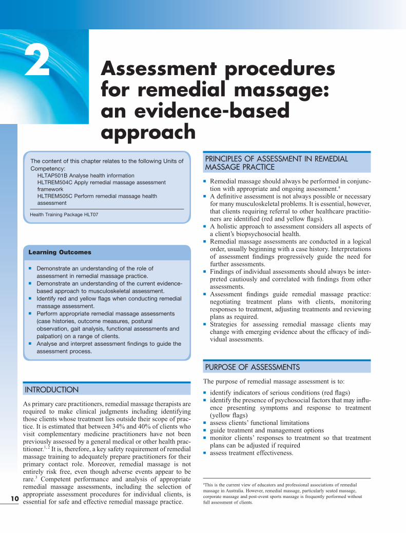
Assessment procedures for remedial massage: an evidence-based approach
2
PRINCIPLES OF ASSESSMENT IN REMEDIAL MASSAGE PRACTICE
� Remedial massage should always be performed in conjunc-tion with appropriate and ongoing assessment. a
� A defi nitive assessment is not always possible or necessary for many musculoskeletal problems. It is essential, however, that clients requiring referral to other healthcare practitio-ners are identifi ed (red and yellow fl ags).
� A holistic approach to assessment considers all aspects of a client ’ s biopsychosocial health.
� Remedial massage assessments are conducted in a logical order, usually beginning with a case history. Interpretations of assessment fi ndings progressively guide the need for further assessments.
� Findings of individual assessments should always be inter-preted cautiously and correlated with fi ndings from other assessments.
� Assessment fi ndings guide remedial massage practice: negotiating treatment plans with clients, monitoring responses to treatment, adjusting treatments and reviewing plans as required.
� Strategies for assessing remedial massage clients may change with emerging evidence about the effi cacy of indi-vidual assessments.
PURPOSE OF ASSESSMENTS
The purpose of remedial massage assessment is to:� identify indicators of serious conditions (red fl ags) � identify the presence of psychosocial factors that may infl u-
ence presenting symptoms and response to treatment (yellow fl ags)
� assess clients ’ functional limitations � guide treatment and management options � monitor clients ’ responses to treatment so that treatment
plans can be adjusted if required � assess treatment effectiveness.
INTRODUCTION
As primary care practitioners, remedial massage therapists are required to make clinical judgments including identifying those clients whose treatment lies outside their scope of prac-tice. It is estimated that between 34% and 40% of clients who visit complementary medicine practitioners have not been previously assessed by a general medical or other health prac-titioner. 1, 2 It is, therefore, a key safety requirement of remedial massage training to adequately prepare practitioners for their primary contact role. Moreover, remedial massage is not entirely risk free, even though adverse events appear to be rare.3 Competent performance and analysis of appropriate remedial massage assessments, including the selection of appropriate assessment procedures for individual clients, is essential for safe and effective remedial massage practice.
Learning Outcomes
� Demonstrate an understanding of the role of assessment in remedial massage practice.
� Demonstrate an understanding of the current evidence-based approach to musculoskeletal assessment.
� Identify red and yellow fl ags when conducting remedial massage assessment.
� Perform appropriate remedial massage assessments (case histories, outcome measures, postural observation, gait analysis, functional assessments and palpation) on a range of clients.
� Analyse and interpret assessment fi ndings to guide the assessment process.
a This is the current view of educators and professional associations of remedial massage in Australia. However, remedial massage, particularly seated massage, corporate massage and post-event sports massage is frequently performed without full assessment of clients.
The content of this chapter relates to the following Units of Competency:
HLTAP501B Analyse health information HLTREM504C Apply remedial massage assessment framework HLTREM505C Perform remedial massage health assessment
Health Training Package HLT07

A N E V I D E N C E - B A S E D A P P R O A C H 2
11
applying any technique (e.g. for pain tolerance, for age, to meet clients ’ expectation). A fl owchart of remedial massage assessments is illustrated in Figure 2.1 .
CLIENT HISTORY Taking a comprehensive client history is one of the most important assessment tools of healthcare practitioners. It is an important opportunity to establish relationships with clients that enhance positive outcomes of treatment. 4, 5 It also enables the practitioner to learn about the biopsychosocial infl uences on the client ’ s health. The fi rst responsibility of remedial massage therapists is to determine clients ’ suitability for remedial massage or for referral to another healthcare practi-tioner. The term red fl ag refers to clinical features (signs or symptoms) that may indicate the presence of serious underly-ing medical conditions requiring further investigation. Such conditions include cancers, fractures and infections (see Table 2.1 on p 12 ). Clients who choose to complement medical treatment of serious conditions with remedial massage therapy require supervision by their medical practitioner and liaison between practitioners (or at the very least awareness of the entirety of their clients ’ treatment). Clients should always be encouraged to disclose their remedial massage treatment to
DEMONSTRATING TREATMENT EFFECTIVENESS To monitor clients ’ responses to treatment, remedial massage therapists must fi rst collect suitable baseline data, including information collected from case histories and observed or measured by practitioners. The pain scale is commonly used for this purpose. Repeated collection of pain scale data enables changes in clients ’ perceptions of pain to be recorded and may demonstrate the perceived effectiveness of treatment. The amount of medication that clients require to manage symp-toms can also be used in this way. Recording details of medi-cation at the initial consultation and changes in medication use over the ensuing period of treatment provides evidence of progress towards treatment goals.
ASSESSMENT PROCEDURES
The most commonly used assessment tools in remedial massage therapy are presented below in the order in which they are usually applied and easiest to learn. In reality, assess-ments are overlapping and ongoing throughout the consulta-tion from fi rst encounters with clients in waiting rooms to the continual assessments and adjustments that are required when
Fig. 2.1 Assessment procedures in Remedial Massage Therapy

12
T E X T B O O K O F R E M E D I A L M A S S A G E
factors that could affect clients ’ responses to treatment (yellow fl ags). Questions should proceed logically and presenting con-ditions prioritised. A sound grounding in physiology, pathol-ogy and symptomatology is required for analysing the health information collected and for recognising deviations from normal fi ndings and signs and symptoms of common patho-physiologies. Practitioners are not required to read X-rays, MRIs or other diagnostic imaging but they are expected to interpret their reports. Figure 2.2 is a typical remedial massage case history.
Assessing the well client Remedial massage has long played a role in health mainte-nance and preventive medicine. Assessment of the well client is based on lifestyle history, postural and gait observations, functional tests, particularly those for muscle and joint func-tion, and palpation. Clients who present for massage therapy without symptoms are sometimes surprised when massage elicits pain or tenderness or when they feel the ropiness of fi brotic tissues or the tightness of a muscle being stretched.
Fig. 2.2 A typical case history form for remedial massage
PHONE B/H ________________A/H__________________Mobile __________________
FAX ___________ ______________EMAIL ____________________________________
OCCUPATION
SPORTS / HOBBIES
REFERRED BY
PRESENTING SYMPTOMS How can I help you? Record nature of complaint and duration.Inquire and record: nature of pain, frequency and intensity of symptoms, first occurrence,mode of onset. If recurrent, previous treatment. Associated symptoms. Aggravating andrelieving factors. Any other symptoms. IDENTIFY POSSIBLE RED AND YELLOW FLAGS.
PAST HISTORY Have you ever had any major illnesses or diseases? Any hospitalisations?Any major accidents? IDENTIFY POSSIBLE RED AND YELLOW FLAGS.
FAMILY HISTORY Has any member of your family had a major illness or disease?
OBSTETRICS HISTORY Ask any female client about pregnancies and births.
SOCIAL HISTORY Do you drink or smoke? Recreational drugs? MEDICATION Are you taking any medication? Record all details.
ALLERGIES Do you have any allergies? Record all details.
SYSTEMS REVIEW Either by questionnaire or orally, ask a few questions relating to eachsystem as required. (Central nervous: headaches, dizziness, numbness. Cardiovascular:chest pain, shortness of breath. Respiratory system: shortness of breath, wheezing, cough.Alimentary: nausea, vomiting, indigestion, abdominal pain, change in bowel habit.Urogenital: pain on urination, frequency of urination. Endocrine: Heat or cold intolerance,change in sweating, excessive thirst.)
NAME DATE OF BIRTH
ADDRESS
POSTCODE
Red fl ags Yellow fl ags
Clinical features that may be associated with the presence of a serious physical condition.
Psychosocial and occupational factors that may infl uence presenting symptoms, treatment approach and outcomes.
Table 2.1 Red and yellow fl ags
their doctor. However, many remedial massage clients do not have serious medical conditions. If there are no signs or symp-toms associated with specifi c diseases or serious conditions, further investigations (such as X-rays, MRIs or blood tests) are generally not indicated. 6, 7
Once practitioners have dismissed the likelihood of serious conditions, they gather information to guide further assess-ments: identifying likely contributing factors, types of tissues that could be involved, clients ’ responses to previous treat-ments, aggravating and relieving factors and psychosocial

A N E V I D E N C E - B A S E D A P P R O A C H 2
13
Table 2.2 ( p 15 ) is designed to assist practitioners identify red fl ags associated with pain of the lumbar, thoracic and cervical regions.
Identifying yellow fl ags Yellow fl ags are indicators of psychosocial and occupational factors that may affect clients ’ presenting symptoms and responses to treatment. Identifying the presence of yellow fl ags may prompt early interventions and better outcomes for clients. For example, clients with high stress levels may benefi t from referral to a counsellor; clients with concern about job security after extended absences from work may benefi t from reassurance from their employer. The presence of yellow fl ags is not a contraindication for remedial massage. Rather, yellow fl ags alert the practitioner to possible compli-cating factors which may prompt referral to other health prac-titioners or delay the normal recovery process.
Yellow fl ags include: 6
� belief that pain and activity are harmful � sickness behaviours (e.g. extended rest) � low or negative moods, social withdrawal � problems with claims and compensation � overprotective or unsupportive family � apportioning blame � overt anxiety or depression � non-compliance with rehabilitation program � long time between injury and referral.
Informed consent As part of the initial interview clients need to have proposed assessments and treatments and any associated risks fully explained. Informed consent must be obtained for all assess-ment and treatment procedures. 9
OUTCOME MEASURES Outcome measures are standardised questionnaires in which clients record perceived changes to their health status. They are used to assess clients ’ perceived health status and also to demonstrate the benefi ts of treatment. Three common outcome measures for musculoskeletal pain and disability are the Oswestry Disability Index, the Vernon Mior Neck Disability Questionnaire, and the Patient Specifi c Scale which can be used for disability in any region of the body. Research into the use of Outcome Measures demonstrate high validity c and reliabilityd, 10 – 12 for some questionnaires which may account for their increasing use in clinical trials and in clinical prac-tice. They are simple to administer and their standardised scoring systems allow comparisons of clients ’ responses over time. They are also readily interpreted by healthcare practitio-ners from other disciplines.
Completing questionnaires takes time and clients with low levels of English literacy will need assistance. Appointment times may need to be adjusted accordingly or clients invited to come to the clinic a few minutes before their scheduled appointment to complete the questionnaires. Outcome
They are being made aware of potential muscle imbalances that could lead to future problems (sub-clinical indicators). Early interventions (e.g. correcting posture, improving manual handling techniques, or modifying an exercise program) may divert many potential problems.
THE NATURE OF PAIN Pain is a common presenting symptom of remedial massage clients. Perceptions of pain are complicated by its multi-dimensional nature and its changing nature over time. Ques-tioning clients about pain helps determine their suitability for remedial massage and possible sources of pain. Ask clients about:� the location of the pain � the character of the pain, e.g. sharp shooting pain in a rela-
tively narrow band (radicular pain), a dull ache that is hard to localise (referred pain), burning pain (from trauma to sympathetic and somatic sensory nerves)
� the severity of the pain� A number of pain intensity measures are available
although the validity and reliability of such measures continues to be debated. 8 A simple procedure is to ask clients to rate their pain on a pain scale from 0 to 10, where 0 is no pain and 10 is extreme pain. Alternatively, practitioners can use a Visual Analogue Scale of Pain Intensity (VAS), a 10 cm line on which clients are asked to put a mark to show their current pain level.
� the onset of the pain � the duration of the pain � the course of the pain, e.g. intermittent, constant, episodic � aggravating and relieving factors.
Questions designed to distinguish between pain of local origin and pain referred from a distant site (in particular, from a spinal segment) are useful in directing treatment to the source of pain. As a guide for determining the nature of pain, con-sider the following:� local pain
� usually sharp � well localised
� referred pain� dull � poorly localised � often refers in a predetermined pattern (e.g. nerve
impingement at the spinal segment will refer pain in dermatomal patterns, trigger points refer in predictable patterns).
If a client reports numbness in an area of skin, the nerves supplying that dermatome b could be damaged. Similarly, if a client reports weak muscles in a particular myotome, the motor neuron in that spinal segment may be damaged. Knowing the spinal cord segment which supplies each derma-tome and myotome can help locate the source of pain (see Figure 2.3 ).
b Every spinal nerve contains both sensory and motor neurons. Somatic sensory neurons carry impulses from the skin to the spinal cord and brain stem. A dermatome is the area of skin that provides sensory input to one pair of spinal nerves. Somatic motor neurons carry impulses from the spinal cord to skeletal muscles. All muscles innervated by the motor neurons in a single spinal segment constitute a myotome. In general, dermatomes overlie corresponding myotomes. d Reliability is the consistency of repeated applications of a measurement.
c Validity is the extent to which a measure estimates the true nature of what it purports to measure.

14
T E X T B O O K O F R E M E D I A L M A S S A G E
Fig. 2.3 Dermatomes: anterior and posterior view
measures should be used at every opportunity by remedial massage therapists to demonstrate perceived effectiveness of their treatments.
POSTURAL OBSERVATION Postural observation forms an important part of the physical assessment of clients. It involves observing clients standing erect to assess structural and habitual postures and their asso-ciated muscle imbalances. Observing the whole client, regard-less of the complaint, is consistent with the holistic approach of complementary medicine. Postural observation is not an exact science. However, fi ndings from postural observation, along with case history and other assessment fi ndings, can contribute useful information to the development of treatment plans.
By observing erect postures from the posterior and lateral aspects, practitioners can compare one side of the body with the other, the upper half with the lower half, the left foot with the right foot and so on to identify postural patterns and/or factors that could contribute to presenting conditions or
predispose to future symptoms. Postural observation can provide information about the effects on the musculoskeletal system of right or left dominance, of regular sports and hobbies, of work-related activities and of adaptations to dis-eases and traumas. For example, cervicogenic headaches can be perpetuated by a scoliosis and without postural observation this connection could remain undetected. Failing to take such predisposing factors into account could result in short-lived treatment effects. Moreover conditions and postural tenden-cies identifi ed (sometimes incidentally) can prompt appropri-ate advice from therapists. For example, advising a client with bilateral pes planus about appropriate footwear, or a client who works at a computer all day about the importance of regular pectoralis major stretches, may prevent compensatory muscle imbalances and reduce susceptibility to injury.
Identifying anatomical landmarks To accurately describe and record postural observations, prac-titioners should be familiar with the following anatomical landmarks (see Figure 2.4 on p 16 ):

A N E V I D E N C E - B A S E D A P P R O A C H 2
15
Table 2.2 Red fl ags of serious conditions associated with acute low back, thoracic and neck pain
Red fl ags Possible condition
For acute low back pain
Symptoms and signs of infection (e.g. fever) InfectionRisk factors for infection (e.g. underlying disease, immunosuppression, penetrating wound)
History of trauma FractureMinor trauma (if > 50 years, history of osteoporosis, taking corticosteroids)
Past history of malignancy CancerAge > 50 yearsFailure to improve with treatmentUnexplained weight lossPain at multiple sitesPain at rest
Absence of aggravating factors Aortic aneurysm Other serious condition
Widespread neurological symptoms and signs in the lower limb (e.g. gait abnormality, saddle area numbness) ± urinary retention, faecal incontinence
Cauda equina syndrome (a medical emergency and requires urgent hospital referral)
For acute thoracic pain
Minor trauma (if > 50 years, history of osteoporosis, taking corticosteroids) FractureMajor trauma
Symptoms and signs of infection (e.g. fever, night sweats) InfectionRisk factors for infection (e.g. underlying disease, immunosuppression, penetrating wound)
Past history of malignancy CancerAge > 50 yearsFailure to improve with treatmentUnexplained weight lossPain at multiple sitesPain at restNight pain
Chest pain or heaviness Other serious conditionMovement/change in posture has no effect on painAbdominal painShortness of breath, cough
For acute neck pain
Symptoms and signs of infection (e.g. fever, night sweats) InfectionRisk factors for infection (e.g. underlying disease, immunosuppression, penetrating wound)
History of trauma FractureMinor trauma (if taking corticosteroids)
Past history of malignancy CancerAge > 50 yearsFailure to improve with treatmentUnexplained weight lossDysphagia, headache, vomiting
Neurological symptoms in the limbs Neurological condition
Cerebrovascular symptoms or signs, anticoagulant use Cerebral or spinal haemorrhage
Cardiovascular risk factors; transient ischaemic attack Vertebral or carotid aneurysm
Note that acute pain in this context refers to pain of less than three months ’ duration. Adapted from the New Zealand Guidelines Group 6 and the Acute Musculoskeletal Pain Guidelines Group 7 .

16
T E X T B O O K O F R E M E D I A L M A S S A G E
ANTERIOR AND LATERAL � The nipple line is approximately level with the 5th rib or
4th intercostal space. � The umbilicus is approximately level with the L3/4 disc. � The iliac crests are approximately level with L4.
POSTERIOR � The spine of the scapula is approximately level with T2
spinous process. � The inferior angle of the scapula is approximately level
with T7. � The insertion of the trapezius muscle is approximately level
with T12. � The PSISs are approximately level with S2.
Performing postural observation Clothing that does not obscure posture is required for the most accurate postural assessments. Swimwear or sportswear are suitable. Alternatively clients may disrobe to their underwear and wear a gown that opens at the back. Stand two or three metres behind the client. Ask the client to walk up and down on the spot a few times with their eyes closed and then to open their eyes, and stand comfortably, hands by their sides.
A plumb line (either real or imaginary) is used to project a centre of gravity line onto the external surface of the body. In the ideally aligned erect posture, the plumb line should pass as follows (see Figure 2.5 ).
In the posterior view:� equidistant through the heels, ankles, calves, knees and
thighs� through the midline of the trunk � through the midline of the neck and head.
In the lateral view:� anterior to the lateral malleolus e
Fig. 2.4 Anatomical landmarks for postural observation.
Fig. 2.5 Ideally aligned erect posture
� anterior to the axis of the knee joint � posterior to the hip joint � through the bodies of L1 and L5 � bisecting the shoulder joint � bisecting the ear.
e Some controversy exists among authors about the exact location of the centre of gravity line in the lateral view. It is sometimes said to bisect the lateral malleolus, rather than pass anteriorly to it.

A N E V I D E N C E - B A S E D A P P R O A C H 2
17
the level of the pelvis, the following describes the pattern of postural changes most often observed when the pelvis tilts. In this example, the client ’ s pelvis is lower on the left than the right (see Figure 2.6 ). (For a client whose pelvis tilts lower on the right, the reverse would follow.) For a client whose pelvis is lower on the left than the right:� the PSIS is usually lower on the left � the gluteal fold is usually lower on the left � the gluteal cleft usually deviates to the left � the iliac crest is usually lower on the left � the waist curve usually fl attens on the left � the lumbar scoliosis is usually convex on the left � the left foot is usually anterior and everted. If the level of the pelvis is not easily determined by observation, palpate the pelvis by placing your thumbs on the PSISs and fi ngers (held horizontally) on the iliac crests. Use your palpatory fi ndings to confi rm the pres-ence (or absence) of a pelvic tilt.
2. The level of the shoulders Look at the contour of the trapezius muscle between the neck and the acromion and compare both sides.
3. Determine the nature of the scoliosis. The exact nature of scolioses can only be determined by diagnostic imaging. However, estimates of the shape of the curve can be ascertained from the presence of pelvic or shoulder tilt. Figure 2.7 illustrates common C-shaped and S-shaped spinal curvatures. Most thoracic scolioses are convex to the right (see Chapter 11 ).
4. Any other features of the back, neck or head that stand out (e.g. hypertonic muscles mid thoracic area, asymmetrical development from sports, work, or right or left dominance, muscle wasting).
5. Any lower limb features (e.g. ankle pronation, genu valgus or varus, oedema, feet everted) (see Figure 2.8 ).
General observations A common reason that remedial massage therapists fi nd pos-tural observation diffi cult is that they look for detail before making general observations. Always begin by asking, ‘ What stands out? Are there any regions of the body that appear to be overloaded, under stress or very different from normal? ’ Next, focus on pelvic alignment which is useful for identify-ing common posture deviations like hyperlordosis or scolio-sis. Learning to observe details like colour changes (e.g. the pallor of poor circulation, butterfl y rash across the cheeks in rheumatoid conditions, a hairy tuft in the lumbosacral area in spondylolisthesisf ), and unusual skin markings (e.g. pete-chiae, surgical scars, stretch marks, moles g ) can be developed after the basics of postural observation have been mastered. Remember that postural observations are never defi nitive. They only indicate possibilities. In most cases clients present with minor deviations from normal standing alignment and communication with clients about postural observations should always make this clear.
In the posterior view, observe:
1. The level of the pelvis. One of the most important fi nd-ings of postural observation is the level of the pelvis, as many treatment plans address imbalances associated with pelvic tilt. The pelvis comprises two ilia which articulate with each other at the symphysis pubis anteri-orly and with the sacrum at the sacroiliac joints. Observe the level of the gluteal folds, PSISs, iliac crests, deviation of the gluteal cleft, the waist curvature and the distance between arms and trunk. To assist your determination of
Fig. 2.6 Left pelvic tilt
g Massage therapists can play an important role in the detection of skin cancers among their clients. See Appendix 2.2 for a useful checklist.
f Spondylolisthesis is the anterior displacement of a vertebra in relation to the vertebra below (see Chapter 10 ).

18
T E X T B O O K O F R E M E D I A L M A S S A G E
Scoliosis Scoliosis refers to a lateral curvature of the spine, observed when the client is viewed anteriorly or posteriorly. Diagnostic imaging is required to determine the exact nature and degree of spinal curvature. Postural observation enables practitioners to estimate large changes in spinal curvature and to suggest pos-tural factors that may contribute or predispose to symptoms.
There are two major types of scoliosis:� Structural (idiopathic) scoliosis. This is a curvature of the
spine of unknown cause. It is relatively fi xed and does not straighten on forward bending of the trunk (Adam ’ s test) (see Figure 2.9 on p 20 ). It is often associated with vertebral and rib abnormalities and in severe cases can require bracing and surgery. Scoliosis screening tests in schools are designed to detect severe idiopathic scolioses that require such medical interventions. As a general rule, refer any child under 16 with a moderate or severe rib hump on forward fl exion (Adam ’ s test) who has not been previously screened by a medical practitioner, chiropractor, osteopath, physiotherapist or in a school screening program.
� Functional scoliosis. This is a compensatory curvature that may be associated with pelvic tilt and imbalance of muscles due to work and sport activities. This type of scoliosis usually straightens on forward bending of the trunk (i.e. Adam ’ s test is negative). It is usually correctable with such interventions as exercise programs, remedial massage, spinal manipulation, and the use of orthotics in shoes.
Scoliosis is recorded on a posterior posture diagram by drawing three lines: i) a straight line at the level of the iliac
In the lateral view, note:
1. the relation of the body to the centre of gravity line: assess the alignment of the ear, shoulder, hips and lateral malleoli
2. the level of the pelvis (e.g. hyperlordosis, hypolordosis) 3. the shape of the kyphosis (e.g. hyperkyphosis, fl attened
thoracic curvature) 4. the position of the head (ear position in relation to shoul-
der, cervical hyper or hypolordosis) 5. the position of shoulders (rounded), arms and hands 6. any lower limb features (e.g. genu recurvatum, pes
planus, pes cavus, hallux valgus, claw and hammer toes).
Method of recording Postural observations are most easily recorded by marking diagrams of the posterior and lateral aspects of the erect body. A standardised recording system is yet to be developed for remedial massage. Only the most obvious and important fi nd-ings need be recorded. These include the level of the pelvis, any major posture distortion (e.g. scoliosis, hyperlordosis), and any possible factors that could predispose to current or future symptoms. In the following section ways of recording common posture observations are presented. Figure 2.8 on p 19 shows how common posture observations of the lower limb can be recorded.
COMMON POSTURE OBSERVATIONS In the posterior view:
Fig. 2.7 C-shaped and S-shaped scoliosis

A N E V I D E N C E - B A S E D A P P R O A C H 2
19
Fig. 2.8 Postural observations: Lower limb
crests angled to indicate the pelvic tilt. Try to make the angle of the straight line indicate the degree of pelvic tilt: an almost horizontal line would represent a small degree of pelvic tilt; a more diagonal line would represent a greater degree of pelvic tilt; ii) a straight line indicating the level of the shoul-ders; and iii) a curved line estimating the scoliosis.
In the lateral view :
Lumbar hyperlordosis This is a commonly observed posture (see Figure 2.11 ). Its causes include an anteriorly tilted pelvis and muscular imbal-ance such as weak abdominal muscles relative to tight erector spinae muscles, and/or weak hamstring muscles relative to tight hip fl exor muscles. Hyperlordosis is commonly associ-ated with lower crossed syndrome (see Chapter 10 ).
Standing hyperlordosis test
Another quick test for hyperlordosis: The client removes their shoes and stands with their shoulders, buttocks and heels against a wall. Without adjusting the position, the client places one hand into the space between their waist and the wall. One fl at palm in the space between the back and the wall is usual. Any more than this could indicate lumbar hyperlordosis. (Some clients can fi t two fi sts placed one on top of the other.)
(Note: There could be other reasons for excessive space between the wall and the client ’ s body, such as overdevel-oped gluteus maximus muscles.)
A N E V I D E N C E - B A S E D A P P R O A C H 2
23
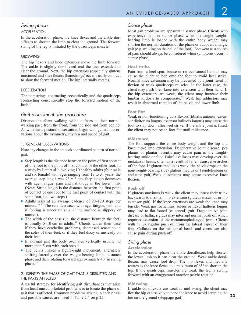
Stance phase Most gait problems are apparent in stance phase. Clients who experience pain in stance phase when the single weight-bearing limb is loaded with the entire body weight may shorten the normal duration of the phase or adopt an antalgic gait (e.g. walking on the ball of the foot). Footwear as a source of pain should always be considered when symptoms occur in stance phase.
Heel strike Pain from a heel spur, bruise or retrocalcaneal bursitis may cause the client to hop onto the foot to avoid heel strike. Normal knee extension may be prevented by a joint fused in fl exion or weak quadriceps muscles. In the latter case, the client may push their knee into extension with their hand. If the hip extensors are weak, the client may increase their lumbar lordosis to compensate. 19 Weak hip adductors may result in abnormal rotation of the pelvis and lower limb.
Foot fl at Weak or non-functioning dorsifl exors (tibialis anterior, exten-sor digitorum longus, extensor hallucis longus) may cause the foot to slap down after heel strike. If the ankle joint is fused, the client may not reach foot fl at until midstance.
Midstance The foot supports the entire body weight and the hip and knee move into extension. Degenerative joint disease, pes planus or plantar fasciitis may cause pain in the weight-bearing ankle or foot. Painful calluses may develop over the metatarsal heads, often as a result of fallen transverse arches of the foot. If gluteus medius is weak, the pelvis drops on the non-weight-bearing side (gluteus medius or Trendelenburg or abductor gait).Weak quadriceps may cause excessive knee fl exion.
Push off If gluteus maximus is weak the client may thrust their trunk backwards to maintain hip extension (gluteus maximus or hip extensor gait). If the knee extensors are weak the knee may buckle. Weak gastrocnemius, soleus or fl exor hallucis longus may lead to fl at-footed (calcaneal) gait. Degenerative joint disease or hallux rigidus may interrupt normal push off which requires extension of the metatarsophalangeal joint. Clients with hallux rigidus push off from the lateral aspect of their foot. Calluses on the metatarsal heads and corns can also cause pain during push off.
Swing phase Acceleration In the acceleration phase the ankle dorsifl exors help shorten the lower limb so it can clear the ground. Weak ankle dorsi-fl exors may cause foot drop. The hip fl exes and medially rotates as the knee fl exes to a maximum of 65 ° to shorten the leg. If the quadriceps muscles are weak the leg is swung forward with an exaggerated anterior pelvic rotation.
Midswing If ankle dorsifl exors are weak in mid swing, the client may fl ex the hip excessively to bend the knee to avoid scraping the toe on the ground (steppage gait).
Swing phase ACCELERATION In the acceleration phase, the knee fl exes and the ankle dor-sifl exes to shorten the limb to clear the ground. The forward swing of the leg is initiated by the quadriceps muscle.
MIDSWING The hip fl exors and knee extensors move the limb forward. The ankle is slightly dorsifl exed and the toes extended to clear the ground. Next, the hip extensors (especially gluteus maximus) and knee fl exors (hamstrings) eccentrically contract to slow the forward motion. The hip internally rotates.
DECELERATION The hamstrings contracting eccentrically and the quadriceps contracting concentrically stop the forward motion of the limb. 15
Gait assessment: the procedure Observe the client walking without shoes at their normal walking pace from the front, from the side and from behind. As with static postural observation, begin with general obser-vations about the symmetry, rhythm and speed of gait.
1. GENERAL OBSERVATIONS Note any changes in the smooth coordinated pattern of normal gait.� Step length is the distance between the point of fi rst contact
of one foot to the point of fi rst contact of the other foot. In a study by Latt et al 16 involving 10 healthy adults (four male and six female) with ages ranging from 17 to 31 years, the average step length was 73 ± 3 cm. Step length decreases with age, fatigue, pain and pathology in the lower limb. (Note: Stride length is the distance between the fi rst point of contact of one foot to the fi rst point of contact with the same foot, i.e. two steps.)
� Adults walk at an average cadence of 90 – 120 steps per minute.16, 17 The rate decreases with age, fatigue, pain and if footing is uncertain (e.g. if the surface is slippery or uneven).
� The width of the base (i.e. the distance between the feet) is usually 5 – 10 cm in adults. 18 Clients widen their base if they have cerebellar problems, decreased sensation in the soles of their feet, or if they feel dizzy or unsteady on their feet.
� In normal gait the body oscillates vertically usually no more than 5 cm with each step. 17
� The pelvis makes a fi gure-eight movement, alternately shifting laterally over the weight-bearing limb in stance phase and then rotating forward approximately 40 ° in swing phase.14
2. IDENTIFY THE PHASE OF GAIT THAT IS DISRUPTED AND THE PARTS AFFECTED A useful strategy for identifying gait disturbances that arise from local musculoskeletal problems is to locate the phase of gait that is affected. Common problems arising in each phase and possible causes are listed in Table 2.4 on p 25 .

24
T E X T B O O K O F R E M E D I A L M A S S A G E
Table
2.3
P
hase
s o
f g
ait
Phase
s of
gait
Sta
nce p
hase
Sw
ing p
hase
Heel
stri
ke
Foot
fl at
Mid
stan
ce
Pu
sh o
ff
Acc
ele
rati
on
Mid
sw
ing
Dece
lera
tion
The
foot
mak
es fi
rst
co
ntac
t w
ith t
he g
roun
d
and
the
lim
b d
ecel
erat
es .
A c
lose
d k
inet
ic c
hain
fo
r th
e lo
wer
lim
b is
in
itiat
ed.
Foot
ad
apts
to
surf
ace
and
tak
es t
he w
eigh
t of
th
e b
ody.
The
pel
vis
is
stab
ilise
d.
Trun
k is
ca
rrie
d f
orw
ard
by
mom
entu
m.
The
knee
rem
ains
st
abili
sed
. M
omen
tum
co
ntin
ues
to c
arry
the
b
ody
forw
ard
.
The
bod
y is
acc
eler
ated
in
to f
orw
ard
mot
ion.
The
low
er li
mb
is
sho
rten
ed
and
sw
ung
forw
ard
. Th
e in
nom
inat
e m
oves
ant
erio
rly.
The
foot
cle
ars
the
grou
nd.
On
the
wei
ght-
bea
ring
sid
e th
e in
nom
inat
e m
oves
p
oste
riorly
; th
e sa
crum
tilt
s la
tera
lly t
owar
ds
the
sid
e of
lo
adin
g.
Dec
eler
ate
the
limb
, an
d
pre
par
e fo
r fi r
st c
onta
ct
with
the
gro
und
.
Majo
r m
usc
le g
roups
Con
cen
tric
A
nkle
dor
sifl e
xors
E
ccen
tric
H
ip e
xten
sors
Ti
bia
lis a
nter
ior
Con
cen
tric
K
nee
exte
nsor
s A
nkle
pla
ntar
fl e
xors
Is
omet
ric
Hip
ab
duc
tors
E
ccen
tric
H
ip e
xten
sors
Q
uad
ricep
s Ti
bia
lis a
nter
ior
Isom
etri
c A
nkle
pla
ntar
fl e
xors
H
ip a
bd
ucto
rs
Ecc
entr
ic
Pla
ntar
fl e
xors
Con
cen
tric
A
nkle
pla
ntar
fl e
xors
E
ccen
tric
To
e fl e
xors
Con
cen
tric
H
ip fl
exo
rs
Kne
e fl e
xors
A
nkle
d
orsi
fl exo
rs
Con
cen
tric
H
ip fl
exo
rs
Ank
le d
orsi
fl exo
rs
Ecc
entr
ic
Hip
ext
enso
rs
Con
cen
tric
Q
uad
ricep
s A
nkle
dor
sifl e
xors
E
ccen
tric
K
nee
fl exo
rs
Hip
ext
enso
rs

A N E V I D E N C E - B A S E D A P P R O A C H 2
25
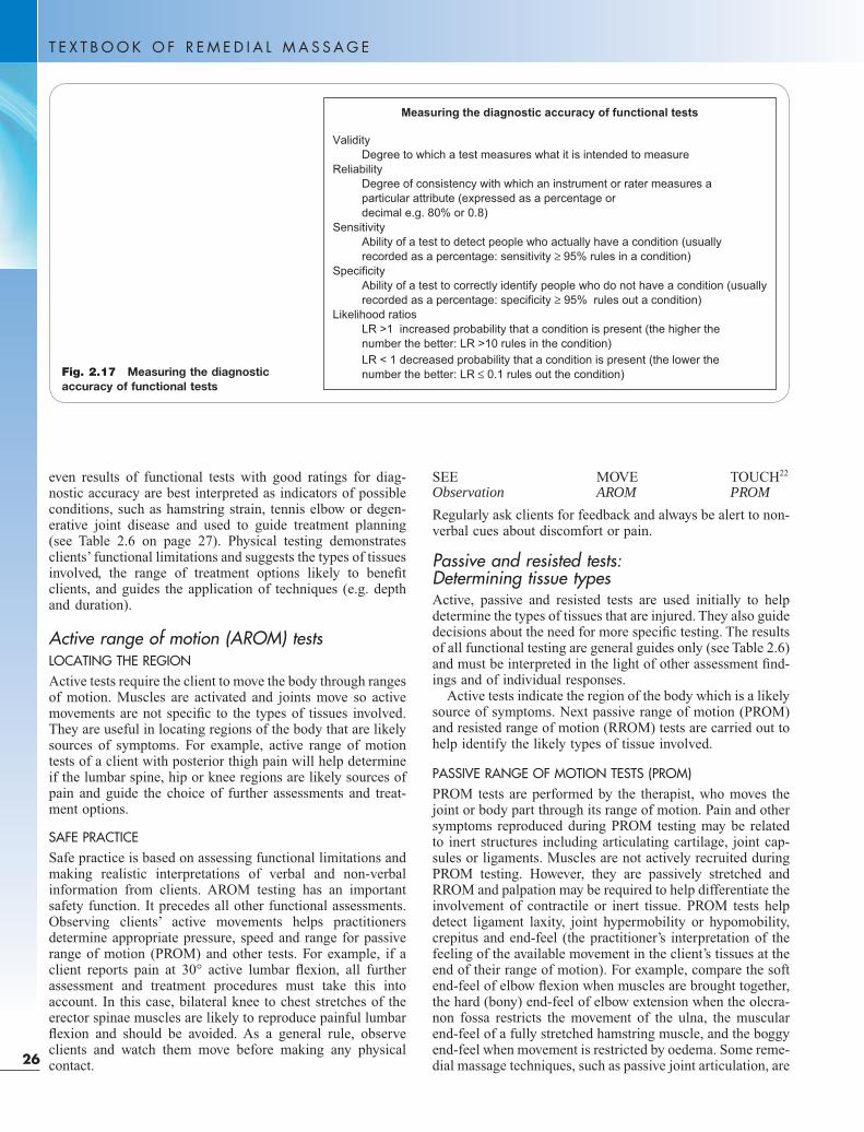
Guidelines Group 7 has urged practitioners to interpret clinical signs elicited during physical assessment cautiously. ‘ Eyeball-ing ’ a client ’ s active range of motion (i.e. measuring by eye without the use of a goniometer or other measuring tool) is particularly unreliable in clinical practice. Many functional tests have not been fully researched; however, increasingly, information about their diagnostic accuracy is becoming available. Measures of sensitivity (the probability of a posi-tive test among clients with the condition) and specifi city (the probability of a negative test among clients who don ’ t have the condition) are available for some tests, however such measures are really only useful when they are very high (≥ 95%) to help rule a condition in or out. 15
Likelihood ratios (LRs) are another measure of the useful-ness of diagnostic tests. LRs greater than 1 indicate an increased probability that a condition is present. LRs of 10 or more usually rule in a condition. LRs less than 1 indicate a decreased probability that a condition is present. LRs of 0.1 or less usually rule out a condition. 21 Figure 2.17 summarises common measures used to rate the diagnostic accuracy of functional tests.
The decision to perform diagnostic tests is based on client history and other fi ndings that indicate the likelihood of a condition and also whether it is important to have a defi nitive diagnosis. Remember that in many cases it is not possible or necessary to have a defi nitive diagnosis of the cause of acute musculoskeletal pain to have effective management of the condition. 7 Where diagnostic tests are appropriate, tests with the highest diagnostic accuracy are preferred. However,
Deceleration If the hamstrings are weak heel strike can be excessively hard and the knee may hyperextend (back knee gait).
Findings from gait analysis should be correlated with fi nd-ings from other assessments, particularly length and strength tests for specifi c muscles which are detailed in Chapters 10 – 20 and assessment of the sacroiliac joints, and hip, knee, ankle and foot regions ( Chapters 10, 18, 19 and 20 respectively). Further assessments such as muscle fi ring patterns (see Fritz et al 14 ) and neuromuscular treatments like muscle energy techniques (see Chaitow 20 ) are beyond the scope of this book but will be of interest to remedial massage therapists who use advanced soft tissue techniques.
FUNCTIONAL TESTS As part of their assessment procedures, remedial massage therapists use a range of functional tests. These include active and passive range of motion, resisted muscle tests and other tests of functional limitation (e.g. tests for the strength of specifi c muscles, muscle length tests and other special tests including straight leg raising, Valsalva, Adam ’ s test), and less commonly vital signs (blood pressure, temperature, pulse rate). Active, passive and resisted tests are usually performed before more specifi c tests.
Although functional tests are widely used by physical therapists including physiotherapists, chiropractors and osteo-paths, several studies have found that many tests lack validity and reliability. The Australian Acute Musculoskeletal Pain
Table 2.4 Common gait problems
Stance Phase
Antalgic gait (often minimising the duration of stance phase)
Any condition causing pain, especially in the weight-bearing limb or lower back (e.g. shoe problems, DJD ankle, knee, hip, lumbar disc herniation)
Avoiding heel strike (e.g. hopping onto fl at foot) Heel spurs, bruise, bursitis
Drop foot gait Weak tibialis anterior
Trendelenburg gait The pelvis drops and the trunk lurches to the unaffected side to maintain balance
Weak gluteus medius
Hip extensor gait Immediately after heel strike, the hip is thrust forward and the trunk thrown into extension
Weak gluteus maximus
Back knee gait to lock the knees into extension, pushing knee into extension with hand
Weak quadriceps
Unstable knee which may suddenly buckle into fl exion Dislocating knee cap, torn menisci, torn collateral ligaments
Walking on the balls of the feet Gastrocnemius/soleus shortening or tear; heel or foot pain (antalgic gait)
Flat foot gait, no forceful toe-off Weak gastrocnemius/soleus; hallux rigidus
Swing Phase
Steppage gait (lifting knee high) Weak dorsifl exors
Abnormal hip rotation in acceleration Weak quadriceps
Heavy heel strike Weak hamstrings
26
T E X T B O O K O F R E M E D I A L M A S S A G E
SEE MOVE TOUCH 22
Observation AROM PROM
Regularly ask clients for feedback and always be alert to non-verbal cues about discomfort or pain.
Passive and resisted tests: Determining tissue types Active, passive and resisted tests are used initially to help determine the types of tissues that are injured. They also guide decisions about the need for more specifi c testing. The results of all functional testing are general guides only (see Table 2.6 ) and must be interpreted in the light of other assessment fi nd-ings and of individual responses.
Active tests indicate the region of the body which is a likely source of symptoms. Next passive range of motion (PROM) and resisted range of motion (RROM) tests are carried out to help identify the likely types of tissue involved.
PASSIVE RANGE OF MOTION TESTS (PROM) PROM tests are performed by the therapist, who moves the joint or body part through its range of motion. Pain and other symptoms reproduced during PROM testing may be related to inert structures including articulating cartilage, joint cap-sules or ligaments. Muscles are not actively recruited during PROM testing. However, they are passively stretched and RROM and palpation may be required to help differentiate the involvement of contractile or inert tissue. PROM tests help detect ligament laxity, joint hypermobility or hypomobility, crepitus and end-feel (the practitioner ’ s interpretation of the feeling of the available movement in the client ’ s tissues at the end of their range of motion). For example, compare the soft end-feel of elbow fl exion when muscles are brought together, the hard (bony) end-feel of elbow extension when the olecra-non fossa restricts the movement of the ulna, the muscular end-feel of a fully stretched hamstring muscle, and the boggy end-feel when movement is restricted by oedema. Some reme-dial massage techniques, such as passive joint articulation, are
even results of functional tests with good ratings for diag-nostic accuracy are best interpreted as indicators of possible conditions, such as hamstring strain, tennis elbow or degen-erative joint disease and used to guide treatment planning (see Table 2.6 on page 27 ). Physical testing demonstrates clients ’ functional limitations and suggests the types of tissues involved, the range of treatment options likely to benefi t clients, and guides the application of techniques (e.g. depth and duration).
Active range of motion (AROM) tests LOCATING THE REGION Active tests require the client to move the body through ranges of motion. Muscles are activated and joints move so active movements are not specifi c to the types of tissues involved. They are useful in locating regions of the body that are likely sources of symptoms. For example, active range of motion tests of a client with posterior thigh pain will help determine if the lumbar spine, hip or knee regions are likely sources of pain and guide the choice of further assessments and treat-ment options.
SAFE PRACTICE Safe practice is based on assessing functional limitations and making realistic interpretations of verbal and non-verbal information from clients. AROM testing has an important safety function. It precedes all other functional assessments. Observing clients ’ active movements helps practitioners determine appropriate pressure, speed and range for passive range of motion (PROM) and other tests. For example, if a client reports pain at 30 ° active lumbar fl exion, all further assessment and treatment procedures must take this into account. In this case, bilateral knee to chest stretches of the erector spinae muscles are likely to reproduce painful lumbar fl exion and should be avoided. As a general rule, observe clients and watch them move before making any physical contact.
Fig. 2.17 Measuring the diagnostic accuracy of functional tests
Measuring the diagnostic accuracy of functional tests
ValidityDegree to which a test measures what it is intended to measure
ReliabilityDegree of consistency with which an instrument or rater measures aparticular attribute (expressed as a percentage ordecimal e.g. 80% or 0.8)
SensitivityAbility of a test to detect people who actually have a condition (usuallyrecorded as a percentage: sensitivity ≥ 95% rules in a condition)
SpecificityAbility of a test to correctly identify people who do not have a condition (usuallyrecorded as a percentage: specificity ≥ 95% rules out a condition)
Likelihood ratiosLR >1 increased probability that a condition is present (the higher thenumber the better: LR >10 rules in the condition)LR < 1 decreased probability that a condition is present (the lower thenumber the better: LR ≤ 0.1 rules out the condition)

A N E V I D E N C E - B A S E D A P P R O A C H 2
27
Mobilisers tend to be superfi cial, span two joints and contain fast twitch fi bres for power rather than endurance. Stabilisers, on the other hand, are usually deep, cross one joint and are made up of slow twitch fi bres for endurance; they contribute to habitual postures. Mobilisers tend to become short and tight and can inhibit the action of stabilisers that may have become weak and long. Commonly shortened muscles include upper trapezius, pectoralis major, sternocleidomastoid, erector spinae, quadratus lumborum, iliopsoas, hamstring muscles
performed near the end of normal physiological range to stretch joint capsules and other tissues around the joint, to prevent the formation of scar tissue and to improve mobility. Stretching beyond the normal physiological limit may result in tissue damage. Sensitivity to tissue tightening near the limits of normal physiological movement is important for safe practice. Remedial massage therapists are strongly urged to observe AROM before PROM and other assessment and treatment procedures (See-Move-Touch) to determine broad parameters of clients ’ comfortable movement ranges. Crepitus is sometimes heard and felt and, when related to joint move-ment, can indicate cartilage wear in the joint space.
Many texts describe capsular patterns of joint limitations in clients with degenerative joint disease or joint infl ammation (see Table 2.5 ). They are included here because they are com-monly reported in clinical practice. However, the concept of capsular pattern does not appear to be supported by research evidence. 23, 24
RESISTED RANGE OF MOTION TESTS (RROM) RROM tests are used to test contractile tissue in specifi c ranges of motion. Position the client ’ s body region or joint (usually in mid-range) and instruct them to hold the position against your resistance. Resistance normally lasts for 3 – 5 seconds. RROM tests help locate muscle groups according to their actions. For example, performing an RROM test of knee extension tests the integrity of the knee extensor muscles as a group but will not differentiate between the rectus femoris, vastus medialis, intermedialis or lateralis muscles. Further testing, such as resisted tests for specifi c muscles and palpa-tion, are required to help identify individual muscles.
Tests for specifi c muscles Resisted tests and length tests for specifi c muscles are of particular interest to remedial massage therapists who often focus their treatments on assessing and treating muscle imbal-ances (e.g. relationships between agonists and antagonists, patterns of kinetic dysfunction, muscle tightness or weakness, impacts on postural alignment, and compensatory patterns). Muscle imbalances have been associated with the develop-ment of almost all musculoskeletal pain and dysfunction. Muscles can be classifi ed as mobilisers and stabilisers.
Table 2.5 Capsular patterns
Joint Limitation of range of motion
Cervical spine Lateral fl exion = rotation > extension
Thoracic spine Lateral fl exion = rotation > extension
Lumbar spine Lateral fl exion = rotation > extension
Shoulder Lateral rotation > abduction > medial rotation
Elbow Flexion > extension
Wrist Flexion = extension
Metacarpophalangeal Flexion > extension
Distal interphalangeal Flexion > extension
Proximal interphalangeal Flexion > extension
Hip Flexion, abduction, medial rotation > extension
Knee Flexion > extension
Ankle Plantarfl exion > dorsifl exion
Subtalar Inversion > eversion
2nd – 5th metatarsophalangeal
Flexion > extension
1st metatarsophalangeal Extension > fl exion
Table 2.6 Direction of treatment following active, passive and resisted range of motion tests
Type of test Indications Implications for treatment
Active range of motion (AROM)
Helps locate musculoskeletal region that is the likely source of symptom, e.g. helps determine if symptoms in the upper limb emanate from the cervical spine or shoulder joint
Long-term management usually involves treatment of the source of symptoms. Client education about the likely source of symptoms
Passive range of motion (PROM)
Inert tissue (e.g. the articulation itself, ligaments, joint capsule, bursae) (Note: muscles are also passively stretched during PROM testing)
Treatment focuses on inert structures: Swedish massage, thermotherapy, passive joint articulation, myofascial release, range of motion exercises for joint mobility, joint stabilisation, etc
Resisted range of motion (RROM)
Contractile tissue (e.g. major muscles performing a movement tested as a group) Normally followed by specifi c muscle testing to isolate involved muscle(s)
Treatment focuses on muscle function: Swedish massage, thermotherapy, deep transverse friction, trigger point therapy, soft tissue releasing techniques, muscle stretching and strengthening

28
T E X T B O O K O F R E M E D I A L M A S S A G E
(e.g. hip fl exors may become shortened after prolonged periods of sitting), adaptive responses to other conditions (e.g. the presence of a trigger point, scoliosis or a leg length dis-crepancy) and ageing. When a shortened muscle is identifi ed, muscle-stretching techniques are usually applied to restore its normal length. Stretching can be performed as part of consul-tation and/or prescribed as home exercises. For example, the length of the hamstring muscles can be assessed by straight leg raising when the client is supine. Shortness is assessed by comparison to population norms (normal hamstring length permits approximately 65 – 80 ° straight leg raising) and/or by bilateral comparison. A range of stretching techniques can be applied to the posterior thigh after Swedish massage or heat application has warmed the area (see Chapter 5 ). Length tests of some of the major muscles of the body are included in Table 2.7 .
Positioning the client � Muscle should be placed in the fully elongated position. � As far as possible isolate the muscle across one joint. � Bony landmarks used to measure should be palpable and
in proper alignment.
and hip adductors. Shortened hamstrings, adductors and hip fl exors are often associated with weakened and lengthened transversalis abdominis and posterior gluteal muscles.
Specifi c muscle testing to identify correctable abnorma-lities of muscle strength and length may be prompted by (1) postural observation (e.g. hyperlordotic posture may prompt testing bilateral iliopsoas muscles for shortness), or (2) resisted muscle testing (RROM) in an initial screening that suggests contractile tissue as a source of symptoms.
RESISTED TESTS FOR SPECIFIC MUSCLES Resisted tests are used by many healthcare professionals to test muscle strength, and grading systems, such as the one described in the box below, have been developed. However, in remedial massage, resisted tests for specifi c muscles are more commonly used to locate possible lesions in specifi c muscles. Grading muscle strength is often restricted to bilat-eral comparisons or judgments based on clinical experience.
A typical grading system rates muscle strength on a scale from 5 to 0: 5 Normal strength: movement is possible against maximum
resistance by the examiner 4 Good: movement is possible against gravity and some
resistance by the examiner 3 Fair: movement is possible against gravity but not against
resistance by the examiner 2 Poor: movement is possible when gravity is eliminated
(i.e. testing joint in its horizontal plane) 1 Trace: muscle tightens but no movement 0 Zero: no contraction
Fig. 2.18 Differentiating (a) middle deltoid and (b) supraspinatus strains
A
B In the following example, resisted testing is used to isolate the specifi c muscles that are likely sources of the presenting symptoms in a client with shoulder pain.
� The client would be tested for shoulder range of motion (AROM, PROM, RROM). AROM tests help identify the region of the body that is the likely source of symptoms. If active shoulder abduction reproduces the pain, the injury is likely to be related to the shoulder joint or associated muscles (and not referred from the cervical spine, for example). PROM and RROM tests are required to help determine the likely tissues involved. If PROM tests do not reproduce the pain (i.e. inert structures are probably not involved) but RROM testing for shoulder abduction does, then con-tractile tissue is implicated. The client ’ s injury is probably related to the shoulder abductor muscles. Further resisted testing is required to dis-criminate between the two abductor muscles (deltoid and supraspinatus muscles) (see Figure 2.18 ).
Isolating the injured muscle(s) enables practitioners to develop targeted treatment plans which are likely to produce faster and better outcomes for clients. Resisted tests for muscles implicated in commonly occurring conditions are detailed in Chapters 10 – 20 . (For further information see Kendall McCreary et al 25 and Hislop and Montgomery 26 .)
MUSCLE LENGTH TESTS Muscle length and fl exibility both refer to the ability of a muscle to be lengthened to the end of its range of motion. 27
There are many reasons why muscles become shortened, including maintaining postures for excessively long periods

A N E V I D E N C E - B A S E D A P P R O A C H 2
29
Table 2.7 Length tests of major muscles 27, 28
Muscle – upper extremity Length test
Levator scapulae The client lies supine. The practitioner stabilises the client ’ s shoulder with one hand and fl exes, sidebends and rotates the occiput to the opposite side.
Upper trapezius The client lies supine. The practitioner stabilises the client ’ s shoulder with one hand. With the other the occiput is laterally fl exed to the opposite side and rotated to the side of testing.
Latissimus dorsi The client lies supine and fully fl exes the extended arm. Observe the distance between the extended arm and the table.
Pectoralis major The client lies supine with hand behind head. Observe the distance between the elbow and the table.
Pectoralis major – sternal (lower) portion
The client lies supine and abducts the extended arm to about 135 ° . The arm is allowed to fall into maximum horizontal abduction.
Pectoralis major – clavicular (upper portion)
As above except that the arm is abducted to 90 ° .
Continued

A N E V I D E N C E - B A S E D A P P R O A C H 2
33
� Movement should not be blocked by external supports or pillows.
� Client must be able to assume the position.
Stabilisation of proximal bony segment of the associated joint � Required to isolate intended motion. � Avoids the client substituting another muscle to perform the
motion.
End-feel � Bony (e.g elbow extension). � Capsular (e.g. medial hip rotation). � Muscular (e.g. knee extension with hip fl exion). � Soft tissue (e.g. knee fl exion).
Special (orthopaedic) tests A range of special (orthopaedic) tests can be used by remedial massage therapists to help identify specifi c conditions. Remember that fi ndings from special tests should be inter-preted cautiously, especially where the specifi city or sensitiv-ity of the tests are less than 95%, or where likelihood ratios (LR) are in the range 0.1 ≥ LR ≥ 10.
Some commonly used special tests with measures of diag-nostic accuracy are listed in Table 2.8 . Details of relevant tests will be discussed in Chapters 10 – 20 .
PALPATION Palpation is considered by many to be the most important assessment tool of massage therapists. Palpatory assessment continues throughout the treatment as an inherent function of manual therapy. Beginning students can rapidly learn to detect differences in muscle tone as they massage. With practice, palpation can be used to identify specifi c tissues (see Table 2.9 ) and to assess tissues for pathological states and stages of healing using the following indicators:� Temperature: use the dorsum of the hand just above or
lightly touching the skin to identify changes in temperature (e.g. increased temperature in acute infl ammation, decreased temperature in impaired circulation).
� Changes in normal tissue (e.g. fi brotic cords in muscles, hard pea-shaped painful trigger points, muscle fl accidity, micro-tears in musculotendinous junctions and muscle bellies).
� Presence of oedema (e.g. sponginess of tissues in acute infl ammation, pitting and non-pitting i ).
The chiropractic, osteopathic and physiotherapy literature contains a number of studies on the effi cacy of palpation. One annotated bibliography found only slight interexaminer reli-ability and moderate intraexaminer reliability for palpation. 30
Inter- and intraexaminer discrepancies were also found in a small study in which two physiotherapists identifi ed 12 ana-tomical landmarks that enabled measurement of eight joint
angles.31 Joensen et al 32 , on the other hand, found strong agreements between palpation for tendon hypertrophy. As with many other aspects of massage assessment practices, further research is needed to confi rm the effi cacy of palpation as an assessment strategy for remedial massage practice.
Palpatory fi ndings must also be recorded on clients ’ fi les. There have been several attempts at categorising palpatory fi ndings but none has been widely taken up. Recording the tissue type and any abnormal temperature, tissue quality or water content is usually suffi cient (e.g. fi brotic left upper trapezius, hot swollen right lateral ankle joint, trigger point right supraspinatus muscle).
LEGAL AND ETHICAL REQUIREMENTS FOR REMEDIAL MASSAGE ASSESSMENT
Issues of privacy, confi dentiality, child protection and the safe and effi cient use of equipment and resources are of paramount importance in all aspects of remedial massage practice, including assessment. Remedial massage assessments must be recorded accurately and in a well-organised manner so as to be easily interpreted by others. Lack of standard abbrevia-tions of key terms and recordings of remedial massage assess-ments confounds the transparency of record keeping. SOAP jand other similar charting methods are useful guides for massage therapists, however they often lack the capacity for recording the range of assessments commonly used in reme-dial massage practice. Practice management software can often be used to record assessments, copies of X-ray and CT scans and other medical reports and correspondence.
Figure 2.19 is an extract from a sample client assessment form which accommodates commonly used remedial massage assessments.
Another simple method for recording trunk ranges of motion involves drawing a star diagram to represent cervical, thoracic or lumbar motion as if observed from above (see Figure 2.20 ). Each line represents normal ROM for the move-ment. Restrictions in ranges of motion are indicated by drawing dashes, and pain is indicated by recording P on the lines of the star diagram. The closer to the centre of the star, the more marked the restriction or the earlier the onset of pain.
KEY MESSAGES
Remedial massage assessments determine the suitability of clients for remedial massage through screening for red fl ags. Those deemed suitable for remedial massage undergo further assessments to identify potential sources of symptoms, to understand clients ’ perceptions of their own functional impair-ments and to develop effective therapy plans.
For safe practice, all assessments should adhere to the fol-lowing principles:
1. See-Move-Touch; that is, clients should be observed and perform active range of motion tests fi rst before any physical contact is made with clients.
2. Assessments should be performed slowly and carefully especially when using tests that can reproduce symptoms.
i Pitting oedema results when a swollen area is pressed and an indentation persists after the pressure is released. This is the most common form of oedema. It is caused by any form of pressure (e.g. from tight socks). In non-pitting oedema pressure applied to a swollen area does not create a persistent indentation. Non-pitting oedema occurs in disorders of the lymphatic system (e.g. lymphoedema after lymph-node surgery or congenitally). It is usually observed in the arms or legs. It can also be associated with hyperthyroidism.
j SOAP charting encourages therapists to record subjective (S) and objective (O) assessments, assessment or analysis (A) and their therapy plan (P). 34

34
T E X T B O O K O F R E M E D I A L M A S S A G E
Table 2.8 Special tests commonly used by remedial massage therapists (see Chapters 10 – 20 for specifi c regional orthopaedic tests)
Adam ’ s test Test to discriminate idiopathic and functional scoliosis
Anterior draw (knee) Test for integrity of anterior cruciate ligament of the knee(SN 18 – 95, SP 86 – 100, LR + 1.8 – 8.3, LR − 0.1 – 0.5)
Anterior draw (ankle) Test for anterior talofi bular instability(SN 78 – 95, SP 74 – 84, LR + 3.1, LR − 0.29)
Apley ’ s test Test for meniscal tear in the knee(R 0.95, SN 13 – 16, SP 86 – 100, LR + 0.8 – 5.9, LR − 0.63 – 1.1)
Cervical compression test Test for pressure on a nerve root (increased peripheral pain), joint damage or ligament strain (increased local pain)
Cervical distraction Pressure on a nerve root relieved by distraction. Increased local pain suggests muscle, ligament or joint capsule damage (R 0.82, SN 40 – 50, SP 80 – 100, LR + 4.4; LR − 0.62)
Drop arm test Useful to rule in rotator cuff tear(R 0.28 – 0.66, SN 8 – 27, SP 88 – 100, LR + 2.6 – 5 LR − 0.83 – 0.94)
Elbow extension test Useful to rule out bony or joint injury(SN 97, SP 69, LR + 3.1, LR − 0.04) 29
Elevated arm stress test (Roo ’ s test) Test for thoracic outlet syndrome(SN 82, SP 100)
Empty can test Pain or weakness suggests supraspinatus damage(SN 44 – 89, SP 50 – 90, LR + 1.8 – 4.2, LR − 0.22 – 0.63)
Finkelstein ’ s test Test for tenosynovitis of the abductor pollicis longus and extensor pollicis brevis(SN 81, SP 50, LR + 1.6, LR − 0.38)
Iliac compression test Test for sacroiliac lesion
Impingement test Test for shoulder impingement syndrome: shoulder impingement suggested if pain during active fl exion; supraspinatus impingement suggested if pain when shoulder internally rotated; biceps long head implicated if pain when shoulder externally rotated.
Kemp ’ s/quadrant test Leg pain suggests nerve root compression or radiculopathy; low back pain suggests local sprain/strain or facet syndrome
Morton ’ s test Test for Morton ’ s neuroma, metatarsal joint arthritis, fracture of the metatarsal heads or metatarsalgia
Painful arc Test for impingement syndrome, rotator cuff or AC pathology (SN 33 – 98, SP 10 – 81, LR + 1.1 – 3.9, LR − 0.2 – 0.82)
Patella apprehension test Test for dislocation or subluxation of the patella(SN 7 – 37, SP 86 – 100, LR + 0.9 – 2.3, LR − 0.8 – 1.0)
Patellofemoral grind test Test for patellofemoral syndrome, DJD, osteochondritis of the patella, patella fracture (SN 29 – 49, SP 67 – 75, LR + 0.9 – 1.9, LR − 0.7 – 1.1)
Patrick ’ s/FABERE test Test for hip joint pathology, DJD, sprain/strain, fracture, tight hip adductors (SN 88)
Phalen ’ s test Test for carpal tunnel syndrome (compression of median nerve) (R 0.79, SN 10 – 88, SP 33 – 100, LR + 0.7 – 41.5, LR − 0.1 – 1)
Posterior draw (knee) Test for the integrity of the posterior cruciate ligament (SN 22 – 100, SP 99, LR + 90, LR − 0.1)
Posterior draw (ankle) Test for the integrity of the posterior ankle ligaments
PSIS asymmetry Test for sacroiliac lesion, anatomical variation(R 0.7, SN 69)
Shoulder (anterior) apprehension test Test for anterior glenohumeral instability (SN 53, SP 99, LR + 53, LR − 0.47)

A N E V I D E N C E - B A S E D A P P R O A C H 2
35
Table 2.8 Special tests commonly used by remedial massage therapists—cont’d
SI compression test Test for SI sprain, fracture, joint dysfunction (R 0.64 – 0.79, SN 25 – 69, SP 63 – 100, LR + 1.6 – 2.2, LR − 0.4 – 0.63)
SI distraction test Test for anterior SI or pubic symphysis sprain, pelvic fracture (R 0.36 – 0.94, SN 11 – 60, SP 74 – 100, LR + 1.1 – 3.2, LR − 0.5 – 0.98)
Sign of the buttock Test to distinguish whether pain on straight leg raising arises from SI joint dysfunction, bursitis, gluteal muscle strain or from lumbar spine pathology, nerve root pressure or hamstring tension
Slump test Leg symptoms suggest nerve root tension, radiculopathy, disc herniation. Local lumbosacral pain suggests lumbosacral sprain or strain (SN 83, SP 55, LR + 1.8, LR − 0.3)
Straight leg raising Test to distinguish sciatic nerve impingement, disc herniation, piriformis syndrome from SI/lumbar strain or sprain (R 0.92, SN 78 – 98, SP 11 – 64, LR + 1.03 – 2.2, LR − 0.05 – 0.86)
Thomas test Test for hip contracture, tight hip fl exors (R 0.91)
Tinel ’ s sign Test for neuropathyTinel ’ s sign at elbow (SN 70, SP 98, LR + 35, LR − 0.31)Tinel ’ s sign at wrist (R 0.5 – 0.8, SN 23 – 97, SP 53 – 100, LR + 0.7 – 11, LR − 0.03 – 1.0)
Trendelenburg test Test for gluteus medius weakness or paralysis, Legg-Calve-Perthes disease, poliomyelitis, hip dislocation, fracture (R 0.68, SN 73, SP 77, LR + 3.15, LR − 0.36)
Valgus/varus elbow stress test Test for medial/lateral collateral ligament sprain
Valgus/varus stress test (knee) Test for medial/lateral collateral ligament sprain(R 0.16, SN 86 – 100, SP 100)
Valsalva Test for space occupying lesion, disc herniation, IVF encroachment (R 0.69, SN 22, SP 94, LR + 3.67, LR − 0.82)
Yergason ’ s test Test for biceps brachii tendonitis or strain, glenoid labrum pathology, torn transverse humeral ligament(SN 9 – 50, SP 79 – 93, LR + 1.3 – 3.0, LR − 0.72 – 0.94)
(R) = reliability, (SN) = sensitivity, (SP) = specifi city, (LR) = likelihood ratios Adapted from Visniak 15
Table 2.9 Palpating tissue layers
Epidermis Place dorsum of hands lightly on the skin and compare the temperature of different parts of the body.Slide hand over the skin and note the dryness/moistness and texture of the skin.Slide fi ngers over the skin so that it wrinkles and note any resistance between the epidermis and the underlying tissue.
Muscle Muscle tissue feels fi rmer than subcutaneous tissue. Postural muscles often palpate as tight bundles of fi bres. Fibrotic tissue feels ropey.
Tendons Tendons feel like thin fi rm movable cords.
Ligaments Ligaments feel like fl at bands of tissue, less mobile and elastic than tendons.
Joint capsules Not palpable unless thickened. Articular disorders are often associated with joint line tenderness.
Peripheral nerves Peripheral nerves feel like small threads, thinner and less elastic than tendons, often fairly mobile.
Lymph glands Small soft rubbery nodes, rarely bigger than 0.5 cm.
Blood vessels Superfi cial blood vessels feel like soft tubes, pulses are often detected.
Adapted from Chaitow 33

36
T E X T B O O K O F R E M E D I A L M A S S A G E
Gait assessment
Postural observation
Rate Rhythm Symmetry
Phase
Heel strike Foot flat Midstance Push off Acceleration Swing DecelerationJoint
Muscle
Palpatory findings
Cervical
AROM Flexion Extension R lat flexion L lat flexion R rotation L rotation
PROM Flexion Extension R lat flexion L lat flexion R rotation L rotation
RROM Flexion Extension R lat flexion L lat flexion R rotation L rotation
Specificmuscle tests
Special tests Compression Distraction Valsalva
Fig. 2.19 Sample client assessment form

A N E V I D E N C E - B A S E D A P P R O A C H 2
37
Continued
Thoracic
AROM Flexion Extension R lat flexion L lat flexion R rotation L rotation
PROM Flexion Extension R lat flexion L lat flexion R rotation L rotation
RROM Flexion Extension R lat flexion L lat flexion R rotation L rotation
Specificmuscle tests
Special tests Adam’s test Deep breathing Elevated arm stress test
Shoulder
Elbow
Wrist
AROM Flexion Extension Abduction Horiz adduc Med rot Lat rot
L R L R L R L R L R L R
PROM L R L R L R L R L R L R
RROM L R L R L R L R L R L R
Specificmuscletests
Specialtests
Apley’s scratch L R Impingement L R Apprehension L RDrop arm L R Yergason’s L R Empty can test L R
AROM Flexion Extension Supination Pronation
L R L R L R L R
PROM L R L R L R L R
RROM L R L R L R L R
Specificmuscletests
Specialtests
Valgus stress L R Varus stress L R Extension test L RResisted wrist flexion L R Resisted wrist extension L R
AROM Flexion Extension Abduction Adduction Supination PronationL R L R L R L R L R L R
PROM L R L R L R L R L R L R
RROM L R L R L R L R L R L R
Specificmuscletests
Specialtests
Phalen’s test L R Tinel’s test L R Finkelstein’s test L RFig. 2.19, cont’d

38
T E X T B O O K O F R E M E D I A L M A S S A G E
Lumbar
Hip
AROM Flexion Extension R lat flexion L lat flexion R rotation L rotation
PROM Flexion Extension R lat flexion L lat flexion R rotation L rotation
RROM Flexion Extension R lat flexion L lat flexion R rotation L rotation
Specificmuscletests
Specialtests
Kemp’s test L R Valsalva SLR L R
Centralisationperipheralisation
Patrick’s test L R SI compression L R
SI distraction L R Short leg L R
AROM Flexion Extension Abduction Adduction Med rot Lat rotL R L R L R L R L R L R
PROM L R L R L R L R L R L R
RROM L R L R L R L R L R L R
Specificmuscletests
Specialtests
Patrick’s test L R Thomas test L R Trendelenburg L ROber’s test L R
Knee
AROM Flexion Extension Med rot Lat rot
L R L R L R L R
PROM L R L R L R L R
RROM L R L R L R L R
Specificmuscletests
Specialtests
Patellofemoral grind L R Patella apprehension L R Valgus stress test L R
Varus stress test L R Anterior draw L R Posterior draw L RApley’s compression L RFig. 2.19, cont’d

A N E V I D E N C E - B A S E D A P P R O A C H 2
39
Fig. 2.20 Recording trunk ranges of motion: the star diagram
A
B C
Lumbar: mildly restricted flexion moderatelyrestricted right lateral flexion
Ankle and foot
AROM Flexion Extension Inversion Eversion
L R L R L R L R
PROM L R L R L R L R
RROM L R L R L R L R
Specificmuscletests
Specialtests
Anterior draw L R Posterior draw L R Lat stability L RMorton’s test L R
Fig. 2.19, cont’d
� Functional tests� Active range of motion (AROM)
– to locate source of lesion � Passive range of motion (PROM) � Resisted tests (RROM) � Specifi c muscle tests (where appropriate)
– Resisted tests and length tests for specifi c muscles � Special (orthopaedic) tests
� Palpation� Identify tissues involved, stage of healing
Remedial massage assessments include:� Case history
� Identify red and yellow fl ags � Is the client ’ s condition mild, moderate or severe?
� Outcome measures� Monitor progress towards treatment goals
� Postural observation� Look for underlying predisposing factors to presenting
symptoms� Gait assessment
� Observe disturbances of symmetry, rate and rhythm of normal gait

40
T E X T B O O K O F R E M E D I A L M A S S A G E
Review questions
1. Complete the following sentences with a word or phrase: a) Red fl ags are ________________________________________ b) A common cause of increased kyphosis of the
cervicothoracic spine is _______________________________ c) Passive range of motion tests are used to test __________ d) A dermatome is ______________________________________ e) __________ test is used to discriminate between idiopathic
and functional scoliosis. 2. What would you do if a client came to your clinic for
assessment and treatment of pain in the upper thoracic spine, bilateral shoulders and hands, and with a history of night sweats?
3. Circle the correct answer: A client is observed to have a mild scoliosis with the right iliac crest lower than the left. It is likely that: a) the right gluteal fold is lower/higher on the right b) the gluteal cleft deviates to the left/right c) the left PSIS is lower/higher than the right PSIS d) the right foot is anterior/posterior to the left foot e) the left/right knee is slightly fl exed f ) the waist curve on the right appears to increase/decrease
4. Indicate True or False for each of the following statements. a) Using the principle of See-Move-Touch, passive tests are
conducted before active tests. True/False b) The centre of gravity passes anterior to the lateral
malleolus in the ideally aligned erect posture. True/False c) Referred pain is sharp and poorly localised. True/False d) The inferior angle of the scapula is approximately at the
level of T5 spinous process. True/False 5. What type of tissue is implicated if active shoulder fl exion is
positive and resisted shoulder fl exion is positive but passive shoulder fl exion is not? What further testing, if any, is required?
6. What type of tissue is implicated if active hip abduction is positive and passive hip abduction is positive but resisted hip abduction is not? What further testing, if any, is required?
7. How would you interpret the following assessment fi ndings for the lumbar spine: active right lateral fl exion positive, resisted right lateral fl exion positive, and passive right lateral fl exion positive? What further testing, if any, is required?
8. Describe a test for the length of the hamstring muscle. 9. What type of tissue is suggested by the following palpatory
fi ndings: a) Soft, rubbery, pea-shaped, less than 0.5 cm diameter b) Tight cord c) Small threads d) Small pulsing tubes
10. Name a possible cause for the following observations of gait: a) steppage gait b) heavy heel strike c) back knee gait d) excessive hip rotation in acceleration phase
11. What are the implications for practice of using a functional test with a sensitivity of 95%?
12. How would you interpret the lumbar ranges of motion indicated by the following star diagram?
APPENDIX 2.1 Skin cancer In Australia 70 – 80% of all skin cancers are basal cell carci-nomas, 15 – 20% are squamous cell carcinomas and less than 5% are melanomas. 35
Basal cell carcinomas usually occur in people over 40 years of age, although they may occur in younger people. They are slow growing and present as either small, raised pearly tumours with small blood vessels on the surface or a red plaque. The latter is more common on the trunk.
Squamous cell carcinomas may develop as a slowly enlarging, scaly nodule or an ulcerated sore with raised, rolled edges or a red, scaly plaque.
Malignant melanoma is a malignant change in the pigment cells of the skin. It can develop in normal skin as a fl at, coloured spot or appear as a change in an existing pigmented
lesion such as a mole. They can appear anywhere on the body. In the early stages, a melanoma is a fl at, coloured spot.
Encourage clients to seek medical attention for pigmented lesions which are either new or changing, especially if their size, colour or shape is changing.
The following checklist may be useful: 36
a. Asymmetry – one half of the lesion is dissimilar to the other
b. Border irregularity – lack of distinct edge, spread of pig-mentation into apparently normal skin
c. Colour change – red, white, blue, sudden darkening, par-ticularly shades of brown or black
d. Changes in surface – scaliness, erosion, bleeding, oozing, crusting
e. Diameter enlarging Seborrhoeic keratoses are raised, pigmented lesions which are common benign tumours occurring in people over 40

A N E V I D E N C E - B A S E D A P P R O A C H 2
41
What to look for
Any mole or freckle that has: 1. changed colour. Melanomas often develop a blue or black
colour. Sometimes areas may become lighter and many different colours such as reds, greys, blues may be found.
2. changed shape or increased in size. The change in shape is usually from an oval or round mole to an irregular ‘ coastline ’ appearance. The increase in size can be overall or simply an elevation above surrounding skin. React quickly to a fl at mole that becomes elevated, especially if the elevation is dark or a different colour from the original mole.
3. an irregular border. Most harmless moles have smooth, regular borders. Melanomas often have irregular borders.
4. become itchy or bleeds. A mole that bleeds without any signifi cant injury should be examined by a doctor. Itch may be an important symptom but only if there are other changes.
5. appeared recently from normal-looking skin, especially if it is dark or is rapidly growing.
Any pigmented lesion that looks different from the others on an individual ’ s skin should be examined closely and biop-sied if diagnosis is uncertain. (Most moles or pigmented spots on an individual resemble each other – show a family resemblance.)
Clinical features suggestive of malignancy � size greater than 6 mm � irregular shape � variation in colour within the mole � noticeable increase in size over 3 – 18 months � unexplained infl ammation in and around the mole (redness
and/or tenderness around a mole for longer than two months is suspicious)
� bleeding and itching (repeated bleeding with minor injury is unusual)
REFERENCES
1. Chow R . Complementary medicine: impact on medical practice . Curr Ther 2000 ; 41 ( 7 ): 76 – 9 .
2. Grace S , Vemulpad S , Beirman R . Training in and use of diagnostic techniques among CAM practitioners: an Australian study . J Altern Complement Med 2006 ; 12 ( 7 ): 695 – 700 .
3. Ernst E . The safety of massage therapy . Rheumatology 2003 ; 42 : 1101 – 6 .
4. Anderson T , Ogles B , Patterson C , et al . Therapist effects: facilitative interpersonal skills as a predictor of therapist success . J Clin Psychol 2009 ; 65 ( 7 ): 755 – 68 .
5. Weiss L , Blustein J . Faithful patients: the effect of long-term physician-patient relationships on the costs and use of health care by older Americans . Am J Public Health 1996 ; 86 ( 12 ): 1742 – 7 .
6. New Zealand Guidelines Group . New Zealand acute low back pain guide . Online. Available : www.nzgg.org.nz/guidelines/0072/acc1038_col.pdf ; 2004 .
7. Australian Acute Musculoskeletal Pain Guidelines Group . Evidence-based management of acute musculoskeletal pain: a guide for clinicians . Bowden Hills, Qld : Australian Academic Press ; 2004 .
8. WorkCover NSW . Training manual for providers of remedial massage therapy to injured workers . Gosford : WorkCover NSW ; 2010 .
9. Weir M . Complementary Medicine: ethics and law . Ashgrove : Prometheus Publications ; 2000 .
10. WorkCover NSW . Delivering Physical Treatment to Injured Workers – using evidence-based practice and treatment outcomes . Sydney : WorkCover NSW ; 2005 .
11. Bayar K , Bayar B , Yakut E , et al . Reliability and construct validity of the Oswestry Low Back Pain Disability Questionnaire in the elderly with low back pain . The Pain Clinic 2003 ; 15 ( 1 ): 55 – 9 .
12. Harcourt B , Wijesinha M , Harcourt G . Subjective and Objective Numerical Outcome Measure Assessment. A combined outcome measure tool:
fi ndings on a study of reliability . J Manipulative Physiol Ther 2003 ; 26 ( 8 ): 481 – 92 .
13. Beirman R . Handbook of clinical diagnosis . Sydney : R. Beirman ; 2003 .
14. Fritz S , Chaitow L , Hymel GM . Clinical massage in the healthcare setting . St Louis, MI : Mosby Elsevier ; 2008 .
15. Vizniak N . Physical assessment . Burnaby, B.C. : Professional Health Systems ; 2008 .
16. Latt M , Menz H , Fung V , et al . Walking speed, cadence and step length are selected to optimize the stability of head and pelvic accelerations . Exp Brain Res 2008 ; 184 : 201 – 9 .
17. Hoppenfeld S . Physical examination of the spine and extremities . 2nd edn . Upper Saddle River, NJ : Prentice Hall ; 2010 .
18. Magee D . Orthopedic Physical Assessment . 5th edn . Philadelphia : Saunders ; 2008 .
19. Rattray F , Ludwig L . Clinical massage therapy: Understanding, assessing and treating over 70 conditions . Elora, Ont : Talus ; 2000 .
20. Chaitow L . Muscle energy techniques . 2nd edn . Edinburgh : Churchill Livingstone ; 2001 .
21. McGee S . Simplifying likelihood ratios . J Gen Intern Med 2002 ; 17 ( 8 ): 646 – 9 .
22. Esposito S , Phillipson S . Spinal adjusting technique: The chiropractic art . Sydney : Well-Adjusted Publishing ; 2005 .
23. Klassbo M , Harms-Ringdahl K , Larsson G , et al . Examination of passive ROM and capsular patterns in the hip . Physiother Res Int (Research Support, Non-U.S. Gov ’ t) 2003 ; 8 ( 1 ): 1 – 12 .
24. Bijl D , Dekker J , van Baar ME , et al . Validity of Cyriax ’ s concept capsular pattern for the diagnosis of osteoarthritis of hip and/or knee . Scand J Rheumatol (Clinical Trial Randomized Controlled Trial Research Support, Non-U.S. Gov ’ t) 1998 ; 27 ( 5 ): 347 – 51 .
25. Kendall McCreary E , Provance P , McIntyre Rodgers M , et al . Muscles, testing and function, with posture and pain . 5th edn . Philadelphia, PA : Lippincott Williams and Wilkins ; 2005 .
26. Hislop H , Montgomery J . Daniels and Worthingham ’ s muscle testing: Techniques of manual examination . 8th edn . Oxford : Saunders Elsevier ; 2007 .
27. Reese N , Bandy W . Joint range of motion and muscle length testing . Philadelphia : W.B. Saunders ; 2002 .
28. Reiman M , Manske R . Functional testing in human performance . Champaign, IL : Human Kinetics ; 2009 .
29. Cleland J , Koppenhaver S . Netter ’ s orthopaedic clinical examination: an evidence-based approach . 2nd edn . Philadelphia : Saunders ; 2011 .
30. Haneline M , Cooperstein R , Young M , et al . An annotated bibliography of spinal motion palpation reliability studies . J Can Chiropr Assoc 2009 ; 53 ( 1 ): 40 – 58 .
31. Moriguchi C , Carnaz L , Silva L , et al . Reliability of intra- and inter-rater palpation discrepancy and estimation of its effects on joint angle measurements . J Can Chiropr Assoc 2009 ; 53 ( 1 ): 40 – 58 .
32. Joensen J , Couppe C , Bjordal J . Increased palpation tenderness and muscle strength defi cit in the prediction of tendon hypertrophy in symptomatic unilateral shoulder tendinopathy: an ultrasonographic study . Physiotherapy 2009 ; 95 : 83 – 93 .
33. Chaitow L . Palpatory literacy: they complete instruction manual for the hands on therapist . London : Thorsons Element ; 1991 .
34. Fritz S . Mosby ’ s Fundamentals of Therapeutic Massage . 4th edn . St Louis, MI : Mosby Elsevier ; 2009 .
35. Australian Cancer Council . Cancer types 2009 : Online. Available from : www.cancer.org.au .
36. Berwick M , Begg C , Fine J , et al . Screening for cutaneous melanoma by skin self-examination . J Natl Canc Inst 1996 ; 88 ( 1 ): 17 – 23 .
Useful Websites http://www.pennchiro.org/fi les/clinical/doctor/Oswestry.
years old. Solar keratoses are scaly lesions which may come and go. They occur on the sun-exposed areas of the body and are seen in 40 – 50% of people over 40 years old. These lesions are often confused with melanomas. However, their
presence indicates that a person has had suffi cient sun exposure to be at high risk of having either a basal cell or squamous cell carcinoma somewhere on the sun-exposed areas of their body.