TEXT BOOK OF BIOMECHANICS AND EXERCISE THERAPY · TEXT BOOK OF BIOMECHANICS AND EXERCISE THERAPY...
109
TEXT BOOK OF BIOMECHANICS AND EXERCISE THERAPY Dr. C.NAGAVANI, M.P.T (Neuro) Assistant professor Susruta college of physiotherapy Dilshuknagar, Hyderabad. by
Transcript of TEXT BOOK OF BIOMECHANICS AND EXERCISE THERAPY · TEXT BOOK OF BIOMECHANICS AND EXERCISE THERAPY...

TEXT BOOK OF BIOMECHANICS AND EXERCISE THERAPY
Dr. C.NAGAVANI, M.P.T (Neuro) Assistant professor Susruta college of physiotherapy Dilshuknagar, Hyderabad.
by
DEDICATED TO
My Father C.SATYAM
AND
My mother C.USHA RANI

ACKNOWLEDGEMENT
First and foremost I want to thank Almighty and My Parentsbecause of whom I’am in this position. Thanks to my brothers Subash,Suresh and Prudhvi who are always beside me in every deed I do andhelped me in completing this book. It is my pleasure and privilege to recordmy deep sense of gratitude to Mrs. Sandhya Rani for giving me oppurtunityfor writting this book.

CONTENTS
CHAPTER 1: MECHANICAL PRINCIPLES........................................1
CHAPTER 2: GAIT................................................................................12
CHAPTER 3: INTRODUCTION TO PHYSIOTHERAPY..................16
CHAPTER 4: INTRODUCTION TO EXERCISE THERAPY...........18
CHAPTER 5: STARTING POSITIONS...............................................26
CHAPTER 6: PASSIVE MOVEMENTS.............................................31
CHAPTER 7: ACTIVE MOVEMENTS..............................................36
CHAPTER 8: RELAXATION..............................................................39
CHAPTER 9: JOINT MOBILITY.......................................................48
CHAPTER 10: MUSCLE STRENGTH..............................................54
CHAPTER 11: STRETCHING............................................................56
CHAPTER 12: FRENKLE’S EXERCISES........................................59
CHAPTER 13: PNF TECHNIQUES....................................................64
CHAPTER 14: HYDROTHERAPY......................................................67
CHAPTER 15: BREATHING EXERCISES........................................71
CHAPTER 16: POSTURE....................................................................79
CHAPTER 17: EXERCISE THERAPY EQUIPMENT......................82
CHAPTER 18: WALKING AIDS AND GAIT TRAINING................96
CHAPTER 19: MASSAGE................................................................100
CHAPTER 1
MECHANICAL PRINCIPLES
DEFINITIONS:
1. The study of mechanics in the human body is referred to as biomechanics.
2. The study of the effects of internal and external forces on the human body inmovement and rest is called biomechanics.
3. Mechanical principles applied to the study of biological functions; theapplication of mechanical laws to living structures; the study and knowledge ofbiological function from an application of mechanical principles is calledbiomechanics.
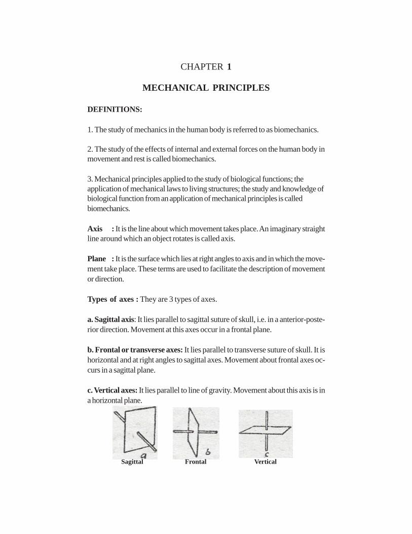
Axis : It is the line about which movement takes place. An imaginary straightline around which an object rotates is called axis.
Plane : It is the surface which lies at right angles to axis and in which the move-ment take place. These terms are used to facilitate the description of movementor direction.
Types of axes : They are 3 types of axes.
a. Sagittal axis: It lies parallel to sagittal suture of skull, i.e. in a anterior-poste-rior direction. Movement at this axes occur in a frontal plane.
b. Frontal or transverse axes: It lies parallel to transverse suture of skull. It ishorizontal and at right angles to sagittal axes. Movement about frontal axes oc-curs in a sagittal plane.
c. Vertical axes: It lies parallel to line of gravity. Movement about this axis is ina horizontal plane.
Sagittal Frontal Vertical

2 EXERCISE THERAPY
Planes: There are three planes.
1. Movement in horizontal plane (transverse plane): This plane divides thebody into upper and lower halves. Movements in transverse plane occur parallelto ground. For example in rotation of the head, the nose moves parallel to ground.Rotatory movements in a transverse plane occur around a vertical axis of mo-tion. Movement in the horizontal plane is not affected by gravity hence it is statedas gravity free movement. Weak muscles which unable to produce movementagainst gravity can often succeed in this plane.
2. Movement in frontal plane (coronal plane): the frontal plane divides thebody into front and back halves. Movements in the frontal plane occur side toside movements such as bringing the head to each of the shoulders. Rotatorymotion in the frontal plane occurs around an anterior posterior axis.
3. Movement in vertical plane (sagittal plane): An anteroposterior verticalplane passing through the body from front to back, dividing it in half. It is theplane that divides the body or body segment into the right and left parts. Move-ments in this plane include forward and backward motions such as nodding ofthe head. Rotatory motion in the sagittal plane occurs around a coronal axis.
Frontal plane Sagittal plane Transverse plane
MECHANICAL PRINCIPLES 3
KINEMATICS
Kinematics is the area of biomechanics that include description of motion with-out regard for the forces producing it. They include.
i. Types of motion.ii. Location of motion.iii. Direction of motion.iv. Magnitude of motion.
(i) Types of motion: four types of motion are there.
a. Rotatory motion: It is the movement of an object around a fixed axis in acurved path. Each point on the object or segment moves through the same angleat same distance. In human body the goal of most muscles appear to rotate abony lever around a fixed axis.
b. Translatory motion: It is the movement of an object in a straight line. Eachpoint of an object moves through the same distance, at the same time, in parallelpaths.
c. Curvilinear motion: Both rotatory and translatory motions combine to pro-duce this motion. It is the most common form of motion produced in humanjoints Ex: thrown ball, where the ball both moves through space and rotates onits own axis.
d. General plane motion: Here the object is segmented and free to move.
(ii) Location of motion: Motion at a joint may occur in transverse, frontal orsagittal planes.
(iii) Direction of Motion: Movement may occur either in clockwise oranticlockwise direction. Flexion and extension generally occur in sagittal planearound coronal axis. Flexion refers to rotation of one or both bony levers arounda joint axis so those ventral surfaces are being approximated. Rotation in thesame plane in the opposite direction is termed extension.

4 EXERCISE THERAPY
Abduction and adduction occur in frontal plane around Antero-poste-rior axis. Abduction is rotation of one or both segments of a joint around an axisso that the distal segment moves away from the midline of the body. Adductionin same plane but in opposite direction.
Rotation occurs in horizontal plane around vertical axis. Medial rotationrefers to rotation toward the body’s midline. Lateral rotation refers to the oppo-site direction.
(iv) Magnitude of Motion can be given either in degrees or radians.One radian = 57.30
10 = 0.01745 radiansGoniometer is most widely used measure for joint range in degrees.
KINETICS
Kinetics means it is the area of biomechanics concerned with the forces produc-ing motion or maintaining equilibrium. All forces are described as either Externalor Internal forces. External forces are pushes or pulls on the body arise fromsources outside the body. Ex: Gravity. Internal forces are forces act on the bodyarise from sources within human body Ex: Muscles, bones etc.
Gravity: It is force by which all the bodies are attracted to earth. It is the mostconsistent force encountered by human body and behaves in a predictable man-ner.
CENTER OF GRAVITY (COG): It is the point through which the earth’sattraction effectively acts regardless of position of body.The center of gravity (COG) of human body lies approximately at S2, anteriorto sacrum.
LINE OF GRAVITY (LOG):- It is the vertical line through centre of gravity.When the human body is in fundamental standing position, the line of gravity(LOG) pass through vertex and a point between the feet, level with transversetarsal joints.

MECHANICAL PRINCIPLES 5
BASE OF SUPPORT: It is the area which is supported. In human body baseof support is the area bounded posteriorly by tips of heels and anteriorly by aline joining the tips of toes.
EQUILIBRIUM: It results when the forces acting upon a body are balancedand the body remains at rest.
Types of equilibrium are• Stable equilibrium: If the forces acting upon a body at rest tend to
restore it to its original position after it has been displaced, the body issaid to be in stable equilibrium.
• Unstable equilibrium: If a body is given an initial displacement and theforces acting upon it increase this initial displacement, the body is said tobe in unstable equilibrium.
• Neutral equilibrium: If, inspite of displacement of a body, the heightand position of its centre of gravity remain the same in relation to thebase, the body is said to be in neutral position.
FIXATION AND STABILISATION• Fixation is the state of immobility• Stabilization is the state of relative immobility• Active fixation of joints is obtained by co-combination of muscles.• Passive fixation is by manual pressure straps, sand bags etc.
FORCE: It is that which alters the state of rest of a body or its uniform motionin a straight line
The force applies to a body is specified bya) direction of forceb) Magnitude of force
Forces are classified as external force, internal force.External force: It is supplied from a source outside the body, i.e. the force ofgravity or the pressure of physiotherapist hand.
Internal force: It is supplied by forces developed within the body i.e. by mus-cular contraction.

6 EXERCISE THERAPY
LEVER: It is a rigid bar that rotate around on axis- Forces applied to levers will produce either equilibrium or movement
such as rotation or translation.- There are 3 orders or classes of levers.- Lever is capable of producing a movement about a fixed point called
fulcrum (F).- Work is done when a force or effort (E), applied at one point on the
lever, acts upon another force or weight (W), acting at second point onlever.
- The perpendicular distance from fulcrum to effort (E) is called as effortarm and from fulcrum to weight (W) is called as weight’s arm.
Ist Order Lever: Here fulcrum is in between the effort and weight; it may besituated centrally, or towards either the effort or the weight, consequently theefforts and the weight arms may be equal, or may exceed the other in length.Ex: Nodding movements of head.Skull represents lever atlanto-occipital joints represents fulcrum, the weight issituated anteriorly in the face and the effort is supplied by contraction of poste-rior neck muscles.
First order lever

MECHANICAL PRINCIPLES 7
2nd Order Lever: The weight is in between fulcrum and effort, and the effort’sarm must therefore always exceed the weight’s arm. It helps in taking mechani-cal advantage, thus known as lever of power.
Ex: Rising of heels to stand on toesTarsal and metatarsal bones are stabilized to form lever. Fulcrum is metatar-sophalangeal joints weight of the body is transmitted to ankle joint by talus.Effort is applied by combination of calf muscles.
Second order lever
3rd Order Lever: Effort is in between fulcrum and weight, and weight arm musttherefore exceed the effort arm. It severs as mechanical disadvantage. It is con-sidered as lever of velocity as it offers more velocity and less stability.
Ex: When lever is forearm, fulcrum is elbow joint effort is supplied by contrac-tion of brachialis muscle and weight is some object held in hand.
Third order lever

8 EXERCISE THERAPY
MECHANICAL ADVANTAGE: Efficacy of force in relation to lever de-pends on two factors.They arei) Force exerted (W) or (E)ii) Perpendicular distance from fulcrum to the weight’s arm or efforts arm.
- When both weights arm and efforts arm are of equal length no advan-tage is gained.
- However if the length of effort arm exceeds weight arm an advantagewill be gained by the use of lever. This is known as Mechanical advan-tage.
- Here less effort is required to lift a weight.- Mechanical advantage is obtained in 1st order lever when fulcrum is
nearer to weight than to effort, and in all levers of the 2nd order. It isnever obtained in 3rd order lever.
- It is the ratio of weight to effortM.A = W/E
PULLEYS: Pulley is a grooved wheel which rotates about a fixed axis by arope which passes round it. The axis is supported by a frame work or block.
Types of pulleysi) Fixed pulleysii) Movable pulleys
i) Fixed Pulleys: These are used to alter the direction of force. The pulleyblock is fixed and the rope which passes round the wheel is attached to theweight at one end and the effort is applied at the other.
ii) Movable pulleys: These are used to gain mechanical advantage when liftingheavy weights. Commonly used for lifting the trunk for suspension exercises.The upper pulley is fixed to an overhead support, to which one end of rope isattached. The rope is then wound round the movable pulley, to which the weightis attached, and round the fixed pulley, the effort being applied at the free end.

MECHANICAL PRINCIPLES 9
Single Fixed Pulley Two Fixed Pulley
SPRINGS:Spiral springs are used either to resist or to assist the force of muscular contrac-tion, or to produce passive movement of joint, consist of a uniform coil of wirewhich is extensible.
Springs in Parallel Springs in Series
Springs used in parallel: When a spring of a specific weight is not availabletwo equal springs of half the required weight may be used in parallel to pro-duce the same result.

10 EXERCISE THERAPY
Springs used in series: The weight of two equal springs arranged in series issame as that of a single spring, but the amount by which they must be extendedin order to reach the limit of extension is double that required for a single spring.
ELASTICITY: It is the property of a body which regains its original shape afterthe application of force.
TYPES OF MUSCLE CONTRACTION:• Isometric• Isotonic
Isometric contraction involves the development of force by an increase inintramuscular tension without any change in length of the muscle.
Isotonic contraction increases intramuscular tension accompanied by changein length of the muscle. It may either shorten or lengthen the muscle.
Types of Muscle work:Work is the product of force and distance through which the force acts
Types arei) Static Muscle work: Muscles contract isometrically to balance
opposing forces and maintain stability. Therefore no work is done.ii) Concentric Muscle Work: the muscles contract isotonically in
shortening to produce movement.iii) Eccentric Muscle Work: the muscles contract isotonically in length-
ening. The muscle attachments are drawn apart.
Range of Muscle Work: The amount of shortening or lengthening of muscleduring contraction is about 50 percent of muscles maximum length.
Types of range are• Inner range – muscle in its shortest position• Outer range – muscle in fully extended position• Middle range – muscle is neither fully shortened nor fully extended.

MECHANICAL PRINCIPLES 11
Group Action of Muscles: Integrated activity of many muscle groups is re-quired for production of efficient functional movement. They are
i) Agonists: Group of muscles which contract to provide the force required toproduce the movement.ii) Antagonists: These muscles oppose the action of agonists and relax pro-gressively for permitting the movement.iii) Synergists: These groups of muscles work with agonists to provide a suit-able activity and facilitates the movementsiv) Fixators: These muscles stabilize the bones of origin of the agonists andincreases their efficiency for production of movement.
Limb Length Measurements:• True shortening of leg is measured from the anterior superior iliac spine
or upper margin of greater trochanter to lateral malleolus.• Apparent shortening of leg is measured from umbilicus or xiphisternum
to the level of knee joint or the tip of medial malleolus.

CHAPTER 2
GAIT
Gait may be described as a translatory progression of the body as a whole,produced by coordinated, rotatory movements of body segments.
Stages of gait:
I. stance phase: the stance phase begins at the instant that one extremity con-tacts the ground and continuous only as long as some portion of the foot is incontact with the ground.
Heel strike: the beginning of the stance phase when the heel contacts the ground.Foot flat: It occurs immediately following heel strike, when sole of the footcontacts the floor.Mid stance: the point at which the body passes directly over the referenceextremity.Heel off: the point following midstance at which time the heel of the referenceextremity leaves the ground.Toe off: the point following heel off when only the toe of the reference extremityis in contact with the ground.

GAIT 13
II. Swing phase: the swing phase begins as soon as the toe of one extremityleaves the ground and ceases just before heel strike or contact of the sameextremity.Acceleration: the portion of beginning swing from the moment the toe of thereference extremity leaves the ground to the point when the reference extremityis directly under the body.Midswing: portion of the swing phase when the reference extremity passesdirectly below the body. Midswing extends from the end of acceleration to thebeginning of deceleration.Deceleration: the swing portion of the swing phase when the reference ex-tremity is decelerating in preparation for heel strike.
Acceleration Midswing Deceleration

14 EXERCISE THERAPY
Variables:
Stance time: It is the amount of time that elapses during stance phase of oneextremity in a gait cycle.
Single-support time: It is the amount of time that elapses during the periodwhen only one extremity is on supporting surface in the gait cycle.
Double-support time: It is the amount of time that a person spends with boththe feet on the ground during one gait cycle.
Stride length: It is the linear distance from the point of heel strike of one lowerextremity to the next heel strike of the same extremity.
Step length: It is the linear distance from the point of heel strike of one lowerextremity to the next heel strike of the opposite extremity.
Stride duration: It refers to amount of time it takes to accomplish one stride.
Step duration: It refers to the amount of time spent during a single step.
Cadence: It is the number of steps taken by a person per unit of time.
Pathological gaits:
1. Antalgic or painful hip gait: this is the gait of a person with a painful condi-tion in the hip joint. To minimize the pain the person shortens the time duration ofthe stance phase on the painful side and quickly transfers the weight to the pain-less leg.
2. Stiff hip gait: when one hip is ankylosed, it is not possible to flex at the hipjoint during walking to clear the ground in the swing phase.
3. Unstable hip gait:The stability of the hip in walking is provided by the bony components of thejoint being kept in stable position by the muscles and ligaments around the joint.Any problem in these structures causes instability of hip.

GAIT 15
a. Trendelenberg gait: eg. Anatomical disruption on the right side Ex: nonunion fracture neck of femur. The action of gluteus medius in pulling the pelvisdownwards in the stance phase is ineffective or weak due to lack of a stablefulcrum. The pelvis drops on the opposite (i.e. left) side causing instability.
b. Gluteus medius gait: when the right gluteus medius is paralyzed, it is unableto pull down the pelvis on the right due to a functional deficiency of the abductormechanism in the stance phase.
4. Gluteus maximus gait: when the gluteus maximus muscle is paralyzed, thestabilizing factor is lost and the patient leans backward at the hip to passivelyextend it and keep the centre of gravity over the stance leg. This causes thebackward lurch in the gluteus maximus gait.
5. Quadriceps gait: when quadriceps power is weak or paralyzed; the lockingis done by passively pushing the knee backward by the patient putting his handover the front of the lower thigh. This results in a limp and may even cause genurecurvatum.
6. High stepping gait: when there is a foot drop, the foot slaps on the groundon heel strike and then drops in the swing phase. To get the foot clear the ground,the hip is flexed more and this causes the high stepping gait.
7. Short leg gait: inequality of the legs is obvious when the shortening of one legis more than 1”. It leads to gait with a marked pelvic tilt downwards and anequines deformity at the foot.
8. Scissoring gait: this is characteristic gait of a spastic child with marked bilat-eral spasm at the hips and equines spasm in the ankle.
CHAPTER 3
INTRODUCTION TO PHYSIOTHERAPY
Physiotherapy (also known as physical therapy) is a health profession con-cerned with and the assessment, diagnosis and treatment of disease and disabil-ity through physical means. It is based upon principles of medical science, and isgenerally held to be within the sphere of conventional (rather than alternative)medicine.
ROLE OF PHYSIOTHERAPY1. It provides psychological support for the patient in depression.2. It is useful in treating psychosomatic conditions.3. It helps in treating deformities and making the functionally independent.4. It plays a major role in treating neuro-muscular disorders.5. It improves the walking abilities.6. It relieves the pain, spasm etc.
PRINCIPLES OF TREATMENT1. To relieve pain and spasm.2. To increase joint range of motion.3. To reduce the stiffness or contractures.4. To improve muscle power.5. To prevent deformities.6. To relieve from spasticity.7. To remove the secretions from the lungs.8. To improve the breathing capacity.9. To improve the aerobic capacity.10. Improve gait pattern etc.
Methods and effects:1. Heat therapy: By heating tissues there will be rise in the temperature and
increase in the metabolitic activity. As a result of increase in the metabo-lism there is an increased demand for oxygen and foodstuffs, and anincreased output of waste products. There is blood flow to the part. Allthese physiological effects can be used to relieve the pain and spasm byincreasing the circulation and carrying the waste products. e.g. superfi-cial heating modalities are wax bath, Hydrocollateral packs etc. Deepheating modalities are SWD, IFT etc.

INTRODUCTION TO PHYSIOTHERAPY 17
2. Cryotherapy: The initial of skin to collng is vasoconstriction of bloodvessels which is useful in reducing the inflammation. Cryotherapy alsohelps in reducing the spasticity by reducing the nerve conduction veloc-ity of muscle spindles. It also reduces pain by stimulating cold receptorswhich inhibits pain carrying fibers.
3. Ultra Violet radiation thrapy is used to control skin diseases.4. Breathing exercises and postural drainage are used to reduce secretions
and improve breathing capacity.5. Passive and active exercises are used to increase joint ROM, deformi-
ties by preventing adhesion formation and maintaining circulation.6. Resisted exercises to improve strength: To improve muscle strength re-
sisted exercises can be given with the help springs, pulleys, weights etc.7. Gait training to improve gait: after any musculoskeletal and neurological
deficits the patient tends to develop abnormal gait. To correct this wehave teach gait training to the patient by using assistive devices such ascrutches, frames, orthotics etc.
These are some of the means of physiotherapy. There are so many otheruses which be dealt later in the text.
CHAPTER 4
AN INTRODUCTION TO EXERCISE THERAPY
Exercise therapy is a means of accelerating the patient’s recovery from injuriesand diseases which have altered his normal way of living.
The aims of exercise therapy1. To promote activity and minimize the effects of inactivity.2. To increase the normal range of motion.3. To strength the weak muscles.4. To improve the performance in daily activities.
The techniques of exercise therapyMovement used in treatment may be classified as follows.
I. Active movements1. Voluntary: (i) assisted (ii) Free (iii) Assisted-Resisted (iv) Resisted2. Involuntary reflex
II. Passive movementsa. (i) Relaxed Passive Movements and accessory movements
b. Passive Manual Mobilization Techniques
(i) Mobilizations of joints
(ii) Manipulations of joints performed by
(iii) Controlled sustained stretching of tightened structures
Posture: movement begins and ends in posture which is classified either asactive or passive.Active movement and posture is achieved by muscular contraction in responseto demands which are suitable to the patient’s ability.Passive movement and posture result from the application of external forceswhen the muscles are unable to contract voluntarily to permit movement or allowsupport.

INTRODUCTION TO EXERCISE THERAPY 19
The techniques which are most effective for obtaining the aims of treatment arethose which(i) Demand as much activity as possible.(ii) Based on patterns of movement which are the same as those used by thepatient for his normal functional activities.
The approach to the patient problemThe problems arising from loss of function are different for each patient thereforetreatment must be planned according to the patient need.
Assessment of the patient’s condition1. Functional tests: these are used to assess the patient’s needs and abilities
with regard to functional activities e.g. mobility, personal care etc.2. Test of range of motion: it is measured with the help of goniometer.3. Tests for neuromuscular efficiency: these may be carried out electrically,
manually or mechanically.Electrically: these may be carried out with the help of electro-myography.Manually: it can be done with manual muscle testing.Mechanically: these are done with tape measurement.
Static power test: It may be recorded by means of a spring balance capable ofregistering up to 50 or 100 lbs.
Dynamic power test: the maximum weight which can be lifted once through aprescribed range is called the one repetition maximum (1 R.M).
4. Endurance test.5. Speed test.6. Tests for co-ordination.7. Tests of sensation.8. Measurement of vital capacity.

20 EXERCISE THERAPY
GONIOMETRY
The term goniometry is derived from two greek words, gonio, meaning angle,and metron, meaning measure. Therefore, goniometry refers to the measure-ment of angles, in particular the measurement of angles created by human joints.
Types of goniometry
1. Universal goniometry: These are most commonly used instrument. Thebody of a universal goniometer resembles a protractor and may from full or halfcircle. Measurement scales are located on the body (0-180 or 0-360). It con-sists of two arms stationary or fixed arm and movable arm. Stationary can notbe moved. Movable arm is attached to the fulcrum which is the center of thebody and it can be moved. It contain a black line extend the length of the arm formeasuring the angle.
2. Gravity dependent goniometers: These are sometimes called inclinom-eters. They use gravity’s effect on pointers and fluid levels to measure joint po-sition and motion.
3. Electro goniometers: These are used primarily in research to obtain dy-namic joint measurements. It is similar to that of universal goniometer.
4. Visual estimation: Although some examiners make visual estimates of jointposition and motion but it is not a recommended position.
5. Pendulum goniometer: It consists of a 360 degree protractor with a weightedpointer hanging from the center of the protractor.
6. Fluid goniometer: It has fluid filled circular chamber containing an air bubble.It is similar to a carpenter’s level, but being circular, has a 360 degree scale.
INTRODUCTION TO EXERCISE THERAPY 21
I. UPPER LIMBShoulder joint range of motion1. FlexionRecommended testing position: supine lyingNormal ROM: 0-1800
Fulcrum : acromial processMovable arm: middle line of humerusFixed arm : midaxillary line of thorax
2. ExtensionRecommended testing position: prone lyingNormal ROM: 0-60Fulcrum : coracoid processMovable arm: lateral midline of the humerus.Fixed arm : midaxillary line of thorax
3. Abduction and AdductionRecommended testing position: supine lyingNormal ROM: 0-180Fulcrum : acromial processMovable arm: medial midline of humerusFixed arm : parallel to the midline of the anterior aspect of the sternum.
4. AdductionNormal ROM: 180-0Rest is same as abduction
5. Medial rotationRecommended testing position: supine lying, with the arm placed at 90 of ab-ductionNormal ROM: 0-70Fulcrum : olecranon processMovable arm : parallel to ulnaFixed arm : parallel or perpendicular to the floor
6. Lateral rotationNormal ROM: 0-90Rest is same as medial rotation

22 EXERCISE THERAPY
ELBOW1. Flexion and ExtensionRecommended testing position: supine lyingNormal ROM: 0-135Fulcrum : Lateral epicondyle of humerusMovable arm : lateral midline of the humerusFixed arm : midline of the humerus
2. ExtensionNormal ROM: 135-0Rest is same as flexion
FOREARM1. SupinationRecommended testing position: sitting with upper arm at the side of the body,elbow flexed to 90 and forearm supportedNormal ROM: 0-80Fulcrum : lateral to the ulnar styloid processMovable arm: ventral aspect of the forearm, proximal to styloid processFixed arm : anterior midline of humerus
2. PronationRecommended testing position: same as supinationNormal ROM: 0-80Fulcrum : lateral to the ulnar styloid processMovable arm : dorsal aspect of the forearm, proximal to styloid process ofradiusFixed arm : anterior midline of humerus
WRIST1. FlexionRecommended testing position: sitting next to a supporting surface and handfacing the ground.Normal ROM: 0-80Fulcrum : lateral aspect of the wrist over the triquetrumMovable arm : lateral midline of the fifth metacarpalFixed arm : lateral midline of the ulna

INTRODUCTION TO EXERCISE THERAPY 23
2. ExtensionRecommended testing position: same as flexionNormal ROM: 0-70Fulcrum : at the level of capitateMovable arm : volar midline of the third metacarpalFixed arm : volar midline of the forearm
3. Radial deviationRecommended testing position: same as flexionNormal ROM: 0-20Fulcrum : at the level of capitateMovable arm : dorsal midline of the third metacarpalFixed arm : dorsal midline of the humerus
4. Ulnar deviation:Normal ROM: 0-30Rest is same as radial deviation
II. LOWER LIMB
HIP JOINT1. FlexionRecommended testing position: supine lyingNormal ROM: 0-120Fulcrum : lateral aspect of the hip jointMovable arm: lateral midline of the femurFixed arm : lateral midline of the pelvis
2. ExtensionRecommended testing position: prone lyingNormal ROM: 0-30Rest is same as flexion.
3. AbductionRecommended testing position: supine lyingNormal ROM : 0-45Fulcrum : anterior superior iliac spine(ASIS) of the extremity being mea-sured
24 EXERCISE THERAPY
Movable arm : anterior midline of the femurFixed arm : horizontal line extending from one ASIS to other ASIS
4. AdductionNormal ROM: 0-30Rest is same as abduction
5.Medial rotation and Lateral rotationRecommended testing position: sitting on a supporting surfaceNormal ROM: 0-45Fulcrum : anterior of the patellaMovable arm: anterior midline of lower legFixed arm : parallel to leg
KNEE JOINT1. FlexionRecommended testing position: prone lyingNormal ROM: 0-145Fulcrum : Lateral epicondyle of the femurMovable arm : lateral midline of the femurFixed arm : lateral midline of the fibula
2. ExtensionNormal ROM:145-0Rest is same as flexion.
ANKLE JOINT1. Dorsi flexionRecommended testing position: sitting or supineNormal ROM: 0-20Fulcrum : lateral aspect of lateral malleolusMovable arm : lateral aspect of fifth metatarsalFixed arm : lateral midline of the fibula
2. Plantar flexionNormal ROM: 0-50Rest is same as dorsi flexion

INTRODUCTION TO EXERCISE THERAPY 25
3. InversionRecommended testing position: sitting with knee flexed to 90 and the lower legover the edge of supporting surfaceNormal ROM: 0-35Fulcrum : anterior aspect of the ankle midway between the malleoliMovable arm : anterior midline of the second metatarsalFixed arm : anterior midline of the lower leg
4. EversionNormal ROM: 0-15Rest is same as inversion.

CHAPTER 5
STARTING POSITIONS
Sherrington stated that every moment begins in posture and ends in posture. Thepostures from which movement is initiated are known as starting positions. Thereare five fundamental starting positions they are STANDING, KNEELING, SIT-TING, LYING and HANGING. Equilibrium and stability is maintained in thesepositions by balance of forces acting upon the body.
FUNDAMENTAL POSITIONS
1. STANDINGThis is the most difficult of all the fundamental positions to maintain as
the whole body must be balanced on a small base of support and by the coordi-nated work of many muscles. The position may be described as follows.(i) The heels are together and on the same line.(ii) The knees are together and straight.(iii) The hips are extended and laterally rotated slightly.(iv) The pelvis is balanced on the femoral heads.(v) The spine is stretched to its maximum length.(vi) The vertex is thrust upwards, the ears are levels and the eyes look
straight forwards.(vii) The shoulders are down and back.(viii) The arms hang loosely to the sides, palms facing inwards towards the
body.
Muscle work: the muscle work required to maintain the position varies with thecircumstances. It is reduced considerably when the body segments are in goodalignment and perfectly balanced.1. The intrinsic muscles of the feet working to stabilize the feet.2. The plantar flexors and dorsiflexors of the ankle working to keep the ankle
in neutral.3. The evertors, working to counterbalance the action of the invertors.4. The extensors of the knee may work slightly.5. The extensors of the hip, working to maintain hip extension and to pelvis on
the femoral heads.6. The extensors and flexors of the spine are working to keep the trunk up-
right.7. The pre-vertebral neck muscles, working to control excessive extension of
the neck.

STARTING POSITIONS 27
8. The flexors and extensors of atlanto-occipital joint, working reciprocally tobalance the head.9. The retractors of the scapulae, working to draw the scapula backwards.10. The arms relaxed by the side of the body.
Effects and uses:As the base of support is small and centre of gravity high standing is a difficultposture so this position should be given only to those who can maintain it. Bypractice in attaining and holding standing posture reduces fatigue. It is appositionof alertness, joy and efficiency.
Standing
2. KNEELING
The body is supported on the knees which may be together or slightly apart. Thelower leg rests on the floor with feet plantar-flexed. The rest of the body is heldas in standing.
Muscle work:The lower leg is relaxed; the body must be stabilized on the knees.
1. There is interplay between the flexors and extensors of the knee, tobalance the femora vertically on the knees.
2. The extensors of the hip and the flexors of the lumbar spine workmore strongly to maintain the correct angle of pelvic tilt.

28 EXERCISE THERAPY
Kneeling
3. SITTINGThe position is taken on a chair or stool, the thighs are fully supported, the hipsand knees to be flexed to a right angle and feet rest on the floor.Muscle work:The flexors of the hips work to maintain a right angle at these joints. The musclework of rest of the body is same as in standing.Effects and uses:This is a comfortable, natural and very stable position. Many non-weight bearingknee and foot exercises and lateral and rotatory movements of the spine can beperformed in this position.
Sitting

STARTING POSITIONS 29
4. LYINGIt is easiest of the fundamental positions as the body can be completely sup-ported in the supine position.Muscle Work:There is minimal muscle work in this position. When the lying position is used asthe starting position for exercise it is usually taken on a form surface.
1. Head rotators of both the sides work reciprocally to stabilizethe position of the head.
2. The extensors of the hips and flexors of the lumbar spine workto counter balance the hallowing of the back.
3. The medial rotators of the hips work to keep the legs in theneutral position.
Effects and Uses:This is an easy position so it is a suitable position for many exercises. The spineis relieved of the weight of the head and shoulders therefore it tends to elongateand straighten, hence it is used in treatment of spinal deformities. Breathing isimpeded slightly by pressure on the thorax and abdomen, so this position isunsuitable for those suffering from respiratory or heart diseases.
Lying
5. HANGINGThe body is suspended by grasping over a horizontal bar, the forearm beingpronated, the arm straight. The head is held high. The trunk and legs hang straightwith the heels together and ankle plantarflexed.Muscle Work:
1. The flexors of the fingers work strongly to grasp the bar.2. All the muscles around the wrist and elbows work to reduce the
strain on the joints.3. The adductors of the shoulders work strongly to lift the body on
the arms.4. The pre vertebral and posterior neck muscles work reciprocally
to maintain the position of the head and neck.5. The flexors of the lumbar spine and extensors of the hips work
to correct the tendency to arch the back.6. The adductors of the hips work to keep the legs together.

30 EXERCISE THERAPY
Effects and Uses:As the weight of the shoulders is taken on the spine and weight of the legs exertstraction upon it, it is straightened and elongated. Breathing is difficult in this po-sition. Therefore the position is unsuitable for respiratory or cardiac connec-tions. This position is enjoyed especially by children.
Hanging

CHAPTER 6
PASSIVE MOVEMENT
These movements are produced by an external force during muscular inactivityor when muscular activity is voluntarily reduced as much as possible to permitmovement.
Classification
a. Relaxed Passive Movements, including accessory movements.
b. Passive Manual Mobilization Techniques
(i) Mobilizations of joints
(ii) Manipulations of joints
(iii) Controlled sustained stretching of tightened structures
Specific Definitions
a. (i) Relaxed Passive Movements
These are movements performed accurately and smoothly by the Physiothera-pist. A knowledge of the anatomy of joints is required. The movements are per-formed in the same range and direction as active movements. The joint is movedthrough the existing free range and within the limits of pain.
(ii) Accessory movements
These occur as part of any normal joint movement but may be limited or. absentin abnormal joint conditions. They consist of gliding or rotational movementswhich cannot be performed in isolation as a voluntary movement but can beisolated by the physiotherapist.
b. Passive Manual Mobilization Techniques
(i) Mobilizations of joints
These are usually small repetitive rhythmical oscillatory, localized accessory, orfunctional movements performed by the physiotherapist in various amplitudeswithin the available range, and under the patient’s control. These can be donevery gently or quite strongly, and are graded according to the part of the avail-able range in which they are performed.

32 EXERCISE THERAPY
(ii) Manipulations of joints performed by
a. Physiotherapists
These are accurately localized, single, quick decisive movements of small ampli-tude and high velocity completed before the patient can stop it.
b. Surgeon/Physician
The movements are performed under anesthesia by a surgeon, or physician togain further range. The increase in movement must be maintained by the physio-therapist.
(iii) Controlled sustained stretching of tightened structures
Passive stretching of muscles and other soft tissues can be given to increaserange of movement. Movement can be gained by stretching adhesions in thesestructures or by lengthening of muscle due to inhibition of the tendon protectivereflex.
PRINCIPLES OF GIVING RELAXED PASSIVE MOVEMENTS
Relaxation. A brief explanation of what is to happen is given to the patient, whois then taught to relax voluntarily, except in cases of flaccid paralysis when this isunnecessary. The selection of a suitable starting position ensures comfort andsupport, and the bearing of the physiotherapist will do much to inspire confi-dence and co-operation in maintaining relaxation through the movement.
Fixation. Where movement is to be limited to a specific joint, the bone whichlies proximal to it is fixed by the physiotherapist as close to the joint line aspossible to ensure that the movement is localized to that joint; otherwise anydecrease in the normal range is readily masked by compensatory movementsoccurring at other joints in the vicinity.
Support. Full and comfortable support is given to the part to be moved, so thatthe patient has confidence and will remain relaxed. The physiotherapist graspsthe part firmly but comfortably in her hand, or it may be supported by axialsuspension in slings. The latter method is particularly useful for the trunk or heavylimbs, as it frees the physiotherapist’s hands to assist fixation and to perform themovement. The physiotherapist’s stance must be firm and comfortable. Whenstanding, her feet are apart and placed in the line of the movement.
Traction. Many joints allow the articular surfaces to be drawn apart by traction,which is always given in the long axis of a joint, the fixation of the bone proximalto the joint providing an opposing force to a sustained pull on the distal bone.

PASSIVE MOVEMENTS 33
Traction is thought to facilitate the movement by reducing interarticular friction.
Range. The range of movement is as full as the condition of the joints permitswithout eliciting pain or spasm in the surrounding muscles. In normal joints slightover pressure can be given to ensure full range, but in flail joints care is needed toavoid taking the movement beyond the normal anatomical limit.
As one reason for giving full-range movement is to maintain the extensibility ofmuscles which pass over the joint, special consideration must be given to muscleswhich pass over two or more joints. These muscles must be progressively ex-tended over each joint until they are finally extended to their normal length overall the joints simultaneously, e.g. the Quadriceps is fully extended when the hipjoint is extended with the knee flexed.
Speed and Duration. As it is essential that relaxation be maintained throughoutthe movement, the speed must be uniform, fairly slow and rhythmical. The num-ber of times the movement is performed depends on the purpose for which it isused.
Effects and Uses of Relaxed Passive Movements
(i) Adhesion formation is prevented and the present free range of movementmaintained. One passive movement, well given and at frequent intervals, is suffi-cient for this purpose, but the usual practice is to put the joint through two move-ments twice daily.
(ii) When active movement is impossible, because of muscular in efficiency, thesemovements may help to preserve the memory of movement patterns by stimulat-ing the receptors of kinesthetic sense.
(iii) When full-range active movement is impossible the extensibility of muscle ismaintained, and adaptive shortening prevented.
(iv) The venous and lymphatic return may be assisted slightly by mechanicalpressure and by stretching of the thin-walled vessels which pass across the jointmoved. Relatively quick rhythmical and continued passive movements are re-quired to produce this effect. They are used in conjunction with elevation of thepart to relieve oedema when the patient is unable or unwilling, to perform suffi-cient active exercise.
(v) The rhythm of continued passive movements can have a soothing effect andinduce further relaxation and sleep. They may be tried in training relaxation and,if successful the movement is made imperceptibly and progressively slower asthe patient relaxes.
.s

34 EXERCISE THERAPY
PRINCIPLES OF GIVING ACCESSORY MOVEMENTS
The basic principles of relaxation and fixation apply to accessory movements asto relaxed passive movements. Full and comfortable support is given and therange of the movement is as full as the condition of the joint permits. They arecomparatively small movements.
Effects and Uses of Accessory Movements
Accessory movements contribute to the normal function of the joint in whichthey take place or that of adjacent joints.
In abnormal joint conditions there may be limitation of these movements due toloss of full active range caused by stiffness of joints from contracture of softtissue, adhesion formation or muscular inefficiency. Accessory movements areperformed by the Physiotherapist to increase lost range of movement and tomaintain joint mobility. Hence they form an important part of the treatment of apatient who is unable to perform normal active movement.
PRINCIPLES OF PASSIVE MANUAL MOBILISATIONS AND
MANIPULATIONS
These techniques, together with their effects and uses, cover a very wide fieldwhich is beyond the scope of this book. Specific reference to books by Maitland,Grieve, Kaltenborn and other authorities on the subject is given in the bibliogra-phy.
Manipulations performed by a surgeon or physician are usually given under ageneral or local anesthetic which eliminates pain and protective spasm, and al-lows the use of greater force. Even well-established adhesions can be brokendown; but when these are numerous, it is usual to regain full range progressively,by a series of manipulations, to avoid excessive trauma and marked exudation.Maximum effort on the part of the patient and the physiotherapist must be ex-erted after manipulation to maintain the range of movement gained at each ses-sion, otherwise fibrous deposits from the inevitable exudation will form new ad-hesions.
PRINCIPLES OF GIVING CONTROLLED SUSTAINED STRETCH-ING OF TIGHTENED STRUCTURES
The patient is comfortably supported and as relaxed as possible in an appropri-ate position. With suitable fixation the part is grasped by the physiotherapist andmoved in such a way that a sustained stretch can be applied to the contracted.

PASSIVE MOVEMENTS 35
structures for a period of time within a functional pattern of movement. Me-chanical means can be used, e.g. turnbuckle plaster.
Effects and Uses of Controlled Sustained Stretching
(i) Steady and sustained stretching may be used to overcome spasticity patternsof limbs, e.g. a hemiplegic patient. The slow stretch produces a relaxation andlengthening of the muscle.
(ii) A steady and prolonged passive stretch can overcome the resistance of short-ened ligaments, fascia and fibrous sheaths of muscles as, for example, in con-trolled stretching and progressive spintage of talipes equino varus.

CHAPTER 7
ACTIVE MOVEMENTSDefinitionMovement performed or controlled by the voluntary action of muscles, workingin opposition to an external force.
Classification:Free exercise: the working muscles are subject only to forces of gravity actingupon the part moved or stabilized.Assisted exercise: when muscle strength or co-ordination is inadequate to per-form a movement an external force is applied to compensate for deficiency.Assisted-resisted exercises: muscles may be strong enough to work against re-sistance in part of the range and not in others. External forces applied are adaptedin every part of the range to the abilities of the muscles.Resisted exercises: resistance is applied to the working muscles are artificiallyand systematically increased to develop the power and endurance of muscle.
FREE EXERCISEFree exercises are those which are performed by the patient’s own muscularefforts without the assistance or resistance of any external force, other that ofgravity.Advantage: helps in maintaining range of motion by the patient itself withoutrelying on others for this purpose.Disadvantage: they frequently make insufficient demands on neuromuscular sys-tem to elicit the maximal response required for redevelopment of weak muscles.
Classification1. Localized: These exercises are designed primarily to produce some local andspecific effect, for example to mobilize a particular joint or to strengthen particu-lar muscle groups.
2. General: These exercises usually involve the use of many joints and muscles allover the body and the effect is wide spread.The technique of free exercises
1. The starting position is selected and taught to the patient with care.2. Instruction is given in a manner which will gain the interest and co-op-
eration of the patient.3. The speed at which exercise is given depends on the effect required.4. The duration of the exercise depends very largely on the patient’s ca-
pacity.

ACTIVE MOVEMENTS 37
The effect and uses of free exercises1. Relaxation: rhythmical swinging movements assist in relaxation of hyper-
tonic muscles.2. Joint mobility: normal ROM is maintained by exercises performed in full
range.3. Muscle power and tone: it is increased by tension created by the muscles.4. Neuromuscular coordination: it is improved by repetition of exercises.5. Improves confidence of patient.
Assisted exercisesWhen the force exerted on one of the body levers by muscular action is insuffi-cient for the production or control of movement, an external force may be addedto augment it. As the power of muscle increases, the assistance given must de-crease.
Technique1. Starting position and pattern of movement: this must be well known and un-derstood by the patient.2. Fixation: adequate fixation of the bone origin of prime movers improves theirefficiency.3. Support: the part of the body moved is supported throughout to reduce theload on weakened muscles.4. Antagonistic muscles: every effort must be made to reduce tension in theantagonistic muscles.5. Traction: preliminary stretching of the weak muscles to elicit the stretch reflex.6. Assisting force: the force used to augment the action of the muscles is appliedin the direction of the movement.7. The character of the movement: the movement should be smooth.8. Repetitions: the number of times the movement is repeated depends on thecondition of the patient.9. The cooperation of the patient is essential during this exercise.
Effects and uses of assisted exercise1. There will be production of movement which they are incapable of achiev-
ing.2. The memory of the pattern of co-coordinated movement is stimulated
by the correct performance.3. Patient’s confidence is increased.

38 EXERCISE THERAPY
Assisted-resisted exerciseThis type of exercise constitutes a combination of assistance and resistance dur-ing a single movement.
Resisted exerciseThe external force may be applied to the body levers to oppose the force ofmuscular contraction and there will be increase in muscle power and hypertro-phy.
Technique1. Starting position and pattern of movement: this must be well known and un-derstood by the patient.2. Fixation: adequate fixation of the bone origin of prime movers improves theirefficiency.3. Support: the part of the body moved is supported throughout to reduce theload on weakened muscles.5. Traction: preliminary stretching of the weak muscles to elicit the stretch reflex.6. Resisting force: a variety of means may be employed to supply the force usedto resist the contraction of the working muscles, e.g. manual pressure, weightsetc.7. The character of the movement: the movement should be smooth and con-trolled.8. Repetitions: the number of times the muscles are thrown into action against aresistance varies according to the condition of the patient.9. The cooperation of the patient is essential during this exercise.
Resistances: a resisting force other than that provided by gravity and frictionmay be provided by
1. Physiotherapist 2. The patient 3.weights 4. Pulleys 5. springs6. Water 7. Substances which are malleable
Effects and uses of resisted exercises1. Muscle power can only be maintained or increased by contraction.2. The blood flow to the working muscles is increased.3. There will be a general rise in blood pressure.4. Heat, which is produced as the result of strenuous muscular activity.
CHAPTER 8
RELAXATION
MUSCLES which are relatively free from tension and at rest are said to be re-laxed. Tension develops in muscles as they work during contraction and this ten-sion is reduced to a variable degree as the muscles come to rest during relaxation.
Muscle Tone
Under ordinary circumstances living muscles are never completely free from ten-sion, as they retain a quality of firmness known as muscle tone even when they areas relaxed as possible.
Muscle tone, which represents a state of preparedness in resting muscles, is nowthought to be maintained through the activity of the muscle spindle circuit. Theefferent fibres of this small fibre nervous reflex pathway transmit impulses whichproduce a sustained contraction of the small intrafusal muscle fibres of the musclespindles, while the large extrafusal fibres concerned in the production of voluntarymovement remain relaxed.
Postural Tone
The contraction which persists in the muscles concerned with the maintenance ofposture (chiefly the anti-gravity muscles) is called postural tone. Postural tone ismaintained and regulated by a reflex mechanism, the fundamental basis of which isthe myotatic or stretch reflex, although the higher centers also exert a controllinginfluence, Any stretching of the muscles by an external force, such as the force ofgravity, stimulates sensory receptors situated within the muscles themselves andso gives rise to a discharge of motor impulses to the same muscles. These motorimpulses bring about a contraction of a sufficient number of the muscles motorunits to increase the tension sufficiently to enable the effects of the force whichproduced the stretching to be counterbalanced.
As tension in these muscles is increased in response to stretching of their constitu-ent fibres by an external force, and in proportion to the degree of stretching towhich they are subjected, it follows that the use of measures tending to reduce oreliminate the effect of this force assists in promoting their relaxation.
The degree and location of postural tone varies with any alteration in posture. It isgreater in the upright positions, in which the force of gravity tends to stretch themuscles more strongly, than it is in recumbent positions, in which the effects of theforce of gravity upon them is adequately counterbalanced by full support of thebody. Those recumbent positions which provide full support for all segments ofthe body are therefore most suitable for obtaining general relaxation.

40 EXERCISE THERAPY
Voluntary Movement
Specific muscles contract as they work to initiate or control movement, but at thecompletion of the movement in question they relax and come to rest. There is arecognized biological principle that activity of living cells tends to be followed byinhibition of that activity. Con traction in any one group of muscles is accompaniedby a reciprocal relaxation of the antagonistic group to allow movement to takeplace smoothly. These facts are of importance during consideration of methodsdesigned to obtain relaxation of a particular group of muscles.
Mental Attitudes
Mental attitudes such as fear, anger and excitement give rise to a general increasein muscular tension which serves a useful purpose by preparing the muscles forrapid or forceful action.
Normally this tension, developed to serve a useful purpose, is relaxed when theneed for it no longer exists, but in some cases it persists and becomes habitualwhich may lead to alterations in normal posture.
Recognition of a state of tension followed by voluntary relaxation of the muscles inwhich it is present provide a means of helping the patient to economize in nervousenergy, and in cases where the tension has resulted in the reduction of the normalrange of movement in a joint, an increase in mobility can be achieved.
As fear in one form or another is the most usual cause of persistent tension, thephysiotherapist must do her best to reassure the patient and to gain his confidenceand co-operation. An atmosphere conducive to rest, both mental and physical,contributes much for success in helping the patient to acquire the art of voluntaryrelaxation.
Degrees of Relaxation
The degree to which muscular tension can be reduced is very variable and it isbetter to regard the term ‘Relaxation’ merely as an indication that some reductionin tension has taken place. It is often possible to estimate the degree of relaxationachieved by gentle passive movement or by palpating the muscles, as for instanceduring massage, and the fact that a patient falls to sleep during treatment is ampleproof that the method of obtaining general relaxation has been successful.
Pathological Tension in Muscles
A marked, persistent increase in muscular tension or tone is a feature of manypathological conditions which affect the nervous system. Lesions of the highermotor centres, and those which interfere with the normal function of the nervous

RELAXATION 41
pathways which connect them with the spinal reflex arc, commonly result in anabnormal state of muscular tension which varies from hypertonicity to spasticity orrigidity. A temporary reduction in this tension in the affected area can be achievedin some cases by suitable means which promote relaxation, and this allows re-education of any functional activity which remains to take place.
TECHNIQUE
GENERAL RELAXATION
Support, comfort and a restful atmosphere are basic conditions for general relax-ation and may prove effective without additional methods.
a. Support
Various forms and modifications of the lying position are used, to achieve fullsupport of the body, the relative suitability of each one varying according to thecondition of the patient and to individual preference. The weight of the body isthus effectively counterbalanced by the uniform upward pressure of a reciprocalsurface, or by suspension, in a position of semi-flexion which obviates all me-chanical tension on muscles or ligaments.
(i) Lying Supine. A firm surface is essential, and if resilient also, as in the case of agood spring mattress, it is ideal, as it will mould itself to the body contours and giveeven press and comfort. At all costs plinths or beds which sag are to be avoidedas they cramp the thorax and so throw additional strain on the inspiratory muscles.A head pillow is required which is sufficiently soft to prevent the head from rollingto either side, and to be well moulded to support the neck posteriorly. A smallpillow under the knees relieves tension on the Hamstrings and the ilio-femoralligament, and consequently allows the pelvis to roll backwards so that the lumbarspine is straightened and supported. The feet are held in the mid-position by asandbag or similar device, and each arm, slightly abducted at the shoulder andflexed at the elbow, rests on a pillow.
Lying

42 EXERCISE THERAPY
(ii) Half Lying. This is similar to the previous position but breathing is easier asthere is less weight on the back and abdominal pressure on the under surface ofthe Diaphragm is reduced.
Half Lying
An armchair makes quite a good substitute for a plinth or bed, the thighs are fullysupported and the feet rest on the floor, or a footstool, or a T footrest.
(iii) Prone Lying. The head is turned to one side and may rest on a small pillow, ifmore comfortable. A firm pillow under the hips and the lower abdomen preventshollowing of the back, and for women it should extend higher to avoid too muchpressure on the breasts; the lower leg is elevated so that the knees are slightly bentand the toes free. A degree of medial rotation at the hips, causing the heels to fallapart, still further induces relaxation of legs. Many find this position comfortableand use it for sleeping; others dislike it because of the rotated position of the head.
Prone Lying
(iv) Side Lying. The measure of relaxation obtained is governed by the efficiencywith which the shoulder and pelvic gjrdles are stabilized. The arm and leg whichare uppermost may be rested on the supporting surface instead of on pillows, butsome of the weight then falls on the trunk and this impedes respiration. The headpillow supports the neck and head in alignment with the body, and must not be toohigh. The majority of people sleep on the side, but few are conscicous of the partsuitable positioning for relaxation plays in promoting it.

RELAXATION 43
Side Lying
b. comfort
In addition to support and individual preference in positioning, for which somesuggestions have already been made, the ingredients of comfort include freedomto breathe deeply, warmth, abdominal quiescence and a mild degree of physicalfatigue. Removal of constrictive clothing, such as corsets and belts, is essential andany garters, buttons or suspenders liable to cause pressure must be removed. Theroom should be warm, but should have a free supply of fresh air; in winter addi-tional warmth can be supplied by light but warm blankets, a covered hot-waterbottle at the feet, an electric blanket or by non- luminous infra-red irradiation, butcare being taken to avoid over heating, as this leads to restlessness. For home usea warm bath gives the most even and pleasing type of heat, but its soothing effectmust not be ruined subsequently by vigorous rubbing with a towel. A light well-balanced meal, rhythmical physical activity of short duration, such as a brisk walkin the open- air, and attention to emptying the bladder before treatment are allconducive to general relaxation.
c. Restful Atmosphere
As physical and mental relaxations are interdependent, an effort must bemade to secure a state of mental rest. The treatment-room should be as quiet as-possible, as many people for whom training in relaxation is prescribed are highlysusceptible to the disturbing influence of noise. A few are worried by completesilence, but in general it is the high-pitched intermittent sound produced close athand which is to be avoided; the continuous low-pitched ‘hum’ of distant traffictends to be soothing. Bright lights and strong colours, such as red and brightyellow, are said to be stimulating, whereas a room with low well-diffused light withfor instance green and peach furnishings gives a soft and warm glow and providesan ideal setting for relaxation. This is indeed a counsel of perfection, but much canbe done with screens and shades used with a little imagination, even in a busydepartment.
44 EXERCISE THERAPY
The most difficult and important factor in the creation of a restful atmo-sphere, and one which determines the ultimate success or failure of the treatment,is the manner and bearing of the physiotherapist. She must inspire confidence, asfear, in one form or another, is at the root of much of the tension which she canhelp to relieve. Her appearance must be tidy and her dress suitable; she must bepunctual and move calmly without hurry or hesitation. Her manner must be cour-teous, pleasant and understanding and her voice low-pitched and. clear. A simpleexplanation of the routine and any instructions required are given to the patient inlanguage and terms which he can understand, so that any anxiety or fear of theunknown is removed.
It must be remembered that situations and routines with which one be-comes very familiar often appear strange and terrifying when encountered for thefirst time. Conversation, apart from these instructions, should direct the patient’sthoughts to contemplation of restful and pleasant topics.
Confidence in the physiotherapist and the treatment is gradually built up over aperiod of time; immediate results are not to be expected and are rarely achieved,often because of psychological factors beyond the control of the physiotherapistor patient. In successful cases a habit of relaxation is built up in place of a habit oftension, but the formation of new habits takes time. Regular and frequent practiceon the part of the patient is essential, until finally he becomes, an expert in the art of‘letting go’ or relaxing, and the normal rhythm of life, in which activity alternateswith relaxation, can be re-established.d. Additional Methods of promoting Relaxation
Tension may persist in spite of the provision of conditions conducive to relaxation,in which case additional methods to help the patient may be employed. Very littleshould be attempted at first, the period of time being extended as the ability torelax improves.
Consciousness of Breathing. Under conditions of quiet and comfort the patient’smind may remain active and turn to mundane problems and anxieties, with associ-ated physical tension; in this case it may help him to concentrate on his own rhythmof breathing, which must be deep with a slight pause at the end of expiration.Expiration is a phase of relaxation and should be accompanied by a feeling of‘letting go’ in the whole body.
Progressive Relaxation, A method by which relaxation may be achieved pro-gressively was devised and practiced by Jacobson of Chicago, and somethingsimilar pears in modern literature on the Yoga System as the ‘Savasana’ or ‘StillPose’.

RELAXATION 45
Savasana - The Still Pose
Contrast Method. Difficulty in appreciating the sensation of relaxation is notuncommon; the patient does not know that the muscles are tense or what to doin order to relax them. This can often be taught by demonstrating the contrastbetween maximal contraction and the degree of relaxation which follows it, thepatient being told to contract any group or series of muscles as strongly aspossible and then to ‘let go’ and ‘continue to let go’.
Success may be achieved by another method by which the patient is urged to stepup this preliminary contraction until he is so tired that he has to let go; there is alarge element of suggestion about this as it is unlikely and undesirable that a stateof fatigue should actually be produced. This method follows the biological prin-ciple that activity of living cells tends to be followed by inhibition of that activity.
Routine contraction followed by relaxation is carried out in each area of the body,the attention traveling in logical sequence from limb to limb and to the trunk andhead including the neck and face muscles until all areas can remain relaxed at oneand the same time. Much practice may be necessary before this is accomplished,and it is not unusual for the muscles of the leg, for instance, to again become tensewhile attention has been focused on relaxation of the face muscles. Before theroutine has been completed, the patient frequently drops off to sleep and generalrelaxation is obtained. When possible he should be allowed to wake naturally;alternatively, he must be wakened gently in sufficient time for getting up and dress-ing to be unhurried. Later, the patient learns to relax the muscles at will from thestate of tension in which they are normally maintained, and without previous vol-untary contraction.
Physiological Relaxation. This method of relieving tension was devised by LauraMitchell, MC.S.P., Dip.T.P., in 1957. It is based on the physiological principle ofreciprocal relaxation.
The position of tension of the whole body is defined in detail, viz. raised shoulders,bent-up elbows and hands, head and trunk flexed, etc. The patient changes theposition of every joint in turn, by exact voluntary orders which he is taught to giveto his own body, e.g., ‘Stretch the fingers out long’, ‘Stop’, ‘Feel the straight-ened-out fingers and the fingertips touching the support’.

46 EXERCISE THERAPY
In this way, the patient induces, firstly, reciprocal relaxation in the muscles that hadbeen working to maintain the tense positions, and then in the opposite group whichhe used to change that position. He registers the new position of the joints and skinpressures associated with them because these two senses reach the cortex. In thisway, he changes the pattern of tension of the whole body to one of ease by meansof a method which he can use by himself at any time, and so can stop mountingtension.
Passive Movement. Rhythmical passive movements of the limbs and head mayassist the degree of general relaxation in some cases. These movements aregenerally given as a sequel to massage.Group movements of joints, e.g. flexionand extension of hip, knee and ankle, are preferable, but a very high standard ofperformance on the part of the physiotherapist is required to obtain results. Therhythm of small pendular movements pleases some patients.The ability to promote a state of relaxation depends very largely on the individualphysiotherapist and the particular patient with whom she is dealing, and details ofsuccessful methods employed vary widely. Ideal conditions are rarely obtainableand, indeed, are hardly desirable, for many patients must eventually learn to relaxwhere and when they opportunity presents itself, e.g. in the train or on a mountaintop after a strenuous climb. General relaxation can sometimes be carried out ef-fectively in groups, as in the case of pregnant women, who tend to relax easily, andwith some asthmatic and bronchitic sufferers who have had previous individualinstruction.
LOCAL RELAXATION
General relaxation takes time and is not always essential or desirable. Methods ofobtaining local relaxation depend to some extent on the cause and distribution ofthe tension.
Preparatory to Massage and Passive Movement
Massage and passive movement both presuppose relaxation of the area undertreatment. Relaxation is obtained of a specific area by the application to that areaof the general principles already described for the whole body. A general attitudeof rest, however, will assist the process, e.g. the abducted and flexed arm sup-ported by a table or slings is more inclined to relax when the patient lies or reclinesin a chair, than when he sits bolt upright
For the Relief of Spasm
Spasm due to pain is protective and is most effectively reduced by the relief of thepain which caused it. However, if it persists because of fear of pain, techniques

RELAXATION 47
which ensure pain-free movement are often successful. Hold-relax is applicable inthese circumstances, or pendular movements which start in the free range andgradually increase in amplitude may restore confidence and achieve relaxation.
The relief of pathological spasm resulting from lesions affecting the centralnervous system is only temporary unless some voluntary control remains andcan be re-established. Temporary relief is useful to permit the re-developmentof voluntary control which is masked by the spasm and to maintain joint rangeand circulation in the affected area. The initiation of reflex movements by the useof the stretch reflex applied at the same time as a command for the patientvoluntary effort of contraction can be used for this purpose but care must betaken to ensure that spasm which is useful is not reduced by hyperactivity of theantagonistic reflex unless sufficient voluntary power is present, e.g. extensorspasm of leg which makes it possible for the patient to stand.
In preventing and combating Adaptive Shortening
Persistent tension or hypertonicity of muscles acting upon one aspect of a jointproduces a state of muscular imbalance which leads to adaptive shortening of thetense muscles and progressive lengthening and weakening of the antagonists onthe opposing aspect of the joint. Both agonistic and antagonistic muscles are inef-ficient when this situation develops. Relaxation techniques for the shortened musclesand strengthening techniques for their antagonists are followed by integration oftheir reciprocal action to establish the increase in the range of movement.

CHAPTER 9
JOINT MOBILITY
Skeletal movements occurring at joints, type of movement, range of movementdepends on anatomical structure of joint and position of muscles controlling it.
Joints are classified into synovial and nonsynovial joints.
1. Non synovial joints: are subclassified intoa) Fibrous joints: Bony components in this joint are united by thin fibrous tis-sue.Ex: coronal suture
b) Cartilaginous joints: Bony components in this joint are connected by eitherhyaline cartilage or fibro cartilage.Ex: symphysis pubis, 1st sternocoastal joint.
2) Synovial joints: are subclassified intoi) Uniaxial: Movement takes place about one axis. In a hinge joint it is flexionand extension eg: inter-phalangeal joints. In a pivot joint it is rotatory eg: atlanto-axial joint.
ii) Bi-axial: movement takes place about two axes. Ellipsoid joint-allow flex-ion, extension, abduction, adduction and circumduction. Saddle joint – such ascarpometacarpal joint of thumb.
iii) Polyaxial: Movements about many axes occurs in ball and socket joints.They allow flexion, extension, abduction, adduction, circumduction and rota-tion.
LIMITATION OF JOINT RANGE OF MOTIONThe following factors are responsible for limitation.i) Tightness of skin, superficial fascia.ii) Muscular weakness or insufficiency.iii) Adhesion formation.iv) Presence of foreign bodies in the joint.v) Tearing of intracapsular fibrocartilage.vi) Cartilaginous or bony destruction.vii) Sometimes the cause is unknown.

JOINT MOBILITY 49
MOBILISING METHODS
1. Relaxation: when spasm causes limitation of movement, relaxation leads to anincrease in range.
2. Relaxed passive movements including accessory movements: accessory move-ment is necessary to maintain or regain full joint function.
3. Passive manual mobilization techniquesi) Mobilization of jointsii) Manipulationsiii) Controlled sustained stretching.These techniques increase the mobility in joints and are followed by active exer-cise to maintain acquired range.
4. Active exercisei) Assisted exercises: rhythmical movement, in which muscular contraction andassistance combine at the limit of the free range against the resistance of thelimiting structures, is successful in increasing range.
ii) Free exercises: pendular movement is used with an attempt to increase theamplitude, series of contractions or pressing movements are performed at thelimit of the range.
iii) Resisted exercises: techniques of PNF and strengthening exercises are effec-tive for mobilization of stiff joints.
iv) Objective, occupational and diversional activities such as ball exercises, scrub-bing, hiking etc. These techniques increase the circulation, improve patient inter-est and are useful for maintaining range and to increase joint mobility.
Techniques of joint mobilization.
I. ANKLE JOINT:
Relaxed passive movements
Half lying with the patient’s knee bent over a firm pillow or across aphysiotherapist’s knee, leaving the heel unsupported. The physiotherapist placesone hand above the joint and other hand round the foot.

50 EXERCISE THERAPY
Assisted exercises for the foot
Manual assistance can be given using the same grasp as passive movements.Self assistance given by means of a rope and pulley.
Free exercises
Non-weight bearing exercises for ankle joint
1. Legs crossed sitting; foot dorsiflexion and plantarflexion.
1. Inclined long sitting; alternate foot dorsiflexion and plantarflexion.2. Sitting; alternate heel and toe raising.
Weight bearing exercises
1. Reach grasp high toe standing; heel raising and lowering.2. Reach grasp standing; foot inversion and eversion.3. High standing; walk up inclined form.
Leg cross sitting Inclined long sitting Sitting
II. KNEE JOINT
Relaxed passive movements
1. Hip and knee flexion and extension: patient in lying position, the physio-therapist gives support under the thigh with one hand and other hand graspsround the ankle. The hip and knee are then moved into flexion and exten-sion.
Assisted and assisted-resisted exercises for knee joint
1. Knee flexion and extension: Manual assistance may be given for theflexors or extensors of the knee from side lying with the limb supportedin the hands or on the surface of the plinth.

JOINT MOBILITY 51
Free exercises for the knee joint
Non-weight bearing exercises
1. Lying; one hip and flexion and extension.2. Side lying; one hip and knee flexion and extension.3. Prone lying; alternate knee flexion and extension.
Partial weight bearing exercises
1. Bicycling on free or stationary bicycle.2. Rowing on rowing machine.3. Long sitting; receive and pass ball.
Weight bearing exercises
1. Crouch position; alternate leg stretching, with or without spring.2. Prone kneeling; sit back on heels.
High sitting Prone lying
Bicycling Rowing machine

52 EXERCISE THERAPY
Long sitting; receive and pass ball
III. HIP JOINT
Relaxed passive movements
1. Hip abduction and adduction, medial and lateral rotation, flexion and exten-sion: the leg which is not to be moved is fully abducted and fixed, either by asandbag or by bending the knee over the side of the plinth, and the patientrelaxes. With the forearm supinated, one of the physiotherapist hands supportsunder the thigh, and with the other pronated she supports the lower leg at theankle joint. Traction is given and the leg is moved into abduction and adduction.Medial and lateral rotation can be performed by giving traction on the heel androlling the knee inwards and outwards with a stroking movement.
Assisted exercises for hip joint
As the limb to be moved is heavy, suspension and the use of roller skates arevaluable means of assistance.
Free exercises
Non weight bearing exercises:
1. Grasp high half standing; leg swinging forwards and backwards.2. Prone lying; leg medial and lateral rotation.
Partial weight bearing exercises
1. Heave grasp high half standing; arm stretching and one knee bending.2. Crouch position; step or spring to stride prone falling.
Weight bearing exercises
1. Half kneeling; or step standing; forward pressing.2. Stride standing; pelvis and trunk rotation.

JOINT MOBILITY 53
Grasp high half standing
Prone kneeling

MUSCLE STRENGTH 55
The prevention of muscle wastingIn flaccid paralysis
1. The affected muscles must be protected over stretching by adequatesupport and splintage.
2. The circulation of area must be maintained to ensure adequate nutritionto the paralyzed muscles by active exercise for other normal muscles inthe area, contrast baths etc.
3. The range of movement in joints immobilized by the paralysis and exten-sibility of the affected muscles must be maintained by passive move-ments.
4. Remembrance of pattern of movement must be stimulated and kept aliveby passive movement while active movement is impossible.
5. The strength and use of normal muscles in the area must be maintainedby resisted exercise.
In spastic paralysis: It can be treated by proprioceptive neuromuscular facilita-tion techniques, controlled sustained passive stretching, active or passive mobi-lization may be preceded by massage or packing with ice.
The initiation of muscle contraction:1. Warmth: moderate warmth improves the quality of contraction.2. Stabilization: stabilization of the bones of origin of the affected muscles
and of joints distal to those over which this muscles work, improvesthere efficiency.
3. Grip or manual contact: The therapist’s hands give pressure only in thedirection of the movement and give sensory stimulation.
4. Stretch: stimulation of the muscle spindles elicit reflex contraction of thatmuscle provided the reflex arc is intact.
Types of exercises used to strengthen muscles
Assisted-resisted exercises: these are rarely used to strengthen muscles exceptin cases of marked weakness.
Free exercises: free exercises are valuable as they can be practiced at regularand frequent intervals and at home.
Resisted exercises: these exercises create the tension in muscles essential forincrease in power and hypertrophy.

CHAPTER 11
STRETCHING
Stretching is a general term used to describe any therapeutic maneuver designedto increase mobility of soft tissues and subsequently improve ROM.
Flexibility is the ability to move a single joint or series of joints smoothly andeasily through an unrestricted, pain free ROM.Hypomobility refers to decreased mobility or restricted motion.Contracture is defined as the adaptive shortening of the muscle-tendon unit andother soft tissues that cross or surround a joint.
Indications for stretching1. When ROM is limited due to adhesions, contractures etc.2. Structural deformities.3. When there is muscle weakness and shortening of opposing tissue.4. Muscle soreness.
Contraindications1. Recent fracture.2. Acute inflammation.3. Bony blocks joint motion.4. When a haematoma exist.5. Hypermobility exists.
Types of stretching
Self stretching: any stretching exercise that is carried out independently by apatient after instruction and supervision by a therapist is referred to as self stretch-ing.
Static stretching: it is most common term used to describe a method by whichsoft tissues are lengthened just past the point of tissue resistance and then held ina lengthened position for an extended period of time with a sustained stretchforce.
Static progressive stretching: the shortened soft tissues are held in a com-fortably lengthened position until a degree of relaxation is felt by the patient ortherapist. Then the shortened structures are lengthened even further and againheld in the new end range position.

STRETCHING 57
Cyclic (intermittent) stretchingA relatively short-duration stretch force that is repeatedly but gradually applied,released, and then reapplied is described as a cyclic stretching.
Ballistic stretchingA rapid, forceful intermittent stretch, that is, a high speed and high intensity stretchis commonly called ballistic stretching.
Mechanical stretchingThere are many ways to use equipment to stretch a contracture and increasejoint ROM. The equipment can be simple as a cuff weight or weight pulleysystem or as sophisticated as some orthosis or automated stretching machines.
Manual stretchingA therapist applies an external force to move the involved body segment slightlybeyond the point of tissue resistance and available ROM.
Neuromuscular inhibition techniques: these procedures reflexively relax ten-sion in shortened muscles prior to or during stretching.
Hold –Relax (HR)
Procedure1. Start with the range limiting muscle in a comfortably lengthened position.2. Ask the patient to isometrically contract the tight muscle against resistance.3. Then have the patient voluntarily relax.4. The therapist moves the extremity through the gained range.5. Repeat the entire procedure.
Precautions1. The isometric contraction of the range should not be painful.2. Multiple repetitions of prestretch isometric contractions can lead to an
acute increase in arterial blood pressure.
Hold relax with agonist contraction (HR-AC)Procedure
1. Follow the same procedure as done for hold relax.2. After the patient contracts the range-limiting muscle, have the patient
perform a concentric contraction.
58 EXERCISE THERAPY
PrecautionsSame as for hold relax.
Agonist contraction (AC)Procedure
1. Passively lengthen the range-limiting muscle to a comfortable position.2. Have the patient perform a concentric contraction of the agonist muscle.3. Apply mild resistance to the contracting muscle.4. The range-limiting muscle will relax and lengthen as a result of reciprocal
inhibition.Precautions
1. Do not apply excessive resistance to the contracting muscle.2. The AC technique is least effective if the patient has already achieved
nearly full ROM.
Joint mobilization/manipulation: these are stretching techniques specificallyapplied to joint structures and are used to stretch capsular restrictions.Soft tissue mobilization and manipulation: various techniques, including fric-tion massage, myofascial release etc are designed to improve tissue mobility.
Selective stretching: it is a process whereby the overall function of a patientmay be improved by applying stretching techniques.
Determinants of stretching exercises
Intensity: The lower the intensity, the longer the time the patient will toleratestretching and soft tissues can be held in a lengthened position. The higher theintensity, the less frequently the stretching intervention can be applied.
Duration: stretch duration of 15, 30, 45, or 60 seconds or 2 minutes to lowerextremity musculature have produced significant gains in ROM.
Frequency: frequency of stretching needs to occur a minimum of two times perweek.
Dr. H. S. Frenkel was Medical Superintendent of the Sanatorium ‘Freihof’ inSwitzerland towards the end of the last century. He made a special study oftabes dorsalis and devised a method of treating the ataxia, which is a prominentsymptom of the disease, by means of systematic and graduated exercises. Sincethen his methods have been used to treat the incoordination which results frommany other diseases, e.g. disseminated sclerosis.
He aimed at establishing voluntary control of movement by the use of any part ofthe sensory mechanism which remained intact, notably sight, sound and touch,to compensate, for the loss of kinaesthetic sensation. The process of learningthis alternative method of control is similar to that required to learn any newexercise, the essentials being—
a. Concentration of the attention.
b. Precision.
c. Repetition.
The ultimate aim is to establish control of movement so that the patient is ableand confident in his ability to carry out those activities which are essential forindependence in everyday life.
Technique
1. The patient is positioned and suitably clothed so that he can see the limbsthroughout the exercise.
2. A concise explanation and demonstration of the exercise is given before move-ment is attempted, to give the patient a clear mental picture of it.
3. The patient must give his full attention to the performance of the exercise tomake the movement smooth and accurate.
4. The speed of movement is dictated by the physiotherapist by means of rhyth-mic counting, movement of her hand, or the use of suitable music.
5. The range of movement is indicated by marking the spot on which the foot orhand is to be placed.
6. The exercise must be repeated many times until it is perfect and easy. It is thendiscarded and a more difficult one is substituted.
7. As these exercises are very tiring at first, frequent rest periods must be al-lowed. The patient retains little or no ability to recognize fatigue, but it is usuallyindicated by deterioration in the quality of the movement, or by a rise in
CHAPTER 12
FRENKEL’S EXERCISES

60 EXERCISE THERAPY
in the pulse rate.
Progression
Progression is made by altering the speed, range and complexity of the exercise.Fairly quick movements require less control than slow ones. Later, alteration inthe speed of consecutive movements and interruptions which involve stoppingand starting to command, are introduced. Wide range and primitive movements,in which large joints are used, gradually give way to those involving the use ofsmall joints, limited range and a more frequent alteration of direction. Finallysimple movements are built up into sequences to form specific actions whichrequire the use and control of a number of joints and more than one limb, e.g.walking.
According to the degree of disability, re-education exercises start in lying withthe head propped up and with the limbs fully supported and progress is made toexercises in sitting, and then in standing.
Examples of Frenkel’s exercises
Exercise for the legs in lying.
a. lying (Head raised); Hip abduction and adduction.
The leg is fully supported throughout on the smooth surface of a plinth or on a re-education board.
b. lying (Head raised); one Hip and Knee flexion and extension.The heel is sup-ported throughout and slides on the plinth to a position indicated by the physio-therapist.
c. lying (Head raised); one Leg raising top/ace Heel on specified mark. Themark may be made on the plinth, on the patient’s other foot or shin, or the heelmay be placed in the palm of the physiotherapist’s hand.
Exercise for the legs in lying

FRENKEL’S EXERCISES 61
d. lying (Head raised); Hip and Knee flexion and extension, abduction and ad-duction.
The legs may work alternately or in opposition to each other. Stopping andstarting during the course of the movement may be introduced to increase thecontrol required to perform any of these exercises.
Exercise for the legs in sitting.
e. sitting; one Leg stretching, to slide Heel to a position indicated by a mark onthe floor.
f. sitting; alternate Leg stretching and lifting to place Heel or Toe on specifiedmark.
g. stride sitting; change to standing and then sit down again.
The feet are drawn back and the trunk inclined forwards from the hips to get thecentre of gravity over the base. The patient then extends the legs and drawshimself up with the help of his hands grasping the wall-bars or other suitableapparatus.
Exercise for the legs in sittingExercise for the legs in standing.
h. stride standing; transference -of weight from Foot to Foot.
i. stride standing; walking sideways placing Feet on marks on the floor.
Some support may be necessary, but the patient must be able to see his feet.
j. standing; walking placing Feet on marks.
The length of the stride can be varied by the physiotherapist according to thepatient’s capacity.
k. standing; turn round.

62 EXERCISE THERAPY
Patients find this difficult and are helped by marks on the floor.
1. Standing; walking and changing direction to avoid obstacles.
Group work is of great value as control improves, as it teaches the patient toconcentrate on his own efforts without being distracted by those of other people.In walking, he gains confidence and becomes accustomed to moving about withothers, to altering direction and stopping if he wishes, to avoid bumping intothem. The ability to climb stairs and to step on and off a kerb helps him toindependence.
Diversional activities such as plaiting, building with toy bricks, or drawing on ablackboard, lead to more useful movements such as using a knife and fork,doing up buttons and doing the hair.
Exercises for the arms.
m. Sitting (one Arm supported on a table or in slings); Shoulder flexion or exten-sion to place Hand on a specified mark.
n. Sitting; one Arm stretching, to thread it through a small hoop or ring.
o. Sitting; picking up objects and putting them down on specified marks.
EXERCISES TO PROMOTE MOVEMENT AND RHYTHM
All exercises are repeated continuously to a rhythmic count, or to suitable music.
a. sitting; one Hip flexion and adduction (to cross one Thigh over the other), themovement is then reversed and repeated.
b. half lying; one Leg abduction to bring Knee to side of plinth, followed by oneKnee bending to put Foot on floor, the movement is then reversed and repeated.
c. sitting, lean forward and take weight on Feet (as if to stand), then sit downagain. Later this can be done progressing along the seat as if moving up to makeroom for someone else to sit.
d. standing; Arm swing forwards and backwards (with partner, holding twosticks).
e. standing or walking; bounce and catch, or throw and catch a ball.
Marching to music, ballroom dancing or swimming, if possible, should be en-couraged.

FRENKLE’S EXERCISE 63
standing arm swinging forward and backward
Sitting hip flexion and adduction sitting, lean forward and take weight on feet

CHAPTER 13
PROPRIOCEPTIVE NEUROMUSCULARFACILITATION
Definition:Proprioceptive: sensory receptors that give information about movement andposition of the body.Neuromuscular: involving the nerves and muscles.Facilitation: making easy.PNF is an approach in which treatment is directed at a total human being, notjust at a specific problem or body segment. This method was developed bydoctor Herman kabat and miss Margaret knott in 1946 and 1951.
Basic techniques.
1. Patterns of facilitation:Mass movement patterns are used as the basis for all the techniques of PNF asthese movements are similar to the normal functional movements. The pattern ofmovement is spiral or diagonal and they are observed in everyday use eg: intaking the hand to the mouth. Each pattern of movement has 3 components.Two components of the movement are angular and the third is rotatory eg: flex-ion adduction with lateral rotation of the leg or extension with rotation to the rightof the lower trunk.Effects and uses:This pattern of movement represents the normal movements they can be re-peated to improve movements which needs.
2. Manual contacts:Pressure of the physiotherapist’s manual contact on the patient provides a meansof applying maximal resistance to movement in patterns. Touch contributes tofacilitation by stimulating the exteroceptors. Manual contacts must be
(i) Purposeful: Pressure must be firm, so that the patient must be awareof it.
(ii) Directional: Pressure is applied only in the direction of the move-ment.
(iii) Comfortable: Manual contacts which produce painful stimuli mustbe avoided.

PROPRIOCEPTIVE NEUROMUSCULAR FACILITATION 65
3. Stretch stimulus and the Stretch reflex:Proprioceptors situated in the muscles are stimulated by stretching, which in-creases the intramuscular tension which produces reflex contraction of themuscle.A sharp but controlled stretch reflex of the muscles at the limit of theirextended range if given along with the dynamic command increases patient’seffort to perform the movement.Effects and uses:Stretch reflex when applied to weak muscles increases their response and im-proves the strength.
4. Traction and approximation:Traction and approximation may be effectivein stimulating proprioceptive impulses arising from joint structures. Traction iseffective for facilitating flexion movements whereas approximation is effectivefor facilitating extension movements.
5. Commands to the patient:The physiotherapist’s voice is used as a verbal stimulus to improve patient’seffort to perform movement. Commands should be simple, brief, accurate andwell timed.
6. Normal timing:Timing is defined as the sequence of muscle contraction occurring motor activity.
7. Maximal resistance:It is defined as the greatest amount of resistance which can be given to muscularcontraction. Maximal resistance is given both in isometric and isotonic contrac-tions and for all the three components of the movement.Effects and uses:Resistance increases the motor units of the muscles and increases the strengthand endurance of the muscles.
8. Re-inforcement:Innumerable combitions of movements are utilized in every day life. So the prop-rioceptive stimulation which results from tension in strongly contracting musclesleads to spread or overflow of excitation to the weak muscles. Therefore, re-inforcement is used as a means of obtaining the contraction of weak muscles andto correct the imbalance.

66 EXERCISE THERAPY
TECHNIQUES OF EMPHASIS
1. Repeated contractionsRepetition of activity against resistance is essential for the development ofmuscle strength and endurance. The contraction of specific weak muscles of apattern is repeated while they are being re-inforced by maximal isotonic orisometric contraction of stronger muscles.

CHAPTER 14
HYDROTHERAPYHydrotherapy refers to the use of multi-depth immersion pools or tanks thatfacilitate the application of various established therapeutic interventions includingstretching, joint mobilization, strengthening etc.
Application: To cure musculo-skeletal problems.
Indications for hydrotherapy:
1. Muscular problems: Muscular weakness, Muscle spasm, Tightness or con-tracture of muscles.
2. Bony or skeletal problems: Inflammatory conditions of spine, Arthritis ofvarious joints, Post fracture stiffness.
3. Neurological Problems: Paralytic condition like polio, spinal cord injury,brain injury.
Goals:1. Facilitate ROM exercises.2. Initiate resistance exercises.3. Facilitate weight exercises.4. Facilitate cardiovascular exercises.5. Enhance patient relaxation.
Properties of water:
Buoyancy: it is the upward force that works opposite to gravity. Buoyancyprovides the patient with relative weightlessness and joint unloading allowingperformance of active motion easily.
Hydrostatic pressure: it is the pressure exerted on the immersed objects. Thisincreased pressure reduces or limits effusion, assists venous return, induces Bradycardia, and centralizes peripheral blood flow.
Viscosity: it is friction occurring between molecules of liquid resulting in resis-tance to flow. It creates resistance to all active movements.
Surface tension: the surface of the fluid acts a membrane under tension. Sur-face tension is measured as force per unit length. An extremity that moves through

68 EXERCISE THERAPY
the surface will perform more work than if kept under water.
Hydromechanics: Hydromechanics is the physical properties and characteris-tics of fluid in motion.
Components:Laminar flow: Movements where all molecules move parallel to each other,typically slow movement.Turbulent flow: movement where molecules do not move parallel to each other,typically faster movements.Drag: the cumulative effects of turbulence and fluid viscosity acting on a objectin motion.
Center of buoyancy: the center of buoyancy is the reference point of an im-mersed object upon which buoyant forces of fluid act. In vertical position, thehuman center is located at the sternum.
Laminar flow
Turbulent flow

HYDROTHERAPY 69
Application of hydrotherapy in treatment:
1. Strengthening of muscle: the water provides a remarkable environment toproduce very fine exercise progression and it provides more resistance than air.Muscles are strengthened by resistance may be offered by upward force, turbu-lence force etc. Even manual resistance can be applied along with it.
2. Endurance training: muscular endurance refers to how many times the pa-tient can repeat a particular activity inside water. The endurance activity can beperformed against buoyancy, turbulence.
3. Joint mobility: relief of pain and muscle spasm by the warmth of the waterand by the support of buoyancy can restore free movement of joint.
4. Co-ordination and balance: the buoyancy of water relieves the patient fromweight and makes the activities like walking and step climbing easy.
5. Pain relief: hydrotherapy pool improves circulation and enable tissue fluid toflow through the tissues thus facilitates removal of metabolites and improvesnutrition.
Moment of buoyancy causing rotation of the body

70 EXERCISE THERAPY
CONTRAINDICATION:1. Infective wounds2. Hyperpyrexia3. cardiac failure4. Deep vein thrombosis.5. Gastro intestinal disorder6. Hypo or Hypertension7. Epilepsy8. Low vital lung capacity
PRECAUTIONS:
1. AIDS – The person suffering from AIDS should not be allowed in pool if anyhave a recent cut.
2. If the person is mentally retarded.
3. If the person is wearing contact lens.
4. If the person is wearing any hearing aids.
5. Patients have fear of water.
6. Cardiac dysfunction: patients with angina and abnormal blood pressure re-quire close monitoring.
7. Patients with epilepsy.

CHAPTER 15
BREATHING EXERCISES
Breathing exercises are designed to retrain the muscles of respiration, improveventilation, lessen the work of breathing, and improve gaseous exchange andpatient’s overall function in daily living activities.
Indications1. Acute or chronic lung disease: pneumonia, atelectasis, COPD etc.2. After surgeries.3. Airway obstruction due to retained secretions.4. Deficits in CNS: spinal cord injury, myopathies etc.5. As relaxation procedure.
Aims of breathing exercises:1. Improve ventilation.2. Increase the effectiveness of the cough mechanism.3. Improve the strength, endurance and coordination of respiratory muscles.4. Promote relaxation.5. Improve chest and thoracic spine mobility.
Types of breathingAll the breathing patterns should be deep, voluntarily controlled and relaxed.1. Diaphragmatic breathing:Place the patient in a relaxed position such as reclined sitting. Place your handson the rectus abdomen just below the anterior costal margin. Ask the patient tobreathe in slowly and deeply through the nose. Then tell the patient to slowly letthe air out through the mouth. Practice this for 3 or 4 times. Then ask the patientto keep his or her hand on the abdomen and practice.
2. Ventilatory muscle trainingThe process of improving strength and endurance of muscles of breathing isknown as ventilatory muscle training (VMT). This technique usually focuses onmuscles of inspiration.
a. Diaphragmatic training using weightsHave the patient assume a supine lying. Place a small weight over the epigastricregion of the abdomen. Tell the patient to breathe in deeply while trying to keepthe upper chest quiet.

72 EXERCISE THERAPY
Diaphragmatic breathing
b. Inspiratory resistance trainingThe patient inhales through a hand held resistive device that he or she place in themouth. These devices are narrow tubes of varying diameters that provide resis-tance to airflow during inspiration and improve strength of inspiratory muscles.Gradually the time is increased to 20 to 30 minutes.
c. Incentive respiratory spirometryIt is form of low-level resistance training. The patient inhales through a spirom-eter that provides visual or auditory feedback as the patient breathes in asdeeply as possible. Place the patient in a comfortable position. Have thepatient take 3 to 4 breaths and exhale with the fourth breath.Then have thepatient place the spirometer in the mouth and maximally inhale through thespirometer and hold the inspiration for several seconds.
3. Segmental breathinga. Lateral costal expansionThe patient will be in a hook-lying position. Place your hands along the lateralaspect of the lower ribs and ask the patient to breathe out, and feel the rib cagemove downward and inward.b. posterior basal expansionHave the patient sit and lean forward on a pillow. Place your hands over theposterior aspect of the lower ribs. Follow the same procedure as above.c. right middle-lobe or lingula expansionPatient is sitting. Place your hands at either the right or left side of the patient’schest just below the axilla. Follow the same procedure as above.

BREATHING EXERCISES 73
Lateral costal breathing
d. apical expansionPatient is sitting. Apply the pressure below the clavicle with the finger tips.
4. Glossopharyngeal breathingThe patient takes several gulps of air. Then the mouth is closed and the tonguepushes the air back and traps it in the pharynx. The air is then forced into thelungs and the glottis is opened. This increases the depth of the inspiration.
5. Pursed-lip breathingHave the patient assume a comfortable position. Explain to the patient that expi-ration must be relaxed and contraction abdominals must be avoided. Instruct thepatient to breathe slowly and deeply then have the patient purse the lips andexhale.

74 EXERCISE THERAPY
CoughingAn effective cough is necessary to eliminate respiration obstructions and keepthe lungs clear.
The cough mechanismThe following series of action occur when a person coughs.
1. Deep inspiration.2. Glottis closes and vocal cords tighten.3. Abdominal muscles contract and the diaphragm elevates.4. Glottis opens.5. Explosive expiration of air occurs.
Additional means of facilitating a cough
Manual assisted coughIf a patient has abdominal weakness, manual pressure on the abdominal areawill assist in developing greater intra-abdominal pressure for a more forcefulcough.
Therapist assisted techniquesWith the patient in a supine or semi-recycling position, the therapist places theheel of one hand on the patient’s abdomen at the epigastric area just distal to thexiphiod process. The other hand is kept on the first. After the patient inhales asdeeply as possible, the therapist manually assists the patient as he or she at-tempts to cough.

BREATHING EXERCISE 75
POSTURAL DRAINAGE
POSTURAL DRAINAGE is a means of mobilizing secretions in one or more lungsegments to the central airways by placing the patient in various positions so thatthe gravity assists in the drainage process. Postural drainage therapy includes themanual techniques such as percussion, vibration and voluntary coughing.
Aims1. Prevent accumulation of secretions: Chronic bronchitis, cytic fibrosis,
prolong bed rest, post surgical patients etc.,2. Remove secretions already accumulated: COPD, pneumonia, atelecta-
sis etc.,
Contraindications1. Hemorrhage2. Untreated acute conditions: congestive heart failure, pleural effusion,
pulmonary embolism etc.,3. Cardiovascular instability: Hypertension, myocardial infraction4. Recent neurosurgery
Manual techniques used during postural drainage therapy
PercussionPercussion is performed with cupped hands over the lung segment being drained.It is continued for several minutes or until the patient needs to alter position tocough.
VibrationVibration is applied by placing both the hands directly on the skin and over thechest wall and gently compressing and rapidly vibrating the chest wall as thepatient breathes out.
ShakingShaking is a more vigorous from of vibration applied during exhalation using aintermittent bouncing maneuver coupled with the wide movement of therapist’shands.

76 EXERCISE THERAPY
TREATMENT PROCEDURES
Right and left upper lobes.1. Anterior apical segments: Percussion is applied directly under the
clavicle2. Posterior apical segments: Percussion is applied directly above the
scapulae.
3. Anterior segments: Patient lies supine and percussion is applied directlyabove the nipple.
Anterior segments
4. Posterior segment (left): patient lies one-quarter turn from prone and restson right side. Head end is elevated to 18 inches. Percussion is applied over theleft scapula.

BREATHING EXERCISES 77
5. Posterior segment (right): patient lies flat one-quarter turn from prone onside. Percussion is applied over the right scapula.
1. Lateral segment (left): patient lies on the right side in a 45 head downposition. Percussion is applied over the lower lateral aspect of the left rib cage.

78 EXERCISE THERAPY
2. Lateral segment (right): patient lies on the left side in a 45 head downposition. Percussion is applied over the lower lateral aspect of the right ribcage.
3. Superior segments: patient lies prone, pillows under the abdomen to flattenthe back. Percussion is applied bilaterally, directly below the scapula.
CHAPTER 16
POSTURE
Posture is the attitude assumed by the body either with support during muscularinactivity, or by means of co-ordinated action of many muscles working to main-tain stability.
Inactive postures: these are attitudes adopted for resting or sleeping, and theyare most suitable for this purpose when all the essential muscular activity re-quired to maintain life is reduced to minimum.
Active postures: the integrated action of many muscles is required to maintainactive postures, which may be static or dynamic.Static postures: a constant pattern of posture is maintained by the interaction ofgroups of muscles which work more or less statically.Dynamic postures: the pattern of the posture is constantly modified and ad-justed to meet the changing circumstances which arise as a result of movement.
The postural mechanismThe muscles: the intensity and distribution of muscle work which is required forboth static and dynamic postures varies considerably with the pattern of pos-ture, and physical characteristic of the individual. The group of muscle mostfrequently employed are anti-gravity muscles.
Nervous control: postures are maintained are adopted as a result of neuromus-cular coordination, the appropriate muscles being innervated by means of a verycomplex reflex mechanism.The postural reflexes: a reflex is by definition, an efferent response to an afferentstimulus. The efferent response in this instance is motor one. The afferent stimu-lus is from variety of sources of the body such as muscles, eyes, ears and jointstructures.
1. Muscles: neuromuscular and neurotendinous spindles within the musclesrecord changing tension.
2. The eyes: visual sensation records any alteration in the position of thebody with regard to the surroundings.
3. The ears: stimulation of the receptors of the vestibular nerve results fromthe movement of fluid contained in the semicircular canals of the internalear.
4. Joint structures: in the weight bearing position approximation of bonesstimulates receptors in joint structures and elicits reflex reactions.

80 EXERCISE THERAPY
Impulses from all these receptors are conveyed and coordinated in the centralnervous system.
Good posture: posture is said to be good when it fulfils the purpose for which itis used with maximum efficiency and minimum effort.Development of good posture: the chief factors which predispose to the healthand development of the muscles and the postural reflex are-
1. A stable psychological background.2. Good hygienic conditions.3. Opportunity for plenty of natural free movement.
Poor posture: posture is poor when it is inefficient, that is, when it fails to servethe purpose for which it was designed, or if unnecessary amount of musculareffort is used to maintain it.Factors which predispose to poor posture:General causes: mental attitude, poor hygienic conditions, prolonged fatigue etc.Local factors: localized pain, muscular weakness, occupational stresses etc.
Principles of re-educationMental attitude and poor hygienic conditions: it can be only remedied perma-nently by an alteration in the habitual mental attitude and by improvement of thehygienic conditions.Postural defects: it may lead to structural changes such as marked lengthening ofmuscles and ligaments and may lead to limitation joint ROM. Relaxation, jointmobility exercises and repeated presentation of a satisfactory postural patternwill help in improvement.

POSTURES 81
Pain and muscle weakness: specific exercise to restore the balance of musclepower, local relaxation methods etc are given.Occupational strains can sometimes be relieved by analysis of the movementrequired and substitution of a new pattern which is more satisfactory mechani-cally.
Technique of re-educationRelaxationThe ability to relax is an important factor in re-education.Examples:
1. Crook lying, lying or prone lying; general relaxation.2. Crook lying; relax shoulders to supporting surface, with expiration.3. Sitting; shoulder shrugging and retraction followed by relaxation.
Mobility: the maintenance of normal mobility is essential to enable a wide vari-ety of postures to be assumed. Normal mobility is maintained by general freeexercises which are rhythmical in character and include full-range movement ofall joints. Exercises and agilities which increase the respiratory excursions are ofgreat importance and those which involve hanging positions give good alignmentof the body.
Muscle power: if there is any muscular weakness it can be controlled by mus-cular development and helps to maintain their muscle tone and efficiency.
Presentation of a good postureHead: an upward thrust of the vertex in the erect positions may be sufficient toachieve satisfactory alignment of the whole body.
1. Crook lying or lying with feet support; body lengthening.2. Half lying, sitting or standing; head stretching upwards.
The pelvic tilt:1. Crook lying; gluteal and abdominal contraction, followed by relaxation,
then hollowing of back.2. Low wing sitting; pelvis tilting and adjustment.
The feet:1. Sitting; bracing of the longitudinal arch and pressing the toes to the floor.2. Standing; hip rotation outwards.

CHAPTER 17
EXERCISE THERAPY EQUIPMENT
MAT EXERCISES
ACTIVITIES ON THE MAT/BED
Rolling — the roll over from lying supine to side lying
This requires a total flexion-with-rotation of the body which is initiated from andled by the head and neck. Strong limb activity is recruited to assist whenever it isavailable, e.g. to roll forward and to the left.
a. trunk rotation is facilitated by a strong pull on the left hand which grasps afixture at the side of the bed. (Retraction of the left shoulder facilitates the pro-traction of the right.)
b. the right arm may be used to pull, thrust or swing across towards the oppositehip
c. the left leg can be hooked over the side of the bed and by flexing and adduct-ing can assist rotation of the lower trunk and pelvis.
d. with the knee bent so that pressure on the right foot can lift and push the rightside of the pelvis upwards and over, the rolling movement of the lower trunk canbe completed.
A reversal of the movement returns the body to the supine position.
Purposes and use of the roll
1. He has the freedom to make the decision as to when he shall roll over to geta different view of his surroundings, ease the pressure on his back or stiffness ofhis legs; may be he can also reach and use a more comfortable sleeping posture.
2. This roll is the first part of an integrated series of movements which leadsdirectly to a sitting position and to getting out of bed.
Rolling from supine to side lying

EXERCISE THERAPY EQUIPMENT 83
Rolling — the roll over from lying supine through side lying to prone or toroll forwards and to the left (alternative method)
The arm initiates and gives direction to the movement which enables the patientto roll forwards to lie on the left side and when the movement is continued theprone position is reached. The patient extends and rotates the head to watch theright hand as the arm is lifted to a position obliquely across the face and reachingout towards the head of the bed. Extension-rotation of the upper trunk followsthe arm movement and brings the body to the side lying position.
Purpose and use of the roll
The most important aspect of this activity and of the prone position is that it helpsto combat and counteract the effects of long-term recumbency in bed, sitting upin bed or reclining in a lounge chair.
Rolling from supine to prone
Bridging
From the crook lying position t pelvis is lifted to form the keystone to an arch thesupports of which are the shoulders and the feet.
Purposes and use of bridging
a. For the bed-bound patient bridging makes bedpan routines easier for every-one concerned.
b. By lifting the lower back from the bed, sensitive pressure areas are relieved ofthe body weight. When elements of rotation and side flexion added to the liftingmovement the weight can be transferred to one buttock or the other as it islowered to the bed (preliminary training for transfers and ambulation).
Bridging
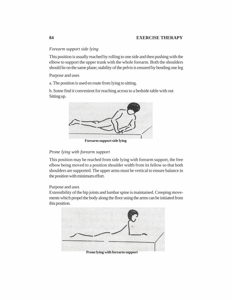
84 EXERCISE THERAPY
Forearm support side lying
This position is usually reached by rolling to one side and then pushing with theelbow to support the upper trunk with the whole forearm. Both the shouldersshould lie on the same plane; stability of the pelvis is ensured by bending one leg
Purpose and uses
a. The position is used en route from lying to sitting.
b. Some find it convenient for reaching across to a bedside table with outSitting up.
Forearm support side lying
Prone lying with forearm support
This position may be reached from side lying with forearm support, the freeelbow being moved to a position shoulder width from its fellow so that bothshoulders are supported. The upper arms must be vertical to ensure balance inthe position with minimum effort.
Purpose and usesExtensibility of the hip joints and lumbar spine is maintained. Creeping move-ments which propel the body along the floor using the arms can be initiated fromthis position.
Prone lying with forearm support

EXERCISE THERAPY EQUIPMENT 85
Sitting — on the side of the mat/bed
From lying on one side propped up by the elbow the body is pushed upright byextension of the elbow as the legs are lifted and swung over the side of the bed.During the movement the body is pivoted on one buttock until the sitting positionis reached when the weight is equally distributed through both buttocks.
Sitting
Hitching and Hiking
The ability to take the weight on the arms, lift and move the pelvis is essentialfor transfers for wheelchair patients, e.g. from b to chair. Blocks, sandbags orshort crutches help to make these easier for the patient to practice.
Side Sitting
Unlike the push up to sitting on the side of the bed, the push up to side sitting onthe floor includes little or no rotation as the trunk is pivoted to sit upright. Thelegs are bent and remain resting on the floor.
Purpose and uses
This is an elegant way to sit either on the floor or out of doors for those whofind it possible and comfortable. It can be practiced safely and it is easy toreturn to a resting position.
Hitching Hiking

86 EXERCISE THERAPY
Side Sitting
Prone Kneeling
Prone kneeling or the ‘four foot position’ may be reached from prone lying whenfle is initiated by bending the head forward to put the chin on the chest, then bywalking the hands backwards as the hips and knees bend.
Purpose and uses
This is the starting position for ‘crawling’ which gives the patient mobility at doorlevel in any direction he wishes. It may prove very use for patients with vertigoand others who cannot bear weight on the feet for the time being.
Prone Kneeling
Crawling
When balance and stability have been established in prone kneeling patients canbegin to practice lifting a hand or a knee from the floor to balance ‘on three legs’.
Purposes and uses of crawling
Crawling activities build up co-ordination of the whole body including reciprocalmovement of the arms and legs as required in walking. The direction of thecrawl, i.e. forwards, backwards; sideways determines the distribution and em-phasis of the neuromuscular activity employed.

EXERCISE THERAPY EQUIPMENT 87
Crawling
Kneeling
One can kneel down or get up to kneeling.
Before the patient attempts to kneel it is advisable to make sure that,
a. the surface on which to kneel is sufficiently comfortable for the patient totolerate pressure on the knees;
b. there is sufficient range of knee flexion, i.e. a minimum of about 100°.
c. any furniture to be used for support is in the right position, firm and immobile.A chair or bed is better pushed back against a wall;
d. any other disability which affects the patient has been taken into account, e.g.restriction of ankle joint movement, painful toes. Toes inadvertently beat underor pressed against a hard surface can be extremely painful.
Half Kneeling
To reach this position from kneeling the body weight is supported on one kneewhile the other leg is lifted and brought forwards to put the foot on the floor.From standing the half kneeling position can be assured either by stepping for-wards to kneel or by stepping backwards to kneel on one knee. Good balanceor some support is essential for stability.
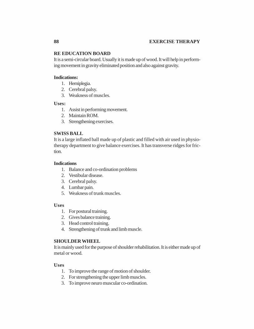
88 EXERCISE THERAPY
RE EDUCATION BOARDIt is a semi-circular board. Usually it is made up of wood. It will help in perform-ing movement in gravity eliminated position and also against gravity.
Indications:1. Hemiplegia.2. Cerebral palsy.3. Weakness of muscles.
Uses:1. Assist in performing movement.2. Maintain ROM.3. Strengthening exercises.
SWISS BALLIt is a large inflated ball made up of plastic and filled with air used in physio-therapy department to give balance exercises. It has transverse ridges for fric-tion.
Indications1. Balance and co-ordination problems2. Vestibular disease.3. Cerebral palsy.4. Lumbar pain.5. Weakness of trunk muscles.
Uses1. For postural training.2. Gives balance training.3. Head control training.4. Strengthening of trunk and limb muscle.
SHOULDER WHEELIt is mainly used for the purpose of shoulder rehabilitation. It is either made up ofmetal or wood.
Uses1. To improve the range of motion of shoulder.2. For strengthening the upper limb muscles.3. To improve neuro muscular co-ordination.

EXERCISE THERAPY EQUIPMENT 89
Indications1. Frozen shoulder.2. Periarthritis shoulder.3. Post traumatic stiffness of shoulder.4. Weakness of shoulder muscles.
CONTINUOUS PASSIVE MOVEMENT (CPM)It provides continues passive motion to the applied joint. The apparatus can beused immediately after the operation to improve the range of motion, reducepain, discomfort and healing etc. this machine is adjustable, easily controlled,versatile and usually electrically operated.
Benefits:1. Improves range of motion.2. Improves fluid dynamics.3. Prevents adhesion formation.4. Enhances nutritional status of the joint.5. Reduces joint diffusion.6. Facilitates healing.
FINGER LADDERIt is a wooden device which gives the objective reinforcement and motivation topatient for improving shoulder range of motion. It also feed back to the patientabout improvement.
Uses1. For improving range of motion.2. For improving neuromuscular co-ordination of upper limb.
Indication1. Frozen shoulder.2. Periarthritis shoulder.3. Post traumatic stiffness of shoulder.4. Weakness of shoulder muscles.
PARALLEL BARIt is equipment used in physiotherapy gym. It has got two horizontal frameswhich are mounted on a four vertical frames and a walking platform with onecentral divider to prevent crossing of leg. A postural correction mirror will beplaced at the end of the board. The main purpose is to improve

90 EXERCISE THERAPY
(i) Standing tolerance.(ii) Gait training.(iii) Postural correction.
Uses1. Gait training2. Postural training.3. Trunk control training.4. Balance training with and without support.5. Strengthening and mobility management of lower limb.
Indications1. Hemiplegia.2. Cerebral palsy.3. Post fracture and post traumatic gait training.4. Paraplegia.
MEDICINE BALLSIt is a leather ball which has got many layers of different materials. It has gotouter layer made up of thick leather and second layer made of foam and coir.Innermost layer is filled with stone chips and sands. The coir and foam is forpreventing injury to patient on direct hit.
Uses(i) Strengthening of upper limb muscles.(ii) Eye hand co-ordination.(iii) Neuro-muscular co-ordination.
Indications1. Paraplegia.2. Patient with stiff hip.3. Hand eye co-ordination.4. Maintenance of upper limb motion.
EQUILIBRIUM BOARDIt is a board made up of wood or metal used in the physiotherapy department.
Indications(i) Imbalance and co-ordination.(ii) Stiffness of ankle.(iii) Cerebral functioning.

EXERCISE THERAPY EQUIPMENT 91
(iv) Sensory ataxia.(v) Hemiplegia.(vi) Weakness of ankle muscle.
Uses1. Strengthening of ankle muscle.2. Balance and co-ordination.3. Relaxation.4. Neuro-muscular co-ordination.
SUSPENSION THERAPY
Suspension: it is the means whereby the parts of the body are supported inslings and elevated by the use of variable length ropes fixed to a point above thebody. It is also called as Guthre Smith’s apparatus. Suspension frees the bodyfrom the friction of the material upon which the body components may be rest-ing.
Principles of suspension apparatus:1. Free movement, frictionless movement.2. Gravity eliminated supported movement.3. Works on pendulum principle.
The supporting ropes: Ropes should be of 3-ply hemp so that they will not slip and may beof 3 arrangements.
1. Single rope system: this has a ring fixed at one end by which it’s hung. Theother end of rope then passes through one end of a wooden cleat then throughthe ring of a dog clip and through the other end of the cleat and is then knottedwith a half hitch.
The cleat is for altering the length of the rope and should be held horizontally formovement and pulled oblique when supporting is required. The length of therope is 1.5 m. This type of arrangement is used for suspending the limb partsonly.
2. Pulley Rope SystemThis has a dog clip attached at one end of the rope which is passed over thewheel of a pulley. The rope then passes through the cleat and the second dogclip..

92 EXERCISE THERAPY
his rope is also of 1.5 m length. Finally, rope will pass from the end of a woodencleat and a half hitch regular knot can be used. This type of arrangement is usedfor suspending the limb parts and for its movement also.
3. Double Rope SystemThis consists of a ring and a cleat at its upper end by which the rope is hung,which creates a corresponding device to persist a certain amount of swivel onthe rope. The rope then passes through one side of a cleat, round a pulley wheelat the lower end of the rope which is attached to a dog clip. This system isusually used to support the heavy parts of the body.
4. SLINGSSINGLE SLING: Made up of canvas bound with a D-ring at each end. This isused to support the limbs or foot. Measurement is 68 cm long ×17 cm breadth.
DOUBLE SLING: These are broad slings. Dimension is 68cm long and 59cmwide with D rings at each end and the slings are used to support the pelvis, trunkand thigh.
THREE-RING SLINGS: They are webbing slings 71cm long and 4cm widethree D rings, one of which is freely movable and is placed in the middle. Theseslings are usually used for wrist, hand, ankle and feet.
HEAD AND SLING: This is a short; split sling with its two halves stitchedtogether to an angle is create a central slit. This slit allows the head is rest under
the lower and upper part of skull.
Single pulley rope Double pulley rope

EXERCISE THERAPY EQUIPMENT 93
Single sling Double sling Head sling Three ring sling
A. Dog clip B. Karabiner clip S hook
TYPES OF SUSPENSION
1. VERTICAL: In this suspension, the rope is fixed so that it hangs verticallyabove the C.O.G. Vertical suspension is used for the movement of a part througha small range like pendular movement on each side.
2. AXIAL: Axial occurs when all ropes are supporting a part as attached to ‘S’hook of the joint which is to be moved. If some resistance is a muscle is re-quired, then the whole fixed pt. is moved away from the muscle which requiredresistance.INDICATIONS OF SUSPENSION:
i) For re-education of weak muscleii) For strengthening muscle in gravity eliminated friction posi-
tion.

94 EXERCISE THERAPY
iii) For improving and maintaining range of movementiv) For early neuro-muscular co-ordination training
CONTRA-INDICATIONSi) Hyper mobile jointii) Acute trauma or fractureiii) Burniv) Generalized Oedemav) Skin diseasevi) Open wounds and sinuses
SUSPENSION FOR UPPER LIMB
THE SHOULDER JOINTAbduction and Adduction:Position of the patient: supine.Fixation point: Over the shoulder joint, single sling is attached to the elbow andone is a Three-ring sling applied to the wrist and hand.
Flexion and Extension:Position of the patient: side lyingFixation point: Over the shoulder joint, single sling is attached to the elbow andone is a Three-ring sling applied to the wrist and hand.
ELBOW JOINTPosition of the patient: sitting on a low backed chair.Fixation point: A single sling and rope supports the arm in vertical fixation, and athree-ring sling and single rope fixed to point above the elbow joint.
WRISTFlexion and extensionThis is more usually performed on a polished board.
HIP JOINTAbduction and AdductionPosition of the patient: supine lying.Fixation point: Over the hip joint, single sling is attached to the thigh and one is aThree-ring sling applied to the ankle and foot and they are attached to the ropehung from the fixation point.

EXERCISE THERAPY EQUIPMENT 95
Flexion and ExtensionPosition of the patient: Side lying.Fixation point: Over the hip joint, single sling is attached to the thigh and one is aThree-ring sling applied to the ankle and foot.During flexion both the hip and knee should be flexed together to overcomepassive insufficiency of hamstring muscle. When extension is performed the kneeshould be extended to over come the active insufficiency of hamstring muscle.
Internal and External rotationPosition of the patient: supine lying with Hip and knee flexed at 90 degrees.Fixation point: Over the hip joint, single sling is attached to the thigh and one is aThree-ring sling applied to the ankle and foot.
KNEE JOINT
Flexion and ExtensionPosition of the patient: Side lying with thigh slightly flexed.Fixation point: Over the knee joint, single sling is attached to the thigh and one isa Three-ring sling applied to the ankle and foot.
ANKLE JOINTIt is rarely necessary to use suspension as in this case it is easier to performsupported movements by using a polished board.

CHAPTER 18
WALKING AIDS AND GAIT TRAININGWalking aids are useful to assist people who have difficulty in walking or peoplewho cannot walk independently. This include crutches, sticks and frames.
1. Crutches: these are used to reduce weight bearing on one or both legs andalso give support where balance is impaired and strength is inadequate.
Types:a. Axillary crutches: They are made of wood with an axillary pad, a hand pieceand a rubber ferrule.
The position of the hand piece and the total length are usually adjustable.The axillary pad should rest beneath the apex of axilla and hand grip in slightflexion when weight is not being taken. When weight is being taken throughaxillary pad, the elbow will go into extension and weight is transmitted down thearm to hand piece.
Crutch Elbow crutch
b. Elbow crutches: They are made of metal and have a metal or plastic forearmband. They are usually adjustable in length by means of a press clip or metalbutton and have a rubber ferrule. These crutches are suitable for patients withgood balance and strong arms. Weight is transmitted exactly the same way asfor axillary crutches.
c. Forearm crutches (gutter crutches): They are made of metal with a paddedforearm support and strap, an adjustable hand piece and a rubber ferrule.

WALKING AIDS 97
These are used for patients with rheumatoid disease for providing support. Theycannot take weight through hands, wrists and elbows because of deformity orpain.
Forearm crutch
Preparation for crutch walking:a. Arms: shoulder extensors, adductors and elbow extensors must be assessedand strengthened before the patient starts walking. The hand grip must also betested to see that the patient has sufficient power and mobility to grasp handpiece.
b. Legs: the strength and mobility of both legs should be assessed and strength-ened if necessary. Main attention to the hip abductors and extensor, the kneeextensors and the plantar flexors of the ankle should be given.
c. Balance: sitting and standing balance must be tested.
Demonstration: the physiotherapist should demonstrate appropriate crutch walkingto the patient.
Crutch walking: during first time, when the patient is to stand and walk, thephysiotherapist should have an assistant for supporting the patient.
i. Non-weight bearing: patient should always stand with a triangular base i.e.crutches either infront or behind the weight bearing leg

98 EXERCISE THERAPY
ii. Partial weight bearing: the crutches and the affected leg are taken forward andput down together. Weight is then taken through the crutches and the affectedleg, while the unaffected leg is brought through.
Sticks: sticks may be made up of either wood or metal with curved or straighthand piece. Metal ores are adjustable while the wooden ones are non-adjust-able.Uses: sticks allow more weight to be taken through the leg than crutches. Onestick may be used on the unaffected side, so that the stick and affected leg areplaced forward together, taking some of the weight through the stick.
Tripod or quadripod: metal sticks with three or four prolonged bases and givesmore stable support than stick.
Tripod Quadripod

WALKING AIDS 99
Frames: they are light weight with four feet which can be adjustable in height.Patient lifts the frame forward then leans on it and takes steps. The patient shouldtake even steps, keeping the frame forward. Rotator frames which can be pushedor reciprocal frame where each side moves independently are useful for ataxicpatients.
Rollator Frames
Safety: the physiotherapist must check the safety of all walking aids not onlywhen giving to them to a patient, but regularly throughout the treatmentprogramme.

CHAPTER 19
MASSAGE
Massage signifies a group of procedures, which are usually done with hand onthe external tissue of the body in a variety of ways either with a curative, pallia-tive or hygienic point of view.
Classification of technique:
I. On the basis of character of technique:
1. Stroking manipulationi. superficial stroking.ii. Deep stroking.
2. Pressure manipulation
i. kneading. a. palmar kneading. b. digital kneading. c. ironing.
ii. Petrissage.a. Picking up.b. Wringing.c. Skin rolling.
iii. Friction.a. Circular friction.b. Transverse friction.
3. Percussion manipulationsi. Clapping.ii. Hacking.iii. Tapping.iv. Beating.v. Pounding.vi. Tenting.vii. Contact heel percussion.

MASSAGE 101
4. Vibratory manipulationsi. Vibrations.ii. Shaking.
II. On the basis of depth of tissue
1. Light massage.2. Deep massage.
III. On the basis of part of body massagedi. General massage.ii. Local massage.
Therapeutic usesMassage is one of the oldest from of treatment for human ills. It has been used asa therapeutic modality in various conditions since ancient times.
1. To improve mobility of the soft tissues.2. To reduce muscle spasm and pain under abnormal conditions.3. To reduce oedema.4. To increase circulation.5. To mobilize secretions in the lung.6. To induce local and general relaxations.
Contraindications
I. General contraindications:1. High fever.2. Renal diseases.3. Cardiac diseases.4. Deep x-ray therapy.5. Osteoporosis.6. Severe spasticity.
II. Local contraindications:1. Acute inflammation.2. Skin diseases.3. Recent fractures.4. Varicose veins.

102 EXERCISE THERAPY
1. Atherosclerosis.2. Thrombosis.3. Open wound.
Physiological effects:
1. Effect of massage on the circulatory system: massage aids in the me-chanical emptying of the veins and the lymphatics. It facilitates the forward move-ment of the venous blood and the lymph and there by reduces the chances ofstagnation of the blood and the lymph in the tissue space.
On the arterial flow: massage improves the blood supply of the area beingmassaged. A definite vasodilation along with an increase in the peripheral bloodflow is usually observed after massage. This moderate, consistent and definiteincrease in the arterial flow may be attributed to the following events happeningduring massage.Release of vasodilators, activation of axon reflex, decrease of venous conges-tion.
2. Effects on blood: massage is found to increase RBC and haemoglobin countand may increase the oxygen carrying capacity of blood.
3. Effects on the exchange of metabolites: massage speeds up the lymphaticand venous flow, which promotes rapid disposal of the waste products of me-tabolism. These changes make the exchange of waste products between theblood and the tissue at cellular level more efficiently.
4. Effects on the nervous system: the nervous system consists of sensory,motor and autonomic component. Different technique of massage produces ef-fects on all these components.
Sensory system: massage has a sedative effect on the central nervous systemif applied monotonously with slow rhythm. The use of massage for the relief ofpain of various origins is an age-old practice.
Facilitatory effects of massage on motor system: it is said that massage canreflexly increase the muscle tone by stimulation of the skin receptor or stretch ofthe muscle spindle. Superficial stroking, taping, hacking etc. are commonly usedfor this purpose.

MASSAGE 103
Inhibitory effects of massage on motor system: massage techniques canalso reduce the tone of muscle. It has been claimed that petrissage or massage inwhich muscles are kneaded can exert an inhibitory effect on motorneuron. It issaid that effleurage is capable of producing both stimulating and relaxing effects.
On autonomic nervous system: massage has definite reflex effect and it caninfluence the functioning of visceral organ by modulating the autonomic nervoussystem through peripheral sensory stimulation.
5. Effects on the soft tissue: Massage has significant effect on certain proper-ties of the soft tissues like elasticity, plasticity and mobility. The adhesions presentbetween fibres are broken and maximum mobility between fibres and adjacentstructures is ensured.
6. Effects on the respiratory system: percussion and vibration techniques ofmassage assist the removal of secretions from the larger airways. After the re-moval of secretion gas exchange becomes more efficient.
7. Effects on the skin: massage in general improves the nutritive status of skin,the skin temperature of skin rises. Massage facilitates the movement of skin overthe subcutaneous structures. As a result skin becomes soften, more supple andfiner.
Techniques of massageI. Stroking: the uninterrupted linear movement of hand along the whole lengthof segment is called stroke.Superficial stroking: it is the rhythmic movement of hand or parts thereof overthe skin with the lightest amount of pressure in order to obtain sensory stimula-tion. The strokes can be applied from proximal to the distal or vice versa.
Effleurage or deep stroking: it is the movement of the palmar aspect of handover the external surface of the body with constant moderate pressure, in thedirection of the venous and lymphatic drainage.
II. Pressure manipulations: in this group of techniques, the hand of the thera-pist and skin of the patient move together as one and fairly deep localized pres-sure is applied to the body. It is divided into
i. Kneading.ii. Petrissage.iii. Friction.

104 EXERCISE THERAPY
i. kneading: in this group of techniques, the tissues are pressed down on to theunderlying firm structure and intermittent pressure is applied in circular direction,parallel to the long axis of bone.a. digital kneading: pressure is applied with the fingers (finger kneading) orthumb (thumb kneading).b. palmar kneading: pressure is applied with the palm.c. reinforced kneading: both the hands, placed over one another, are used toapply pressure.
III. Petrissage: in this the tissues are grasped and lifted away from the underly-ing structures and intermittent pressure is applied to the tissues in the directionthat is perpendicular to the long axis of the bone. It is divided intoi. Picking up: tissues are lifted away from underlying structures, squeezed andthen released using one or both the hands.ii. Wringing: using both the hands, tissues are lifted away from the underlyingstructures, squeezed, twisted and then released.iii. Skin rolling: the skin and fascia are lifted up with both the hands and movedover the subcutaneous tissues by keeping a roll of lifted tissue continuously aheadof the moving thumb.
IV. Friction: in this technique the tissue are subjected to small range of to andfro movement performed with constant deep pressure of the finger or thumb. Itis divided in toi. Circular friction: direction of movement is circular.ii. Transverse friction: to and fro movement is performed across the length ofstructure.
V. Vibratory Manipulations: In this group of techniques, the mechanical en-ergy is transmitted to the body by the vibrations of the distal part of upper limb,i.e. hand and/or fingers, which are in constant contact with the subjects skin,using the body weight and generalized co-contraction of the upper limb muscles.This technique is mainly directed towards the lung and other hollow cavities.
Depending upon the direction and frequency of vibration it is divided into twotechniques:
Vibration In this technique, the fine vibrations are produced, which tend toproduce fine movement of hand in upwards and downward direction.
Shaking In this technique, coarse vibrations are produced, which tend to pro-duce fine movement of hand in sideway direction.

MASSAGE 105
VI. Percussion/tapotment manipulations
In this group of techniques, a succession of soft, gentle blows are applied overthe body, which produce a characteristic sound. The striking hands are not inconstant contact with the skin and strike the body part at regular interval. Thisresults in the application of an intermittent touch and pressure to the body duringthese manipulations.
The different parts of hand are used to strike the subject’s skin and accordinglythe techniques are named:
Technique Administered with
Clapping Cupped palm
Hacking Ulnar border of the 5th, 4th and 3rd digits
Beating Anterior aspect of the clenched fist
Tapping Pulp of the fingers
Pounding Medial aspect of the clenched fist
On the Basis of Depth of Tissue Approached
Depending upon the depth of tissue approached during manipulations massagetechniques can be classified as:
Light Massage Techniques
The force applied during the manoeuvre are light, so that the effect of massage isconfined to the superficial tissue only, e.g. stroking, tapping, etc.
Deep Massage Techniques
The forces applied during the massage are moderate to deep so that the effect ofmassage reaches to the deeper tissues like muscle, e.g. friction, kneading, etc.
On the Basis of Region Massaged
Massage can also be classified as below, according to the region to which it isgiven.
General massage
Massage applied to the entire body is usually termed as general massage. How-ever, massage administered to a large body segment like the back, lower limb,etc. can also be included in this category.

106 EXERCISE THERAPY
It is usually administered in debilitated persons following prolonged recumbencyand on athletes after exhaustive physical work to bring a sense of well-being andcomfort.
Local Massage
When massage is administered in a particular area of the body segment it istermed as local massage. This is used in the treatment of the local pathologicalconditions. For example, massage of wrist in tenosynovitis, friction to lateralligament of ankle following sprain, etc. can be considered as local massage.
On the Basis of Means of Administration of Technique
On this basis, the massage can be classified into the following two categories.
Manual Massage
Word manual refers to the ‘lying on’ of hand over the subject’s body. The mas-sage administered with the hand or other body part of the therapist is calledmanual massage, e.g. technique of classical massage, connective tissue mas-sage, trigger point massage, accupressure massage, etc.
Mechanical Massage
When the mechanical devices based on the principles of massage, administer themechanical energy to the patient’s body, in order to manipulate soft tissue, it maybe termed as mechanical massage, e.g. vibrator, compression devices, pneu-matic massage, etc.