Texas A & M University Zeiss...
14
Microscopy and Imaging Center Texas A & M University http://microscopy.tamu.edu 1 Zeiss Axiophot Microscope User Guide Modified April 12, 2013 Contents: Acknowledgment of the use of MIC facilities ........................................................................................ 1 Mercury Lamp Precautions ..................................................................................................................... 1 Biosafety requirements and rules for work in the MIC .......................................................................... 2 Biological Spill Response: BL1 Laboratory ............................................................................................ 4 Turning the system ON: .......................................................................................................................... 5 Turning OFF: .......................................................................................................................................... 5 Using the Microscope ............................................................................................................................. 6 If you use oil immersion objectives, read this! ....................................................................................... 6 Cleaning oil immersion objectives.......................................................................................................... 6 Setting up Köhler illumination................................................................................................................ 7 Transmitted-light observartion................................................................................................................ 8 Epiillumination techniques: .................................................................................................................. 10 Where to Save Image Files ................................................................................................................... 12 Storing your data ................................................................................................................................... 12 Working with your images.................................................................................................................... 12 Using your images in Powerpoint presentations:.................................................................................. 13 Further training in Image Analysis and Processing: ............................................................................. 13 Adding a scale bar to your images: ....................................................................................................... 13 Acknowledgment of the use of MIC facilities MIC guidelines mandate that the use of the microscope must be acknowledged in any publication (including web pages): “The use of the Zeiss Axiophot microscope in the Microscopy and Imaging Center at Texas A&M University is acknowledged.” Users are also required to file a copy of any relevant publication containing the acknowledgment with the MCF administrative office. Mercury Lamp Precautions The lamp emits strong UV and visible radiation. Do not look into the source or disassemble the lamp housing. DO NOT LOOK INTO THE OCULARS WHEN MOVING TO A DIFFERENT FLUORESCENCE FILTER. THE EPI-POLARIZING FILER SET (Slider #2) WILL REFLECT UV TO YOUR EYES. Keeping track of mercury lamp usage is vital. Make sure to record the used time and the total hours accumulated usage. Mercury lamp lifetime is rated at 100 hr. If used beyond that point, risk of explosion and mercury contamination of the room sharply increases. Do not turn the lamp on if the lamp reached its expected lifetime! Frequent switching ON/OFF shortens the mercury lamp's life considerably. It is better to leave it on if the next user is going to need it within 1-2 hours. After turned on, it takes ~ 15 min for the lamp to reach full brightness. Lamp must be ON for at least 30 min before it can be switched OFF. After the lamp has been switched OFF, it must cool down (at least 30 min) before it may be switched ON again.
Transcript of Texas A & M University Zeiss...
Microscopy and Imaging Center Texas A & M University http://microscopy.tamu.edu
1
Zeiss Axiophot Microscope User Guide Modified April 12, 2013
Contents:
Acknowledgment of the use of MIC facilities ........................................................................................ 1 Mercury Lamp Precautions ..................................................................................................................... 1 Biosafety requirements and rules for work in the MIC .......................................................................... 2 Biological Spill Response: BL1 Laborator y ............................................................................................ 4 Turning the system ON: .......................................................................................................................... 5
Turning OFF: .......................................................................................................................................... 5 Using the Microscope ............................................................................................................................. 6 If you use oil immersion objectives, read this! ....................................................................................... 6 Cleaning oil immersion objectives .......................................................................................................... 6 Setting up Köhler illumination................................................................................................................ 7
Transmitted-light observartion................................................................................................................ 8 Epiillumination techniques: .................................................................................................................. 10
Where to Save Image Files ................................................................................................................... 12 Storing your data ................................................................................................................................... 12
Working with your images.................................................................................................................... 12 Using your images in Powerpoint presentations:.................................................................................. 13 Further training in Image Analysis and Processing: ............................................................................. 13
Adding a scale bar to your images: ....................................................................................................... 13
Acknowledgment of the use of MIC facilities MIC guidelines mandate that the use of the microscope must be acknowledged in any publication
(including web pages): “The use of the Zeiss Axiophot microscope in the Microscopy and Imaging
Center at Texas A&M University is acknowledged.”
Users are also required to file a copy of any relevant publication containing the acknowledgment with
the MCF administrative office.
Mercury Lamp Precautions
The lamp emits strong UV and visible radiation. Do not look into the source or disassemble the
lamp housing. DO NOT LOOK INTO THE OCULARS WHEN MOVING TO A DIFFERENT
FLUORESCENCE FILTER. THE EPI-POLARIZING FILER SET (Slider #2) WILL REFLECT
UV TO YOUR EYES.
Keeping track of mercury lamp usage is vital. Make sure to record the used time and the total
hours accumulated usage.
Mercury lamp lifetime is rated at 100 hr. If used beyond that point, risk of explosion and
mercury contamination of the room sharply increases. Do not turn the lamp on if the lamp
reached its expected lifetime!
Frequent switching ON/OFF shortens the mercury lamp's life considerably. It is better to leave it
on if the next user is going to need it within 1-2 hours.
After turned on, it takes ~ 15 min for the lamp to reach full brightness.
Lamp must be ON for at least 30 min before it can be switched OFF.
After the lamp has been switched OFF, it must cool down (at least 30 min) before it may be
switched ON again.

Microscopy and Imaging Center Texas A & M University http://microscopy.tamu.edu
2
Biosafety requirements and rules for work in the MIC Selected rooms in the Microscopy and Imaging Center (MIC) have been approved as BL-1 space. In
order to be able to bring the active BL-1 material to the MIC, the MIC facility and room number MUST
be listed in the investigator’s IBC permit, in Section F, Agent use and Storage Locations. The
investigator is required to send a copy of the IBC permit listing the relevant MIC lab and the BL1
organisms, to MIC office ([email protected]), BEFORE bringing the BL-1 samples.
The MIC is neither equipped nor allowed to deal with samples that are Biosafety level 2 (BL-2) or
higher. If such samples need to be examined in the MIC, they must be rendered inactive prior to their
transport to MIC. The investigator should first contact the University Biosafety Committee (IBC) and
have an approved operation procedure for sample inactivation and containment. The investigator should
consult with MIC to make sure that the procedure is compatible with microscopy imaging.
F. Agent use and storage locations.
Location
ID
Campus
Building
Number
Room
Number
Room
Use
Current Bio-
safety Level
Shared
Lab?
Other
PIs
1 Texas A&M 1530 1116 Multiphoton
microscopy
BSL-1 Yes *
2 Texas A&M 1530 1117 Cell and
tissue culture
BSL-1 Yes *
3 Texas A&M 1530 1118 Confocal
microscopy
BSL-1 Yes *
4 Texas A&M 1530 1121 laboratory BSL-1 Yes *
5 Texas A&M 1530 1120 Micropscopy
room
BSL-1 Yes *
* The list of other PIs is maintained by the MIC
All users of rooms listed in table F must follow the rules. This applies even to those users that do not
work with samples requiring IBC permit:
I) For users that DO NOT work with BL-1 agents:
- Closed toe shoes are required in the confocal microscope room. Upon exiting the microscope room,
users are required to wash hands. The sink in the Bioprep lab (Rm. 1121) or in the Culture room
(Rm. 1117) may be used.
II) For users working with active BL1-agents:
- The MIC facility and room number MUST be listed in the investigator's IBC permit, in Section F,
Agent use and Storage Locations; The PI is required to send a copy of the IBC permit, listing the
relevant MIC lab and the BL-1 organisms, to [email protected] BEFORE the BL-1 samples
can be brought to the MIC. Without the MIC facility listed in the PI’s permit, no BL-1 work is
allowed in the MIC.
- Samples being brought to the MIC must be contained, in accordance with the operating procedure in
investigator’s IBC permit, to prevent spills during transport
- Closed toe shoes must be worn
- The use of gloves should be restricted to only handling the sample to avoid contaminating general
work area. Touching the microscope or the control computer keyboard and mouse with gloved hands
should be avoided. Gloves may not be worn outside of the rooms listed in table F.
Microscopy and Imaging Center Texas A & M University http://microscopy.tamu.edu
3
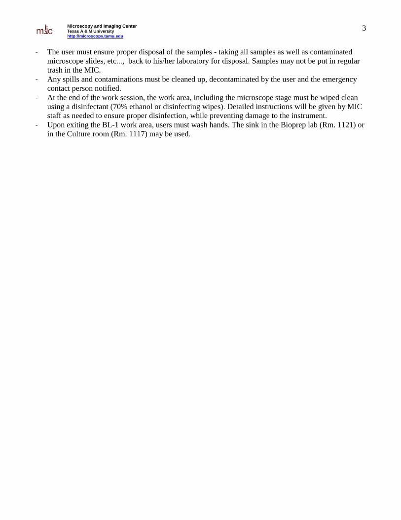
- The user must ensure proper disposal of the samples - taking all samples as well as contaminated
microscope slides, etc..., back to his/her laboratory for disposal. Samples may not be put in regular
trash in the MIC.
- Any spills and contaminations must be cleaned up, decontaminated by the user and the emergency
contact person notified.
- At the end of the work session, the work area, including the microscope stage must be wiped clean
using a disinfectant (70% ethanol or disinfecting wipes). Detailed instructions will be given by MIC
staff as needed to ensure proper disinfection, while preventing damage to the instrument.
- Upon exiting the BL-1 work area, users must wash hands. The sink in the Bioprep lab (Rm. 1121) or
in the Culture room (Rm. 1117) may be used.
Microscopy and Imaging Center Texas A & M University http://microscopy.tamu.edu
4
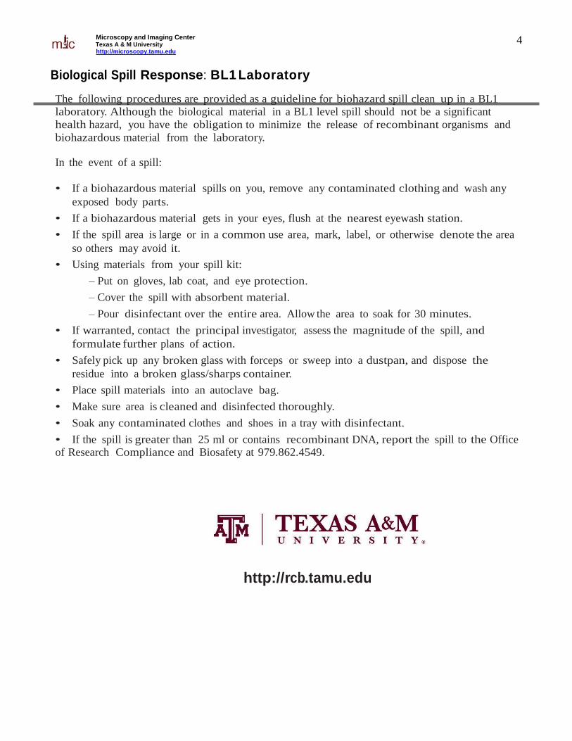
Biological Spill Response: BL1 Laborator y
The following procedures are provided as a guideline for biohazard spill clean up in a BL1
laboratory. Although the biological material in a BL1 level spill should not be a significant
health hazard, you have the obligation to minimize the release of recombinant organisms and
biohazardous material from the laboratory. In the event of a spill:
• If a biohazardous material spills on you, remove any contaminated clothing and wash any
exposed body parts.
• If a biohazardous material gets in your eyes, flush at the nearest eyewash station.
• If the spill area is large or in a common use area, mark, label, or otherwise denote the area
so others may avoid it.
• Using materials from your spill kit:
– Put on gloves, lab coat, and eye protection.
– Cover the spill with absorbent material.
– Pour disinfectant over the entire area. Allow the area to soak for 30 minutes.
• If warranted, contact the principal investigator, assess the magnitude of the spill, and
formulate further plans of action.
• Safely pick up any broken glass with forceps or sweep into a dustpan, and dispose the
residue into a broken glass/sharps container.
• Place spill materials into an autoclave bag.
• Make sure area is cleaned and disinfected thoroughly.
• Soak any contaminated clothes and shoes in a tray with disinfectant.
• If the spill is greater than 25 ml or contains recombinant DNA, report the spill to the Office
of Research Compliance and Biosafety at 979.862.4549.
http://rcb.tamu.edu
Microscopy and Imaging Center Texas A & M University http://microscopy.tamu.edu
5
Turning the system ON: 1. Sign-in, fill out the instrument time sheet.
2. Take the protective cover off the microscope.
3. If planning to use fluorescence, turn ON the mercury lamp power. Since the power spike from the
power supply could damage other equipment, it must be turned on before anything else.
4. Turn on the motorized focus control box.
5. Turn on the halogen lamp
6. If you plan to take pictures, start the computer and turn on the monochrome camera. The color
camera does not have a power switch, it ison as long as the computer is on.
Turning OFF: 1. Remove the specimen and turn off the focus control box.
2. Turn the camera OFF.
3. If oil immersion was used, clean the objectives (see detailed instructions below) and the stage.
4. Change to the smallest magnification (2.5x or 10x objective)
5. Transfer/save your images.
6. If nobody is scheduled to use fluorescence after you, turn OFF the computer, then the mercury lamp
power supply and record the usage in the logbook.
7. Allow the mercury lamp to cool down for ~ 10 min, then replace the plastic dust cover. Protecting
the optics from dust is very important – dust is the biggest enemy of your images, dirty optics
produces blurry pictures.
8. Clean after yourself. Wipe the working area with wet paper towel, properly dispose of all used
slides/coverslips, material, etc.

Microscopy and Imaging Center Texas A & M University http://microscopy.tamu.edu
6
Using the Microscope 1. When switching between objectives, hold on the collar of the nosepiece; NEVER turn the
nosepiece turret by grabbing the objectives. The objectives are precision-mounted and this
force could disturb their alignment.
2. With the lowest-magnification objective engaged, place the slide on the stage and locate the
specimen. Select the proper filter set or other appropriate filter cube and adjust the illumination
as necessary.
3. The oil immersion objectives are kept in the retracted position. In this position you will not be
able to focus. To use, set them to the fully extended position.
For those who did not pay attention: After using oil immersion, you cannot switch back to the 40x dry objective to view the same area,
because the oil would get on the lens! You could use the 10x objective, but to get there, you must
turn the objective turret in the direction that will avoid contaminating the 40x lens.
Adjusting the oculars for optimal viewing 1. Adjust the interpupillary distance of the binoculars so that you can see with both eyes
2. Set both oculars to “0” focal correction (see scale on the side of the ocular tube)
3. Use the microscope focus knob to bring a specimen in focus for your left eye.
4. Now, take your hand off the focus knob. Turning the ocular focus correction, bring the specimen
in focus for your right eye.
If you use oil immersion objectives, read this! Because the dry 40x objective has a very small working distance, it will get contaminated with
immersion oil if there is some on the slide. Therefore, if you are using the 63x or 100x oil immersion
objectives and want to switch to the dry 10x, you must turn the objective turret in such a direction to
avoid swinging the 40x lens through the oil. You cannot use the 40x objective for observation at all,
unless the slide has been cleaned and all oil removed from the coverslip.
Cleaning oil immersion objectives 1. Use only the lens paper (e.g. Fisher 11-996). NEVER USE KIMWIPES OR PAPER TOWELS
TO CLEAN THE OBJECTIVE LENS. Do NOT drag the paper across the lens, just blot off
the oil. The front lens of the objective is very delicate and must be protected from scratching.
2. Lock the objective in retracted position
3. Thorough cleaning of the objectives will be performed periodically by the MIC staff.
Microscopy and Imaging Center Texas A & M University http://microscopy.tamu.edu
7
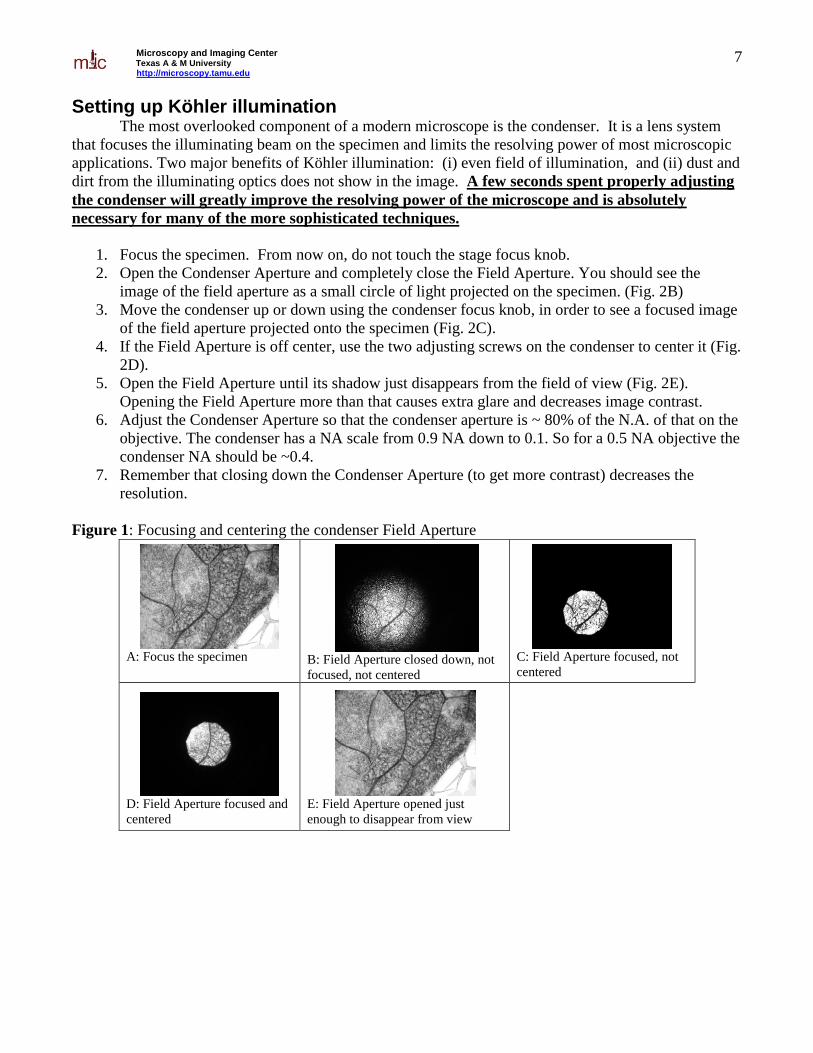
Setting up Köhler illumination The most overlooked component of a modern microscope is the condenser. It is a lens system
that focuses the illuminating beam on the specimen and limits the resolving power of most microscopic
applications. Two major benefits of Köhler illumination: (i) even field of illumination, and (ii) dust and
dirt from the illuminating optics does not show in the image. A few seconds spent properly adjusting
the condenser will greatly improve the resolving power of the microscope and is absolutely
necessary for many of the more sophisticated techniques.
1. Focus the specimen. From now on, do not touch the stage focus knob.
2. Open the Condenser Aperture and completely close the Field Aperture. You should see the
image of the field aperture as a small circle of light projected on the specimen. (Fig. 2B)
3. Move the condenser up or down using the condenser focus knob, in order to see a focused image
of the field aperture projected onto the specimen (Fig. 2C).
4. If the Field Aperture is off center, use the two adjusting screws on the condenser to center it (Fig.
2D).
5. Open the Field Aperture until its shadow just disappears from the field of view (Fig. 2E).
Opening the Field Aperture more than that causes extra glare and decreases image contrast.
6. Adjust the Condenser Aperture so that the condenser aperture is ~ 80% of the N.A. of that on the
objective. The condenser has a NA scale from 0.9 NA down to 0.1. So for a 0.5 NA objective the
condenser NA should be ~0.4.
7. Remember that closing down the Condenser Aperture (to get more contrast) decreases the
resolution.
Figure 1: Focusing and centering the condenser Field Aperture
A: Focus the specimen B: Field Aperture closed down, not
focused, not centered
C: Field Aperture focused, not
centered
D: Field Aperture focused and
centered
E: Field Aperture opened just
enough to disappear from view

Microscopy and Imaging Center Texas A & M University http://microscopy.tamu.edu
8
Transmitted-light observartion
Brightfield 1. The condenser should be in position “H”
2. Make sure the Analyzer slider (= polarizer above the nosepiece, on the right side) is pulled out.
3. For maximum light intensity, also swing out the Polarizer below the condenser out of the light
path.
4. Set up Köhler illumination
5. Adjust the oculars for optimal viewing
Polarization Works best with the 10x dry objective.
1. Perform setup as for brightfield, including Köhler illumination
2. Make sure there is no DIC prism in a slot above the objective.
3. Remove the specimen and engage both the Polarizer (below the condenser) and the Analyzer
(above the nosepiece). The field should be very dark. If it is not, rotate the Polarizer (located at
the bottom of the condenser) for maximum extinction (the darkest image) –the polarizer should
click into position.
4. View your specimen. The stage can be rotated after loosening the knob on the front.
Differential Interference Contrast (Nomarski) This mode is possible with the 10x, 40x, 63x, and 100x lenses. It can be used without a fluorescence
filter set (dummy filter holder) or with most fluorescence filter sets.
1. Perform setup for brightfield, including Köhler illumination, but leave the condenser aperture
fully open.
2. Make sure the Polarizer below the condenser is engaged and in correct orientation (the lever
towards you in the click position), and that the Analyzer slider above the objective is engaged
(pushed in).
3. According to the numerical aperture of the objective used, select the proper DIC position (RED
label) on the condenser: There are two positions, “DIC .3-.4” for objectives with NA up to 0.4,
and “DIC.5-1.3” for NA between 0.5 and 1.3 (use also for the 63x/1.4 oil immersion objective).
4. Fully open the condenser aperture.
5. Insert a Nomarski prism slider in the slot above the objective. Each slider is clearly marked for
which objective it is to be used (the 100x slider can also be used with the 63x objective).
6. Adjustment of the image can be made by turning a knob on the Nomarski prism. If there is not
enough contrast in the image, try closing the condenser aperture slightly.

Microscopy and Imaging Center Texas A & M University http://microscopy.tamu.edu
9
Phase Contrast This mode is possible only with objectives marked “Ph”, i.e, the 20x, 40x, 63x, and 100x.
1. Set up as for Brightfield
2. Turn the condenser to the appropriate Phase Contrast position, Ph2 or Ph3, corresponding to the
label on the objective.
3. Remove the specimen from the stage or move to the clear area of the slide
4. Engage the Bertrand lens (turning the Optovar on the right side above the nosepiece to the “Ph”
position) and focus the Bertrand lens (knurled ring on the lower part of the Optovar turret) to see
the phase ring at the back focal plane of the objective.
5. The dark phase ring and the bright condenser annular aperture s should coincide (Figure 1). If
necessary, align the condenser annular aperture with the dark phase ring , using two small hex
keys inserted in the slots on the back of the condenser turret.
6. Disengage the Bertrand lens by turning the Optovar to the 1.25x position.
7. The alignment must be re-checked every time the condenser or the objective turret has been
turned, or if new specimen is viewed.
Figure 2. Alignment for phase contrast observation, view of the back focal plane of the objective. Left: condenser phase
annulus not aligned with the dark phase ring of the objective Right: correct alignment, the two rings coincide.
Darkfield: Currently limited to the 10x objective. It uses the annular aperture Ph3 to illuminate the specimen.
1. Set up as for brightfield, using the 10x objective
2. Turn the condenser to the Ph3 position

Microscopy and Imaging Center Texas A & M University http://microscopy.tamu.edu
10
Epiillumination techniques: These utilize either the mercury lamp (for epifluorescence) or the upper halogen lamp (for reflected
modes). Switch between the halogen and mercury by flipping the lever in between the two housing in
the back of the microscope. In order to get the specimen properly illuminated, the auxiliary lens
positioned behind the fluorescent filter slider should always be used. The only exception is when
using the stand-alone mirror cube “H”, which already has the auxiliary lens built in. For all other
observations, including fluorescence, the auxiliary lens must be present.
Reflected brightfield. Possible with all objectives; however, with the 2.5x objective some hot spots and internal reflections are
visible. There are two filter cubes that can be used for brightfield observation; the stand-alone “H” cube,
or the Polarizing filter set in the filter cube slider..
1) Select halogen as the light source
2) Select the upper halogen lamp,(switch on the right side of the scope next to the lamp voltage
knob)
3) Insert the filter cube for brightfield observation: either the “H” cube, or use the polarizing filter
set on the 3-FL filter slider. Read the above note regarding the auxiliary lens
4) Make sure the Analyzer slider is pulled out (disengaged).
5) Turn the lamp on using the voltage control knob
6) Pull out the shutter slider in the epiillumination path to a desired position (ND filter 0.6, ND
filter 0.3, no filter)
7) Fully open the condenser aperture (push in the control lever)
8) Fully open the field aperture on the epiillumination condenser (push in the control lever)
9) Focus on the specimen
Reflected Polarization 1. Set up as for reflected brightfield. You must use the Polarizing filter set, not the “H” cube.
2. Make sure there is no Nomarski prism in the slot above the objective.
3. Engage the Analyzer
Reflected DIC (Nomarski) 1. Set up as for polarization
2. Insert appropriate Nomarski prism to the slot above the objective. Turn the adjustment knob on
the Nomarski prism for best image.
Microscopy and Imaging Center Texas A & M University http://microscopy.tamu.edu
11
Epifluorescence We have are two filter sliders, each holds three filter sets:
Slider 1:
I. .DAPI set (ex. 365 nm, em. 420-470 nm)
II. Texas Red set (ex. 530-585, em. 615 nm longpass)
III. GFP set (ex. 450-490 nm, em. 500-550 nm)
Slider 2:
I. Alexa 430 set (ex. 436 nm, em. 520 nm longpass)
II. EPI-POLARIZATION (do not use with fluorescence)
III. Cy3 set (ex. 546 nm, em. 590 nm longpass)
CAUTION: DO NOT USE THE EPI-POLARIZATION FILTER SET WHEN THE MERCURY
LAMP IS USED. This set does not have a barrier filter, thus both UV and visible light reflected
from the specimen can enter the eyepieces. When switching from the Alexa 430 to the Cy3 set,
close the fluorescence light shutter and do not look in the oculars..
1. Use brightfield or other reflected or transmitted techniques to locate your specimen.
2. Switch to the Mercury lamp as the light source
3. Select the proper filter cube for your fluorochrome. Make sure to have the auxiliary lens in place.
4. Make sure the Analyzer slider is pulled out (disengaged), otherwise signal intensity is reduced.
5. Make sure the Nomarski prism is not inserted in the slot above the objective, since it would
cause loss of fluorescence image quality.
6. Pull out the shutter slider (in the epiillumination path) to a desired position (ND filter 0.6, ND
filter 0.3, no ND filter)
7. Fully open the condenser aperture (push in the control lever)
8. Fully open the field aperture on the epiillumination condenser (push in the control lever)
9. Focus on the specimen.
10. When not viewing the specimen, close the fluorescence shutter (push the shutter slider in) to
minimize photobleaching of the specimen.

Microscopy and Imaging Center Texas A & M University http://microscopy.tamu.edu
12
Where to Save Image Files Create a folder with your name in “C:\Users\” folder. Create sub-folders as needed.
When finished, copy your files onto your media (CD or Zip). Blank CD may be purchased from
the microscopy center. It is recommended that you create two copies of your files to avoid data
loss. Once you confirmed that the data are saved on your media, erase the files from the
computer.
Please note that the imaging computer is not connected to the internet
Old image files will be purged periodically to free disk space.
Storing your data All hard drives may fail. The disk on your computer is not a permanent storage solution.
If using CDs or DVDs, store the disks in protective cover in a cool, dark place – the CD-burning
layer is heat and light sensitive (after all this is how the data is recorded).
Always create two copies of the data files, store them in different locations.
Avoid using acidic markers (Sharpie) for labeling CD/DVD disks. Use a non-acidic archival felt-
tip marker
Most CDs and DVDs are not an archival medium; they can decay and be unreadable in few
years, especially if not stored properly.
Working with your images Images from the monochrome camera are saved as 16-bit grayscale TIFFs with. Users have recently
experienced problems opening the images with Photoshop, probably due to incompatible file headers
from the MetaVue software. Therefore it is recommended to use ImageJ for viewing and processing the
images, and/or use the BatchConverter plugin in ImageJ, or the Process-Batch-Convert command in
the newer versions of ImageJ, to resave files as TIFF with the correct file header. This plugin converts
all files in a specified folder and saves them in a user-specified destination folder. ImageJ is an image
processing and analysis freeware (http://rsb.info.nih.gov/ij/). It also allows opening Metamorph .” stk”
image stacks acquired with our microscope (e.g. when doing z-sectioning or time lapse imaging). These
are standard multi-image TIFF files.
Please note that the monochrome camera acquires images at 12-bits-per-pixel (4096 gray
levels). However, the image files are saved as 16-bits per pixel (65535 gray kevels), with the image data
in the first 4096 intensity levels of the 65536. Thus, if the 16-bit images are opened in Photoshop, they
will look all black Use the “Image- Adjust-Levels” command to lower the input max intensity value
(see arrow in Fig. 3). You may need to do this twice for a more precise control.
Figure 3: Scaling grayscale intensities in Adobe Photoshop. the original image is on the left. A copy of the image is
adjusted using the “Levels” command by lowering the max input intensity value (arrow).
Microscopy and Imaging Center Texas A & M University http://microscopy.tamu.edu
13
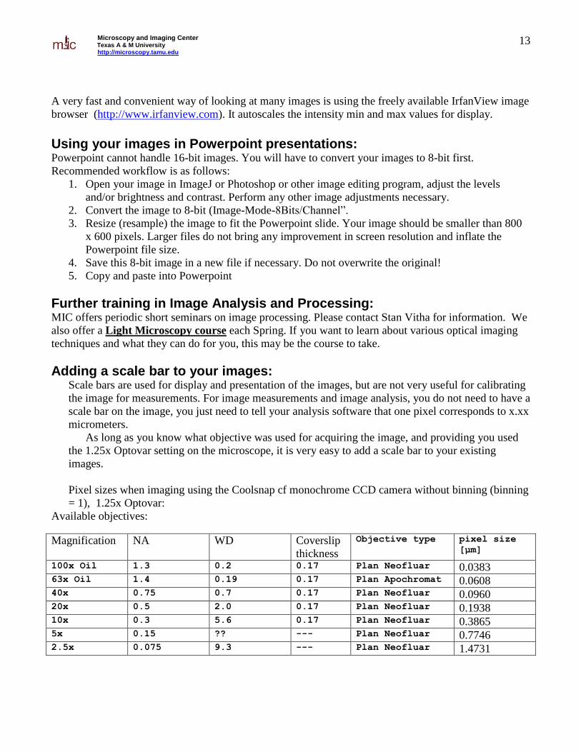
A very fast and convenient way of looking at many images is using the freely available IrfanView image
browser (http://www.irfanview.com). It autoscales the intensity min and max values for display.
Using your images in Powerpoint presentations: Powerpoint cannot handle 16-bit images. You will have to convert your images to 8-bit first.
Recommended workflow is as follows:
1. Open your image in ImageJ or Photoshop or other image editing program, adjust the levels
and/or brightness and contrast. Perform any other image adjustments necessary.
2. Convert the image to 8-bit (Image-Mode-8Bits/Channel”.
3. Resize (resample) the image to fit the Powerpoint slide. Your image should be smaller than 800
x 600 pixels. Larger files do not bring any improvement in screen resolution and inflate the
Powerpoint file size.
4. Save this 8-bit image in a new file if necessary. Do not overwrite the original!
5. Copy and paste into Powerpoint
Further training in Image Analysis and Processing: MIC offers periodic short seminars on image processing. Please contact Stan Vitha for information. We
also offer a Light Microscopy course each Spring. If you want to learn about various optical imaging
techniques and what they can do for you, this may be the course to take.
Adding a scale bar to your images: Scale bars are used for display and presentation of the images, but are not very useful for calibrating
the image for measurements. For image measurements and image analysis, you do not need to have a
scale bar on the image, you just need to tell your analysis software that one pixel corresponds to x.xx
micrometers.
As long as you know what objective was used for acquiring the image, and providing you used
the 1.25x Optovar setting on the microscope, it is very easy to add a scale bar to your existing
images.
Pixel sizes when imaging using the Coolsnap cf monochrome CCD camera without binning (binning
= 1), 1.25x Optovar:
Available objectives:
Magnification NA WD Coverslip
thickness
Objective type pixel size
[µm]
100x Oil 1.3 0.2 0.17 Plan Neofluar 0.0383 63x Oil 1.4 0.19 0.17 Plan Apochromat 0.0608 40x 0.75 0.7 0.17 Plan Neofluar 0.0960 20x 0.5 2.0 0.17 Plan Neofluar 0.1938 10x 0.3 5.6 0.17 Plan Neofluar 0.3865 5x 0.15 ?? --- Plan Neofluar 0.7746 2.5x 0.075 9.3 --- Plan Neofluar 1.4731

Microscopy and Imaging Center Texas A & M University http://microscopy.tamu.edu
14
Adding scale bars using ImageJ freeware
Start ImageJ, open the image of interest.
“Analyze-Set Scale”. Enter the pixel size and units. If you check “Global”, the spatial
calibration will be applied to all non-calibrated images currently open or opened afterwards.
“Analyze – Tools – Scale Bar” . Set the size and color of the bar, position and font size. If
your image has a busy background in the bar area, you can fill the area around the bar
(“Background” option in the dialog window).


![Microscopy] Microscope - Basics and Beyond](https://static.fdocuments.net/doc/165x107/552155ba497959842f8b5500/microscopy-microscope-basics-and-beyond.jpg)


