Testing for 65 Confirmed Disease-Associated Mitochondrial ... · T3291C MELAS, Myopathy, Deafness...
6
Test Information Sheet 207 Perry Parkway, Gaithersburg, MD 20877 | P: 301-519-2100 | F: 201-421-2010 | E: [email protected] www. genedx.com Page 1 of 6, Updated: Jan-18 Testing for 65 Confirmed Disease-Associated Mitochondrial DNA (mtDNA) Point Variants and mtDNA DeletionTesting Variant List: G583A, C1494T, A1555G, G1606A, G1644A, A3243G, C3256T, A3260G, T3271C, T3291C, A3302G, C3303T, G3376A, G3460A, G3635A, G3697A,G3700A, G3733A, G3733C, G3890A, C4171A, G4298A, A4300G, G4308A, G4332A, A5537insT, G5650A, G5703A, A7445G, C7471CC (='7472insC'), G7497A, T7511C, A8344G, T8356C, G8363A, T8528C, T8993C, T8993G, T9176C, T9176G, T9185C, T10010C, T10158C, T10191C, G10197A, T10663C, C11777A, G11778A, G12147A, G12315A, T12706C, G13051A, G13513A, A13514G, G14459A, C14482G, C14482A, T14484C, T14487C, A14495G, C14568T, T14674C, T14709C, T14849C, T14864C and large deletions Clinical Features: Mitochondrial disorders are clinically heterogeneous and result from dysfunction of the mitochondrial respiratory chain, which can be caused by pathogenic variants in mitochondrial DNA (mtDNA) or in nuclear genes. Some affected individuals exhibit clinical features that fall into a discrete clinical syndrome, such as Leber’s Hereditary Optic Neuropathy (LHON), chronic progressive external ophthalmoplegia (CPEO), mitochondrial encephalomyopathy with lactic acidosis and stroke-like episodes (MELAS), myoclonic epilepsy with ragged-red fibers (MERRF), neurogenic weakness with ataxia and retinitis pigmentosa (NARP) or Leigh syndrome (LS). Although the majority of mtDNA variants are rare, common mtDNA variants have been identified and are often associated with discrete clinical syndromes, whereas, other mtDNA variants have been confirmed as pathogenic because they have been described in multiple independent families. The table below lists the 65 confirmed disease-associated variants according to MITOMAP that are included in this panel. 15 Genetics: Variants in mtDNA arise de novo or are maternally inherited. In most cases, mtDNA point variants are inherited, whereas gross deletions arise de novo 12 . Each mitochondrion has multiple copies of mtDNA and there are hundreds to thousands of mitochondria per cell, depending on the cell type. Usually, mtDNA variants affect only a fraction of the mtDNA; the coexistence of normal and mutant mtDNA is called heteroplasmy. When the percentage of mutant mtDNA (variant load) reaches a certain threshold that varies by tissue type, age, and specific variant the function of that tissue may become impaired. 12 As the variant load varies within and between tissues, the manifestation of mitochondrial disease may reflect the tissue- specific variant load. 13 Many factors can affect the percent heteroplasmy these include physiologic processes that are affected by the mtDNA variant, the function of the tissue, and the rate of cell division in that tissue. Variants in mtDNA may only be identified in specific
Transcript of Testing for 65 Confirmed Disease-Associated Mitochondrial ... · T3291C MELAS, Myopathy, Deafness...
Test Information Sheet
207 Perry Parkway, Gaithersburg, MD 20877 | P: 301-519-2100 | F: 201-421-2010 | E: [email protected]
www. genedx.com Page 1 of 6, Updated: Jan-18
Testing for 65 Confirmed Disease-Associated Mitochondrial DNA
(mtDNA) Point Variants
and mtDNA DeletionTesting
Variant List: G583A, C1494T, A1555G, G1606A, G1644A, A3243G, C3256T, A3260G,
T3271C, T3291C, A3302G, C3303T, G3376A, G3460A, G3635A, G3697A,G3700A, G3733A,
G3733C, G3890A, C4171A, G4298A, A4300G, G4308A, G4332A, A5537insT, G5650A,
G5703A, A7445G, C7471CC (='7472insC'), G7497A, T7511C, A8344G, T8356C, G8363A,
T8528C, T8993C, T8993G, T9176C, T9176G, T9185C, T10010C, T10158C, T10191C,
G10197A, T10663C, C11777A, G11778A, G12147A, G12315A, T12706C, G13051A,
G13513A, A13514G, G14459A, C14482G, C14482A, T14484C, T14487C, A14495G,
C14568T, T14674C, T14709C, T14849C, T14864C and large deletions
Clinical Features:
Mitochondrial disorders are clinically heterogeneous and result from dysfunction of the
mitochondrial respiratory chain, which can be caused by pathogenic variants in mitochondrial
DNA (mtDNA) or in nuclear genes. Some affected individuals exhibit clinical features that fall
into a discrete clinical syndrome, such as Leber’s Hereditary Optic Neuropathy (LHON),
chronic progressive external ophthalmoplegia (CPEO), mitochondrial encephalomyopathy with
lactic acidosis and stroke-like episodes (MELAS), myoclonic epilepsy with ragged-red fibers
(MERRF), neurogenic weakness with ataxia and retinitis pigmentosa (NARP) or Leigh
syndrome (LS). Although the majority of mtDNA variants are rare, common mtDNA variants
have been identified and are often associated with discrete clinical syndromes, whereas, other
mtDNA variants have been confirmed as pathogenic because they have been described in
multiple independent families. The table below lists the 65 confirmed disease-associated
variants according to MITOMAP that are included in this panel.15
Genetics:
Variants in mtDNA arise de novo or are maternally inherited. In most cases, mtDNA point
variants are inherited, whereas gross deletions arise de novo12. Each mitochondrion has
multiple copies of mtDNA and there are hundreds to thousands of mitochondria per cell,
depending on the cell type. Usually, mtDNA variants affect only a fraction of the mtDNA; the
coexistence of normal and mutant mtDNA is called heteroplasmy. When the percentage of
mutant mtDNA (variant load) reaches a certain threshold that varies by tissue type, age, and
specific variant the function of that tissue may become impaired.12 As the variant load varies
within and between tissues, the manifestation of mitochondrial disease may reflect the tissue-
specific variant load.13 Many factors can affect the percent heteroplasmy these include
physiologic processes that are affected by the mtDNA variant, the function of the tissue, and
the rate of cell division in that tissue. Variants in mtDNA may only be identified in specific
Test Information Sheet
207 Perry Parkway, Gaithersburg, MD 20877 | P: 301-519-2100 | F: 201-421-2010 | E: [email protected]
www. genedx.com Page 2 of 6, Updated: Jan-18
tissues, particularly those with a lower rate of cell division such as skeletal muscle, heart and
brain.12 Large deletions of mtDNA associated with Pearson syndrome are detectable in
blood, while large deletions associated with KSS and CPEO are detectable in skeletal muscle.
Test Methods:
Using genomic DNA, the entire mitochondrial genome is amplified by long-range PCR and
sequenced for the detection of 65 confirmed disease-associated variants and large mtDNA
deletions using a novel solid-state sequencing-by-synthesis process that allows sequencing a
large number of amplicons in parallel.14 DNA sequences are assembled and compared to the
published mitochondrial genome reference sequences for analysis. The presence of any
variants is confirmed by conventional dideoxy sequence analysis or other methods.
Test Sensitivity:
The 65 mtDNA point variants include all mtDNA point variants that have been confirmed to be
disease-associated to date. These variants account for >80% of MELAS, >80% of MERRF,
>95% of LHON, >50% of MIDD, approximately 50% of NARP and approximately 20% of LS
cases.15 Approximately 90% of individuals with Pearson syndrome or KSS, and 50% of
patients with CPEO have a large-scale (2-10 kb) mtDNA deletion.18 MtDNA deletions larger
than 2 kb account for >95% of the reported disease causing mtDNA deletions and are
responsible for >99% cases of mtDNA deletion-associated mitochondrial disease.15 Overall,
this test can detect pathogenic primary mtDNA defects in approximately 85% of patients. For
the 65 point variants, heteroplasmy as low as 1.5% is expected to be detected and for large-
scale mtDNA deletions (2 kb or larger) heteroplasmy as low as 2.5%-5% is expected to be
detected. However, for large-scale mtDNA deletions observed at less than 15% heteroplasmy
a quantitative value will not be provided.
Specimen Requirements and Shipping/Handling:
Special Considerations for Mitochondrial Disorders: While variants in nuclear genes are easily
detectable in whole blood specimens, some mtDNA variants and deletions/duplications may
only be detectable in other tissues. Tissue biopsies are preferable for mtDNA analysis,
therefore, sending a blood sample together with a tissue biopsy from the same patient is
recommended.
PREFERRED: TISSUE BIOPSIES (muscle or liver) AND BLOOD SPECIMEN: For tissue,
please submit ≥50 mg, frozen within minutes after collection, stored at -80°C and shipped on
dry ice with overnight delivery. Whole blood in EDTA; Adults: 8-10 ml; Children: 4-6 ml;
Infants: 2-3 ml. Ship blood separately, overnight at ambient temperature, using a cool pack in
hot weather. Blood specimens may be refrigerated for up to 7 days prior to shipping.
DO NOT FREEZE BLOOD. BLOOD: Whole blood in EDTA; Adults: 8-10 ml; Children: 4-6 ml;
Infants: 2-3 ml. Ship blood overnight at ambient temperature, using a cool pack in hot
weather. Blood specimens may be refrigerated for up to 7 days prior to shipping. EXTRACTED
Test Information Sheet
207 Perry Parkway, Gaithersburg, MD 20877 | P: 301-519-2100 | F: 201-421-2010 | E: [email protected]
www. genedx.com Page 3 of 6, Updated: Jan-18
DNA: A minimum amount of 5 micrograms of high quality DNA, with a concentration of at least
50 ng/ul (50 nanograms per microliter). Buccal Brushes: NOT accepted for this test. Cultured
fibroblasts: NOT accepted for this test
mtDNA pathogenic variants Examples of Associated Disorders
G583A MELAS, Mitochondrial Myopathy and Exercise Intolerance15
C1494T Maternally Inherited Deafness or Aminoglycoside-Induced Deafness15
A1555G Maternally Inherited Deafness or Aminoglycoside-Induced Deafness15
G1606A Ataxia, Myoclonus and Deafness15
G1644A Hypertrophic Cardiomyopathy Plus MELAS15
A3243G MELAS (3243A>G present in ~80% of cases)1
Maternally Inherited Diabetes and Deafness (MIDD) (3243A>G present in ~ 2%-7% of patients)2 Leigh Syndrome1 Hypertrophic Cardiomyopathy (3243A>G present in ~10% of Finnish patients)2
Sensorineural Hearing Loss, Focal Segmental Glomerulosclerosis, Cardiac Plus Multi-Organ Dysfunction15
Chronic Progressive External Ophthalmoplegia / Mitochondrial myopathy19
C3256T MELAS15
A3260G Maternal Myopathy and Cardiomyopathy15
T3271C MELAS (3271T>C present in ~7.5% of cases)3
T3291C MELAS, Myopathy, Deafness plus Cognitive Impairment15
A3302G Mitochondrial Myopathy15
C3303T Maternal Myopathy and Cardiomyopathy15
G3376A LHON-MELAS Overlap Syndrome15
G3460A LHON (Together 3460G>A, 11778G>A and 14484T>C account for 95% of patients with LHON)4
G3635A LHON15
G3697A MELAS/ Leigh Syndrome/ LHON and Dystonia15
G3700A LHON15
G3733A LHON15
G3733C LHON16
G3890A Progressive Encephalomyopathy / Leigh Syndrome / Optic Atrophy15
C4171A LHON15
G4298A Chronic Progressive External Ophthalmoplegia/ Multiple Sclerosis15
A4300G Maternally Inherited Hypertrophic Cardiomyopathy (MICM)5
G4308A Chronic Progressive External Ophthalmoplegia15
G4332A Encephalopathy/ MELAS15
A5537insT Leigh Syndrome15
G5650A Myopathy15
G5703A Chronic Progressive External Ophthalmoplegia/ Mitochondrial Myopathy15
A7445G Sensorineural Hearing Loss15
C7471CC (='7472insC') Progressive Encephalopathy/ Ataxia, Myoclonus and Deafness/ Moto Neuron Disease-Like15
G7497A Mitochondrial Myopathy/ Exercise Intolerance15

Test Information Sheet
207 Perry Parkway, Gaithersburg, MD 20877 | P: 301-519-2100 | F: 201-421-2010 | E: [email protected]
www. genedx.com Page 4 of 6, Updated: Jan-18
T7511C Sensorineural Hearing Loss15
A8344G MERRF (8344A>G present in over 80% of patients)6
T8356C MERRF15
G8363A MERRF6
Maternally Inherited Cardiomyopathy Plus Deafness (MICM)6, 15
Autism/ Leigh Syndrome/ Ataxia Plus Lipomas15
T8528C Infantile Cardiomyopathy15
T8993C Leigh Syndrome (LS) (~10-20% of patients have either 8993T>C or 8993T>G)7 NARP (A mutation at nucleotide 8993 is estimated to be present in 20% to greater than 50% of patients. 8993T>C is less common than 8993T>G.)7
T8993G Leigh Syndrome (LS) (~10-20% of patients have either 8993T>G or 8993T>C)7 NARP (A mutation at nucleotide 8993 is estimated to be present in 20% to greater than 50% of patients. 8993T>G is more common than 8993T>C.)7
T9176C Leigh Syndrome (LS)/NARP (present in ~ 1-5% of patients)7/ Familial Bilaterial Striatal Necrosis15
T9176G Leigh Syndrome (LS) (present in ~ 1-5% of patients)7 NARP (present in ~ 1-5% of patients)7
Spastic Paraplegia15
T9185C Leigh Disease/ Ataxia Syndromes/ NARP-Like Disease15
T10010C Progressive Encephalopathy15
T10158C Leigh Disease15
T10191C Leigh Disease/ Leigh-Like Disease/ Epilepsy, Strokes, Optic Atrophy and Cognitive Decline15
G10197A Leigh Disease/ Dystonia/ Stroke/ LHON and Dystonia15
T10663C LHON15
C11777A Leigh Disease15
G11778A LHON (Together 11778G>A, 3460G>A and 14484T>C account for 95% of patients with LHON. Of the three 11778G>A is the most common, present in ~70% of Caucasian patients and 90% of Asian patients)4
Progressive Dystonia15
G12147A MERFF-MELAS/ Encephalopathy15
G12315A Chronic Progressive External Ophthalmoplegia/ Kearns Sayre Syndrome15
T12706C Leigh Disease15
G13051A LHON15
G13513A MELAS (rare)8
Leigh Disease/ MELAS/ LHON-MELAS Overlap Syndrome15, 17 Leigh Syndrome (LS) (present in ~ 1-5% of patients)7
A13514G Leigh Disease/ MELAS15
G14459A LHON (rare)9/ Leigh Disease15
C14482G LHON15
C14482A LHON15
T14484C LHON (Together 14484T>C, 3460G>A and 11778G>A account for 95% of patients with LHON.4 14484T>C is the most common cause of LHON in French Canadians10)
T14487C Dystonia/ Leigh Disease/ Ataxia15
A14495G LHON15, 16
C14568T LHON15
Test Information Sheet
207 Perry Parkway, Gaithersburg, MD 20877 | P: 301-519-2100 | F: 201-421-2010 | E: [email protected]
www. genedx.com Page 5 of 6, Updated: Jan-18
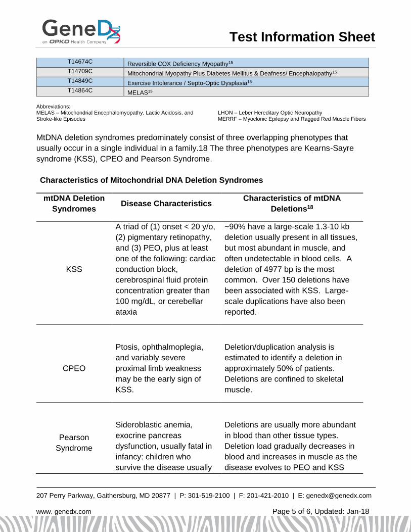
T14674C Reversible COX Deficiency Myopathy15
T14709C Mitochondrial Myopathy Plus Diabetes Mellitus & Deafness/ Encephalopathy15
T14849C Exercise Intolerance / Septo-Optic Dysplasia15
T14864C MELAS15
Abbreviations: MELAS – Mitochondrial Encephalomyopathy, Lactic Acidosis, and Stroke-like Episodes
LHON – Leber Hereditary Optic Neuropathy MERRF – Myoclonic Epilepsy and Ragged Red Muscle Fibers
MtDNA deletion syndromes predominately consist of three overlapping phenotypes that
usually occur in a single individual in a family.18 The three phenotypes are Kearns-Sayre
syndrome (KSS), CPEO and Pearson Syndrome.
Characteristics of Mitochondrial DNA Deletion Syndromes
mtDNA Deletion
Syndromes Disease Characteristics
Characteristics of mtDNA
Deletions18
KSS
A triad of (1) onset < 20 y/o,
(2) pigmentary retinopathy,
and (3) PEO, plus at least
one of the following: cardiac
conduction block,
cerebrospinal fluid protein
concentration greater than
100 mg/dL, or cerebellar
ataxia
~90% have a large-scale 1.3-10 kb
deletion usually present in all tissues,
but most abundant in muscle, and
often undetectable in blood cells. A
deletion of 4977 bp is the most
common. Over 150 deletions have
been associated with KSS. Large-
scale duplications have also been
reported.
CPEO
Ptosis, ophthalmoplegia,
and variably severe
proximal limb weakness
may be the early sign of
KSS.
Deletion/duplication analysis is
estimated to identify a deletion in
approximately 50% of patients.
Deletions are confined to skeletal
muscle.
Pearson
Syndrome
Sideroblastic anemia,
exocrine pancreas
dysfunction, usually fatal in
infancy: children who
survive the disease usually
Deletions are usually more abundant
in blood than other tissue types.
Deletion load gradually decreases in
blood and increases in muscle as the
disease evolves to PEO and KSS

Test Information Sheet
207 Perry Parkway, Gaithersburg, MD 20877 | P: 301-519-2100 | F: 201-421-2010 | E: [email protected]
www. genedx.com Page 6 of 6, Updated: Jan-18
go on to develop KSS. over time.
References: 1. Longo, N. (2003) Neurol Clin N Am 21:817-831.
2. Majamaa et al., (1998) Am J Hum Genet 63:447-454.
3. Goto et al., (1991) Biochim Biophys Acta 1097:238-40.
4. Mackey et al., (1996) Am J Hum Genet 59:481-485
5. Taylor et al., (2003) J Am Coll Cardiol 41:1786-96.
6. DiMauro, S. Gene Reviews (2005) MERRF.
7. Thorburn, D. Gene Reviews (2006) Mitochondrial DNA-Associated Leigh Syndrome and NARP.
8. DiMauro, S. Gene Reviews (2005) MELAS.
9. Yu-Wai-Man, P and Chinnery, P. Gene Reviews (2008) Leber Hereditary Optic Neuropathy.
10. Macmillan et al., (1998) Neurology 50:417-22.
11. Crispim et al., (2008) Arq Bras Endocrinol Metab 52:1228-1235.
12. Zhu et al., (2009) Acta Biochim Biophys Sin 41:179-187.
13. Chinnery, P. Gene Reviews (2006) Mitochondrial Disorders Overview.
14. Bennett S.(2004) Pharmacogenomics 5:433-8.
15. MITOMAP: A Human Mitochondrial Genome Database. http://www.mitomap.org, 2008.
16. Achilli et al., (2012) PLoS One 7:e42242.
17. Pulkes et al., (1999) Ann Neurol 46:916-9.
18. DiMaruo, S. and Hirano, M. (Updated [May 3, 2011]). Mitochondrial DNA Deletion Syndromes In: GeneReviews at Genetests:
Medical Genetics Information Resource (database online). Copyright, University of Washington, Seattle. 1997-2012. Available at
http://www.genetests.org
19. Jeppesen TD,et al. (2003) Ann Neurol 54(1):86-92.