Tendon injury by dr yash
18
Sri Siddhartha Medical College Agalkote, Tumakuru. Department of Orthopaedics . SUBJECT SEMINAR ON: Flexors & extensor tendon injuries of hand. Chairperson& Moderator: Prof, & HOD: Dr Kiran kalaiah Presented by: Dr Yashavardhan T.M On: 30/03/2017 Introduction. Tendon injuries are the second most common injuries of the hand and therefore an important topic in trauma and orthopaedic patients.
-
Upload
yashavardhan-yashu -
Category
Healthcare
-
view
140 -
download
2
Transcript of Tendon injury by dr yash
Sri Siddhartha Medical
College Agalkote,
Tumakuru.
Department of Orthopaedics.
SUBJECT SEMINAR ON: Flexors &
extensor tendon injuries of hand.
Chairperson& Moderator: Prof, & HOD: Dr Kiran kalaiah
Presented by: Dr Yashavardhan T.M
On: 30/03/2017
Introduction. Tendon injuries are the second most common injuries of the hand and therefore an important topic in trauma and orthopaedic patients.
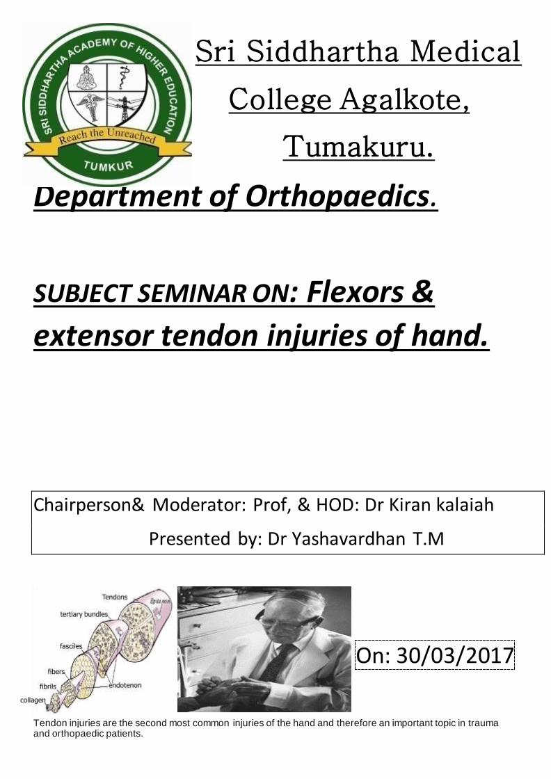
“A glistening structure between muscle & bone which transmit force from muscle to the bone’”
Paratenon: Loose areolar tissue encasing tendon in low mechanical stress area
Tendon sheath: a dense fibrous tissue tunnel enclosing tendon in high mechanical stress area 70% collagen (Type I) Extracellular components
Elastin
Mucopolysaccharides (enhance water-binding capability) Endotenon – around collagen bundles Epitenon – covers surface of tendon Paratenon – visceral/parietal adventitia surrounding tendons in hand.
Most injuries are open injuries to the flexor or extensor tendons, but less frequent injuries, e.g., damage to the functional system tendon sheath and pulley or dull avulsions, also need to be considered.
After clinical examination, ultrasound and magnetic resonance imaging have proved to be important diagnostic tools. Tendon injuries mostly require surgical repair, dull avulsions of the distal phalanges extensor tendon can receive conservative therapy.
Injuries of the flexor tendon sheath or single pulley injuries are treated conservatively and multiple pulley injuries receive surgical repair. In the postoperative course of flexor tendon injuries, the principle of early passive movement is important to trigger an "intrinsic" tendon healing to guarantee a good outcome.
Many substances were evaluated to see if they improved tendon healing; however, little evidence was found. Nevertheless, hyaluronic acid may improve intrinsic tendon healing.
Anatomical position
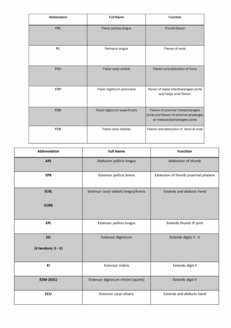
Flexors muscles: FDP and FDS tendons have fibrous sheaths on the palmar aspect of the digits
Fibrous arches and cruciate (cross-shaped) ligaments, which are attached posteriorly to the margins of the
phalanges and to the palmar ligaments hold the tendons to the bony plane and prevent the tendons from bowing
when the digits are flexed. The tendons are surrounded by a synovial sheath.
Flexor tendon system consists of intrinsic and extrinsic components
Extrinsics:
FDP: flexing the DIP joint
FDS: Flexing the PIP Joint
FPL: Flexing the IP joint of the thumb
Intrinsics:
Lumbricals: Flex the MCP joints and Extend the IP joints
FDP inserts on base of distal phalanx
FDS inserts on sides of middle phalanx
FPL inserts on proximal portion of the distal phalanx
INTEROSSEIUS
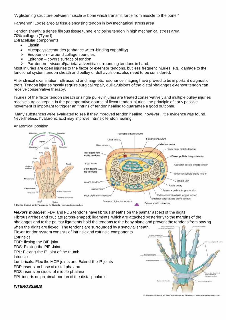
BLOOD SUPPLY
Pulleys of flexiours Synovial sheath is reinforced by a system of fibrous pulleys
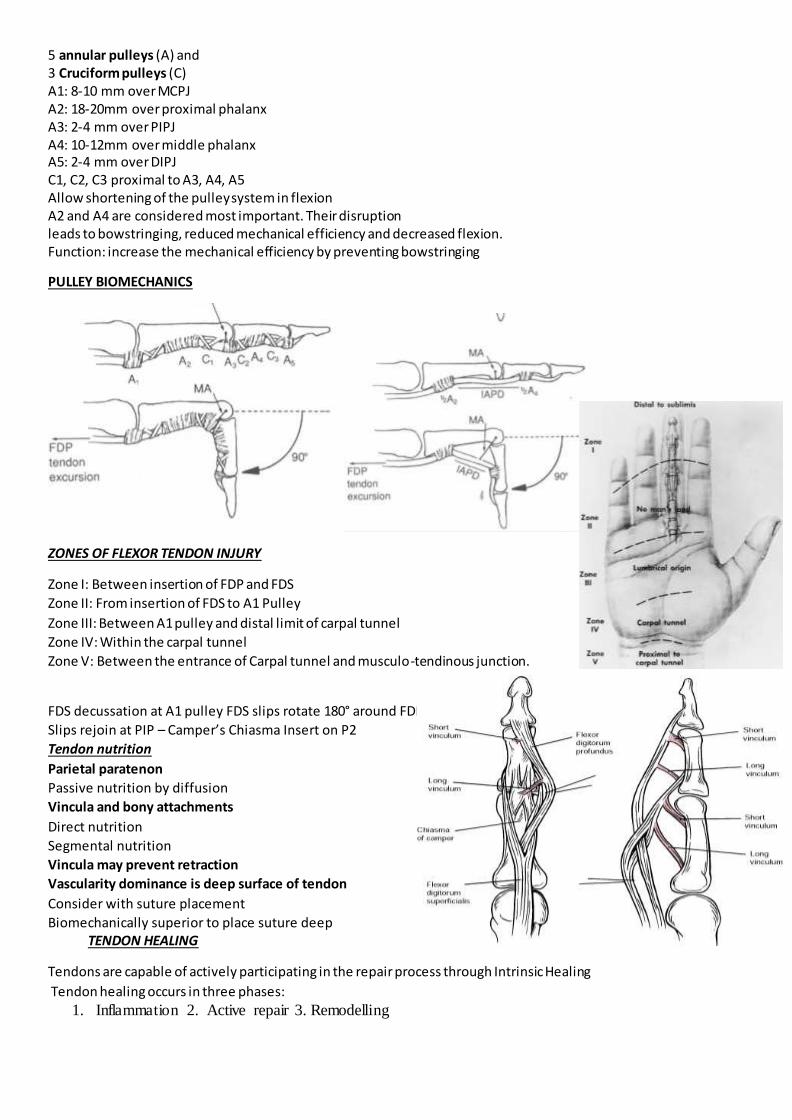
5 annular pulleys (A) and 3 Cruciform pulleys (C) A1: 8-10 mm over MCPJ A2: 18-20mm over proximal phalanx A3: 2-4 mm over PIPJ A4: 10-12mm over middle phalanx A5: 2-4 mm over DIPJ C1, C2, C3 proximal to A3, A4, A5 Allow shortening of the pulley system in flexion A2 and A4 are considered most important. Their disruption leads to bowstringing, reduced mechanical efficiency and decreased flexion. Function: increase the mechanical efficiency by preventing bowstringing
PULLEY BIOMECHANICS
ZONES OF FLEXOR TENDON INJURY
Zone I: Between insertion of FDP and FDS
Zone II: From insertion of FDS to A1 Pulley
Zone III: Between A1 pulley and distal limit of carpal tunnel
Zone IV: Within the carpal tunnel
Zone V: Between the entrance of Carpal tunnel and musculo-tendinous junction.
FDS decussation at A1 pulley FDS slips rotate 180° around FDP
Slips rejoin at PIP – Camper’s Chiasma Insert on P2
Tendon nutrition
Parietal paratenon
Passive nutrition by diffusion
Vincula and bony attachments
Direct nutrition
Segmental nutrition
Vincula may prevent retraction
Vascularity dominance is deep surface of tendon
Consider with suture placement
Biomechanically superior to place suture deep TENDON HEALING
Tendons are capable of actively participating in the repair process through Intrinsic Healing
Tendon healing occurs in three phases:
1. Inflammation 2. Active repair 3. Remodelling
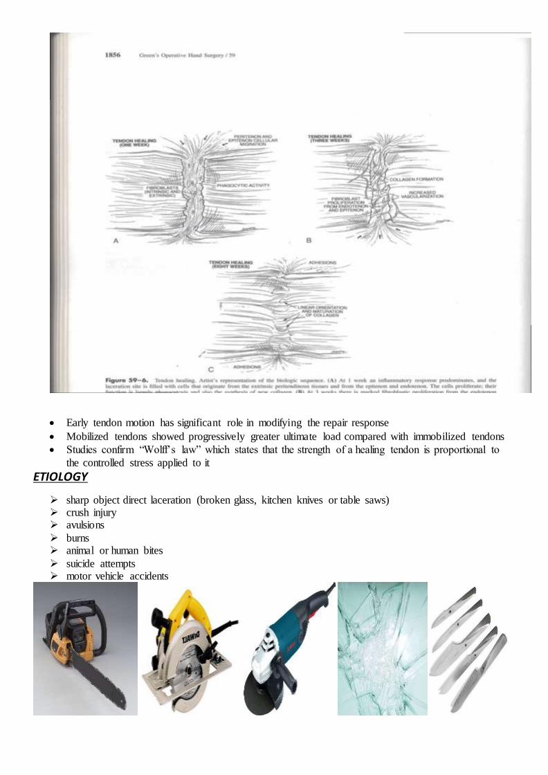
Early tendon motion has significant role in modifying the repair response
Mobilized tendons showed progressively greater ultimate load compared with immobilized tendons
Studies confirm “Wolff’s law” which states that the strength of a healing tendon is proportional to
the controlled stress applied to it
ETIOLOGY
sharp object direct laceration (broken glass, kitchen knives or table saws) crush injury avulsions
burns animal or human bites
suicide attempts motor vehicle accidents
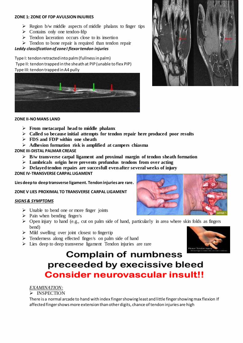
ZONE 1: ZONE OF FDP AVULSION INJURIES
Region b/w middle aspects of middle phalanx to finger tips Contains only one tendon-fdp
Tendon laceration occurs close to its insertion Tendon to bone repair is required than tendon repair
Leddy classification of zone I flexor tendon injuries
Type I: tendon retracted into palm (fullness in palm)
Type II: tendon trapped in the sheath at PIP (unable to flex PIP)
Type III: tendon trapped in A4 pully
ZONE II-NO MANS LAND
From metacarpal head to middle phalanx
Called so because initial attempts for tendon repair here produced poor results
FDS and FDP within one sheath
Adhesion formation risk is amplified at campers chiasma
ZONE III-DISTAL PALMAR CREASE
B/w transverse carpal ligament and proximal margin of tendon sheath formation
Lumbricals origin here prevents profundus tendons from over acting
Delayed tendon repairs are succesfull even after several weeks of injury
ZONE IV-TRANSVERSE CARPAL LIGAMENT
Lies deep to deep transverse ligament. Tendon injuries are rare.
ZONE V LIES PROXIMAL TO TRANSVERSE CARPAL LIGAMENT
SIGNS & SYMPTOMS
Unable to bend one or more finger joints Pain when bending finger/s Open injury to hand (e.g., cut on palm side of hand, particularly in area where skin folds as fingers
bend) Mild swelling over joint closest to fingertip
Tenderness along effected finger/s on palm side of hand Lies deep to deep transverse ligament Tendon injuries are rare
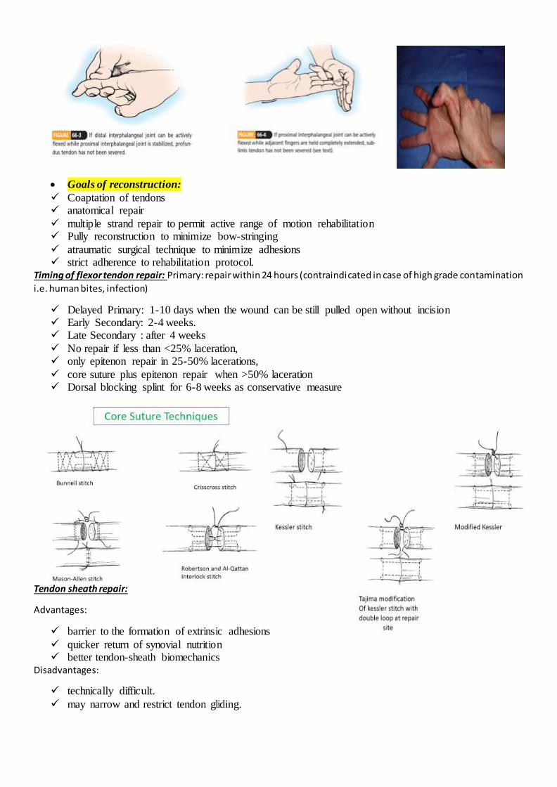
EXAMINATION: INSPECTION
There is a normal arcade to hand with index finger showing least and little finger showing max flexion If affected finger shows more extension than other digits, chance of tendon injuries are high
Goals of reconstruction:
Coaptation of tendons anatomical repair
multiple strand repair to permit active range of motion rehabilitation Pully reconstruction to minimize bow-stringing
atraumatic surgical technique to minimize adhesions strict adherence to rehabilitation protocol.
Timing of flexor tendon repair: Primary: repair within 24 hours (contraindicated in case of high grade contamination
i.e. human bites, infection)
Delayed Primary: 1-10 days when the wound can be still pulled open without incision
Early Secondary: 2-4 weeks. Late Secondary : after 4 weeks
No repair if less than <25% laceration, only epitenon repair in 25-50% lacerations,
core suture plus epitenon repair when >50% laceration Dorsal blocking splint for 6-8 weeks as conservative measure
Tendon sheath repair:
Advantages:
barrier to the formation of extrinsic adhesions
quicker return of synovial nutrition better tendon-sheath biomechanics
Disadvantages:
technically difficult.
may narrow and restrict tendon gliding.

ZONE 1 REPAIR
WOUND EXTENDED PROXIMALLY AND DISTALLY PROXIMAL TENDON RETRIEVED, CORE SUTURES ARE PLACED
KEITH NEEDLES USED TO PASS THE SUTURES AROUND THE DISTAL PHALANX EXITING THROUGH NAIL PLATE DISTALLY
REMAINING DISTAL END OF TENDON SUTURED TO THE RE-ATTACHED PROXIMAL PORTION.
Direct repair: if laceration is more than 1 cm from FDP insertion
Tendon advancement: if the laceration is less then 1 cm from insertion
ZONE II REPAIRS
REPAIR BOTH TENDON LACERATIONS
TENDON SHEATH MAY BE OPENED FOR EXPOSURE BUT A2 AND A4 ARE PRESERVED AS MUCH AS POSSIBLE
FDS IS REPAIRED FIRST FOLLOWED BY FDP Try to milk the tendon with the wrist flexed Morris and Martin. single skin hook is carefully inserted into sheath, then the hook is then turned
toward the tendons and when it is secured to the tendon, withdrawal of the hook should retrieve both tendons
Sourmelis and McGrouther. a small catheter is passed into the sheath and is delivered proximally into a small wound in the palm, just proximal to the A1 pulley, the catheter is sutured to both tendons 2 cm proximal to A1 pulley, which is then pulled distally to deliver the tendons into the synovial
window ZONE III REPAIRS
If both tendons are lacerated, both are repaired, end to end with
circumferential re-enforcing sutures May affect lumbricals in addition to flexor tendons Damaged lumbrical is either repaired or excised depending on severity of injury and the location of
the laceration ZONE IV REPAIR
Lacerations of flexor tendons within the carpal canal are typically associated with partial or complete laceration of median nerve
Here median nerves should be repaired first and the tendons last. ZONE V REPAIR
In this area there may be concomitant ulnar nerve & artery damage as well as radial artery & median
nerve damage. Primary repair of the arteries is usually indicated If wound is contaminated, arteries are repaired and delayed repair of tendons and nerves is planned

Zone 3 injuries
Lumbrical muscle bellies usually are not sutured because this can increase the tension
of these muscles and result in a “lumbrical plus” finger (paradoxical proximal
interphalangeal extension on attempted active finger flexion).
Quadriga effect Tendon advancement shortens the FDP & completes the grip before the normal fingers, if the tension on tendon
graft is set too high, and limit their flexion and thus week grip
Tendon Repair:
Silfverskiöld
Fish-Mouth End-to-End Suture (Pulvertaft)
End-to-Side method

Most of them involve active motion exercises. Then the suture strength has to increase
Active Extention-Rubber Band Flexion Method: e.g. Kleinert , and Brooke-Army
Immobilization Controlled Passive Motion Methods: e.g. Duran’s protocol Strickland: Early active ROM
Kleinert Protocol
Combines dorsal extension block with rubber-band traction proximal to wrist
Originally, included a nylon loop placed through the nail, and around the nail is placed a rubber band This passively flexes fingers, & the patient actively extends within the limits of the splint
Duran protocol
At surgery, a dorsal extension-block splint is applied with the wrist at 20-30° of flexion, the MCP joints at 50-60° of flexion, and the IP joints straight
Active range of motion rehabilitation
Kleinert !!
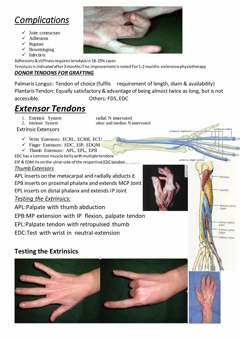
Complications Joint contracture Adhesions
Rupture Bowstringing Infection
Adhesions & stiffness requires tenolysis in 18-25% cases
Tenolysis is indicated after 3 months if no improvement is noted For 1-2 months extensive physiotherapy
DONOR TENDONS FOR GRAFTING
Palmaris Longus: Tendon of choice (fulfils requirement of length, diam & availability)
Plantaris Tendon: Equally satisfactory & advantage of being almost twice as long, but is not
accessible. Others: FDS, EDC
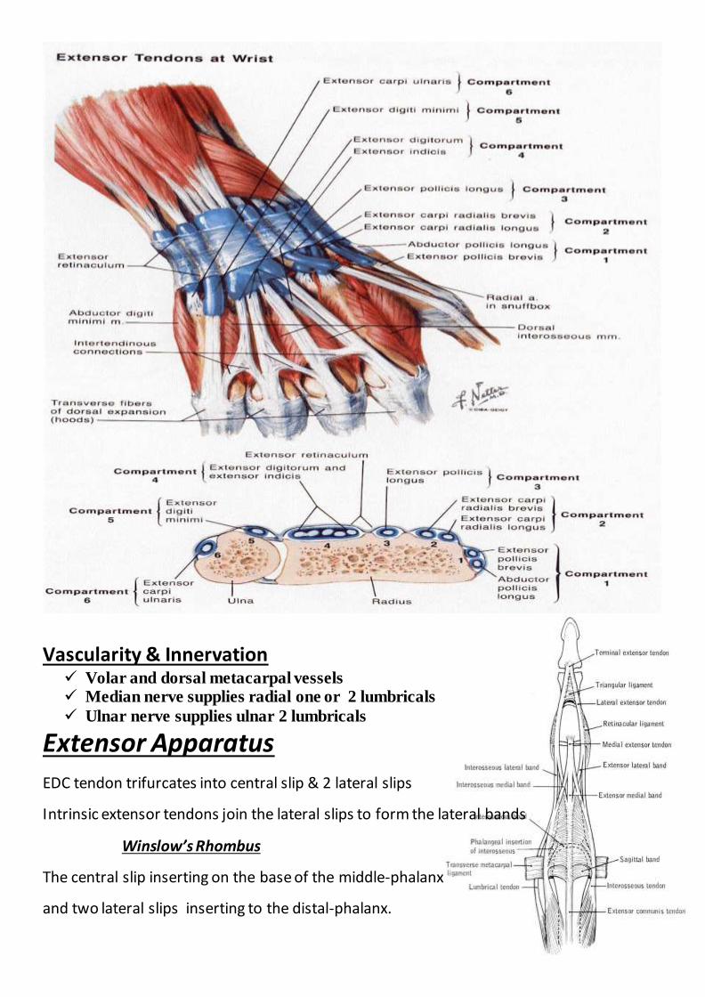
Extensor Tendons 1. Extrinsic System radial N innervated 2. Intrinsic System ulnar and median N innervated
Extrinsic Extensors
Wrist Extensors: ECRL, ECRB, ECU
Finger Extensors: EDC, EIP, EDQM Thumb Extensors: APL, EPL, EPB
EDC has a common muscle belly with multiple tendons
EIP & EDM lie on the ulnar side of the respective EDC tendon
Thumb Extensors
APL inserts on the metacarpal and radially abducts it
EPB inserts on proximal phalanx and extends MCP Joint
EPL inserts on distal phalanx and extends IP Joint
Testing the Extrinsics:
APL:Palpate with thumb abduction
EPB:MP extension with IP flexion, palpate tendon
EPL:Palpate tendon with retropulsed thumb
EDC:Test with wrist in neutral-extension
Testing the Extrinsics
Vascularity & Innervation Volar and dorsal metacarpal vessels Median nerve supplies radial one or 2 lumbricals
Ulnar nerve supplies ulnar 2 lumbricals
Extensor Apparatus
EDC tendon trifurcates into central slip & 2 lateral slips
Intrinsic extensor tendons join the lateral slips to form the lateral bands
Winslow’s Rhombus
The central slip inserting on the base of the middle-phalanx
and two lateral slips inserting to the distal-phalanx.
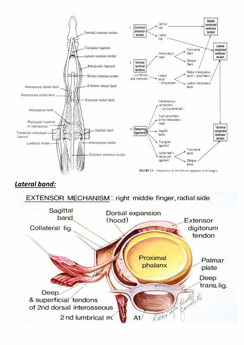
Lateral band:
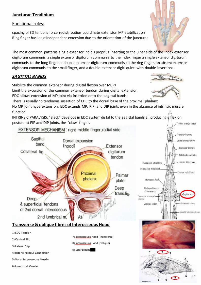
Juncturae Tendinium
Functional roles:
spacing of ED tendons force redistribution coordinate extension MP stabilization
Ring finger has least independent extension due to the orientation of the juncturae
The most common patterns single extensor indicis proprius inserting to the ulnar side of the index extensor
digitorum communis a single extensor digitorum communis to the index finger a single extensor digitorum
communis to the long finger, a double extensor digitorum communis to the ring finger, an absent extensor digitorum communis to the small finger, and a double extensor digiti quinti with double insertions.
SAGITTAL BANDS
Stabilize the common extensor during digital flexion over MCPJ
Limit the excursion of the common extensor tendon during digital extension
EDC allows extension of MP joint via insertion onto the sagittal bands
There is usually no tendinous insertion of EDC to the dorsal base of the proximal phalanx
No MP joint hyperextension: EDC extends MP, PIP, and DIP joints even in the absence of intrinsic muscle
function.
INTRINSIC PARALYSIS: “slack” develops in EDC system distal to the sagittal bands all producing a flexion
posture at PIP and DIP joints, the “claw” finger.
Transverse & oblique fibres of Interosseous Hood
1) EDC Tendon
2) Centra l Slip
3) Latera l Slip
4) Intertendinous Connection
5) Volar Interosseous Muscle
6) Lumbrical Muscle

Triangular Ligament
Connects both lateral bands over the middle phalanx.
Limits the volar and lateral shifting of the
lateral conjoined extensor tendon during digital flexion
In boutonniere deformity; elongated
In fixed swan neck deformity; retracted
Retinacular Ligament
Lateral continuation of the triangular ligament extending from the lateral margin of the lateral conjoined
extensor tendon to PIPJ articular volar plat
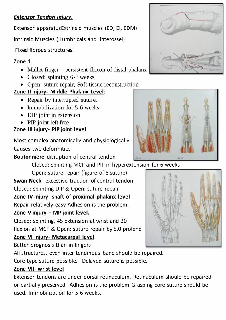
Extensor Tendon Injury.
Extensor apparatusExtrinsic muscles (ED, EI, EDM)
Intrinsic Muscles ( Lumbricals and Interossei)
Fixed fibrous structures.
Zone 1
Mallet finger – persistent flexon of distal phalanx
Closed: splinting 6-8 weeks
Open: suture repair, Soft tissue reconstruction
Zone II injury- Middle Phalanx Level:
Repair by interrupted suture.
Immobilization for 5-6 weeks
DIP joint in extension
PIP joint left free
Zone III injury- PIP joint level
Most complex anatomically and physiologically
Causes two deformities
Boutonniere disruption of central tendon
Closed: splinting MCP and PIP in hyperextension for 6 weeks
Open: suture repair (figure of 8 suture)
Swan Neck excessive traction of central tendon
Closed: splinting DIP & Open: suture repair
Zone IV injury- shaft of proximal phalanx level
Repair relatively easy Adhesion is the problem.
Zone V injury – MP joint level.
Closed: splinting, 45 extension at wrist and 20
flexion at MCP & Open: suture repair by 5.0 prolene
Zone VI injury- Metacarpal level
Better prognosis than in fingers
All structures, even inter-tendinous band should be repaired.
Core type suture possible. Delayed suture is possible.
Zone VII- wrist level
Extensor tendons are under dorsal retinaculum. Retinaculum should be repaired
or partially preserved. Adhesion is the problem Grasping core suture should be
used. Immobilization for 5-6 weeks.
IMMOBILIZATION
INJURIES IN ZONES PROXIMAL TO MCPs May be immobilized for 3 weeks.
Afterwards, finger may be placed in removable volar splint between exercise periods for 2 weeks
Progressive ROM after 3 weeks
If full flexion is not regained rapidly, dynamic flexion may be started after 6 weeks
INJURIES IN ZONES DISTAL TO MCPs
Require a longer period of immobilization (usually 6 weeks)
A progressive exercise program is initiated
Dynamic splinting during day and static splinting at night to maintain extension
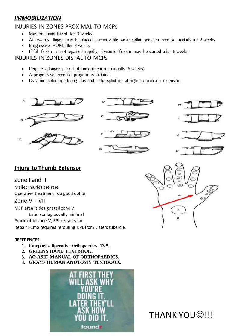
Injury to Thumb Extensor
Zone I and II Mallet injuries are rare
Operative treatment is a good option
Zone V – VII MCP area is designated zone V
Extensor lag usually minimal
Proximal to zone V, EPL retracts far
Repair >1mo requires rerouting EPL from Listers tubercle.
REFERENCES.
1. Campbel's 0perative 0rthopaedics 13th.
2. GREENS HAND TEXTBOOK.
3. AO-ASIF MANUAL OF ORTHOPAEDICS.
4. GRAYS HUMAN ANOTOMY TEXTBOOK.
THANK YOU!!!