Tendon Cell Behavior and Matrix Remodeling in Degenerative Tendinopathy Marieke de.pdf ·...
144
Tendon Cell Behavior and Matrix Remodeling in Degenerative Tendinopathy Marieke de Mos
-
Upload
nguyendiep -
Category
Documents
-
view
218 -
download
0
Transcript of Tendon Cell Behavior and Matrix Remodeling in Degenerative Tendinopathy Marieke de.pdf ·...

Tendon Cell Behaviorand Matrix Remodeling in
Degenerative Tendinopathy
Marieke de Mos

Printing of this thesis was financially supported by Stichting Anna Fonds, Nederlandse Vereni-
ging voor Matrix Biologie, Biomet, Reumafonds.
ISBN: 978-90-8559-453-6
Cover by: Marieke de Mos
Layout and printing: Optima Grafische Communicatie, Rotterdam, The Netherlands
© M. de Mos, The Netherlands, 2008. All rights reserved. No part of this thesis may be reproduced
or transmitted in any form or by any means, without prior written permission by the author.

Tendon Cell Behaviorand Matrix Remodeling in
Degenerative Tendinopathy
Peescelgedrag en matrix remodellering indegeneratieve tendinopathie
Proefschrift
ter verkrijging van de graad van doctor aan de
Erasmus Universiteit Rotterdam
op gezag van de
rector magnificus
Prof.dr. S.W.J. Lamberts
en volgens besluit van het College voor Promoties.
De openbare verdediging zal plaatsvinden op
woensdag 7 januari 2009 om 11.45 uur
door
Marieke de Mos
geboren te Voorburg

PRoMoTie CoMMissie
Promotor: Prof.dr. J.A.N. Verhaar
Overige leden: Prof.dr. J.M.W. Hazes
Prof.dr. S.E.R. Hovius
Prof.dr. P.R. Van Weeren
Copromotor: Dr. G.J.V.M. van Osch

Voor mama


Contents
List of Abbreviations 9
Chapter 1 General introduction 11
Chapter 2 Achilles tendinosis: changes in biochemical composition and
collagen turnover rate
25
Chapter 3 Tendon degeneration is not mediated by regulation by Toll-like
receptors 2 and 4 in human tenocytes
43
Chapter 4 Intrinsic differentiation potential of adolescent human tendon
tissue: an in-vitro cell differentiation study
55
Chapter 5 In-vitro model to study chondrogenic differentiation in
tendinopathy
73
Chapter 6 Can platelet-rich plasma enhance tendon repair: a cell culture study 89
Chapter 7 General discussion 105
Chapter 8 Summary 117
Appendices
Nederlandse samenvatting
Dankwoord
Curriculum Vitae
PhD Portfolio Summary
List of Publications
Color figures
123
125
129
133
135
137
139


List of Abbreviations
α-SMA alpha smooth muscle actin
AA Achilles tendon tissue adjacent to tendinotic lesion
AGC1 aggrecan
AGE advanced glycation endproduct
AH healthy Achilles tendon tissue
AT tendinotic Achilles tendon tissue
BGLAP bone gamma-carboxyglutamate = osteocalcin
BMSC bone marrow-derived stromal cell
CBFA1 core-binding factor 1 = RUNX2
COL10A1 collagen type X, alpha 1 chain
COL2A1 collagen type II, alpha 1 chain
D7-FIB 11-fibrau, fibroblast antibody
DMEM Dulbecco’s modified Eagle’s medium
ECM extracellular matrix
EGF epidermal growth factor
FABP adipocyte fatty acid binding protein
FACS fluorescense activated cell sorter
FCS fetal calf serum
FGF fibroblast growth factor
GAG glycosaminoglycan
HGF hepatocyte growth factor
HP hydroxylysylpyridinoline
HPLC high-performance liquid chromatography
Hyl hydroxylysine
Hyp hydroxyproline
IGF insulin-like growth factor
IHC immunohistochemistry
IL interleukin
LP lysylpyridinoline
MEC medical ethical committee
MMP matrix-metalloproteinase
OA osteoarthritis
PDGF platelet-derived growth factor
PPARG peroxisome proliferative activated receptor gamma
PPCR platelet-poor clot releasate
PPP platelet-poor plasma

10
List
of A
bbre
viat
ions
PRCR platelet-rich clot releasate
PRP platelet-rich plasma
QPCR quantitative polymerase chain reaction
RA rheumatoid arthritis
RFU relative fluorescence unit
RT-PCR reverse transcriptase polymerase chain reaction
RUNX2 RUNT-related transcription factor 2 = CBFA1
SOX9 SRY-box 9
SP7 Sp7 transcription factor = osterix
TDF tendon-derived fibroblast
TGF transforming growth factor
TLR Toll-like receptor
TNF tumor necrosis factor
VEGF vascular endothelial-derived growth factor A

Chapter 1
General Introduction

General Introduction 13
TenDinoPaThy
Tendon injuries are common in human athletes [1-4]. Furthermore, such injuries are also
prevalent in the ageing sedentary population [5-7]. In recent decades, the incidence of tendon
injuries has risen due to both an increase in an elderly population and a rise in participation in
recreational and competitive sporting activities. In the general population the lifetime cumula-
tive incidence of Achilles tendinopathy is 5.9 % among sedentary people and 50 % among elite
endurance athletes [2]. Despite the high frequency, there are still many unsolved questions and
differences of opinion concerning pathology, etiology, and even terminology.
Until several years ago the most often used word for tendon disease in the clinical practice of
orthopaedic and sports medicine was ’tendonitis/ tendinitis’, literally meaning tendon inflam-
mation, reflecting the general idea that overuse tendinopathies were due to inflammation.
However, this common wisdom was challenged by that time, as the histopathological feature
usually described in tendinopathies was a degenerative process and inflammation was not
typically seen [8-11]. Therefore Nicola Maffulli suggested to use the term ‘tendinopathy’ as a
general descriptor of the clinical conditions in and around tendons arising from overuse [12,
13]. In addition the term ‘tendinosis’, literally meaning tendon degeneration, should be used
after histopathological examination. This nomenclature is gradually being integrated now in
research communication and clinicial practice.
The clinical presentation of tendinopathy is characterized by a combination of pain, swell-
ing, and impaired performance. A variety of tendons in humans may be affected including the
supraspinatus tendon in the shoulder, the forearm extensor and flexor muscle tendons in the
elbow, and the Achilles tendon and the patellar tendon in the lower limb. The respons of tendi-
nopathy to the currently available treatment options is often unsatisfactory requiring lengthy
periods of rehabilitation or even surgical intervention [14, 15].
TenDons
Function
Tendons are most often thought of as the bright white, parallel fibred connective tissues that
join muscles to bones. While they exert no pulling force of their own, they enable locomotion
by transferring muscle contractions to the bones. Energy storing tendons, like the Achilles
tendon [16], not only transmit muscle-generated tensile forces to bone in order to move joints,
but they additionally act like springs to store elastic energy, which enhances the efficiency of
locomotion.
Chap
ter 1
14
structure
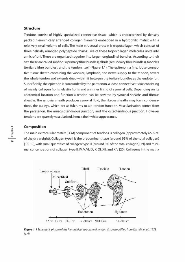
Tendons consist of highly specialized connective tissue, which is characterized by densely
packed hierarchically arranged collagen filaments embedded in a hydrophilic matrix with a
relatively small volume of cells. The main structural protein is tropocollagen which consists of
three helically arranged polypeptide chains. Five of these tropocollagen molecules unite into
a microfibril. These are organized together into larger longitudinal bundles. According to their
size these are called subfibrils (primary fibre bundles), fibrils (secundairy fibre bundles), fascicles
(tertiairy fibre bundles), and the tendon itself (Figure 1.1). The epitenon, a fine, loose connec-
tive-tissue sheath containing the vascular, lymphatic, and nerve supply to the tendon, covers
the whole tendon and extends deep within it between the tertiary bundles as the endotenon.
Superficially, the epitenon is surrounded by the paratenon, a loose connective tissue consisting
of mainly collagen fibrils, elastin fibrils and an inner lining of synovial cells. Depending on its
anatomical location and function a tendon can be covered by synovial sheaths and fibrous
sheaths. The synovial sheath produces synovial fluid, the fibrous sheaths may form condensa-
tions, the pulleys, which act as fulcrums to aid tendon function. Vascularisation comes from
the paratenon, the musculotendinous junction, and the osteotendinous junction. However
tendons are sparsely vascularised, hence their white appearance.
Composition
The main extracellular matrix (ECM) component of tendons is collagen (approximately 65-80%
of the dry weight). Collagen type I is the predominant type (around 95% of the total collagen)
[18, 19], with small quantities of collagen type III (around 3% of the total collagen)[19] and mini-
mal concentrations of collagen type II, IV, V, VI, IX, X, XI, XII, and XIV [20]. Collagens in the matrix
Figure 1.1 Schematic picture of the hierarchical structure of tendon tissue (modified from Kastelic et al., 1978 [17]).

General Introduction 15
are stabilized by cross-links. These cross-links influence the mechanical properties of the tendon
tissue. Some cross-links are formed after enzymatic modifications. The best characterised of
these are the hydroxylysylpyridinoline (HP) and lysylpyridinoline (LP) cross-links. Tendons have
high HP content compared to other connective tissues. The amount of cross-links in a tendon
is related to its mechanical function [21]. A greater cross-link concentration is generally found
in compressed tendons associated with the fibrocartilaginous composition found at these sites
[21, 22]. A second mechanism of intermolecular cross-linking of collagen is via non-enzymatic
reactions with glucose. A well-identified non-enzymatic glycation endproduct is pentosidine.
Pentosidine cross-links can be used to asses the remodeling rate and biological age of collagen
networks because of the relatively slow collagen turnover rate and the linear time-related
increase of spontaneously formed irreversible pentosidine cross-links [23, 24].
The major non-collagenous constituents of tendons are water (around 70% of the wet
weight) and proteoglycans. The strongly hydrophilic glycosaminoglycan molecules attract
water molecules and the collagenous network prevents the tissue from expanding beyond a
certain limit, thus giving the tissue its strength. Differences in proteoglycan composition within
and between tendons have been described during maturation and ageing, after immobilisa-
tion and exercise, in response to compressional mechanical forces, and after tendon injury [22,
25, 26].
Tenocytes are sparsely distributed within the ECM, constituting about 90-95% of the cell
population. They can be considered highly specialized fibroblasts, but their phenotype has
been poorly defined to date [27]. They are responsible for the production and maintainance
of all the ground substances of the extracellular matrix. Adjacent cells are connected via large
cytoplasmatic extensions with gap junctions, which are thought to be involved in effecting a
coordinated cellular response to the environment. The cells can change their metabolic activity
with respect to production of extracellular matrix components and matrix degrading enzymes
[28]. For instance different types of loading (compression, tension) elicit different types of
response [25, 29, 30].
The achilles tendon
The thickest and strongest tendon in the body is the Achilles tendon (Figure 1.2, see also color
inlay). It is an energy-storing tendon [16] that is subjected to tensional forces as large as 6-12
times the body weight [31, 32]. It originates from the calf muscles (gastrocnemius and soleus)
where collagen fibers from within the muscles are continuous with those of the tendon. It is
about 15 cm long and is inserted into the posterior surface of the calcaneus where its collagen
fibers are mineralized and integrated into bone tissue. The tendon spreads out somewhat at
its distal end so that the narrowest part, also called the mid-portion, is about 4-6 cm proximal
to the insertion site. Along their run from proximal to distal the tertiairy bundles twist approxi-
mately 90 degrees around the tendons longitudinal axis, the largest part of this twist taking
place in the mid-portion area [33]. The mid-portion area is also the most hypovascular region

Chap
ter 1
16of the Achilles tendon, hence also called the watershed region [34]. Besides insertional Achilles
tendinopathies especially this mid-portion area of the Achilles tendon is frequently subject to
degenerative pathology [1, 11, 14].
PaThoPhysiology oF TenDinoPaThy
histology
Histological alterations described in tendinotic lesions include an abnormal fiber structure and
fiber arrangement, variations in cellular distribution with hypercellular and hypocellular areas,
rounding of the tenocyte nuclei, decreased collagen stainability, increased GAG-stainability,
and increased vascularity (Figure 1.3, see also color inlay)[8-11]. Occasionally signs of bleeding,
fibrin deposits, calcifications, and lipid accumulations can be seen. Inflammatory cell infiltrates
and acellular necrotic areas are exceptional and not regarded as normal elements of the degen-
erative process [9].
Biochemistry and molecular biology
There have been relatively few biochemical and molecular studies of tendinopathy, and most
of them have been of material collected at the endstage after tendon rupture. Nonetheless, the
Figure 1.2 Anatomy of the Achilles tendon (adapted from Wolf-Heidegger’s Atlas of Human Anatomy, 4th edition).
Figure 1.2 Anatomy of the Achilles tendon (adapted from Wolf-Heidegger’s Atlas of Human Anatomy, 4th edition). (See color inlay for a full color version of this figure.)

General Introduction 17
histological absence of inflammation in the chronic phase was confirmed using microdialysis
techniques [35] and cDNA microarrays [36]. Due to lack of material from early tendinopathy
possible inflammatory events in the early stages cannot be excluded yet.
The importance of matrix turnover in tendon pathology was demonstrated in many ways.
In degenerative supraspinatus tendons a small but significant decrease in total collagen was
found, with an increased proportion of type III collagen relative to type I, resembling fibrotic
repair tissue [19]. A decreased accumulation of non-enzymatic pentosidine cross-links was
found in supraspinatus tendinopathy suggesting a relatively high matrix remodeling rate
linked to the onset of degenerative pathology [21]. Concurrent with the biochemical data, on
gene expression level a large increase was found in collagen type I and III expression [36]. Also
changes in gene expression level and activity of members of the matrix-metalloproteinase
(MMP) family of matrix degrading enzymes were found [37-39].
It has been long recognized that in tendinopathy the amount of glycosaminoglycans is
increased and the composition of these non-collagenous matrix components is changed
towards a fibrocartilaginous composition [8-11, 22, 26]. These changes in tendinopathy have
been considered a functional adaptation to compressional loading in these areas [25, 40-43]
although it may have negative effects on the tendons tensional strength and ability to repair
after injury [22].
hypotheses
Extensive research during the past decades regarding the aetiological factors, the tissue
alterations, and the cellular behavior associated with degenerative tendinopathy has led to the
formulation of three main hypotheses regarding the pathophysiology, all three with propos-
edly synergistic interactions contributing to the disease:
Figure 1.3 Histological picture of (A) healthy and (B) tendinotic Achilles tendon tissue (Magnification 200x).
Figure 1.3 Histological picture of (A) healthy and (B) tendinotic Achilles tendon tissue (Magnification 200x). (See color inlay for a full color version of this figure.)

Chap
ter 1
18
1. Contribution of mechanical overuse (repetitive overload) [5, 44]
Classically, the aetiology of tendinopathy has been linked to the performance of repetitive activi-
ties, eliciting an overuse injury. A proposed algorithm for the onset of tendinopathy involves
repetitive tensional loading with repeated strains below the injury threshold of the tendon,
inducing microdamage. The microdamage and subsequent processes to repair the matrix
composition and organisation cause a transient weakness of the tissue, which makes the tissue
more susceptible to damage from continued loading. This damage then accumulates until the
overt pathology of tendinopathy develops. Increased exercise levels increase the amount of
microdamage. Ageing alters the tendons mechanical properties making the tissue more prone
to microdamage. Poor vascularisation and low tenocyte metabolism contribute to the overload
pathology by extending this vulnerable repair period. This theory explains how chronic repeti-
tive damage to tendons could accumulate over time and perhaps why tendinopathy would be
degenerative and not inflammatory in nature. However, it does not explain the pain associated
with tendinopathy or the prevalence of tendinopathy in the sedentary population. Also it is
somewhat counterintuitive that load well within the physiological range can actually harm the
tendon.
2. Contribution of ageing [5, 45]
Tendon mechanical properties deteriorate with ageing. Tendons become stiffer and less elastic.
There is substantial decrease in the solubility of the collagen, thought to be associated with
the accumulation of advanced glycation endproduct (AGE) cross-links, such as pentosidine.
Accumulated physical damage is seen in ageing tendons, with increased amounts of dena-
tured collagen and increased proteolytic cleavage of matrix components. These changes are
all associated with deterioration in the physical properties of the tendon. Also, tendon blood
flow declines with age and after skeletal maturity the tendon cells appear to decrease matrix
synthesis. More importantly, the cells decrease responsiveness to loading or exercise regarding
their matrix synthesis. Though degeneration is not an inevitable consequence of ageing, the
tissue changes in ageing tendons might at least partially explain the increased prevalence of
degenerative changes associated with ageing. However this theory does not fully explain why
certain ageing individuals do get tendinopathy and others do not. A synergistic effect of ageing
and exercise in initiating tendon degeneration has been proposed.
3. Contribution of vascularisation [44, 46, 47]
During development, tendons are highly cellular and metabolically active and supplied with
a rich capillary network. Mature tendons however are sparsely vascularised, hence their white
appearance. The vascularity is even more compromised at junctional zones, and sites of torsion,
friction, or compression. For example two hypovascular regions of the Achilles tendon are the
insertional area and the mid-portion area. These avascular zones are commonly associated with
degeneration and rupture. Furthermore tendon blood flow generally declines with increasing

General Introduction 19
age and mechanical loading. In many cases of chronic tendinopathy there is an angiofibroblastic
response. The vasculoneural ingrowth may be a contributory factor to the pain in tendinopa-
thy. This theory may explain why tendons have specific vulnerable sections and explain the
relatively high prevalence of tendinopathy in hypovascular tendon regions like the mid-portion
of the Achilles tendon. However, the role of neovascularisation in the pathology remains poorly
understood.
TReaTMenT oF TenDinoPaThy
Current treatments
There are numerous different treatment types in the management of tendinopathy. No good
evidence so far is presented for physical therapies (cryotherapy, therapeutic ultrasound, and
low-intensity laser treatment), popular manual therapies (deep transverse friction massage and
soft tissue mobilisation stimulating blood supply), and biomechanical alterations (heelpads
and other orthotics). Some pain relief may be achieved by oral and local non-steroidal anti-
inflammatory drugs, possibly independent of the anti-inflammatory action. However, many
consider the use of local intratendinous corticosteroid injections contraindicated given the
high risk of spontaneous rupture of the tendon post-injection, particularly at the Achilles ten-
don. Injection of heparin, dextrose, aprotinin, or sclerosing agents also remains controversial.
There is little evidence of benefit from extracorporeal shock wave therapy for tendon condi-
tions other then calcifying tendinopathy of the shoulder and chronic heel pain [48, 49]. Topical
application of glyceryl trinitrate showed improvement at 6 months, but these results are yet to
be repeated [50]. Eccentric training for the treatment of mid-portion Achilles tendinopathy has
gained popularity following recent successful randomized controlled trials [51, 52]. The success
of this treatment for Achilles tendinopathy has led to efforts to see whether the results can be
extended to other tendon disorders like the patella tendon and supraspinatus tendon. Early
results look promising [53, 54], but why the treatment is successful remains uncertain.
The Dutch guidelines for treatment of mid-portion Achilles tendinopathy formulated in
2007 suggest maximal conservative treatment for a minimal duration of 6 months with at
least 3 months of eccentric exercises [55]. After this period 20-25% of the patients will have not
returned to their original exercise level and surgical treatment can be considered. Surgical inter-
vention may include debridement of the lesional area, multiple longitudinal tenotomies, and
cleaving of the peritendineum. Percutaneous tenotomy resulted in 75% of patients reporting
good or excellent results at 18 months follow-up [56]. Open surgery for Achilles tendinopathy
has shown that 67 % had returned to physical activity at 7 months after surgery [57].
In summary, tendinopathy responds poorly to the currently available treatment options of
which very few are evidence-based. The nowadays abundant amount of treatment options for
Chap
ter 1
20
tendinopathy inversily reflects the little amount of evidence regarding the working mechanisms
and success rates of the practiced interventions.
Future treatments
As a result of the deficiencies of current treatments there is a great interest in investigating new
therapies. Two treatments that hold promise for the future of tendon disease management
are injection of autologous platelet-rich plasma (PRP) and a tissue engineering approach to
regenerate healthy tendon tissue.
Clinical applications of autologous PRP in human medicine include periodontal and maxil-
lofacial surgery, plastic surgery, treatment of bone fractures, and treatment of chronic skin and
soft tissue ulcers. Numerous publications on PRP yielded excellent clinical outcomes [58, 59].
The only published cohort study in tendon research reported 93% reduction of pain for PRP-
treated patients with chronic elbow tendinosis [60]. In natural healing processes in the body,
platelets actively participate by rapidly releasing a variety of growth factors. PRP might provide
an autologous source of these growth factors that play a key role in tendon repair mechanisms.
Both controlled clinical studies and in-vitro studies are required to investigate in detail the pos-
sibly beneficial effects of PRP in tendon disorders.
Cell therapy with use of stem cells represents a potentially exciting alternative treatment
in the future of tendon management. Whilst healing was seen when mesenchymal stem cells
seeded in a collagen matrix were placed in a rabbit Achilles tendon defect, on histology the
new cells exhibited morphology more similar to fibroblasts than tenocytes [61]. This illustrates
the major challenge in tendon tissue engineering: mastering the knowledge on what drives
a stem cell or fibroblast towards tenogenic differentiation and how to provide these biome-
chanical, biochemical, or biophysical cues in vivo or in vitro to facilitate healthy tendon tissue
development or tendon repair. Early results in this research area are exciting and highlight the
potential of cell therapy for tendinopathy in the future [62, 63].
aiMs anD ouTline
Tendinopathy often responds poorly to the currently available treatment options requiring
lengthy periods of rehabilitation or even surgical intervention. The increasing scientific interest
in tendon disorders and the concurrent application of biochemical and molecular techniques
has led to rapid developments in the understanding of degenerative tendinopathies in the last
decades. We believe that more insight in the behavior of the tendon cells and the biomechani-
cal, biochemical, and biophysical signals that influence their conduct is a conditio sine qua non
for the development of more effective mechanism-based therapeutical interventions for both
prevention and repair of degenerative tendon lesions. The work in this thesis aims to improve
fundamental knowledge of tendon cell behavior and matrix remodeling in tendinopathy, both

General Introduction 21
during the disease process as well as in reaction to treatment modalities. The focus in this
research is on mid-portion Achilles tendinopathy.
We started out purely describing the changes observed in cell behavior, biochemical composi-
tion, and collagenous matrix turnover rate in vivo (chapter 2). We collected tendinosis biopsies
and macroscopically healthy tendon biopsies adjacent to the lesion from patients undergoing
surgery for mid-portion Achilles tendinopathy and healthy tendon biopsies from donors with
asymptomatic Achilles tendons. The results were compared with findings in literature regard-
ing supraspinatus tendinopathy.
From here hypotheses regarding pathogenic processes that might play a role in the devel-
opment and persistance of tendinopathy were explored in vivo and in vitro. Firstly, we were
intrigued by the analogy between tendon and cartilage tissue in both healthy and degenera-
tive state. Therefore in chapter 3 we hypothesized a role for Toll-like receptors (TLRs) in tendi-
nopathy, a role that might be quite similar to their role in osteoarthritis and their even more
distict role in rheumatoid and inflammatory arthritis. We explored this idea by examining both
in vivo mid-portion Achilles tendinopathy samples as well as by performing in-vitro culture
experiments stimulating healthy tendon explants with inflammatory mediators.
Mid-portion Achilles tendinotic lesions display an increased amount of glycosaminoglycans,
resembling cartilage tissue in this aspect, and sometimes also contain calcifications and lipid
accumulations. We hypothesized that alterations in tendon cellular differentiation might
contribute to these tissue changes associated with tendinopathy. In chapter 4 we studied
whether a population of cells with intrinsic differentiation potential is present in tendon tissue.
We performed in-vitro experiments with tendon-derived cells from non-degenerative human
tendon tissue trying to differentiate these cells towards cells with chondrogenic, adipogenic, or
osteogenic characteristics.
Considering the excessive or inappropriate fibrocartilaginous matrix production in tendons
as a remarkable feature of the pathological process in mid-portion Achilles tendinopathy, we
were especially interested in chondrogenic differentiation possibilities of the native tendon
cells. In chapter 5 this chondrogenic differentiation pattern was studied in mid-portion Achil-
les tendinopathy in vivo. Reasoning that opposing or removing the stimulus that causes the
metaplasia in the diseased tendons might help the tenocytes to return to their normal tendon
matrix production, we developed a tendon explant culture model to induce a chondrogenic
differentiation quite similar to the in-vivo situation in tendinopathy. We explored the usefulness
of this model to investigate early chondrogenic differentiation as a possible target for drug
treatment of tendinopathic lesions. We also studied the effects of adding triamcinolone or
platelet-rich plasma (PRP) to the chondrogenic differentiation model.
The following chapter (chapter 6) explored in further detail the working mechanisms of this
platelet-rich plasma (PRP), being considered in the present literature as a clinically promising
treatment intervention for tendinopathy although the exact effects on tendon cell behavior

Chap
ter 1
22
are not known yet. We studied the effects of platelet-rich plasma (PRP) on the metabolism of
explanted human tendon cells in-vitro.
In the final chapter (chapter 7) the main results described in this thesis are discussed in rela-
tion to eachother and in the context of current scientific knowledge. Also, certain limitations of
the in-vitro work are summarized. Suggestions for follow-up experiments and future research
directions are given throughout this general discussion section.
ReFeRenCes
1. Maffulli N, Wong J, Almekinders LC: Types and epidemiology of tendinopathy. Clin Sports Med 2003, 22(4):675-692.
2. Kujala UM, Sarna S, Kaprio J: Cumulative incidence of achilles tendon rupture and tendinopathy in male former elite athletes. Clin J Sport Med 2005, 15(3):133-135.
3. Ferretti A: Epidemiology of jumper’s knee. Sports Med 1986, 3(4):289-295. 4. Hume PA, Reid D, Edwards T: Epicondylar injury in sport: epidemiology, type, mechanisms, assess-
ment, management and prevention. Sports Med 2006, 36(2):151-170. 5. Smith RK, Birch HL, Goodman S, Heinegard D, Goodship AE: The influence of ageing and exercise on
tendon growth and degeneration--hypotheses for the initiation and prevention of strain-induced tendinopathies. Comp Biochem Physiol A Mol Integr Physiol 2002, 133(4):1039-1050.
6. Sayana MK, Maffulli N: Eccentric calf muscle training in non-athletic patients with Achilles tendinopa-thy. J Sci Med Sport 2007, 10(1):52-58.
7. Rolf C, Movin T: Etiology, histopathology, and outcome of surgery in achillodynia. Foot Ankle Int 1997, 18(9):565-569.
8. Movin T, Gad A, Reinholt FP, Rolf C: Tendon pathology in long-standing achillodynia. Biopsy findings in 40 patients. Acta Orthop Scand 1997, 68(2):170-175.
9. Astrom M, Rausing A: Chronic Achilles tendinopathy. A survey of surgical and histopathologic find-ings. Clin Orthop Relat Res 1995(316):151-164.
10. Tallon C, Maffulli N, Ewen SW: Ruptured Achilles tendons are significantly more degenerated than tendinopathic tendons. Med Sci Sports Exerc 2001, 33(12):1983-1990.
11. Puddu G, Ippolito E, Postacchini F: A classification of Achilles tendon disease. Am J Sports Med 1976, 4(4):145-150.
12. Maffulli N, Khan KM, Puddu G: Overuse tendon conditions: time to change a confusing terminology. Arthroscopy 1998, 14(8):840-843.
13. Maffulli N, Kahn KM: Clinical nomenclature for tendon injuries. Med Sci Sports Exerc 1999, 31(2):352-353.
14. Kvist M: Achilles tendon injuries in athletes. Sports Med 1994, 18(3):173-201. 15. Paavola M, Kannus P, Paakkala T, Pasanen M, Jarvinen M: Long-term prognosis of patients with achilles
tendinopathy. An observational 8-year follow-up study. Am J Sports Med 2000, 28(5):634-642. 16. Lichtwark GA, Wilson AM: In vivo mechanical properties of the human Achilles tendon during one-
legged hopping. J Exp Biol 2005, 208(Pt 24):4715-4725. 17. Kastelic J, Galeski A, Baer E: The multicomposite structure of tendon. Connect Tissue Res 1978, 6(1):11-
23. 18. von der Mark K: Localization of collagen types in tissues. Int Rev Connect Tissue Res 1981, 9:265-324. 19. Riley GP, Harrall RL, Constant CR, Chard MD, Cawston TE, Hazleman BL: Tendon degeneration and
chronic shoulder pain: changes in the collagen composition of the human rotator cuff tendons in rotator cuff tendinitis. Ann Rheum Dis 1994, 53(6):359-366.
General Introduction 23
20. Riley G: The pathogenesis of tendinopathy. A molecular perspective. Rheumatology (Oxford) 2004, 43(2):131-142.
21. Bank RA, TeKoppele JM, Oostingh G, Hazleman BL, Riley GP: Lysylhydroxylation and non-reducible crosslinking of human supraspinatus tendon collagen: changes with age and in chronic rotator cuff tendinitis. Ann Rheum Dis 1999, 58(1):35-41.
22. Riley GP, Harrall RL, Constant CR, Chard MD, Cawston TE, Hazleman BL: Glycosaminoglycans of human rotator cuff tendons: changes with age and in chronic rotator cuff tendinitis. Ann Rheum Dis 1994, 53(6):367-376.
23. Lin YL, Brama PA, Kiers GH, van Weeren PR, DeGroot J: Extracellular matrix composition of the equine superficial digital flexor tendon: relationship with age and anatomical site. J Vet Med A Physiol Pathol Clin Med 2005, 52(7):333-338.
24. Verzijl N, DeGroot J, Bank RA, Bayliss MT, Bijlsma JW, Lafeber FP, Maroudas A, TeKoppele JM: Age-related accumulation of the advanced glycation endproduct pentosidine in human articular cartilage aggrecan: the use of pentosidine levels as a quantitative measure of protein turnover. Matrix Biol 2001, 20(7):409-417.
25. Vogel KG, Koob TJ: Structural specialization in tendons under compression. Int Rev Cytol 1989, 115:267-293.
26. Corps AN, Robinson AH, Movin T, Costa ML, Hazleman BL, Riley GP: Increased expression of aggrecan and biglycan mRNA in Achilles tendinopathy. Rheumatology (Oxford) 2006, 45(3):291-294.
27. Yao L, Bestwick CS, Bestwick LA, Maffulli N, Aspden RM: Phenotypic drift in human tenocyte culture. Tissue Eng 2006, 12(7):1843-1849.
28. McNeilly CM, Banes AJ, Benjamin M, Ralphs JR: Tendon cells in vivo form a three dimensional network of cell processes linked by gap junctions. J Anat 1996, 189 ( Pt 3):593-600.
29. Arnoczky SP, Lavagnino M, Egerbacher M: The mechanobiological aetiopathogenesis of tendinopathy: is it the over-stimulation or the under-stimulation of tendon cells? Int J Exp Pathol 2007, 88(4):217-226.
30. Wall ME, Banes AJ: Early responses to mechanical load in tendon: role for calcium signaling, gap junc-tions and intercellular communication. J Musculoskelet Neuronal Interact 2005, 5(1):70-84.
31. Scott SH, Winter DA: Internal forces of chronic running injury sites. Med Sci Sports Exerc 1990, 22(3):357-369.
32. Komi PV, Fukashiro S, Jarvinen M: Biomechanical loading of Achilles tendon during normal locomo-tion. Clin Sports Med 1992, 11(3):521-531.
33. O’Brien M: Functional anatomy and physiology of tendons. Clin Sports Med 1992, 11(3):505-520. 34. Theobald P, Benjamin M, Nokes L, Pugh N: Review of the vascularisation of the human Achilles tendon.
Injury 2005, 36(11):1267-1272. 35. Alfredson H, Lorentzon R: Chronic tendon pain: no signs of chemical inflammation but high con-
centrations of the neurotransmitter glutamate. Implications for treatment? Curr Drug Targets 2002, 3(1):43-54.
36. Ireland D, Harrall R, Curry V, Holloway G, Hackney R, Hazleman B, Riley G: Multiple changes in gene expression in chronic human Achilles tendinopathy. Matrix Biol 2001, 20(3):159-169.
37. Riley GP: Gene expression and matrix turnover in overused and damaged tendons. Scand J Med Sci Sports 2005, 15(4):241-251.
38. Riley GP, Curry V, DeGroot J, van El B, Verzijl N, Hazleman BL, Bank RA: Matrix metalloproteinase activities and their relationship with collagen remodelling in tendon pathology. Matrix Biol 2002, 21(2):185-195.
39. Alfredson H, Lorentzon M, Backman S, Backman A, Lerner UH: cDNA-arrays and real-time quantitative PCR techniques in the investigation of chronic Achilles tendinosis. J Orthop Res 2003, 21(6):970-975.
40. Robbins JR, Evanko SP, Vogel KG: Mechanical loading and TGF-beta regulate proteoglycan synthesis in tendon. Arch Biochem Biophys 1997, 342(2):203-211.
41. Vogel KG: What happens when tendons bend and twist? Proteoglycans. J Musculoskelet Neuronal Interact 2004, 4(2):202-203.

Chap
ter 1
24
42. Vogel KG: The effect of compressive loading on proteoglycan turnover in cultured fetal tendon. Con-nect Tissue Res 1996, 34(3):227-237.
43. Archambault JM, Jelinsky SA, Lake SP, Hill AA, Glaser DL, Soslowsky LJ: Rat supraspinatus tendon expresses cartilage markers with overuse. J Orthop Res 2007, 25(5):617-624.
44. Rees JD, Wilson AM, Wolman RL: Current concepts in the management of tendon disorders. Rheuma-tology (Oxford) 2006, 45(5):508-521.
45. Tuite DJ, Renstrom PA, O’Brien M: The aging tendon. Scand J Med Sci Sports 1997, 7(2):72-77. 46. Fenwick SA, Hazleman BL, Riley GP: The vasculature and its role in the damaged and healing tendon.
Arthritis Res 2002, 4(4):252-260. 47. Alfredson H, Ohberg L, Forsgren S: Is vasculo-neural ingrowth the cause of pain in chronic Achilles
tendinosis? An investigation using ultrasonography and colour Doppler, immunohistochemistry, and diagnostic injections. Knee Surg Sports Traumatol Arthrosc 2003, 11(5):334-338.
48. Wang CJ, Yang KD, Wang FS, Chen HH, Wang JW: Shock wave therapy for calcific tendinitis of the shoulder: a prospective clinical study with two-year follow-up. Am J Sports Med 2003, 31(3):425-430.
49. Ogden JA, Alvarez RG, Marlow M: Shockwave therapy for chronic proximal plantar fasciitis: a meta-analysis. Foot Ankle Int 2002, 23(4):301-308.
50. Paoloni JA, Appleyard RC, Nelson J, Murrell GA: Topical glyceryl trinitrate treatment of chronic non-insertional achilles tendinopathy. A randomized, double-blind, placebo-controlled trial. J Bone Joint Surg Am 2004, 86-A(5):916-922.
51. Kingma JJ, de Knikker R, Wittink HM, Takken T: Eccentric overload training in patients with chronic Achilles tendinopathy: a systematic review. Br J Sports Med 2007, 41(6):e3.
52. Alfredson H, Pietila T, Jonsson P, Lorentzon R: Heavy-load eccentric calf muscle training for the treat-ment of chronic Achilles tendinosis. Am J Sports Med 1998, 26(3):360-366.
53. Young MA, Cook JL, Purdam CR, Kiss ZS, Alfredson H: Eccentric decline squat protocol offers superior results at 12 months compared with traditional eccentric protocol for patellar tendinopathy in vol-leyball players. Br J Sports Med 2005, 39(2):102-105.
54. Jonsson P, Wahlstrom P, Ohberg L, Alfredson H: Eccentric training in chronic painful impingement syn-drome of the shoulder: results of a pilot study. Knee Surg Sports Traumatol Arthrosc 2006, 14(1):76-81.
55. van Linschoten R, den Hoed PT, de Jongh AC: [Guideline ‘Chronic Achilles tendinopathy, in particular tendinosis, in sportsmen/sportswomen’]
Richtlijn ‘Chronische achillestendinopathie, in het bijzonder de tendinosis, bij sporters’. Ned Tijdschr Geneeskd 2007, 151(42):2319-2324.
56. Alfredson H, Cook J: A treatment algorithm for managing Achilles tendinopathy: new treatment options. Br J Sports Med 2007, 41(4):211-216.
57. Paavola M, Kannus P, Orava S, Pasanen M, Jarvinen M: Surgical treatment for chronic Achilles tendi-nopathy: a prospective seven month follow up study. Br J Sports Med 2002, 36(3):178-182.
58. Roukis TS, Zgonis T, Tiernan B: Autologous platelet-rich plasma for wound and osseous healing: a review of the literature and commercially available products. Adv Ther 2006, 23(2):218-237.
59. Eppley BL, Pietrzak WS, Blanton M: Platelet-rich plasma: a review of biology and applications in plastic surgery. Plast Reconstr Surg 2006, 118(6):147e-159e.
60. Mishra A, Pavelko T: Treatment of chronic elbow tendinosis with buffered platelet-rich plasma. Am J Sports Med 2006, 34(11):1774-1778.
61. Young RG, Butler DL, Weber W, Caplan AI, Gordon SL, Fink DJ: Use of mesenchymal stem cells in a collagen matrix for Achilles tendon repair. J Orthop Res 1998, 16(4):406-413.
62. Smith RK, Korda M, Blunn GW, Goodship AE: Isolation and implantation of autologous equine mes-enchymal stem cells from bone marrow into the superficial digital flexor tendon as a potential novel treatment. Equine Vet J 2003, 35(1):99-102.
63. Smith RK, Webbon PM: Harnessing the stem cell for the treatment of tendon injuries: heralding a new dawn? Br J Sports Med 2005, 39(9):582-584.

Chapter 2
Achilles tendinosis: changes in biochemical composition and collagen turnover rate
M. de Mos, B. van El, J. DeGroot, H. Jahr, H.T.M. van Schie, E.R. van Arkel, J.L. Tol, M.P. Heijboer, G.J.V.M. van Osch, J.A.N. Verhaar
Am J Sports Med. 2007 Sep;35(9):1549-56

Chap
ter 2
26
aBsTRaCT
Background
Understanding of biochemical and structural changes of the extracellular matrix in Achilles
tendinosis might be important for developing mechanism-based therapies.
hypothesis
In Achilles tendinosis changes occur in biochemical composition and collagen turnover rate.
Methods
From ten patients undergoing surgery for Achilles tendinopathy, one tendinosis biopsy (AT) and
one biopsy of macroscopically healthy tendon tissue adjacent to the lesion (AA) were collected.
Furthermore, biopsies were collected from three donors with asymptomatic Achilles tendons
(AH). Water content, collagen content, percentage of denatured collagen, amount of lysine
hydroxylation, number of enzymatic and non-enzymatic cross links, matrix metalloproteinase
(MMP) activity, and MMP and collagen gene-expression levels were analysed.
Results
In AT lesions the water content was highest, collagen content was subnormal with higher
amounts of denatured/damaged collagen. Low pentosidine levels in AT tissue indicated the
presence of relatively young collagenous matrix. More hydroxylated lysine residues were pres-
ent in AT samples, but enzymatic crosslinks revealed no differences between AT, AA, and AH
samples. In AT specimens MMP activity was higher, MMP gene-expression profile was altered,
collagen type I and III gene expression were upregulated.
Conclusions
In Achilles tendinosis the collagen turnover rate is increased and the natural biochemical com-
position of the collagenous matrix is compromised.
Clinical Relevance
Although tendon tissue directly adjacent to an Achilles tendinosis lesion looks macroscopically
healthy, histological and biochemical degenerative changes in adjacent tissue are evident,
which may have implications for surgical interventions.

Achilles tendinosis: changes in biochemical composition and collagen turnover rate 27
inTRoDuCTion
Tendinopathy is a tendon disorder that occurs most frequently in athletes and middle-aged
people [20]. Tendinopathy of the mid-portion of the Achilles tendon is one of the main causes
of chronic Achilles tendon pain [5, 7]. Generally, treatments of Achilles tendinopathy are con-
servative and is mainly aimed at relief of symptoms, with frequently unsatisfying results due to
a remarkable lack of knowledge concerning the underlying pathological mechanisms [24, 28].
On one hand, a prostaglandin-mediated inflammatory cascade does not seem to play a major
role in the pathogenesis of tendinopathy [2]. Moreover, there is growing evidence that tendi-
nopathy of the Achilles tendon mid-portion is often the clinical result of multiple degenerative
processes in the tendon matrix called tendinosis [25]. In order to develop a mechanism-based
therapy, it is crucial to obtain a better insight in the biochemical and structural changes of the
extracellular matrix that are involved in the pathogenesis of Achilles tendinosis.
Maintenance and regeneration of the physiological biochemical composition of the collag-
enous and non-collagenous extracellular matrix is essential for optimal structure and function
of the tendon [29]. There have been some studies on the compositional changes of the tendon
in supraspinatus tendinopathy. However, there are no such studies available on the difference
in biochemical collagen composition between healthy and degenerated mid-portion Achilles
tendon tissue.
Assumptions on the biochemical collagen compositional changes in Achilles tendinosis can
only be extrapolated from supraspinatus tendinopathy studies. This extrapolation might be
justified by the observation that histological features in Achilles tendinosis and supraspinatus
tendinopathy are similar [7, 16, 22, 26]. The alterations described in supraspinatus tendinopathy
include: 1) a significantly increased water content [32]; 2) a small, but significant decrease in
total collagen content [11, 32]; 3) an increased proportion of type III collagen compared to type
I collagen [32]; 4) an altered mode of collagen crosslinking, namely an increase in the amount
of hydroxylysine residues per collagen triple helix, and an increase of the enzymatic cross links
hydroxylysylpyridinoline (HP) and lysylpyridinoline (LP) [11], which is also reported in fibrotic
tissue [36]; 5) a significant decrease in the amount of non-enzymatic pentosidine cross links per
collagen molecule, which can be interpreted as a young biological age of the collagenous tissue
[11]. All these findings taken into account, it seems likely that in supraspinatus tendinopathy
the previously functional and carefully constructed native matrix is replaced by an aberrant
collagen network.
The process of collagen network turnover is a dynamic equilibrium between synthesis and
degradation [29]. Degradation of the extracellular matrix (ECM) is principally mediated by the
enzymatic activity of matrix metalloproteinases (MMPs). MMP activity is the product of MMP
synthesis, activation, inhibition, and degradation. MMP gene expression levels have been
measured in healthy, tendinotic, and ruptured Achilles tendon [3, 19, 21, 27], but the actual
MMP activity has not.

Chap
ter 2
28
We hypothesized that changes in biochemical collagen composition and collagen turnover
rate were present in Achilles tendinosis lesions. We report the results of a comparison between
(1) Achilles tendinosis lesions, (2) less affected Achilles tendon tissue surrounding those lesions
in the same individual, and (3) healthy Achilles tendon tissue, concerning histology, total water
content, biochemical collagen composition (including total collagen content, percentage of
degraded collagen, number of enzymatic and non-enzymatic cross links), MMP activity, and
gene expression levels of MMPs and collagens.
MaTeRials anD MeThoDs
Patient characteristics and sample collection
Tissue specimens of 10 consecutive patients with chronic mid-portion Achilles tendinopathy
were harvested during surgical debridement. Chronic Achilles tendinopathy was defined as
Achilles tendon pain for at least 3 months in combination with clinically and / or radiologically
(either MRI or ultrasonography) suspected mid-portion Achilles tendinopathy. Only patients
without previous tendon injury, without radiologically evident partial tendon rupture, with-
out use of chinolone antibiotics in the past three years, and with sufficient biopsy material
to perform all analyses were included. At surgery, one biopsy specimen was taken from the
macroscopically affected tendinotic lesion (AT) and a second biopsy specimen was taken from
macroscopically healthy tendon tissue adjacent to the lesion (AA). Approval for this study was
obtained from the Medical Ethical Committee of the Erasmus MC University Medical Center
(MEC-2005-100). All patients signed informed consent.
Of the ten patients that underwent surgical debridement for mid-portion Achilles tendinop-
athy, five patients were male and five were female. The average age was 46 years (range 36-58).
Seven of ten patients underwent surgery for their left Achilles tendon and three patients for
their right Achilles tendon. Mean duration of symptoms was 22.5 months (range 9-48 months).
Five of ten patients had contralateral tendon complaints as well. Two patients were competitive
athletes before the beginning of symptoms (skating, football), seven were recreational athletes
(swimming, running, squash, tennis, fitness), and one patient did not participate in any sports
activity. Three of nine athletes had reduced their sports activities whereas six of nine had com-
pletely stopped. All patients underwent at least 3 months of eccentric training as described
by Alfredson et al [4]. Although local corticosteroid treatment for conservative treatment of
Achilles mid-portion tendinopathy is not recommended because of elevated risk of rupture
15, 34, two of ten patients (patient number 7 and number 9) reported to have received one
corticosteroid injection for their Achilles tendon pain in the course of pre-operative treatment
by a physician not related to this study.
Healthy Achilles tissue specimens of 3 donors (1 man, 2 women; average age 58, range 25-78)
without clinically evident Achilles tendinopathy were collected as control (AH) (MEC-2006-069).

Achilles tendinosis: changes in biochemical composition and collagen turnover rate 29
Surgical procedures for these patients included arthrodesis of the talocrural joint, upper leg
amputation because of a septic revised total knee prosthesis, and extension of the Achilles
tendon for spastic diplegia.
Tissue preparation
Specimens were divided into four samples for separate analyses, such as histology, biochem-
istry, and gene expression analyses. For histology, tissue was fixed overnight in 10% formalin
and then embedded in paraffin. For the biochemical analyses, two wet tissue samples of each
specimen were weighted, frozen, and then freeze dried until no further weight change was
recorded. The water content of the tendon specimens was calculated as follows: (wet weight
- dry weight ) / wet weight x 100%. For gene expression analyses, tissue was snap frozen and
stored at -80°C until further use.
histology
Longitudinal sections (6 μm) of the paraffin embedded tendon samples were stained with
haematoxylin and eosin or with thionin (for glycosaminoglycans). Two different researchers
performed a blinded examination of the slides using a modified semiquantitative grading scale
for tendinosis adapted from Åström et al. [7], including subscores for fiber structure / arrange-
ment, regional differences in cell density, roundness of cell nuclei, collagen stainability, and
GAG-stainability. Each item was scored 0 (normal), 1 (mildly deviant), 2 (moderately deviant), or
3 (severely affected). The scores of the two examiners were added up yielding a total sum of 0
for minimal histological severity and 30 for maximal severity.
Collagen composition
1. Collagen content, hydroxylysine, and cross links
After MMP extraction, tendon samples were hydrolyzed (108° C, 18–20 h) with 6 M HCl for
high-performance liquid chromatography (HPLC) of amino acids (hydroxyproline (Hyp) and
hydroxylysine (Hyl)), enzymatic collagen cross links (HP and LP) and non-enzymatic glycation
cross links (pentosidine). The hydrolyzed samples were vacuum-dried and redissolved in an
internal standard solution (2.4 mM homo-arginine, 10 μM pyridoxine (Fluka Buchs, Switserland)
in water). Tissue collagen levels (Hyp) and Hyl were determined in 250 times diluted hydro-
lysates after FMOC labelling by reversed-phase HPLC as described before [9]. In the same tissue
hydrolysates the HP, LP, and pentosidine levels were determined by reversed-phase HPLC after
5-fold dilution as described before [8]. The total collagen content, expressed as % of dry weight,
was calculated assuming 300 Hyp residues per collagen molecule and molecular mass of 300
kDa. HP, LP, and pentosidine levels were expressed as the total amount of residues per collagen
molecule.

Chap
ter 2
30
2. Degraded collagen
The assay for degraded collagen is based on the selective proteolysis of denatured collagen by
α-chymotrypsin (αCT) as described before [10]. Briefly, the tendon samples were extracted two
times with 1 mL 4M guanidine-HCl to remove proteoglycans and make the tissue more acces-
sible. After removal of the guanidine-HCl the denatured collagen was digested overnight at 37º
C using 0.25 mg of αCT. After treatment with αCT, the digested collagen containing supernatant
was separated from the remaining insoluble matrix containing the intact collagen. Collagen
contents in both supernatant and pellet were determined after acid hydrolysis by measuring
hydroxyproline levels using a colorimetric assay as described before [17]. The concentration of
degraded collagen was calculated as follows: Hyp supernatant / (Hyp supernatant + Hyp pellet)
x 100%.
MMP activity
Tendon tissue was extracted with 200 μL extraction buffer (overnight at 4° C under constant
agitation). The supernatant obtained after centrifugation was used for measurement of
MMP activity. General MMP activity and MMP3 activity were measured based on methods
described previously [12, 13]. Briefly, MMP activity in 4 times diluted tendon extract was
determined using fluorogenic MMP-specific substrates TNO211-F (Dabcyl-Gaba-Pro-Gln-Gly-
Leu-Cys[Fluorescein]-Ala-Lys-NH2; 5 μM) and TNO003-F (Dabcyl-Gaba-Arg-Pro-Lys-Pro-Val-
Glu-Nva-Trp-Arg-Cys[Fluorescein]-Gly-Lys-NH2; 5 μM) for general MMP assay and MMP3 assay
respectively. EDTA-free Complete™ solution (Roche, Mannhein, Germany; 1 tablet per 10 mL)
was added to all conditions to reduce non-MMP substrate conversion. All incubations and
measurements were performed in sealed, black, clear-bottom 384-well plates. The increase in
fluorescence, which results from cleavage of the substrates was measured for 6 hours at 30°
C using Cytofluor 4000 (Applied Biosystems, Foster City, CA, USA) at 485/530 nm (excitation/
emission). For both MMP activity assays the substrate conversion rate was determined with and
without the selective MMP inhibitor BB-94 (10 μM), the difference between the two was called
MMP activity. The amount of MMP activity was expressed as the relative increase in fluorescence
per second (ΔRFU/s) per g tendon dry weight.
MMP and collagen gene expression
Specimens were snap frozen and quickly homogenized in a Mikro-Dismembrator® (BioTech
International Inc, Needville, Texas) and suspended in 1.8mL/100mg RNA-BeeTM (TEL-TEST,
Friendswood, TX, USA). RNA was isolated and purified using RNeasy® Micro Kit (Qiagen,
Hilden, Germany), and 1 µg total RNA of each sample was reverse-transcribed into cDNA using
RevertAidTM First Strand cDNA Synthesis Kit (MBI Fermentas, St. Leon-Rot, Germany). Primers
were designed using PrimerExpress 2.0 software (Applied Biosystems, Foster City, CA, USA) to
meet Taqman® or SYBR®Green requirements and were designed to bind to separate exons to
avoid co-amplification of genomic DNA. BLASTn ensured gene specificity of all primers listed
Achilles tendinosis: changes in biochemical composition and collagen turnover rate 31
in Table 2.1. Amplification was performed in 20 ml reactions using either TaqMan® Universal
PCR MasterMix (ABI, Branchburg, New Jersey, USA) or qPCRTM Mastermix Plus for SYBR®Green
I (Eurogentec, Nederland B.V., Maastricht, The Netherlands) according to the manufacturer’s
guidelines. Real-Time RT-PCR (QPCR) was done using an ABI PRISM® 7000 with SDS software
version 1.7. Data were normalized to 18SrRNA which was shown to be stably expressed across
samples. Relative expression was calculated according to the 2-ΔCT formula [23].
statistical analysis
To determine sample size, a power analysis was performed using PS Power and Sample Size
software version 2.1.31. Calculation was based on the collagen contents measured by Bank et
al. in supraspinatus tendons [11]. A difference (p<0.05) between the paired AA and AT samples
concerning total collagen content could be demonstrated with a power of 0.90 if we used 10
samples per groups.
Further statistical analyses were performed using SPSS 11.0 software (SPSS Inc., Chicago,
USA). A Wilcoxon Signed Ranks Test was used to assess the differences between tendinotic (AT)
and adjacent (AA) tissue. A Mann-Whitney U Test was used to assess the differences between
healthy (AH) and tendinotic (AT), and between healthy (AH) and adjacent (AA) tissue. Signifi-
cance level was set at p<0.05.
Table 2.1 Primer and probe nucleotide sequences of the tested genes
Gene Accession no. Primer Probe
18SrRNA M10098.1F: AGTCCCTGCCCTTTGTACACAR: GATCCGAGGGCCTCACTAAAC
CGCCCGTCGCTACTACCGATTGG
COL1 NM_000088.3F: CAGCCGCTTCACCTACAGCR: TTTTGTATTCAATCACTGTCTTGCC
CCGGTGTGACTCGTGCAGCCATC
COL3 NM_000090.3.F: TACTTCTCGCTCTGCTTCATCCR: GAACGGATCCTGAGTCACAGAC
*
MMP1 NM_002421F: CTCAATTTCACTTCTGTTTTCTGR: CATCTCTGTCGGCAAATTCGT
CACAACTGCCAAATGGGCTTGAAGC
MMP2 NM_004530F: TCAAGTTCCCCGGCGATR: TGTTCAGGTATTGCACTGCCA
TCGCCCCCAAAACGGACAAAGA
MMP3 NM_002422F: TTTTGGCCATCTCTTCCTTCAR: TGTGGATGCCTCTTGGGTATC
AACTTCATATGCGGCATCCACGCC
MMP9 NM_004994F: TGAGAACCAATCTCACCGACAGR: TGCCACCCGAGTGTAACCAT
CAGCTGGCAGAGGAATACCTGTACCGC
MMP13 NM_002427F: AAGGAGCATGGCGACTTCTR: TGGCCCAGGAGGAAAAGC
CCCTCTGGCCTGCTGGCTCA
F: forward; R: reverse; * SYBR®Green assay.
Chap
ter 2
32
ResulTs
histology
None of the tendon samples showed macroscopically or microscopically evidence of tendon
rupture or tendon inflammation. Histological severity of the AA and AT samples ranged from
4 to 24 out of 30, thus representing a range from mild to moderate stages of tendinosis (Table
2.2). Although the tissue specimens adjacent to the lesions were macroscopically healthy, in
all patients not only the lesion biopsy but also the adjacent tissue specimen showed histo-
logical signs of degeneration. The median histological severity score of the 10 tendinotic (AT)
specimens was 18 (range 9-24) and of their adjacent (AA) specimens 14.5 (range 4-19). This
difference was statistically significant (p=0.01).
Histological severity of the three healthy (AH) samples ranged from 1 to 4 out of 30, which was
significantly different from AT (p=0.011) as well as from AA (p=0.014) samples. Microscopically
normal tendon tissue morphology was found in all AH samples: a minimal tendinosis grade, no
Table 2.2 Histological severity score of tendon specimens of patients (n=10) and controls (n=3: A,B,C)
Patient Specimen Score per examiner Total score
I II
1 AA 9 10 19
AT 8 10 18
2 AA 7 8 15
AT 7 9 16
3 AA 3 5 8
AT 5 4 9
4 AA 6 8 14
AT 9 11 20
5 AA 4 3 7
AT 9 4 13
6 AA 8 8 16
AT 9 9 18
7* AA 6 9 15
AT 9 11 20
8 AA 6 5 11
AT 6 6 12
9* AA 3 1 4
AT 9 9 18
10 AA 10 7 17
AT 14 10 24
A AH 1 2 3
B AH 0 1 1
C AH 1 3 4
AH: healthy specimen. AA: biopsy specimen adjacent to tendinosis lesion. AT: tendinosis lesion. *patient received corticosteroid injection before surgery. Total score can range from 0 (normal) to 30 (maximal severity).

Achilles tendinosis: changes in biochemical composition and collagen turnover rate 33
inflammatory cell infiltration, no granulation, no small ruptures, no chondroid metaplasia, and
no calcifications were seen.
Total water content
Mean water content, expressed as the percentage of wet weight, was 76.3% in the tendinotic
tissue (AT) which was significantly higher than 73.3% in the adjacent less affected samples (AA)
(p=0.028). In the three healthy samples (AH) the mean water content was lowest, namely 66.2%
(p=0.028 for the difference between AH and AT)(Table 2.3).
Collagen composition
Although the total collagen content per tendon dry weight was not significantly different
between AT and AA specimens (p=0.114), a trend was seen towards a subnormal collagen
content in the AT specimens (Table 2.3). This trend was largely due to the relatively low col-
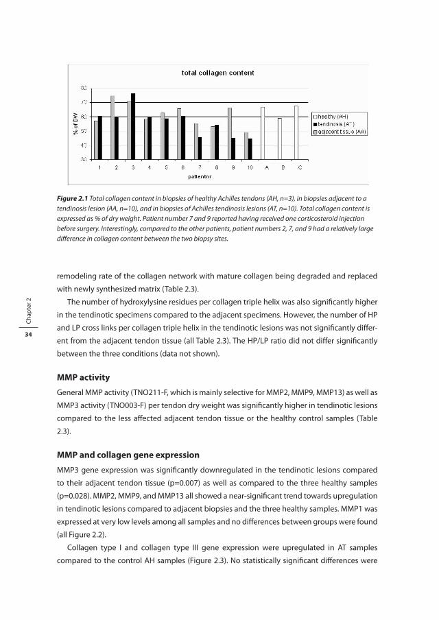
lagen content in the tendinotic specimens of patients 2, 7, and 9, compared to their adjacent
tissue biopsies (Figure 2.1). The percentage of denatured collagen was significantly higher in
AT specimens (Table 2.3). The number of pentosidine cross links per collagen triple helix was
significantly lower in tendinotic lesions compared to the adjacent tissue, reflecting an increased
Table 2.3 Biochemical analyses of healthy Achilles tendons (AH), Achilles tendon tissue adjacent to tendinosis lesion (AA), and Achilles tendinosis lesions (AT).
Biochemical parameter Healthy tendon tissue (AH) n=3
Adjacent tendon tissue (AA) n=10
Tendinosis lesion(AT) n=10
mean +/- stdev mean +/- stdev mean +/- stdev
Watercontent % of wet weight
66.2 +/- 5.5 73.3 +/- 4.7 76.3 +/- 6.3 † §
Total collagen % of dry weight
64.5 +/- 4.6 61.4 +/- 8.1 56.6 +/- 9.7
Denatured collagen % of total collagen
6.00 +/- 7.97 7.51 +/- 2.65 10.40 +/- 2.85 †
Pentosidine mol/mol collagen triple helix
0.0172 +/- 0.0127 0.0073 +/- 0.0027 0.0047 +/- 0.0032 †
Hydroxylysine mol/mol collagen triple helix
29.2 +/- 3.2 35.9 +/- 2.6 § 37.7 +/- 3.4 † §
HP crosslinks mol/mol collagen triple helix
1.10 +/- 0.19 1.30 +/- 0.19 1.26 +/- 0.29
LP crosslinks mol/mol collagen triple helix
0.075 +/- 0.012 0.077+/- 0.026 0.079 +/- 0.032
MMP activity TNO211-F ∆RFU/sec/ g of dry weight
51.7 +/- 21.9 88.2 +/- 107.9 308.6 +/- 392.6 †
MMP activity TNO003-F ∆RFU/sec/ g of dry weight
20.1 +/- 22.0 65.8 +/- 43.8 103.9 +/- 64.8 §
∆RFU: Relative fluorescence unit; p-value<0.05 is considered statistically significant. § p<0.05 compared to AH in an unpaired non-parametric test (mann-whitney u test). † p< 0.05 compared to AA in a paired non-parametric test (wilcoxon signed ranks test)
Chap
ter 2
34
remodeling rate of the collagen network with mature collagen being degraded and replaced
with newly synthesized matrix (Table 2.3).
The number of hydroxylysine residues per collagen triple helix was also significantly higher
in the tendinotic specimens compared to the adjacent specimens. However, the number of HP
and LP cross links per collagen triple helix in the tendinotic lesions was not significantly differ-
ent from the adjacent tendon tissue (all Table 2.3). The HP/LP ratio did not differ significantly
between the three conditions (data not shown).
MMP activity
General MMP activity (TNO211-F, which is mainly selective for MMP2, MMP9, MMP13) as well as
MMP3 activity (TNO003-F) per tendon dry weight was significantly higher in tendinotic lesions
compared to the less affected adjacent tendon tissue or the healthy control samples (Table
2.3).
MMP and collagen gene expression
MMP3 gene expression was significantly downregulated in the tendinotic lesions compared
to their adjacent tendon tissue (p=0.007) as well as compared to the three healthy samples
(p=0.028). MMP2, MMP9, and MMP13 all showed a near-significant trend towards upregulation
in tendinotic lesions compared to adjacent biopsies and the three healthy samples. MMP1 was
expressed at very low levels among all samples and no differences between groups were found
(all Figure 2.2).
Collagen type I and collagen type III gene expression were upregulated in AT samples
compared to the control AH samples (Figure 2.3). No statistically significant differences were
Figure 2.1 Total collagen content in biopsies of healthy Achilles tendons (AH, n=3), in biopsies adjacent to a tendinosis lesion (AA, n=10), and in biopsies of Achilles tendinosis lesions (AT, n=10). Total collagen content is expressed as % of dry weight. Patient number 7 and 9 reported having received one corticosteroid injection before surgery. Interestingly, compared to the other patients, patient numbers 2, 7, and 9 had a relatively large difference in collagen content between the two biopsy sites.
Figure 2.1 Total collagen content in biopsies of healthy Achilles tendons (AH, n=3), in biopsies adjacent to a tendinosis lesion (AA, n=10), and in biopsies of Achilles tendinosis lesions (AT, n=10). Total collagen content is expressed as % of dry weight. Patient number 7 and 9 reported having received one corticosteroid injection before surgery. Interestingly, compared to the other patients, patient numbers 2, 7, and 9 had a relatively large difference in collagen content between the two biopsy sites.

Achilles tendinosis: changes in biochemical composition and collagen turnover rate 35
Figure 2.3 Collagen gene expression levels in biopsies of healthy Achilles tendons (AH, n=10), in biopsies adjacent to a tendinosis lesion (AA, n=10), and in biopsies of Achilles tendinosis lesions (AT, n=10).
Figure 2.3 Collagen gene expression levels in biopsies of healthy Achilles tendons (AH, n=10), in biopsies adjacent to a tendinosis lesion (AA, n=10), and in biopsies of Achilles tendinosis lesions (AT, n=10).
Figure 2.2 MMP gene expression levels in biopsies of healthy Achilles tendons (AH, n=3), in biopsies adjacent to a tendinosis lesion (AA, n=10), and in biopsies of Achilles tendinosis lesions (AT, n=10).
Figure 2.2 MMP gene expression levels in biopsies of healthy Achilles tendons (AH, n=3), in biopsies adjacent to a tendinosis lesion (AA, n=10), and in biopsies of Achilles tendinosis lesions (AT, n=10).
Chap
ter 2
36
found between AT and AA samples. The ratio between collagen type I and collagen type III gene
expression did not differ between the three conditions (results not shown).
DisCussion
This comparison of several tissue characteristics between healthy Achilles tendon tissue, tendi-
notic Achilles tendon tissue, and its adjacent less affected Achilles tendon tissue revealed vari-
ous differences concerning biochemical composition, MMP activity, and MMP gene expression
levels. These differences indicate an aberrant collagenous matrix composition and a relatively
high collagenous matrix turnover rate in degenerated Achilles tendon tissue. On one hand,
the significant increases of both MMP activity and degraded collagen are signs of increased
matrix degradation. On the other hand, higher collagen gene expression levels and a low
pentosidine level, reflecting a relatively young collagenous matrix, are indicative for increased
matrix synthesis.
Our results are in line with the findings in supraspinatus tendinopathy [11, 32], indicating
that both Achilles and supraspinatus tendinopathies result at least partly from similar underly-
ing pathological processes. First, we found an increased water content in tendinotic Achilles
tendon specimens, which is consistent with previous biochemical findings in supraspinatus
tendinopathy [32]. Probably, the combination of matrix disintegration and an increased water
content due to increased amounts of glycosaminoglycans [31], leads to the tissue swelling and
signal intensity changes as visualized in MRI images of Achilles tendinopathies [6]. Second,
we found lower levels of non-enzymatic pentosidine cross-links in tendinotic Achilles tendon
specimens, representing a young collagenous matrix. This was also observed in previous stud-
ies of supraspinatus tendinopathy [11]. Other results require more elaboration and/or were
different from the supraspinatus tendinopathy studies, and are therefore addressed in more
detail in the following paragraphs.
The absence of a significant change in total collagen content in Achilles tendinotic tissue,
is consistent with findings in equine superficial digital flexor tendon degeneration [14]. In
supraspinatus tendinopathy, however, total collagen content has been reported to be lower
than in the healthy specimens [11, 32].
One explanation for this discrepancy might be the different preoperative treatments given
to Achilles and supraspinatus tendinopathy patients. In the supraspinatus studies, all patients
received at least one local corticosteroid injection before surgery, whereas the control patients
(cadaver material from subjects with no known history of tendinopathy or shoulder pathology)
did not. In our study, the AT and AA samples were harvested from the same Achilles tendon
from the same patient, and had therefore also undergone exactly the same treatment. Of the
three patients in our study who had a relatively low collagen content in their tendinotic sample
(patients 2, 7, and 9), two patients (patient number 7 and 9) reported having received a local

Achilles tendinosis: changes in biochemical composition and collagen turnover rate 37
corticosteroid injection pre-operatively. In-vitro culture in the presence of corticosteroids at
pharmacological concentrations decreases collagen accumulation (and proliferation) by teno-
cytes within two weeks [33]. In this way, a higher tissue turnover rate in AT samples compared
to AA samples, as was seen in this study, together with impaired collagen synthesis due to
corticosteroid treatment, might lead to lower total collagen content in tendinosis samples
subjected to corticosteroids. We are not aware of any literature reporting on the in-vivo effects
of local corticosteroids application on collagen content.
An alternative explanation for this discrepancy might be that a change in collagen content
does take place during early stages of tendinosis development. In our study, the tissue adjacent
to the lesion (AA), despite of being macroscopically normal, was already mildly affected (median
histological severity score 14.5, range 4-19). It could thus be argued that in the adjacent samples
(AA) the total collagen content had already reached a lower level (see Table 2.3 for mean collagen
contents in AH, AA, and AT samples), making it impossible to ascertain a difference between
affected (AT) and less affected (AA) tissue. The number of healthy control tissues (AH, n=3) from
our extra control group is too small to reach statistically significant differences between healthy
and tendinotic tissue concerning this parameter. This number of AH samples was severy limited
by our choice of material for healthy tendon tissue. However, we preferred to obtain the fresh
tendon tissue instead of post-mortem material for this extra control group because we do not
know how the biochemical parameters change post-mortem. Furthermore, gene expression
analysis cannot be performed on post-mortem material because RNA is lost.
At gene expression level we saw an upregulation of collagen type I and collagen type III in
Achilles tendinosis (AT) specimens without a change in ratio between the two collagen types.
Ireland et al. also found an increased expression of both collagen types in Achilles tendinosis but
did not report on the ratio between them [19]. At protein level others have found an increase in
collagen type III protein relative to collagen type I protein in degenerated equine tendon tissue
[14], human supraspinatus tendinopathy [32], and ruptured human Achilles tendon [18]. With
our method of measuring total collagen content, we cannot discriminate between collagen
type I and collagen type III at protein level in our Achilles tendinosis specimens.
MMP activity levels have been reported to differ significantly between supraspinatus and
biceps tendon, representing a higher level of protein turnover in supraspinatus tendons
in response to the higher mechanical demands and/or repeated injury exerted upon the
supraspinatus tendon [30]. The increase in total MMP activity in our study potentially mediates
the higher collagen turnover rate that we ascertained in the affected Achilles tendon speci-
mens. Affirming the collective activity of MMPs, we also found a higher amount of denatured/
damaged collagen.
Consistent with the increase in general MMP activity (TNO211-F, which is mainly converted
by MMP2, MMP9, and MMP13) is the trend towards an increase in gene expression of MMP2,
MMP9, and MMP13 in the tendinotic tissue. Although upregulation of MMP9 and MMP13 gene
expression has been described in ruptured Achilles tendon [19, 21], the studies in question

Chap
ter 2
38
reported no upregulation in painful Achilles tendons. In our tendinotic samples, this might sug-
gest that even though the tendon matrix did not yet show evidence of a macroscopic rupture,
microruptures may already have taken place. The decrease of MMP3 gene-expression level
confirms earlier results on MMP3 gene expression in painful as well as ruptured Achilles ten-
dons [3, 19, 21]. Although MMP3 gene expression was downregulated, we found an increase in
MMP3 activity in Achilles tendinotic specimens. MMP3 activity therefore appears to be mainly
regulated on posttranscriptional level (increased activation or less inhibition). MMP3 is active
against a broad range of substrates and is also capable of activating other MMPs [37], thus
playing an important role in the MMP cascade. The elevated MMP3 activity may also explain the
relatively large increase in general MMP activity that we found in the tendinotic specimens as
compared to the gene expression levels for MMP2, MMP9, and MMP13, which merely showed a
non-significant trend towards upregulation. Generally, a fibrotic repair process is accompanied
by increased formation of hydroxylysine residues and of enzymatic HP cross links [36]. Both
changes were seen in supraspinatus tendinopathy [11], but only an increase in the amount of
hydroxylysine residues was found in our Achilles tendinosis samples. Corticosteroid injection
in the rotator cuff can cause fragmentation of collagen bundles, inflammatory cell infiltra-
tion and necrosis [1, 35]. In response to damage or inferior tissue repair, fibrosis is a common
pathophysiological process. This fibrotic repair process might be much more advanced in the
supraspinatus tendinopathy samples than in our Achilles tendinosis samples. In this case, the
trend towards upregulation of MMP2, MMP9, and MMP13 gene expression in our tendinotic
Achilles specimens, might be interpreted as a sign of microruptures taking place (see previ-
ous paragraph) and the increase in hydroxylysine residues as an early sign of the start of a
concomitant fibrotic process.
To develop methods for intervention and thereby improve the clinical management of
tendon degeneration, it is crucial to obtain a detailed understanding of the biochemical and
structural changes involved in the development and worsening of tendinotic lesions. Similar
pathological processes appear to underlie both Achilles and supraspinatus tendinopathy in
middle-aged people. It remains unclear whether these results can be applied to a younger
athletic populations as well. For studying the general disease process there may be three
advantages of using the Achilles tendon: 1) corticosteroid injections that presumably influence
the pathological process are less often used in conservative treatment of Achilles tendinosis
than of supraspinatus tendinopathy; 2) the Achilles tendon has a more accessible anatomical
localisation and a larger diameter; 3) it is easier to clinically monitor the effect of intervention
studies in the Achilles tendon. With respect to the third point mentioned, an ultrasonographic
imaging technique, facilitating the discrimination of various stages of integrity of tendon
tissue by means of computerized ultrasonographic tissue characterisation, is currently being
evaluated as a tool for diagnosis and subsequent monitoring of tendon pathology, in order to
improve clinical management of Achilles tendinopathy [38-42].

Achilles tendinosis: changes in biochemical composition and collagen turnover rate 39
We conclude that in Achilles tendinosis an increase in tissue turnover rate as part of an exag-
gerated repair process, possibly resulting from a failure to regulate specific MMP activities, leads
to the deposition of a compromised, non-physiological tendon matrix. Also, the tendon tissue
directly surrounding a tendinotic lesion, despite of looking macroscopically healthy, reveals
both histologically and biochemically degenerative changes. Changes in extracellular matrix
composition seen in Achilles tendinosis and supraspinatus tendinopathy are largely alike and
may be the result of similar underlying pathological processes. The MMP gene expression
pattern in our early and mild stages of Achilles tendinosis suggests the occurrence of microrup-
tures, and our crosslink analyses may at least be suggestive for an early fibrotic repair process.
aCknowleDgeMenTs
The authors would like to thank Harrie Weinans for his interest in this work and his valuable
suggestions; Nicole Kops for preparing and staining the histological sections; and Anna van der
Windt for examining and scoring the histological sections.
ReFeRenCes
1. Akpinar S, Hersekli MA, Demirors H, et al. Effects of methylprednisolone and betamethasone injec-tions on the rotator cuff: an experimental study in rats. Adv Ther. Jul-Aug 2002;19(4):194-201.
2. Alfredson H. The chronic painful Achilles and patellar tendon: research on basic biology and treat-ment. Scand J Med Sci Sports. Aug 2005;15(4):252-259.
3. Alfredson H, Lorentzon M, Backman S, et al. cDNA-arrays and real-time quantitative PCR techniques in the investigation of chronic Achilles tendinosis. J Orthop Res. Nov 2003;21(6):970-975.
4. Alfredson H, Pietila T, Jonsson P, et al. Heavy-load eccentric calf muscle training for the treatment of chronic Achilles tendinosis. Am J Sports Med. May-Jun 1998;26(3):360-366.
5. Astrom M. Partial rupture in chronic achilles tendinopathy. A retrospective analysis of 342 cases. Acta Orthop Scand. Aug 1998;69(4):404-407.
6. Astrom M, Gentz CF, Nilsson P, et al. Imaging in chronic achilles tendinopathy: a comparison of ultra-sonography, magnetic resonance imaging and surgical findings in 27 histologically verified cases. Skeletal Radiol. Oct 1996;25(7):615-620.
7. Astrom M, Rausing A. Chronic Achilles tendinopathy. A survey of surgical and histopathologic find-ings. Clin Orthop Relat Res. Jul 1995(316):151-164.
8. Bank RA, Beekman B, Verzijl N, et al. Sensitive fluorimetric quantitation of pyridinium and pentosidine crosslinks in biological samples in a single high-performance liquid chromatographic run. J Chro-matogr B Biomed Sci Appl. Dec 5 1997;703(1-2):37-44.
9. Bank RA, Jansen EJ, Beekman B, et al. Amino acid analysis by reverse-phase high-performance liquid chromatography: improved derivatization and detection conditions with 9-fluorenylmethyl chloro-formate. Anal Biochem. Sep 5 1996;240(2):167-176.
10. Bank RA, Krikken M, Beekman B, et al. A simplified measurement of degraded collagen in tissues: application in healthy, fibrillated and osteoarthritic cartilage. Matrix Biol. Nov 1997;16(5):233-243.

Chap
ter 2
40
11. Bank RA, TeKoppele JM, Oostingh G, et al. Lysylhydroxylation and non-reducible crosslinking of human supraspinatus tendon collagen: changes with age and in chronic rotator cuff tendinitis. Ann Rheum Dis. Jan 1999;58(1):35-41.
12. Beekman B, Drijfhout JW, Bloemhoff W, et al. Convenient fluorometric assay for matrix metalloprotei-nase activity and its application in biological media. FEBS Lett. Jul 22 1996;390(2):221-225.
13. Beekman B, van El B, Drijfhout JW, et al. Highly increased levels of active stromelysin in rheumatoid synovial fluid determined by a selective fluorogenic assay. FEBS Lett. Dec 1 1997;418(3):305-309.
14. Birch HL, Bailey AJ, Goodship AE. Macroscopic ‘degeneration’ of equine superficial digital flexor tendon is accompanied by a change in extracellular matrix composition. Equine Vet J. Nov 1998;30(6):534-539.
15. Blanco I, Krahenbuhl S, Schlienger RG. Corticosteroid-associated tendinopathies: an analysis of the published literature and spontaneous pharmacovigilance data. Drug Saf. 2005;28(7):633-643.
16. Chard MD, Cawston TE, Riley GP, et al. Rotator cuff degeneration and lateral epicondylitis: a compara-tive histological study. Ann Rheum Dis. Jan 1994;53(1):30-34.
17. Creemers LB, Jansen DC, van Veen-Reurings A, et al. Microassay for the assessment of low levels of hydroxyproline. Biotechniques. Apr 1997;22(4):656-658.
18. Eriksen HA, Pajala A, Leppilahti J, et al. Increased content of type III collagen at the rupture site of human Achilles tendon. J Orthop Res. Nov 2002;20(6):1352-1357.
19. Ireland D, Harrall R, Curry V, et al. Multiple changes in gene expression in chronic human Achilles tendinopathy. Matrix Biol. Jun 2001;20(3):159-169.
20. Jarvinen TA, Kannus P, Maffulli N, et al. Achilles tendon disorders: etiology and epidemiology. Foot Ankle Clin. Jun 2005;10(2):255-266.
21. Jones GC, Corps AN, Pennington CJ, et al. Expression profiling of metalloproteinases and tissue inhibi-tors of metalloproteinases in normal and degenerate human achilles tendon. Arthritis Rheum. Mar 2006;54(3):832-842.
22. Kjellin I, Ho CP, Cervilla V, et al. Alterations in the supraspinatus tendon at MR imaging: correlation with histopathologic findings in cadavers. Radiology. Dec 1991;181(3):837-841.
23. Livak KJ, Schmittgen TD. Analysis of relative gene expression data using real-time quantitative PCR and the 2(-Delta Delta C(T)) Method. Methods. Dec 2001;25(4):402-408.
24. Maffulli N, Kader D. Tendinopathy of tendo achillis. J Bone Joint Surg Br. Jan 2002;84(1):1-8. 25. Maffulli N, Khan KM, Puddu G. Overuse tendon conditions: time to change a confusing terminology.
Arthroscopy. Nov-Dec 1998;14(8):840-843. 26. Maffulli N, Testa V, Capasso G, et al. Similar histopathological picture in males with Achilles and patel-
lar tendinopathy. Med Sci Sports Exerc. Sep 2004;36(9):1470-1475. 27. Magra M, Maffulli N. Matrix metalloproteases: a role in overuse tendinopathies. Br J Sports Med. Nov
2005;39(11):789-791. 28. Paavola M, Kannus P, Jarvinen TA, et al. Achilles tendinopathy. J Bone Joint Surg Am. Nov 2002;84-
A(11):2062-2076. 29. Riley G. The pathogenesis of tendinopathy. A molecular perspective. Rheumatology (Oxford). Feb
2004;43(2):131-142. 30. Riley GP, Curry V, DeGroot J, et al. Matrix metalloproteinase activities and their relationship with col-
lagen remodelling in tendon pathology. Matrix Biol. Mar 2002;21(2):185-195. 31. Riley GP, Harrall RL, Constant CR, et al. Glycosaminoglycans of human rotator cuff tendons: changes
with age and in chronic rotator cuff tendinitis. Ann Rheum Dis. Jun 1994;53(6):367-376. 32. Riley GP, Harrall RL, Constant CR, et al. Tendon degeneration and chronic shoulder pain: changes in
the collagen composition of the human rotator cuff tendons in rotator cuff tendinitis. Ann Rheum Dis. Jun 1994;53(6):359-366.
33. Scutt N, Rolf CG, Scutt A. Glucocorticoids inhibit tenocyte proliferation and Tendon progenitor cell recruitment. J Orthop Res. Feb 2006;24(2):173-182.

Achilles tendinosis: changes in biochemical composition and collagen turnover rate 41
34. Seeger JD, West WA, Fife D, et al. Achilles tendon rupture and its association with fluoroquinolone antibiotics and other potential risk factors in a managed care population. Pharmacoepidemiol Drug Saf. Feb 3 2006.
35. Tillander B, Franzen LE, Karlsson MH, et al. Effect of steroid injections on the rotator cuff: an experi-mental study in rats. J Shoulder Elbow Surg. May-Jun 1999;8(3):271-274.
36. van der Slot AJ, Zuurmond AM, van den Bogaerdt AJ, et al. Increased formation of pyridinoline cross-links due to higher telopeptide lysyl hydroxylase levels is a general fibrotic phenomenon. Matrix Biol. Jul 2004;23(4):251-257.
37. van Meurs J, van Lent P, Stoop R, et al. Cleavage of aggrecan at the Asn341-Phe342 site coincides with the initiation of collagen damage in murine antigen-induced arthritis: a pivotal role for stromelysin 1 in matrix metalloproteinase activity. Arthritis Rheum. Oct 1999;42(10):2074-2084.
38. van Schie HT, Bakker EM. Structure-related echoes in ultrasonographic images of equine superficial digital flexor tendons. Am J Vet Res. Feb 2000;61(2):202-209.
39. van Schie HT, Bakker EM, Jonker AM, et al. Computerized ultrasonographic tissue characterization of equine superficial digital flexor tendons by means of stability quantification of echo patterns in contiguous transverse ultrasonographic images. Am J Vet Res. Mar 2003;64(3):366-375.
40. van Schie HT, Bakker EM, Jonker AM, et al. Efficacy of computerized discrimination between structure-related and non-structure-related echoes in ultrasonographic images for the quantitative evaluation of the structural integrity of superficial digital flexor tendons in horses. Am J Vet Res. Jul 2001;62(7):1159-1166.
41. van Schie HT, Bakker EM, Jonker AM, et al. Ultrasonographic tissue characterization of equine super-ficial digital flexor tendons by means of gray level statistics. Am J Vet Res. Feb 2000;61(2):210-219.
42. van Schie JT, Bakker EM, van Weeren PR. Ultrasonographic evaluation of equine tendons: a quantita-tive in vitro study of the effects of amplifier gain level, transducer-tilt, and transducer-displacement. Vet Radiol Ultrasound. Mar-Apr 1999;40(2):151-160.


Chapter 3
Tendon degeneration is not mediated by regulation of Toll-like receptors 2 and 4 in human tenocytes
M. de Mos, L.A.B. Joosten, B. Oppers-Walgreen, H.T.M. van Schie, H. Jahr, G.J.V.M. van Osch, J.A.N. Verhaar
Manuscript under revision by J Orthop Res

Chap
ter 3
44
aBsTRaCT
objective
We hypothesized that expression of Toll-like receptors (TLRs) 2 and 4 by tenocytes is involved
in the catabolic processes of tendon degeneration. We investigated TLR2 and TLR4 expression
by tenocytes in healthy and tendinotic tendons. We also investigated whether TLR2 and TLR4
could be upregulated in tendon explants using proinflammatory cytokines IL-1β and TNFα.
Methods
Peroperatively harvested healthy (n=5) and tendinotic (n=13) Achilles tendon samples were
examined by real-time RT-PCR and immunohistochemical staining for TLR2 and TLR4. In addi-
tion the catabolic process in tendinopathy was analysed by real-time RT-PCR for matrix metal-
loproteinases MMP1, MMP3, MMP9, and MMP13. Furthermore, healthy tendon explants were
cultured in the presence of 20 ng/ml IL-1β (n=10) or 10 ng/ml TNFα (n=8) for 4, 24, 48, and 72
hours before analysis of TLR and MMP expression levels.
Results
Although mRNA levels for both TLR2 and TLR4 were detected in healthy and tendinotic Achilles
tendons, we could not confirm expression of these receptors by immunohistochemical staining
in neither healthy or tendinotic tendon samples. Both receptors didn’t show significant tran-
scriptional regulation in tendinopathy, although MMP3 was downregulated and MMP9 was
upregulated in tendinopathy. In tendon explant cultures TLR2 mRNA was upregulated by TNFα
(p<0.05) and IL-1β (not significant). TLR4 gene expression was not altered by addition of IL-1β
or TNFα.
Conclusion
Tendon tissue can be stimulated to increase TLR2 gene expression by addition of ‘catabolic
factors’ TNFα or IL-1β. However, the catabolic processes in tendinopathy can not be attributed
to regulation of TLR2 and TLR4 by tenocytes.

Tendon degeneration is not mediated by regulation of Toll-like receptors 2 and 4 in human tenocytes 45
inTRoDuCTion
Toll-like receptors (TLRs) are phylogenetically conserved transmembrane receptors belonging
to the family of pattern-recognition receptors (PRR). In humans, eleven TLR homologs have
been identified thus far, expressed by numerous cell types like monocytes and dendritic cells
[1], and for example by chondrocytes [2]. TLRs detect microbial components, but TLR2 and
TLR4 also recognize endogenous ligands like heat-shock proteins, necrotic cells, hyaluronan,
and fibronectin [1]. Ligand recognition elicits an immune response with upregulation and
activation of proinflammatory cytokines and matrix-metalloproteinases (MMPs)[2]. Hence, via
endogenous ligand recognition the TLR-mediated catabolic signalling pathways can directly
contribute to degradative tissue reactions even in the absence of microbial components. TLRs
are implicated in inflammatory and autoimmune diseases, but also in disorders like atheroscle-
rosis and cancer [3, 4]. Furthermore they play a role in non-infected rheumatoid arthritis (RA)
and in the primarily degenerative joint disease osteoarthritis (OA)[1-3, 5, 6].
In degenerative tendinopathies the balance between matrix synthesis and degradation is
disturbed and increased MMP activity as well as an increased percentage of degraded collagen
are found [7-9]. An increase in NO synthase expression was found in a supraspinatus tendon
overuse model [10] and an increase in COX2, a key regulator of prostaglandin synthesis was
found in patellar tendinopathy [11]. These catabolic activities can be influenced by TLR signal-
ling, as shown in experiments with chondrocytes [2, 6]. Furthermore in Achilles tendinopathy
increased expression of subunit B of the endogenous TLR ligand fibronectin has been found in
Achilles tendinopathy [12]. The presence of other endogenous TLR ligands known so far (heat-
shock proteins, necrotic cells, hyaluronan) in tendinopathy is very likely considering the nature
of the pathology.
We hypothesized that TLR2 and 4 expression on tenocytes is involved in the abovemen-
tioned catabolic processes of tendon degeneration. Therefore, we investigated whether TLR2
and TLR4 were constitutively expressed by tenocytes in healthy tendon and were higher
expressed by tenocytes in tendinotic lesions. Also, we investigated whether TLR2 and TLR4
could be upregulated in healthy tendon explant cultures by adding proinflammatory cytokines
IL-1β and TNFα.
MaTeRials anD MeThoDs
Tendon biopsies
Tissue specimens of 13 patients with chronic mid-portion Achilles tendinopathy (6 men, 8
women; average age 46, range 36-58) were harvested from the macroscopically affected ten-
dinotic lesion during surgical debridement. Approval was obtained from the Medical Ethical
Committee of the Erasmus MC University Medical Center (MEC-2005-100). All patients signed

Chap
ter 3
46
informed consent. Healthy Achilles tissue specimens of 5 donors (3 men, 2 women; average
age 44, range 20-78) without clinically evident Achilles tendinopathy were collected as control
(MEC-2006-069). Of every patient one specimen was frozen in liquid nitrogen for RNA extrac-
tion, one was embedded in Tissue-Tek (Sakura Finetek Europe, Zoeterwoude, the Netherlands)
for cryosectioning and immunohistochemical staining of TLR2 and TLR4, and one was fixed
overnight in 10% formalin and embedded in paraffin for histological evaluation of tendinosis
severity.
Culture experiments
We obtained non-degenerative hamstring tendon tissue from healthy adolescents (all under
18 yrs of age) undergoing hamstring-tendon release for treatment of knee-contractures (MEC-
2006-069). After peritendineum and fat tissue were carefully removed, tendons were cut into 3
mm3 sections and cultured at 37˚C in a humidified atmosphere of 5% CO2 in Dulbecco’s modified
Eagle’s medium (DMEM) with 10% fetal calf serum (FCS). After 24 hours, medium was replaced
with DMEM containing 2% FCS with or without either 20 ng/ml IL-1β (Peprotech, Rocky Hill, NJ)
or 10 ng/ml TNFα (Biosource, Camarillo, CA). All media contained 50 μg/ml gentamycin, and 1.5
μg/ml fungizone (both Invitrogen). Explants were frozen in liquid nitrogen for RNA extraction
after 4, 24, and 48 hours for gene expression analysis and after 72 hours for immunohistochemi-
cal staining of TLR2 and TLR4.
histology
Longitudinal frozen sections (5 μm) were blocked with normal swine serum followed by 60
minutes of incubation with polyclonal antibodies against human TLR2 (H-175)(10µg/ml) or
TLR4 (H-80)(12µg/ml)(both Santa Cruz Biotechnologies, Santa Cruz, California). The second-
ary antibody, biotinylated swine anti-rabbit Ig (Dako Diagnostics, Glostrup, Denmark), was
incubated for 30 minutes. Slides were stained with streptavidin peroxidase, developed with
Diaminobenzidine (DAB)(Sigma, St. Louis, MO), and counterstained with hematoxylin for 30
seconds. Control sections were stained with irrelevant primary isotype-specific IgG antibodies.
Longitudinal paraffin sections (6 μm) were stained with haematoxylin and eosin or with
thionin for glycosaminoglycans. Two different researchers performed a blinded examination
of the slides using a modified semiquantitative grading scale for tendinosis [7], yielding a total
sum of 0 for minimal histological tendinosis severity and 30 for maximal severity.
Rna isolation and real-time RT-PCR
Specimens were snap frozen and quickly homogenized in a Mikro-Dismembrator® (BioTech
International Inc, Needville, Texas) and suspended in 1.8mL/100mg RNA-BeeTM (TEL-TEST,
Friendswood, TX, USA). RNA was isolated and purified using RNeasy® Micro Kit (Qiagen, Hilden,
Germany), and 1 µg total RNA of each sample was reverse-transcribed into cDNA using Rever-
tAidTM First Strand cDNA Synthesis Kit (MBI Fermentas, St. Leon-Rot, Germany). Primers were

Tendon degeneration is not mediated by regulation of Toll-like receptors 2 and 4 in human tenocytes 47
designed using PrimerExpress 2.0 software (Applied Biosystems, Foster City, CA, USA) to meet
SYBR®Green (TLRs only) or Taqman® requirements. BLASTn ensured gene specificity of the
following primers: TLR2 (F: 5’-CTACTGGGTGGAGAACCTTATGGT-3’; R: 5’-CCGCTTATGAAGACA-
CAACTTGA-3’ ), TLR4 (F: 5’-GGCATGCCTGTG
CTGAGTT-3’; R: 5’-CTGCTACAACAGATACTACAAGCACACT-3’), MMP1, MMP3, MMP9, MMP13
[7], and GAPDH [13] as calibrator. Amplification was performed using an ABI PRISM® 7000 with
SDS software version 1.7 in 20 µl reactions using either qPCRTM Mastermix Plus for SYBR®Green I
(Eurogentec, Nederland B.V., Maastricht, The Netherlands) or TaqMan® Universal PCR MasterMix
(ABI, Branchburg, New Jersey, USA) according to the manufacturer’s guidelines. Data were
normalized to GAPDH. Relative expression was calculated according to the 2-ΔCT formula.
statistical analysis
Statistical analysis was performed using GraphPad Prism 5.0 software (GraphPad Software, Inc
1994-2007, www.graphpad.com). A Mann-Whitney U Test was used to assess the differences
between healthy and tendinotic tendon tissue. A Kruskall-Wallis H test and post-hoc Dunn’s
multiple comparison test was used to asses the differences between growth factor stimulated
cultures and control cultures. Significance level was set at p<0.05.
ResulTs
achilles tendinopathy
Tendinosis severity ranged from 1 to 10 (median 3) in healthy Achilles tendon samples, and
from 9 to 24 (median 18) in tendinotic Achilles tendon samples (p=0.02). MMP activity was
significantly higher in tendinotic Achilles samples than in healthy controls [7]. On gene expres-
sion level MMP3 was significantly downregulated, MMP13 significantly upregulated (both p<
0.05), and MMP1 and MMP9 were not altered (Figure 3.1A).
Transcripts of both TLR2 and TLR4 were detected in healthy Achilles tendon tissue at basal
levels, but no significant differences in TLR2 or TLR4 expression were found between healthy
and tendinotic Achilles tendon tissue (Figure 3.1B).
Immunohistochemical staining for TLR2 and TLR4 were performed on frozen sections of
healthy and tendinotic Achilles tendon biopsies. Human RA synovium was used as positive
control. None of the tenocytes in either healthy or tendinotic Achilles tendon samples stained
positive for TLR2 or TLR4 (Figure 3.2, see also color inlay).
explant cultures
Healthy tendon explants were harvested after 4, 24, and 48 hours of culture in the presence
of IL-1β or TNFα. MMP1, MMP3, and MMP13 gene expression levels were significantly upregu-
lated by IL-1β treatment after 24 hours (all p< 0.05). MMP1 and MMP9 gene expression were

Chap
ter 3
48
A
B
Figure 3.1 Gene expression of A. MMP1, MMP3, MMP9, MMP13, and B. TLR2 and TLR4 in healthy (n=5) and tendinotic (n=13) Achilles tendon samples. Ct values were corrected for GAPDH, which was stably expressed across samples. Mean +/- standard deviation are displayed. * Indicates a p-value<0.05.
Figure 3.1 Gene expression of A. MMP1, MMP3, MMP9, MMP13, and B. TLR2 and TLR4 in healthy (n=5) and tendinotic (n=13) Achilles tendon samples. Ct values were corrected for GAPDH, which was stably expressed across samples. Mean +/- standard deviation are displayed. * Indicates a p-value<0.05.

Tendon degeneration is not mediated by regulation of Toll-like receptors 2 and 4 in human tenocytes 49
significantly upregulated by TNFα after 24 hours (both p< 0.05). MMP3 and MMP13 showed a
clear but non-significant trend towards upregulation by TNFα treatment. These data indicate
that IL-1β and TNFα treatments in culture were effective (Figure 3.3A).
Gene expression of TLR2 appeared upregulated by IL-1β and TNFα at all time points, but this
general trend reached significance (p<0.05) only for TNFα stimulation (at all time points; Figure
3.3B). Gene expression of TLR4 was not significantly affected by either IL-1β treatment or TNFα
treatment (Figure 3.3C).
Immunohistochemical staining on frozen sections of explants cultured for 3 days in the pres-
ence or absence of IL-1β or TNFα showed no staining for TLR 2 or TLR4 in any of the tenocytes.
DisCussion
We hypothesized that TLR2 and TLR4 presentation by tenocytes is involved in tendon degen-
eration. However, in tendon samples in vivo no TLR2 or TLR4 expression by tenocytes was seen
in any of the healthy or tendinotic tendon samples on immunohistochemical staining. Basal
mRNA levels of TLR2 and TLR4 were detected in healthy Achilles tendon tissue, but were not
upregulated in tendinotic Achilles tendon lesions. The catabolic factors TNFα and IL-1β were
able to stimulate TLR2 mRNA expression in tendon explant cultures (significantly for TNFα).
Therefore we conclude that tendons can be stimulated to increase TLR2 expression by addi-
Figure 3.2 Immunohistochemical staining for TLR2 and TLR4. Representative photomicrographs are shown from frozen longitudinal sections of healthy Achilles tendon samples (n=5) and tendinotic Achilles tendon samples (n=13). Human rheumatoid arthritis (RA) synovium was used as positive control. Magnification 400x.
Figure 3.2 Immunohistochemical staining for TLR2 and TLR4. Representative photomicrographs are shown from frozen longitudinal sections of healthy Achilles tendon samples (n=5) and tendinotic Achilles tendon samples (n=13). Human rheumatoid arthritis (RA) synovium was used as positive control. Magnification 400x. (See color inlay for a full color version of this figure.)
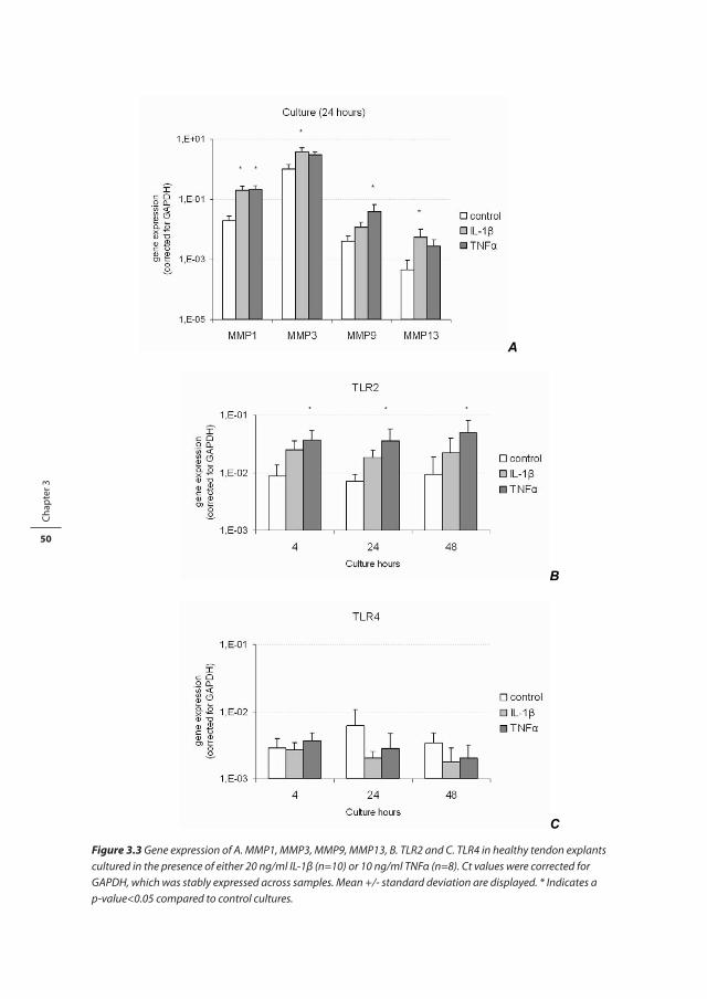
Chap
ter 3
50
A
B
C Figure 3.3 Gene expression of A. MMP1, MMP3, MMP9, MMP13, B. TLR2 and C. TLR4 in healthy tendon explants cultured in the presence of either 20 ng/ml IL-1 (n=10) or 10 ng/ml TNF (n=8). Ct values were corrected for GAPDH, which was stably expressed across samples. Mean +/- standard deviation are displayed. * Indicates a p-value<0.05 compared to control cultures.
Figure 3.3 Gene expression of A. MMP1, MMP3, MMP9, MMP13, B. TLR2 and C. TLR4 in healthy tendon explants cultured in the presence of either 20 ng/ml IL-1β (n=10) or 10 ng/ml TNFα (n=8). Ct values were corrected for GAPDH, which was stably expressed across samples. Mean +/- standard deviation are displayed. * Indicates a p-value<0.05 compared to control cultures.
Tendon degeneration is not mediated by regulation of Toll-like receptors 2 and 4 in human tenocytes 51
tion of the ‘catabolic’ cytokines TNFα or IL-1β, but the catabolic processes demonstrated in
tendinopathy can not be attributed to regulation of TLR2 and TLR4 by the tenocytes.
The exact molecular processes involved in tendinopathy are largely unknown. Our hypoth-
esis was mainly based on the catabolic processes known so far to occur in tendinopathy. Tendon
tissue has many aspects in common with cartilage tissue. Both are embryologically derived
from the mesenchyme with the basic components of the extracellular matrix (ECM) consisting
of large collagen bundles and a glycosaminoglycan(GAG)-rich viscous substance interspersed
between the collagenous matrix. The main cell type in tendons are tenocytes, the only cell
type in cartilage are chondrocytes. These specialized cells are responsible for maintenance
of the ECM. However, in both tissues they are distributed over the ECM at very low density
and they function under hypoxic conditions because tendon and cartilage respectively have
limited to no blood supply. Consequently, both tissues have very limited regeneration capacity,
rendering them susceptible to degenerative diseases. Cartilage degeneration in osteoarthritis
(OA) and tendon degeneration in tendinopathy appear to have many aspects in common as
well. Both OA and tendinopathy are considered overuse injuries characterized by catabolic
tissue reactions associated with increased expression of mediators of inflammation [14, 15].
The central event in both diseases seems to be degradation of the ECM, although the changes
in the complex balance between activities of matrix degrading enzymes (e.g. MMPs), cytokines,
growth factors, and other signalling molecules have been studied in far more detail in cartilage
pathology than in tendon pathology.
The interplay between TLRs and catabolic processes has been studied in joint pathology
(cartilage, synovium). Human articular chondrocytes were shown to express a variety of TLRs
in vivo [6]. In cartilage degeneration TLR2 and TLR4 reappear on the chondrocyte membrane
[2, 6], both underlining the involvement of an inflammatory component and concurrently
reminiscing TLR expression during embryological developmental stages. In primary OA chon-
drocytes in culture this upregulation of TLR2 (and TLR4, though not significantly) was induced
by the proinflammatory cytokines IL-1β and TNFα [2]. Our study is the first to investigate TLR
expression in tendon tissue. We found basal mRNA levels for both TLR2 and TLR4 in Achilles
tendon tissue, but no upregulation was seen in tendinotic Achilles tendon samples. Moreover
immunohistochemical staining for TLR2 and TLR4 remained negative for all tenocytes in the
healthy and degenerated Achilles tendon samples.
Although we conclude that TLR expression on tenocytes does not contribute to the tendon
degeneration process, we do not exclude a role for TLRs in tendinopathy. The presence and
regulation of TLRs on cells in the peritendineum and endotenon might be able to contribute
to the degradative tissue reactions seen in the tendinotic lesions. Analogous to the role of TLR-
presenting cells in synoviitis accompanying OA and RA [5], monocytes and other inflammatory
cells can invade the peritendineum and the endotenon in peritendinitis, which is a frequently
seen co-morbidity with tendinopathy. TLR presentation by these cells and the subsequently
induced catabolic pathways might influence the degenerative process in tendinopathy.

Chap
ter 3
52
In conclusion, based on the degenerative processes present in tendinopathy we hypoth-
esized that TLR2 and TLR4 are involved in the pathology of tendon degeneration, analogous to
their involvement in OA. However, our results add to the current knowledge indicating that the
catabolic processes in tendinopathy can not be attributed to regulation of TLR2 and TLR4 by
tenocytes. Nonetheless tendon tissue can be stimulated to increase mRNA expression of TLR2
by addition of “catabolic factors” TNFα or IL-1β. Therefore, further investigation is warranted into
the role of TLR-presenting cells that invade the endotendon/peritenon, hereby inducing gen-
eral catabolic responses and thus contributing to the degenerative process in tendinopathy.
aCknowleDgeMenTs
The authors would like to thank Erik Lubberts for his interest in this work and kindly providing
the TNFα, and Anna van der Windt for her technical support.
ReFeRenCes
1. Roelofs MF, Abdollahi-Roodsaz S, Joosten LA, van den Berg WB, Radstake TR. The orchestra of Toll-like receptors and their potential role in frequently occurring rheumatic conditions. Arthritis Rheum 2008;58(2):338-48.
2. Kim HA, Cho ML, Choi HY, Yoon CS, Jhun JY, Oh HJ, et al. The catabolic pathway mediated by Toll-like receptors in human osteoarthritic chondrocytes. Arthritis Rheum 2006;54(7):2152-63.
3. Pandey S, Agrawal DK. Immunobiology of Toll-like receptors: emerging trends. Immunol Cell Biol 2006;84(4):333-41.
4. Huang B, Zhao J, Unkeless JC, Feng ZH, Xiong H. TLR signaling by tumor and immune cells: a double-edged sword. Oncogene 2008;27(2):218-24.
5. Radstake TR, Roelofs MF, Jenniskens YM, Oppers-Walgreen B, van Riel PL, Barrera P, et al. Expression of toll-like receptors 2 and 4 in rheumatoid synovial tissue and regulation by proinflammatory cytokines interleukin-12 and interleukin-18 via interferon-gamma. Arthritis Rheum 2004;50(12):3856-65.
6. Su SL, Tsai CD, Lee CH, Salter DM, Lee HS. Expression and regulation of Toll-like receptor 2 by IL-1beta and fibronectin fragments in human articular chondrocytes. Osteoarthritis Cartilage 2005;13(10):879-86.
7. de Mos M, van El B, DeGroot J, Jahr H, van Schie HT, van Arkel ER, et al. Achilles tendinosis: changes in biochemical composition and collagen turnover rate. Am J Sports Med 2007;35(9):1549-56.
8. Riley GP, Curry V, DeGroot J, van El B, Verzijl N, Hazleman BL, et al. Matrix metalloproteinase activities and their relationship with collagen remodelling in tendon pathology. Matrix Biol 2002;21(2):185-95.
9. Jones GC, Corps AN, Pennington CJ, Clark IM, Edwards DR, Bradley MM, et al. Expression profiling of metalloproteinases and tissue inhibitors of metalloproteinases in normal and degenerate human achilles tendon. Arthritis Rheum 2006;54(3):832-42.
10. Szomor ZL, Appleyard RC, Murrell GA. Overexpression of nitric oxide synthases in tendon overuse. J Orthop Res 2006;24(1):80-6.
11. Fu SC, Wang W, Pau HM, Wong YP, Chan KM, Rolf CG. Increased expression of transforming growth factor-beta1 in patellar tendinosis. Clin Orthop Relat Res 2002(400):174-83.

Tendon degeneration is not mediated by regulation of Toll-like receptors 2 and 4 in human tenocytes 53
12. Alfredson H, Lorentzon M, Backman S, Backman A, Lerner UH. cDNA-arrays and real-time quantitative PCR techniques in the investigation of chronic Achilles tendinosis. J Orthop Res 2003;21(6):970-5.
13. de Mos M, Koevoet WJ, Jahr H, Verstegen MM, Heijboer MP, Kops N, et al. Intrinsic differentiation potential of adolescent human tendon tissue: an in-vitro cell differentiation study. BMC Musculosk-elet Disord 2007;8:16.
14. Fredberg U, Stengaard-Pedersen K. Chronic tendinopathy tissue pathology, pain mechanisms, and etiology with a special focus on inflammation. Scand J Med Sci Sports 2008;18(1):3-15.
15. Burrage PS, Mix KS, Brinckerhoff CE. Matrix metalloproteinases: role in arthritis. Front Biosci 2006;11:529-43.


Chapter 4
Intrinsic differentiation potential of adolescent human tendon tissue: an in-vitro cell differentiation study
M. de Mos, J.L.M. Koevoet, H. Jahr, M.M.A.Verstegen, M.P. Heijboer, N. Kops, J.P.T.M. van Leeuwen, H. Weinans, J.A.N. Verhaar, G.J.V.M. van Osch
BMC Musculoskelet Disord. 2007 Feb 23;8:16

Chap
ter 4
56
aBsTRaCT
Background
Tendinosis lesions show an increase of glycosaminoglycan amount, calcifications, and lipid
accumulation. Therefore, altered cellular differentiation might play a role in the etiology of ten-
dinosis. This study investigates whether adolescent human tendon tissue contains a population
of cells with intrinsic differentiation potential.
Methods
Cells derived from adolescent non-degenerative hamstring tendons were characterized
by immunohistochemistry and FACS-analysis. Cells were cultured for 21 days in osteogenic,
adipogenic, and chondrogenic medium and phenotypical evaluation was carried out by
immunohistochemical and qPCR analysis. The results were compared with the results of similar
experiments on adult bone marrow-derived stromal cells (BMSCs).
Results
Tendon-derived cells stained D7-FIB (fibroblast-marker) positive, but α-SMA (marker for
smooth muscle cells and pericytes) negative. Tendon-derived cells were 99% negative for CD34
(endothelial cell marker), and 73% positive for CD105 (mesenchymal progenitor-cell marker).
In adipogenic medium, intracellular lipid vacuoles were visible and tendon-derived fibroblasts
showed upregulation of adipogenic markers FABP4 (fatty-acid binding protein 4) and PPARG
(peroxisome proliferative activated receptor γ). In chondrogenic medium, some cells stained
positive for collagen type II and tendon-derived fibroblasts showed upregulation of collagen
type II and collagen type X. In osteogenic medium Von Kossa staining showed calcium depo-
sition although osteogenic markers remained unaltered. Tendon-derived cells and BMSCs
behaved largely comparable, although some distinct differences were present between the
two cell populations.
Conclusions
This study suggests that our population of explanted human tendon cells has an intrinsic differ-
entiation potential. These results support the hypothesis that there might be a role for altered
tendon-cell differentiation in the pathophysiology of tendinosis.

Intrinsic differentiation potential of adolescent human tendon tissue: an in-vitro cell differentiation study 57
BaCkgRounD
Tendinosis is a chronic degenerative tendon disorder occurring particularly among athletes
and middle-aged people [1]. As its pathophysiology is still largely unknown, only symptomatic
treatment options are available, with limited success rates [1, 2]. A better understanding of the
cellular processes involved in the development of tendinosis lesions may ultimately improve
treatment and prevention.
Histopathological findings in tendinosis have been described in detail [3, 4]. In brief, hyper-
cellularity and rounding of the cell nuclei indicate a relatively high metabolic activity. Likewise,
altered extracellular matrix composition reflects changes in cellular behavior. For instance,
in tendinosis lesions there is a higher amount of glycosaminoglycans [3]. Lipid accumulation
and calcium deposition have also been described [5]. Thus, the histopathological findings may
indicate the presence of cells with diverse phenotypes, different from that of tenocytes under
healthy conditions.
Cells with multilineage differentiation potential likely play an important role in the body’s
capacity to naturally remodel, repair, and regenerate various tissue types where necessary [6].
However, the multilineage differentiation potential of cells might also be involved in patho-
logical processes. Although the pathophysiology of tendinosis is largely unclear, histological
findings suggest that multipotent cells might be implicated in its development. The origin of
these multipotent cells is unknown. They may be recruited from the bone marrow in response
to tendon tissue injury, and migrate through the circulation to the site of tissue damage [7].
They might also be present in the tendon tissue itself.
Local progenitor cells with multilineage potential have previously been found in many
locations within the musculoskeletal system, e.g. in bone marrow, skin, periosteum, bone,
muscle and adipose tissue [8-15]. On the other hand, progenitor cells are not the only cells with
multilineage potential: some highly differentiated cells are capable of transdifferentiation, i.e.
switching their phenotype to another lineage. This transdifferentiation has been demonstrated
for highly differentiated chondrocytes [16, 17].
Multipotent cells have been found in virtually all tissues of the musculoskeletal system, but
it is not known if tendon tissue has a cell population with multilineage potential. In this study,
we investigated whether the population of cells derived from non-degenerative tendon tissue
has differentiation potential similar to bone marrow-derived stromal cells (BMSCs). Specifically,
we characterized human tendon-derived fibroblasts by immunohistochemical staining and
FACS-analysis. Then, after a culture period of 21 days in adipogenic, chondrogenic, and osteo-
genic medium, we evaluated changes in their phenotype using immunohistochemical and
histochemical stainings as well as gene expression analysis.
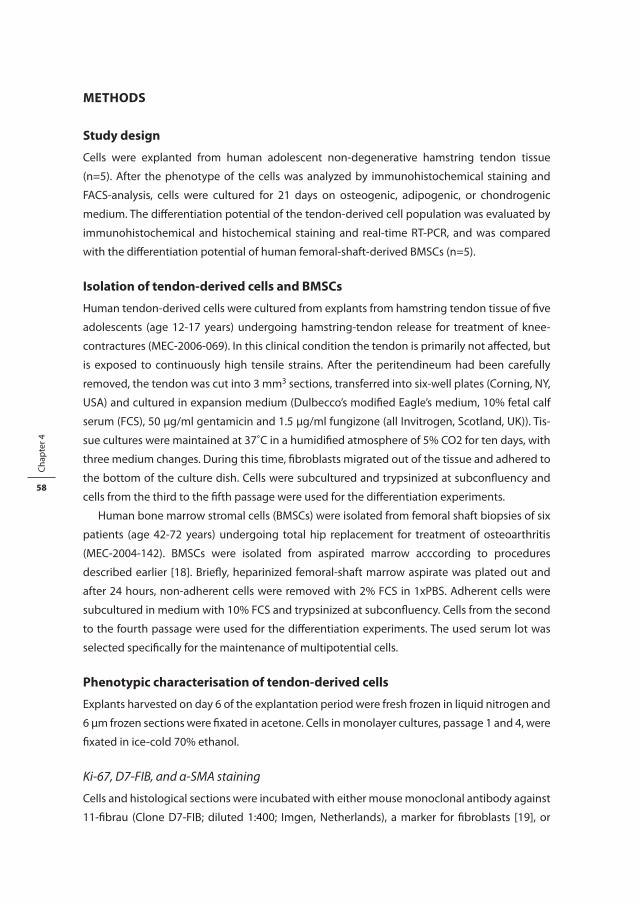
Chap
ter 4
58
MeThoDs
study design
Cells were explanted from human adolescent non-degenerative hamstring tendon tissue
(n=5). After the phenotype of the cells was analyzed by immunohistochemical staining and
FACS-analysis, cells were cultured for 21 days on osteogenic, adipogenic, or chondrogenic
medium. The differentiation potential of the tendon-derived cell population was evaluated by
immunohistochemical and histochemical staining and real-time RT-PCR, and was compared
with the differentiation potential of human femoral-shaft-derived BMSCs (n=5).
isolation of tendon-derived cells and BMsCs
Human tendon-derived cells were cultured from explants from hamstring tendon tissue of five
adolescents (age 12-17 years) undergoing hamstring-tendon release for treatment of knee-
contractures (MEC-2006-069). In this clinical condition the tendon is primarily not affected, but
is exposed to continuously high tensile strains. After the peritendineum had been carefully
removed, the tendon was cut into 3 mm3 sections, transferred into six-well plates (Corning, NY,
USA) and cultured in expansion medium (Dulbecco’s modified Eagle’s medium, 10% fetal calf
serum (FCS), 50 μg/ml gentamicin and 1.5 μg/ml fungizone (all Invitrogen, Scotland, UK)). Tis-
sue cultures were maintained at 37˚C in a humidified atmosphere of 5% CO2 for ten days, with
three medium changes. During this time, fibroblasts migrated out of the tissue and adhered to
the bottom of the culture dish. Cells were subcultured and trypsinized at subconfluency and
cells from the third to the fifth passage were used for the differentiation experiments.
Human bone marrow stromal cells (BMSCs) were isolated from femoral shaft biopsies of six
patients (age 42-72 years) undergoing total hip replacement for treatment of osteoarthritis
(MEC-2004-142). BMSCs were isolated from aspirated marrow acccording to procedures
described earlier [18]. Briefly, heparinized femoral-shaft marrow aspirate was plated out and
after 24 hours, non-adherent cells were removed with 2% FCS in 1xPBS. Adherent cells were
subcultured in medium with 10% FCS and trypsinized at subconfluency. Cells from the second
to the fourth passage were used for the differentiation experiments. The used serum lot was
selected specifically for the maintenance of multipotential cells.
Phenotypic characterisation of tendon-derived cells
Explants harvested on day 6 of the explantation period were fresh frozen in liquid nitrogen and
6 μm frozen sections were fixated in acetone. Cells in monolayer cultures, passage 1 and 4, were
fixated in ice-cold 70% ethanol.
Ki-67, D7-FIB, and α-SMA staining
Cells and histological sections were incubated with either mouse monoclonal antibody against
11-fibrau (Clone D7-FIB; diluted 1:400; Imgen, Netherlands), a marker for fibroblasts [19], or

Intrinsic differentiation potential of adolescent human tendon tissue: an in-vitro cell differentiation study 59
monoclonal antibody against α-SMA (Clone 1A4; diluted 1:1000; Sigma, St.Louis, Missouri,
USA), a marker for smooth muscle cells and pericytes [20], for two hours. Cells were rinsed in
1xPBS and IHC detection was performed using Link-Label (Biotin-based) Multilink® IHC Detec-
tion Kit (Biogenex, San Ramon, CA). Finally, a new fuchsin substrate was added to obtain a pink
signal in positive cultures. Cells were counterstained with Gill’s haematoxilin (Sigma). For Ki-67
staining histological sections were pre-incubated in 1% H2O2 (Sigma) in methanol (Sigma) and
then incubated with mouse monoclonal antibody Ki-67 (M7187; diluted 1:25; Dako, Glostrup,
Denmark). IHC detection was performed using StrAviGen Multilink® Kit (Biogenex, San Ramon,
CA), substrate development was performed using the SK-4800 Vector® NovaREDTM Substrate
kit (Vector Laboratories, Burlingame, CA), and no counterstaining was performed.
FACS-analysis
Trypsinized first to fifth passage cells were incubated at 4ºC for 30 minutes with saturating
amounts of human antibodies CD105-PE (dilution 1:20; BD Biosciences, San Jose, USA), a
marker for mesenchymal progenitor cells [21], and CD34-PE (dilution 1:20; Ancell, Bayport,
USA), a marker that remains negative in non-hematogenic progenitor cells [20] and is positive
for hematogenic progenitor cells, endothelial cells, and pericytes [22-24]. Cells were washed
and resuspended in 300 µl HBN buffer (Hank’s Balanced Salt Solution (HBSS; GIBCO, Breda, The
Netherlands) + 0.5% (wt/vol) Bovine Serum Albumin + 0.05% (wt/vol) sodium azide) and ana-
lyzed by flow cytometric analysis using a FACSCalibur flow cytometer and Cellquest software
(BD Biosciences, San Jose, USA) with a minimum of 10,000 events acquired.
Differentiation experiment
After trypsinisation, cells were seeded in six-well plates and cultured in a modified version of
three differentiation media described earlier [18]. Briefly, cells were seeded at 3,000 cells/cm2
to induce osteogenic differentiation and then cultured in an osteogenic induction medium
containing DMEM plus 10% FCS and freshly added β-glycerophosphate 10 mM (Sigma, St.
Louis, USA), dexamethasone 0.1 μM (Sigma) and L-ascorbic acid 2 phosphate 0.5 mM (Sigma).
Cells were seeded at 20,000 cells/cm2 to induce adipogenic differentiation, and cultured in
adipogenic induction medium containing DMEM with 10% FCS, supplemented with dexam-
ethasone 1 μM, indo-methacin 0.2 mM, insulin 0.01 mg/ml, and 3-isobutyl-l-methyl-xanthine
0.5 mM (all from Sigma). To induce chondrogenic differentiation, cells were cultured in 1.2% low
viscosity alginate beads at a density of 4 × 10⁶ cells/ml in serum-free chondrogenic induction
medium containing DMEM supplemented with TGF-β2 10 ng/ml (R&D Systems, UK), L-ascorbic
acid 2 phosphate 25 μg/ml (Sigma), sodium pyruvate 100 μg/ml (Invitrogen), proline 40 μg/ml
(Sigma) and ITS+ (diluted 1:100; BD Biosciences, Bedford, MA). All media contained 50 μg/ml
gentamicin and 1.5 μg/ml fungizone. Cells were cultured in differentiation media for 21 days,
with media changes twice a week. On day 21 of culture, two wells were harvested for RNA
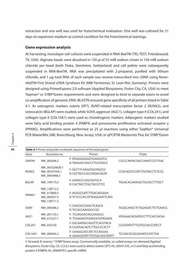
Chap
ter 4
60
extraction and one well was used for histochemical evaluation. One well was cultured for 21
days on expansion medium as control condition for the histochemical stainings.
gene expression analysis
At harvesting, monolayer cell cultures were suspended in RNA-BeeTM (TEL-TEST, Friendswood,
TX, USA). Alginate beads were dissolved in 150 μl of 55 mM sodium citrate in 150 mM sodium
chloride per bead (both Fluka, Steinheim, Switzerland) and cell pellets were subsequently
suspended in RNA-BeeTM. RNA was precipitated with 2-propanol, purified with lithium
chloride, and 1 µg total RNA of each sample was reverse-transcribed into cDNA using Rever-
tAidTM First Strand cDNA Synthesis Kit (MBI Fermentas, St. Leon-Rot, Germany). Primers were
designed using PrimerExpress 2.0 software (Applied Biosystems, Foster City, CA, USA) to meet
Taqman® or SYBR®Green requirements and were designed to bind to separate exons to avoid
co-amplification of genomic DNA. BLASTN ensured gene specificity of all primers listed in Table
4.1. As osteogenic markers osterix (SP7), RUNT-related transcription factor 2 (RUNX2), and
osteocalcin (BGLAP) were studied, while SOX9, aggrecan (AGC1), collagen type II (COL2A1), and
collagen type X (COL10A1) were used as chondrogenic markers. Adipogenic markers studied
were fatty acid binding protein 4 (FABP4) and peroxisome proliferative activated receptor γ
(PPARG). Amplifications were performed as 25 µl reactions using either TaqMan® Universal
PCR MasterMix (ABI, Branchburg, New Jersey, USA) or qPCRTM Mastermix Plus for SYBR®Green
Table 4.1 Primer and probe nucleotide sequences of the tested genes
Gene Accession no. Primer Probe
GAPDH NM_002046.2F: ATGGGGAAGGTGAAGGTCGR: TAAAAGCAGCCCTGGTGACC
CGCCCAATACGACCAAATCCGTTGAC
RUNX2NM_001024630.1NM_00101505.1NM_0043468.3
F: GCCTTCAAGGTGGTAGCCCR: CGTTACCCGCCATGACAGTA
CCACAGTCCCATCTGGTACCTCTCCG
BGLAP NM_199173.2F: GAAGCCCAGCGGTGCAR: CACTACCTCGCTGCCCTCC
TGGACACAAAGGCTGCACCTTTGCT
PPARG*
NM_138712.2NM_015869.3NM_005037.4NM_138711.2
F: AGGGCGATCTTGACAGGAAAR: TCTCCCATCATTAAGGAATTCATG
SOX9 NM_000346.2F: CAACGCCGAGCTCAGCAR: TCCACGAAGGGCCGC
TGGGCAAGCTCTGGAGACTTCTGAACG
AGC1NM_001135.1NM_013227.1
F : TCGAGGACAGCGAGGCCR : TCGAGGGTGTAGCGTGTAGAGA
ATGGAACACGATGCCTTTCACCACGA
COL2A1 NM_033150F: GGCAATAGCAGGTTCACGTACAR: CGATAACAGTCTTGCCCCACTT
CCGGTATGTTTCGTGCAGCCATCCT
COL10A1 NM_000493.2F: CAAGGCACCATCTCCAGGAAR: AAAGGGTATTTGTGGCAGCATATT
TCCAGCACGCAGAATCCATCTGA
F: forward; R: reverse; * SYBR®Green assay. Commercially available, so-called assays-on-demand (Applied Biosystems, Foster City, CA, U.S.A.) were used to detect osterix (SP7; Hs_00541729_m1) and fatty acid binding protein 4 (FABP4; Hs_00609791)-specific mRNA.

Intrinsic differentiation potential of adolescent human tendon tissue: an in-vitro cell differentiation study 61
I (Eurogentec, Nederland B.V., Maastricht, The Netherlands) according to the manufacturer’s
guidelines. Real-Time RT-PCR (QPCR) was done using an ABI PRISM® 7000 with SDS software
version 1.7. Data were normalized to GAPDH which was stably expressed across sample condi-
tions (not shown). Relative expression was calculated according to the 2-ΔCT formula [25] using
averages of duplicate samples.
histochemical and immunohistochemical stainings
Von Kossa staining
Cells were fixed in formalin, hydrated in milliQ water, immersed in 5% silver nitrate solution
(Sigma) for 10 minutes, rinsed and exposed to light for 10 minutes. Excess silver nitrate was
removed with 5% sodium-thiosulphate (Sigma) and cells were rinsed in distilled water, followed
by a counterstaining with azophloxine (Sigma).
Oil Red O staining
Cells were fixed in 10% formalin, treated with 0.3% Oil red O solution (Sigma) for 15 min, and
then repeatedly washed with tap water.
Collagen type II staining
Alginate beads were dissolved in sodium citrate, cytospins were prepared and stored at -80˚C.
Cytospins were fixed in acetone and treated with 1% hyaluronidase (Sigma) for 20 min. Cell
monolayers were fixed with 70% ethanol, treated with 50 mM NH4Cl (Sigma), and permea-
bilised in a 0.1% Triton X-100 (Sigma) solution. Cells were incubated with mouse monoclonal
antibody against collagen type II (II-II6B3, diluted 1:100; Developmental Studies Hybridoma
Bank) for 2 hours. Anti-mouse Fab fragments conjugated with alkaline phosphatase (GAMAP,
diluted 1:100; Immunotech, Marseille, France) were added. Finally, alkaline phosphatase con-
jugated anti-mouse antibodies in combination with a new fuchsin substrate were added to
obtain a pink signal in positive cultures. Counterstaining with Gill’s haematoxilin (Sigma) was
performed.
ResulTs
Characterisation of tendon-derived fibroblasts
Histological examination of the adolescent hamstring tendon explants confirmed normal tis-
sue morphology. Specifically, no degenerative lesions, inflammatory cell infiltration, (partial)
ruptures, chondroid metaplasia, or calcifications were seen.
During the explant culture period, proliferating cells (Ki-67 positive) were located between
the highly organized collagen fibres of the tendon tissue and also in the connective tissue of
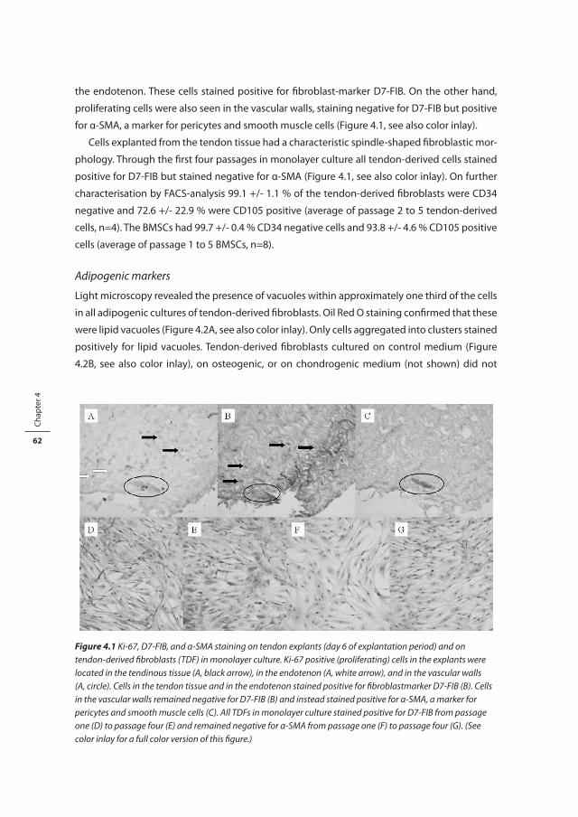
Chap
ter 4
62
the endotenon. These cells stained positive for fibroblast-marker D7-FIB. On the other hand,
proliferating cells were also seen in the vascular walls, staining negative for D7-FIB but positive
for α-SMA, a marker for pericytes and smooth muscle cells (Figure 4.1, see also color inlay).
Cells explanted from the tendon tissue had a characteristic spindle-shaped fibroblastic mor-
phology. Through the first four passages in monolayer culture all tendon-derived cells stained
positive for D7-FIB but stained negative for α-SMA (Figure 4.1, see also color inlay). On further
characterisation by FACS-analysis 99.1 +/- 1.1 % of the tendon-derived fibroblasts were CD34
negative and 72.6 +/- 22.9 % were CD105 positive (average of passage 2 to 5 tendon-derived
cells, n=4). The BMSCs had 99.7 +/- 0.4 % CD34 negative cells and 93.8 +/- 4.6 % CD105 positive
cells (average of passage 1 to 5 BMSCs, n=8).
Adipogenic markers
Light microscopy revealed the presence of vacuoles within approximately one third of the cells
in all adipogenic cultures of tendon-derived fibroblasts. Oil Red O staining confirmed that these
were lipid vacuoles (Figure 4.2A, see also color inlay). Only cells aggregated into clusters stained
positively for lipid vacuoles. Tendon-derived fibroblasts cultured on control medium (Figure
4.2B, see also color inlay), on osteogenic, or on chondrogenic medium (not shown) did not
Figure 4.1 Ki-67, D7-FIB, and -SMA staining on tendon explants (day 6 of explantation period) and on tendon-derived fibroblasts (TDF) in monolayer culture. Ki-67 positive (proliferating) cells in the explants were located in the tendinous tissue (A, black arrow), in the endotenon (A, white arrow), and in the vascular walls (A, circle). Cells in the tendon tissue and in the endotenon stained positive for fibroblastmarker D7-FIB (B). Cells in the vascular walls remained negative for D7-FIB (B) and instead stained positive for -SMA, a marker for pericytes and smooth muscle cells (C). All TDFs in monolayer culture stained positive for D7-FIB from passage one (D) to passage four (E) and remained negative for -SMA from passage one (F) to passage four (G).
Figure 4.1 Ki-67, D7-FIB, and α-SMA staining on tendon explants (day 6 of explantation period) and on tendon-derived fibroblasts (TDF) in monolayer culture. Ki-67 positive (proliferating) cells in the explants were located in the tendinous tissue (A, black arrow), in the endotenon (A, white arrow), and in the vascular walls (A, circle). Cells in the tendon tissue and in the endotenon stained positive for fibroblastmarker D7-FIB (B). Cells in the vascular walls remained negative for D7-FIB (B) and instead stained positive for α-SMA, a marker for pericytes and smooth muscle cells (C). All TDFs in monolayer culture stained positive for D7-FIB from passage one (D) to passage four (E) and remained negative for α-SMA from passage one (F) to passage four (G). (See color inlay for a full color version of this figure.)
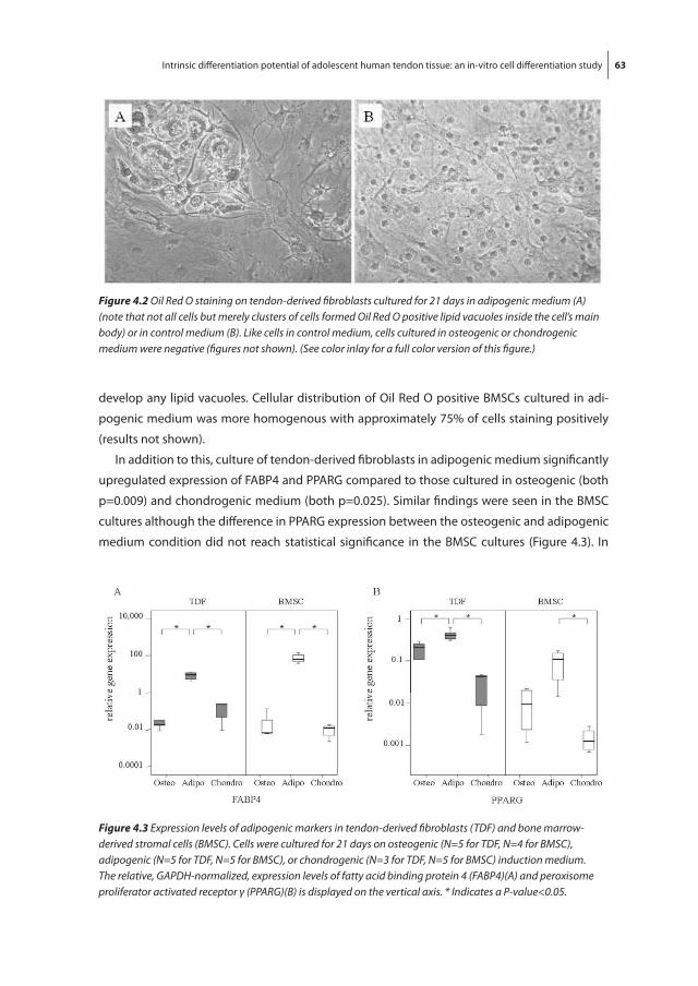
Intrinsic differentiation potential of adolescent human tendon tissue: an in-vitro cell differentiation study 63
develop any lipid vacuoles. Cellular distribution of Oil Red O positive BMSCs cultured in adi-
pogenic medium was more homogenous with approximately 75% of cells staining positively
(results not shown).
In addition to this, culture of tendon-derived fibroblasts in adipogenic medium significantly
upregulated expression of FABP4 and PPARG compared to those cultured in osteogenic (both
p=0.009) and chondrogenic medium (both p=0.025). Similar findings were seen in the BMSC
cultures although the difference in PPARG expression between the osteogenic and adipogenic
medium condition did not reach statistical significance in the BMSC cultures (Figure 4.3). In
Figure 4.2 Oil Red O staining on tendon-derived fibroblasts cultured for 21 days in adipogenic medium (A) (note that not all cells but merely clusters of cells formed Oil Red O positive lipid vacuoles inside the cell’s main body) or in control medium (B). Like cells in control medium, cells cultured in osteogenic or chondrogenic medium were negative (figures not shown).
Figure 4.2 Oil Red O staining on tendon-derived fibroblasts cultured for 21 days in adipogenic medium (A) (note that not all cells but merely clusters of cells formed Oil Red O positive lipid vacuoles inside the cell’s main body) or in control medium (B). Like cells in control medium, cells cultured in osteogenic or chondrogenic medium were negative (figures not shown). (See color inlay for a full color version of this figure.)
Figure 4.3 Expression levels of adipogenic markers in tendon-derived fibroblasts (TDF) and bone marrow-derived stromal cells (BMSC). Cells were cultured for 21 days on osteogenic (N=5 for TDF, N=4 for BMSC), adipogenic (N=5 for TDF, N=5 for BMSC), or chondrogenic (N=3 for TDF, N=5 for BMSC) induction medium. The relative, GAPDH-normalized, expression levels of fatty acid binding protein 4 (FABP4)(A) and peroxisome proliferator activated receptor (PPARG)(B) is displayed on the vertical axis. * Indicates a P-value<0.05.
Figure 4.3 Expression levels of adipogenic markers in tendon-derived fibroblasts (TDF) and bone marrow-derived stromal cells (BMSC). Cells were cultured for 21 days on osteogenic (N=5 for TDF, N=4 for BMSC), adipogenic (N=5 for TDF, N=5 for BMSC), or chondrogenic (N=3 for TDF, N=5 for BMSC) induction medium. The relative, GAPDH-normalized, expression levels of fatty acid binding protein 4 (FABP4)(A) and peroxisome proliferator activated receptor γ (PPARG)(B) is displayed on the vertical axis. * Indicates a P-value<0.05.

Chap
ter 4
64
the BMSC cultures PPARG expression was significantly higher in adipogenic medium compared
to chondrogenic medium (p=0.021); FABP4 expression was upregulated in the adipogenic
medium compared to osteogenic medium (p=0.021) and chondrogenic medium (p=0.021).
Chondrogenic markers
Immunohistochemical staining for collagen type II was performed on tendon-derived fibro-
blasts cultured in chondrogenic, adipogenic, osteogenic, and control medium for 21 days. In
all chondrogenic medium conditions approximately 5% of the cells stained positive for col-
lagen type II (Figure 4.4A, see also color inlay). Tendon-derived fibroblasts cultured in control
medium (Figure 4.4B, see also color inlay), as well as adipogenic and osteogenic medium were
immunonegative for collagen type II (not shown). BMSC cultures showed a similar amount of
collagen type II staining in chondrogenic medium (not shown).
Culture of tendon-derived fibroblasts in chondrogenic medium significantly increased
expression of chondrogenic markers COL2A1 and COL10A1 (the latter is considered to be a
marker for hypertrophic cartilage formation) compared to the osteogenic condition (p=0.025
for both genes) and adipogenic condition (p=0.025 for both genes). Expression of SOX9 and
AGC1 in chondrogenic medium compared to osteogenic and adipogenic medium was not
significantly different (Figure 4.5).
BMSC cultures also showed a significantly higher expression of COL2A1 and COL10A1 in
chondrogenic medium compared to osteogenic medium (p=0.014 for both genes) and adi-
pogenic medium (p=0.028 for COL2A1 and p=0.009 for COL10A1). SOX9 expression in BMSCs
showed the same trend as in the tendon-derived fibroblast cultures, but the differences only
reached significance in the BMSC cultures (osteogenic versus adipogenic medium p=0.014
and osteogenic versus chondrogenic medium p=0.014). Expression of AGC1 in the BMSCs did
not differ significantly between the three medium conditions. Interestingly, BMSCs cultured in
Figure 4.4 Immunohistochemical staining for collagen type II on tendon-derived fibroblasts. 5% of the cells cultured for 21 days in alginate beads in chondrogenic medium stained positive (A). Cells cultured in monolayer in control medium remained negative (B) as did cells in adipogenic or osteogenic media (figures not shown).
Figure 4.4 Immunohistochemical staining for collagen type II on tendon-derived fibroblasts. 5% of the cells cultured for 21 days in alginate beads in chondrogenic medium stained positive (A). Cells cultured in monolayer in control medium remained negative (B) as did cells in adipogenic or osteogenic media (figures not shown). (See color inlay for a full color version of this figure.)

Intrinsic differentiation potential of adolescent human tendon tissue: an in-vitro cell differentiation study 65
osteogenic medium had significantly upregulated COL10A1 compared to the adipogenic con-
dition (p=0.027). This phenomenon was not seen in the tendon-derived fibroblasts (Figure 4.5).
Osteogenic markers
Von Kossa staining of tendon-derived fibroblasts in the osteogenic condition showed clustered
areas of calcium deposition, whereas the tendon-derived fibroblast cultures in control medium
had no calcium deposition (Figure 4.6, see also color inlay). Also, tendon-derived fibroblast
cultures in adipogenic and chondrogenic medium remained negative for calcium (results not
shown). Similarly, in BMSC cultures, calcium deposition was found only in the osteogenic condi-
tion (not shown).
Although we did find expression of osteogenic markers RUNX2, osterix, and osteocalcin,
culture of tendon-derived fibroblasts in osteogenic medium did not induce statistically signifi-
cant upregulation of any of these genes. Similar results were found by QPCR of these markers
Figure 4.5 Expression levels of chondrogenic markers in tendon-derived fibroblasts (TDF) and bone marrow-derived stromal cells (BMSC). SOX9 (A), aggrecan (AGC1)(B), collagen type II (COL2A1)(C) and collagen type X (COL10A1)(D). See figure 3 for reminder of key.
Figure 4.5 Expression levels of chondrogenic markers in tendon-derived fibroblasts (TDF) and bone marrow-derived stromal cells (BMSC). SOX9 (A), aggrecan (AGC1)(B), collagen type II (COL2A1)(C) and collagen type X (COL10A1)(D). See figure 3 for reminder of key.

Chap
ter 4
66
Figure 4.6 Von Kossa staining on tendon-derived fibroblasts cultured for 21 days in osteogenic (A) or control medium (B). Calcium deposition was seen in osteogenic medium (A), not in control medium (B) or in adipogenic or chondrogenic media (figures not shown).
Figure 4.6 Von Kossa staining on tendon-derived fibroblasts cultured for 21 days in osteogenic (A) or control medium (B). Calcium deposition was seen in osteogenic medium (A), not in control medium (B) or in adipogenic or chondrogenic media (figures not shown). (See color inlay for a full color version of this figure.)
Figure 4.7 Expression levels of osteogenic markers in tendon-derived fibroblasts (TDF) and bone marrow-derived stromal cells (BMSC). RUNT-related transcription factor 2 (RUNX2) (A), SP7 (B), and BGLAP (C). See figure 3 for reminder of key.
Figure 4.7 Expression levels of osteogenic markers in tendon-derived fibroblasts (TDF) and bone marrow-derived stromal cells (BMSC). RUNT-related transcription factor 2 (RUNX2) (A), SP7 (B), and BGLAP (C). See figure 3 for reminder of key.

Intrinsic differentiation potential of adolescent human tendon tissue: an in-vitro cell differentiation study 67
in BMSCs cultured in osteogenic medium (Figure 4.7). In the tendon-derived fibroblast cultures
SP7 and RUNX2 (both also known to play an important role in chondrogenic differentiation
and hypertrophic cartilage formation [26]) were significantly upregulated in the chondrogenic
medium compared to the osteogenic (p=0.025 for both genes) and adipogenic medium
(p=0.025 for both genes)(Figure 4.7). BMSCs also showed an upregulation of SP7 and RUNX2
in the chondrogenic medium. RUNX2 upregulation was significant (p=0.016 for the difference
in gene expression of RUNX2 between adipogenic and chondrogenic medium in BMCSs), but
SP7 upregulation in chondrogenic medium did not reach significance (Figure 4.7). In summary,
chondrogenic medium not only stimulated expression of chondrogenic marker COL2A1, but
also of COL10A1, RUNX2, and SP7.
DisCussion
This in-vitro differentiation study suggests that a proportion of the cell population explanted
from adolescent human tendon tissue may have adipogenic and chondrogenic differentiation
potential. In adipogenic medium lipid vacuoles were visible and tendon-derived fibroblasts
showed upregulation of FABP4 and PPARG. In chondrogenic medium, positive collagen type
II staining was visible around some of the tendon-derived fibroblasts and the tendon-derived
fibroblasts showed upregulation of COL2A1 and COL10A1. In osteogenic medium Von Kossa
staining showed calcium deposition, although osteogenic markers remained unaltered, as
assessed by qPCR. Compared to the BMSCs, the diffentiation capacity of our tendon-derived
fibroblasts was similar, although some differences were visible, mainly concerning the number
of Oil Red O positive cells.
To our knowledge, this is the first study evaluating the intrinsic differentiation potential of
human tendon cells in vitro. Previously, Salingcarnboriboon et al [27] established three murine
tendon cell lines by clonal expansion and showed that these single cell clones could differenti-
ate towards multiple mesenchymal lineages upon culture in appropriate differentiation media.
Therefore, they suggested that cells with mesenchymal stem-cell-like characteristics might
exist in murine tendon tissue. Our experiments cannot distinguish between individual cells
with multilineage potential and a cell population containing more or less strongly commit-
ted cells. We did find that not all of the tendon-derived fibroblasts appeared to be capable of
differentiating towards other lineages, e.g. not all fibroblasts but merely clusters of fibroblasts
created lipid vacuoles in adipogenic medium and only a small proportion of approximately 5%
of the cells stained positive for collagen type II. In addition to this observation, only a subpopu-
lation of 72.6 +/- 22.9 % of these tendon-derived fibroblasts stained positive for CD105 and this
subpopulation might be responsible for the observed differentiation potential.
Due to their spindle-shaped morphology in monolayer culture and because all explanted
cells stained D7-FIB positive in passage one through passage four, we identified these cells

Chap
ter 4
68
as tendon-derived fibroblasts. Based on the results of the Ki-67 staining, it could be surmised
that this mixed population may be partly derived from the tendon tissue and partly from the
endotenon. It is possible that these cells were already preselected for during the explantation
procedure, based on cellular motility, chemotactic responses or plastic adherence characteris-
tics. Within this culture population, mature tendon-derived fibroblasts with transdifferentiation
capacity or a specific subpopulation of tendon-derived progenitor cells might exist. Several
authors have found that pericytes isolated from different tissues can be induced to differentiate
into various connective tissue phenotypes [8]. It seems unlikely that the presence of vascular
pericytes in tendon tissue, which might be another multipotent cell source in tendon tissue
[28], can account for our findings. Not only is tendon a poorly vascularized tissue, but also the
tendon-derived fibroblasts remained negative for pericyte marker α-SMA through the first four
passages. Furthermore, our explanted cell population was 98.5 +/- 0.7 % negative for CD34 on
FACS-analysis. It seems unlikely that the small portion of 1.5% CD34-positive tendon-derived
fibroblasts accounts for the results of the immunohistochemical staining and the changes in
gene expression pattern.
A cell population with multilineage potential that might be present in tendon tissue, is likely
involved in tendon repair. Such a population might also contribute to the development of
tendinosis, as this tendon disorder is associated with fatty degeneration, glycosaminoglycan
accumulation, and calcifications. In addition to these internal multipotent cells other cells with
multilineage potential may arrive at the site of overuse or tendon damage through the vascular
system and contribute to the development or repair of tendinosis: upregulation of VEGF was
found in human Achilles tendinosis lesions [29] and VEGF can act as a chemotactic stimulus for
mesenchymal cells [30]. In-vivo control of differentiation of cells with multilineage potential
might prove useful in the future for prevention of tendinosis lesions or induction of in-situ
repair of these lesions.
The exact changes in the tendon microenvironment outside the cells that play a role in cel-
lular differentiation are still the subject of many investigations. First, the capability of specific
growth factors, cytokines, and other inflammatory mediators to influence the cellular differen-
tiation process has been demonstrated. Changes in the concentration of various growth factors
have also been found in tendinosis lesions: for instance, a higher number of cells expressing
TGF-β2 and TGF-βRII (a TGF-β receptor) in chronic Achilles tendinosis lesions [31] and increased
expression of TGF-β1 in patellar tendinosis [32] have been reported. TGF-β molecules are also
used in vitro to induce chondrogenic differentiation of mesenchymal progenitor cells [21].
Second, changes in the degree of vascularisation of the tissue, as reported in Achilles tendinosis
lesions [33], might influence the tendon cell differentiation state in vivo. For instance, oxygen
tension influences the redifferentiation potential of dedifferentiated chondrocytes in vitro [34]
and hypoxia not only promotes the differentiation of bone mesenchymal stem cells along a
chondrocyte pathway [35], but can also promote the formation of an adipocyte-like pheno-
type with cytoplasmic lipid inclusions in human MSCs [36]. Third, following repetitive tendon

Intrinsic differentiation potential of adolescent human tendon tissue: an in-vitro cell differentiation study 69
overload and its resulting microruptures in tendinosis lesions [37], tendon cells may experience
an altered mechanical microenvironment, which in turn might influence chondrogenic, osteo-
genic, or tenogenic differentiation [38].
Our findings demonstrate that an intrinsic differentiation capacity is present in tendon tissue
of adolescent individuals. However, age plays an important role in the response of musculoskel-
etal tissues in response to environmental changes. It has been demonstrated that adult but not
juvenile cartilage has lost its ability to regenerate (cited by Hunter [39]) and BMSCs gradually
loose their differentiation potential as subjects grow older [40]. Therefore, the adolescent ten-
don samples used in this study might not be representative of tendon tissue in adult tendinosis
lesions. Since tendon cell populations derived from adult and from late fetal equine tendons
have demonstrated similar levels of a weak progenitor cell ability [41], it might be justified to
speculate that tendon-derived fibroblasts from older subjects may still have some differentia-
tion capacity. However, this certainly needs further investigation.
A tendon-cell population with intrinsic differentiation capacity might be used in vivo for
repair of lesions and might play a role in tendinosis. However, extrapolating results from
in-vitro cultures to the in-vivo situation must be done with tremendous caution, particularly
as the expansion-culture period prior to experimentation may have led to the loss of the
original tendon fibroblast phenotype (due to dedifferentiation): the latter being well known in
chondrocyte-cultures [42]. Whether cells in vivo can be stimulated to display this differentiation
potential remains to be elucidated.
ConClusions
Obtaining insight in the cellular behavior and pathogenesis in tendinosis is crucial in order to
develop mechanism-based therapies. Our study suggests that adolescent tendon tissue has
an intrinsic differentiation potential. This study conducted on human tenocytes corroborates
the findings that cells with mesenchymal stem-cell-like characteristics might exist in murine
tendon tissue. Our results support the hypothesis that altered tendon-cell differentiation might
play a role in the pathophysiology of tendinosis.
aCknowleDgeMenTs
The authors would like to thank the orthopedic surgeon Ad Diepstraten for providing the
tendon tissue and Han van Neck and Aleko Chatojev for their support and interest in this work.
The antibody II-116B3 was obtained from the Developmental Studies Hybridoma Bank, under
contract N01-HD-6–2915 from the NICHD.

Chap
ter 4
70
ReFeRenCes
1. Paavola M, Kannus P, Jarvinen TA, Khan K, Jozsa L, Jarvinen M: Achilles tendinopathy. J Bone Joint Surg Am 2002, 84-A(11):2062-2076.
2. Maffulli N, Kader D: Tendinopathy of tendo achillis. J Bone Joint Surg Br 2002, 84(1):1-8. 3. Movin T, Gad A, Reinholt FP, Rolf C: Tendon pathology in long-standing achillodynia. Biopsy findings
in 40 patients. Acta Orthop Scand 1997, 68(2):170-175. 4. Tallon C, Maffulli N, Ewen SW: Ruptured Achilles tendons are significantly more degenerated than
tendinopathic tendons. Med Sci Sports Exerc 2001, 33(12):1983-1990. 5. Jarvinen M, Jozsa L, Kannus P, Jarvinen TL, Kvist M, Leadbetter W: Histopathological findings in chronic
tendon disorders. Scand J Med Sci Sports 1997, 7(2):86-95. 6. Caplan AI, Bruder SP: Mesenchymal stem cells: building blocks for molecular medicine in the 21st
century. Trends Mol Med 2001, 7(6):259-264. 7. Roufosse CA, Direkze NC, Otto WR, Wright NA: Circulating mesenchymal stem cells. Int J Biochem Cell
Biol 2004, 36(4):585-597. 8. Muschler GF, Midura RJ: Connective tissue progenitors: practical concepts for clinical applications.
Clin Orthop Relat Res 2002(395):66-80. 9. Gregory CA, Prockop DJ, Spees JL: Non-hematopoietic bone marrow stem cells: molecular control of
expansion and differentiation. Exp Cell Res 2005, 306(2):330-335. 10. Morasso MI, Tomic-Canic M: Epidermal stem cells: the cradle of epidermal determination, differentia-
tion and wound healing. Biol Cell 2005, 97(3):173-183. 11. Nakahara H, Dennis JE, Bruder SP, Haynesworth SE, Lennon DP, Caplan AI: In vitro differentiation of
bone and hypertrophic cartilage from periosteal-derived cells. Exp Cell Res 1991, 195(2):492-503. 12. Nakahara H, Goldberg VM, Caplan AI: Culture-expanded human periosteal-derived cells exhibit
osteochondral potential in vivo. J Orthop Res 1991, 9(4):465-476. 13. Bosch P, Musgrave DS, Lee JY, Cummins J, Shuler T, Ghivizzani TC, Evans T, Robbins TD, Huard: Osteo-
progenitor cells within skeletal muscle. J Orthop Res 2000, 18(6):933-944. 14. Seale P, Rudnicki MA: A new look at the origin, function, and “stem-cell” status of muscle satellite cells.
Dev Biol 2000, 218(2):115-124. 15. Rodriguez AM, Elabd C, Amri EZ, Ailhaud G, Dani C: The human adipose tissue is a source of multipo-
tent stem cells. Biochimie 2005, 87(1):125-128. 16. Kahn AJ, Simmons DJ: Chondrocyte-to-osteocyte transformation in grafts of perichondrium-free
epiphyseal cartilage. Clin Orthop Relat Res 1977(129):299-304. 17. Barbero A, Ploegert S, Heberer M, Martin I: Plasticity of clonal populations of dedifferentiated adult
human articular chondrocytes. Arthritis Rheum 2003, 48(5):1315-1325. 18. Caterson EJ, Nesti LJ, Danielson KG, Tuan RS: Human marrow-derived mesenchymal progenitor cells:
isolation, culture expansion, and analysis of differentiation. Mol Biotechnol 2002, 20(3):245-256. 19. van Osch GJ, van der Veen SW, Marijnissen WJ, Verhaar JA: Monoclonal antibody 11-fibrau: a use-
ful marker to characterize chondrocyte differentiation stage. Biochem Biophys Res Commun 2001, 280(3):806-812.
20. Shi S, Gronthos S: Perivascular niche of postnatal mesenchymal stem cells in human bone marrow and dental pulp. J Bone Miner Res 2003, 18(4):696-704.
21. Tuli R, Tuli S, Nandi S, Wang ML, Alexander PG, Haleem-Smith H, Hozack WJ, Manner PA, Danielson KG, Tuan RS: Characterization of multipotential mesenchymal progenitor cells derived from human trabecular bone. Stem Cells 2003, 21(6):681-693.
22. Zocchi MR, Poggi A: PECAM-1, apoptosis and CD34+ precursors. Leuk Lymphoma 2004, 45(11):2205-2213.
23. Middleton J, Americh L, Gayon R, Julien D, Mansat M, Mansat P, Anract P, Cantagrel A, Cattan P, Reimund JM et al: A comparative study of endothelial cell markers expressed in chronically inflamed

Intrinsic differentiation potential of adolescent human tendon tissue: an in-vitro cell differentiation study 71
human tissues: MECA-79, Duffy antigen receptor for chemokines, von Willebrand factor, CD31, CD34, CD105 and CD146. J Pathol 2005, 206(3):260-268.
24. Howson KM, Aplin AC, Gelati M, Alessandri G, Parati EA, Nicosia RF: The postnatal rat aorta contains pericyte progenitor cells that form spheroidal colonies in suspension culture. Am J Physiol Cell Physiol 2005, 289(6):C1396-1407.
25. Livak KJ, Schmittgen TD: Analysis of relative gene expression data using real-time quantitative PCR and the 2(-Delta Delta C(T)) Method. Methods 2001, 25(4):402-408.
26. Wang Y, Belflower RM, Dong YF, Schwarz EM, O’Keefe RJ, Drissi H: Runx1/AML1/Cbfa2 mediates onset of mesenchymal cell differentiation toward chondrogenesis. J Bone Miner Res 2005, 20(9):1624-1636.
27. Salingcarnboriboon R, Yoshitake H, Tsuji K, Obinata M, Amagasa T, Nifuji A, Noda M: Establishment of tendon-derived cell lines exhibiting pluripotent mesenchymal stem cell-like property. Exp Cell Res 2003, 287(2):289-300.
28. Katenkamp D, Stiller D, Schulze E: Ultrastructural cytology of regenerating tendon--an experimental study. Exp Pathol (Jena) 1976, 12(1):25-37.
29. Alfredson H, Lorentzon M, Backman S, Backman A, Lerner UH: cDNA-arrays and real-time quantitative PCR techniques in the investigation of chronic Achilles tendinosis. J Orthop Res 2003, 21(6):970-975.
30. Fiedler J, Leucht F, Waltenberger J, Dehio C, Brenner RE: VEGF-A and PlGF-1 stimulate chemotactic migration of human mesenchymal progenitor cells. Biochem Biophys Res Commun 2005, 334(2):561-568.
31. Fenwick SA, Curry V, Harrall RL, Hazleman BL, Hackney R, Riley GP: Expression of transforming growth factor-beta isoforms and their receptors in chronic tendinosis. J Anat 2001, 199(Pt 3):231-240.
32. Fu SC, Wang W, Pau HM, Wong YP, Chan KM, Rolf CG: Increased expression of transforming growth factor-beta1 in patellar tendinosis. Clin Orthop Relat Res 2002(400):174-183.
33. Pufe T, Petersen WJ, Mentlein R, Tillmann BN: The role of vasculature and angiogenesis for the patho-genesis of degenerative tendons disease. Scand J Med Sci Sports 2005, 15(4):211-222.
34. Murphy CL, Polak JM: Control of human articular chondrocyte differentiation by reduced oxygen tension. J Cell Physiol 2004, 199(3):451-459.
35. Robins JC, Akeno N, Mukherjee A, Dalal RR, Aronow BJ, Koopman P, Clemens TL: Hypoxia induces chondrocyte-specific gene expression in mesenchymal cells in association with transcriptional activation of Sox9. Bone 2005, 37(3):313-322.
36. Fink T, Abildtrup L, Fogd K, Abdallah BM, Kassem M, Ebbesen P, Zachar V: Induction of adipocyte-like phenotype in human mesenchymal stem cells by hypoxia. Stem Cells 2004, 22(7):1346-1355.
37. Wang JH, Iosifidis MI, Fu FH: Biomechanical basis for tendinopathy. Clin Orthop Relat Res 2006, 443:320-332.
38. Forslund C, Aspenberg P: CDMP-2 induces bone or tendon-like tissue depending on mechanical stimulation. J Orthop Res 2002, 20(6):1170-1174.
39. Hunter W: Of the structure and diseases of articulating cartilages. 1743. Clin Orthop Relat Res 1995(317):3-6.
40. Muraglia A, Cancedda R, Quarto R: Clonal mesenchymal progenitors from human bone marrow dif-ferentiate in vitro according to a hierarchical model. J Cell Sci 2000, 113 ( Pt 7):1161-1166.
41. Strassburg S, Goodship A, Hardingham T, Clegg P: Adult and late fetal equine tendons contain cell populations with weak progenitor properties in comparison to bone marrow derived mesenchymal stem cells. In: 52nd Annual Meeting Orthopaedic Research Society: 2006; Chicago.
42. Mandl EW, Jahr H, Koevoet JL, van Leeuwen JP, Weinans H, Verhaar JA, van Osch GJ: Fibroblast growth factor-2 in serum-free medium is a potent mitogen and reduces dedifferentiation of human ear chondrocytes in monolayer culture. Matrix Biol 2004, 23(4):231-241.


Chapter 5
In-vitro model to study chondrogenic differentiation in tendinopathy
M. de Mos, J.L.M. Koevoet, H.T.M. van Schie, N. Kops, H. Jahr, J.A.N. Verhaar, G.J.V.M. van Osch
Manuscript under revision by Am J Sports Med

Chap
ter 5
74
aBsTRaCT
Background
Fibrocartilaginous metaplasia can be a sign of tendinopathy. We investigated whether chon-
drogenic differentiation is present in mid-portion Achilles tendinopathy and subsequently
developed an in-vitro model for chondrogenic differentiation of tendon cells to investigate
possible targets for future treatment.
Methods
Peroperatively harvested tissue from human mid-portion Achilles tendinotic lesions (n=12) and
healthy Achilles tendons (n=5) were analysed by microscopy and real-time reverse transcription
polymerase chain reaction (QPCR). Secondly, chondrogenic differentiation of non-degenerative
human tendon explants was induced using transforming growth factor beta (TGF-β) to develop
an in-vitro model for chondrogenic differentiation of tendon tissue. To evaluate the usefulness
of the model, we investigated the possibility to modulate the chondrogenic differentiation in
vitro by removing the chondrogenic stimulus or adding triamcinolone or platelet-rich plasma
(PRP).
Results
Mid-portion Achilles tendinotic lesions had significantly increased glycosaminoglycan staining
and more rounded cell nuclei than healthy tendons. Chondrogenic markers (sex-determining
region Y)-box9 (SOX9), aggrecan (AGC1), collagen type II (COL2A1), RUNT-related transcription
factor 2 (RUNX2), but not collagen type X (COL10A1) were upregulated. Non-degenerative
tendon explants cultured on chondrogenic medium for 7, 14, and 21 days had significantly
higher expression of AGC1, COL2A1, and COL10A1, but not SOX9 and RUNX2. Removing the
chondrogenic stimulus at day 14 resulted in a reduced expression of AGC1, COL2A1, and
COL10A1 at day 21. Adding triamcinolone (10–4 M) from day 14 resulted in less COL2A1 expres-
sion, whereas other genes were not affected. PRP (20% vol/vol) also appeared to likely influence
chondrogenic gene expression pattern.
Conclusions
In conclusion, mid-portion Achilles tendinopathy showed chondrogenic differentiation and
the in-vitro model that was developed in order to investigate chondrogenic differentiation
appeared to be useful for the evaluation of innovative therapies.

In-vitro model to study chondrogenic differentiation in tendinopathy 75
inTRoDuCTion
Chronic tendinopathies are a common problem in orthopaedic and rheumatologic practice,
occurring in athletes as well as in the general population. Chronic Achilles tendinopathy
involves either mid-portion tendinopathy, insertional tendinopathy, or peritendinitis, or any
combination of these. Chronic mid-portion Achilles tendinopathies are often difficult to treat,
and surgical interventions may be necessary [1, 2]. Although several hypotheses have been
formulated during the last decade [3-7], the pathophysiology of tendinopathies in general, and
of mid-portion Achilles tendinopathy in particular, is still largely unknown.
Considerable alterations in the extracellular matrix (ECM) composition have been described
in chronic mid-portion Achilles tendinopathy [8]. Firstly, an increased amount of glycosamino-
glycans is present in tendinotic tissue compared to healthy tendon tissue [9, 10]. Secondly, the
tendon cells in chronic mid-portion Achilles tendinopathy display an altered gene expression
pattern of the major proteoglycans towards that seen in fibrocartilaginous tissue [11, 12]. These
findings led us to consider whether in Achilles mid-portion tendinopathy some of the tendon
cells may deviate from tendon matrix production towards fibrocartilaginous matrix produc-
tion. Fibrocartilaginous differentiation along the length of the mid-portion area of the Achilles
tendon, which is a ‘direct’ tendon area [12] (an area that runs a straight course and experiences
mainly tensile forces), most probably results in impaired elasticity and tensile stress resistance
of the tendon. This can compromise the tendon’s function as elastic energy-saving spring and
force transmitter, and thus can make the tissue more prone to degeneration and rupture.
Fibrocartilaginous metaplasia is a normal feature of insertional tendon areas (entheses)
and of compressed areas of ‘wrap-around’ tendons that bend around a bony pulley [12]. This
process is considered to be a functional adaptation to compressive loads and/or stress shield-
ing (lack of tensile loads) [3, 13]. However, it can also be a sign of tendon pathology. Maffulli et
al. found chondral metaplasia in calcific insertional Achilles tendinopathy [14]. Archambault et
al. found cartilage-specific markers to be upregulated in an overuse model of the rat supraspi-
natus tendon [15]. Excessive or inappropriate fibrocartilaginous matrix production in tendons
may therefore be considered part of the pathological processes involved in developing tendi-
nopathies. Consequently, removing or ceasing the stimulus that causes the fibrocartilaginous
metaplasia might help tenocytes to return to their normal tendon matrix production. From
this perspective, understanding the mechanical and/or molecular signals that can influence
chondrogenic differentiation may be key factors to eventually improve prevention and treat-
ment opportunities of developing tendinopathies.
Because tendinopathies are exposed to triamcinolone and PRP in clinical as well as experi-
mental settings, both compounds are of interest to study in relation to chondrogenic differ-
entiation of tendon cells. Triamcinolone is a synthetic analogue of cortisol which has a potent
anti-inflammatory effect [16]. Although inflammation does not appear to play a major role in
chronic tendinopathies [15, 17], triamcinolone is used as a symptomatic treatment for various
tendinopathies and for peritendinitis. As peritendinitis is a frequently diagnosed co-morbidity

Chap
ter 5
76
of tendinopathy, tendinotic tendon tissue may be exposed to rather high local steroid con-
centrations at some point during their disease course. Platelet-rich plasma (PRP) has yielded
promising outcomes in numerous clinical applications. A cohort study reported 93% reduction
of pain for PRP-treated patients with chronic elbow tendinosis [18], although the exact working
mechanism still remains to be elucidated.
We hypothesized that chondrogenic differentiation, demonstrated in insertional and
compressed tendon areas, also takes place in mid-portion Achilles tendinopathy. Therefore, we
investigated whether a chondrogenic differentiation pattern was seen in mid-portion Achilles
tendinopathy in patients. Secondly, we hypothesized that this chondrogenic differentiation
might be used as a target for drug treatment of developing tendinotic lesions. Therefore, the
second part of this research aimed at developing an in-vitro culture model of chondrogenic
tendon differentiation, for future use in evaluating treatment opportunities for tendinopathy.
MaTeRials anD MeThoDs
study design
To investigate whether mid-portion Achilles tendinotic lesions display a chondrogenic differ-
entiation pattern, peroperatively harvested biopsies of human mid-portion Achilles tendinotic
lesions and of macroscopically healthy Achilles tendons were analysed by microscopy and real-
time RT-PCR. Secondly, to develop a culture model for chondrogenic differentiation of tendons,
healthy adolescent hamstring tendon explants and healthy adult Achilles tendon explants
were stimulated in-vitro with TGF-β2. The subsequently induced chondrogenic differentiation
was modulated by switching to non-chondrogenic medium, or by adding either triamcinolone
or platelet-rich plasma (PRP) to the chondrogenic culture medium.
Tendon biopsies
For the in-vivo study, macroscopically affected tissue specimens of 12 consecutive patients
with chronic mid-portion Achilles tendinopathy (6 male and 6 female; average age 46 years,
range 36-58; mean duration of symptoms 25 months, range 9-54 months) were harvested dur-
ing surgical debridement of the lesion. All lesions were located between 3-5 cm proximal from
the insertion. Chronic Achilles tendinopathy was defined as Achilles tendon pain for at least
3 months in combination with clinically and/or radiologically (either MRI or ultrasonography)
suspected mid-portion Achilles tendinopathy. Patients with previous tendon injury, radiologi-
cally evident partial tendon rupture, or use of chinolone antibiotics in the past three years were
excluded. Approval for this study was obtained from the Medical Ethical Committee of the
Erasmus MC University Medical Center (MEC-2005-100). All patients signed informed consent.
Healthy Achilles tissue specimens (all between 3-5 cm proximal from the insertion) of 5 donors
(2 male, 3 female; average age 55, range 24-78) without clinically evident Achilles tendinopathy

In-vitro model to study chondrogenic differentiation in tendinopathy 77
were collected as control. Surgical procedures for these patients included arthrodesis of the
talocrural joint, upper leg amputation because of a septic revised total knee prosthesis, lower
leg amputation after trauma, and extension of the Achilles tendon for spastic diplegia (MEC-
2006-069).
For the differentiation culture studies we harvested a) non-degenerative hamstring tendon
tissue from 9 adolescents (all under 18 yrs of age) undergoing hamstring-tendon release for
treatment of knee-contractures (MEC-2006-069) and b) non-degenerative Achilles tendon tis-
sue from 2 adults (ages 24 and 61 years) undergoing leg amputation following acute trauma
(MEC-2006-069).
Chondrogenic differentiation experiment
After the peritendineum and the fat tissue had been carefully removed, the tendons were cut
into 3 mm3 sections, transferred into six-well plates (Corning, NY, USA), and cultured at 37˚C in
a humidified atmosphere of 5% CO2 in Dulbecco’s modified Eagle’s medium (DMEM) with 10%
FCS or in chondrogenic induction medium containing DMEM supplemented with TGF-β2 10 ng/
ml (R&D Systems, UK), L-ascorbic acid-2-phosphate 25 μg/ml (Sigma), sodium pyruvate 100 μg/
ml (Invitrogen), proline 40 μg/ml (Sigma) and ITS+ (diluted 1:100; BD Biosciences, Bedford, MA).
All media contained 50 μg/ml gentamycin and 1.5 μg/ml fungizone (both Invitrogen). Medium
was refreshed twice a week. Explants were frozen in liquid nitrogen for RNA extraction at day
7 (adolescent hamstring tendons n=5 donors) and day 14 (adolescent hamstring tendons n=5
donors, adult Achilles tendons n=2 donors).
Modulation of chondrogenic differentiation
Explants were cultured during 21 days (adolescent hamstring tendons from 2 donors, adult
Achilles tendons from 2 donors). Explants were first maintained for 14 days on chondrogenic
medium. At day 14, either the chondrogenic medium was switched to control medium, or tri-
amcinolone acetonide (10–4 M; Sigma, T6501) was added to the chondrogenic medium, or PRP
(20% vol/vol) was added to the chondrogenic medium. In addition, explants were cultured for
21 days on chondrogenic medium or for 21 days on control medium. Explants were harvested
in duplicate at day 21 for RNA extraction.
PRP preparation
After informed consent, whole blood (500 mL) from a healthy, male donor was collected in
70 ml of anticoagulants (citrate-phosphate-dextrose [CPD], Sanquin Blood Supply Founda-
tion) and processed within 24 hours. Briefly, whole blood was centrifuged at 300xg for 10 min.
The buffy coat was centrifuged again at 300xg for 10 min to separate PRP (supernatant) from
erythrocytes and leucocytes (bottom layer). The PRP was then centrifuged at 480xg for 20 min
to precipitate the platelets. After removal of the supernatant, the platelets were resuspended in
a smaller volume to increase platelet concentration. Clotting upon addition of 22.8 mM CaCl2

Chap
ter 5
78
at 37°C for 1 hr activated platelets to release their growth factors. The soluble releasate from the
clotted preparation, containing growth factors, was aspirated, stored at 4°C, and used within
two weeks. Platelet concentration in PRP, measured on an ABC Animal Blood Counter (Scil,
Viernheim, Germany), was 2.8 times higher than baseline concentration in whole blood.
Rna isolation and Real-Time RT-PCR
Explants and biopsies were quickly homogenized in a Mikro-Dismembrator® (BioTech Inter-
national Inc, Needville, Texas) and suspended in >1.8ml/100mg RNA-BeeTM (TEL-TEST, Friend-
swood, TX, USA). RNA was further purified using RNeasy® Micro Kit (Qiagen, Hilden, Germany),
and 1 µg of total RNA of each sample was reverse-transcribed into cDNA using RevertAidTM
First Strand cDNA Synthesis Kit (MBI Fermentas, St. Leon-Rot, Germany). Primers were designed
using PrimerExpress 2.0 software (Applied Biosystems, Foster City, CA, USA) to meet Taqman®
requirements and were designed to bind to separate exons to avoid co-amplification of genomic
DNA. BLASTN ensured gene specificity of the following primers and probes: (sex-determining
region Y)-box9 (SOX9) [19], aggrecan (AGC1) [19], and collagen type II (COL2A1) [19] were used
as chondrogenic markers; RUNT-related transcription factor 2 (RUNX2) [19] and collagen type
X (COL10A1) [19] were used as markers for early and late hypertrophic cartilage formation
respectively; gene expression of transforming growth factor beta (TGF-β)
(F: 5’-GTGACAGCAGGGATAACACACTG-3’; R: 5’-CATGAATGGTGGCCAG GTC-3’; Probe:
5’-ACATCAAGGGGTTCACTACCGGC-3’) was assessed in the patient samples. Amplifications
were performed as 20 µl reactions using TaqMan® Universal PCR MasterMix (ABI, Branchburg,
New Jersey, USA) according to the manufacturer’s guidelines. Real-Time RT-PCR (QPCR) was
performed using an ABI PRISM® 7000 with SDS software version 1.7. Two housekeeping genes,
GAPDH and 18SrRNA, were tested on both the in-vivo and the in-vitro datasets. In-vivo data
were normalized to 18SrRNA [20] which was shown to be most stably expressed across those
samples. In-vitro data were normalized to GAPDH [19] which was shown to be most stably
expressed across these samples. Relative expression was calculated according to the 2-ΔCT
formula [21].
histology
Longitudinal sections (6 μm) of paraffin embedded tendon specimens were stained with
hematoxylin and eosin or with thionin (for glycosaminoglycans). Two different researchers each
performed a blinded examination of the sections twice. Both GAG-stainability of the tendon
matrix and morphology of the cell nuclei were scored 0 (normal), 1 (mildly deviant), 2 (moder-
ately deviant), or 3 (severely affected). For each section the mean scores of the two examiners
were added up yielding a total minimum score of 0 and total maximum score of 6.

In-vitro model to study chondrogenic differentiation in tendinopathy 79
statistical analysis
Statistical analysis was performed using SPSS 11.5 software (SPSS Inc., Chicago, IL, USA). A Mann-
Whitney U Test was used to assess the differences between healthy and tendinotic Achilles
tendon samples as well as to assess the differences between explants cultured on chondrogenic
and control medium. The modulation experiments were analysed using a Kruskall-Wallis H test
and post-hoc Dunn’s multiple comparison test. For all tests p<0.05 was considered statistically
significant.
ResulTs
in vivo chondrogenic differentiation pattern
To investigate whether mid-portion Achilles tendinotic lesions display a chondrogenic differ-
entiation pattern, peroperatively harvested biopsies of human mid-portion Achilles tendinotic
lesions and of macroscopically healthy Achilles tendons were analysed.
Microscopically, the healthy Achilles tendons were confirmed to have normal tendon mor-
phology: uni-axially arranged tendon bundles interspersed with tenocytes with slim nuclei, and
no signs of degeneration, inflammatory cell infiltration, granulation, small ruptures, fibrocar-
tilaginous metaplasia, or calcifications. Tendinotic specimens appeared to have a remarkably
less organized matrix and cells had a significantly rounder and prominent nucleus (Figure 5.1,
see also color inlay). Thionin staining revealed that tendinotic tendon samples had significantly
higher GAG scores compared to the healthy tendon samples (Figure 5.2, see also color inlay).
Gene expression analysis revealed that the tendinotic samples expressed significantly
higher levels of chondrogenic markers SOX9 (p=0.005), AGC1 (p=0.031), and COL2A1 (p=0.007),
and of early marker for hypertrophic cartilage formation RUNX2 (p=0.043). However, COL10A1
expression was not significantly upregulated in the tendinosis samples (p=0.429) (Figure 5.3).
Figure 5.1 Changes in cell morphology in mid-portion Achilles tendinosis. Hematoxilin and eosin staining of tendon samples. Representative photomicrographs (400x magnification) of healthy Achilles tendon samples (n=5, A) and of Achilles tendinotic samples (n=12, B) are shown; rounding of the cell nuclei was scored for each individual sample; total score can range from 0 (normal) to 6 (maximum deviant), line represents median score (C).
Figure 5.1 Changes in cell morphology in mid-portion Achilles tendinosis. Hematoxilin and eosin staining of tendon samples. Representative photomicrographs (400x magnification) of healthy Achilles tendon samples (n=5, A) and of Achilles tendinotic samples (n=12, B) are shown; rounding of the cell nuclei was scored for each individual sample; total score can range from 0 (normal) to 6 (maximum deviant), line represents median score (C). (See color inlay for a full color version of this figure.)

Chap
ter 5
80
Gene expression of TGF-β was significantly upregulated in the tendinotic samples compared
to the healthy samples (p=0.005) (Figure 5.3).
in-vitro culture model for chondrogenic tendon differentiation
To simulate chondrogenic differentiation of tendon tissue in an in-vitro model, we cultured
the hamstring tendon explants on chondrogenic and control medium for 7 days (Figure 5.4).
The expression level of early chondrogenic marker SOX9 was not significantly upregulated
Figure 5.2 Fibrocartilaginous metaplasia in mid-portion Achilles tendinosis demonstrated by thionin staining for glycosaminoglycans (GAGs). Representative photomicrographs (200x magnification) of healthy Achilles tendon samples (n=5, A), and of Achilles tendinotic samples (n=12, B); GAG stainability was scored for each individual sample, total score can range from 0 (normal) to 6 (maximum GAG staining), line represents median GAG score (C).
Figure 5.2 Fibrocartilaginous metaplasia in mid-portion Achilles tendinosis demonstrated by thionin staining for glycosaminoglycans (GAGs). Representative photomicrographs (200x magnification) of healthy Achilles tendon samples (n=5, A), and of Achilles tendinotic samples (n=12, B); GAG stainability was scored for each individual sample, total score can range from 0 (normal) to 6 (maximum GAG staining), line represents median GAG score (C). (See color inlay for a full color version of this figure.)
Figure 5.3 Gene expression of chondrogenic markers and of TGF- in mid-portion Achilles tendinosis. Healthy (n=5) and tendinotic (n=12) Achilles tendons were harvested peroperatively. Ct values were corrected for 18SrRNA, which was stably expressed across samples (data not shown). * Indicates a p-value<0.05 compared to healthy Achilles tendons.
Figure 5.3 Gene expression of chondrogenic markers and of TGF-β in mid-portion Achilles tendinosis. Healthy (n=5) and tendinotic (n=12) Achilles tendons were harvested peroperatively. Ct values were corrected for 18SrRNA, which was stably expressed across samples (data not shown). * Indicates a p-value<0.05 compared to healthy Achilles tendons.

In-vitro model to study chondrogenic differentiation in tendinopathy 81
in chondrogenic medium compared to control medium (p=0.110). However, all other chon-
drogenic differentiation markers were significantly upregulated: AGC1 (p=0.009), COL2A1
(p=0.009), RUNX2 (p=0.006), and COL10A1 (p=0.002).
To examine whether this chondrogenic differentiation in culture could be extended and was
not limited to adolescent hamstring tendons (easily available for research), we repeated the
experiment with different tendon types. Therefore we cultured non-degenerative adolescent
hamstring tendons (n=5) and non-degenerative adult mid-portion Achilles tendon explants
(scarcely available, n=2) for 14 days on chondrogenic medium (Figure 5.5). Expression of
AGC1 (p=0.021), COL2A1 (p=0.009), and COL10A1 (p=0.009) was significantly upregulated in
chondrogenic medium compared to control medium at day 14. SOX9 and RUNX2 upregula-
tion were not significant (p=0.117 and p=0.347 respectively, Figure 5.5). Gene expression of
Achilles tendon explants showed a chondrogenic differentiation pattern very similar to that of
hamstring tendon explants.
Modulation of the chondrogenic differentiation model
To test whether the chondrogenic differentiation in this model could be modulated, we used
two different approaches. Firstly, after 14 days of culture in chondrogenic medium, we removed
Figure 5.4 Chondrogenic differentiation in vitro. Gene expression of chondrogenic markers in non-degenerative adolescent hamstring tendon tissue explants (n=5) cultured for 7 days on chondrogenic medium. Relative gene expression levels (corrected for GAPDH) are expressed as n-fold changes compared to control cultures. * Indicates a statistically significant difference between the chondrogenic and control medium (p-value<0.05).
Figure 5.4 Chondrogenic differentiation in vitro. Gene expression of chondrogenic markers in non-degenerative adolescent hamstring tendon tissue explants (n=5) cultured for 7 days on chondrogenic medium. Relative gene expression levels (corrected for GAPDH) are expressed as n-fold changes compared to control cultures. * Indicates a statistically significant difference between the chondrogenic and control medium (p-value<0.05).
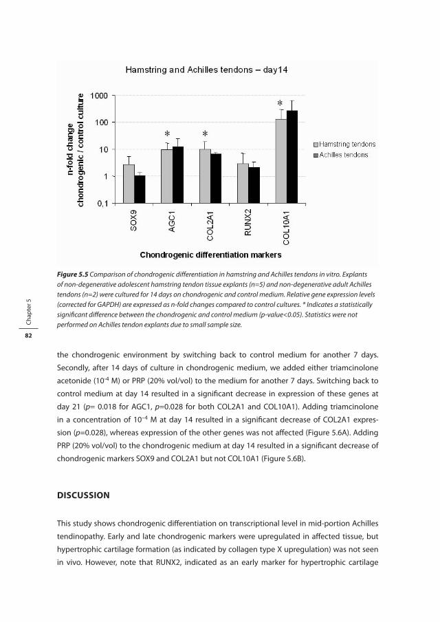
Chap
ter 5
82
the chondrogenic environment by switching back to control medium for another 7 days.
Secondly, after 14 days of culture in chondrogenic medium, we added either triamcinolone
acetonide (10-4 M) or PRP (20% vol/vol) to the medium for another 7 days. Switching back to
control medium at day 14 resulted in a significant decrease in expression of these genes at
day 21 (p= 0.018 for AGC1, p=0.028 for both COL2A1 and COL10A1). Adding triamcinolone
in a concentration of 10–4 M at day 14 resulted in a significant decrease of COL2A1 expres-
sion (p=0.028), whereas expression of the other genes was not affected (Figure 5.6A). Adding
PRP (20% vol/vol) to the chondrogenic medium at day 14 resulted in a significant decrease of
chondrogenic markers SOX9 and COL2A1 but not COL10A1 (Figure 5.6B).
DisCussion
This study shows chondrogenic differentiation on transcriptional level in mid-portion Achilles
tendinopathy. Early and late chondrogenic markers were upregulated in affected tissue, but
hypertrophic cartilage formation (as indicated by collagen type X upregulation) was not seen
in vivo. However, note that RUNX2, indicated as an early marker for hypertrophic cartilage
Figure 5.5 Comparison of chondrogenic differentiation in hamstring and Achilles tendons in vitro. Explants of non-degenerative adolescent hamstring tendon tissue explants (n=5) and non-degenerative adult Achilles tendons (n=2) were cultured for 14 days on chondrogenic and control medium. Relative gene expression levels (corrected for GAPDH) are expressed as n-fold changes compared to control cultures. * Indicates a statistically significant difference between the chondrogenic and control medium (p-value<0.05). Statistics were not performed on Achilles tendon explants due to small sample size.
Figure 5.5 Comparison of chondrogenic differentiation in hamstring and Achilles tendons in vitro. Explants of non-degenerative adolescent hamstring tendon tissue explants (n=5) and non-degenerative adult Achilles tendons (n=2) were cultured for 14 days on chondrogenic and control medium. Relative gene expression levels (corrected for GAPDH) are expressed as n-fold changes compared to control cultures. * Indicates a statistically significant difference between the chondrogenic and control medium (p-value<0.05). Statistics were not performed on Achilles tendon explants due to small sample size.

In-vitro model to study chondrogenic differentiation in tendinopathy 83
formation, was upregulated in the affected tissue. In an in-vitro model simulating this chon-
drogenic tendon differentiation, the early chondrogenic marker SOX9 was not upregulated
anymore, whereas besides collagen type II also the hypertrophic marker collagen type X was
upregulated in both tendon types. This suggests that the model in its current state may slightly
overstimulate the cells towards a hypertrophic chondrogenic lineage compared to the in-vivo
situation. However, all upregulated chondrogenic markers (including collagen type X) could still
be significantly downregulated by removing the chondrogenic stimulus. Importantly, we also
showed that chondrogenic differentiation of tendon cells could be modulated by addition of a
biochemical signal in the presence of the chondrogenic environment. The model can therefore
be used to test substances for their potentially therapeutic role in controlling chondrogenic
differentiation in tendinopathy.
Figure 5.6 Modulation of chondrogenic differentiation in vitro. Tendon explants were maintained for 14 days on chondrogenic medium. (A) At day 14, the chondrogenic medium was switched to control medium, or triamcinolone acetonide (10–4 M) was added to the chondrogenic medium (n=1 adolescent hamstring tendon in duplo and n=2 adult Achilles tendons in duplo were pooled for statistical analysis). (B) At day 14, the chondrogenic medium was switched to control medium, or PRP (20% vol/vol) was added to the chondrogenic medium (n=3 adolescent hamstring tendons in duplo were pooled for statistical analysis). Relative gene expression levels (corrected for GAPDH) are expressed as n-fold changes compared to control cultures. * Indicates a statistically significant difference between two conditions (p-value<0.05). # Indicates a statistically significant difference between the condition and 21 days on control medium (p-value<0.05).
Figure 5.6 Modulation of chondrogenic differentiation in vitro. Tendon explants were maintained for 14 days on chondrogenic medium. (A) At day 14, the chondrogenic medium was switched to control medium, or triamcinolone acetonide (10–4 M) was added to the chondrogenic medium (n=1 adolescent hamstring tendon in duplo and n=2 adult Achilles tendons in duplo were pooled for statistical analysis). (B) At day 14, the chondrogenic medium was switched to control medium, or PRP (20% vol/vol) was added to the chondrogenic medium (n=3 adolescent hamstring tendons in duplo were pooled for statistical analysis). Relative gene expression levels (corrected for GAPDH) are expressed as n-fold changes compared to control cultures. * Indicates a statistically significant difference between two conditions (p-value<0.05). # Indicates a statistically significant difference between the condition and 21 days on control medium (p-value<0.05).

Chap
ter 5
84
We used both healthy adolescent hamstring tendons and healthy adult Achilles tendons and
found no striking difference between these two tissue sources regarding their behavior in the
model. Without stating that chondrogenic differentiation capacity is independent of age or
tissue source, we think our results justify the use of easily available non-degenerative adoles-
cent hamstring tendons as a standard tissue source for the model. This is especially interesting
because it may often be difficult to obtain large amounts of non-degenerative rotator cuff,
elbow, and Achilles tendon tissue In this context, it might even be interesting to test the ben-
efits of an animal tissue source for the experimental model.
Three main theories exist about the origin of cells that play a role in fibrocartilaginous meta-
plasia in vivo. Firstly, differentiated cells may change their pattern of differentiation in a process
termed transdifferentiation. Secondly, local progenitor cells may become reprogrammed by
changing molecular or biomechanical signals. Thirdly, circulating stem cells may be recruited
from the blood into the tissue and develop into a chondrocyte-like phenotype. Recently, evi-
dence for the presence of cells with multilineage potential in tendon tissue has been collected
by several groups. Salingcarnboriboon et al. [22] showed that single cell clones from murine
tendon cell lines could differentiate towards multiple mesenchymal lineages upon culture in
appropriate differentiation media. In-vitro differentiation experiments from our group have
shown that third to fifth passage tendon-derived fibroblasts explanted from adolescent ham-
string tendons, have a differentiation capacity similar to that of adult bone marrow-derived
stromal cells [19]. Our chondrogenic differentiation model suggests that at least part of the
resident cells, either local progenitor cells or local differentiated cells, are involved. It cannot be
excluded, however, that in the in-vivo situation, also circulating progenitor cells are recruited
from the periphery.
Our findings indicate that in mid-portion Achilles tendinopathy at least part of the tendon
cells deviate in their metabolism towards fibrocartilaginous matrix production. However, what
can cause this chondrogenic differentiation? Mid-portion Achilles tendinopathy has histori-
cally been attributed to tensional overload, but the unique anatomical structure of the triceps
surae muscle-tendon unit, being a fusion of several muscles bodies into one tendon, might
also implicate a role for compressional overload in two ways. Firstly, the tertiairy bundles in
the Achilles tendon twist approximately 90 degrees around the tendon’s longitudinal axis, the
largest part of this twist taking place in the mid-portion area [23]. During tensional loading, the
mid-portion may experience torsional compressive and shearing forces (similar to wringing
out a towel) [23, 24]. Secondly, individual force contributions of the triceps surae components
cause non-uniform stress in the Achilles tendon, which may result in frictional and compressive
forces between tendon fibres [25]. Anatomical variations and exercise level, both influencing
the magnitude and duration of these intratendinous forces, may predispose to develop a
lesion. Apart from mechanical factors, also biochemical changes in the environment of the cells
can exert an effect on chondrogenic differentation. Growth factors, like TGF-ß in this study, can
influence chondrogenic differentiation. In mid-portion Achilles tendinopathy, such biochemical

In-vitro model to study chondrogenic differentiation in tendinopathy 85
signals could be due to ageing, inflammatory processes, wound healing processes, or mechani-
cal alterations. In summary, the pathways involved in tensional mechanical overuse leading to
microdamage of the fibrils rendering them inactive and causing tensional stress deprivation
of tenocytes [26] might act simultaneously or consecutively with the pathways involved in
compressional mechanical overload, possibly even enhancing eachother, ultimately leading to
cumulative tendon damage and degeneration.
In an in-vitro culture system to simulate chondrogenic differentiation in tendon explants,
we applied TGF-β as chondrogenic stimulus in a stress-deprived culture condition. TGF-β is a
well accepted chemical substance to induce chondrogenic differentiation in stem cell research,
even without loading [27]. Addition of TGF-ß can be done easily in a (high-throughput) model
system. Furthermore, an increase in TGF-β on protein level has been demonstrated in Achilles
and patellar tendinopathy [28, 29], which is confirmed on transcriptional level in the present
study. Also mechanical loading effects on chondrogenic behavior have been demonstrated to
be dependent on TGF-β mediated pathways [30-32]. New insights on the role of stress depriva-
tion in the development of tendinopathy provide good reason for the use of a stress deprived
culture condition in a model for tendinopathy [33]. However, caution is required to extrapolate
data derived from the model directly to the in-vivo situation.
The outcome parameters in our model were restricted to analyses on transcriptional regula-
tion level, which makes the model useful to detect early cellular responses in relatively short
term experiments and creates the possibility for high throughput screening. Follow-up with
longer culture periods would be necessary to detect changes in matrix composition using
histology and biochemistry. Furthermore, the model is intended for testing of early interven-
tions. Advanced degenerative tissue or cartilage tissue formation can very likely not be treated
with drugs. In this model we intended to simulate chondrogenic celldifferentiation that is still
reversible with drug treatment.
Triamcinolone was studied as a first example to see whether the chondrogenic tendency
could be influenced by adding a biochemical substance. Triamcinolone, and glucocorticoids in
general, have been studied in the light of chondrogenic differentiation before. Glucocorticoids
appear to promote chondrogenic differentiation in general [34-36]; on the other hand, triam-
cinolone acetonide led to a decrease in the proliferative rate of chondroprogenitor cells along
with an inhibition of the chondrogenic differentiation pathway in an in-vitro study with neonatal
mouse cartilage explants [37]. Therefore, triamcinolone was expected to exert a certain effect
on the chondrogenic differentiation state of tendon cells (either stimulatory or inhibitory). In
our model, triamcinolone acetonide successfully downregulated COL2A1 expression, but had
no effect on any of the other chondrogenic markers. As a second example we tested the effect
of platelet-rich plasma, a promising new substance in the treatment of tendinopathy [18, 38],
in our model. In autologous PRP applications, the effects thus far have been attributed to the
presence of high concentrations of multiple growth factors and cytokines that promote wound
healing by stimulating cell migration, proliferation, collagen production, and differentiation

Chap
ter 5
86
[38-41]. In this experiment, heterologous platelet-rich plasma appeared to decrease the chon-
drogenic differentiation state of tendon explants. However, the exact working mechanism
remains to be elucidated.
In conclusion, we have shown that chondrogenic differentiation is present in mid-portion
Achilles tendinopathy. Our study also demonstrates the use of an in-vitro model to investigate
early chondrogenic differentiation as a possible target for drug treatment of tendinopathic
lesions.
aCknowleDgeMenTs
The authors would like to thank the orthopedic surgeons A.F. Diepstraten, M.P. Heijboer, J.H.J.M.
Bessems, and E.R.A. van Arkel for providing the tendon tissue, and H.Weinans, and A.E. van der
Windt for their technical support and interest in this work. The antibody II-II6B3 was obtained
from the Developmental Studies Hybridoma Bank, under contract N01-HD-6–2915 from the
NICHD.
ReFeRenCes
1. Maffulli, N., Kader, D. (2002). Tendinopathy of tendo achillis. J Bone Joint Surg Br. 84, 1-8. 2. Wilson, J. J., Best, T. M. (2005). Common overuse tendon problems: A review and recommendations
for treatment. Am Fam Physician. 72, 811-8. 3. Pufe, T., Petersen, W. J., Mentlein, R., Tillmann, B. N. (2005). The role of vasculature and angiogenesis for
the pathogenesis of degenerative tendons disease. Scand J Med Sci Sports. 15, 211-22. 4. Wang, J. H., Iosifidis, M. I., Fu, F. H. (2006). Biomechanical basis for tendinopathy. Clin Orthop Relat Res.
443, 320-32. 5. Wilson, A. M., Goodship, A. E. (1994). Exercise-induced hyperthermia as a possible mechanism for
tendon degeneration. J Biomech. 27, 899-905. 6. Almekinders, L. C., Weinhold, P. S., Maffulli, N. (2003). Compression etiology in tendinopathy. Clin
Sports Med. 22, 703-10. 7. Smith, R. K., Birch, H. L., Goodman, S., Heinegard, D., Goodship, A. E. (2002). The influence of ageing
and exercise on tendon growth and degeneration--hypotheses for the initiation and prevention of strain-induced tendinopathies. Comp Biochem Physiol A Mol Integr Physiol. 133, 1039-50.
8. Riley, G. (2004). The pathogenesis of tendinopathy. A molecular perspective. Rheumatology (Oxford). 43, 131-42.
9. Movin, T., Gad, A., Reinholt, F. P., Rolf, C. (1997). Tendon pathology in long-standing achillodynia. Biopsy findings in 40 patients. Acta Orthop Scand. 68, 170-5.
10. Tallon, C., Maffulli, N., Ewen, S. W. (2001). Ruptured Achilles tendons are significantly more degener-ated than tendinopathic tendons. Med Sci Sports Exerc. 33, 1983-90.
11. Corps, A. N., Robinson, A. H., Movin, T., Costa, M. L., Hazleman, B. L., Riley, G. P. (2006). Increased expres-sion of aggrecan and biglycan mRNA in Achilles tendinopathy. Rheumatology (Oxford). 45, 291-4.
12. Benjamin, M., Ralphs, J. R. (1998). Fibrocartilage in tendons and ligaments--an adaptation to compres-sive load. J Anat. 193 ( Pt 4), 481-94.

In-vitro model to study chondrogenic differentiation in tendinopathy 87
13. Pauwels, F. (1960). [A new theory on the influence of mechanical stimuli on the differentiation of supporting tissue. The tenth contribution to the functional anatomy and causal morphology of the supporting structure.]. Z Anat Entwicklungsgesch. 121, 478-515.
14. Maffulli, N., Reaper, J., Ewen, S. W., Waterston, S. W., Barrass, V. (2006). Chondral metaplasia in calcific insertional tendinopathy of the Achilles tendon. Clin J Sport Med. 16, 329-34.
15. Archambault, J. M., Jelinsky, S. A., Lake, S. P., Hill, A. A., Glaser, D. L., Soslowsky, L. J. (2007). Rat supraspi-natus tendon expresses cartilage markers with overuse. J Orthop Res. 25, 617-24.
16. See, Y. (1998). Intra-synovial corticosteroid injections in juvenile chronic arthritis--a review. Ann Acad Med Singapore. 27, 105-11.
17. Astrom, M., Rausing, A. (1995). Chronic Achilles tendinopathy. A survey of surgical and histopatho-logic findings. Clin Orthop Relat Res. 151-64.
18. Mishra, A., Pavelko, T. (2006). Treatment of chronic elbow tendinosis with buffered platelet-rich plasma. Am J Sports Med. 34, 1774-8.
19. de Mos, M., Koevoet, W. J., Jahr, H., Verstegen, M. M., Heijboer, M. P., Kops, N., et al. (2007). Intrinsic differentiation potential of adolescent human tendon tissue: an in-vitro cell differentiation study. BMC Musculoskelet Disord. 8, 16.
20. de Mos, M., van El, B., Degroot, J., Jahr, H., van Schie, H. T., van Arkel, E. R., et al. (2007). Achilles Tendi-nosis: Changes in Biochemical Composition and Collagen Turnover Rate. Am J Sports Med.
21. Livak, K. J., Schmittgen, T. D. (2001). Analysis of relative gene expression data using real-time quantita-tive PCR and the 2(-Delta Delta C(T)) Method. Methods. 25, 402-8.
22. Salingcarnboriboon, R., Yoshitake, H., Tsuji, K., Obinata, M., Amagasa, T., Nifuji, A., et al. (2003). Estab-lishment of tendon-derived cell lines exhibiting pluripotent mesenchymal stem cell-like property. Exp Cell Res. 287, 289-300.
23. O’Brien, M. (1992). Functional anatomy and physiology of tendons. Clin Sports Med. 11, 505-20. 24. Clement, D. B., Taunton, J. E., Smart, G. W. (1984). Achilles tendinitis and peritendinitis: etiology and
treatment. Am J Sports Med. 12, 179-84. 25. Arndt, A. N., Komi, P. V., Bruggemann, G. P., Lukkariniemi, J. (1998). Individual muscle contributions to
the in vivo achilles tendon force. Clin Biomech (Bristol, Avon). 13, 532-541. 26. Arnoczky, S. P., Lavagnino, M., Egerbacher, M. (2007). The mechanobiological aetiopathogenesis of
tendinopathy: is it the over-stimulation or the under-stimulation of tendon cells? Int J Exp Pathol. 88, 217-26.
27. Caterson, E. J., Nesti, L. J., Danielson, K. G., Tuan, R. S. (2002). Human marrow-derived mesenchymal progenitor cells: isolation, culture expansion, and analysis of differentiation. Mol Biotechnol. 20, 245-56.
28. Fenwick, S. A., Curry, V., Harrall, R. L., Hazleman, B. L., Hackney, R., Riley, G. P. (2001). Expression of transforming growth factor-beta isoforms and their receptors in chronic tendinosis. J Anat. 199, 231-40.
29. Fu, S. C., Wang, W., Pau, H. M., Wong, Y. P., Chan, K. M., Rolf, C. G. (2002). Increased expression of trans-forming growth factor-beta1 in patellar tendinosis. Clin Orthop Relat Res. 174-83.
30. Heinemeier, K., Langberg, H., Kjaer, M. (2003). Exercise-induced changes in circulating levels of transforming growth factor-beta-1 in humans: methodological considerations. Eur J Appl Physiol. 90, 171-7.
31. Ohno, S., Tanaka, N., Ueki, M., Honda, K., Tanimoto, K., Yoneno, K., et al. (2005). Mechanical regulation of terminal chondrocyte differentiation via RGD-CAP/beta ig-h3 induced by TGF-beta. Connect Tissue Res. 46, 227-34.
32. Terraciano, V., Hwang, N., Moroni, L., Park, H. B., Zhang, Z., Mizrahi, J., et al. (2007). Differential Response of Adult and Embryonic Mesenchymal Progenitor Cells to Mechanical Compression in Hydrogels. Stem Cells.

Chap
ter 6
88
33. Arnoczky, S. P., Lavagnino, M., Egerbacher, M., Caballero, O., Gardner, K. (2007). Matrix Metalloprotei-nase Inhibitors Prevent a Decrease in the Mechanical Properties of Stress-Deprived Tendons: An In Vitro Experimental Study. Am J Sports Med.
34. Derfoul, A., Perkins, G. L., Hall, D. J., Tuan, R. S. (2006). Glucocorticoids promote chondrogenic differ-entiation of adult human mesenchymal stem cells by enhancing expression of cartilage extracellular matrix genes. Stem Cells. 24, 1487-95.
35. Tanaka, H., Murphy, C. L., Murphy, C., Kimura, M., Kawai, S., Polak, J. M. (2004). Chondrogenic differ-entiation of murine embryonic stem cells: effects of culture conditions and dexamethasone. J Cell Biochem. 93, 454-62.
36. Grigoriadis, A. E., Aubin, J. E., Heersche, J. N. (1989). Effects of dexamethasone and vitamin D3 on cartilage differentiation in a clonal chondrogenic cell population. Endocrinology. 125, 2103-10.
37. Weiss, A., Livne, E., Silbermann, M. (1988). Glucocorticoid hormone adversely affects the growth and regeneration of cartilage in vitro. Growth Dev Aging. 52, 67-75.
38. Schnabel, L. V., Mohammed, H. O., Miller, B. J., McDermott, W. G., Jacobson, M. S., Santangelo, K. S., et al. (2007). Platelet rich plasma (PRP) enhances anabolic gene expression patterns in flexor digitorum superficialis tendons. J Orthop Res. 25, 230-40.
39. van den Dolder, J., Mooren, R., Vloon, A. P., Stoelinga, P. J., Jansen, J. A. (2006). Platelet-rich plasma: quantification of growth factor levels and the effect on growth and differentiation of rat bone marrow cells. Tissue Eng. 12, 3067-73.
40. Celotti, F., Colciago, A., Negri-Cesi, P., Pravettoni, A., Zaninetti, R., Sacchi, M. C. (2006). Effect of platelet-rich plasma on migration and proliferation of SaOS-2 osteoblasts: role of platelet-derived growth factor and transforming growth factor-beta. Wound Repair Regen. 14, 195-202.
41. Gruber, R., Karreth, F., Kandler, B., Fuerst, G., Rot, A., Fischer, M. B., et al. (2004). Platelet-released supernatants increase migration and proliferation, and decrease osteogenic differentiation of bone marrow-derived mesenchymal progenitor cells under in vitro conditions. Platelets. 15, 29-35.

Chapter 6
Can platelet-rich plasma enhance tendon repair: a cell culture study
M. de Mos, A.E. van der Windt, H. Jahr, H.T.M. van Schie, H. Weinans, J.A.N. Verhaar, G.J.V.M. van Osch
Am J Sports Med Jun;36(6):1171-8, 2008

Chap
ter 6
90
aBsTRaCT
Background
Autologous platelet-rich plasma (PRP) application appears to improve tendon healing in trau-
matic tendon injuries, but basic knowledge on how PRP promotes tendon repair is needed.
hypothesis
PRP has a positive effect on cell proliferation and collagen production, and induces the produc-
tion of matrix-degrading enzymes and endogenous growth factors by human tenocytes.
Methods
Human tenocytes were cultured 14 days in 2% fetal calf serum medium complemented with
0%, 10%, or 20% vol/vol platelet-rich clot releasate (PRCR, the active releasate of PRP) or
platelet-poor clot releasate (PPCR). At day 4, 7, and 14, cell amount, total collagen, and gene
expression of collagen Iα1 (COL1) and IIIα1 (COL3), matrix metalloproteinases (MMP1, MMP3,
MMP13), vascular endothelial-derived growth factor A (VEGF-A) and transforming growth fac-
tor β1 (TGFβ1) were analysed.
Results
Platelet numbers in PRP increased to 2.55 times baseline. Growth-factor concentrations of VEGF
and PDGF-BB were higher in PRCR than PPCR. Both PRCR and PPCR increased cell number and
total collagen, whereas they decreased gene expression of COL1 and COL3 without affecting
COL3/COL1 ratio. PRCR, but not PPCR, showed upregulation of MMP1 and MMP3 expression.
MMP13 expression was not altered by either treatment. PRCR increased VEGF-A expression at
all timepoints and TGFβ1 expression at day 4.
Conclusions
In human tenocyte cultures PRCR, but also PPCR, stimulates cell proliferation, total collagen
production. PRCR, but not PPCR, slightly increases the expression of matrix degrading enzymes
and endogenous growth factors.
Clinical Relevance
In-vivo use of PRP, but also of PPP to a certain extent, in tendon injuries might accelerate the
catabolic demarcation of traumatically injured tendon matrix and promote angiogenesis and
formation of a fibro-vascular callus. Whether this will also be beneficial for degenerative tendi-
nopathies remains to be elucidated.

Can platelet-rich plasma enhance tendon repair: a cell culture study 91
inTRoDuCTion
Traumatic tendon injuries and tendinopathies are a growing problem in sports medicine and
orthopedic practice [4, 17]. Most tendons have the ability to heal after injury, but the repair
tissue is functionally inferior to normal tendon tissue and is accompanied by increased risk of
further injury [25]. The poor vascularisation seems to be a major reason for this limited healing
capacity [14, 28]. Treatment of tendon lesions, either primary traumatic or degenerative tendi-
nopathies, is often hampered by contradictory descriptions of the underlying pathology, with a
limited repertoire of successful and evidence-based treatments [31]. New treatment strategies,
such as the use of platelet-rich plasma (PRP), might improve healing.
Clinical applications of autologous PRP in human medicine include periodontal and maxil-
lofacial surgery, plastic surgery, treatment of bone fractures, and treatment of chronic skin and
soft tissue ulcers [3, 10, 15, 20, 23, 27]. Numerous publications on PRP yielded excellent clinical
outcomes [3, 10, 15, 20, 23, 27, 32]. The only published cohort study in tendon research reported
93% reduction of pain for PRP-treated patients with chronic elbow tendinosis [24]. However,
this was a pilot study with a small number of patients and without randomized control group.
Platelets actively participate in healing processes in the body [4]. Platelets contain different
growth factors, such as PDGF [4], TGFβ [4, 27], IGF [27], EGF [27], VEGF [27], and FGF [27], which
are released from their α-granules upon platelet activation and delivered to the injured site
to facilitate healing [4]. Platelet-rich plasma is, by clinical definition, a volume fraction of the
plasma, having a platelet concentration above baseline (whole blood) [27]. In activated PRP,
compared to activated whole blood, significant increases of growth factors can be observed,
e.g. VEGF (6.2-fold), PDGF-BB (5.1-fold), EGF (3.9-fold), and TGFβ1 (3.6-fold) [12]. Specific roles
of growth factors in tendon and ligament healing have been studied before. PDGF, peaking
shortly after tendon damage, plays a central role in the healing process [12, 25] by chemotaxis,
proliferation of fibroblasts, collagen synthesis, and the stimulation of TGFβ1 and VEGF. TGFβ1
increases collagen production and cell viability [25]. VEGF is a powerful stimulator of angiogen-
esis [25].
To summarize, platelets rapidly release a variety of growth factors and PRP might provide
an autologous source of these growth factors that play a key role in tendon repair mechanisms
[25]. Not only controlled clinical studies but even more in-vitro studies are required to investi-
gate in detail the effects of PRP on human tendon cell metabolism.
In in-vitro tendon research, the effects of culturing equine flexor digitorum superficialis ten-
don explants with 100% PRP (vol/vol) and other blood products were examined [30]. Enhanced
anabolic gene expression patterns (COL1A1, COL3A1 and COMP), with no concomitant increase
in catabolic genes (MMP3 and MMP13), after 3 days of 100% PRP (vol/vol) treatment were
reported. The only study with human tendon cells (tenocytes) cultured in 20% PRP (vol/vol)
reported an increase in cell proliferation and in VEGF and HGF production [4]. Effects of PRP on
collagen production and degradation of human tenocyte cultures remain to be elucidated.

Chap
ter 6
92
The purpose of this study was to investigate the effects of releasates from 10% (vol/vol) and
20% (vol/vol) platelet-rich and platelet-poor plasma on human tenocytes in culture. We exam-
ined whether platelet-rich plasma releasate affects cell proliferation, collagen production, and
production of matrix-degrading enzymes and endogenous growth factors by human tendon
cells.
MaTeRials anD MeThoDs
isolation of tendon cells
Human tendon-derived cells were explanted from hamstring tendon tissue of three children
(age 13-15 years) undergoing hamstring-tendon release for treatment of knee-contractures.
Approval was obtained from the Medical Ethical Committee of the Erasmus MC, University
Medical Center Rotterdam. After removal of peritendineum, the tendon was cut into 3 mm3
sections, transferred into six-well plates (Corning, NY, USA) and cultured in expansion medium
(Dulbecco’s modified Eagle’s medium, 10% FCS, 50 μg/ml gentamycin and 1.5 μg/ml fungizone
(all Invitrogen, Scotland, UK)). Cultures were maintained at 37˚C in humidified atmosphere of
5% CO2 for ten days. During this time, fibroblasts migrated out of the tissue and adhered to the
bottom of the culture dish. Cells were subcultured and trypsinized at subconfluency. Cells from
fifth and sixth passage were used.
Plasma preparations
After informed consent, whole blood (500 ml) from nine healthy, male donors (median age 47
years, range 31-69 years) was collected in 70 ml of anticoagulants (citrate-phosphate-dextrose
[CPD], Sanquin Blood Supply Foundation) and processed within 24 hours as a non-autologous
source of platelets. None of the donors used medication that is known to influence platelet
function. Processing protocols for platelet-rich plasma (PRP) and platelet-poor plasma (PPP)
were adopted from literature [4-6] and tested in different combinations. The best combination
of these protocols (with highest platelet counts and lowest blood cell counts) was used in this
study.
Briefly, whole blood was centrifuged at 300xg for 10 minutes. The supernatant was centri-
fuged at 4500xg for 12 minutes, to obtain a superficial layer of PPP. The buffy coat of the first cen-
trifugation was centrifuged at 300xg for 10 minutes again to separate it into PRP (supernatant)
and erythrocytes and leucocytes (bottom layer). In order to increase the platelet concentration,
PRP was then centrifuged at 480xg for 20 min to precipitate the platelets. Half the superficial
plasma layer was removed and the platelet pellet was suspended in the remaining half of the
plasma volume. Clotting upon addition of 22.8 mM CaCl2 at 37°C for 1 hr activated platelets
to release their growth factors. The soluble releasate from the clotted preparations (PRCR and
PPCR), containing growth factors, was aspirated, stored at 4°C and used within two weeks.

Can platelet-rich plasma enhance tendon repair: a cell culture study 93
Concentrations of platelets and red and white blood cells were measured on ABC Animal Blood
Counter (Scil, Viernheim, Germany) in samples of whole blood, PRP and PPP (before clotting),
and PRCR and PPCR (after clotting). For growth factor measurements, 1 ml of freshly prepared
PPCR and PRCR was collected separately and immediately centrifuged at 1000xg for 10 minutes
at 4˚C, and stored at –80˚C until further use. Growth factor concentrations in PRCR and PPCR
were measured in triplicate using commercially available sandwich ELISA kits for VEGF and
PDGF-BB (both R&D systems, Abingdon, UK) according to manufacturers protocol.
Cell culture experiment
Trypsinized tenocytes were plated at a density of 4,000 cells/cm2 and maintained in 10%
FCS for 24 hours prior to replacement by medium with 2% FCS and 0.1 mM L-ascorbic acid
2-phosphate (Sigma-Aldrich, St. Louis, Missouri, USA), with or without (a) PPCR 10% (vol/vol),
(b) PRCR 10% (vol/vol) (c) PPCR 20% (vol/vol), or (d) PRCR 20% (vol/vol). Cells were cultured at
37˚C in humidified atmosphere of 5% CO2 for 14 days. Medium was refreshed at day 4, 7 and 11.
At day 4, 7 and 14, amount of cells (DNA assay), total collagen (hydroxyproline assay), and gene
expression of collagen types Iα1 (COL1) and IIIα1 (COL3), matrix metalloproteinases (MMP1,
MMP3, and MMP13), vascular endothelial-derived growth factor A (VEGF-A) and transforming
growth factor β1 (TGFβ1) were analysed.
Dna assay
Cells were suspended in 0.1% phosphate buffered saline (PBS) / Triton X-100 (Sigma-Aldrich,
St. Louis, USA). Samples were sonificated for 10 seconds, incubated with 200 μl of 8.3 IU/ml
heparin solution (LEO Pharma BV, Breda, Netherlands) and 100 μl of 0.05 mg/ml ribonuclease A
for 30 min at 37˚ C. This was followed by adding 100 μl ethidium bromide solution (25 μg/ml)
(Sigma-Aldrich). Quantification of incorporated dye was performed in triplicate on the Wallac
1420 victor2 (PerkinElmer, Wellesley, MA) using an extinction filter of 340 nm and an emission
filter of 590 nm [11]. For standards, calf thymus DNA (Sigma) was used.
hydroxyproline assay
Cells were suspended in milli-Q, hydrolyzed at 108°C for18-20 hours in 6 M HCl, dried and redis-
solved in 100 µL water. Hydroxyproline contents were measured by colorimetric method [8]
(extinction 570 nm), with chloramine-T and dimethylaminobenzaldehyde (DMBA) as reagentia
and hydroxyproline as standard (all Merck, Damstadt, Germany).
gene expression analysis
Cells were suspended in RNA-BeeTM (TEL-TEST, Friendswood, TX, USA). Downstream processing
and real-time PCR is described elsewhere [9]. Briefly, RNA was purified using RNeasy® Micro Kit
(Qiagen, Hilden, Germany), and 1 µg of total RNA of each sample was reverse-transcribed into
cDNA using RevertAidTM First Strand cDNA Synthesis Kit (MBI Fermentas, St. Leon-Rot, Germany).

Chap
ter 6
94
Primers were designed to meet Taqman® or SYBR®Green requirements and ensure gene speci-
ficity. COL1 [9] and COL3 [9] were studied as markers for collagen production. MMP1 [9], MMP3
[9] and MMP13 [9] were used as indicators of collagen degradation. Also, gene expression of
growth factor VEGF-A (F:5’-CTTGCCTTGCTGCTCTACC-3’; R:5’-CACACAGGATGGCTTGAAG-3’)
and TGFβ1 (F:5’-GTGACAGCAGGGATAACACACTG -3’; R:5’-CATGAA TGGTGGCCAGGTC-3’;
PROBE:5’-ACATCAACGGGTTCACTACCGGC-3’) were assessed. Amplifications were performed as
20 µL reactions using TaqMan® Universal PCR MasterMix (ABI, Branchburg, NJ, USA) or qPCRTM
Mastermix Plus for SYBR® Green I (Eurogentec, Maastricht, Netherlands) according to the
manufacturer’s guidelines on an ABI PRISM® 7000 with SDS software version 1.7. Data were nor-
malized to 18SrRNA (F:5’-AGTCCCTGCCCTTTGTACACA-3’; R:5’-GATCCGAGGGCCTCACTAAAC-3’;
PROBE:5’-CGCCCGTCGCTACTACCGATTGG-3’), which was stably expressed across samples (not
shown). Relative expression was calculated according to 2-ΔCT method [21].
statistical analysis
The experiment was performed in duplicate for all three donor explants (n=6). 2% FCS condition
without PRCR or PPCR was used as control and was set to 1 at each timepoint. All conditions
were expressed as n-fold difference from the control at the corresponding timepoint. Statistical
analysis was performed using SPSS 13.0 software (SPSS Inc., Chicago, IL, USA). A Kruskall-Wallis
H test and post-hoc Dunn’s multiple comparison test was used. p<0.05 was considered to indi-
cate statistically significant differences.
ResulTs
Plasma preparations
Whole blood baseline platelet concentrations were all within physiological range (119 x106
to 195 x106 platelets/ml) (Table 6.1). The platelet concentration procedure increased platelet
numbers in the PRP group on average to 2.55 times baseline concentration in whole blood.
The PPP group showed an average decrease in platelet number to 0.02 times baseline. After
clotting PRP and PPP, platelet numbers decreased respectively to 0.08 and 0.05 times baseline.
In addition, the centrifugation and clotting procedure decreased white blood cell counts to
0.02 and red blood cell counts to 0.01 in PRCR, and in PPCR to respectively 0.02 and 0.00 times
baseline.
VEGF concentration in PPCR was below the detection limit of 20 pg/ml in all preparations.
On the other hand, VEGF concentration in PRCR was 107 ± 83 pg/ml (mean ± SD). PDGF-BB
concentration in PPCR was 123 ± 151 pg/ml (mean ± SD) and in PRCR 3114 ± 2709 pg/ml (mean
± SD).
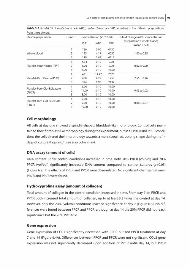
Can platelet-rich plasma enhance tendon repair: a cell culture study 95
Cell morphology
All cells at day one showed a spindle-shaped, fibroblast-like morphology. Control cells main-
tained their fibroblast-like morphology during the experiment, but in all PRCR and PPCR condi-
tions the cells altered their morphology towards a more stretched, oblong shape during the 14
days of culture (Figure 6.1, see also color inlay).
Dna assay (amount of cells)
DNA content under control conditions increased in time. Both 20% PRCR (vol/vol) and 20%
PPCR (vol/vol) significantly increased DNA content compared to control cultures (p<0.05)
(Figure 6.2). The effects of PRCR and PPCR were dose-related. No significant changes between
PRCR and PPCR were found.
hydroxyproline assay (amount of collagen)
Total amount of collagen in the control condition increased in time. From day 7 on PRCR and
PPCR both increased total amount of collagen, up to at least 3.3 times the control at day 14.
However, only the 20% (vol/vol) conditions reached significance at day 7 (Figure 6.3). No dif-
ferences were found between PRCR and PPCR, although at day 14 the 20% PPCR did not reach
significance but the 20% PRCR did.
gene expression
Gene expression of COL1 significantly decreased with PRCR but not PPCR treatment at day
7 and 14 (Figure 6.4A). Differences between PRCR and PPCR were not significant. COL3 gene
expression was not significantly decreased upon addition of PPCR untill day 14, but PRCR
Table 6.1 Platelet (PLT), white blood cell (WBC), and red blood cell (RBC) numbers in five different preparations from three donors.Plasma preparation Donor Concentration (x106 / ml) n-fold change in PLT concentration
(preparation / whole blood)(mean ± SD)PLT WBC RBC
Whole blood123
186195119
5.004.173.63
443040504313
1.00 ± 0.25
Platelet-Poor Plasma (PPP)123
4.335.003.00
0.100.100.10
0.000.00
10.000.02 ± 0.00
Platelet-Rich Plasma (PRP)123
451488324
14.474.278.90
257017502437
2.55 ± 0.16
Platelet-Poor Clot Releasate (PPCR)
123
6.0011.008.00
0.100.100.10
10.0010.0010.00
0.05 ± 0.02
Platelet-Rich Clot Releasate (PRCR)
123
7.007.00
19.00
0.100.100.10
10.0010.0090.00
0.08 ± 0.07

Chap
ter 6
96
Fig. 6.1 Photomicropgraphs of tenocyte cultures with or without PRCR or PPCR. Representative photomicrographs (200x magnification) are shown of control, 20% PPCR, and 20% PRCR conditions on day 1, 4, and 14 of the experimental culture period.
Fig. 6.1 Photomicropgraphs of tenocyte cultures with or without PRCR or PPCR. Representative photomicrographs (200x magnification) are shown of control, 20% PPCR, and 20% PRCR conditions on day 1, 4, and 14 of the experimental culture period. (See color inlay for a full color version of this figure.)
Fig. 6.2 DNA content of tenocyte cultures with or without PRCR or PPCR. Cells were harvested at day 4, 7, and 14. Control cultures (2% FCS) are set to 1 at each timepoint. DNA content for each condition is expressed as n-fold difference from control cultures at the corresponding timepoint. Results represent mean ± SD (n=6). * p<0.05 as compared to control; # p<0.05 comparison between two specified groups.
Fig. 6.2 DNA content of tenocyte cultures with or without PRCR or PPCR. Cells were harvested at day 4, 7, and 14. Control cultures (2% FCS) are set to 1 at each timepoint. DNA content for each condition is expressed as n-fold difference from control cultures at the corresponding timepoint. Results represent mean ± SD (n=6). * p<0.05 as compared to control; # p<0.05 comparison between two specified groups.

Can platelet-rich plasma enhance tendon repair: a cell culture study 97
Fig. 6.3 Total amount of collagen synthesized by tenocytes cultured with or without PPCR or PRCR. Collagen was measured at day 4, 7, and 14. Total amount of collagen for each condition is expressed as n-fold difference from control cultures at the corresponding timepoint. Control is set at 1 and results represent mean ± SD (n=6). * p<0.05 as compared to control.
Fig. 6.3 Total amount of collagen synthesized by tenocytes cultured with or without PPCR or PRCR. Collagen was measured at day 4, 7, and 14. Total amount of collagen for each condition is expressed as n-fold difference from control cultures at the corresponding timepoint. Control is set at 1 and results represent mean ± SD (n=6). * p<0.05 as compared to control.
Fig. 6.4 Gene expression levels of (A) COL1 and (B) COL3 in tenocytes cultured with or without PPCR or PRCR. Cells were harvested at day 4, 7, and 14. Gene expression was normalized to 18SrRNA and expressed as n-fold difference from control cultures at the corresponding timepoint. Control is set at 1 and results represent mean ± SD (n=6). * p<0.05 as compared to control; # p<0.05 comparison between two specified groups.
Fig. 6.4 Gene expression levels of (A) COL1 and (B) COL3 in tenocytes cultured with or without PPCR or PRCR. Cells were harvested at day 4, 7, and 14. Gene expression was normalized to 18SrRNA and expressed as n-fold difference from control cultures at the corresponding timepoint. Control is set at 1 and results represent mean ± SD (n=6). * p<0.05 as compared to control; # p<0.05 comparison between two specified groups.

Chap
ter 6
98
Fig. 6.5 Gene expression levels of (A) MMP1, (B) MMP3 and (C) MMP13 in tenocytes cultured with or without PPCR or PRCR. Cells were harvested at day 4, 7, and 14. Gene expression was normalized to 18SrRNA and expressed as n-fold difference from control cultures at the corresponding timepoint. Control is set at 1 and results represent mean ± SD (n=6). * p<0.05 as compared to control; # p<0.05 comparison between two specified groups.
Fig. 6.5 Gene expression levels of (A) MMP1, (B) MMP3 and (C) MMP13 in tenocytes cultured with or without PPCR or PRCR. Cells were harvested at day 4, 7, and 14. Gene expression was normalized to 18SrRNA and expressed as n-fold difference from control cultures at the corresponding timepoint. Control is set at 1 and results represent mean ± SD (n=6). * p<0.05 as compared to control; # p<0.05 comparison between two specified groups.

Can platelet-rich plasma enhance tendon repair: a cell culture study 99
decreased COL3 gene expression at all points in time with exception of 20% PRCR at day 4
(Figure 6.4B). The apparent difference between PRCR and PPCR treated groups at days 4 and 7
did not reach significance. COL3/COL1 ratio did not significantly change in any condition.
PRCR significantly upregulated MMP1 expression at day 7 and 14 in the 20% condition
(Figure 6.5A). The apparent dose-related respons in MMP1 expression on day 7 and 14 reached
significance only at day 14. PRCR also increased MMP3 gene expression (significant at day 4,
Figure 6.5B). No significant differences in MMP1 and MMP3 gene expression were found with
PPCR-treatment. Neither PRCR nor PPCR affected MMP13 gene expression significantly (Figure
6.5C).
PRCR highly increased VEGF-A gene expression at all time points (significantly for the 10%
PRCR condition at day 7, for the 20% PRCR condition at day 4 and 14). This increase reached
up to 30 ±14 times the control level at day 14 in the 20% PRCR condition (Figure 6.6A, note
the logarithmic scale). VEGF-A expression in PRCR-treated cells appeared higher than in
PPCR-treated cells but this difference did not reach significance. TGFβ1 gene expression was
significantly increased only at day 4 in the 20% PRCR condition (Figure 6.6B).
Fig. 6.6 Gene expression levels of (A) VEGF-A and (B) TGF 1 in tenocytes cultured with or without PPCR or PRCR. Cells were harvested at day 4, 7, and 14. Gene expression was normalized to 18SrRNA and expressed as n-fold difference from control cultures at the corresponding timepoint. Control is set at 1 and results represent the mean ± SD (n=6). VEGF expression in figure 6.5A is presented on a logarithmic scale. * p<0.05 as compared to control; # p<0.05 comparison between two specified groups.
Fig. 6.6 Gene expression levels of (A) VEGF-A and (B) TGFβ1 in tenocytes cultured with or without PPCR or PRCR. Cells were harvested at day 4, 7, and 14. Gene expression was normalized to 18SrRNA and expressed as n-fold difference from control cultures at the corresponding timepoint. Control is set at 1 and results represent the mean ± SD (n=6). VEGF expression in figure 6.5A is presented on a logarithmic scale. * p<0.05 as compared to control; # p<0.05 comparison between two specified groups.

Chap
ter 6
100
DisCussion
In this in-vitro study we tested our hypothesis that the releasate from platelet-rich plasma, PRCR,
has a positive effect on proliferation and matrix metabolism of human tendon cells to enhance
tendon repair. Our results show that PRCR, but also PPCR, enhances cell proliferation and total
collagen production by human tendon cells in culture, despite a possible decrease of collagen
production per cell. PRCR, but not PPCR, slightly increases the expression of matrix degrading
enzymes and endogenous growth factors. In vivo these effects of platelet-rich plasma, but also
of platelet-poor plasma to a certain extent, on tenocyte behavior might accelerate the catabolic
demarcation of traumatically injured tendon matrix and promote angiogenesis and the forma-
tion of a fibro-vascular callus. Whether these working mechanisms will also be beneficial in case
of degenerative tendinopathies remains to be elucidated.
To examine the effects of PRCR and PPCR on human tendon cells in culture provides an
interesting model to study the cooperative effects of a mixture of growth factors. We applied
PRCR and PPCR to our cultures in two concentrations that are most frequently used in in-vitro
studies with PRP [1, 4, 5], namely 10% and 20% (vol/vol), allowing comparison of our results
with literature [4]. We did not apply a 100% concentration of the plasma product to our cultures
[30] because we feel that this might be less comparable to the concentration of PRP reached
during in-vivo administration. Upon injection of 100% PRP into a tendon in vivo, it is unlikely
that tendon cells are exposed for more than several minutes to a 100% PRP concentration,
because the PRP will be diluted in extracellular fluids immediately after injection. However,
a major problem might be the fact that not only the platelets counts but even more so the
growth factor concentrations in the respective releasates, depending on the platelet activation,
can not be standardized, rendering comparison of experimental results rather complicated.
The only in-vitro study with human tendon cells reported that, in contrast to un-clotted
PPP, both 20% PRCR and 20% PPCR stimulated tendon cell proliferation [4]. In line with their
results, we also found that PRCR and PPCR increased cell number as well as total amount of
collagen, the latter probably being a direct consequence of increased cell numbers. COL1 and
COL3 transcripts decreased with both treatments similarly, suggesting a decrease in collagen
production per cell. The COL3 versus COL1 ratio of the tendon cells is known to shift towards
COL3 in case of tendinosis, in early stages of tendon repair, and in tendon scarring [13, 22]. No
significant changes were found in COL3/COL1 ratio in this study, suggesting no negative side
effects of PRCR and PPCR on this ratio.
Expression levels of MMP1 and MMP3 were upregulated by PRCR in some conditions, while
no significant differences were found with PPCR treatment. Neither PRCR nor PPCR changed
MMP13 expression. Gene expression of MMP1 shows no change in chronic Achilles tendinosis
[16, 18], but increases in ruptured Achilles and supraspinatus tendons [18, 19]. While expression
levels of MMP3 decreases in both degenerative and ruptured Achilles tendon [2, 16, 18, 29], in
most studies the gene expression of MMP13 doesn’t change significantly. However, conclusions

Can platelet-rich plasma enhance tendon repair: a cell culture study 101
on MMP activity based on gene expression only must be drawn with utmost precaution. With
current knowledge, it is difficult to state whether or not increased gene expression levels of
MMP1 and MMP3, as found in our experiment, will be of benefit in degenerative or ruptured
tendons. The secretion of MMPs facilitates the ingrowth of neovessels by dissolution of the
extracellular matrix [7]. Angiogenesis contributes on one hand to the repair and remodeling of
the injured tendon, but, on the other hand the proteolysis of the extracellular matrix by invad-
ing endothelial cells results in impaired mechanical stability [26]. Therefore, the application of
PRP in already degenerative tendons needs further investigation as progressive weakening of
the matrix might predispose for spontaneous rupture [19].
The amount of growth factors VEGF and HGF synthesized by the tendon cells is significantly
higher with PRCR than with PPCR treatment, as demonstrated by Anitua et al. [4]. In addition
to this, we found that PRCR highly increased VEGF-A gene expression by tendon cells. VEGF is
active after inflammation, most notably during proliferation and remodeling phases where it
has been shown to be a powerful stimulator of angiogenesis [25]. Increased VEGF expression
and concentration could be an intrinsic mechanism for inducing angiogenesis as part of a tis-
sue repair process [33]. Furthermore, TGFβ1 expression increased by PRCR at day 4. TGFβ1 is
thought to play an important role in the initial inflammatory response to tissue damage having
a positive effect on collagen production and viability of tendon cells [25].
In our experiment we aimed for a PRCR-product with the following characteristics: 1) con-
taining high platelet numbers before clotting as well as a high level of platelet activation, 2)
containing mainly growth factors released from the platelets, 3) absence of leucocytes in order
to minimize risk of graft versus host reaction in our cultures (even more for the reason that
the platelet-concentrate used in this experiment was heterologous). After the concentration
procedure, platelet numbers in PRP were 2.55 times baseline (whole blood), which corresponds
well to the platelet concentration in the study of Anitua et al., who also applicated their product
in 20% (vol/vol) concentration on human tendon cells in vitro [4]. After clotting, the platelet
numbers in PRCR decreased to 0.08 times baseline, which indicates that most platelets were
actually trapped in the clot. The higher concentrations of VEGF and PDGF-BB measured in PRCR
compared to PPCR indicate that the platelets were not only trapped in the clot but also acti-
vated to release their growth factors. A possible release of growth factors from white blood cells
scarcely affected our results, because up to 98 % of the white blood cells was already eliminated
from our preparations before clotting. This simultaneously minimized the risk of a graft versus
host reaction in our experiments.
We found that not only PRCR but also PPCR affected the outcome parameters when compared
to the control condition. This might be caused by the many unavoidable handling procedures
performed to obtain PPCR and PRCR, like drawing and centrifugating the blood which could
theoretically have activated some of the platelets leading to untimely release. In this way also
PPCR could contain a small but sufficient amount of growth factors, which might have induced
the effects that we found in PPCR conditions. However, other studies examining the difference

Chap
ter 6
102
in growth factor concentrations between whole blood, PRCR, and PPCR indicated that this is
unlikely: concentrations of TGFβ1 and PDGF-BB significantly increased in PRCR compared to
PPCR and whole blood, at least 2-fold [4, 30] and 3-fold [4, 30] respectively; in addition, growth
factor concentrations in PPCR were not significantly different from whole blood [30]. A more
plausible explanation for why both PRCR and PPCR affected the outcome parameters compared
with control conditions therefore seems to be the fact that all experimental conditions basically
consisted of adding extra serum (in the quality of 10% or 20% clot releasates) compared to the
control cultures. The higher serum concentrations could account for the higher cell amounts
in the experimental conditions, because cells proliferate faster in higher serum concentrations.
Notwithstanding the aforementioned observation that PPCR already exerted a considerable
effect on the tendon cells, particularly the effects on MMP and growth factor expression were
still more pronounced in PRCR conditions than in their equivalent PPCR conditions. These addi-
tive PRCR-effects might be attributed to higher concentrations of growth factors present in
PRCR compared to PPCR. This is supported by the results of our growth factor concentration
measurements in PPCR and PRCR.
Our tendon donors were of relatively young age, which might be a limitation of our study.
Although unlikely, it is not known whether adult tendon tissue might respond very differently
from adolescent tendon tissue regarding the effects of platelet-rich plasma on tendon cell
behavior. Also, the behavior of explanted and passaged tendon cells in an artificial culture
environment cannot be considered identical to the behavior of tendon cells in their natural
matrix environment in-vivo. Therefore one should always be cautious about translating culture
data directly to the in-vivo situation.
Finally, the platelet-rich plasma in our study was applied heterologously to the tendon cell
cultures, although in clinical settings this plasma product is usually prepared from autologous
blood. For this reason we aimed at and succeeded in minimizing the number of leucocytes so
that no graft versus host reactions occurred in our cultures.
Autologous platelet-rich plasma application appears promising in healing of traumatic ten-
don injuries and tendinopathies, but how PRP might improve or accelerate the tendon repair
process remains to be elucidated. We found that platelet-rich plasma clot releasate stimulates
cell proliferation, collagen deposition, and enhances the gene expression of matrix degrading
enzymes and endogenous growth factors by human tendon cells in vitro. This suggests that
in vivo PRP application could lead to accelerated remodeling and angiogenesis in the injured
matrix, which may promote repair of traumatic tendon injuries.
aCknowleDgeMenTs
The authors would like to thank orthopedic surgeon Ad Diepstraten for providing the tendon
tissue and Femke Verseijden for her help performing the ELISA’s.

Can platelet-rich plasma enhance tendon repair: a cell culture study 103
ReFeRenCes
1. Akeda K, An HS, Okuma M, et al. Platelet-rich plasma stimulates porcine articular chondrocyte prolif-eration and matrix biosynthesis. Osteoarthritis Cartilage. Jun 30 2006.
2. Alfredson H, Lorentzon M, Backman S, et al. cDNA-arrays and real-time quantitative PCR techniques in the investigation of chronic Achilles tendinosis. J Orthop Res. Nov 2003;21(6):970-975.
3. Anitua E. Plasma rich in growth factors: preliminary results of use in the preparation of future sites for implants. Int J Oral Maxillofac Implants. Jul-Aug 1999;14(4):529-535.
4. Anitua E, Andia I, Sanchez M, et al. Autologous preparations rich in growth factors promote prolif-eration and induce VEGF and HGF production by human tendon cells in culture. J Orthop Res. Mar 2005;23(2):281-286.
5. Annunziata M, Oliva A, Buonaiuto C, et al. In vitro cell-type specific biological response of human periodontally related cells to platelet-rich plasma. J Periodontal Res. Dec 2005;40(6):489-495.
6. Aspenberg P, Virchenko O. Platelet concentrate injection improves Achilles tendon repair in rats. Acta Orthop Scand. Feb 2004;75(1):93-99.
7. Conway EM, Collen D, Carmeliet P. Molecular mechanisms of blood vessel growth. Cardiovasc Res. Feb 16 2001;49(3):507-521.
8. Creemers LB, Jansen DC, van Veen-Reurings A, et al. Microassay for the assessment of low levels of hydroxyproline. Biotechniques. Apr 1997;22(4):656-658.
9. de Mos M, van El B, Degroot J, et al. Achilles Tendinosis: Changes in Biochemical Composition and Collagen Turnover Rate. Am J Sports Med. May 3 2007.
10. DelRossi AJ, Cernaianu AC, Vertrees RA, et al. Platelet-rich plasma reduces postoperative blood loss after cardiopulmonary bypass. J Thorac Cardiovasc Surg. Aug 1990;100(2):281-286.
11. Eijken M, Hewison M, Cooper MS, et al. 11beta-Hydroxysteroid dehydrogenase expression and glucocorticoid synthesis are directed by a molecular switch during osteoblast differentiation. Mol Endocrinol. Mar 2005;19(3):621-631.
12. Eppley BL, Woodell JE, Higgins J. Platelet quantification and growth factor analysis from platelet-rich plasma: implications for wound healing. Plast Reconstr Surg. Nov 2004;114(6):1502-1508.
13. Eriksen HA, Pajala A, Leppilahti J, et al. Increased content of type III collagen at the rupture site of human Achilles tendon. J Orthop Res. Nov 2002;20(6):1352-1357.
14. Fenwick SA, Hazleman BL, Riley GP. The vasculature and its role in the damaged and healing tendon. Arthritis Res. 2002;4(4):252-260.
15. Hee HT, Majd ME, Holt RT, et al. Do autologous growth factors enhance transforaminal lumbar inter-body fusion? Eur Spine J. Aug 2003;12(4):400-407.
16. Ireland D, Harrall R, Curry V, et al. Multiple changes in gene expression in chronic human Achilles tendinopathy. Matrix Biol. Jun 2001;20(3):159-169.
17. Jarvinen TA, Kannus P, Maffulli N, et al. Achilles tendon disorders: etiology and epidemiology. Foot Ankle Clin. Jun 2005;10(2):255-266.
18. Jones GC, Corps AN, Pennington CJ, et al. Expression profiling of metalloproteinases and tissue inhibi-tors of metalloproteinases in normal and degenerate human achilles tendon. Arthritis Rheum. Mar 2006;54(3):832-842.
19. Kannus P, Jozsa L. Histopathological changes preceding spontaneous rupture of a tendon. A con-trolled study of 891 patients. J Bone Joint Surg Am. Dec 1991;73(10):1507-1525.
20. Knighton DR, Ciresi K, Fiegel VD, et al. Stimulation of repair in chronic, nonhealing, cutaneous ulcers using platelet-derived wound healing formula. Surg Gynecol Obstet. Jan 1990;170(1):56-60.
21. Livak KJ, Schmittgen TD. Analysis of relative gene expression data using real-time quantitative PCR and the 2(-Delta Delta C(T)) Method. Methods. Dec 2001;25(4):402-408.
22. Maffulli N, Ewen SW, Waterston SW, et al. Tenocytes from ruptured and tendinopathic achilles tendons produce greater quantities of type III collagen than tenocytes from normal achilles tendons. An in vitro model of human tendon healing. Am J Sports Med. Jul-Aug 2000;28(4):499-505.

Chap
ter 6
104
23. Margolis DJ, Kantor J, Santanna J, et al. Effectiveness of platelet releasate for the treatment of diabetic neuropathic foot ulcers. Diabetes Care. Mar 2001;24(3):483-488.
24. Mishra A, Pavelko T. Treatment of chronic elbow tendinosis with buffered platelet-rich plasma. Am J Sports Med. Nov 2006;34(11):1774-1778.
25. Molloy T, Wang Y, Murrell G. The roles of growth factors in tendon and ligament healing. Sports Med. 2003;33(5):381-394.
26. Petersen W, Pufe T, Zantop T, et al. Expression of VEGFR-1 and VEGFR-2 in degenerative Achilles tendons. Clin Orthop Relat Res. Mar 2004(420):286-291.
27. Pietrzak WS, Eppley BL. Platelet rich plasma: biology and new technology. J Craniofac Surg. Nov 2005;16(6):1043-1054.
28. Pufe T, Petersen WJ, Mentlein R, et al. The role of vasculature and angiogenesis for the pathogenesis of degenerative tendons disease. Scand J Med Sci Sports. Aug 2005;15(4):211-222.
29. Riley GP, Curry V, DeGroot J, et al. Matrix metalloproteinase activities and their relationship with col-lagen remodelling in tendon pathology. Matrix Biol. Mar 2002;21(2):185-195.
30. Schnabel LV, Mohammed HO, Miller BJ, et al. Platelet rich plasma (PRP) enhances anabolic gene expression patterns in flexor digitorum superficialis tendons. J Orthop Res. Nov 14 2006.
31. Scott A, Ashe MC. Common tendinopathies in the upper and lower extremities. Curr Sports Med Rep. Sep 2006;5(5):233-241.
32. Tozum TF, Demiralp B. Platelet-rich plasma: a promising innovation in dentistry. J Can Dent Assoc. Nov 2003;69(10):664.
33. Yoshikawa T, Tohyama H, Katsura T, et al. Effects of local administration of vascular endothelial growth factor on mechanical characteristics of the semitendinosus tendon graft after anterior cruciate liga-ment reconstruction in sheep. Am J Sports Med. Dec 2006;34(12):1918-1925.

Chapter 7
General Discussion


General Discussion 107
inTRoDuCTion
Tendon injuries are a common problem in the general population as well as in athletes. The
precise aetiopathogenesis of degenerative tendinopathies is largely unknown. An abundance
of treatment options are available for tendinopathies, none of which have evidence-based
satisfactory clinical results. Patients often require lengthy rehabilitation periods or even surgical
intervention and they frequently end up with remaining limitation in their everyday life or their
sport activities.
The increasing scientific interest in tendon disorders and the concurrent application of
biochemical and molecular techniques has led to rapid developments in the understanding
of degenerative tendinopathies in the last decades. Notwithstanding the current knowledge
many questions remain unanswered yet. The work in this thesis aimed at improving fundamen-
tal knowledge of tendon cell behavior and matrix remodeling in tendinopathy, both during
the disease process as well as in reaction to treatment modalities. Thus we hope to contribute
to the increasing knowledge that will ultimately help to develop improved mechanism-based
strategies for the prevention and treatment of tendinopathy.
ChonDRogeniC DiFFeRenTiaTion in TenDinoPaThy
Healthy tendons at sites of compressive loading are more fibrocartilaginous in nature [1, 2].
In degenerative tendon disease it has been recognized that there is an accumulation of gly-
cosaminoglycans (GAGs) as well [3] and this was confirmed in this thesis in chapter 6 in which
we found an increased GAG staining on histological samples from Achilles tendinotic tissues
compared to biopsies from healthy Achilles tendon tissue. In human tendinopathy in vivo we
found that the gene expression profile in mid-portion Achilles lesions shifts towards an early
chondrogenic phenotype (chapter 6). Later on this gene expression pattern has been confirmed
in horses, describing a fibrochondrogenic differentiation pattern as the specific phenotype in
chronic tendon disease [4]. Also, in a rat model for supraspinatus tendon overuse, cDNA micro-
arrays showed seventeen cartilage specific genes upregulated in the supraspinatus tendon
following running [5]. Recent analyses corroborated our results as the gene expression pattern
of proteoglycans in tendinopathy was more similar to a chondrogenic profile [6] and human
Achilles insertional tendinopathies displayed increased collagen type II staining compared to
healthy insertion sites [7]. All this raises the question whether tendon cells have the capacity to
differentiate to other lineages and whether the fibrocartilaginous matrix composition is thus
the effect of chondrogenic differentiation of the cells present in the tendon tissue.
In vitro we were able to induce chondrogenic cell differentiation as well as adipogenic and
osteogenic differentiation of human non-degenerative adolescent tendon-derived fibroblasts
(chapter 5). This multilineage differentiation potential of tendon-derived cells was confirmed in

Chap
ter 7
108
adult horses [8]. Equine tendon-derived cells had some degree of multipotent differentiation
capacity, though they were less capable than adult equine bone marrow-derived mesenchymal
cells. Furthermore, murine tendon tissue has been suggested to contain cells with some degree
of multipotent differentiation capability, as specific single cell clones derived from murine
tendon tissue were demonstrated to undergo multipotent differentiation with the appropriate
stimuli [9].
Because early chondrogenic differentiation takes place in tendinotic lesions and tendon-
derived cells showed the capacity to differentiate towards the chondrogenic lineage, we
developed an in-vitro model to simulate this differentiation process for the purpose of testing
the effect of therapeutical interventions on chondrogenic differentiation of tendon tissue
(chapter 6). The model in its current state may slightly overstimulate the cells towards a hyper-
trophic chondrogenic lineage compared to the in-vivo situation. However, even in the stage
of hypertrophic differentiation all upregulated chondrogenic markers (including type X) could
still be significantly downregulated by removing the chondrogenic stimulus. Importantly, we
also showed that chondrogenic differentiation could be modulated for instance by adding
platelet-rich plasma or triamcinolone. This model can therefore be used to test substances for
their potentially therapeutic role in controlling chondrogenic differentiation in tendinopathy
(chapter 6).
These studies implicate that chondrogenic differentiation might be an interesting target for
drug treatment of tendinotic lesions. However, we have to bear in mind that these differentia-
tion processes are most likely part of the pathological events but might simultaneously be an
adaptive protective mechanism to prevent the tissue from further microdamage.
Biomechanical, biophysical, and biochemical stimuli can contribute to the chondrogenic
differentiation in tendinopathy. Compressive forces have already been linked to chondrogenic
differentiation [5, 10]. It would be very interesenting to increase our understanding of the
molecular pathways that control cellular differentiation in tendinopathy and to investigate
which signals influence these pathways. Such signals might arise during wound healing pro-
cesses, mechanical (compressional) loading, neovascularisation, or inflammatory processes
surrounding the tendon tissue.
CellulaR DiveRsiTy in TenDinoPaThy
The repair of injured and degenerated tendons remains a great challenge, largely owing to a
lack of in-depth characterisation of the tendon cells. In healthy adult tendon tissue the teno-
cytes are sparsely but homogenously distributed within the collagen bundles and the cells look
morphologically alike having slender spindle-shaped nuclei. Many studies suggest that these
are relatively quiescent native cells functioning at low metabolic activity level to maintain the

General Discussion 109
tendon matrix [11-13]. The endotenon separating the large tendon bundles consists of a thin
reticular network containing fibroblasts and occasional vasculoneural units.
In contrast, degenerative tendon tissue shows a range of histological appearances concern-
ing cellular morphology and distribution within the tendon bundles [14-16]. One can distin-
guish areas of hypercellularity and of hypocellularity. In some areas the cell nuclei are more
rounded as opposed to the spindle-shaped ‘healthy’ cell nuclei, possibly reflecting changes in
metabolic activity. For example, a high metabolic cellular activity is suggested in chapter 3 and
a change in cellular differentiation is suggested in chapters 5 and 6. Some cells are aligned
like trains in a longitudinal direction in certain areas whereas in other areas they are still single
and randomly distributed. The remarkable morphological variation between the cells raises
objection against the conventional presumption that there is only one tendon cell phenotype.
In contrast, the cellular diversity probably reflects functional differences between different cells
during the disease process.
In our differentiation experiments on tendon-derived fibroblasts in vitro, not all cells had
the same differentiation potential in culture, which argues for a diversity in cellular phenotype
within the tissue (chapter 5). This was also concluded from the differentiation experiments with
single cell clones derived from rat tendons [9]. Certain tendon cell populations appear to have
the capacity to differentiate towards other lineages whereas others might be highly specialized
tenocytes that lost their multilineage potential. Even specific quiescent stem cell niches might
exist in tendon tissue which might become active during degeneration and repair processes,
quite similar to other connective tissues [17]. To date one recent study has addressed this issue
and may have identified tendon stem cell niches in tendon tissue [18]. It should be mentioned
in this context that the absence of specific tenogenic markers remains an important limitation
in tendon cell differentiation research. Collagen type I is the main collagen type in tendon tissue,
but it is also produced by other fibroblasts, for example in fibrocartilaginous tissue, skin, and
bone. Tenascin C and Cartilage oligomeric matrix protein are not only present in tendon but
also in (fibro)cartilage. Scleraxis and Tenomodulin might be the most appropriate tenogenic
markers at this moment as the specificity of these molecules for tendon cells and tendon tissue
is quite well-defined. However, at the time of our own differentiation studies the antibodies and
the primers for these markers were not available yet for analyses of human tissues.
Although cells with multilineage differentiation potential appear to exist in tendon tissue, it
cannot be excluded that cells from the periphery also contribute to the phenotypical cell diver-
sity in tendinotic tissue. Cell tracking studies with Green Fluorescent Protein (GFP) chimeric rats
for instance have shown that circulation-derived cells invade injured patellar tendons within
24 hours [19].
All this suggests that a variety of phenotypically different cells contribute to tendinopa-
thy. However, this issue has barely been addressed in tendon literature yet. Specific markers
to discriminate between different tendon fibroblast subtypes are only starting to be used.
Efforts on defining specific cellular phenotypes, functional differences in metabolic behavior

Chap
ter 7
110
and contribution to tendon degeneration could provide us with crucial information on the
pathophysiology of tendinopathies. Furthermore, certain cell phenotypes might be interesting
targets for treatment by either stimulating or ceasing their specific metabolic activities. Sug-
gestions for investigation involve either in situ hybridisation [2] or laser capture microscopy
followed by gene expression profiling of tendon cell subtypes in vivo. Sequential enzymatic
digestion protocols could be developed to acquire different tendon cell populations from per-
itenon, endotenon, and tendon bundles to study their phenotypic characteristics separately or
in co-cultures in vitro [20].
Collagenous MaTRix ReMoDeling in TenDinoPaThy
The extracellular matrix (ECM) in tendons is in a state of dynamic equilibrium between synthe-
sis and degradation. This balance is disturbed in tendinopathy. In healthy adult tendons the
collagen remodeling rate is considered very low. Little collagen turnover was seen in healthy
biceps brachii tendons, showing a linear accumulation of spontaneous pentosidine cross links
with increasing age [21]. In contrast, supraspinatus tendons obtained from asymptomatic
shoulders showed relatively high levels of collagen turnover, with lower levels of pentosidine
than expected for the age of the tissue, and there were correspondingly high levels of matrix
metalloproteinase (MMP) activity. It was suggested that this high remodeling rate in supraspi-
natus tendons compared to biceps tendons represents either a repair function or a maintenance
function occurring in normal supraspinatus tendons as a result of the high mechanical loads
placed upon this tendon (as compared to the biceps tendon). It may also be associated with an
underlying degenerative process which was common in the asymptomic supraspinatus tendon
biopsies. Supporting this latter explanation, in chapter 2 we found in microscopically degener-
ated mid-portion Achilles tendons an increased collagen remodeling rate compared to less
affected tissue biopsies from the same tendon, adjacent to the macroscopically lesional area.
Although a lot is already known about the differences in collagenous matrix composition
and turnover in tendinopathies, a very important question remains unanswered: to what
extent the changes in collagenous matrix remodeling in tendinopathy represent a limited
repair response to microscopic fiber damage or an adaptive response to changes in loading
patterns? The answer is of crucial importance for our understanding of the tendinopathy and
the development of mechanism-based treatment strategies to prevent or treat tendinotic
lesions. For example, as shown in chapter 5 and 6, the in-vitro effects of platelet-rich plasma on
human tendon cells or explants in culture can be studied and they exist of increased tendon cell
proliferation, increased expression of collagens, MMPs, VEGF and TGFβ, as well as a modulatory
effect on chondrogenic differentiation. Therefore, in-vivo use of PRP in tendon injuries might
for instance accelerate the catabolic demarcation of traumatically injured tendon matrix and

General Discussion 111
promote angiogenesis and formation of a fibro-vascular callus. This sounds very promising, but
whether it will also be beneficial for degenerative tendinopathies remains to be elucidated.
Tendons consist predominantly of collagen, but there are many other important matrix
constituents, including proteoglycans. In this thesis the emphasis was on collagenous matrix
composition and turnover. Very little is known about the changes in proteoglycan composi-
tion in tendinopathy. The proteoglycan content might be considered as an indicator of the
‘mechanical history’ of the tendon, with regions exposed to predominantly tensional loading
displaying a different proteoglycan composition than regions exposed to compressional load-
ing or shear forces. Since levels of proteoglycans are increased in tendon degeneration (see
also the increased GAG-staining in tendinotic Achilles mid-portion biopsies chapter 2) and
the proteoglycan composition is also changed [1, 3, 6], it will be very interesting to determine
whether this is caused by an increase in proteoglycan synthesis or a decrease in proteoglycan
degradation or both. Also, the question to what extent these alterions are an adaptive response
to changes in loading patterns in tendinopathy should be elucidated in future studies.
ThoughTs on The vulneRaBiliTy oF The aChilles TenDon MiD-PoRTion
The focus in this work was on mid-portion Achilles tendinopathy. The Achilles tendon is the
strongest tendon in the body but at the same time this tendon is frequently subject to overuse
pathology [22, 23]. Two distinct locations are specifically at risk. Firstly, enthesional pathol-
ogy is seen at the insertion of the tendon into the calcaneus. In this area the collagen fibers
are mineralized and integrated into bone tissue. The area is subjected to both tensional and
compressional forces, but not perfectly equipped for either one. Additionally Haglund deformi-
ties of the calcaneus can contribute to insertional pathology as well. The insertional area can
be considered a weak spot in the Achilles tendon design because of the transition in tissue
composition and tissue function. Thus, the occurrence of insertional pathology is considered
‘understandable’. A completely different story is told by the mid-portion of the Achilles tendon.
Why would this specific anatomical area 4 to 6 centimetres proximal of the insertion be so
frequently subject to pathology? Some thoughts on this are embedded deep in the scientists’
general view on mid-portion Achilles tendinopathy:
(1) Firstly the mid-portion has the smallest diameter of the whole tendon. This implies that
the tensional stress experienced per cross-sectional area is largest in the mid-portion, making
specifically this region vulnerable for microdamage following non-physiological mechanical
loading.
(2) Concurrently the mid-portion area has very limited blood supply, even for the norms
of tendon tissue [24]. Whether this is due to extrinsic factors such as tissue compression or
to a simple lack of blood vessels because the mid-portion is essentially more avascular than

Chap
ter 7
112
other areas, remains an interesting and important question to be answered. The result of a lack
of blood supply either way would be a lack of oxygen and nutrients resulting in an impaired
tissue repair capacity. To repair the microdamage following non-physiological overloading
the tenocytes have to increase their metabolic activity and accelerate tissue turnover rate.
We demonstrated this adaptation of tendon cell performance to increase matrix remodeling
rate in chapter 3. The tendon cells need extra energy to upgrade their performance. This could
account for the high lactate levels in mid-portion tendinopathy indicating anaerobic condi-
tions [25], and it may be a triggers for the cells to increase VEGF production [26] to stimulate
neovascularisation. In this context a curative effect of platelet-rich plasma might be expected,
because these platelet concentrates have high levels of VEGF and platelet-rich plasma is also
able to increase VEGF gene expression of tendon cells in culture (chapter 7).
(3) Mid-portion tendinopathy has historically been attributed to tensional overload, but
arguments have risen for increased compressional loading in this area as well. The unique
anatomical structure of the triceps surae muscle-tendon unit, being a fusion of several muscles
bodies into one tendon, might implicate a role for compressional overload in two ways. Firstly,
the tertiary bundles in the Achilles tendon twist approximately 90 degrees around the tendon’s
longitudinal axis, the largest part of this twist taking place in the mid-portion area [27]. During
tensional loading, the mid-portion may experience torsional compressive and shearing forces
(similar to wringing out a towel) [27, 28]. Secondly, individual force contributions of the triceps
surae components cause non-uniform stress in the Achilles tendon. This may result in frictional
and compressive forces between tendon fibres [29]. Lastly, the frequent co-morbidity of a locally
thickened paratenon/peritendineum, might act as a constrictor increasing compressional
forces within the tendon body. These unique features of anatomy are subject to individual
variations. More insight in the anatomical variations concurrent with individual differences in
loading duration and intensity might help to explain why some individuals will easily and some
will never develop mid-portion Achilles tendinopathy. The presumed compressional forces as
well as the lack of blood supply (low oxygen levels) might contribute to the chondrocyte-like
cell response showed in chapter 6 in vivo in mid-portion Achilles tendinopathy.
Challenges anD liMiTaTions oF in-viTRo TenDon ReseaRCh
For a considerable part of this research project we performed in-vitro studies both in monolayer
cultures as well as explant cultures. However when interpreting experimental in-vitro data one
should always be careful to directly extrapolate the findings on cellular behavior to the in-vivo
situation. The following paragraphs describe the challenges and limitations of in-vitro tendon
research due to the artificial handling procedures and culture environment which can markedly
affect the behavior of the cells.

General Discussion 113
Culturing in monolayer in an appropriate medium for fibroblasts excludes the presence of
other cell types like endothelial cells and monocytes, simply because those cells will not sur-
vive under the fibroblast culture conditions. However in the case of tendon-derived fibroblast
cultures we cannot make a distinction between the various tendon cell subtypes. For example
fibroblasts derived from the endotenon and fibroblasts derived from within the tendon bundles
are cultured and analysed collectively although they probably have a different behavior and
function in vivo. It would be a very interesting challenge to develop aforementioned sequential
digestion methods or develop cell surface markers for discriminating between the different cell
types present in tendon tissue. This will create the opportunity to study their specific behavior
under controlled circumstances either separately or in co-culture systems.
The two most frequently used cellular extraction methods are explantation of the cells and
enzymatic digestion of the ECM. Both methods have the disadvantage of strongly selecting
for certain cell populations. Using the explantation method a relatively small number of cells is
able to crawl out of the tissue explants in culture. The majority of the cells simply remain in the
discarded tissue. During the outgrowth period another selection is made in favor of those cells
that achieve to adhere to the culture well and start proliferating. In contrast to explantation,
enzymatic digestion has the advantage of every cell ending up in the suspension after the ECM
is completely dissolved by the enzymes. However in both methods the cells that adhere to the
culture well and proliferate fastest are selected for during subsequent culturing. In chapter 5
for instance, it cannot be concluded that specifically the adhering and fastly proliferating cells
are the ones that have the capacity to differentiate and thus the percentage of cells in tendon
tissue that have multilineage differentiation potential is overestimated.
Apart from the practically unavoidable selection for fastly proliferating cells, another disad-
vantage of monolayer expansion cultures is the phenotypic drift described in tendon-derived
cells cultured in monolayer [30]. Both rapid proliferation as well as the non-physiological
mechanical and biochemical environment markedly influence the cells behavior. This might be
partially overcome by using explant cultures like we did in chapters 4 and 6. In such an explant
culture system the cells are not attached to the culture well in monolayer fashion but instead
remain surrounded by their natural tendon ECM. Also an expansion period is not needed to
acquire enough cells for the experiments.
Nonetheless, even in explant cultures the cells are still subject to a non-physiological bio-
chemical, biophysical, and biomechanical environment. Studying tendon cell behavior and
matrix remodeling under physiological mechanical loading conditions in-vitro, as well as under
more physiological oxygen tension, pH levels, and metabolic activity are important future
experimental directions. For the purpose of mimicking the low metabolic activity of tendon
cells in vivo, experiments tend to shift towards culturing in the presence of 5% foetal calf serum
(FCS) or less as opposed to 10% FCS. The exact temperature, pH, and oxygen level in healthy
tendon tissue in vivo is not known, nor the changes in these parameters in tendinotic tissues,
although these factors most probably contribute to the development of tendinotic lesions,
Chap
ter 7
114
considering for instance the neovessel formation and high lactate levels found in tendinopathy
[25, 31].
These and other challenges deserve attention in future research because of the many advan-
tages of in-vitro research over animal studies or human trials. For in-vitro experiments tendon
material from surgery, post-mortem material, or slaughterhouses is often sufficient. Generally
a smaller number of humans or animals is needed to perform the research, and the studies do
not involve any suffering of testsubjects. In-vitro the effect of changing one or two parameters
can be studied under controlled circumstances, i.e. with all other parameters unchanged. Also,
it is easier to study basal cellular and molecular mechanisms under controlled culture condi-
tions than in animal experiments. Finally, biological variation and systemic influences can be
avoided or minimized in culture experiments.
FuTuRe PeRsPeCTives anD ConCluDing ReMaRks
Tendinopathies are a common clinical problem. They tend to heal slowly rarely attaining the
structural integrity and mechanical strength of normal tendons. The development of new treat-
ment options for degenerated tendons has been hampered by a limited understanding of basic
tendon biology. The work in this thesis contributed to the current understanding of cellular
behavior and matrix remodeling in tendinopathies and some of the biochemical signals that can
influence these processes, both during the disease process as well as in reaction to treatment
modalities. Suggestions for future research directions include (1) studying and influencing the
causes, the presence, and the effects of chondrogenic differentiation in tendinotic lesions; (2)
characterizing the tendon cell subtypes in the tendon as well as the peritendinous structures;
and (3) studying the effects of mechanical loading, oxygen tension, and pH on tendon cell
behavior and matrix composition in vivo and in vitro. This will gain more insight in the function
and regulation of the cells in the tendon bundles and the connective tissues surrounding the
tendon bundles during the development, maintenance, and repair of tendinopathic lesions.
This should ultimately lead to the development of more successful mechanism-based treat-
ment and prevention options for tendinopathies.
ReFeRenCes
1. Vogel KG, Koob TJ: Structural specialization in tendons under compression. Int Rev Cytol 1989, 115:267-293.
2. Perez-Castro AV, Vogel KG: In situ expression of collagen and proteoglycan genes during develop-ment of fibrocartilage in bovine deep flexor tendon. J Orthop Res 1999, 17(1):139-148.
3. Riley GP, Harrall RL, Constant CR, Chard MD, Cawston TE, Hazleman BL: Glycosaminoglycans of human rotator cuff tendons: changes with age and in chronic rotator cuff tendinitis. Ann Rheum Dis 1994, 53(6):367-376.

General Discussion 115
4. Clegg PD, Strassburg S, Smith RK: Cell phenotypic variation in normal and damaged tendons. Int J Exp Pathol 2007, 88(4):227-235.
5. Archambault JM, Jelinsky SA, Lake SP, Hill AA, Glaser DL, Soslowsky LJ: Rat supraspinatus tendon expresses cartilage markers with overuse. J Orthop Res 2007, 25(5):617-624.
6. Corps AN, Robinson AH, Movin T, Costa ML, Hazleman BL, Riley GP: Increased expression of aggrecan and biglycan mRNA in Achilles tendinopathy. Rheumatology (Oxford) 2006, 45(3):291-294.
7. Maffulli N, Reaper J, Ewen SW, Waterston SW, Barrass V: Chondral metaplasia in calcific insertional tendinopathy of the Achilles tendon. Clin J Sport Med 2006, 16(4):329-334.
8. Strassburg S. SR, Goodship A., Hardingham T., Clegg P.: Adult and late fetal equine tendons contain cell populations with weak progenitor properties in comparison to bone marrow derived mesenchymal stem cells. In: Trans Orthop Res Soc Annu Meet 2006.
9. Salingcarnboriboon R, Yoshitake H, Tsuji K, Obinata M, Amagasa T, Nifuji A, Noda M: Establishment of tendon-derived cell lines exhibiting pluripotent mesenchymal stem cell-like property. Exp Cell Res 2003, 287(2):289-300.
10. Almekinders LC, Weinhold PS, Maffulli N: Compression etiology in tendinopathy. Clin Sports Med 2003, 22(4):703-710.
11. Goodman SA, May SA, Heinegard D, Smith RK: Tenocyte response to cyclical strain and transforming growth factor beta is dependent upon age and site of origin. Biorheology 2004, 41(5):613-628.
12. Stanley RL, Fleck RA, Becker DL, Goodship AE, Ralphs JR, Patterson-Kane JC: Gap junction protein expression and cellularity: comparison of immature and adult equine digital tendons. J Anat 2007, 211(3):325-334.
13. Stanley RL, Goodship AE, Edwards B, Firth EC, Patterson-Kane JC: Effects of exercise on tenocyte cel-lularity and tenocyte nuclear morphology in immature and mature equine digital tendons. Equine Vet J 2007.
14. Movin T, Gad A, Reinholt FP, Rolf C: Tendon pathology in long-standing achillodynia. Biopsy findings in 40 patients. Acta Orthop Scand 1997, 68(2):170-175.
15. Astrom M, Rausing A: Chronic Achilles tendinopathy. A survey of surgical and histopathologic find-ings. Clin Orthop Relat Res 1995(316):151-164.
16. Tallon C, Maffulli N, Ewen SW: Ruptured Achilles tendons are significantly more degenerated than tendinopathic tendons. Med Sci Sports Exerc 2001, 33(12):1983-1990.
17. Fuchs E, Tumbar T, Guasch G: Socializing with the neighbors: stem cells and their niche. Cell 2004, 116(6):769-778.
18. Bi Y, Ehirchiou D, Kilts TM, Inkson CA, Embree MC, Sonoyama W, Li L, Leet AI, Seo BM, Zhang L et al: Identification of tendon stem/progenitor cells and the role of the extracellular matrix in their niche. Nat Med 2007, 13(10):1219-1227.
19. Kajikawa Y, Morihara T, Watanabe N, Sakamoto H, Matsuda K, Kobayashi M, Oshima Y, Yoshida A, Kawata M, Kubo T: GFP chimeric models exhibited a biphasic pattern of mesenchymal cell invasion in tendon healing. J Cell Physiol 2007, 210(3):684-691.
20. Banes AJ, Donlon K, Link GW, Gillespie Y, Bevin AG, Peterson HD, Bynum D, Watts S, Dahners L: Cell populations of tendon: a simplified method for isolation of synovial cells and internal fibroblasts: confirmation of origin and biologic properties. J Orthop Res 1988, 6(1):83-94.
21. Riley GP, Curry V, DeGroot J, van El B, Verzijl N, Hazleman BL, Bank RA: Matrix metalloproteinase activities and their relationship with collagen remodelling in tendon pathology. Matrix Biol 2002, 21(2):185-195.
22. Maffulli N, Wong J, Almekinders LC: Types and epidemiology of tendinopathy. Clin Sports Med 2003, 22(4):675-692.
23. Kujala UM, Sarna S, Kaprio J: Cumulative incidence of achilles tendon rupture and tendinopathy in male former elite athletes. Clin J Sport Med 2005, 15(3):133-135.
24. Theobald P, Benjamin M, Nokes L, Pugh N: Review of the vascularisation of the human Achilles tendon. Injury 2005, 36(11):1267-1272.

Chap
ter 7
116
25. Alfredson H, Bjur D, Thorsen K, Lorentzon R, Sandstrom P: High intratendinous lactate levels in pain-ful chronic Achilles tendinosis. An investigation using microdialysis technique. J Orthop Res 2002, 20(5):934-938.
26. Alfredson H, Lorentzon M, Backman S, Backman A, Lerner UH: cDNA-arrays and real-time quantitative PCR techniques in the investigation of chronic Achilles tendinosis. J Orthop Res 2003, 21(6):970-975.
27. O’Brien M: Functional anatomy and physiology of tendons. Clin Sports Med 1992, 11(3):505-520. 28. Clement DB, Taunton JE, Smart GW: Achilles tendinitis and peritendinitis: etiology and treatment. Am
J Sports Med 1984, 12(3):179-184. 29. Arndt AN, Komi PV, Bruggemann GP, Lukkariniemi J: Individual muscle contributions to the in vivo
achilles tendon force. Clin Biomech (Bristol, Avon) 1998, 13(7):532-541. 30. Yao L, Bestwick CS, Bestwick LA, Maffulli N, Aspden RM: Phenotypic drift in human tenocyte culture.
Tissue Eng 2006, 12(7):1843-1849. 31. Fenwick SA, Hazleman BL, Riley GP: The vasculature and its role in the damaged and healing tendon.
Arthritis Res 2002, 4(4):252-260.

Chapter 8
Summary

Summary 119
Tendon injuries are a common problem in the general population athletes as well as in athletes.
Despite the high prevalence, there are still many unsolved questions and differences of opinion
concerning pathology and etiology of degenerative tendinopathies. A significant amount of
knowledge is available on the clinical presentation and pathological changes associated with
tendinopathy, but the precise aetiopathogenesis is not yet elucidated. Resulting from this,
the currently available treatment options are merely aimed at symptom relief. Tendinopathy
often responds poorly to conservative treatments requiring lengthy periods of rehabilitation or
even surgical intervention. With the work in this thesis we hope to contribute to the increasing
fundamental knowledge of tendon pathology that will ultimately help to develop improved
mechanism-based strategies for the prevention and treatment of tendinopathy.
An overview of the composition, structure, and function of healthy tendon tissue as well as
the pathological alterations in tendinopathy is provided in chapter 1. This chapter also includes
the presently most important hypotheses on the pathogenesis of tendinopathy, reflecting the
research directions at this moment. An overview of the current and promising future treat-
ment options is given. This is followed by the general aims and outline of the thesis. This work
aimed at increasing fundamental knowledge of tendon cell behavior and matrix remodeling in
tendinopathy, both during the disease process as well as in reaction to treatment modalities.
The focus in this work was on mid-portion Achilles tendinopathy.
Describing the biochemical and structural changes of the extracellular matrix in Achilles
tendinopathy is an important first step for understanding the pathology of tendinopathies.
Assumptions on the biochemical collagen compositional changes in Achilles tendinopathy
could thus far only be extrapolated from supraspinatus tendinopathy studies. Therefore we
focused in chapter 2 on mid-portion Achilles tendinopathy, purely describing the changes
we observed in cell behavior, biochemical composition, and collagen turnover rate in vivo. We
collected tendinotic biopsies and biopsies from macroscopically healthy tendon tissue adja-
cent to the lesion from patients undergoing surgery for Achilles tendinopathy. Furthermore,
healthy tendon biopsies were collected from donors with asymptomatic Achilles tendons. The
mildly and moderately severe tendinotic samples appeared to be metabolically more active
with a higher collagen turnover rate and the presence of an early fibrotic repair process was
suggested. A clinically relevant finding was that the degenerative changes anatomically extend
further than macroscopically is visible for the surgeon by visual inspection of the tendon tissue
during surgery.
We formulated a number of hypotheses regarding pathogenic processes that might play
a role in the development and persistence of tendinopathy. In the following chapters these
processes were explored in vivo and in vitro for their potential contribution to tendinopathy.
Firstly, we focused on the catabolic processes in tendinopathy and asked ourselves the question
whether Toll-like receptors are involved in these processes, similarly to cartilage degeneration
in osteoarthritis. Toll-like receptors (TLRs) are cell surface receptors that have been shown to
re-appear on the surface of chondrocytes during inflammation of the joint and degeneration
Chap
ter 8
120
of the cartilage. Exploring the similarities between cartilage and tendon overuse pathology,
we hypothesized in chapter 3 a role for TLRs in tendinopathy, possibly quite similar to the role
of TLRs in osteoarthritis and their even greater role in rheumatoid and inflammatory arthritis.
The most likely TLR candidates TLR2 and TLR4 could be up regulated in vitro by inflammatory
cytokines TNFα or IL-1β. However there was no up regulation on transcriptional level and no
immunohistochemical staining of TLR2 and TLR4 in the tendon cells in tendinopathic samples.
Therefore the catabolic processes in tendinopathy cannot be attributed to regulation of TLR2
and TLR4 by tenocytes.
Tendinotic lesions display an increased amount of glycosaminoglycans and may also
contain calcifications and lipid accumulations. We therefore hypothesized that alterations in
cellular differentiation stage might contribute to the development of tendinopathy. In chapter
4 we studied whether a population of cells with intrinsic differentiation potential is present in
tendon tissue. In culture experiments with cells explanted from non-degenerative adolescent
human tendon tissue, we tried to differentiate these tendon-derived fibroblasts towards cells
with more chondrogenic, adipogenic, or osteogenic characteristics. This study explicitly does
not describe the behavior of cells that invade from the periphery into a lesional area. We specifi-
cally studied the cells resident in the tendon tissue itself and we showed on protein level and
gene expression level that a proportion of these explanted tendon-derived fibroblasts had an
intrinsic differentiation potential in vitro, especially towards the chondrogenic phenotype.
Following up on this in chapter 5, we confirmed the presence of a chondrogenic differ-
entiation pattern in mid-portion Achilles tendinopathy in vivo. Considering excessive or inap-
propriate fibrocartilaginous matrix production in tendons as part of the pathological process,
removing or opposing the stimulus that causes the metaplasia might help the tenocytes to
return to their normal tendon matrix production. Therefore, we developed a tendon explant
culture model in which we were able to induce a chondrogenic differentiation quite similar
to the in-vivo situation in tendinopathy. By studying the effects of adding triamcinolone or
platelet-rich plasma (PRP) to the model, we demonstrated that this model can be a useful tool
to investigate early chondrogenic differentiation as a possible target for drug treatment of
tendinotic lesions.
In the present literature local injection of the aforementioned PRP is considered clinically
as a promising treatment intervention for tendinopathy although the exact effects on tendon
cell behavior are largely unknown. The effects of PRP in the previous culture model inspired us
to explore the metabolic reaction of tendon cells to this treatment modality in more detail. In
chapter 6 platelet-rich plasma was added to explanted non-degenerative adolescent human
tendon cells for 7 days. We found that PRP, but also the control condition platelet-poor plasma
(PPP) to a certain extent, stimulates cell proliferation and collagen deposition, and enhances
the gene expression of matrix degrading enzymes and endogenous growth factors by these
human tendon-derived cells in vitro. This suggests that in-vivo use of PRP, but also of PPP to
a certain extent, might accelerate the catabolic demarcation of injured tendon matrix and

Summary 121
promote angiogenesis and formation of a fibro-vascular callus. Whether this will ultimately be
beneficial for degenerative tendinopathies remains to be elucidated.
In chapter 7 the main results described in this thesis are discussed in relation to each other
and in the context of current scientific knowledge. Also, certain challenges and limitations of
in-vitro tendon research are reflected upon. Furthermore, suggestions for follow-up experi-
ments and future research directions are given throughout the general discussion section.
Suggestions for future research directions include (1) studying and influencing the causes, the
presence, and the effects of chondrogenic differentiation in tendinotic lesions; (2) character-
izing the tendon cell subtypes in the tendon as well as the peritendinous structures; and (3)
studying the effects of mechanical loading, oxygen tension, and pH on tendon cell behavior
and matrix composition in vivo and in vitro. This will gain more insight in the function and
regulation of the cells during the development, maintenance, and repair of tendinopathic
lesions and it should ultimately lead to the development of more successful mechanism-based
treatment and prevention options for tendinopathies.


Appendices
Nederlandse samenvatting
Dankwoord
Curriculum Vitae
PhD Portfolio Summary


Nederlandse samenvatting 125
neDeRlanDse saMenvaTTing
Peesletsels zijn een veel voorkomend probleem zowel in de algemene populatie als onder
atleten. Ondanks de hoge prevalentie zijn er nog altijd veel vragen onbeantwoord en ver-
schillen de meningen wat de pathologie en de etiologie van degeneratieve tendinopathieën
betreft. Er is een aanzienlijke hoeveelheid kennis met betrekking tot de klinische presentatie
en de pathologische veranderingen die worden geassocieerd met tendinopathie, maar de
precieze etiopathogenese is nog niet opgehelderd. Als gevolg hiervan zijn de huidige behan-
delmogelijkheden vooral gericht op symptoomverlichting. Tendinopathieën reageren vaak
matig op conservatieve therapie, waarbij langdurige revalidatie en uiteindelijk soms zelfs een
chirurgische interventie nodig kan zijn. Met dit proefschrift hopen we bij te dragen aan de
toenemende fundamentele kennis omtrent peespathologie die uiteindelijk zal leiden tot de
ontwikkeling van effectievere methoden voor preventie en behandeling van tendinopathie,
omdat zij gericht zullen zijn op het ontstaansmechanisme van de afwijking.
In hoofdstuk 1 van dit proefschrift treft u een overzicht van de opbouw, de structuur en de
functie van gezond peesweefsel, alsmede van de pathologische veranderingen die kenmerkend
zijn voor een tendinopathie. In dit hoofdstuk komen tevens de belangrijkste hypotheses van dit
moment omtrent de pathogenese van tendinopathie aan de orde, hypotheses die een afspie-
geling vormen van de actuele richtingen van het biomedische onderzoek naar tendinopathie.
Daarnaast wordt een overzicht gegeven van de huidige en van veelbelovende toekomstige
behandelmogelijkheden. Het hoofdstuk sluit af met een formulering van het specifieke doel
van dit proefschrift en een omschrijving van de inhoud van het proefschrift: dit werk heeft tot
doel de fundamentele kennis te vergroten van het gedrag van peescellen en van de samenstel-
ling en veranderingen van de extracellulaire peesmatrix in het geval van tendinopathie. Dit
betreft de peescellen en de peesmatrix zowel tijdens de ontwikkeling van de pathologie als
in het kader van de reactie op diverse behandelingen. In dit proefschrift ligt het focus van de
onderzoeken op de tendinopathie van de mid-portion van de achillespees.
Het beschrijven van de biochemische en structurele veranderingen van de extracellulaire
matrix bij achillespeestendinopathie is een belangrijke eerste stap naar het begrip van het ziek-
tebeeld. Tot nu toe was in het geval van achillespeestendinopathieën slechts mogelijk om aan-
names ten aanzien van de biochemische veranderingen in collageenopbouw te extrapoleren
vanuit onderzoek naar tendinopathie van de schouderpees, de supraspinatus-tendinopathie.
In hoofdstuk 2 van dit proefschrift leest u over een in vivo observationeel onderzoek naar mid-
portion achillespeestendinopathie. Hierin worden de veranderingen beschreven die gevonden
zijn in het celgedrag, de biochemische samenstelling en de snelheid van de ombouw van
het zieke achillespeesweefsel. We hebben hiertoe peroperatief peesbiopten genomen en op
deze wijze materiaal verzameld van tendinotische haarden en van macroscopisch gezond
App
endi
ces
126
ogend peesweefsel aangrenzend aan de tendinotische haard. Het betrof dan patiënten die
een chirurgische behandeling ondergingen voor hun achillespeestendinopathie. Tevens
werden peesbiopten afgenomen van gezonde donoren met asymptomatische achillespezen,
patiënten die om een andere reden een operatie aan het been ondergingen. De milde en matig
ernstige tendinotische weefselmonsters bleken metabool actiever dan gezond peesweefsel,
met een hogere ombouwsnelheid van het collageen. Tevens vonden wij aanwijzingen voor de
aanwezigheid van een vroeg reparatieproces met fibrosevorming, zoals ook kenmerkend voor
littekengenezing. Een klinisch relevante bevinding vormde het feit dat de degeneratieve ver-
anderingen anatomisch verder uitgebreid zijn dan macroscopisch zichtbaar is voor de chirurg
bij visuele inspectie van het peesweefsel tijdens de operatie.
Vervolgens hebben wij een aantal hypothesen geformuleerd ten aanzien van de pathogene-
tische processen die een rol zouden kunnen spelen in de ontwikkeling en het voortduren van
een tendinopathie. In opeenvolgende hoofdstukken worden deze processen onder de loep
genomen in het kader van hun mogelijke bijdrage aan het ziekteproces.
Allereerst hebben we ons gericht op de katabole processen die een rol spelen bij tendinopathie
en ons de vraag gesteld of er bij deze processen Toll-like receptoren betrokken zijn, analoog aan
de kraakbeendegeneratie zoals deze aan de orde is in het geval van artrose. Toll-like receptoren
(TLRs) zijn receptoren aan het celoppervlak waarvan gebleken is dat deze verschijnen op onder
andere het oppervlak van kraakbeencellen bij ontsteking van het gewricht en bij degeneratie
van het kraakbeen. De overeenkomsten tussen de pathologie van overbelast kraakbeenweef-
sel en overbelast peesweefsel explorerend, hebben we in hoofdstuk 3 de hypothese gevormd
dat TLRs een rol zouden kunnen spelen in tendinopathie. Een rol die mogelijk overeenkomstig
is met de rol van TLRs in artrose en hun nog grotere aandeel in reumatoïde en inflammatoire
artritis. In vitro kon door middel van stimulatie met inflammatoire cytokinen TNFα of IL-1β
een toename in genexpressie worden bewerkstelligd van twee TLR-varianten, namelijk TLR2
en TLR 4. Er was echter in vivo geen toename waarneembaar op genexpressieniveau, maar
ook niet bij immunohistochemische kleuring: TLR2 en TLR4 kleurden niet aan in de peescellen
van tendinopathisch weefsel. Gesteld werd dat de katabole processen van tendinopathie niet
kunnen worden toegeschreven aan upregulatie van TLR2 en TLR4 door peescellen.
Tendinosehaarden vertonen een toegenomen hoeveelheid glycosaminoglycanen en kunnen
ook calcificaties en ophopingen van lipiden bevatten. We veronderstelden daarom dat veran-
deringen in celdifferentiatie zouden kunnen bijdragen aan de ontwikkeling van tendinopathie.
In hoofdstuk 4 onderzochten we of er een celpopulatie met een intrinsiek differentiatiepoten-
tieel aanwezig is in peesweefsel. In kweekexperimenten met cellen, fibroblasten, die werden
geoogst uit niet-degeneratief humaan peesweefsel afkomstig van adolescenten, hebben we
getracht om deze fibroblasten afkomstig uit het peesweefsel in hun differentiatie te sturen

Nederlandse samenvatting 127
in de richting van cellen met meer chondrogene, adipogene of osteogene eigenschappen.
Deze studie beschrijft uitdrukkelijk niet het gedrag van cellen die vanuit de periferie een lesie
invaderen. We hebben specifiek cellen bestudeerd uit het peesweefsel zelf en we hebben op
het niveau van zowel eiwitexpressie als genexpressie aangetoond dat een deel van deze fibro-
blasten afkomstig uit het peesweefsel in kweek een intrinsiek differentiatiepotentieel hebben,
in het bijzonder in de richting van het chondrogene (kraakbeen) phenotype.
Hierop doorgaande hebben we in hoofdstuk 5 in vivo de aanwezigheid van een chondro-
geen differentiatiepatroon in mid-portion achillespees-tendinopathie bevestigd. Indien wij
de overmatige of ongepaste productie van fibrocartilagineuze matrix in pezen beschouwen
als een onderdeel van het pathologische proces, dan zou het opheffen of tegengaan van de
stimulus die deze metaplasie veroorzaakt ervoor kunnen zorgen dat de peescellen teruggaan
naar hun normale peesmatrixproductie. Volgens deze gedachte hebben wij een kweekmodel
ontwikkeld waarbij we in stukjes peesweefsel chondrogene differentiatie konden induceren
die zeer veel leek op de in vivo chondrogene differentiatie situatie bij tendinopathie. Met een
onderzoek naar de effecten van het toevoegen van triamcinolon of plaatjes-rijk plasma (PRP)
aan het model, hebben we aangetoond dat dit model een functioneel instrument kan zijn om
vroege chondrogene differentiatie te onderzoeken. Dit niet alleen om meer kennis te verkijgen
over dit aspect van het ziekteproces, maar ook in het kader van een mogelijk aangrijpingspunt
voor medicamenteuze behandeling van tendinotische lesies.
In de huidige literatuur wordt de locale injectie van PRP in klinische setting beschouwd als een
veelbelovende behandeling van tendinopathie, alhoewel de precieze effecten op het gedrag
van peescellen nog grotendeels onbekend zijn. De effecten van PRP in het bovengenoemde
kweekmodel brachten ons ertoe om de metabole reactie van peescellen op deze behandel-
methode uitgebreider te bestuderen. In hoofdstuk 6 werd plaatjes-rijk plasma gedurende
7 dagen toegevoegd aan niet-deneratieve humane peescellen afkomstig van adolescenten.
We ontdekten dat het PRP (maar ook tot op zekere hoogte het plaatjesarme plasma (PPP) uit
de controlegroep) de proliferatie van cellen en de depositie van collageen stimuleert en de
genexpressie van matrixafbrekende enzymen en endogene groeifactoren doet toenemen. Dit
doet vermoeden dat het in vivo gebruik van PRP (maar ook van PPP, zij het in mindere mate) de
katabole demarcatie van een beschadigde peesmatrix zou kunnen versnellen en angiogenese
en de vorming van een fibro-vasculaire callus zou kunnen bevorderen. Of dit mechanisme kan
bijdragen aan genezing van degeneratieve tendinopathieën, moet nog blijken uit klinische
vervolgstudies.
Tot slot worden in de discussie van hoofdstuk 7 de belangrijkste resultaten van dit proefschrift
besproken in relatie tot elkaar en in het kader van de actuele wetenschappelijke kennis over
dit onderwerp. Daarnaast wordt aandacht besteed aan de uitdagingen en beperkingen van

App
endi
ces
128
peesonderzoek in vitro en worden suggesties gedaan ten aanzien van hierop aansluitende
experimenten en mogelijke richtingen voor het vervolg van het tendinopathie-onderzoek.
Mogelijkheden voor toekomstig onderzoek zijn (1) het bestuderen en beïnvloeden van de oor-
zaken, de aanwezigheid en de gevolgen van chondrogene differentiatie in tendinotische lesies;
(2) het identificeren van subtypes van cellen zowel binnen de pees alsook in de peritendineuze
structuren; en (3) het bestuderen van de effecten van mechanische belasting en veranderingen
in zuurstofspanning op het gedrag van peescellen en matrixcompositie in vivo en in vitro. Ken-
nis hieromtrent zal meer inzicht verschaffen in de functie en regulatie van de cellen tijdens de
ontwikkeling, de instandhouding en het herstel van tendinopathische lesies, en zal uiteindelijk
leiden tot de ontwikkeling van effectievere behandel- en preventiemethoden, juist omdat deze
direct zullen aangrijpen op het ontstaansmechanisme van tendinopathie.

Dankwoord 129
DankwooRD
Het is een fijn besef dat zoveel mensen mijn leven en werk zo aangenaam maken en ieder heeft
op zijn/haar eigen manier ook bijgedragen aan de totstandkoming van dit boekje. Graag wil ik
al die mensen hier kort daarvoor bedanken.
Prof. dr. J.A.N. Verhaar, mijn promotor. Beste professor, dank dat u mij deze promotieplaats heeft
geboden. Dank ook voor uw enthousiasme en uw steun voor het peesproject. In de voetsporen
van uw eigen promotieonderzoek ligt hier het eerste basaal wetenschappelijke peesproefschrift
van het laboratorium van de afdeling Orthopedie van het Erasmus MC.
Dr. G.J.V.M. van Osch, mijn co-promotor. Beste Gerjo, bijna vier jaar lang stond jij voor het
peesproject klaar met razendsnelle, praktische, grondige en prikkelende wetenschappelijke
feedback. Maar ook was je er altijd voor een peptalk of coachend gesprek. Je hebt hiermee een
grote invloed gehad op mijn ontwikkeling, misschien wel meer dan je zelf beseft. Ik had deze
jaren onder jouw hoede niet willen missen en ben je erg dankbaar.
Prof. dr. H. Weinans. Beste Harrie, je enthousiasme voor de wetenschap straalt van je af tijdens
elke discussie. Bedankt voor je kritische blik, je ongezouten mening, je vertrouwen in mijn werk
en niet in de laatste plaats je grote inzet voor het peesproject.
De paardendokters.
Beste Hans van Schie, niet alleen jouw passie voor peesonderzoek, maar ook de gastvrij-
heid en hartelijkheid van jou en Koosje op de Raaphorst hebben mij geraakt. Beste René van
Weeren, de samenwerking tussen Utrecht en Rotterdam is inmiddels stevig verankerd, wat ik
zie als meer dan een verdubbeling van de mogelijkheden. Beste Gerco Bosch, mijn lotgenoot
en sparring partner in Utrecht: ook jij succes met de afronding van jouw project.
De sportartsen van Antoniushove.
Hans Tol en Don de Winter, dank voor jullie inzet om weefsel voor mijn onderzoek te helpen
verzamelen. Ook met jullie afdeling is intussen een samenwerking opgezet die wat mij betreft
zeer welkom was en die zeker niet beperkt blijft tot jullie bijdrage aan dit proefschrift.
Dank aan alle orthopedisch chirurgen en assistenten die interesse toonden in mijn werk en
peesweefsel hebben aangeleverd vanuit vele operatiekamers. Mijn speciale dank hiervoor aan
dokter Diepstraten en aan dokter Heijboer die tevens nauw betrokken was bij dit project. Dank
ook aan de patienten die hebben meegewerkt aan mijn onderzoek.

App
endi
ces
130
Dank aan Jeroen de Groot voor zijn bijdrage aan het tendinose-project vanuit TNO Leiden.
Benno van El, ook jij bedankt voor alle analyses en wetenschappelijke feedback.
Dank aan Leo Joosten voor zijn visie op de tendinose-problematiek vanuit de reumatologie en
zijn gastvrijheid op het lab in Nijmegen. Birgitte Oppers-Walgreen, dank voor je hulp met de
immunokleuringen.
Ik kan me nauwelijks voorstellen dat er een leukere afdeling bestaat om je promotieonderzoek
te doen. Een dynamisch en snel groeiend orthopaedisch lab met een continu komen en gaan
van collega’s en studenten. Altijd een flinke dosis humor, serieuze en onserieuze wetenschap-
pelijke gesprekken, persoonlijke gesprekken, support bij onderzoek(sfrustraties), medeleven
en interesse voor en door iedereen. Wendy, Nicole, Holger en Han, aan jullie mijn speciale dank
voor alle ondersteuning en wijze lablessen aan die beginnende AIO met trillende hand in het
kweekhok. Anna, mijn eerste student, dankjewel voor je werk aan het PRP project en succes
met je eigen project nu. Renée, dankjewel voor het opzetten van de equine PCR primerset.
Robert-Jan en Jennifer, wat een feest om jullie tegen het einde te hebben mogen verwelko-
men als voortzetters van het peesonderzoek: ik wens jullie het allerbeste. Sander, dankjewel
voor je vriendschap en binnenkort ben jij ook aan de beurt! Eric (“dude”) you’re the best. Mijn
mede-AIO’s Ruud (“Stud”), Yvonne S, Katja, Olav, Tom, Femke, Martine, Gerben dank ik voor
jullie feedback op mijn werk en bijdrage aan de labvreugde; succes met jullie eigen projecten.
Erwin (“roomy”), Jacqueline, Carola, Predrag, Martha, Aleko, Sandra, Petra, Judd, Elian, Justus,
Jarno, Inez, Arthur, Christiaan, Paul, Daniel, Stefan, en allen die ik nu nog vergeet: dank voor
jullie bijdrage aan mijn promotiebeleving.
Niels, Margriet, Pál, Victoria, Emily, Etelka, Wim, Tünde, Paul: dank voor jullie liefde, zorgzaam-
heid en aanwezigheid in mijn leven.
Sam, Rob, Wouter, Emiel, Ilana: dank voor jullie vriendschap, ik ben blij dat ik jullie ken en al
zoveel goede en mooie herinneringen met jullie deel.
Mijn paranymfen:
Lieve Marissa, mijn grote zus. Al jaren zit ik je op de hielen, maar inhalen zal ik je wel nooit.
De laatste drie jaar werkten we tegelijkertijd aan ons promotieonderzoek in Rotterdam, slechts
vijf verdiepingen van elkaar verwijderd. “Een dag die begint met een treinritje met jou verloopt
beter dan de andere dagen” schreef jij. Het gaf ook mij ongelooflijk veel steun en plezier om
met jou alle promotieperikelen samen te beleven. Ik ben blij dat je ook vandaag weer naast mij
staat.
Lieve Yvonne, mijn collega en goede vriendin. Uit onze ‘klik’ tijdens het ETES congres is een
vriendschap ontstaan die me erg dierbaar is. Maar onze bijna continue meligheid hinderde ons

Dankwoord 131
niet om elkaar ook in het onderzoek dagelijks te steunen, te toetsen, te corrigeren, en uit te
dagen en daarom ben ik blij dat ook jij op deze dag naast mij staat.
Lieve Marianne, mijn jongere zus. Met jou op te trekken, te koken, te lachen, te knutselen,
te klussen, te (winter)sporten, of alleen maar gezellig te niksen: het maakt altijd weer dat ik
helemaal ontspan en mijn (promotie)perikelen in één klap vergeet, dankjewel daarvoor mijn
lieve zusje!
Lieve mama en papa, bedankt voor jullie liefde. Mama, dit boekje is aan jou opgedragen, omdat
van alle mensen jij mij het meest gevormd hebt en mij continu gestimuleerd en gesteund hebt
in mijn ontwikkeling: daarmee legde je de basis waardoor dit boekje tot stand kon komen. Papa
en Trijnie, bedankt ook voor de blijdschap en trots die ik zie in jullie ogen bij iedere kleine en
grotere mijlpaal in mijn leven.
De laatste regels zijn voor mijn lieve, lieve Clio. Vooral aan het begin van mijn promotie is jouw
geduld soms behoorlijk op de proef gesteld. Altijd was je even begrijpend, lief en zorgzaam.
Nu is het dan klaar, ik heb inmiddels een nieuwe baan, jij weer een nieuwe afdeling. Zo rollen
we samen van de ene spannende (opleidings)periode in de volgende, met z’n tweetjes kunnen
we het prima aan. Ik geniet van je elke dag opnieuw en kan niet wachten op ons volgende
avontuur!


Curriculum Vitae 133
CuRRiCuluM viTae
Marieke de Mos is geboren op 19 januari 1980 in het Diaconessenhuis te Voorburg. De lagere
school doorliep zij tot en met groep zeven op de Kijkduinschool in Den Haag. Aansluitend
doorliep zij de middelbare school op het Gymnasium Haganum. Zij behaalde in 1997 het eind-
examen. Het daaropvolgende jaar behaalde zij haar propedeuse Biomedische Wetenschappen
aan het Leids Universitair Medisch Centrum. Aan diezelfde faculteit werd zij het jaar daarop
ingeloot voor de studie Geneeskunde. In september 2004 legde zij het artsexamen af.
Door Prof.dr. Jan Verhaar werd zij ontvangen op de afdeling Orthopedie van het Erasmus
Medisch Centrum te Rotterdam. Aldus werd zij de eerste promovendus op een peesproject
(de peesAIO) aan het orthopaedisch laboratorium van Prof.dr.ir. Harrie Weinans. Zij deed haar
onderzoek onder de dagelijkse begeleiding van haar co-promotor Dr. Gerjo van Osch, hoofd
van de toenmalige kraakbeengroep. Dit heeft na een kleine vier jaar geresulteerd in uitbrei-
ding van het peesproject tot een translationele peesonderzoekslijn in samenwerking met de
onderzoeksgroep van veterinair arts Prof.dr. René van Weeren verbonden aan de Hoofdafdeling
Gezondheidszorg Paard van de faculteit Diergeneeskunde in Utrecht en de onderzoeksgroep
van sportarts Dr. Hans Tol verbonden aan de afdeling Sportgeneeskunde van het Medisch
Centrum Haaglanden. Tevens heeft het geleid tot de totstandkoming van dit proefschrift.
Marieke de Mos is op 21 september 2007 getrouwd met Clio Balázs. Op dit moment is
Marieke de Mos werkzaam als arts bij Achmea Vitale in Rotterdam.


PhD portfolio summary 135
PhD PoRTFolio suMMaRy
Name PhD Student: Marieke de Mos
Erasmus MC Department: Orthopedic Research Laboratory
Research School: MUSC
PhD period: October 2004 - June 2008
Promotor: Prof.dr. J.A.N. Verhaar
Co-promotor: Dr. G.J.V.M. van Osch
1. PhD training
In-depth courses
Cursus Basisdidactiek (OECR, Risbo) 2007
Classical methods for data-analysis (NIHES) 2005-2006
Biomedical english writing and communication (EUR) 2005
Conferences and presentations
Podium presentations
NVMB meeting, Lunteren 2008
VSG Wetenschappelijk Sportmedisch Congres, Noordwijkerhout 2007
ISL&T meeting, San Diego, USA 2007
NOV jaarvergadering, Rotterdam 2007
VSG Wetenschappelijk Sportmedisch Congres, Noordwijkerhout 2006
Awarded best abstract price
NVMB meeting, Lunteren, The Netherlands 2006
ORS meeting, Chicago, USA 2006
NOV jaarvergadering, Amsterdam 2006
NBTE meeting, Lunteren 2005
ETES meeting, Munich, Germany 2005
NVMB meeting, Lunteren 2005
Awarded Pauline van Wachem presentatieprijs
Posterpresentations
ORS meeting, San Francisco, USA (2 posters) 2008
ICRS World Congress, Warsaw, Poland 2007
NVMB and GCTS joint meeting, Münster, Germany 2007
ORS meeting, San Diego, USA 2007
TERMIS-EU meeting, Rotterdam 2006
NVMB meeting, Lunteren 2006
Regenerate World Congress, Pittsburg, Pennsylvania 2006
ORS meeting, Chicago, USA 2006

App
endi
ces
136
2. Teaching activities
Lecturing
“Tendinopathieën - achillespeestendinopathie onder de loep genomen” 2007
College tweedejaars geneeskunde
“Peesweefsel en de gevolgen van overbelasting “ 2006
Invited lecture
Nascholingsdag clubartsen betaald voetbal
“De achillespees in vitro - tendinose onder de loep genomen” 2005
Regionale sportrefereeravond
Supervising practicals and review assignments
Tutoraat eerstejaars geneeskunde 2007
“Het gevaar van steroid injecties bij peesaandoeningen” 2007
Review tweedejaars geneeskunde
“De effectiviteit van ESWT op een tenniselleboog” 2005
Review tweedejaars geneeskunde
Supervising Master’s theses
“The development of an equine real-time RT-PCR primer set, evaluation
of the effects of ESWT on healthy equine tendons and the development
of an in-vitro tendinosis model”
R. van Binsbergen, biologie-studente
Master program Bioveterinary Sciences, 9 months 2006-2007
“Effecten van PRP- en/of PPP-clot-relysaat op peescellen in vitro”
A.E. van der Windt, geneeskunde-studente
Onderzoeksstage doctoraalfase, 8 months 2006

PhD portfolio summary 137
lisT oF PuBliCaTions
1. M. de Mos, J.L.M. Koevoet, H.T.M. van Schie, N. Kops, H. Jahr, J.A.N. Verhaar, G.J.V.M. van Osch.
In-vitro Model To Study Chondrogenic Differentiation In Tendinopathy.
Manuscript under revision by Am J Sports Med
2. M. de Mos, L.A.B. Joosten, B. Oppers-Walgreen, H.T.M. van Schie, H. Jahr, G.J.V.M. van Osch,
J.A.N. Verhaar. Tenocytes do not contribute to tendon degeneration by regulation of TLR2
and TLR4.
Manuscript under revision by J Orthop Res
3. G. Bosch, M. de Mos, R. van Binsbergen, H.T.M. van Schie, C. H. A. van de Lest, René van
Weeren. Extracorporeal Shock Wave Therapy: effect on collagen matrix and gene expres-
sion in sound equine tendons.
Manuscript accepted in Eq Vet J, 2008
4. M. de Mos, A.E. van der Windt, H. Jahr, H.T.M. van Schie, H. Weinans, J.A.N. Verhaar, G.J.V.M.
van Osch. Can platelet-rich plasma enhance tendon repair: a cell culture study.
Am J Sports Med Jun;36(6):1171-8, 2008
5. M. de Mos, J.L.M. Koevoet, H. Jahr, M.M.A.Verstegen, M.P. Heijboer, N. Kops, J.P.T.M. van
Leeuwen, H. Weinans, J.A.N. Verhaar, G.J.V.M. van Osch. Intrinsic differentiation potential of
adolescent human tendon tissue: an in–vitro cell differentiation study.
BMC Musculoskeletal Disorders: 8:16, 2007
6. M. de Mos, B. van El, J. DeGroot, H. Jahr, H.T.M. van Schie, E.R. van Arkel, J.L. Tol, M.P. Heijboer,
G.J.V.M. van Osch, J.A.N. Verhaar. Achilles tendinosis: changes in biochemical composition
and collagen turnover rate.
Am J Sports Med Sep;35(9):1549-56, 2007


Color figures

140
Colo
r figu
res
Figure 1.2 Anatomy of the Achilles tendon (adapted from Wolf-Heidegger’s Atlas of Human Anatomy, 4th edition).
Figure 1.2 Anatomy of the Achilles tendon (adapted from Wolf-Heidegger’s Atlas of Human Anatomy, 4th edition).
Figure 1.3 Histological picture of (A) healthy and (B) tendinotic Achilles tendon tissue (Magnification 200x).
Figure 1.3 Histological picture of (A) healthy and (B) tendinotic Achilles tendon tissue (Magnification 200x).

Color figures 141
Figure 3.2 Immunohistochemical staining for TLR2 and TLR4. Representative photomicrographs are shown from frozen longitudinal sections of healthy Achilles tendon samples (n=5) and tendinotic Achilles tendon samples (n=13). Human rheumatoid arthritis (RA) synovium was used as positive control. Magnification 400x.
Figure 3.2 Immunohistochemical staining for TLR2 and TLR4. Representative photomicrographs are shown from frozen longitudinal sections of healthy Achilles tendon samples (n=5) and tendinotic Achilles tendon samples (n=13). Human rheumatoid arthritis (RA) synovium was used as positive control. Magnification 400x.
Figure 4.1 Ki-67, D7-FIB, and -SMA staining on tendon explants (day 6 of explantation period) and on tendon-derived fibroblasts (TDF) in monolayer culture. Ki-67 positive (proliferating) cells in the explants were located in the tendinous tissue (A, black arrow), in the endotenon (A, white arrow), and in the vascular walls (A, circle). Cells in the tendon tissue and in the endotenon stained positive for fibroblastmarker D7-FIB (B). Cells in the vascular walls remained negative for D7-FIB (B) and instead stained positive for -SMA, a marker for pericytes and smooth muscle cells (C). All TDFs in monolayer culture stained positive for D7-FIB from passage one (D) to passage four (E) and remained negative for -SMA from passage one (F) to passage four (G).
Figure 4.1 Ki-67, D7-FIB, and α-SMA staining on tendon explants (day 6 of explantation period) and on tendon-derived fibroblasts (TDF) in monolayer culture. Ki-67 positive (proliferating) cells in the explants were located in the tendinous tissue (A, black arrow), in the endotenon (A, white arrow), and in the vascular walls (A, circle). Cells in the tendon tissue and in the endotenon stained positive for fibroblastmarker D7-FIB (B). Cells in the vascular walls remained negative for D7-FIB (B) and instead stained positive for α-SMA, a marker for pericytes and smooth muscle cells (C). All TDFs in monolayer culture stained positive for D7-FIB from passage one (D) to passage four (E) and remained negative for α-SMA from passage one (F) to passage four (G).
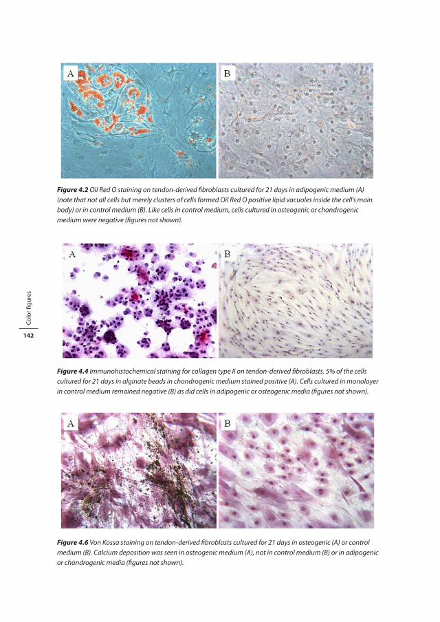
142
Colo
r figu
res
Figure 4.2 Oil Red O staining on tendon-derived fibroblasts cultured for 21 days in adipogenic medium (A) (note that not all cells but merely clusters of cells formed Oil Red O positive lipid vacuoles inside the cell’s main body) or in control medium (B). Like cells in control medium, cells cultured in osteogenic or chondrogenic medium were negative (figures not shown).
Figure 4.2 Oil Red O staining on tendon-derived fibroblasts cultured for 21 days in adipogenic medium (A) (note that not all cells but merely clusters of cells formed Oil Red O positive lipid vacuoles inside the cell’s main body) or in control medium (B). Like cells in control medium, cells cultured in osteogenic or chondrogenic medium were negative (figures not shown).
Figure 4.4 Immunohistochemical staining for collagen type II on tendon-derived fibroblasts. 5% of the cells cultured for 21 days in alginate beads in chondrogenic medium stained positive (A). Cells cultured in monolayer in control medium remained negative (B) as did cells in adipogenic or osteogenic media (figures not shown).
Figure 4.4 Immunohistochemical staining for collagen type II on tendon-derived fibroblasts. 5% of the cells cultured for 21 days in alginate beads in chondrogenic medium stained positive (A). Cells cultured in monolayer in control medium remained negative (B) as did cells in adipogenic or osteogenic media (figures not shown).
Figure 4.6 Von Kossa staining on tendon-derived fibroblasts cultured for 21 days in osteogenic (A) or control medium (B). Calcium deposition was seen in osteogenic medium (A), not in control medium (B) or in adipogenic or chondrogenic media (figures not shown).
Figure 4.6 Von Kossa staining on tendon-derived fibroblasts cultured for 21 days in osteogenic (A) or control medium (B). Calcium deposition was seen in osteogenic medium (A), not in control medium (B) or in adipogenic or chondrogenic media (figures not shown).

Color figures 143
Figure 5.1 Changes in cell morphology in mid-portion Achilles tendinosis. Hematoxilin and eosin staining of tendon samples. Representative photomicrographs (400x magnification) of healthy Achilles tendon samples (n=5, A) and of Achilles tendinotic samples (n=12, B) are shown; rounding of the cell nuclei was scored for each individual sample; total score can range from 0 (normal) to 6 (maximum deviant), line represents median score (C).
Figure 5.1 Changes in cell morphology in mid-portion Achilles tendinosis. Hematoxilin and eosin staining of tendon samples. Representative photomicrographs (400x magnification) of healthy Achilles tendon samples (n=5, A) and of Achilles tendinotic samples (n=12, B) are shown; rounding of the cell nuclei was scored for each individual sample; total score can range from 0 (normal) to 6 (maximum deviant), line represents median score (C).
Figure 5.2 Fibrocartilaginous metaplasia in mid-portion Achilles tendinosis demonstrated by thionin staining for glycosaminoglycans (GAGs). Representative photomicrographs (200x magnification) of healthy Achilles tendon samples (n=5, A), and of Achilles tendinotic samples (n=12, B); GAG stainability was scored for each individual sample, total score can range from 0 (normal) to 6 (maximum GAG staining), line represents median GAG score (C).
Figure 5.2 Fibrocartilaginous metaplasia in mid-portion Achilles tendinosis demonstrated by thionin staining for glycosaminoglycans (GAGs). Representative photomicrographs (200x magnification) of healthy Achilles tendon samples (n=5, A), and of Achilles tendinotic samples (n=12, B); GAG stainability was scored for each individual sample, total score can range from 0 (normal) to 6 (maximum GAG staining), line represents median GAG score (C).

144
Colo
r figu
res
Fig. 6.1 Photomicropgraphs of tenocyte cultures with or without PRCR or PPCR. Representative photomicrographs (200x magnification) are shown of control, 20% PPCR, and 20% PRCR conditions on day 1, 4, and 14 of the experimental culture period.
Fig. 6.1 Photomicropgraphs of tenocyte cultures with or without PRCR or PPCR. Representative photomicrographs (200x magnification) are shown of control, 20% PPCR, and 20% PRCR conditions on day 1, 4, and 14 of the experimental culture period.