Temporal Analysis of Andes Virus and Sin Nombre Virus Infections of Syrian Hamsters
14
JOURNAL OF VIROLOGY, July 2007, p. 7449–7462 Vol. 81, No. 14 0022-538X/07/$08.000 doi:10.1128/JVI.00238-07 Copyright © 2007, American Society for Microbiology. All Rights Reserved. Temporal Analysis of Andes Virus and Sin Nombre Virus Infections of Syrian Hamsters Victoria Wahl-Jensen, 1 Jennifer Chapman, 2 Ludmila Asher, 2 Robert Fisher, 1 Michael Zimmerman, 3 Tom Larsen, 2 and Jay W. Hooper 1 * Virology Division, 1 Pathology Division, 2 and Veterinary Medicine Division, 3 U.S. Army Medical Research Institute of Infectious Diseases, Fort Detrick, Maryland 21702 Received 5 February 2007/Accepted 23 April 2007 Andes virus (ANDV) and Sin Nombre virus (SNV) are rodent-borne hantaviruses that cause a highly lethal hemorrhagic fever in humans known as hantavirus pulmonary syndrome (HPS). There are no vaccines or specific drugs to prevent or treat HPS, and the pathogenesis is not understood. Syrian hamsters infected with ANDV, but not SNV, develop a highly lethal disease that closely resembles HPS in humans. Here, we performed a temporal pathogenesis study comparing ANDV and SNV infections in hamsters. SNV was nonpathogenic and viremia was not detected despite the fact that all animals were infected. ANDV was uniformly lethal with a mean time to death of 11 days. The first pathology detected was lymphocyte apoptosis starting on day 4. Animals were viremic and viral antigen was first observed in multiple organs by days 6 and 8, respectively. Levels of infectious virus in the blood increased 4 to 5 logs between days 6 and 8. Pulmonary edema was first detected ultrastructurally on day 6. Ultrastructural analysis of lung tissues revealed the presence of large inclusion bodies and substantial numbers of vacuoles within infected endothelial cells. Paraendothelial gaps were not observed, suggesting that fluid leakage was transcellular and directly attributable to infecting virus. Taken together, these data imply that HPS treatment strategies aimed at preventing virus replication and dissemination will have the greatest probability of success if administered before the viremic phase; however, because vascular leakage is associated with infected endothelial cells, a therapeutic strategy targeting viral replication might be effective even at later times (e.g., after disease onset). Hantavirus pulmonary syndrome (HPS) was first recognized in North America in 1993 during an outbreak of unexplained illness that led to acute respiratory distress and death in nearly half of the patients. The etiological agent was soon found to be a hantavirus, and an epidemiological link was established with exposure to excreta from infected rodents. Since 1993, numer- ous hantaviruses have been isolated throughout the Americas (5). To date, there have been over 1,900 cases of HPS with an overall case fatality of approximately 40% (29, 39). Sin Nom- bre virus (SNV) was the first hantavirus linked to HPS and is the cause of the majority of North American HPS cases. Andes virus (ANDV) is the cause of the majority of South American HPS cases and is the only hantavirus for which there is com- pelling evidence of person-to-person transmission (17, 24, 30). The rapid onset of HPS, high case fatality, and absence of drugs or vaccines make this disease a significant health concern in regions of endemicity. The recent importation of an ANDV- like virus from Bolivia to British Columbia, Canada (31), fur- ther underscores the need for additional research into the pathogenesis of and development of medical countermeasures to these highly lethal viruses. Hantaviruses represent a diverse group of viruses within a separate genus of the family Bunyaviridae. Members of this genus share morphological similarities with other bunyavi- ruses, including a pleomorphic size (70 to 210 nm) and a generally spherical particle appearance (9). Virions possess a host-derived lipid envelope that is studded with oligomers of the envelope glycoproteins. The genome of hantaviruses is tripartite, negative-sense, single-stranded RNA that encodes three structural proteins (35). The small RNA segment (S) encodes the nucleoprotein (N), the medium segment (M) en- codes the G N and G C envelope glycoproteins (previously known as G1and G2), and the large segment (L) encodes the viral RNA-dependent RNA polymerase. Unlike members of the other four genera within the Bunyaviridae family, hantavi- ruses are not transmitted through an arthropod vector but rather maintain an apathogenic and persistent infection within their natural rodent reservoir species. Human infection is as- sociated with HPS or hemorrhagic fever with renal syndrome (HFRS) (28). Vascular leakage and kidney dysfunction are associated with HFRS, while HPS-associated hantaviruses cause acute pulmonary edema (16, 44). Cardiac dysfunction has also been associated with HPS, and the term hantavirus cardiopulmonary syndrome has been used by some groups to alert clinicians to this disease characteristic. Acute thrombo- cytopenia and changes in vascular permeability are common features of both diseases, and either may include pulmonary or renal features. Previously, we reported the discovery that ANDV is highly lethal to Syrian hamsters (12). The disease in hamsters closely resembled HPS in humans, i.e., the incubation time was ap- proximately 10 days to 2 weeks, onset was rapid, the disease involved infection of endothelial cells, and the organ primarily affected was the lungs. Interestingly, SNV was highly infectious in the hamsters (2 PFU was sufficient to infect 50% of the hamsters) but did not cause disease. Here, we performed a * Corresponding author. Mailing address: Virology Division, USAMRIID, Fort Detrick, MD 21702. Phone: (301) 619-4101. Fax: (301) 619-2439. E-mail: [email protected]. Published ahead of print on 2 May 2007. 7449 on November 15, 2018 by guest http://jvi.asm.org/ Downloaded from
Transcript of Temporal Analysis of Andes Virus and Sin Nombre Virus Infections of Syrian Hamsters

JOURNAL OF VIROLOGY, July 2007, p. 7449–7462 Vol. 81, No. 140022-538X/07/$08.00�0 doi:10.1128/JVI.00238-07Copyright © 2007, American Society for Microbiology. All Rights Reserved.
Temporal Analysis of Andes Virus and Sin Nombre Virus Infectionsof Syrian Hamsters�
Victoria Wahl-Jensen,1 Jennifer Chapman,2 Ludmila Asher,2 Robert Fisher,1 Michael Zimmerman,3Tom Larsen,2 and Jay W. Hooper1*
Virology Division,1 Pathology Division,2 and Veterinary Medicine Division,3 U.S. Army Medical Research Institute ofInfectious Diseases, Fort Detrick, Maryland 21702
Received 5 February 2007/Accepted 23 April 2007
Andes virus (ANDV) and Sin Nombre virus (SNV) are rodent-borne hantaviruses that cause a highly lethalhemorrhagic fever in humans known as hantavirus pulmonary syndrome (HPS). There are no vaccines orspecific drugs to prevent or treat HPS, and the pathogenesis is not understood. Syrian hamsters infected withANDV, but not SNV, develop a highly lethal disease that closely resembles HPS in humans. Here, we performeda temporal pathogenesis study comparing ANDV and SNV infections in hamsters. SNV was nonpathogenic andviremia was not detected despite the fact that all animals were infected. ANDV was uniformly lethal with amean time to death of 11 days. The first pathology detected was lymphocyte apoptosis starting on day 4.Animals were viremic and viral antigen was first observed in multiple organs by days 6 and 8, respectively.Levels of infectious virus in the blood increased 4 to 5 logs between days 6 and 8. Pulmonary edema was firstdetected ultrastructurally on day 6. Ultrastructural analysis of lung tissues revealed the presence of largeinclusion bodies and substantial numbers of vacuoles within infected endothelial cells. Paraendothelial gapswere not observed, suggesting that fluid leakage was transcellular and directly attributable to infecting virus.Taken together, these data imply that HPS treatment strategies aimed at preventing virus replication anddissemination will have the greatest probability of success if administered before the viremic phase; however,because vascular leakage is associated with infected endothelial cells, a therapeutic strategy targeting viralreplication might be effective even at later times (e.g., after disease onset).
Hantavirus pulmonary syndrome (HPS) was first recognizedin North America in 1993 during an outbreak of unexplainedillness that led to acute respiratory distress and death in nearlyhalf of the patients. The etiological agent was soon found to bea hantavirus, and an epidemiological link was established withexposure to excreta from infected rodents. Since 1993, numer-ous hantaviruses have been isolated throughout the Americas(5). To date, there have been over 1,900 cases of HPS with anoverall case fatality of approximately 40% (29, 39). Sin Nom-bre virus (SNV) was the first hantavirus linked to HPS and isthe cause of the majority of North American HPS cases. Andesvirus (ANDV) is the cause of the majority of South AmericanHPS cases and is the only hantavirus for which there is com-pelling evidence of person-to-person transmission (17, 24, 30).The rapid onset of HPS, high case fatality, and absence ofdrugs or vaccines make this disease a significant health concernin regions of endemicity. The recent importation of an ANDV-like virus from Bolivia to British Columbia, Canada (31), fur-ther underscores the need for additional research into thepathogenesis of and development of medical countermeasuresto these highly lethal viruses.
Hantaviruses represent a diverse group of viruses within aseparate genus of the family Bunyaviridae. Members of thisgenus share morphological similarities with other bunyavi-ruses, including a pleomorphic size (70 to 210 nm) and agenerally spherical particle appearance (9). Virions possess a
host-derived lipid envelope that is studded with oligomers ofthe envelope glycoproteins. The genome of hantaviruses istripartite, negative-sense, single-stranded RNA that encodesthree structural proteins (35). The small RNA segment (S)encodes the nucleoprotein (N), the medium segment (M) en-codes the GN and GC envelope glycoproteins (previouslyknown as G1and G2), and the large segment (L) encodes theviral RNA-dependent RNA polymerase. Unlike members ofthe other four genera within the Bunyaviridae family, hantavi-ruses are not transmitted through an arthropod vector butrather maintain an apathogenic and persistent infection withintheir natural rodent reservoir species. Human infection is as-sociated with HPS or hemorrhagic fever with renal syndrome(HFRS) (28). Vascular leakage and kidney dysfunction areassociated with HFRS, while HPS-associated hantavirusescause acute pulmonary edema (16, 44). Cardiac dysfunctionhas also been associated with HPS, and the term hantaviruscardiopulmonary syndrome has been used by some groups toalert clinicians to this disease characteristic. Acute thrombo-cytopenia and changes in vascular permeability are commonfeatures of both diseases, and either may include pulmonary orrenal features.
Previously, we reported the discovery that ANDV is highlylethal to Syrian hamsters (12). The disease in hamsters closelyresembled HPS in humans, i.e., the incubation time was ap-proximately 10 days to 2 weeks, onset was rapid, the diseaseinvolved infection of endothelial cells, and the organ primarilyaffected was the lungs. Interestingly, SNV was highly infectiousin the hamsters (2 PFU was sufficient to infect 50% of thehamsters) but did not cause disease. Here, we performed a
* Corresponding author. Mailing address: Virology Division,USAMRIID, Fort Detrick, MD 21702. Phone: (301) 619-4101. Fax:(301) 619-2439. E-mail: [email protected].
� Published ahead of print on 2 May 2007.
7449
on Novem
ber 15, 2018 by guesthttp://jvi.asm
.org/D
ownloaded from
pathogenesis study comparing ANDV and SNV infections inhamsters. Hamsters were injected with ANDV or SNV by theintramuscular route, and then on days 0, 2, 4, 6, 8, 10, 12, 14,16, and 21, blood and other tissue samples were collected andfull necropsies were performed. The pathology studies in-volved both immunohistological and electron microscopic eval-uations of tissues collected at serial time points after infection.
MATERIALS AND METHODS
Viruses and cells. ANDV strain Chile-9717869 and SNV strain CC107 (34)were propagated in Vero E6 cells (Vero C1008; ATCC CRL 1586). Preparationof ANDV passage 2, twice-plaque-purified virus stocks was previously described(12). The same method was used to prepare a twice-plaque-purified stock ofSNV. Passage 2 stocks of both ANDV and SNV were used in this study.
Intramuscular injection of hamsters with virus. Female Syrian hamstersweighing 150 to 200 g (Harlan, Indianapolis, IN) were anesthetized by an intra-muscular injection with approximately 0.1 ml/100 g of body weight of a ketamine-acepromazine-xylazine mixture. Once anesthetized, hamsters were injected in-tramuscularly (caudal thigh) with 0.2 ml of virus (2,000 PFU) diluted inphosphate-buffered saline, pH 7.4. After viral challenge, hamsters were placedin isolator units (one to four hamsters per cage). All work involving hamstersinfected with ANDV or SNV was performed in a biosafety level 4 (BSL-4)laboratory. Hamsters were observed two to three times daily.
ELISA. There is a high degree of immune serum cross-reactivity to the nu-cleocapsid proteins of ANDV, SNV, and Puumala virus. This cross-reactivitymade it possible to use the Puumala virus nucleocapsid-based enzyme-linkedimmunosorbent assay (ELISA) for detecting antibodies to ANDV and SNVnucleocapsid. Anti-nucleocapsid antibody ELISAs were performed as previouslydescribed (11). The plasmid pPUUSXdelta (kindly provided by F. Elgh) wasexpressed in Escherichia coli BL21(DE3) cells (Novagen, Madison, WI) to gen-erate histidine-tagged truncated Puumala virus nucleocapsid fusion protein. Thefusion protein was subsequently affinity purified in Ni-nitrilotriacetic acid col-umns (QIAGEN, Valencia, CA). Samples were gamma irradiated (on dry ice)with 3 � 106 rads from a 60C source.
PRNT. Plaque reduction neutralization tests (PRNT) were performed as pre-viously described (12). Samples were gamma irradiated as described above.
Blood chemistry. Serum samples were obtained from animals and gammairradiated as described above. Concentrations of albumin, alkaline phosphatase,alanine aminotransferase, amylase, aspartate aminotransferase, total bilirubin,urea nitrogen, calcium, cholesterol, creatinine, glucose, and total protein weredetermined using a general chemistry 12-disc panel rotor and a Piccolo point-of-care blood analyzer (Abaxis, Sunnyvale, CA).
Hematology. Blood samples were collected in Safe-T-Fill capillary blood col-lection tubes containing EDTA (RAM Scientific, Yonkers, NY). Total whiteblood cell count, white blood cell differential count, red blood cell count, hemo-globin concentration, hematocrit, mean corpuscular volume, mean corpuscularhemoglobin, mean corpuscular hemoglobin concentration, and platelet countwere determined from blood samples collected in EDTA by using an Ac T 10Coulter Counter (Coulter Electronics, Hialeah, FL). In addition, blood smearswere prepared and manual counts were obtained by standard methods. Datawere analyzed using the Wilcoxon rank-sum test.
Preparation of tissue for histology. Tissues (lung, liver, spleen, heart, thymus,trachea, thyroid gland, gastrointestinal tract, pancreas, kidneys, adrenal glands,urinary bladder, uterus, ovaries, skin, submandibular salivary gland, multiplelymph nodes, brain, pituitary gland, eyes, and nasal turbinates) were fixed in 10%neutral buffered formalin, trimmed, processed, embedded in paraffin, cut at 5 to6 �m, and stained with hematoxylin & eosin (H&E) according to establishedprotocols (32). Immunolocalization of ANDV and SNV in tissues was performedwith an alkaline phosphatase procedure (EnVision System; DAKO Corp.,Carpinteria, CA) or an immunoperoxidase procedure (horseradish peroxidaseEnVision System; DAKO) according to the manufacturer’s directions. The pri-mary antibody was an anti-SNV nucleocapsid rabbit polyclonal diluted 1:500 forthe alkaline phosphatase procedure or 1:3,000 for the peroxidase procedure(provided by Diagnostic Service Division, USAMRIID). Negative controls in-cluded naı̈ve hamster tissue incubated with nonimmune rabbit immunoglobulinG in place of the primary antibody and naı̈ve hamster tissue exposed to theprimary antibody and to negative serum. Tissue sections were pretreated inproteinase K for 6 min (DAKO) at room temperature and blocked with CAS-BLOCK (Invitrogen, Carlsbad, CA) and 5% goat serum (Vector Laboratories,Burlingame, CA) for 30 min at room temperature. The primary antibody was
incubated on the tissue sections for 1 h at room temperature for the alkalinephosphatase procedure or for 30 min at room temperature for the peroxidaseprocedure. Goat anti-rabbit antibodies conjugated to an alkaline phosphatase-labeled dextran polymer or horseradish peroxidase-labeled dextran polymer pro-vided with the EnVision kit served as the secondary antibody for that system. Thesecondary antibody was incubated for 30 min at room temperature. Sectionswere incubated for 6 min with the DakoCytomation Permanent Red substrate-chromogen system or 5 min with 3,3�-diaminobenzidine (DAKO). Immuno-stained sections were counterstained with hematoxylin and dehydrated, andcoverslips were applied with Permount (Fisher Scientific).
Electron microscopy. Lungs, liver, heart, spleen, and kidneys were analyzed bytransmission electron microscopy by fixing tissues for 24 h with 4% paraformal-dehyde and 1% glutaraldehyde, treated with 1% osmium tetroxide for 1 h,washed, en block stained with 0.5% uranyl acetate, dehydrated in graded ethanoland propylene oxide, and embedded in PolyBed 812 (Polysciences, Warrington,PA). Ultrathin sections were stained with uranyl acetate and lead citrate andexamined at 80 kV on a JOEL 1011 transmission electron microscope.
Tissue samples for immunoelectron microscopy were fixed for 1 month in 2%paraformaldehyde and 0.1% glutaraldehyde, dehydrated through graded eth-anols up to 70%, and embedded in LR White resin (Polysciences, Warrington,PA). Ultrathin sections were placed on nickel grids and prepared for immuno-electron microscopy by an indirect method. The primary antibody was a rabbitanti-SNV nucleocapsid polyclonal antibody (see above) diluted 1:300. The sec-ondary antibody was a goat anti-rabbit serum labeled with 10-nm gold particles(Ted Pella Inc., Redding, CA) and diluted 1:30. As a negative control, sectionsfrom the same block were incubated with normal rabbit serum (Vector Labora-tories, Burlingame, CA) at the same dilution as the primary serum. Grids werefirst floated on the blocking serum, in this case normal goat serum (VectorLaboratories, Burlingame, CA), for 20 min at room temperature and then on theprimary serum for 1 h at room temperature or overnight at 4°C. After rinsinggrids in BTT buffer (20 mM Tris, 0.9% NaCl, 0.1% bovine serum albumin,and 0.5% Tween), sections were incubated with the secondary antibody for1 h, rinsed again, and fixed in 1% glutaraldehyde for 5 min. Grids werestained with uranyl acetate and lead citrate and examined on a JOEL 1011microscope.
Plaque assay. Hantavirus plaque assays were performed as previously de-scribed (12). Briefly, serum samples were centrifuged to pellet any debris beforedilution. Whole blood collected in K2EDTA tubes was transferred to centrifugetubes, rapidly freeze-thawed three times, and then spun in a microcentrifuge at10,000 � g for 10 s to pellet debris. Supernatants were used to prepare initialdilutions. Similarly, throat swabs were processed by first adding 300 �l of Eagle’sminimal essential medium with Earl’s salts containing 10% fetal bovine serum,10 mM HEPES (pH 7.4), penicillin (100 U/ml), streptomycin (100 �g/ml), andgentamycin sulfate (50 �g/ml) to swabs in microcentrifuge tubes for 3 min. Swabswere then swirled to release sample material into the medium. The throat swabsuspension was then freeze-thawed three times and spun in a microcentrifuge at10,000 � g for 10 s. Supernatants were used to prepare dilutions starting at 1:10.Once sample dilutions were prepared, 200 �l was added to each well of a six-wellCostar plate containing 7-day-old Vero E6 monolayers. After a 1-h adsorption at37°C, 3 ml of overlay medium was added to each well as previously described(12). Plates were incubated at 37°C in a humidified 5% CO2 environment for 7days. Plaques were stained on day 7 (ANDV) or day 10 (SNV) by adding 2ml/well of overlay medium containing 5% fetal bovine serum and 5% neutral redsolution (Invitrogen, Carlsbad, CA). Plates were then incubated for an additional2 days at 37°C or until plaques were visible and countable.
Isolation of PBMCs and real-time PCR. Peripheral blood mononuclear cells(PBMCs) were isolated by adding 1 ml ACK lysing buffer (BioSource, Camarillo,CA) to 200 �l EDTA blood, gently mixed by inversion, incubated for 5 min atroom temperature, and then centrifuged at 1,500 � g for 5 min. Supernatantswere discarded and the cell pellet was resuspended in 1 ml phosphate-bufferedsaline. Samples were centrifuged at 1,500 � g for 5 min. Supernatants werediscarded and the cell pellet was resuspended in 1 ml of TRIzol (Invitrogen,Carlsbad, CA) and mixed by pipetting. RNA was extracted from TRIzol samplesas recommended by the manufacturer. Reverse transcriptase (RT) and real-timePCRs were performed as previously described (25). Briefly, 10 ng of RNA wasused for the RT reaction using a primer specific for the negative-sense strands ofthe ANDV and SNV S-segments. The RT primers were as follows: ANDV S350F (5�CATCTC TGA GAT ATG GGA ATG TCC TGG-3�) and SNV S 10F(5�-ACT CCT TGA GAA GCT ACT ACG AC-3�). A 2.5-�l portion of cDNAgenerated from the RT reaction was used in the real-time PCR, which wasperformed as previously described with ANDV-S- and SNV-S-specific primersand probes (26).
7450 WAHL-JENSEN ET AL. J. VIROL.
on Novem
ber 15, 2018 by guesthttp://jvi.asm
.org/D
ownloaded from

RESULTS
Study design. We previously reported that ANDV washighly lethal in hamsters, whereas SNV was highly infectiousbut did not cause disease (12). Moreover, we reported that areassortant virus having an ANDV M genome segment andSNV S and L genome segments was infectious in hamsters butdid not cause disease (26). Here we performed a serial patho-genesis study to (i) determine when and to what tissues and celltypes ANDV and SNV disseminate after challenge, (ii) mea-sure and evaluate hematological changes during the course ofANDV and SNV infections, (iii) determine if blood chemis-tries in ANDV- or SNV-infected hamsters reveal evidenceof organ dysfunction, and (iv) use immunohistochemicaltechniques and electron microscopy to characterize the in-flammation and ultrastructural changes in tissues of ham-sters with HPS.
The study consisted of animals in time course groups (8hamsters injected with ANDV and 8 hamsters injected withSNV) and animals in serial pathology groups (24 hamstersinjected with ANDV and 24 hamsters injected with SNV).Animals in the time course groups were weighed, throatswabbed, and bled 1 week before challenge, at the time ofchallenge, and then every 2 days until day 16. This time coursegroup-based data collection method allowed us to collect sam-ples from the same hamster over time throughout the experi-ment. Animals in the serial pathology groups were weighedand bled 1 week before challenge and assigned predeterminednecropsy dates. On the day of challenge and every 2 daysthereafter for 16 days, three ANDV-injected hamsters andthree SNV-injected hamsters were removed from the serialpathology groups, and full necropsies were performed (includ-ing blood collections and throat swabs for viremia study). Insome cases, moribund animals from the time course groupwere euthanized and necropsies were performed. This wasdone to ensure that at least three hamsters were necropsied ateach of the time points. The fates of the individual animals ineach group are shown in Table 1.
Clinical signs and hematological analysis. The hamstersinfected with ANDV were either found dead, found moribundand euthanized, or killed for necropsy on the indicated day(Table 1). Across all groups, there were 14 hamsters that werefound dead or were found moribund and euthanized. Twohamsters were suspected to have died due to the blood drawprocedure, one on day �7 and one on day 7, and were notincluded in further analysis. Of the remaining 12 hamsters, allhad been injected with ANDV, and the mean time to deathwas 11 days. None of the SNV-injected hamsters displayedsigns of disease, and all survived or were killed for schedulednecropsies. Nevertheless, all of the hamsters injected with SNVwere indeed infected because they had anti-nucleocapsid an-tibody titers as measured by ELISA. ELISA endpoint titers onsera collected on day 28 (terminal bleed) ranged from 200 to6,400, with a geometric mean titer of 872. All sera collectedbefore day 16 were negative by ELISA. Three of the day 28sera were further evaluated for neutralizing antibody byPRNT. All were positive, with 80% PRNT titers of 320, 160,and �640. ANDV-injected animals displayed obvious signs ofillness within a few hours before death. Signs included tachy-pnea, staggered gait, cyanosis (bluing of nares and oral cavity),
and apparent weakness. There was minor (�6%) weight loss inthe ANDV-injected hamsters during the 2 days before death(data not shown).
Blood samples collected from the animals in the serial pa-thology groups were analyzed by Coulter Counter and manualdifferential analysis (smears). ANDV-injected animals exhib-ited an absolute increase in white blood cell counts that was
TABLE 1. Study design
Group HamsterID no.a Fateb Day Necropsy
ANDV time course 201 FD 13202 FD* 7203 FD 11204 FD 11205 SAC 14 Yes206 EUTH 12 Yes207 SAC 14 Yes208 FD 11
SNV time course 209210 Survived211 Survived212 Survived213 Survived214 Survived215 Survived216 Survived
ANDV serial pathology 217 SAC 2 Yes218 SAC 2 Yes219 SAC 2 Yes220 SAC 4 Yes221 SAC 4 Yes222 SAC 4 Yes223 SAC 6 Yes224 SAC 6 Yes225 SAC 6 Yes226 SAC 8 Yes227 SAC 8 Yes228 SAC 8 Yes229 SAC 10 Yes230 SAC 10 Yes231 SAC 10 Yes232 FD 10233 SAC 12 Yes234 FD 14235 FD 10236 FD 11237 EUTH 10238 EUTH 12 Yes239 SAC 14 Yes240 EUTH 10
SNV serial pathology 241 SAC 2 Yes242 SAC 2 Yes243 SAC 2 Yes244 SAC 4 Yes245 FD* �7 Yes246 SAC 4 Yes247 SAC 6 Yes248 SAC 6 Yes249 SAC 6 Yes250 SAC 8 Yes251 SAC 8 Yes252 SAC 8 Yes253 SAC 10 Yes254 SAC 10 Yes255 SAC 10 Yes256 SAC 12 Yes257 SAC 12 Yes258 SAC 12 Yes259 SAC 14 Yes260 SAC 14 Yes261 SAC 14 Yes262 SAC 16 Yes263 SAC 16 Yes264 SAC 16 Yes
No challenge 265 SAC �7 Yes266 SAC �7 Yes267 SAC �7 Yes
a ID, identification.b FD, found dead; FD*, suspect death due to blood draw; SAC, no signs of
disease when sacrificed; EUTH, moribund animal euthanized.
VOL. 81, 2007 PATHOGENESIS OF ANDES VIRUS IN HAMSTERS 7451
on Novem
ber 15, 2018 by guesthttp://jvi.asm
.org/D
ownloaded from
outside of normal reference values (Fig. 1). This was likely dueto a relative neutrophilia with a peak in segmented neutrophilsoccurring at the terminal stages of disease (days 12 to 14). Wedid not observe a significant increase in the percentage ofbanded neutrophils (data not shown). Interestingly, there wasa delayed neutrophilia with SNV, but the increase in seg-mented neutrophils was not as pronounced as what was seenwith ANDV. A relative lymphopenia that was outside normallimits was seen with ANDV on days 12 to 14. Finally, transientthrombocytopenia was observed for both ANDV- and SNV-infected animals, peaking at days 12 and 14, respectively. Thesmall sample size detracted from our ability to assign statisticalsignificance (Wilcoxon rank-sum test) to the observed postin-fection alterations in hematological values. A comparison ofthe ANDV and SNV data for each time point indicated thatthere were no statistically significant differences between those
groups; however, there were significant changes from theprechallenge values (Fig. 1).
Viremia is not detected in SNV-infected hamsters. We pre-viously reported that serum viremia (infectious virus) in ham-sters injected with ANDV could be detected on day 8 postin-fection (12). Here, we included additional time points to gainfurther insight into the kinetics of ANDV viremia in the ham-ster model. In addition, we assayed for the presence of viremiain hamsters injected with SNV. Plaque assays were used tomeasure infectious virus in whole blood and in serum from theanimals in the serial pathology groups. ANDV was first de-tected in whole blood and serum on day 6 postinfection andpeaked by days 8 and 12 postinfection, respectively (Fig. 2Aand B). In stark contrast, infectious SNV was not detected inthe serum or whole blood at any time point. It was possible thatSNV virions had disseminated into the blood but were ren-
FIG. 1. Hematological findings. ANDV and SNV serial pathogenesis groups were analyzed for changes in hematological parameters afterinfection. The area between dotted lines represents the normal reference range for hamsters. ANDV-infected animals exhibited white blood cell(WBC) counts above normal reference values (A) with a corresponding increase in the percent segmented neutrophils (%SEG) beginning on day10 (B). (C and D) Lymphopenia (%LY) and thrombocytopenia (Plt) were also observed with ANDV infection, although similar changes were alsoobserved with SNV that occurred later and to a lesser degree. (E and F) Slight rises in red blood cell (RBC) and hematocrit (Hct) were alsoobserved with ANDV and SNV but remained within normal limits. Symbols represent the mean values for each time point. Error bars representthe standard errors of the means (SEM). Asterisks represent statistically significant (P � 0.05) differences relative to day �7 values.
7452 WAHL-JENSEN ET AL. J. VIROL.
on Novem
ber 15, 2018 by guesthttp://jvi.asm
.org/D
ownloaded from
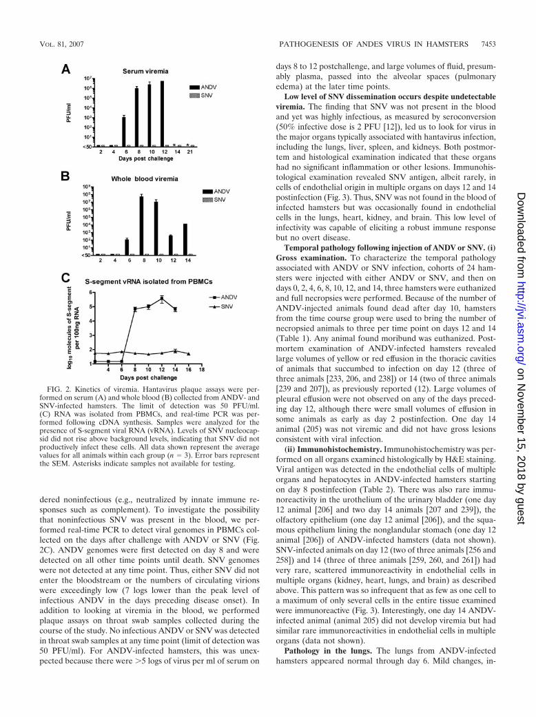
dered noninfectious (e.g., neutralized by innate immune re-sponses such as complement). To investigate the possibilitythat noninfectious SNV was present in the blood, we per-formed real-time PCR to detect viral genomes in PBMCs col-lected on the days after challenge with ANDV or SNV (Fig.2C). ANDV genomes were first detected on day 8 and weredetected on all other time points until death. SNV genomeswere not detected at any time point. Thus, either SNV did notenter the bloodstream or the numbers of circulating virionswere exceedingly low (7 logs lower than the peak level ofinfectious ANDV in the days preceding disease onset). Inaddition to looking at viremia in the blood, we performedplaque assays on throat swab samples collected during thecourse of the study. No infectious ANDV or SNV was detectedin throat swab samples at any time point (limit of detection was50 PFU/ml). For ANDV-infected hamsters, this was unex-pected because there were �5 logs of virus per ml of serum on
days 8 to 12 postchallenge, and large volumes of fluid, presum-ably plasma, passed into the alveolar spaces (pulmonaryedema) at the later time points.
Low level of SNV dissemination occurs despite undetectableviremia. The finding that SNV was not present in the bloodand yet was highly infectious, as measured by seroconversion(50% infective dose is 2 PFU [12]), led us to look for virus inthe major organs typically associated with hantavirus infection,including the lungs, liver, spleen, and kidneys. Both postmor-tem and histological examination indicated that these organshad no significant inflammation or other lesions. Immunohis-tological examination revealed SNV antigen, albeit rarely, incells of endothelial origin in multiple organs on days 12 and 14postinfection (Fig. 3). Thus, SNV was not found in the blood ofinfected hamsters but was occasionally found in endothelialcells in the lungs, heart, kidney, and brain. This low level ofinfectivity was capable of eliciting a robust immune responsebut no overt disease.
Temporal pathology following injection of ANDV or SNV. (i)Gross examination. To characterize the temporal pathologyassociated with ANDV or SNV infection, cohorts of 24 ham-sters were injected with either ANDV or SNV, and then ondays 0, 2, 4, 6, 8, 10, 12, and 14, three hamsters were euthanizedand full necropsies were performed. Because of the number ofANDV-injected animals found dead after day 10, hamstersfrom the time course group were used to bring the number ofnecropsied animals to three per time point on days 12 and 14(Table 1). Any animal found moribund was euthanized. Post-mortem examination of ANDV-infected hamsters revealedlarge volumes of yellow or red effusion in the thoracic cavitiesof animals that succumbed to infection on day 12 (three ofthree animals [233, 206, and 238]) or 14 (two of three animals[239 and 207]), as previously reported (12). Large volumes ofpleural effusion were not observed on any of the days preced-ing day 12, although there were small volumes of effusion insome animals as early as day 2 postinfection. One day 14animal (205) was not viremic and did not have gross lesionsconsistent with viral infection.
(ii) Immunohistochemistry. Immunohistochemistry was per-formed on all organs examined histologically by H&E staining.Viral antigen was detected in the endothelial cells of multipleorgans and hepatocytes in ANDV-infected hamsters startingon day 8 postinfection (Table 2). There was also rare immu-noreactivity in the urothelium of the urinary bladder (one day12 animal [206] and two day 14 animals [207 and 239]), theolfactory epithelium (one day 12 animal [206]), and the squa-mous epithelium lining the nonglandular stomach (one day 12animal [206]) of ANDV-infected hamsters (data not shown).SNV-infected animals on day 12 (two of three animals [256 and258]) and 14 (three of three animals [259, 260, and 261]) hadvery rare, scattered immunoreactivity in endothelial cells inmultiple organs (kidney, heart, lungs, and brain) as describedabove. This pattern was so infrequent that as few as one cell toa maximum of only several cells in the entire tissue examinedwere immunoreactive (Fig. 3). Interestingly, one day 14 ANDV-infected animal (animal 205) did not develop viremia but hadsimilar rare immunoreactivities in endothelial cells in multipleorgans (data not shown).
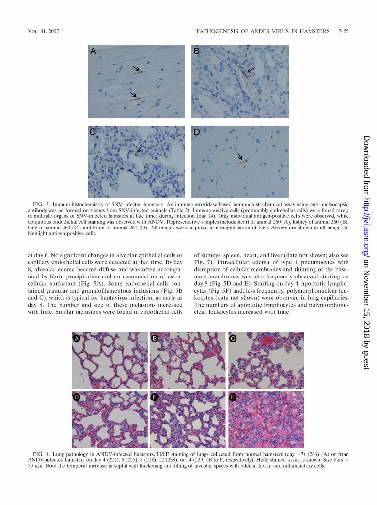
Pathology in the lungs. The lungs from ANDV-infectedhamsters appeared normal through day 6. Mild changes, in-
FIG. 2. Kinetics of viremia. Hantavirus plaque assays were per-formed on serum (A) and whole blood (B) collected from ANDV- andSNV-infected hamsters. The limit of detection was 50 PFU/ml.(C) RNA was isolated from PBMCs, and real-time PCR was per-formed following cDNA synthesis. Samples were analyzed for thepresence of S-segment viral RNA (vRNA). Levels of SNV nucleocap-sid did not rise above background levels, indicating that SNV did notproductively infect these cells. All data shown represent the averagevalues for all animals within each group (n � 3). Error bars representthe SEM. Asterisks indicate samples not available for testing.
VOL. 81, 2007 PATHOGENESIS OF ANDES VIRUS IN HAMSTERS 7453
on Novem
ber 15, 2018 by guesthttp://jvi.asm
.org/D
ownloaded from
cluding thickening of alveolar septal walls, were first observedon day 8 (Table 2). The damage to the lungs increased overtime and culminated with severe pulmonary edema, mononu-clear cell infiltration, and hemorrhage by day 14. Representa-tive images are shown in Fig. 4. The inflammatory componentconsisted primarily of lymphocytes, plasma cells, and fewer
neutrophils that expanded the alveolar, perivascular, and peri-bronchiolar interstitia. H&E-stained lungs from SNV-infectedhamsters were unremarkable at all time points and resembledthe lungs of the negative control animals (data not shown).
The most striking ultrastructural changes were seen in thelungs. The first signs of focal pulmonary edema were noticed
TABLE 2. Summary of temporal histopathological findings
Daypostchallenge Virus Hamster
ID no.a
Temporal histopathological findingb
Lung LiverSpleen
mononuclearinfiltrateIHC Alveolar
edemaInterstitial
inflammation
Perivascular/peribronchialinflammation
IHC Hepatitis Hepatocellularnecrosis
�7 None 265 � � � � � � � �None 266 � � � � � � � �None 267 � � � � � � � �
2 ANDV 217 � � � � � � � �ANDV 218 � � � � � � � �ANDV 219 � � � � � � � �
4 ANDV 220 � � � � � � � �ANDV 221 � � � � � � � �ANDV 222 � � � � � � � �
6 ANDV 223 � � � � � � � �ANDV 224 � � � � � � � �ANDV 225 � � � � � � � �
8 ANDV 226 � ��� �� ��� � ����� ���� �ANDV 227 � � � �� � � � �ANDV 228 � � �� �� � �� � �
10 ANDV 229 �� �� �� �� ��� �� � �ANDV 230 ��� � �� � ��� � � �ANDV 231 �� � �� ��� �� ����� ����� �
12 ANDV 233 ��� � ��� ���� ��� �� ��� �ANDV 238 ��� ����� ����� ���� ��� �� �� �ANDV 206 ��� �� ��� �� ��� ��� ����� �����
14 ANDV 239 ��� ����� ����� ����� �� �� �� �ANDV 205 * � � �� * � � �ANDV 207 ��� � �� �� ��� ���� ���� �����
2 SNV 241 � � � � � � � �SNV 242 � � � � � � � �SNV 243 � � � � � � � �
4 SNV 244 � � � � � � � �SNV 246 � � � � � � � �
6 SNV 247 � � � � � � �� �SNV 248 � � � � � � � �SNV 249 � � � � � ��� � �
8 SNV 250 � � � � � � � �SNV 251 � � � � � � � �SNV 252 � � � � � � � �
10 SNV 253 � � � � � �� � �SNV 254 � � � � � �� � �SNV 255 � � � � � �� �� �
12 SNV 256 * � � � * � � �SNV 257 � � � � � � � �SNV 258 * � � � * � � �
14 SNV 259 * � � �� * � � �SNV 260 * � � �� * � � �SNV 261 * � � � * � � �
16 SNV 262 � � � � � � � �SNV 263 � � � �� � � � �SNV 264 � � � � � � � �
28 SNV 210 � � � � � � � �SNV 213 � � � � � � � �SNV 215 � � � � � � � �
a ID, identification.b IHC, immunohistochemistry. Immunostaining was scored from � to ��� (minimal to maximal staining, respectively). *, Scattered rare endothelial cell
immunoreactivity; �, no immunostaining. Histopathological findings were scored as follows: �, very mild; ��, mild; ���, moderate; ����, marked; �����,severe. *, Scattered rare histopathology; �, negative or minimal finding.
7454 WAHL-JENSEN ET AL. J. VIROL.
on Novem
ber 15, 2018 by guesthttp://jvi.asm
.org/D
ownloaded from
at day 6. No significant changes in alveolar epithelial cells orcapillary endothelial cells were detected at that time. By day8, alveolar edema became diffuse and was often accompa-nied by fibrin precipitation and an accumulation of extra-cellular surfactant (Fig. 5A). Some endothelial cells con-tained granular and granulofilamentous inclusions (Fig. 5Band C), which is typical for hantavirus infection, as early asday 8. The number and size of these inclusions increasedwith time. Similar inclusions were found in endothelial cells
of kidneys, spleen, heart, and liver (data not shown; also seeFig. 7). Intracellular edema of type 1 pneumocytes withdisruption of cellular membranes and thinning of the base-ment membranes was also frequently observed starting onday 8 (Fig. 5D and E). Starting on day 4, apoptotic lympho-cytes (Fig. 5F) and, less frequently, polymorphonuclear leu-kocytes (data not shown) were observed in lung capillaries.The numbers of apoptotic lymphocytes and polymorphonu-clear leukocytes increased with time.
FIG. 3. Immunohistochemistry of SNV-infected hamsters. An immunoperoxidase-based immunohistochemical assay using anti-nucleocapsidantibody was performed on tissues from SNV-infected animals (Table 2). Immunopositive cells (presumably endothelial cells) were found rarelyin multiple organs of SNV-infected hamsters at late times during infection (day 14). Only individual antigen-positive cells were observed, whileubiquitous endothelial cell staining was observed with ANDV. Representative samples include heart of animal 260 (A), kidney of animal 260 (B),lung of animal 260 (C), and brain of animal 261 (D). All images were acquired at a magnification of �60. Arrows are shown in all images tohighlight antigen-positive cells.
FIG. 4. Lung pathology in ANDV-infected hamsters. H&E staining of lungs collected from normal hamsters (day �7) (266) (A) or fromANDV-infected hamsters on day 4 (222), 6 (225), 8 (228), 12 (233), or 14 (239) (B to F, respectively). H&E-stained tissue is shown. Size bars �50 �m. Note the temporal increase in septal wall thickening and filling of alveolar spaces with edema, fibrin, and inflammatory cells.
VOL. 81, 2007 PATHOGENESIS OF ANDES VIRUS IN HAMSTERS 7455
on Novem
ber 15, 2018 by guesthttp://jvi.asm
.org/D
ownloaded from

FIG. 5. Ultrastructural changes to ANDV-infected hamster lungs. Thin-section transmission electron microscopy was performed on ANDV-infected hamster lungs. (A) Fibrin deposition (arrowheads), alveolar airspace edema, and apoptotic lymphocytes were observed for animal 226 at8 days postchallenge. Original magnification, �40,000. (B) At day 12 postchallenge (animal 206), fibrin deposition was increased (arrowheads), andlarge granulofilamentous inclusions that occupied the majority of space in the endothelial cell cytoplasm could be observed. Numerous vacuoleswere often observed in the endothelial cell cytoplasm, adjacent to the large inclusion (arrow). The inclusions were highly structured, as shown inthe enlarged inset. Original magnifications, �15,000 and �80,000 (inset). (C) Endothelial cell inclusions were of viral origin, as demonstrated byimmunoelectron microscopy using an anti-nucleocapsid antibody and a secondary antibody conjugated to 10-nm gold particles. The immunore-active endothelial cell inclusion is enlarged for better visualization of gold particles (inset). Original magnifications, �40,000 and �80,000 (inset).
7456 WAHL-JENSEN ET AL. J. VIROL.
on Novem
ber 15, 2018 by guesthttp://jvi.asm
.org/D
ownloaded from
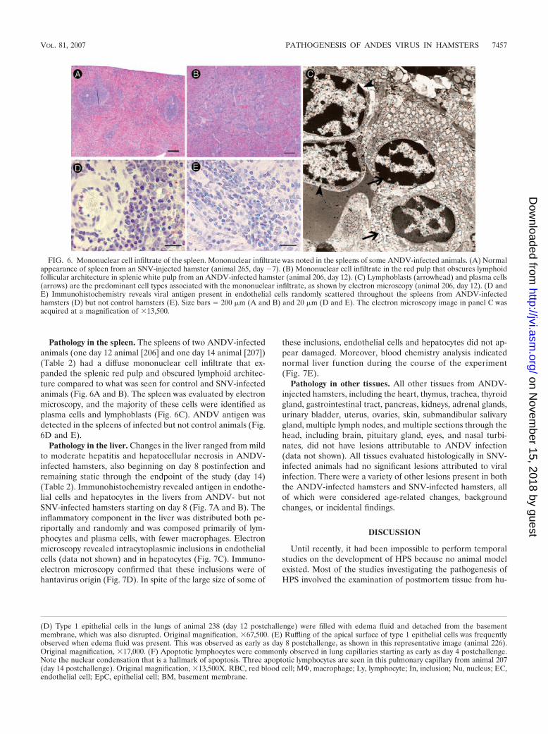
Pathology in the spleen. The spleens of two ANDV-infectedanimals (one day 12 animal [206] and one day 14 animal [207])(Table 2) had a diffuse mononuclear cell infiltrate that ex-panded the splenic red pulp and obscured lymphoid architec-ture compared to what was seen for control and SNV-infectedanimals (Fig. 6A and B). The spleen was evaluated by electronmicroscopy, and the majority of these cells were identified asplasma cells and lymphoblasts (Fig. 6C). ANDV antigen wasdetected in the spleens of infected but not control animals (Fig.6D and E).
Pathology in the liver. Changes in the liver ranged from mildto moderate hepatitis and hepatocellular necrosis in ANDV-infected hamsters, also beginning on day 8 postinfection andremaining static through the endpoint of the study (day 14)(Table 2). Immunohistochemistry revealed antigen in endothe-lial cells and hepatocytes in the livers from ANDV- but notSNV-infected hamsters starting on day 8 (Fig. 7A and B). Theinflammatory component in the liver was distributed both pe-riportally and randomly and was composed primarily of lym-phocytes and plasma cells, with fewer macrophages. Electronmicroscopy revealed intracytoplasmic inclusions in endothelialcells (data not shown) and in hepatocytes (Fig. 7C). Immuno-electron microscopy confirmed that these inclusions were ofhantavirus origin (Fig. 7D). In spite of the large size of some of
these inclusions, endothelial cells and hepatocytes did not ap-pear damaged. Moreover, blood chemistry analysis indicatednormal liver function during the course of the experiment(Fig. 7E).
Pathology in other tissues. All other tissues from ANDV-injected hamsters, including the heart, thymus, trachea, thyroidgland, gastrointestinal tract, pancreas, kidneys, adrenal glands,urinary bladder, uterus, ovaries, skin, submandibular salivarygland, multiple lymph nodes, and multiple sections through thehead, including brain, pituitary gland, eyes, and nasal turbi-nates, did not have lesions attributable to ANDV infection(data not shown). All tissues evaluated histologically in SNV-infected animals had no significant lesions attributed to viralinfection. There were a variety of other lesions present in boththe ANDV-infected hamsters and SNV-infected hamsters, allof which were considered age-related changes, backgroundchanges, or incidental findings.
DISCUSSION
Until recently, it had been impossible to perform temporalstudies on the development of HPS because no animal modelexisted. Most of the studies investigating the pathogenesis ofHPS involved the examination of postmortem tissue from hu-
(D) Type 1 epithelial cells in the lungs of animal 238 (day 12 postchallenge) were filled with edema fluid and detached from the basementmembrane, which was also disrupted. Original magnification, �67,500. (E) Ruffling of the apical surface of type 1 epithelial cells was frequentlyobserved when edema fluid was present. This was observed as early as day 8 postchallenge, as shown in this representative image (animal 226).Original magnification, �17,000. (F) Apoptotic lymphocytes were commonly observed in lung capillaries starting as early as day 4 postchallenge.Note the nuclear condensation that is a hallmark of apoptosis. Three apoptotic lymphocytes are seen in this pulmonary capillary from animal 207(day 14 postchallenge). Original magnification, �13,500X. RBC, red blood cell; M, macrophage; Ly, lymphocyte; In, inclusion; Nu, nucleus; EC,endothelial cell; EpC, epithelial cell; BM, basement membrane.
FIG. 6. Mononuclear cell infiltrate of the spleen. Mononuclear infiltrate was noted in the spleens of some ANDV-infected animals. (A) Normalappearance of spleen from an SNV-injected hamster (animal 265, day �7). (B) Mononuclear cell infiltrate in the red pulp that obscures lymphoidfollicular architecture in splenic white pulp from an ANDV-infected hamster (animal 206, day 12). (C) Lymphoblasts (arrowhead) and plasma cells(arrows) are the predominant cell types associated with the mononuclear infiltrate, as shown by electron microscopy (animal 206, day 12). (D andE) Immunohistochemistry reveals viral antigen present in endothelial cells randomly scattered throughout the spleens from ANDV-infectedhamsters (D) but not control hamsters (E). Size bars � 200 �m (A and B) and 20 �m (D and E). The electron microscopy image in panel C wasacquired at a magnification of �13,500.
VOL. 81, 2007 PATHOGENESIS OF ANDES VIRUS IN HAMSTERS 7457
on Novem
ber 15, 2018 by guesthttp://jvi.asm
.org/D
ownloaded from
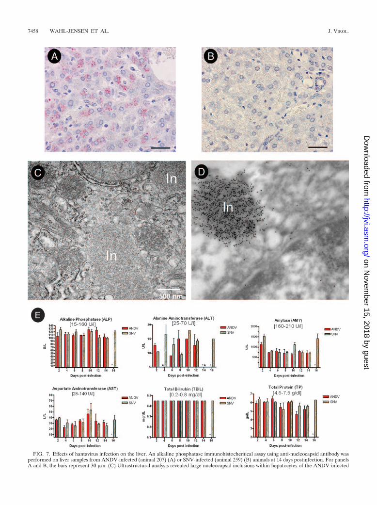
FIG. 7. Effects of hantavirus infection on the liver. An alkaline phosphatase immunohistochemical assay using anti-nucleocapsid antibody wasperformed on liver samples from ANDV-infected (animal 207) (A) or SNV-infected (animal 259) (B) animals at 14 days postinfection. For panelsA and B, the bars represent 30 �m. (C) Ultrastructural analysis revealed large nucleocapsid inclusions within hepatocytes of the ANDV-infected
7458 WAHL-JENSEN ET AL. J. VIROL.
on Novem
ber 15, 2018 by guesthttp://jvi.asm
.org/D
ownloaded from
man HPS cases (43). The ANDV/hamster lethal HPS modelhas made it possible to perform temporal studies to follow thedisease course from exposure to fulminant disease. This modelwas recently used to investigate the possibility that cardiogenicshock is involved in lethality of this disease (4). The authorsused telemetry and found that the heart rate and blood pres-sure slowly dropped during the course of infection and thatsubsequently, during the 12 h preceding death, the blood pres-sure dropped rapidly and the heart rate increased by 20%.Here, we examined the pathogenesis of HPS as it evolved overtime in the hamster and we compared the ANDV and SNVinfections. We confirmed previous findings that ANDV, butnot SNV, is lethal to the Syrian hamster. ANDV-infected ham-sters lost up to 6% of their body weight during the 48 hpreceding death. The mean time to death in this study was 11days, and signs of disease were evident only during the final fewhours before death or euthanasia. A summary of the dayspostchallenge when changes in the hamster were first observedis shown in Fig. 8. The earliest evidence of disease in ANDV-infected hamsters was apoptotic lymphocytes on day 4. Most ofthe changes, including infectious virus in the blood, viral in-
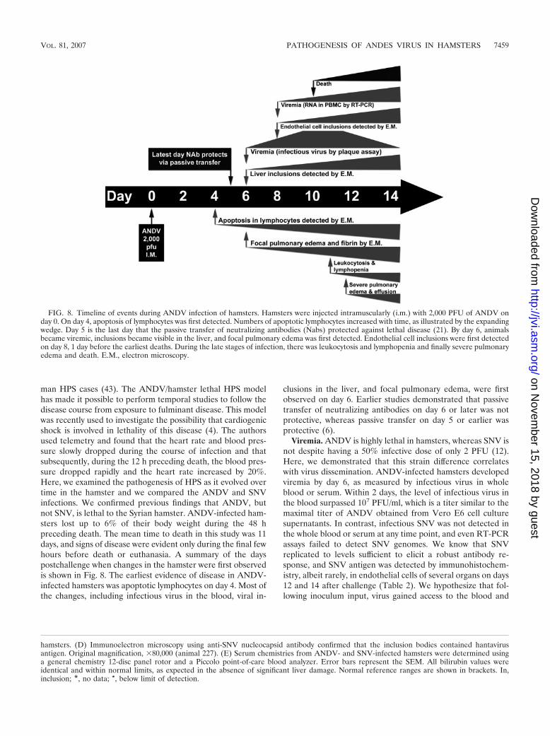
clusions in the liver, and focal pulmonary edema, were firstobserved on day 6. Earlier studies demonstrated that passivetransfer of neutralizing antibodies on day 6 or later was notprotective, whereas passive transfer on day 5 or earlier wasprotective (6).
Viremia. ANDV is highly lethal in hamsters, whereas SNV isnot despite having a 50% infective dose of only 2 PFU (12).Here, we demonstrated that this strain difference correlateswith virus dissemination. ANDV-infected hamsters developedviremia by day 6, as measured by infectious virus in wholeblood or serum. Within 2 days, the level of infectious virus inthe blood surpassed 107 PFU/ml, which is a titer similar to themaximal titer of ANDV obtained from Vero E6 cell culturesupernatants. In contrast, infectious SNV was not detected inthe whole blood or serum at any time point, and even RT-PCRassays failed to detect SNV genomes. We know that SNVreplicated to levels sufficient to elicit a robust antibody re-sponse, and SNV antigen was detected by immunohistochem-istry, albeit rarely, in endothelial cells of several organs on days12 and 14 after challenge (Table 2). We hypothesize that fol-lowing inoculum input, virus gained access to the blood and
hamsters. (D) Immunoelectron microscopy using anti-SNV nucleocapsid antibody confirmed that the inclusion bodies contained hantavirusantigen. Original magnification, �80,000 (animal 227). (E) Serum chemistries from ANDV- and SNV-infected hamsters were determined usinga general chemistry 12-disc panel rotor and a Piccolo point-of-care blood analyzer. Error bars represent the SEM. All bilirubin values wereidentical and within normal limits, as expected in the absence of significant liver damage. Normal reference ranges are shown in brackets. In,inclusion; *, no data; �, below limit of detection.
FIG. 8. Timeline of events during ANDV infection of hamsters. Hamsters were injected intramuscularly (i.m.) with 2,000 PFU of ANDV onday 0. On day 4, apoptosis of lymphocytes was first detected. Numbers of apoptotic lymphocytes increased with time, as illustrated by the expandingwedge. Day 5 is the last day that the passive transfer of neutralizing antibodies (Nabs) protected against lethal disease (21). By day 6, animalsbecame viremic, inclusions became visible in the liver, and focal pulmonary edema was first detected. Endothelial cell inclusions were first detectedon day 8, 1 day before the earliest deaths. During the late stages of infection, there was leukocytosis and lymphopenia and finally severe pulmonaryedema and death. E.M., electron microscopy.
VOL. 81, 2007 PATHOGENESIS OF ANDES VIRUS IN HAMSTERS 7459
on Novem
ber 15, 2018 by guesthttp://jvi.asm
.org/D
ownloaded from
spread to endothelial cells in various organs. Replication inthose cells could evoke an adaptive immune response andcould account for the sporadic antigen-positive cells detectedat late time points by immunohistochemistry. Interestingly,staining was of single cells rather than foci, suggesting that viralspread, even to adjacent cells, was inhibited.
It remains unclear why ANDV was able to readily dissemi-nate in the hamster while the closely related SNV did not. Thiscould be due to differences in growth kinetics between thesetwo viruses. In cell culture, ANDV grows to higher titers withfaster kinetics than SNV (26). However, if a lag in SNV growthis responsible for the decreased virulence in hamsters, then wewould expect to detect SNV viremia at a later time point (e.g.,day 8 or 10), unless this lag delayed dissemination of SNV longenough for the adaptive immune response to neutralize theinfection before dissemination could occur.
An alternative explanation for the absence of a SNV viremiacould be innate immunity. It is possible that the innate immunesystem of hamsters rapidly recognized SNV as a threat andfunctioned to contain and suppress the infection. If this oc-curred, then ANDV must have somehow evaded the innateimmune responses, either by passively replicating as an unrec-ognized threat or by actively disrupting innate immunity. Nu-merous viruses produce proteins that act as interferon antag-onists or otherwise disrupt the innate immune system (2).ANDV was recently shown to interfere with interferon signal-ing in vitro (36). Research investigating the role of innateimmunity in the differential outcome following infection ofhamsters with SNV or ANDV is in progress.
Although viral nucleic acid is routinely detected in the bloodand plasma of HPS patients by RT-PCR (38), infectious virushas never been isolated from an HPS patient. There is a singlereport describing the isolation of an HPS-associated hantavirus(ANDV) from an asymptomatic individual who later devel-oped lethal HPS (8). High loads of viral nucleic acid in humansat hospital admission were associated with severe disease (41).In contrast, high SNV-neutralizing antibody titers at hospitaladmission were associated with a mild disease course (42). Theinability to detect infectious virus in HPS patients could be dueto neutralization of virus by the immune system before theonset of disease symptoms.
Lymphocyte apoptosis. The earliest pathology observed forANDV-infected hamsters was the presence of apoptotic lym-phocytes in lung capillaries starting on day 4 postchallenge asdetected by electron microscopy. Apoptosis is an evolutionarilyconserved and extremely ordered type of cell death that isimportant in regulating normal physiology and under certaincircumstances plays a role in pathology (33). For example,during septic shock, patients exhibit absolute lymphocytecounts below the normal limits due to apoptosis (13). Therehave been several reports of in vitro studies that investigated ifhantaviruses (largely HFRS associated) induce apoptosis ineither Vero E6 cells (10, 15, 18–20), HEK293 cells (23), orperipheral blood lymphocytes (1). While some studies suggestthat induction of apoptosis is replication dependent (15, 18),Markotic et al. suggest a bystander phenomenon (23). Zaki etal. did report the occasional presence of viral antigen withinlymphocytes of humans infected with SNV (43). In our ham-ster model, lymphocyte apoptosis was detected early (2 daysbefore viremia was detected). We did not observe the presence
of viral inclusions within lymphocytes, suggesting a bystandermechanism. In hamsters infected with ANDV, early apoptosisof lymphocytes could result in immune dysfunction allowingdissemination of virus and subsequent fulminant disease. Ashamster-specific reagents become available, it will become pos-sible to test this hypothesis.
Vascular leakage. The primary abnormality in HFRS andHPS is vascular leakage. This leakage could be caused directlyby interaction and/or infection of endothelial cells with hanta-viruses, or indirectly by the immune response, or by a combi-nation of direct and indirect effects on the cells. Direct inter-action of pathogenic hantaviruses with v�3 integrins has beenproposed as a possible mechanism of pathogenesis (22). Thehigh level of viremia that we observed for the hamsters in-fected with ANDV allows the possibility that virus interactionwith endothelial cell receptors, and not infection per se, couldcontribute to vascular leakage. It might be possible to test theeffects of virus-cell interaction but not infection in vivo byinfusion of high levels of inactivated virus, or pseudotypedvirus, over a period of 2 or more days.
One of the hallmarks of in vitro hantavirus infection is theabsence of cytolysis or marked cytopathic effect. However, thisabsence does not rule out the possibility that the cell functionshave been disrupted. For example, infected Vero E6 cells donot take up neutral red in the same way uninfected cells do,and this difference is the basis for the hantavirus plaque assay.Our electron microscopy data confirm earlier H&E observa-tions that cytolyis and necrosis are not occurring in the endo-thelial cells of ANDV-infected hamsters. Ultrastructural anal-ysis did not reveal gaps between adjacent endothelial cells,suggesting that paracellular permeability was not the mecha-nism of leakage. However, the possibility that undetected tran-sient formation of gaps between cells might occur cannot beruled out. Interestingly, we observed increased numbers ofvacuoles in the endothelial cells of infected tissue (Fig. 5B).Many of these cells contained viral inclusions, suggesting thatthe mechanism of leakage might involve transcellular transportof fluid through productively infected cells. If transcellulartransport of plasma across endothelial cells were to occur, thefluid might affect the basement membrane and epithelial cellsthat form the barrier between the capillary lumen and alveolarspace. Supporting this, we observed the presence of edema inlung epithelial cells, and in some cases the basement mem-brane appeared diffuse and possibly disrupted (Fig. 5D and E).It is remarkable that these were the only ultrastructural abnor-malities observed despite the fact that very high volumes offluid crossed from the lung capillaries into the alveolar spacesand pleural cavity.
Despite some evidence for a direct role of hantavirus infec-tion on endothelial cell leakage, there are several lines ofevidence indicating that the immune response plays a role inthe pathogenesis. For example, we observed large numbers ofmononuclear cell infiltrates within the alveoli and in theperivascular interstitium. It is possible that the cytokines re-leased by these cells caused the vascular leakage. Indeed, thereare some compelling data in the hantavirus literature to sup-port this theory. Immunohistological analysis of kidney biop-sies taken during the acute phase of HFRS caused by Puumalavirus revealed an increase in levels of local cytokines includingtumor necrosis factor alpha (TNF-), platelet-derived growth
7460 WAHL-JENSEN ET AL. J. VIROL.
on Novem
ber 15, 2018 by guesthttp://jvi.asm
.org/D
ownloaded from
factor, and transforming growth factor beta (37). In the samestudy, increased expression levels of ICAM-1, VCAM-1, andPECAM were also seen in the same sites of the peritubulararea of distal nephrons. Systemic levels of inflammatory cyto-kines have also been reported for plasma of HFRS patientsand include TNF-, soluble TNF- receptor, interleukin-6 (IL-6), and IL-10 (21). The increased levels of TNF- seen inHFRS may have a profound impact both locally and systemi-cally on vascular integrity. TNF-, along with other proinflam-matory mediators, is known to cause reduced hepatic, renal,pulmonary, and/or cardiac functions during septic shock andviral hemorrhagic fever infection caused by filoviruses (3). El-evated levels of TNF- are capable of altering endothelial celljunctions, actin stress fibers, and ultimately vascular perme-ability (7, 40). Many of the symptoms seen during hantavirusinfection in humans, including fever, chills, myalgia, hypoten-sion, and edema formation, are reminiscent of septic shock.Increased levels of proinflammatory mediators during hanta-virus infection are not limited to HFRS. Mori et al. describedincreased production of vasoactive cytokines (IL-1, IL-1�,IL-6, and TNF-) throughout the lung tissue of HPS patients(27). Gamma interferon and IL-2 were also detected in thelungs of HPS patients, and gamma interferon from activated Tcells may act to exacerbate shock in vivo (27). Finally, a geneticpredisposition to high-level production of TNF- through theTNF2 allele was observed more frequently for hospitalizedHFRS cases than for normal controls (14). As reagents formeasuring hamster cytokines and chemokines become avail-able, it will be possible to determine what role(s), if any, proin-flammatory and/or anti-inflammatory responses play in theHPS caused by ANDV in hamsters.
Concluding remarks. Previous studies by our laboratoryhave shown that the latest time point that passive transfer ofneutralizing antibodies will protect against a lethal ANDVchallenge is day 5 postinfection (6). In this study, we observedthat ANDV-infected hamsters were viremic by day 6 postin-fection, an observation that may explain the lack of efficacy forpassive transfer of neutralizing antibodies at later times. Pa-thology in the lung was first observed at day 8 postinfection,shortly after the viremia. We suspect that ANDV replicated atlow levels during the first 5 days of infection and then rapidlyburst from the initial infection site and spread to multipleorgans, likely by seeding endothelial cells that were uniformlyinfected. Massive nucleocapsid inclusions were observed forendothelial cells of all organs examined; however, endothelialcell architecture was largely unaffected. Furthermore, theseinclusions were observed for hepatocytes and interestingly didnot alter the ability of these cells to function, as measured byblood chemistry analysis (Fig. 7). Taken together, these datasuggest that hantavirus countermeasures aimed at preventingvirus replication and dissemination will have the greatest prob-ability of success if administered within the first 5 days postex-posure (before the viremic phase). Nevertheless, our data sug-gest that because vascular leakage appears to be associatedwith infected endothelial cells, a therapeutic strategy targetingviral replication might be effective in treating this disease evenat later times. This is important because most HPS patients arefirst identified upon admission to the emergency room afterdisease onset. In the present study, we identified previouslyunrecognized similarities between the hamster model and HPS
in humans, including lymphopenia, neutrophilia, mild throm-bocytopenia, and lymphocyte apoptosis. The hamster modelwill be an important system for testing the effectiveness ofcandidate antiviral modalities administered at early and latetimes in the disease course.
ACKNOWLEDGMENTS
We gratefully acknowledge C. Mech for her work on immunohisto-chemistry and G. Krietz, J. Brubaker, N. Davis, S. Moon, K. Kuehl, andJ. Smith for excellent technical assistance. All experiments involving theuse of ANDV or SNV in animals were performed in USAMRIID’s BSL-4laboratory, and all other work involving infectious hantaviruses were per-formed in a BSL-3 laboratory at USAMRIID. Research was conductedin compliance with the Animal Welfare Act and other federal statutesand regulations relating to animals and experiments involving animalsand adheres to principles stated in the Guide for the Care and Use ofLaboratory Animals, National Research Council, 1996 (27a). The fa-cility where this research was conducted is fully accredited by theAssociation for Assessment and Accreditation of Laboratory AnimalCare International.
The research described herein was sponsored by the Military Infec-tious Disease Research Program, U.S. Army Medical and MaterialCommand, project no. AT0002_04_RD. This research was performedwhile V. Wahl-Jensen and R. Fisher held National Research CouncilResearch Associateship Awards at USAMRIID.
Opinions, interpretations, conclusions, and recommendations areours and are not necessarily endorsed by the U.S. Army or the De-partment of Defense.
REFERENCES
1. Akhmatova, N. K., R. S. Yusupova, S. F. Khaiboullina, and S. V. Sibiryak.2003. Lymphocyte apoptosis during hemorragic fever with renal syndrome.Russ. J. Immunol. 8:37–46.
2. Basler, C. F., and A. Garcia-Sastre. 2002. Viruses and the type I interferonantiviral system: induction and evasion. Int. Rev. Immunol. 21:305–337.
3. Bray, M., and S. Mahanty. 2003. Ebola hemorrhagic fever and septic shock.J. Infect. Dis. 188:1613–1617.
4. Campen, M. J., M. L. Milazzo, C. F. Fulhorst, C. J. Obot Akata, and F.Koster. 2006. Characterization of shock in a hamster model of hantavirusinfection. Virology 356:45–49.
5. Centers for Disease Control and Prevention. 2006. Hantavirus pulmonarysyndrome–five states, 2006. Morb. Mortal. Wkly. Rep. 55:627–629.
6. Custer, D. M., E. Thompson, C. S. Schmaljohn, T. G. Ksiazek, and J. W.Hooper. 2003. Active and passive vaccination against hantavirus pulmonarysyndrome with Andes virus M genome segment-based DNA vaccine. J. Virol.77:9894–9905.
7. Feldmann, H., H. Bugany, F. Mahner, H. D. Klenk, D. Drenckhahn, andH. J. Schnittler. 1996. Filovirus-induced endothelial leakage triggered byinfected monocytes/macrophages. J. Virol. 70:2208–2214.
8. Galeno, H., J. Mora, E. Villagra, J. Fernandez, J. Hernandez, G. J. Mertz,and E. Ramirez. 2002. First human isolate of Hantavirus (Andes virus) in theAmericas. Emerg. Infect. Dis. 8:657–661.
9. Goldsmith, C. S., L. H. Elliott, C. J. Peters, and S. R. Zaki. 1995. Ultra-structural characteristics of Sin Nombre virus, causative agent of hantaviruspulmonary syndrome. Arch. Virol. 140:2107–2122.
10. Hardestam, J., J. Klingstrom, K. Mattsson, and A. Lundkvist. 2005. HFRScausing hantaviruses do not induce apoptosis in confluent Vero E6 andA-549 cells. J. Med. Virol. 76:234–240.
11. Hooper, J. W., K. I. Kamrud, F. Elgh, D. Custer, and C. S. Schmaljohn. 1999.DNA vaccination with hantavirus M segment elicits neutralizing antibodiesand protects against Seoul virus infection. Virology 255:269–278.
12. Hooper, J. W., T. Larsen, D. M. Custer, and C. S. Schmaljohn. 2001. A lethaldisease model for hantavirus pulmonary syndrome. Virology 289:6–14.
13. Hotchkiss, R. S., P. E. Swanson, B. D. Freeman, K. W. Tinsley, J. P. Cobb,G. M. Matuschak, T. G. Buchman, and I. E. Karl. 1999. Apoptotic cell deathin patients with sepsis, shock, and multiple organ dysfunction. Crit. CareMed. 27:1230–1251.
14. Kanerva, M., A. Vaheri, J. Mustonen, and J. Partanen. 1998. High-producerallele of tumour necrosis factor-alpha is part of the susceptibility MHChaplotype in severe puumala virus-induced nephropathia epidemica. Scand.J. Infect. Dis. 30:532–534.
15. Kang, J. I., S. H. Park, P. W. Lee, and B. Y. Ahn. 1999. Apoptosis is inducedby hantaviruses in cultured cells. Virology 264:99–105.
16. Lee, J. S., J. Lahdevirta, F. Koster, and H. Levy. 1999. Clinical manifesta-tions and treatment of HFRS and HPS, p. 17–38. In H. W. Lee, C. Calisher,and C. Schmaljohn (ed.), Manual of hemorrhagic fever with renal syndrome
VOL. 81, 2007 PATHOGENESIS OF ANDES VIRUS IN HAMSTERS 7461
on Novem
ber 15, 2018 by guesthttp://jvi.asm
.org/D
ownloaded from

and hantavirus pulmonary syndrome. ASAN Institute for Life Sciences,Seoul, Korea.
17. Levis, S., J. E. Rowe, S. Morzunov, D. A. Enria, and S. St Jeor. 1997. Newhantaviruses causing hantavirus pulmonary syndrome in central Argentina.Lancet 349:998–999.
18. Li, X. D., S. Kukkonen, O. Vapalahti, A. Plyusnin, H. Lankinen, and A.Vaheri. 2004. Tula hantavirus infection of Vero E6 cells induces apoptosisinvolving caspase 8 activation. J. Gen. Virol. 85:3261–3268.
19. Li, X. D., H. Lankinen, N. Putkuri, O. Vapalahti, and A. Vaheri. 2005. Tulahantavirus triggers pro-apoptotic signals of ER stress in Vero E6 cells.Virology 333:180–189.
20. Li, X. D., T. P. Makela, D. Guo, R. Soliymani, V. Koistinen, O. Vapalahti, A.Vaheri, and H. Lankinen. 2002. Hantavirus nucleocapsid protein interactswith the Fas-mediated apoptosis enhancer Daxx. J. Gen. Virol. 83:759–766.
21. Linderholm, M., C. Ahlm, B. Settergren, A. Waage, and A. Tarnvik. 1996.Elevated plasma levels of tumor necrosis factor (TNF)-alpha, soluble TNFreceptors, interleukin (IL)-6, and IL-10 in patients with hemorrhagic feverwith renal syndrome. J. Infect. Dis. 173:38–43.
22. Mackow, E. R., and I. N. Gavrilovskaya. 2001. Cellular receptors and han-tavirus pathogenesis. Curr. Top. Microbiol. Immunol. 256:91–115.
23. Markotic, A., L. Hensley, T. Geisbert, K. Spik, and C. Schmaljohn. 2003.Hantaviruses induce cytopathic effects and apoptosis in continuous humanembryonic kidney cells. J. Gen. Virol. 84:2197–2202.
24. Martinez, V. P., C. Bellomo, J. San Juan, D. Pinna, R. Forlenza, M. Elder,and P. J. Padula. 2005. Person-to-person transmission of Andes virus.Emerg. Infect. Dis. 11:1848–1853.
25. McElroy, A. K., M. Bray, D. S. Reed, and C. S. Schmaljohn. 2002. Andesvirus infection of cynomolgus macaques. J. Infect. Dis. 186:1706–1712.
26. McElroy, A. K., J. M. Smith, J. W. Hooper, and C. S. Schmaljohn. 2004.Andes virus M genome segment is not sufficient to confer the virulenceassociated with Andes virus in Syrian hamsters. Virology 326:130–139.
27. Mori, M., A. L. Rothman, I. Kurane, J. M. Montoya, K. B. Nolte, J. E.Norman, D. C. Waite, F. T. Koster, and F. A. Ennis. 1999. High levels ofcytokine-producing cells in the lung tissues of patients with fatal hantaviruspulmonary syndrome. J. Infect. Dis. 179:295–302.
27a.National Research Council. 1996. Guide for the care and use of laboratoryanimals. National Academy Press, Washington, DC.
28. Nichol, S. T. 2001. Bunyaviruses, p. 1603. In D. M. Knipe, P. M. Howley,and D. E. Griffin, R. A. Lamb, M. A. Martin, B. Roizman, and S. E.Straus (ed.), Fields virology, 4th ed., vol. 1. Lippincott Williams &Wilkins, Philadelphia, PA.
29. PAHO. 2004. Number of cases of hantavirus pulmonary syndrome (HPS)(region of the Americas, 1993–2004). http://www.paho.org/common/Display.asp?Lang�E&RecID�1971.
30. Pinna, D. M., V. P. Martinez, C. M. Bellomo, C. Lopez, and P. Padula. 2004.New epidemiologic and molecular evidence of person to person transmissionof hantavirus Andes Sout. Medicina (Buenos Aires) 64:43–46. (In Spanish.)
31. ProMED-mail. 2006. Hantavirus pulmonary syndrome-Canada ex Bolivia.http://www.promedmail.org/pls/promed/f?p�2400:1001:9753834909080299352::NO::F2400_P1001_BACK_PAGE,F2400_P1001_PUB_MAIL_ID:1010,33838.
32. Prophet, E. B., B. Mills, J. B. Arrington, and L. H. Sobin. 1992. Laboratorymethods for histotechnology. Armed Forces Institute of Pathology, Wash-ington, DC.
33. Raff, M. 1998. Cell suicide for beginners. Nature 396:119–122.34. Schmaljohn, A. L., D. Li, D. L. Negley, D. S. Bressler, M. J. Turell, G. W.
Korch, M. S. Ascher, and C. S. Schmaljohn. 1995. Isolation and initialcharacterization of a newfound hantavirus from California. Virology 206:963–972.
35. Schmaljohn, C., and J. W. Hooper. 2001. Bunyaviridae: the viruses and theirreplication, p. 1581–1602. In B. N. Fields, D. M. Knipe, and P. M. Howley(ed.), Field’s virology. Lippincott-Raven, Philadelphia, PA.
36. Spiropoulou, C. F., C. G. Albarino, T. G. Ksiazek, and P. E. Rollin. 2007.Andes and Prospect Hill hantaviruses differ in early induction of interferonalthough both can down-regulate interferon signaling. J. Virol. 81:2769–2776.
37. Temonen, M., J. Mustonen, H. Helin, A. Pasternack, A. Vaheri, and H.Holthofer. 1996. Cytokines, adhesion molecules, and cellular infiltration innephropathia epidemica kidneys: an immunohistochemical study. Clin. Im-munol. Immunopathol. 78:47–55.
38. Terajima, M., J. D. Hendershot III, H. Kariwa, F. T. Koster, B. Hjelle, D.Goade, M. C. DeFronzo, and F. A. Ennis. 1999. High levels of viremia inpatients with the hantavirus pulmonary syndrome. J. Infect. Dis. 180:2030–2034.
39. Tischler, N. D., H. Galeno, M. Rosemblatt, and P. D. Valenzuela. 2005.Human and rodent humoral immune responses to Andes virus structuralproteins. Virology 334:319–326.
40. Wahl-Jensen, V. M., T. A. Afanasieva, J. Seebach, U. Stroher, H. Feldmann,and H. J. Schnittler. 2005. Effects of Ebola virus glycoproteins on endothe-lial cell activation and barrier function. J. Virol. 79:10442–10450.
41. Xiao, R., S. Yang, F. Koster, C. Ye, C. Stidley, and B. Hjelle. 2006. SinNombre viral RNA load in patients with hantavirus cardiopulmonary syn-drome. J. Infect. Dis. 194:1403–1409.
42. Ye, C., J. Prescott, R. Nofchissey, D. Goade, and B. Hjelle. 2004. Neutralizingantibodies and Sin Nombre virus RNA after recovery from hantavirus car-diopulmonary syndrome. Emerg. Infect. Dis. 10:478–482.
43. Zaki, S. R., P. W. Greer, L. M. Coffield, C. S. Goldsmith, K. B. Nolte, K.Foucar, R. M. Feddersen, R. E. Zumwalt, G. L. Miller, A. S. Khan, et al.1995. Hantavirus pulmonary syndrome. Pathogenesis of an emerging infec-tious disease. Am. J. Pathol. 146:552–579.
44. Zaki, S. R., and K. B. Nolte. 1999. Pathology, immunohistochemistry and insitu hybridization, p. 143–154. In H. W. Lee, C. E. Calisher, and C. Schmal-john (ed.), Manual of hemorrhagic fever with renal syndrome and hantaviruspulmonary syndrome. ASAN Institute for Life Sciences, Seoul, Korea.
7462 WAHL-JENSEN ET AL. J. VIROL.
on Novem
ber 15, 2018 by guesthttp://jvi.asm
.org/D
ownloaded from