Telomere Shortening and Oxidative Stress in Aged ... · the physiological functions required for...
10
of August 25, 2018. This information is current as STAT5a Phosphorylation Aged Macrophages Results in Impaired Telomere Shortening and Oxidative Stress in Lloberas, María A. Blasco and Antonio Celada Carlos Sebastián, Carmen Herrero, Maria Serra, Jorge http://www.jimmunol.org/content/183/4/2356 doi: 10.4049/jimmunol.0901131 July 2009; 2009; 183:2356-2364; Prepublished online 15 J Immunol References http://www.jimmunol.org/content/183/4/2356.full#ref-list-1 , 20 of which you can access for free at: cites 52 articles This article average * 4 weeks from acceptance to publication Fast Publication! • Every submission reviewed by practicing scientists No Triage! • from submission to initial decision Rapid Reviews! 30 days* • Submit online. ? The JI Why Subscription http://jimmunol.org/subscription is online at: The Journal of Immunology Information about subscribing to Permissions http://www.aai.org/About/Publications/JI/copyright.html Submit copyright permission requests at: Email Alerts http://jimmunol.org/alerts Receive free email-alerts when new articles cite this article. Sign up at: Print ISSN: 0022-1767 Online ISSN: 1550-6606. Immunologists, Inc. All rights reserved. Copyright © 2009 by The American Association of 1451 Rockville Pike, Suite 650, Rockville, MD 20852 The American Association of Immunologists, Inc., is published twice each month by The Journal of Immunology by guest on August 25, 2018 http://www.jimmunol.org/ Downloaded from by guest on August 25, 2018 http://www.jimmunol.org/ Downloaded from
Transcript of Telomere Shortening and Oxidative Stress in Aged ... · the physiological functions required for...
of August 25, 2018.This information is current as
STAT5a PhosphorylationAged Macrophages Results in Impaired Telomere Shortening and Oxidative Stress in
Lloberas, María A. Blasco and Antonio CeladaCarlos Sebastián, Carmen Herrero, Maria Serra, Jorge
http://www.jimmunol.org/content/183/4/2356doi: 10.4049/jimmunol.0901131July 2009;
2009; 183:2356-2364; Prepublished online 15J Immunol
Referenceshttp://www.jimmunol.org/content/183/4/2356.full#ref-list-1
, 20 of which you can access for free at: cites 52 articlesThis article
average*
4 weeks from acceptance to publicationFast Publication! •
Every submission reviewed by practicing scientistsNo Triage! •
from submission to initial decisionRapid Reviews! 30 days* •
Submit online. ?The JIWhy
Subscriptionhttp://jimmunol.org/subscription
is online at: The Journal of ImmunologyInformation about subscribing to
Permissionshttp://www.aai.org/About/Publications/JI/copyright.htmlSubmit copyright permission requests at:
Email Alertshttp://jimmunol.org/alertsReceive free email-alerts when new articles cite this article. Sign up at:
Print ISSN: 0022-1767 Online ISSN: 1550-6606. Immunologists, Inc. All rights reserved.Copyright © 2009 by The American Association of1451 Rockville Pike, Suite 650, Rockville, MD 20852The American Association of Immunologists, Inc.,
is published twice each month byThe Journal of Immunology
by guest on August 25, 2018
http://ww
w.jim
munol.org/
Dow
nloaded from
by guest on August 25, 2018
http://ww
w.jim
munol.org/
Dow
nloaded from
Telomere Shortening and Oxidative Stress in AgedMacrophages Results in Impaired STAT5a Phosphorylation1
Carlos Sebastian,* Carmen Herrero,* Maria Serra,* Jorge Lloberas,* María A. Blasco,†
and Antonio Celada2*
Macrophages are an essential component of both innate and adaptive immunity, and altered function of these cells with aging mayplay a key role in immunosenescence. To determine the effect of aging on macrophages, we produced bone marrow-derivedmacrophages in vitro. In these conditions, we analyzed the effect of aging on macrophages without the influence of other cell typesthat may be affected by aging. We showed that telomeres shorten with age in macrophages leading to a decreased GM-CSF butnot M-CSF-dependent proliferation of these cells as a result of decreased phosphorylation of STAT5a. Macrophages from agedmice showed increased susceptibility to oxidants and an accumulation of intracellular reactive oxygen species. In these macro-phages STAT5a oxidation was reduced, which led to the decreased phosphorylation observed. Interestingly, the same cellulardefects were found in macrophages from telomerase knockout (Terc�/�) mice suggesting that telomere loss is the cause for theenhanced oxidative stress, the reduced Stat5a oxidation and phosphorylation and, ultimately, for the impaired GM-CSF-depen-dent macrophage proliferation. The Journal of Immunology, 2009, 183: 2356–2364.
A ging can be defined as the time-related deterioration ofthe physiological functions required for survival and fer-tility. Among the functions affected by aging, the im-
mune system has been shown to be dysregulated with advancingage, thus leading to increased susceptibility to viral and bacterialinfections, reactivation of latent viruses, and decreased response tovaccines (1).
Macrophages from aged humans, rats, and mice display severaldefects in their functional activities (2), which lead to impairmentin the first line of immune defense and to a decreased capacity tocontribute to the development of specific immune responses bypresenting Ags to T cells and producing regulatory cytokines.Thus, these alterations of macrophage functions contribute to theimpairment of the immune system during aging, a process calledimmunosenescence, which is associated with increased mortalityand a major incidence of immune diseases and cancer in theelderly.
Telomeres are chromatin structures that cap and protect the endof chromosomes. In vertebrates, they are formed by tandem re-peats of hexamer sequences (TTAGGG) that are associated withvarious specific proteins involved in the maintenance and regula-tion of telomere length (3). Telomere shortening is involved in theaging process and in the regulation of replicative lifespan (4). Thisage-related shortening and the observation that several prematureaging syndromes are associated with short telomeres support theconcept that telomere length homeostasis is a crucial determinantof human longevity. Moreover, late generations of the telomerase
knockout mice, Terc�/�, which show severe telomere dysfunctioncharacterized by critically short telomeres, suffer from prematureaging and various age-related diseases that affect highly prolifer-ative tissues (5, 6). Among these, the generation and function ofimmune cells has been shown to be affected by telomere attrition.T, B, and NK cells show telomere shortening during aging, whichis associated with impaired proliferation and function of these cells(6, 7).
In this study, we show that telomeres shorten with age in mousemacrophages. We used bone marrow-derived macrophages fromyoung and aged mice grown and differentiated in vitro. This modelallows us to analyze the effect of aging without the influence ofother cell types that may be affected by this process (8). Telomereshortening is associated with impaired GM-CSF- but not M-CSF-dependent proliferation of macrophages, which is caused by a re-duced phosphorylation of STAT5a. These cells also show in-creased intracellular levels of reactive oxygen species (ROS)3 andan enhanced susceptibility to oxidative stress. In addition, we dem-onstrate that STAT5a oxidation is required for its phosphorylationand that this oxidation is reduced in macrophages from aged mice.Macrophages from Terc�/� mice, which have a similar telomerelength as aged macrophages, show the same phenotype as agedmacrophages. Thus, these results point to telomere shortening asthe molecular determinant of some aspects of macrophage agingby modulating oxidative stress.
Materials and MethodsReagents
Recombinant murine cytokines were purchased from Sigma-Aldrich. TheAbs used were anti-STAT5a, anti-STAT5b (R&D Systems), anti-phospho-STAT5a/b Y694/Y699, anti-�-H2AX (Ser139), GM-CSF-R� (UpstateBiotechnology), anti-histone H1, GM-CSR-R� (SantaCruz Biotechnology)anti-�-actin (Sigma-Aldrich). Peroxidase-conjugated anti-rabbit (JacksonImmunoResearch Laboratories) or anti-mouse (Sigma-Aldrich) were usedas secondary Abs. All other chemicals were of the highest purity grade
*Institute for Research in Biomedicine and University of Barcelona, Barcelona,Spain; and †Telomeres and Telomerase Group, Molecular Oncology Program, Span-ish National Cancer Centre, Madrid, Spain
Received for publication April 8, 2009. Accepted for publication June 16, 2009.
The costs of publication of this article were defrayed in part by the payment of pagecharges. This article must therefore be hereby marked advertisement in accordancewith 18 U.S.C. Section 1734 solely to indicate this fact.1 This work was supported by a Grant from the MEC BFU 2007-63712/BMC (toA.C.).2 Address correspondence and reprint requests to Prof. Antonio Celada, Institute ofResearch in Biomedicine, Baldiri Reixac 10, Barcelona Science Park, University ofBarcelona, E-08028 Barcelona, Spain. E-mail address: [email protected]
3 Abbreviations used in this paper: ROS, reactive oxygen species; DCF-DA, dichlo-rofluorescin diacetate; NAC, N-acetylcysteine; WT, wild type.
Copyright © 2009 by The American Association of Immunologists, Inc. 0022-1767/09/$2.00
The Journal of Immunology
www.jimmunol.org/cgi/doi/10.4049/jimmunol.0901131
by guest on August 25, 2018
http://ww
w.jim
munol.org/
Dow
nloaded from
available and were purchased from Sigma-Aldrich. Deionized water furtherpurified with a Millipore Milli-Q system A10 was used.
Cell culture
Bone marrow-derived macrophages were isolated from young (6 wk) oraged (18–24 mo) C57BL/6 mice (Charles River Laboratories and NationalInstitute on Aging) as described (9). Mice C57BL/6 (6 wk old) lacking thegene encoding the telomerase RNA component (Terc�/�) were generatedas described (10). Bone marrow-derived macrophages from wild type(WT) and Terc�/� first and third generation were used. The use of animalswas approved by the Animal Research Committee of the University ofBarcelona (no. 2523).
RNA extraction and real-time RT-PCR
Cells were washed twice with cold PBS, and total RNA was extracted withthe EZ-RNA kit as described by the manufacturer (Biological Industries).RNA was treated with DNase (Roche) to remove contaminating DNA. ForcDNA synthesis, 1 �g of total RNA and M-MLV reverse transcriptaseRNase H minus, Point Mutant, oligo(dT)15 primer and PCR nucleotide mixwere used, as described by the manufacturer (Promega Corporation). Real-time PCR was performed using the power SYBR green master mix (Ap-plied Biosystems), following the manufacturer’s instructions, with the ex-ception that the final volume was 12.5 �l of SYBR green reaction mix.Real-time monitoring of PCR amplification was performed in the ABIPrism 7900 sequence detection system (Applied Biosystems). Data wereexpressed as relative mRNA levels normalized to the �-actin expressionlevel in each sample. The primer sequences can be obtained upon request.
Proliferation assay
Macrophage proliferation was measured by [3H]thymidine incorporation asdescribed (11). Cells were deprived of M-CSF for 16–18 h and then 105
cells were incubated for 24 h in complete medium in the presence of thegrowth factor. After this period, the medium was replaced with mediumcontaining [3H]thymidine. After an additional 6 h of incubation, the me-dium was removed and the cells were fixed in ice-cold 70% methanol.After three washes, cells were solubilized and radioactivity was measured.Each point was performed in triplicate, and the results are expressed as themean � SD. For cell counting we used a hemocytometer.
Cell cycle analysis
Cell cycle was analyzed as described (12). Cells were fixed with EtOH95%, incubated with propidium iodide plus RNase A, and then analyzed byFACS. Cell cycle distributions were analyzed with the Multicycle program(Phoenix Flow Systems).
Protein analysis
Cells were lysed as described (13) in lysis buffer (1% Triton X-100, 10%glycerol, 50 mM HEPES (pH 7.5), 150 mM NaCl, protease inhibitors and1 mM sodium orthovanadate). For immunoprecipitation assays, 150 �g ofcell lysates were mixed with 75 �l of 20% protein A-Sepharose (Sigma-Aldrich) and 2 �l of specific Abs anti-STAT5a and STAT5b to a finalvolume of 500 �l. The reaction was conducted overnight at 4°C in rotation.After three washes in cold washing buffer (PBS, 1% Nonidet P-40, 2 mMsodium orthovanadate) pellets were heated to 95°C in Laemmli SDS load-ing buffer, and proteins were resolved by SDS-PAGE. For histone H2AXWestern blot, an acid extraction of proteins was performed as described bythe anti-phospho-H2AX Ab manufacturer. Briefly, cells were washed incold PBS and lysed with lysis buffer (10 mM HEPES (pH 7.9), 1.5 mMMgCl2, 10 mM KCl, 0.5 mM DTT, and 1.5 mM PMSF). Hydrochloric acidwas added to a final concentration of 0.2 N, and the cell lysate was incu-bated on ice for 30 min. After centrifugation at 11,000 � g for 10 min at4°C, supernatants were dialyzed twice against 200 ml of 0.1 M acetic acidfor 1–2 h and three times against 200 ml of H2O for 1 h, 3 h, and overnight,respectively. Detection of oxidized proteins was performed by immuno-precipitating STAT5a and STAT5b and using the OxiBlot protein oxida-tion detection kit (Chemicon International), which detects the carbonylgroups introduced into proteins by oxidative reactions. These carbonylgroups were derivatized to dinitrophenylhydrazone by reaction with dini-trophenylhydrazine. The dinitrophenylhydrazone-derivatized proteins wereseparated by SDS-PAGE followed by Western blotting.
Nuclear extract preparation and DNA-binding assay
Nuclear extracts were prepared as follows: cell pellets were washed twicewith cold PBS buffer and resuspended in buffer A (10 mM HEPES, 10 mMKCl, 0.1 mM EDTA, 0.1 mM EGTA, 1 mM DTT, 0.5 mM PMSF, 0.01
mg/ml aprotinin, 0.01 mg/ml leupeptin, 0.086 mg/ml iodacetamide, 1 mMsodium orthovanadate) and incubated on ice for 15 min. Cells were lysedby adding 10% Nonidet P-40 and mixing by vortex. Nuclei were collectedat the bottom of the tube by a 30-s centrifugation. After three washes withbuffer A, nuclei were lysed with buffer B (20 mM HEPES, 0.4 M NaCl, 1mM EDTA, 1 mM EGTA, 1 mM DTT, 1 mM PMSF, 5 mM sodiumfluoride, 0.01 mg/ml aprotinin, 0.01 mg/ml leupeptin, 0.086 mg/ml iodac-etamide, 1 mM sodium orthovanadate) at 4°C for 30 min in rotation. Ly-sates were centrifuged for 20 min at 4°C at 13,000 rpm, and supernatantswere stored at �80°C until use. EMSAs were performed as described (14).Briefly, binding reactions were prepared with 6 �g of nuclear extractsand 20,000 cpm 32P-labeled probe in the presence of 2 �g of poly(dI:dC) in a final volume of 15 �l containing 1� binding buffer (12 mMHEPES (pH 7.9), 60 mM KCl, 5 mM MgCl2, 0.12 mM EDTA, 0.3 mMPMSF, 0.3 mM DTT, 12% glycerol). The nuclear extracts were incu-bated for 8 min with poly(dI:dC) and then the radiolabeled probe wasadded and incubated for an additional 15 min at room temperature.Samples were loaded onto 4% acrylamide gel containing 5% glyceroland 0.25% TBE, and electrophoresed at 4°C. Band-shift gels weredried, and bands were visualized by autoradiography. For supershiftexperiments, following the binding reaction, 2 �l of anti-STAT5a andanti-STAT5b Abs were added and incubated for 30 min. The oligonu-cleotides used in the assay were 5�-end labeled using T4 polynucleotidekinase (USB Corporation). The probe was synthesized by Genotek andcorresponds to a GAS-like element from the promoter of the bovine�-casein gene (5�-AGATTTCTAGGAATTCAAATC-3�).
DNA damage susceptibility assay
To analyze macrophage susceptibility to various DNA-damaging agents,we treated the cells with etoposide (Tocris) and hydrogen peroxide (Sigma-Aldrich) at the concentrations indicated during 1 h and UV radiation (� �254 nm) at the energies indicated. Cells were washed and left in completemedium (DMEM � 10% FCS � 5 ng/ml M-CSF) for 5 days. Cells werecounted by trypan blue exclusion, and cell growth was determined as theratio of number of treated cells relative to number of control cells.
Determination of intracellular ROS
ROS levels were determined by FACS analysis. Briefly, cells were treatedwith 25 �M dichlorofluorescin diacetate (DCF-DA) for 20 min at 37°C,and fluorescence intensity was analyzed using an Epics XL flow cytometer(Coulter).
Telomere length quantification by real-time PCR
Telomere length was analyzed as described (15). Briefly, genomic DNAwas isolated using the GFX genomic blood DNA purification kit (Amer-sham Biosciences), and real-time PCR was performed as in RNA expres-sion analysis using 35 ng of DNA/well. Telomere length was calculated asthe ratio of telomere repeat copy number to single copy gene (36B4) copynumber (T/S ratio). This ratio should be proportional to the average telo-mere length. To confirm that the number of copies of the single copy geneper cell that was effectively PCR-amplified was the same in all individualsbeing studied, we quantified the relative ratio of 36B4 copies to �-actincopies. This ratio was �1.0, indicating that equal copy numbers of 36B4per cell were amplified in all DNA samples. Primer sequences and real-time conditions for telomere amplification were as described (15). Primersequences for 36B4 and �-actin can be obtained upon request.
Cell surface staining
This assay was conducted using specific Abs and cytofluorometric analysisas described (12). Cells were incubated for 15 min with 1 �g/106 cells ofanti-CD16/CD32 mAb to block Fc receptors. Then cells were incubated for1 h with specific Abs at 4°C in the darkness. Cells were washed by cen-trifugation through an FCS cushion. Finally, they were fixed with PBS-2%paraformaldehyde. Stained cell suspensions were analyzed using an EpicsXL flow cytometer (Coulter). The results were expressed as themean � SD.
Determination of ERK activity by in-gel kinase assay
ERK activity was analyzed as described (16) using 50–100 �g of totalprotein obtained and separated by 12.5% SDS-PAGE containing 0.1 mg/mlmyelin basic protein (Sigma-Aldrich) as substrate co-polymerized in thegel. After several washes, denaturing, and renaturing, a phosphorylationassay was performed with 50 �M ATP and 100 �Ci of [�-32P]ATP (GEHealthcare Biosciences).
2357The Journal of Immunology
by guest on August 25, 2018
http://ww
w.jim
munol.org/
Dow
nloaded from

Alkaline comet assay
Cells were treated with 250 �M H2O2 for 1 h and led to recover for theindicated times. Cells were then resuspended in 75 �l of 0.5% meltingpoint agarose and led to solidify on clear slides precoated with 1% ofnormal agarose at 4°C. After solidification, the slides are placed in the lysissolution (100 mM EDTA, 2.5 M Trizma Base (pH 10), 1% Triton X-100added just before use) for 1 h at 4°C. To express alkali-labile sites assingle-strand breaks, the slides were incubated in alkaline electrophoresisbuffer (1 mM EDTA, 300 mM NaOH, pH �13) for 20 min. Followingalkaline unwinding, gels were electrophoresed under alkaline conditions(alkaline electrophoresis buffer) at 0.8–1.5 V/cm and 300 mA for 15 min.After electrophoresis, gels were neutralized with Tris buffer (pH 7.5), andcomets were visualized by staining DNA with Hoescht. At least 50 cometsper slide were scored. Images were captured with a Nikon Eclipse E800fluorescence microscope and tail moment was quantified using the Softimaging system (analySIS).
Analysis of oxidative DNA damage
Analysis of oxidative damage in specific gene sequences was performed asdescribed (17). Briefly, genomic DNA was isolated using the GFXgenomic blood DNA purification kit (Amersham Biosciences) in the pres-ence of 20 �M 2,2,6,6-tetramethyl-1-piperidinyloxy, a free radical spintrap, to prevent in vitro oxidation. DNA (250 ng) was digested with FPG(formamidopyrimidine-DNA glycosylase) that selectively releases dam-aged bases from DNA, predominantly affecting the major oxidation prod-uct 8-oxoguanine. FPG creates a single-strand break at the apurinic site,rendering it resistant to PCR amplification. DNA damage was quantified asthe ratio of intact PCR products in cleaved vs uncleaved DNA using real-time PCR with specific primers to exon 16 of Stat5a and exon 18 of Stat5bgenes. The primers sequences can be obtained upon request.
Sequentiation
Genomic DNA from macrophages from three young and three aged micewas amplified using the Certamp enzyme mix (Biotools) and specific prim-ers flanking from exon 15 to exon 17 of Stat5a gene (1085 bp). PCRproducts were purified and sequenced using the BigDye Terminator v3.1cycle sequencing kit (Applied Biosystems) as described by the manufac-turer in the 3730xl DNA analyzer (Applied Biosystems). Sequences wereanalyzed by the GeneWorks software (IntelliGenetics). The primers se-quences can be obtained upon request.
Statistical analysis
Student’s t test and one-way ANOVA test were used to calculate statisticaldifferences between groups.
ResultsTelomere shortening in aged macrophages
Telomere loss has been shown in several cell types during aging (18–21). In particular, cells of the immune system are highly susceptible totelomere attrition because of their high proliferative potential. Al-though mouse strains used in the laboratory have very long telomeres,recent studies have demonstrated that Mus musculus telomeresshorten with age in several stem cell compartments, such as skin,small intestine, cornea, testis and brain, thereby resulting in impairedstem cell functionality in old age (22). As expected the shortening oftelomeres correlates with reduced telomerase activity. These dataprompted us to analyze whether telomere shortening occurs in mac-rophages during aging. For this purpose, we used bone marrow-de-rived macrophages from young (6-wk-old) and aged (19–24-mo-old)mice. The production of macrophages in vitro prevents the presenceof exogenous factors in young or aged mice that could modulate mac-rophage biology. We measured the relative telomere length of thesecells by real-time PCR (15). Macrophages from aged mice had shortertelomeres than those from young mice (Fig. 1A). As a control, wedetermined the telomere length of macrophages from telomerase-de-ficient mice (Terc�/�) (6-wk-old) of different generations (Fig. 1B).These mice show progressive telomere shortening in successive gen-erations, with signs of premature aging between the third and sixthgenerations, depending on the genetic background (5). Telomerelength differences were not significant between controls and G1
Terc�/� macrophages, but a significant difference was found whenwe compared the controls and the G3 Terc�/� mice, as reported inother cell types (23). Noteworthy, no significant differences werefound between telomere lengths from macrophages from young andcontrol mice and the magnitude of telomere shortening during agingwas similar to what we observed in macrophages from G3 Terc�/�
mice (�50%). Thus, macrophage telomeres shorten during mouseaging, reaching the critical length observed in Terc�/� mice.
GM-CSF-dependent proliferation is impaired in macrophagesfrom aged mice
Aging is associated with a decline in the proliferative capacity of cells,which has been shown to be related to the erosion of telomeres (24).Therefore, we decided to measure macrophage proliferation with twogrowth factors that are ligands for two different receptors. M-CSFinduced the proliferation of macrophages from young and aged mice(Fig. 2, A and B). However, in the presence of GM-CSF, macrophagesfrom aged mice did not proliferate even at saturating concentrations(10 ng/ml) (Fig. 2, A and B). A similar result was obtained when weused IL-3, which shares part of the receptor and the signal transduc-tion with GM-CSF (Fig. 2A). These results were also confirmed bycell counting assays (Fig. 2D).
To characterize the proliferative response to GM-CSF, we analyzedthe cell cycle progression of macrophages. After 18 h of growth factordeprivation, macrophages were arrested at G1 phase. Treatment withM-CSF increased the proportion of both young and aged macro-phages in the S phase, thereby indicating that these cells were pro-gressing through the cell cycle (Fig. 2C). However, after GM-CSFstimulation, macrophages from aged mice were unable to progressthrough the cell cycle and remained arrested at G1 phase (Fig. 2C). Itis, in fact possible that the macrophages that respond to M-CSF andGM-CSF are two different cell populations, the GM-CSF responsiveone being not present in old wild type. This possibility seems unre-alistic because such macrophage populations, one with M-CSF recep-tors and another with GM-CSF receptors, has not been described.Together, these results demonstrate that GM-CSF-dependent prolif-eration is impaired in macrophages from aged mice because of a cellcycle blockade at the G1 phase but not because of a defect in themachinery required for proliferation.
STAT5a phosphorylation and DNA-binding activity are reducedin macrophages from aged mice
We next studied the mechanism leading to GM-CSF-dependentimpaired proliferation in aged macrophages. Macrophages fromyoung and aged mice showed the same levels of the GM-CSFreceptor expression of the � and �c subunit (�-chain common toIL-3R) (Fig. 3, A and B). These results indicate that the lack of
FIGURE 1. Telomeres shorten with age in macrophages. Telomere lengthwas measured by real-time PCR in macrophages from young and aged mice(n � 7) (A), and in Terc-deficient mice and the corresponding controls (n � 3)(B). The ratio of relative telomere to single copy gene (36B4) amplification(T/S) was shown. The data represents the mean � SEM.
2358 AGING, TELOMERES, AND MACROPHAGES
by guest on August 25, 2018
http://ww
w.jim
munol.org/
Dow
nloaded from
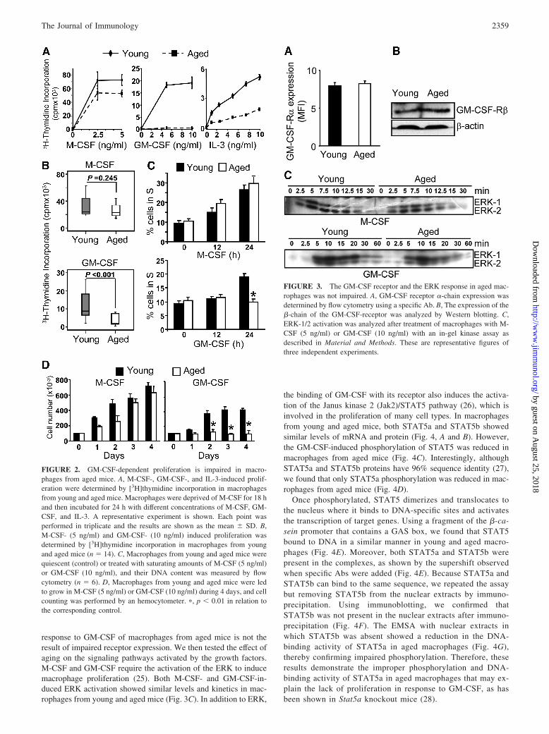
response to GM-CSF of macrophages from aged mice is not theresult of impaired receptor expression. We then tested the effect ofaging on the signaling pathways activated by the growth factors.M-CSF and GM-CSF require the activation of the ERK to inducemacrophage proliferation (25). Both M-CSF- and GM-CSF-in-duced ERK activation showed similar levels and kinetics in mac-rophages from young and aged mice (Fig. 3C). In addition to ERK,
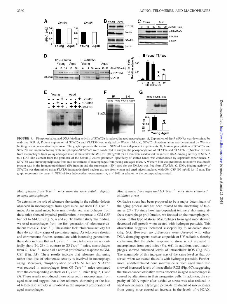
the binding of GM-CSF with its receptor also induces the activa-tion of the Janus kinase 2 (Jak2)/STAT5 pathway (26), which isinvolved in the proliferation of many cell types. In macrophagesfrom young and aged mice, both STAT5a and STAT5b showedsimilar levels of mRNA and protein (Fig. 4, A and B). However,the GM-CSF-induced phosphorylation of STAT5 was reduced inmacrophages from aged mice (Fig. 4C). Interestingly, althoughSTAT5a and STAT5b proteins have 96% sequence identity (27),we found that only STAT5a phosphorylation was reduced in mac-rophages from aged mice (Fig. 4D).
Once phosphorylated, STAT5 dimerizes and translocates tothe nucleus where it binds to DNA-specific sites and activatesthe transcription of target genes. Using a fragment of the �-ca-sein promoter that contains a GAS box, we found that STAT5bound to DNA in a similar manner in young and aged macro-phages (Fig. 4E). Moreover, both STAT5a and STAT5b werepresent in the complexes, as shown by the supershift observedwhen specific Abs were added (Fig. 4E). Because STAT5a andSTAT5b can bind to the same sequence, we repeated the assaybut removing STAT5b from the nuclear extracts by immuno-precipitation. Using immunoblotting, we confirmed thatSTAT5b was not present in the nuclear extracts after immuno-precipitation (Fig. 4F). The EMSA with nuclear extracts inwhich STAT5b was absent showed a reduction in the DNA-binding activity of STAT5a in aged macrophages (Fig. 4G),thereby confirming impaired phosphorylation. Therefore, theseresults demonstrate the improper phosphorylation and DNA-binding activity of STAT5a in aged macrophages that may ex-plain the lack of proliferation in response to GM-CSF, as hasbeen shown in Stat5a knockout mice (28).
FIGURE 2. GM-CSF-dependent proliferation is impaired in macro-phages from aged mice. A, M-CSF-, GM-CSF-, and IL-3-induced prolif-eration were determined by [3H]thymidine incorporation in macrophagesfrom young and aged mice. Macrophages were deprived of M-CSF for 18 hand then incubated for 24 h with different concentrations of M-CSF, GM-CSF, and IL-3. A representative experiment is shown. Each point wasperformed in triplicate and the results are shown as the mean � SD. B,M-CSF- (5 ng/ml) and GM-CSF- (10 ng/ml) induced proliferation wasdetermined by [3H]thymidine incorporation in macrophages from youngand aged mice (n � 14). C, Macrophages from young and aged mice werequiescent (control) or treated with saturating amounts of M-CSF (5 ng/ml)or GM-CSF (10 ng/ml), and their DNA content was measured by flowcytometry (n � 6). D, Macrophages from young and aged mice were ledto grow in M-CSF (5 ng/ml) or GM-CSF (10 ng/ml) during 4 days, and cellcounting was performed by an hemocytometer. �, p 0.01 in relation tothe corresponding control.
FIGURE 3. The GM-CSF receptor and the ERK response in aged mac-rophages was not impaired. A, GM-CSF receptor �-chain expression wasdetermined by flow cytometry using a specific Ab. B, The expression of the�-chain of the GM-CSF-receptor was analyzed by Western blotting. C,ERK-1/2 activation was analyzed after treatment of macrophages with M-CSF (5 ng/ml) or GM-CSF (10 ng/ml) with an in-gel kinase assay asdescribed in Material and Methods. These are representative figures ofthree independent experiments.
2359The Journal of Immunology
by guest on August 25, 2018
http://ww
w.jim
munol.org/
Dow
nloaded from
Macrophages from Terc�/� mice show the same cellular defectsas aged macrophages
To determine the role of telomere shortening in the cellular defectsobserved in macrophages from aged mice, we used G3 Terc�/�
mice. As in aged mice, bone marrow-derived macrophages fromthese mice showed impaired proliferation in response to GM-CSFbut not to M-CSF (Fig. 5, A and B). To further study this finding,we used macrophages from the first generation of telomerase-de-ficient mice (G1 Terc�/�). These mice lack telomerase activity butthey do not show signs of premature aging. As telomeres shortenand chromosome fusions accumulate with increasing generations,these data indicate that in G1 Terc�/� mice telomeres are not crit-ically short (10, 23). In contrast to G3 Terc�/� mice, macrophagesfrom G1 Terc�/� mice had a normal proliferative response to GM-CSF (Fig. 5A). These results indicate that telomere shorteningrather than loss of telomerase activity is involved in macrophageaging. Moreover, phosphorylation of STAT5a but not STAT5bwas reduced in macrophages from G3 Terc�/� mice comparedwith the corresponding controls or G1 Terc�/� mice (Fig. 5, C andD). These results reproduced those observed in macrophages fromaged mice and suggest that either telomere shortening or the lossof telomerase activity is involved in the impaired proliferation ofaged macrophages.
Macrophages from aged and G3 Terc�/� mice show enhancedoxidative stress
Oxidative stress has been proposed to be a major determinant ofthe aging process and has been related to the shortening of telo-meres (24). To study how age-dependent telomere shortening af-fects macrophage proliferation, we focused on the macrophage re-sponse to this type of stress. Macrophages from aged mice showeddecreased cell growth when treated with hydrogen peroxide. Thisobservation suggests increased susceptibility to oxidative stress(Fig. 6A). However, no differences were observed with otherDNA-damaging agents, such as etoposide or UV radiation, therebyconfirming that the global response to stress is not impaired inmacrophages from aged mice (Fig. 6A). In addition, aged macro-phages showed enhanced levels of intracellular ROS (Fig. 6B).The magnitude of this increase was of the same level as that ob-served when we treated the cells with hydrogen peroxide. Further-more, undifferentiated bone marrow cells from aged mice alsoshowed increased levels of intracellular ROS (Fig. 6C), suggestingthat the enhanced oxidative stress observed in aged macrophages iscaused by alterations in their progenitor cells. In addition, the ca-pacity of DNA repair after oxidative stress was also reduced inaged macrophages. Hydrogen peroxide treatment of macrophagesfrom young mice caused an increase in the levels of �-H2AX,
FIGURE 4. Phosphorylation and DNA-binding activity of STAT5a is reduced in aged macrophages. A, Expression of Stat5 mRNAs was determined byreal-time PCR. B, Protein expression of STAT5a and STAT5b was analyzed by Western blot. C, STAT5 phosphorylation was determined by Westernblotting in a representative experiment. The graph represents the mean � SEM of four independent experiments. D, Immunoprecipitation of STAT5a andSTAT5b and immunoblotting with anti-phospho-STAT5a/b were conducted to analyze the phosphorylation of STAT5a and STAT5b. E, Nuclear extractsfrom macrophages from young and aged mice stimulated with GM-CSF (10 ng/ml) for 15 min were used to test the in vitro DNA-binding activity of STAT5to a GAS-like element from the promoter of the bovine �-casein promoter. Specificity of shifted bands was corroborated by supershift experiments. F,STAT5b was immunoprecipitated from nuclear extracts of macrophages from young and aged mice. A Western blot was performed to confirm that Stat5bprotein was in the immunoprecipitated (IP) fraction and the supernatant (SN) used for the EMSAs was free from STAT5b. G, DNA-binding activity ofSTAT5a was determined using STAT5b immunodepleted nuclear extracts from young and aged mice stimulated with GM-CSF (10 ng/ml) for 15 min. Thegraph represents the mean � SEM of four independent experiments. �, p 0.01 in relation to the corresponding control.
2360 AGING, TELOMERES, AND MACROPHAGES
by guest on August 25, 2018
http://ww
w.jim
munol.org/
Dow
nloaded from

which returned to basal levels after 24 h of recovery (Fig. 6D).However, macrophages from aged mice showed sustained levels of�-H2AX, thereby indicating a decreased response to oxidative
FIGURE 5. Macrophages from G3 Terc�/� mice show the same cellulardefects observed in aged macrophages. A, M-CSF- and GM-CSF-induced pro-liferation was determined by [3H]thymidine incorporation in macrophagesfrom WT and G1 or G3 Terc�/� mice. Macrophages were deprived of M-CSFfor 18 h and then incubated for 24 h with different concentrations of M-CSFand GM-CSF. A representative experiment is shown. Each point was per-formed in triplicate and the results are shown as the mean � SD. B, M-CSF-(5 ng/ml) and GM-CSF- (10 ng/ml) induced proliferation was determined by[3H]thymidine incorporation in macrophages from WT and G3 Terc�/� mice(n � 7). C, Immunoprecipitation of STAT5a and STAT5b and immunoblot-ting with anti-phospho-STAT5a/b were conducted to analyze the phosphory-lation of STAT5a and STAT5b. These figures are representative of at leastthree independent experiments. D, Immunoprecipitation of STAT5a and im-munoblotting with anti-phospho-STAT5a/b were conducted to analyze thephosphorylation of STAT5a in macrophages from WT, G1, and G3 Terc�/�
mice. The data is representative of three independent experiments. The graphrepresents the mean � SEM of four independent experiments. �, p 0.01 inrelation to the corresponding control.
FIGURE 6. Enhanced oxidative stress in aged macrophages. A, Mac-rophages from young and aged mice were treated with several DNA-damaging agents and led to recover for 5 days. Cell growth was deter-mined as the ratio of the number of treated cells relative to the numberof control cells. The data represents the mean � SEM of four independentexperiments. Intracellular levels of ROS were quantified in macrophages (B)and bone marrow cells (C) by staining with the ROS-specific dye DCF-DA. InB, hydrogen peroxide was added at 250 �M for 1 h as a positive control. Thedata represents the mean � SEM of three independent experiments. D, Mac-rophages from young and aged mice were treated with hydrogen peroxide (250�M) for 1 h; the cells were washed and led to recover for 6 and 24 h. DNAdamage was determined by �-H2AX immunoblotting (c, untreated cells). Thisfigure is representative of three independent experiments. E, Alkaline Cometassay was performed to analyze DNA damage after oxidative stress in mac-rophages from young and aged mice. Quantification was done using the tailmoment calculation. This figure is representative of three independent exper-iments. �, p 0.01 in relation to the corresponding control.
2361The Journal of Immunology
by guest on August 25, 2018
http://ww
w.jim
munol.org/
Dow
nloaded from

stress. Alkaline comet assay, which detects DNA single-strandbreaks, further confirmed this reduced DNA repair capacity (Fig.6E). Together, these results demonstrate that macrophages fromaged mice suffer enhanced oxidative stress.
In addition to aged mice, macrophages from G3 Terc�/� micealso showed increased susceptibility to oxidative stress but notto other types of stress factors, compared with control mice(Fig. 7A). Strikingly, intracellular levels of ROS were also in-creased in macrophages and bone marrow cells (Fig. 7B) fromG3 Terc�/� mice, thereby linking the two models again, theaged and telomerase-deficient mice.
STAT5a oxidation is reduced in macrophages from aged and G3Terc�/� mice
Next, we addressed whether oxidative stress is related to the re-duction in STAT5a phosphorylation in macrophages from agedand G3 Terc�/� mice. Our first hypothesis was that oxidativestress leads to a mutation in the genomic DNA of STAT5a. Al-though STAT5a and STAT5b are coded by two distinct genes, theyare highly homologous proteins (96% sequence identity) (27), dif-fering only in their COOH-terminal transactivation domain, whichcontains a tyrosine residue that is phosphorylated in response toGM-CSF (29). We sequenced the genomic DNA from three youngand three aged mice using primers flanking the exons containingthis domain in STAT5a (exon 16) but we did not find any differencebetween macrophages from these two groups. However, oxidativedamage may also cause base oxidation, which is not detected bythe sequencing technique. To examine this possibility, we digestedgenomic DNA with formamidopyrimidine-DNA glycosylase. Thisenzyme cleaves the oxidized base, thereby generating an AP site thatprevents PCR amplification. We then quantified the DNA oxidationby real-time PCR. No difference between young and aged macro-phages in the base oxidation in this exon was observed (Fig. 8A).
FIGURE 7. Enhanced oxidative stress in G3 Terc�/� mice. A, Macro-phages from WT and G3 Terc�/� mice were treated with distinct DNA-damaging agents and left to recover for 5 days. Cell growth was determinedas the ratio of the number of treated cells relative to the number of controlcells. The data represents the mean � SEM of four independent experi-ments; �, p 0.01 in relation to the corresponding control. B, Intracellularlevels of ROS were quantified in macrophages and bone marrow cells bystaining with the ROS-specific dye DCF-DA. The data represents themean � SEM (n � 4).
FIGURE 8. Decreased Stat5a oxidation in macrophages from aged andG3 Terc�/� mice. A, Genomic DNA was extracted from young and agedmacrophages and digested with formamidopyrimidine-DNA glycosylase(Fpg). Quantification of oxidative DNA damage was performed by real-time PCR of exon 16 of Stat5a and exon 18 of Stat5b. These figures arerepresentative of four independent experiments. B, STAT5a and STAT5bwere immunoprecipitated from protein extracts from macrophages fromyoung, aged, WT, and G3 Terc�/� mice stimulated with GM-CSF (10ng/ml) and oxidation of Stat5 proteins was determined using an Ab rec-ognizing the carbonyl groups introduced into proteins by oxidative reac-tions. C, The graphs represent the mean � SEM of four independent ex-periments. D, Macrophages from young mice were stimulated with 10ng/ml GM-CSF in the presence or absence of 20 mM NAC for the indi-cated times, and intracellular ROS levels were analyzed by DCF-DA stain-ing. Each point was performed in triplicate and the results are shown as themean � SD. E, Macrophages from young mice were treated with 10 ng/mlGM-CSF for 24 h in the presence or absence of NAC at the concentrationsindicated. ROS levels were measured by DCF-DA staining. The data rep-resents the mean � SEM of four independent experiments. F, STAT5a wasimmunoprecipitated from protein extracts from macrophages treated with10 ng/ml GM-CSF in the presence or absence of 20 mM NAC, and oxi-dation of the protein was determined. This figure is representative of atleast three independent experiments. �, p 0.01 in relation to the corre-sponding control.
2362 AGING, TELOMERES, AND MACROPHAGES
by guest on August 25, 2018
http://ww
w.jim
munol.org/
Dow
nloaded from

In addition to DNA, oxidative stress can directly inflict damageon proteins and lipids (30). Therefore we analyzed the oxidationstatus of STAT5 proteins by immunodetecting the carbonyl groupsintroduced into protein side chains as a consequence of oxidation.Surprisingly, STAT5a was less oxidized in macrophages fromaged mice, whereas no differences were observed in STAT5b (Fig.8, B and C). Moreover, macrophages from G3 Terc�/� mice alsoshowed impaired oxidation of STAT5a but not STAT5b (Fig. 8, Band C). These results suggest that the increased levels of oxidativestress in aged and G3 Terc�/� macrophages cause a reduction inthe oxidation of STAT5a.
STAT5a oxidation is required for this signal transducer to bephosphorylated
Because STAT5a oxidation and phosphorylation were reduced inmacrophages from aged and G3 Terc�/� mice, we addressedwhether these two reactions were related. It has been reported thathematopoietic cytokines induce an increase in intracellular ROSlevels, thereby promoting the phosphorylation of several proteins,including STAT5 (31, 32). After the addition of GM-CSF, ROSproduction showed a gradual increase from 30 min to 24 h (Fig.8D). The addition of the ROS scavenger N-acetylcysteine (NAC)blocked the effect of GM-CSF (Fig. 8D). Interestingly, stimulationof macrophages with GM-CSF induced STAT5a oxidation (Fig.8E) and the treatment of these cells with NAC, which blocked thisoxidation, also inhibited the phosphorylation of STAT5a (Fig. 8F).These results suggest that STAT5a oxidation is required for itsphosphorylation, thereby confirming that the reduced phosphory-lation of STAT5a observed in macrophages from aged and G3Terc�/� mice is due to its impaired oxidation.
DiscussionThe results presented suggest that the loss of telomeric sequencesduring aging is responsible for the enhanced oxidative stress, theimpaired oxidation and phosphorylation of STAT5a and the re-duced proliferation of macrophages. This study provides one of thefew reports that telomere shortening occurs in the mouse strainMus musculus, which has very long telomeres. In addition, ourobservation that the telomere length of macrophages from agedand G3 Terc�/� mice was very similar indicates that normalaging leads to short telomeres, thereby resulting in impairedproliferation.
In this study, we used bone marrow-derived macrophages dif-ferentiated in vitro. Thus, the variations in telomere length in thesecells during aging may reflect changes in telomere length in he-matopoietic progenitor cells. In fact, hematopoietic stem cellsshow telomere shortening during in vitro culture and in vivo aging(21, 33, 34). Hematopoietic stem cells derived from humans andmice lose telomeric DNA with age despite the presence of detect-able telomerase activity (21, 35). Moreover, telomeres shortenwith age in several mouse stem cell compartments, including skin,small intestine, cornea, testis and brain (22).
In addition to telomere shortening, we have shown that macro-phages from aged mice suffer enhanced oxidative stress. This kindof stress has been associated with telomere shortening as reflectedby the observation that cells with enhanced oxidative stress haveshorter telomeres (24, 36). However, our finding that G3 Terc�/�
mice also have an enhanced oxidative stress points to telomereshortening as the cause of the enhanced oxidative stress in mac-rophages. These results correlate with those observed in mouseembryonic fibroblasts derived from Terc�/� mice, which alsoshow increased levels of ROS (37). In fact, telomerase deficiencyreduces catalase activity that determines a redox imbalance (38).Furthermore, it has been shown that cells overexpressing telom-
erase accumulate lower concentrations of peroxides (39). Undif-ferentiated bone marrow cells from aged and G3 Terc�/� micealso show an increase in intracellular ROS, thereby further sup-porting the hypothesis that macrophage aging is due to an intrinsicalteration in their progenitor cells. Recently it has been reportedthat in the absence of telomeres there is a repression of geneslocated within 10 kb (40). Although in mice, catalase is located inthe central area of the chromosome 2, we cannot exclude thattelomeres are required to regulate transcription factors or otherelements necessary for catalase expression.
Telomere shortening is associated with the impaired prolifera-tion of many cell types, including those of the immune system (6,7). Surprisingly, in our studies only GM-CSF- but not M-CSF-dependent proliferation of macrophages was affected by aging.This is attributed to a reduction of STAT5a oxidation, which leadsto impaired phosphorylation. Notably, it has been described thatSTAT5 phosphorylation is reduced during aging in T (41) and Bcells (42) and neutrophils (43), thereby leading to a number offunctional alterations. Aging is associated with a reduction in Band T cell numbers (44), suggesting that impaired STAT5 phos-phorylation is a general effect of aging involved in many aspects ofimmunosenescence. G3 Terc�/� but not G1 Terc�/� macrophagesshowed the same cellular defects as those observed in aged mice.These results indicate that telomere attrition and oxidative stresscause a decrease in the oxidation and phosphorylation of STAT5aand, therefore, impaired proliferation of macrophages. However,the precise mechanism linking enhanced oxidative stress and re-duced STAT5a oxidation and phosphorylation remains to be elu-cidated. One may think that oxidative stress could affect the ac-tivity of tyrosine phosphatases. However, reversible oxidation ofCys residues on these phosphatases leads to their inhibition (45,46), which would result in an increase of the amount of phospho-STAT5a, instead of the reduced levels of phosphorylation that weobserve under oxidative stress conditions. The generation of ROSby GM-CSF stimulation may be through activation of NADPHoxidase, as observed for other cytokines (47, 48). Thus, it is pos-sible that a permanent increase in oxidative stress in aged macro-phages may cause a redox-dependent modification in NADPH ox-idase, leading to an impaired production of ROS and STAT5aphosphorylation. However, further assays are required to provethis hypothesis.
In summary, in this study we describe that macrophage telo-meres shorten during mouse aging, thereby leading to impairedGM-CSF-dependent proliferation of these cells. In addition, wepresent data about the molecular mechanisms involved in this pro-cess. GM-CSF plays a crucial role during inflammation. Proin-flammatory functions of GM-CSF include the recruitment, activa-tion, enhanced survival, proliferation and adhesion of neutrophilsand macrophages (49). Therefore, it is likely that GM-CSF has acentral role in regulating macrophage proliferation at sites of in-flammation (50, 51). Thus, impaired response of macrophages toGM-CSF during aging may have relevant consequences on inflam-matory reactions and on host defenses against a broad spectrum ofinvading organisms. In this regard, it is important to note thathuman aging is generally accompanied by elevated systemic in-flammatory conditions (52) and that macrophages play a key roleduring this process.
AcknowledgmentsWe thank Tanya Yates for editing the manuscript. The help of JavierGonzalez-Linares and Joaquín de Lafuente from the Toxicology Unit at theParc Cientific of Barcelona in the alkaline comet assay is acknowledged.
2363The Journal of Immunology
by guest on August 25, 2018
http://ww
w.jim
munol.org/
Dow
nloaded from

DisclosuresThe authors have no financial conflict of interest.
References1. Miller, R. A. 1996. The aging immune system: primer and prospectus. Science
273: 70–74.2. Plowden, J., M. Renshaw-Hoelscher, C. Engleman, J. Katz, and S. Sambhara.
2004. Innate immunity in aging: impact on macrophage function. Aging Cell 3:161–167.
3. Blackburn, E. H. 2001. Switching and signaling at the telomere. Cell 106:661–673.
4. Iwama, H., K. Ohyashiki, J. H. Ohyashiki, S. Hayashi, N. Yahata, K. Ando,K. Toyama, A. Hoshika, M. Takasaki, M. Mori, and J. W. Shay. 1998. Telomericlength and telomerase activity vary with age in peripheral blood cells obtainedfrom normal individuals. Hum. Genet. 102: 397–402.
5. Samper, E., P. Fernandez, R. Eguia, L. Martin-Rivera, A. Bernad, M. A. Blasco,and M. Aracil. 2002. Long-term repopulating ability of telomerase-deficient mu-rine hematopoietic stem cells. Blood 99: 2767–2775.
6. Blasco, M. A. 2002. Immunosenescence phenotypes in the telomerase knockoutmouse. Springer Semin. Immunopathol. 24: 75–85.
7. Hodes, R. J., K. S. Hathcock, and N. P. Weng. 2002. Telomeres in T and B cells.Nat. Rev. Immunol. 2: 699–706.
8. Herrero, C., L. Marques, J. Lloberas, and A. Celada. 2001. IFN-�-dependenttranscription of MHC class II IA is impaired in macrophages from aged mice.J. Clin. Invest. 107: 485–493.
9. Celada, A., P. W. Gray, E. Rinderknecht, and R. D. Schreiber. 1984. Evidence fora �-interferon receptor that regulates macrophage tumoricidal activity. J. Exp.Med. 160: 55–74.
10. Blasco, M. A., H. W. Lee, M. P. Hande, E. Samper, P. M. Lansdorp,R. A. DePinho, and C. W. Greider. 1997. Telomere shortening and tumor for-mation by mouse cells lacking telomerase RNA. Cell 91: 25–34.
11. Celada, A., F. E. Borras, C. Soler, J. Lloberas, M. Klemsz, C. van Beveren,S. McKercher, and R. A. Maki. 1996. The transcription factor PU. 1 is involvedin macrophage proliferation. J. Exp. Med. 184: 61–69.
12. Xaus, J., M. Cardo, A. F. Valledor, C. Soler, J. Lloberas, and A. Celada. 1999.Interferon � induces the expression of p21waf-1 and arrests macrophage cellcycle, preventing induction of apoptosis. Immunity 11: 103–113.
13. Valledor, A. F., J. Xaus, L. Marques, and A. Celada. 1999. Macrophage colony-stimulating factor induces the expression of mitogen-activated protein kinasephosphatase-1 through a protein kinase C-dependent pathway. J. Immunol. 163:2452–2462.
14. Casals, C., M. Barrachina, M. Serra, J. Lloberas, and A. Celada. 2007. Lipopoly-saccharide up-regulates MHC class II expression on dendritic cells through anAP-1 enhancer without affecting the levels of CIITA. J. Immunol. 178:6307–6315.
15. Cawthon, R. M. 2002. Telomere measurement by quantitative PCR. Nucleic Ac-ids Res. 30: e47.
16. Valledor, A. F., J. Xaus, M. Comalada, C. Soler, and A. Celada. 2000. Proteinkinase C � is required for the induction of mitogen-activated protein kinase phos-phatase-1 in lipopolysaccharide-stimulated macrophages. J. Immunol. 164:29–37.
17. Lu, T., Y. Pan, S. Y. Kao, C. Li, I. Kohane, J. Chan, and B. A. Yankner. 2004.Gene regulation and DNA damage in the ageing human brain. Nature 429:883–891.
18. Canela, A., E. Vera, P. Klatt, and M. A. Blasco. 2007. High-throughput telomerelength quantification by FISH and its application to human population studies.Proc. Natl. Acad. Sci. USA 104: 5300–5305.
19. Harley, C. B., A. B. Futcher, and C. W. Greider. 1990. Telomeres shorten duringageing of human fibroblasts. Nature 345: 458–460.
20. Rufer, N., W. Dragowska, G. Thornbury, E. Roosnek, and P. M. Lansdorp. 1998.Telomere length dynamics in human lymphocyte subpopulations measured byflow cytometry. Nat. Biotechnol. 16: 743–747.
21. Vaziri, H., W. Dragowska, R. C. Allsopp, T. E. Thomas, C. B. Harley, andP. M. Lansdorp. 1994. Evidence for a mitotic clock in human hematopoietic stemcells: loss of telomeric DNA with age. Proc. Natl. Acad. Sci. USA 91: 9857–9860.
22. Flores, I., A. Canela, E. Vera, A. Tejera, G. Cotsarelis, and M. A. Blasco. 2008.The longest telomeres: a general signature of adult stem cell compartments.Genes Dev. 22: 654–667.
23. Herrera, E., E. Samper, J. Martin-Caballero, J. M. Flores, H. W. Lee, andM. A. Blasco. 1999. Disease states associated with telomerase deficiency appearearlier in mice with short telomeres. EMBO J. 18: 2950–2960.
24. von Zglinicki, T. 2002. Oxidative stress shortens telomeres. Trends Biochem. Sci.27: 339–344.
25. Valledor, A. F., M. Comalada, J. Xaus, and A. Celada. 2000. The differentialtime-course of extracellular-regulated kinase activity correlates with the macro-phage response toward proliferation or activation. J. Biol. Chem. 275:7403–7409.
26. de Groot, R. P., P. J. Coffer, and L. Koenderman. 1998. Regulation of prolifer-ation, differentiation and survival by the IL-3/IL-5/GM-CSF receptor family. CellSignal 10: 619–628.
27. Hou, J., U. Schindler, W. J. Henzel, S. C. Wong, and S. L. McKnight. 1995.Identification and purification of human Stat proteins activated in response tointerleukin-2. Immunity 2: 321–329.
28. Feldman, G. M., L. A. Rosenthal, X. Liu, M. P. Hayes, A. Wynshaw-Boris,W. J. Leonard, L. Hennighausen, and D. S. Finbloom. 1997. STAT5A-deficientmice demonstrate a defect in granulocyte-macrophage colony-stimulating factor-induced proliferation and gene expression. Blood 90: 1768–1776.
29. Moriggl, R., D. J. Topham, S. Teglund, V. Sexl, C. McKay, D. Wang,A. Hoffmeyer, J. van Deursen, M. Y. Sangster, K. D. Bunting, et al. 1999. Stat5is required for IL-2-induced cell cycle progression of peripheral T cells. Immunity10: 249–259.
30. Blumberg, J. 2004. Use of biomarkers of oxidative stress in research studies.J. Nutr. 134: 3188S–3189S.
31. Iiyama, M., K. Kakihana, T. Kurosu, and O. Miura. 2006. Reactive oxygen spe-cies generated by hematopoietic cytokines play roles in activation of receptor-mediated signaling and in cell cycle progression. Cell Signal 18: 174–182.
32. Sattler, M., T. Winkler, S. Verma, C. H. Byrne, G. Shrikhande, R. Salgia, andJ. D. Griffin. 1999. Hematopoietic growth factors signal through the formation ofreactive oxygen species. Blood 93: 2928–2935.
33. Zimmermann, S., S. Glaser, R. Ketteler, C. F. Waller, U. Klingmuller, andU. M. Martens. 2004. Effects of telomerase modulation in human hematopoieticprogenitor cells. Stem Cells 22: 741–749.
34. Engelhardt, M., R. Kumar, J. Albanell, R. Pettengell, W. Han, and M. A. Moore.1997. Telomerase regulation, cell cycle, and telomere stability in primitive he-matopoietic cells. Blood 90: 182–193.
35. Allsopp, R. C., S. Cheshier, and I. L. Weissman. 2001. Telomere shorteningaccompanies increased cell cycle activity during serial transplantation of hema-topoietic stem cells. J. Exp. Med. 193: 917–924.
36. Tchirkov, A., and P. M. Lansdorp. 2003. Role of oxidative stress in telomereshortening in cultured fibroblasts from normal individuals and patients with atax-ia-telangiectasia. Hum. Mol. Genet. 12: 227–232.
37. Perez-Rivero, G., M. P. Ruiz-Torres, J. V. Rivas-Elena, M. Jerkic,M. L. Diez-Marques, J. M. Lopez-Novoa, M. A. Blasco, and D. Rodriguez-Puyol.2006. Mice deficient in telomerase activity develop hypertension because of anexcess of endothelin production. Circulation 114: 309–317.
38. Perez-Rivero, G., M. P. Ruiz-Torres, M. L. Diez-Marques, A. Canela,J. M. Lopez-Novoa, M. Rodriguez-Puyol, M. A. Blasco, and D. Rodriguez-Puyol.2008. Telomerase deficiency promotes oxidative stress by reducing catalase ac-tivity. Free Radic. Biol. Med. 45: 1243–1251.
39. Armstrong, L., G. Saretzki, H. Peters, I. Wappler, J. Evans, N. Hole,T. von Zglinicki, and M. Lako. 2005. Overexpression of telomerase confersgrowth advantage, stress resistance, and enhanced differentiation of ESCs towardthe hematopoietic lineage. Stem Cells 23: 516–529.
40. Yang, X., L. M. Figueiredo, A. Espinal, E. Okubo, and B. Li. 2009. RAP1 isessential for silencing telomeric variant surface glycoprotein genes in Trypano-soma brucei. Cell 137: 99–109.
41. Fulop, T., Jr., N. Douziech, A. C. Goulet, S. Desgeorges, A. Linteau,G. Lacombe, and G. Dupuis. 2001. Cyclodextrin modulation of T lymphocytesignal transduction with aging. Mech. Ageing Dev. 122: 1413–1430.
42. Riley, R. L., E. Van der Put, A. M. King, D. Frasca, and B. B. Blomberg. 2005.Deficient B lymphopoiesis in murine senescence: potential roles for dysregulationof E2A, Pax-5, and STAT5. Semin. Immunol. 17: 330–336.
43. Fortin, C. F., A. Larbi, G. Dupuis, O. Lesur, and T. Fulop, Jr. 2007. GM-CSFactivates the Jak/STAT pathway to rescue polymorphonuclear neutrophils fromspontaneous apoptosis in young but not elderly individuals. Biogerontology 8:173–187.
44. Hakim, F. T., and R. E. Gress. 2007. Immunosenescence: deficits in adaptiveimmunity in the elderly. Tissue Antigens 70: 179–189.
45. Salmeen, A., and D. Barford. 2005. Functions and mechanisms of redox regula-tion of cysteine-based phosphatases. Antioxid. Redox. Signal 7: 560–577.
46. Rhee, S. G., Y. S. Bae, S. R. Lee, and J. Kwon. 2000. Hydrogen peroxide: a keymessenger that modulates protein phosphorylation through cysteine oxidation.Sci. STKE 2000: PE1.
47. Bae, Y. S., J. Y. Sung, O. S. Kim, Y. J. Kim, K. C. Hur, A. Kazlauskas, andS. G. Rhee. 2000. Platelet-derived growth factor-induced H2O2 production re-quires the activation of phosphatidylinositol 3-kinase. J. Biol. Chem. 275:10527–10531.
48. Chiarugi, P., and P. Cirri. 2003. Redox regulation of protein tyrosine phospha-tases during receptor tyrosine kinase signal transduction. Trends Biochem. Sci.28: 509–514.
49. Hamilton, J. A. 2002. GM-CSF in inflammation and autoimmunity. Trends Im-munol. 23: 403–408.
50. Bozinovski, S., J. E. Jones, R. Vlahos, J. A. Hamilton, and G. P. Anderson. 2002.Granulocyte/macrophage-colony-stimulating factor (GM-CSF) regulates lung in-nate immunity to lipopolysaccharide through Akt/Erk activation of NFkappa Band AP-1 in vivo. J. Biol. Chem. 277: 42808–42814.
51. Bitterman, P. B., L. E. Saltzman, S. Adelberg, V. J. Ferrans, and R. G. Crystal.1984. Alveolar macrophage replication. One mechanism for the expansion of themononuclear phagocyte population in the chronically inflamed lung. J. Clin. In-vest. 74: 460–469.
52. Franceschi, C., M. Bonafe, S. Valensin, F. Olivieri, M. De Luca, E. Ottaviani, andG. De Benedictis. 2000. Inflamm-aging. An evolutionary perspective on immu-nosenescence. Ann. NY Acad. Sci. 908: 244–254.
2364 AGING, TELOMERES, AND MACROPHAGES
by guest on August 25, 2018
http://ww
w.jim
munol.org/
Dow
nloaded from


![Intrarenal arteriosclerosis and telomere attrition ...€¦ · Telomere length is a well-established marker of biological age [4]. Although telomere length is partly heritable, there](https://static.fdocuments.net/doc/165x107/5f2629fb310cc83259516f06/intrarenal-arteriosclerosis-and-telomere-attrition-telomere-length-is-a-well-established.jpg)