Techniques in Shoulder and Elbow Surgery 8(2):89–97, 2007...
9
| TECHNIQUE | Operative Fixation of Radial Head Fractures John T. Capo, MD and Dan Dziadosz, MD Division of Hand and Microvascular Surgery Department of Orthopaedics New Jersey Medical School Newark, NJ | ABSTRACT The radial head is an important structure that is crucial to the stability of both the elbow and forearm. Fractures of the radial head occur commonly and often are dis- placed or cause impingement to forearm rotation. Recent studies have further demonstrated the impor- tance of maintaining or reconstructing the rigid buttress of the radial head. Advances in implant technology have benefited orthopedic surgeons in their attempts at fixation of the radial head and also in rigid implant arthroplasty. Partial articular fractures of the radial head are relatively simple to repair and can be stabilized with headless or buried screws. A complete articular fracture is more challenging and requires the use of rigid, low- profile, and, often, fixed-angle plates placed in the nonarticular portion of the radial head. Associated ligament tears in the elbow magnify the importance of restoring the stabilizing effect of the radial head. Radial head excision should rarely be done and only after a thorough examination of the ligamentous structures about the elbow and forearm demonstrate complete stability. Keywords: radial head, fractures, ORIF, stability | HISTORICAL PERSPECTIVE Historically, treatment of radial head fractures primarily involved excision of the fracture fragments as well as the remaining pieces of the intact radial head. This treatment potentially created scenarios of instability, motion deficits, proximal migration of the radius, and distal radioulnar joint (DRUJ) dysfunction. 1,2 With the development of advanced techniques in open-reduction and internal fixation (ORIF), the radial head can often be rigidly fixed and the stability of the elbow can be maintained. 3 Several comparisons among ORIF, prosthetic arthro- plasty, excision, and nonoperative treatment have been investigated. 4Y8 It appears that the trend for the treatment of significantly displaced radial head fractures is for stable fixation with plates and/or screws or re- placement with rigid implant arthroplasty. Beingessner et al 9 investigated the biomechanical effects of radial head excision and prosthetic arthroplasty on elbow kinematics in a cadaver model. They demonstrated that replacement of the radial head with a metallic implant improves stability but does not return the elbow to a normal condition. This effect was most clearly seen with associated ligamentous instability and emphasized that rigid implants do not exactly reproduce the dynamics of the intact or rigidly fixed radial head. The native radial head is also elliptical and sits at approxi- mately a 15-degree angle to the radial shaft. These anatomic characteristics are not replicated by implant arthroplasty. For these and other reasons, preservation of the native radial head is desirable, particularly in younger high-demand patients. | FRACTURE CLASSIFICATION One of the common systems for classifying radial head fractures is that of Mason, 10 originally described in the 1950s. This system separated these fractures into 3 types. Type I fractures were described as nondisplaced fractures that were treated most successfully by closed (nonoperative) means. Type II fractures were displaced fractures involving only a portion of the radial head, with some remaining head continuous with the radial shaft or minimally displaced neck fractures. Type III fractures were comminuted fractures involving the entire radial head whose treatment options were histori- cally limited to excision. Since the time of the original classification system several modifications have been made. One of the firsts was by Morrey in the 1980s. This modification included a quantitative description of fracture displacement (a fragment involving 30% or more of the articular surface and 2 mm or more of displacement was deemed unacceptable) and fracture dislocations of the elbow. 11 One of the more recent modifications, made by Hotchkiss, 5 appears to help with treatment decision making. Type I fractures are characterized by minimal displacement, with forearm rotation limited by acute pain and swelling, intraarticular displacement of less Techniques in Shoulder and Elbow Surgery 8(2):89–97, 2007 Ó 2007 Lippincott Williams & Wilkins, Philadelphia Reprints: John T. Capo, MD, 90 Bergen St, DOC 1200, Newark, NJ 07103 (e-mail: [email protected]). Volume 8, Issue 2 89 Copyr ight © Lippincott Williams & Wilkins. Unauthor iz ed reproduction of this article is prohibited.
Transcript of Techniques in Shoulder and Elbow Surgery 8(2):89–97, 2007...

| T E C H N I Q U E |
Operative Fixation of Radial Head FracturesJohn T. Capo, MD and Dan Dziadosz, MDDivision of Hand and Microvascular SurgeryDepartment of OrthopaedicsNew Jersey Medical SchoolNewark, NJ
| ABSTRACT
The radial head is an important structure that is crucial
to the stability of both the elbow and forearm. Fractures
of the radial head occur commonly and often are dis-
placed or cause impingement to forearm rotation.
Recent studies have further demonstrated the impor-
tance of maintaining or reconstructing the rigid buttress
of the radial head. Advances in implant technology have
benefited orthopedic surgeons in their attempts at
fixation of the radial head and also in rigid implant
arthroplasty. Partial articular fractures of the radial head
are relatively simple to repair and can be stabilized with
headless or buried screws. A complete articular fracture
is more challenging and requires the use of rigid, low-
profile, and, often, fixed-angle plates placed in the
nonarticular portion of the radial head. Associated
ligament tears in the elbow magnify the importance of
restoring the stabilizing effect of the radial head. Radial
head excision should rarely be done and only after a
thorough examination of the ligamentous structures about
the elbow and forearm demonstrate complete stability.
Keywords: radial head, fractures, ORIF, stability
| HISTORICAL PERSPECTIVE
Historically, treatment of radial head fractures primarily
involved excision of the fracture fragments as well as
the remaining pieces of the intact radial head. This
treatment potentially created scenarios of instability,
motion deficits, proximal migration of the radius, and
distal radioulnar joint (DRUJ) dysfunction.1,2 With the
development of advanced techniques in open-reduction
and internal fixation (ORIF), the radial head can often
be rigidly fixed and the stability of the elbow can be
maintained.3
Several comparisons among ORIF, prosthetic arthro-
plasty, excision, and nonoperative treatment have been
investigated.4Y8 It appears that the trend for the
treatment of significantly displaced radial head fractures
is for stable fixation with plates and/or screws or re-
placement with rigid implant arthroplasty. Beingessner
et al9 investigated the biomechanical effects of radial
head excision and prosthetic arthroplasty on elbow
kinematics in a cadaver model. They demonstrated that
replacement of the radial head with a metallic implant
improves stability but does not return the elbow to a
normal condition. This effect was most clearly seen
with associated ligamentous instability and emphasized
that rigid implants do not exactly reproduce the
dynamics of the intact or rigidly fixed radial head. The
native radial head is also elliptical and sits at approxi-
mately a 15-degree angle to the radial shaft. These
anatomic characteristics are not replicated by implant
arthroplasty. For these and other reasons, preservation
of the native radial head is desirable, particularly in
younger high-demand patients.
| FRACTURE CLASSIFICATION
One of the common systems for classifying radial head
fractures is that of Mason,10 originally described in the
1950s. This system separated these fractures into 3
types. Type I fractures were described as nondisplaced
fractures that were treated most successfully by closed
(nonoperative) means. Type II fractures were displaced
fractures involving only a portion of the radial head,
with some remaining head continuous with the radial
shaft or minimally displaced neck fractures. Type III
fractures were comminuted fractures involving the
entire radial head whose treatment options were histori-
cally limited to excision.
Since the time of the original classification system
several modifications have been made. One of the firsts
was by Morrey in the 1980s. This modification included
a quantitative description of fracture displacement (a
fragment involving 30% or more of the articular surface
and 2 mm or more of displacement was deemed
unacceptable) and fracture dislocations of the elbow.11
One of the more recent modifications, made by
Hotchkiss,5 appears to help with treatment decision
making. Type I fractures are characterized by minimal
displacement, with forearm rotation limited by acute
pain and swelling, intraarticular displacement of less
Techniques in Shoulder and Elbow Surgery 8(2):89–97, 2007 � 2007 Lippincott Williams & Wilkins, Philadelphia
Reprints: John T. Capo, MD, 90 Bergen St, DOC 1200, Newark, NJ07103 (e-mail: [email protected]).
Volume 8, Issue 2 89
Copyr ight © Lippincott Williams & Wilkins. Unauthorized reproduction of this article is prohibited.

than 2 mm, or a marginal lip fracture. Type II fractures
are displaced fractures with more than 2 mm of
displacement of the head or neck, usually with blocked
or incongruous motion. There is minimal comminution,
but there is more than just a simple marginal lip fracture
of the radial head. These fractures are usually amenable
to ORIF. Type III fractures are severely comminuted.
These are irreparable based on radiographic or intra-
operative appearance, and excision or replacement is
usually required to allow motion.
| OPERATIVE INDICATIONS
The decision to perform ORIF of radial head fractures
involves several factors. The character of the radial head
fracture must be analyzed as well as associated
ligamentous and bony injuries around the elbow. This
may require advanced preoperative diagnostic studies
including specialized radiographs, computed tomogra-
phy scans, and magnetic resonance imaging. A helpful
radiograph is the oblique radiocapitellar, or Coyle, view
that shows the radial head in profile without the
overlying coronoid. Plain films alone have been shown
to be inaccurate in estimation of the fracture size and
displacement.12 In our opinion, radiographic indications
for operative treatment of radial head fractures are neck
fracture with greater than 15 degrees of angulation or
greater than 25% displacement, intra-articular fractures
greater than 25% of the radial head with more than 2
mm of articular step-off or that create a mechanical
block to motion. Although this is our recommendation,
intra-articular displacement greater than 2 mm has been
shown in some long-term studies to not preclude a good
clinical result.13 Other scenarios that would make
operative stabilization necessary include longitudinal
radial instability seen in an Essex-Lopressti injury or
elbow varus-valgus laxity that may be caused by
concomitant collateral ligament injury.
Patient variables that should be factored into the
surgical decision making process include age, hand
dominance, occupation, as well as the physical demand
placed upon the elbow. Operative fixation should be
offered, when indicated, to all patients who can
medically tolerate the operative procedure. Excision
without replacement in a higher demand individual
might lead to proximal migration of the radius with
decreased function at both the elbow and the wrist.14
| CONTRAINDICATIONS
Factors that preclude operative intervention are related
to concomitant injuries, soft tissue integrity, local
healing environment, generalized patient health, as well
as patient demand. In the setting of associated severe
fracture dislocation of the elbow or comminuted
forearm fractures, the long-term result of radial head
repair, especially with regard to restoration of forearm
rotation, may be compromised.15 In this situation,
another method of fixation might be necessary to
stabilize the elbow, such as hinged external fixation.16
If severe soft tissue damage exists (ie, burns, soft tissue
loss, or infection), operative intervention should be
delayed for adequate soft tissue healing. Patient factors
will also dictate the timing and need for surgery. Should
the patient be too medically unstable to undergo
operative intervention, then surgery must be delayed.
Finally, if the radial head is excessively comminuted
and unreconstructable, then implant arthroplasty may be
more appropriate.
| PREOPERATIVE PLANNING
The elbow should be examined for tenderness, neuro-
vascular status, soft-tissue condition, and range of
motion (ROM). Tenderness or ecchymosis at the medial
joint line may represent a medial collateral ligament
injury, whereas tenderness at the DRUJ could reflect
longitudinal instability of the elbow. All 3 major nerves
in the arm are examined, with particular attention to the
posterior interosseous nerve (PIN). Forearm rotation is
assessed to see if a mechanical block to motion exists. If
pain is excessive at the elbow, aspiration of the joint
hematoma followed by injection of a local anesthetic is
helpful in assessing motion and relieving pain.
Associated injuries that are critical include coronoid
fractures, medial and lateral collateral ligament (LCL)
injuries, and DRUJ injuries. If there was an associated
dislocation, then there is often an associated, seemingly
insignificant, coronoid process fracture. Repair of this
coronoid fracture will often provide increased stability
to the elbow and allow earlier ROM. With an incom-
petent coronoid, the resistance to anteroposterior (AP)
instability and tendency for resubluxation is increased,
and the need for a stable radial head is further mag-
nified. Damage to the interosseous ligament of the fore-
arm and DRUJ can induce proximal migration of the
radius with an absent or incompetent radial head.14
Equipment required should include all that is needed
for excision, internal fixation, or replacement arthro-
plasty. Small screws ranging from 1.3 to 2.4 mm, as
well as headless compression screws are often useful for
head fragments. Some type of small plating system,
either a general system or a dedicated radial head
system, should be available in the operating room.
Finally, a rigid implant arthroplasty of a suitable bearing
surface, preferable Cr-Co, and with sufficient modular-
ity is needed. A large fluoroscopic machine, which
allows a wide view of the entire forearm and elbow, is
preferable to the mini units.
Techniques in Shoulder and Elbow Surgery90
Capo and Dziadosz
Copyr ight © Lippincott Williams & Wilkins. Unauthorized reproduction of this article is prohibited.

| INTRAOPERATIVE ASSESSMENT
The elbow should be carefully and systematically
examined in the operating room before the decision
for radial head excision versus fixation is made. Each
elbow is examined in the following manner. Axial
migration is tested with the elbow stabilized on the hand
table, the forearm in neutral rotation, and load placed by
the surgeon on the fisted hand. Proximal migration of
the radius can be appreciated if the radial shaft migrates
into the capitellum. Also, the radial shaft can be grasped
with a tenaculum and pulled proximally and radial
shortening can be observed at the DRUJ on fluoroscopy.
Medial collateral ligament integrity should be evaluated
with the forearm in pronation and the elbow flexed to 30
degrees. The degree of opening, the feel of the end
point, and radiographic appearance are all observed with
this stress maneuver. Radial head incompetence will
exacerbate posterior lateral instability, which is tested in
elbow supination, valgus, and axial load. Finally, AP
stability should be evaluated in progressive extension. If
posterior subluxation of the elbow is seen at more than
30 degrees of flexion, then the radial head should be
repaired or replaced. Also, if a complex proximal ulna
or coronoid fragment undergoes ORIF, reconstitution of
the radial head buttress will help unload the ulna to
allow healing.
| TECHNIQUE
Surgical ExposuresSurgical approaches to the radial head include the
classic posterolateral (Kocher) approach between the
anconeus and extensor carpi ulnaris (ECU) and a more
extensile direct lateral approach. This extensile ap-
proach begins at the Kocher interval but distally extends
to split the extensor muscles, thus allowing access to the
radial shaft.
The Kocher approach starts with an oblique incision
beginning over the posterior surface of the lateral
epicondyle and continuing downward and distally to a
point over the posterior border of the ulna, about 6 cm
distal to the tip of the olecranon. The internervous plane
lies between the anconeus, supplied by the radial nerve
proper, and the ECU, supplied by the PIN. Full
pronation of the forearm moves the PIN away from
the operative field.17,18 The capsule of the elbow joint is
incised longitudinally at the annular ligament to reveal
the underlying capitellum and the radial head. The
capsule should not be dissected too far anteriorly as the
PIN runs over the front of the anterolateral portion of
the elbow capsule. Because of the posterior location of
this approach, the LCL complex is in danger and care
must be taken not to release the origin or insertion of
this ligament. The exposure is adequate for access to the
radial head alone but is not ideal for exposure to the
shaft, because it is difficult to extend distally.19
The extensile lateral approach is made over the
radial head and neck but anterior to the LCL complex.
The skin incision is centered over the lateral epicondyle
and extends distally over the radial head and neck,
anterior to the anconeus (Fig. 1). The deep muscular
interval runs from the lateral epicondyle through the
posterior aspect of the ECU muscle attachment proxi-
mally. It is critical to stay anterior to the posterior
border of the ECU, because it has been shown that
these fibers are integral to the LCL complex.20 The
dissection is carried distally between the ECU and
extensor digitorum communis. The muscle fibers are
spread and retracted as the anterior portion of the
extensor mass is reflected. A useful landmark to look
for at this point is the capitellum, and ensure dissection
should be anterior to its equator (Fig. 2). As the extensor
fibers are spread, the crossing fibers of the supinator
will next be seen. These fibers run obliquely at
approximately a 45-degree angle from the muscle fibers
of the extensor mass (Fig. 3). For better visualization,
the anterior extensor sleeve can be released from the
epicondyle and repaired later. The posterior extensor
mass cannot be safely released because it covers and
includes the attachments of the LCL. The supinator can
be safely divided for a distance of approximately 3 cm
from the articular surface of the radial head with the
forearm in pronation without injuring the PIN. Further
distal dissection requires identification of the PIN. The
FIGURE 1. The incision is centered over the lateralepicondyle and extends distally over the radial head andneck. There is a raphe seen between the anteriorextensor mass and the ECU that can be used as a guidefor the deep dissection.
Volume 8, Issue 2 91
Operative Fixation of Radial Head Fractures
Copyr ight © Lippincott Williams & Wilkins. Unauthorized reproduction of this article is prohibited.

goal of this approach is to protect the posterolateral
ligamentous complex and PIN and still provide ade-
quate exposure for fixation.
If the posterolateral incision needs to be extended
distally, pronation of the forearm is helpful for protec-
tion of the PIN, but it does not completely remove the
danger. A cadaveric study performed by Diliberti et al17
showed that pronation of the forearm allows exposure of
at least the proximal 38 mm of the lateral aspect of the
radius. Additionally, a study performed by Witt and
Kamineni21 showed that at the distal end of the
exposure, the first branches of the PIN are at risk, as
those to the ECU, were on average 6.0 T 1.0 cm (range,
4.0Y8.4 cm) from the articular surface of the radial head.
If a plate is required and more distal dissection is
necessary, it is prudent to identify and protect the PIN.
The nerve is found anteriorly as it enters the proximal
supinator (Fig. 4). It can be dissected through the
FIGURE 3. The extensor fascia and distal muscle fibersare spread and elevated as a sleeve. Distally and deep,the crossing fibers of the supinator will be seen runningobliquely.
FIGURE 4. The PIN is found anteriorly as it enters thesupinator muscle belly at approximately 4.5 cm from thecapitellum and 3.0 cm from the radial head articularsurface.
FIGURE 2. As the anterior extensor mass is elevatedfrom the lateral epicondyle and deep capsule, thecapitellum is next seen. The deep dissection should stayanterior to the capitellum and, thus, the radial head.
FIGURE 5. A, A lateral radiograph of partial articularfracture of the radial head showing a portion of the headintact to the shaft. B, Clinical photograph demonstratingmore articular step-off than appreciated radiographically.
Techniques in Shoulder and Elbow Surgery92
Capo and Dziadosz
Copyr ight © Lippincott Williams & Wilkins. Unauthorized reproduction of this article is prohibited.

supinator, or alternatively, the posterior aspect of the
supinator can be released and reflected anteriorly while
observing the location of the nerve.
Fixation TechniquesOnce the radial head has been adequately exposed
through one of the aforementioned approaches, the
head fragments need to be fixed together, and then, if
necessary, the head is fixed to the shaft. The method
of fixation varies greatly and can range from
Kirschner (K) wires and headless screws to plates
and intramedullary implants. All fixation needs to stay
in the nonarticular ‘‘safe zone’’ of the radius so
rotation is not limited. This zone comprises approx-
imately 100 degrees and is on the dorsal aspect of the
radius, in line with the Lister tubercle of the wrist.
With the forearm in neutral rotation, the direct lateral
surface of the radial neck is the center of the safe
zone. If the boundaries of the safe zone are not
violated, then plates and screws can be used without
fear of causing impingement of the proximal radio-
ulnar joint.5,22 The varying types of hardware used for
fixation include crossed K wires, T plates, locked plates,
and headless screws.15,23,25 Provisional fixation of the
articular fragments is achieved using small (0.028- and
0.035-in) K wires.
Partial Articular Fragments. Once provisional fixa-
tion has been achieved, definitive fixation is per-
formed using screws to compress and rigidly fix the
fragments. This can be done with either headless or low-
profile small headed screws. Screws are either placed in
the non-articular portion in standard fashion, or buried
well below the surface if placed in cartilage. If the
fracture is only a portion of the head, then the fixation
can be done in situ (Fig. 5). With a portion of the radial
head still continuous with the radial shaft, these frac-
tures are relatively straightforward. A Kocher approach
is usually adequate because no distal dissection is
FIGURE 6. Lateral (A) and AP postoperative radiographs(B) demonstrating anatomic fixation of the radial headfracture. A 2.0-mm screw was placed in the neck,whereas two 1.5-mm screws were used to stabilize theradial head.
FIGURE 7. A, Multiple articular fragments of radial head are first provisionally fixed with K wires and buried screws. B, Theassembled head is then attached to an appropriate T plate.
Volume 8, Issue 2 93
Operative Fixation of Radial Head Fractures
Copyr ight © Lippincott Williams & Wilkins. Unauthorized reproduction of this article is prohibited.
required. The LCL still needs to be protected, and the
dissection should stay anterior to the equator of the
radiocapitellar joint. Headed screws of 1.3 to 2.0 mm in
diameter may be placed in a standard fashion in the safe
zone or should be countersunk if inserted into an
articular portion of the radial head (Fig. 6). Care must
be taken not to overpenetrate these screws past the far
cortex, or joint impingement may occur at the proximal
radioulnar joint.
Combined Radial Head and Neck Fractures. If there
exists a complete articular fracture, it may be neces-
sary to remove the entire head and fix the head on the
back table. With the head free, it can be difficult to
tell where the safe zone is; however, careful inspection
can demonstrate the safe zone by its absence of
cartilage. The head fragments can be assembled to
each other with small K wires and headless screws.
The head is next fixed to an appropriate plate, and
the head-plate construct is brought into the operative
field (Fig. 7). The head should be compressed to the
shaft through an eccentric hole in the plate and then
with an interfragmentary screw if possible. Secondary
to the normal angulation of the radial head on the
shaft the plate may lie obliquely when appropriately
placed (Fig. 8). Bone grafting is sometimes necessary
as well as the use of an additional plate.6 Biomechanical
investigations have demonstrated that the addition of
a lag screw across the radial neck significantly im-
proves stability of the construct in both torsion and
bending.24
Once adequate reduction and appropriate fixation
has been achieved, appropriate soft tissue repair and
closure must be performed. The LCL may have been
torn at injury, released during the exposure, or weak-
ened by the retraction necessary during fixation. Repair
of the LCL can be performed using either suture anchors
or transosseus drill holes, with heavy (no. 2 or no. 5)
nonabsorbable sutures (Fig. 9). Repair of the LCL will
also help tighten the annular ligament, and thus, a
separate annular ligament reconstruction is usually
unnecessary.5 The forearm should now be taken through
a full ROM to ensure there is no impingement and that
the elbow is stable.
| RESULTS
Recent studies comparing ORIF to alternative treat-
ments have shown that fixation provides superior
outcome in terms of function to that of resection alone.
Ikeda et al6 compared resection versus ORIF in Mason
type II and III radial head fractures and showed that
those in the ORIF group had significantly better
Broberg and Morrey functional rating scores and ASES
Elbow Assessment scores. In addition, King and
colleagues25 demonstrated that ORIF of these fractures
have good outcomes if stable fixation is achieved that
permits early ROM.
Ring et al15 performed a retrospective review
looking at the functional results after ORIF of fractures
of the radial head to determine which fracture patterns
were most amenable to this treatment. Their conclusions
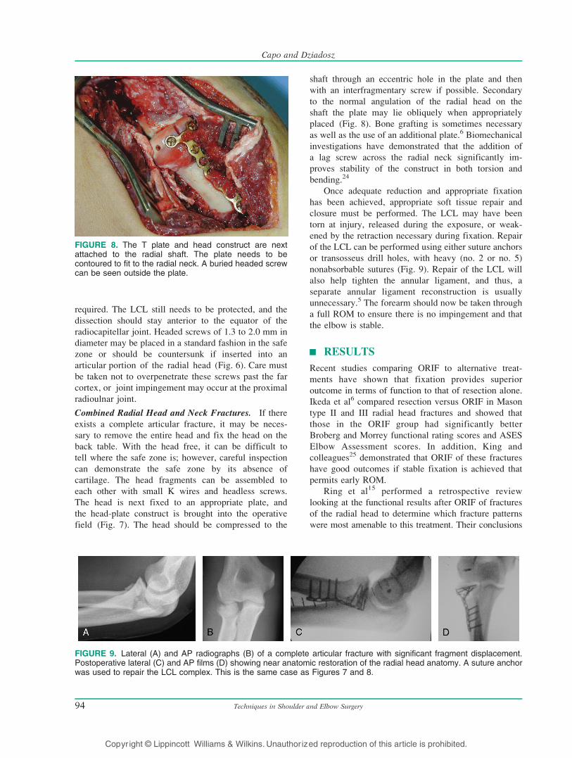
FIGURE 8. The T plate and head construct are nextattached to the radial shaft. The plate needs to becontoured to fit to the radial neck. A buried headed screwcan be seen outside the plate.
FIGURE 9. Lateral (A) and AP radiographs (B) of a complete articular fracture with significant fragment displacement.Postoperative lateral (C) and AP films (D) showing near anatomic restoration of the radial head anatomy. A suture anchorwas used to repair the LCL complex. This is the same case as Figures 7 and 8.
Techniques in Shoulder and Elbow Surgery94
Capo and Dziadosz
Copyr ight © Lippincott Williams & Wilkins. Unauthorized reproduction of this article is prohibited.

revealed that ORIF provided acceptable results in all
cases of noncomminuted Mason type II fractures and in
11 of 12 cases of type III fractures with 3 or fewer
fragments. For fractures with greater comminution than
this, the rates of hardware failure and nonunion were
exceedingly high. It needs to be remembered that this
study was performed before newer implants such as
locking plates and plates dedicated for the radial head
were available.
| COMPLICATIONS
Complications after ORIF of the radial head can occur
both early and late in the postoperative course. Early
complications are seen with injury to the PIN and
inappropriate hardware placement. Late complications
include symptomatic hardware, nonunion, and elbow
stiffness. Failure of fixation is noted with highly
comminuted fractures, as demonstrate by Ring et al15
where 13 of 14 fractures involving the entire radial
head and more than 3 articular fragments had fixa-
tion failure requiring revision surgery. This group
also had 6 nonunions and 4 patients with less than
100 degrees of forearm rotation. Thus, only 1 patient
with this difficult fracture pattern had an acceptable
result.
| POSTOPERATIVE MANAGEMENT
After radial head surgery, elbow motion and forearm
rotation is necessary to prevent adhesions and stiffness.
For these reasons, the goal of surgical repair of a radial
head fracture should be to create a stable construct that
allows early ROM. If the fixation is tenuous and would
not permit early ROM, it is better to convert to a rigid
implant arthroplasty.
If an LCL repair is performed, supination should
be limited to 45 degrees to protect the lateral column
and allow healing. Flexion increases elbow stability
and can be permitted as tolerated, whereas extension
should initially be blocked to 30 degrees. Extension
can usually be increased by 10 degrees every 1 to 2
weeks under therapy guidance. Flexion and extension
are done with the forearm in pronation, whereas
rotation is performed at 90 degrees of elbow flexion.
Another restriction that should be placed on postop-
erative motion is limitation of shoulder abduction in
the presence of an LCL repair. By preventing
abduction, the varus forces placed across the elbow,
and ultimately the LCL, are reduced. Based on the
quality of repair, a minimum of 6 weeks of limited
abduction should allow the LCL to heal.26
| FUTURE OF THE TECHNIQUE
With the advent of locked-plate designs, smaller fixed-
angle devices are now being created to provide more
FIGURE 10. An example of a plate specifically designedfor the radial head. There are multiple locked screws inthe head, an elliptical hole for an interfragmentary or shaftscrew, and the shape is precontoured (Wright MedicalTechnology, Arlington, Tenn).
FIGURE 11. Lateral (A) and AP radiographs (B) showing a multifragmentary articular fracture. There is comminution in theneck portion of the fracture. Lateral (C) and AP radiographs (D) displaying stabilization of the fracture with a radial headplate. Because of the normal angulation of the radial head, a T plate will sit obliquely on the shaft when the radial head isproperly reduced.
Volume 8, Issue 2 95
Operative Fixation of Radial Head Fractures
Copyr ight © Lippincott Williams & Wilkins. Unauthorized reproduction of this article is prohibited.
rigid fixation for these fractures. Additionally, plates
specifically designed for the radial head with multiple
locked screws into the head are now available (Figs. 10
and 11). These small locking plates provide more
secure fixation and may permit earlier ROM and could
result in improved healing rates. Bioabsorbable
implants have shown promise in other areas of fixation
and may be beneficial for the radial head. These are
attractive because they don’t need to be removed and
minimize potential impingement.27Y29 Arthroscopically
assisted fixation has shown promising results, and its
indications should expand as the equipment and
surgeons’ experience improve.30,31
| REFERENCES
1. Hall JA, McKee M. Posterolateral rotatory instability of
the elbow following radial head resection. J Bone JointSurg Am. 2005;87:1571Y1579.
2. Geel CW, Palmer A. Radial head fractures and their effect
on the distal radioulnar joint. A rationale for treatment.
Clin Orthop Relat Res. 1992:79Y84.
3. Ikeda M, Sugiyama K, Kang C, et al. Comminuted
fractures of the radial head: comparison of resection and
internal fixation. Surgical technique. J Bone Joint SurgAm. 2006;88(suppl 1, pt 1):11Y23.
4. Boulas HJ, Morrey BF. Biomechanical evaluation of the
elbow following radial head fracture. Comparison of open
reduction and internal fixation vs. excision, silastic
replacement, and non-operative management. Chir Main.
1998;17:314Y320.
5. Hotchkiss RN. Displaced fractures of the radial head:
internal fixation or excision? J Am Acad Orthop Surg.
1997;5:1Y10.
6. Ikeda M, Sugiyama K, Kang C, et al. Comminuted
fractures of the radial head. Comparison of resection
and internal fixation. J Bone Joint Surg Am. 2005;87:
76Y84.
7. Furry KL, Clinkscales CM. Comminuted fractures of the
radial head. Arthroplasty versus internal fixation. ClinOrthop Relat Res. 1998;353:40Y52.
8. Khalfayan E, Culp R, Alexander A. Mason type II radial
head fractures: operative versus nonoperative treatment.
J Orthop Trauma. 1992;6:283Y289.
9. Beingessner DM, Dunning CE, Stacpoole RA, et al. The
effect of radial head excision and arthroplasty on elbow
kinematics and stability. J Bone Joint Surg Am. 2004;86:
1730Y1739.
10. Mason M. Some observations on fractures of the head of
the radius with a review of one hundred cases. Br J Surg.
1959;42:123Y132.
11. Ring D. In: Bucholz RWH, James D, Court-Brown C, et al,
eds. Fractures and Dislocations of the Elbow. Rockwood
and Green’s Fractures in Adults, Vol. 1, 3rd ed. Philadel-
phia: Lippincott Williams & Wilkins; 2005:1011Y1020.
12. Capo JT, Riaszadeh K, Swan KG, et al. The accuracy of
plain radiographs in assessing fractures of the radial head.
Paper presented at: AAOS Annual Meeting; 2005;
Washington, DC.
13. Akesson T, Herbertsson P, Josefsson P, et al. Primary
nonoperative treatment of moderately displaced two-part
fractures of the radial head. J Bone Joint Surg Am. 2006;
88:1909Y1914.
14. Ikeda M, Oka Y. Function after early radial head resection
for fracture: a retrospective evaluation of 15 patients
followed for 3Y18 years. Acta Orthop Scand. 2000;71:
191Y194.
15. Ring D, Quintero J, Jupiter J. Open reduction and internal
fixation of fractures of the radial head. J Bone Joint SurgAm. 2002;84:1811Y1815.
16. Tan V, Daluiski A, Capo J, et al. Hinged elbow external
fixators: indications and uses. J Am Acad Orthop Surg.
2005;13:503Y514.
17. Diliberti T, Botte MJ, Abrams RA. Anatomical consid-
erations regarding the posterior interosseous nerve during
posterolateral approaches to the proximal part of the
radius. J Bone Joint Surg Am. 2000;82:809Y813.
18. Strauch RJ, Rosenwasser M, Glazer P. Surgical exposure
of the dorsal proximal third of the radius: how vulnerable
is the posterior interosseous nerve? J Shoulder ElbowSurg. 1996;5:342Y346.
19. Hoppenfeld S, deBoer P. The Elbow. Surgical Exposuresin Orthopaedics, The Anatomic Approach, 3rd ed. Phila-
delphia: Lippincott Williams & Wilkins; 2003.
20. Cohen MS, Hastings H II. Post-traumatic contracture of
the elbow. Operative release using a lateral collateral
ligament sparing approach. J Bone Joint Surg Br. 1998;
80:805Y812.
21. Witt JD, Kamineni S. The posterior interosseous nerve
and the posterolateral approach to the proximal radius.
J Bone Joint Surg Br. 1998;80:240Y242.
22. Caputo AE, Mazzocca A, Santoro V. The nonarticulating
portion of the radial head: anatomic and clinical correlations
for internal fixation. J Hand Surg. 1998;23:1082Y1090.
23. Patterson JD, Jones CK, Glisson RR, et al. Stiffness of
simulated radial neck fractures fixed with 4 different
devices. J Shoulder Elbow Surg. 2001;10:57Y61.
24. Capo JT, Asghar J, Sabatino C. Biomechanical stability of
fixation constructs for ORIF of radial head fractures.
Paper presented at: Annual Meeting of the American
Society for Surgery of the Hand; 2003.
25. King G, Evans D, Kellam J. Open reduction and internal
fixation of radial head fractures. J Orthop Trauma. 1991;
5:21Y28.
26. Ring D. Radial head fractures: open reduction internal fixa-
tion. In: Wiss DA, ed. Master Techniques in Orthopaedic
Techniques in Shoulder and Elbow Surgery96
Capo and Dziadosz
Copyr ight © Lippincott Williams & Wilkins. Unauthorized reproduction of this article is prohibited.

Surgery, Fractures, 2nd ed. Philadelphia: Lippincott Wil-
liams & Wilkins; 2006:121Y141.
27. Claes LE, Ignatius AA, Rehm KE, et al. New bioresorb-
able pin for the reduction of small bony fragments: design,
mechanical properties and in vitro degradation. Biomate-rials. 1996;17:1621Y1626.
28. Pelto K, Hirvensalo E, Bostman O, et al. Treatment of
radial head fractures with absorbable polyglycolide pins: a
study on the security of the fixation in 38 cases. J OrthopTrauma. 1994;8:94Y98.
29. Hirvensalo EO, Bostman B, Rokkanen P. Absorbable
polyglycolide pins in fixation of displaced fractures of
the radial head. Arch Orthop Trauma Surg. 1990;109:
258Y261.
30. Rolla PR, Surace MF, Bini A, et al. Arthroscopic
treatment of fractures of the radial head. Arthroscopy.
2006;22:231Y236.
31. Dawson FA, Inostroza F. Arthroscopic reduction and
percutaneous fixation of a radial neck fracture in a child.
Arthroscopy. 2004;20(suppl 2):90Y93.
Volume 8, Issue 2 97
Operative Fixation of Radial Head Fractures
Copyr ight © Lippincott Williams & Wilkins. Unauthorized reproduction of this article is prohibited.