Technique in special examinations - 台灣醫學物理公司 · 2017/5/21 1 Technique in special...
24
2017/5/21 1 Technique in special examinations Contents and category GI tracks UGI LGI Kidney IVP RP AP/PN PCN Breast Mammography Female reproductive system HSG Deterministic: visible, documented, confirmed within a relative short time Skin erythema, hair loss, cataract, infertility, circulatory disease Stochastic: estimated, years or decades to manifest Cancer, genetic effects Health effects of ionizing radiation Have thresholds that are typically quite high Skin erythema Hair loss Cataracts (even in low doses of radiation) 5 Sv for protracted exposures 2 Sv for acute exposures Epidemiological evidence suggesting thresholds (equivalent dose): Lens of eye: 0.5 Gy Circulatory system: 0.5 Gy Tissue reactions Detriment-adjusted nominal risk coefficient at low dose rate: Cancer – 5.5 %/Sv Genetic effects – 0.2 %/Sv (non-human species) Cancer risks are estimated on the basis of probability Organ dose > 100 mGy carcinogenic effects Stochastic risks have no threshold Stochastic effects Tissue weighting factor of gonads: 0.2 0.08 (ICRP, 2007) 1 chest CT scan ~ 8 mSv 20 mGy to breast 5 ~ 15 CT scans carcinogenic effects
Transcript of Technique in special examinations - 台灣醫學物理公司 · 2017/5/21 1 Technique in special...

2017/5/21
1
Technique in special
examinations
Contents and category GI tracks
UGI
LGI
Kidney
IVP
RP
AP/PN
PCN
Breast
Mammography
Female reproductive
system
HSG
Deterministic:
visible, documented, confirmed within a relative short
time
Skin erythema, hair loss, cataract, infertility, circulatory
disease
Stochastic:
estimated, years or decades to manifest
Cancer, genetic effects
Health effects of ionizing radiation
Have thresholds that are typically quite high Skin erythema
Hair loss
Cataracts (even in low doses of radiation) 5 Sv for protracted exposures
2 Sv for acute exposures
Epidemiological evidence suggesting
thresholds (equivalent dose): Lens of eye: 0.5 Gy
Circulatory system: 0.5 Gy
Tissue reactions
Detriment-adjusted nominal risk coefficient
at low dose rate: Cancer – 5.5 %/Sv
Genetic effects – 0.2 %/Sv (non-human species)
Cancer risks are estimated on the basis of
probability Organ dose > 100 mGy carcinogenic effects
Stochastic risks have no threshold
Stochastic effects
Tissue weighting factor of gonads: 0.2 0.08 (ICRP, 2007)
1 chest CT scan ~ 8 mSv 20 mGy to breast
5 ~ 15 CT scans carcinogenic effects

2017/5/21
2
http://www.rerf.jp/radefx/late_e/cancrisk.html
For the average radiation exposure of survivors within 2,500 meters (about 0.2 Gy),
the increase is about 10% above normal age-specific rates. For a dose of 1.0 Gy,
the corresponding cancer excess is about 50% (relative risk = 1.5)
The excess number of solid cancers is estimated as 848 (10.7%)
The dose-response relationship appears to be linear, without any apparent
threshold below which effects may not occur
The probability that an A-bomb survivor will have a cancer caused by A-bomb
radiation (excess lifetime risk) depends on the dose received, age at exposure,
and sex.
Other analyses (not shown) indicate that females have somewhat higher risks of
cancer from radiation exposure than males do.
different tissues and organs have different radiosensitivities
females are generally more radiosensitive than males to cancer induction
young patients are more radiosensitive than older patients
individual genetic differences in susceptibility to radiation-induced cancer
These general aspects of radiosensitivity should be taken into account in the process of justification and optimization of radiological protection in fluoroscopically guided procedures
Individual differences in radiosensitivity
Pre-existing auto-immune and connective tissue disorders predispose patients to the development of severe skin injuries in an unpredictable fashion.
These disorders include scleroderma, systemic lupus erythematosus, and possibly rheumatoid arthritis, although there is controversy regarding whether systemic lupus erythematosus predisposes patients to these effects.
Genetic disorders that affect DNA repair, such as the defect in the ATM gene responsible for ataxia telangiectasia, also predispose individuals to increased radiation sensitivity.
Diabetes mellitus, a common medical condition, does not increase sensitivity to radiation, but does impair healing of radiation injuries
3.3.1 Patient-specific factors Thickness of the body part in the beam
Complexity of the procedure Complexity represents the mental and physical
effort required to perform a procedure.
3.3 Common aspects of patient and occupational
protection
2017/5/21
3
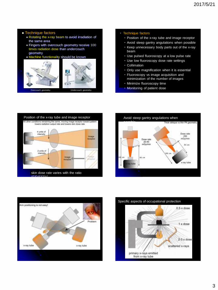
Technique factors Rotating the x-ray beam to avoid irradiation of
the same area
Fingers with overcouch geometry receive 100
times radiation dose than undercouch
geometry
Machine functionality should be known
Overcouch geometry Undercouch geometry
• Technique factors
• Position of the x-ray tube and image receptor
• Avoid steep gantry angulations when possible
• Keep unnecessary body parts out of the x-ray
beam
• Use pulsed fluoroscopy at a low pulse rate
• Use low fluoroscopy dose rate settings
• Collimation
• Only use magnification when it is essential
• Fluoroscopy vs image acquisition and
minimization of the number of images
• Minimize fluoroscopy time
• Monitoring of patient dose
skin dose rate varies with the ratio
(SID/SSD)2
Position of the x-ray tube and image receptor Avoid steep gantry angulations when
possible
Specific aspects of occupational protection
2017/5/21
4
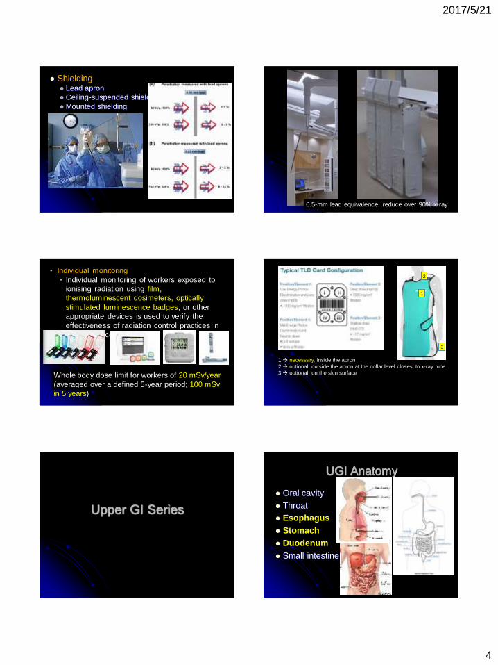
Shielding Lead apron
Ceiling-suspended shielding
Mounted shielding
0.5-mm lead equivalence, reduce over 90% x-ray
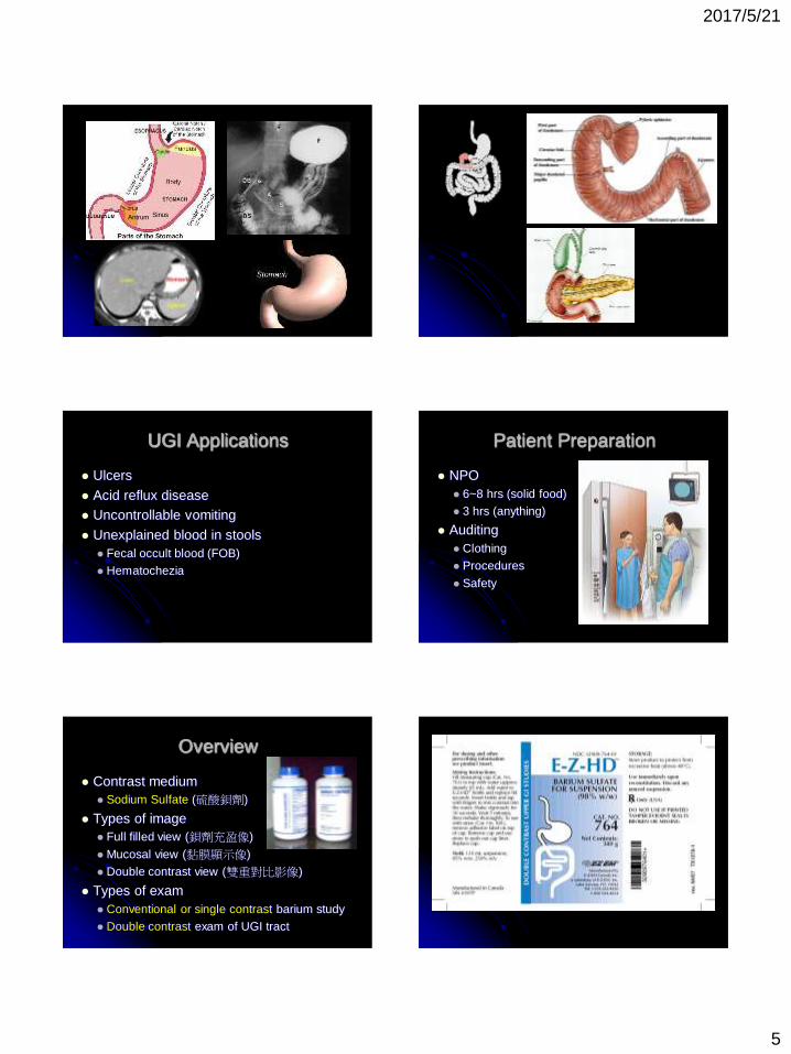
• Individual monitoring
• Individual monitoring of workers exposed to
ionising radiation using film,
thermoluminescent dosimeters, optically
stimulated luminescence badges, or other
appropriate devices is used to verify the
effectiveness of radiation control practices in
the workplace.
Whole body dose limit for workers of 20 mSv/year
(averaged over a defined 5-year period; 100 mSv
in 5 years)
1
2
3
1 necessary, inside the apron
2 optional, outside the apron at the collar level closest to x-ray tube
3 optional, on the skin surface
Upper GI Series
UGI Anatomy
Oral cavity
Throat
Esophagus
Stomach
Duodenum
Small intestine
2017/5/21
5
Sinus Antrum
S
UGI Applications
Ulcers
Acid reflux disease
Uncontrollable vomiting
Unexplained blood in stools
Fecal occult blood (FOB)
Hematochezia
Patient Preparation
NPO
6~8 hrs (solid food)
3 hrs (anything)
Auditing
Clothing
Procedures
Safety
Overview
Contrast medium
Sodium Sulfate (硫酸鋇劑)
Types of image
Full filled view (鋇劑充盈像)
Mucosal view (黏膜顯示像)
Double contrast view (雙重對比影像)
Types of exam
Conventional or single contrast barium study
Double contrast exam of UGI tract

2017/5/21
6
Single Contrast
Sodium Sulfate (硫酸鋇劑),
Semi-solid (250% W/V) for esophagus
70~80% W/V for stomach and duodenum
Image type: Profile view
Techniques:
Rotation of body
Compression of part
Single Contrast 優點:
①一般而言,技術上比較簡單,對於顯影劑之品質要求較不必那麼嚴格。
②對上消化道不正常狹窄、充填缺損(如腔內大型腫塊)、外來性壓迫、瘻管等病變易於顯示出來。對於食道靜脈曲張也比較容易診斷。
③上消化道之生理狀況,如蠕動減少、張性 (tonicity) 消失、不正常攣縮等狀況,不易遺漏。
缺點: ①為使胃腔充分膨滿,大量硫酸鋇劑流入小腸造成重疊。
②胃底 (fundus) 無法壓迫到,cone compression無法做到。
③過份依賴透視:檢查醫師非常依賴X光透視診斷,非透視醫師難發揮會診功能。
④對細微病變,診斷率偏低。
Double Contrast
Contrast medium
140~250% W/V sodium sulfate
Air
Image type: surface, profile
Double Contrast
優點:
①不必用大量硫酸鋇,注入空氣就可達到胃腔膨大之效果。
②例行檢查就可顯示每一部位之粘膜表層之細微構造,對於早期病變多半可以顯示,診斷率高。
③無法做cone compression之處亦可顯示清楚。
④透視主要用於定位和姿勢之調整,故可減少透視時間,病人和醫師的X光接受量較低。
⑤不須太依賴透視診斷能力,放射線科專家可由獲得像片作較深入之分析或會診。透視時能認明病變尤佳。
⑥照像條件kV & mA較低,醫師和病人所受X光照射量較少,對機器之要求亦較低。
Double Contrast
缺點:
①檢查技術困難。對於體質太弱或不合作病人,每一病人花費時間較長。
②診斷醫師更需經驗,人員訓練時間較長。
③顯影劑的品質、濃度和發泡劑對照像品質影響極大。
④使用空氣量不易控制,也不易決定。不足或過度膨脹都會使粘膜下層或粘膜層病灶隱藏。
⑤非常依賴照片來診斷,所以,照像的條件影響診斷極大。
⑥對胃前壁病灶容易遺漏或誤診,所以,檢查時必須包括胃體部和胃竇的cone-down compression film。
Esophagus Exam
Procedures 檢查前5分鐘IM注射Buscopan 20mg。
檢查時令病人用5~10cc開水服下發泡劑,使胃部儘量充氣。
令病人站立,稍向左轉,含一大口硫酸鋇浮懸液,在X光透視監視下,嚥下硫酸鋇劑,當硫酸鋇劑通過Esophagogastric junction時,賁門舒張,胃部空氣立刻反流到食道內,並把食道充氣撐開,此時,立刻攝影取食道之雙重對比X光像。如此,重複吞嚥,攝影取照片。
攝影取頸部食道之正面和側面像。
將X光檢查台放置水平位置,令病人俯臥LPO,X光透視下,令病人再嚥硫酸鋇劑再照像。

2017/5/21
7
Notice 懷疑食道堵塞不通時,不作雙重對比鋇劑X光攝影。
Glucagon對食道之低張性效果不理想。
選擇照像的時機比較困難,照像要快。
食道粘膜對140% W/V高密度硫酸鋇劑之親和力尚稱良好,但因重力關係,很快消退,另一方面,為避免小泡沫在食道內,可令病人多嚥幾口硫酸鋇劑,把小泡沫充分沖掉,並在每次吞嚥時選擇適當時機攝影取像片。
胃部要充飽空氣,才有足夠量之空氣反流到食道把食道張開。
使用低溫 (3~4℃) 之硫酸鋇浮懸液,可能減少食道之蠕動。
檢查過程,若硫酸鋇部分吸入氣管,則檢查應立刻中止。
咽部X光錄影
病人站立,分別吞嚥少量 (3~5cc) 硫酸鋇液,膏劑及含硫酸鋇餅乾
將吞嚥全程錄影,錄取正面、側面、斜位之影像。
檢查過程,若硫酸鋇劑部分吸入氣管,則檢查應立刻中止。
Stomach and Duodenum exam Contrast medium
硫酸鋇劑: 應採用高密度(high density)、高黏質(high viscosity) 且黏膜表面被覆性良好之硫
酸鋇浮懸液,例如140% W/V E-Z HD。
空氣: ①服用發泡劑(effervescent agents):在胃內產生450cc.氣體(如E-Z gas)。
②消泡劑:消除小氣泡顆粒,如gascon(Simethicone)。消泡劑可直接加入到硫酸鋇劑懸液內。
低張劑 Buscopan (Hyoscine-N-butylbromide):(20mg/1ml/Amp.)。
①為一種副交感神經解劑(anticholinergic agent),能有效地使食道、胃、十二指腸之緊張性減低,並使胃幽門鬆弛。
②Buscopan很少引起嚴重之副作用,但是患有前列腺肥大、腸胃道阻塞、toxic megacolon、重症肌無力、青光眼(narrow angle glaucoma) 或心悸、高血壓者宜避免使用。
③使用後將使小腸之transit time顯著延長。若病人要接著做小腸X光攝影時,不採用為宜。
Glucagon ①IV注射作用時間快,但作用時間短。
②IM注射後大約15分鐘才達所要之低張作用。
③絕大多數沒有不良副作用,可是患有糖尿病之病人不宜使用。
④Glucagon對食道低張作用不良。對胃幽門沒有鬆弛作用。藥效過後,小腸馬上恢復蠕動。
Procedures
檢查前10~15分鐘,IM注射glucagon 0.5mg或檢查前5~6分鐘,IM注射glucagon 20mg。
用5~10cc水服下發泡劑,使胃部適當充氣。
病人站立稍向左轉,嚥下1~2口硫酸鋇浮懸液,在適當時機,快速照下食道雙對比X光像。
X光檢查台放平,頭稍低,令病人採俯臥姿,拍攝影胃前壁mucosal view。
站起來再喝鋇劑,總共約150~200cc為原則,之後馬上將X光檢查台放平,令病人在X光檢查台上滾翻一圈,仰臥,透視看,覺得胃粘膜表面被覆(coating) 良好,照下胃正面像。
令病人右側稍抬高 (RAO),攝影取胃體、胃竇、十二指腸之雙對比X光像。
令病人左側抬高約45℃(必要時,控制X光機半立狀況,使病人頭高腳低),取適當角度,攝影取胃底部 (fundus)及cardia之雙對比X光像。
令病人LPO俯臥,攝影取胃和十二指腸全面像。
病人右側臥,攝影取胃和十二指腸側位像。
檢查台立起,病人面向前方站立,稍微LPO,攝影取站立時胃及十二指腸全面像(必要時再喝鋇劑把胃充滿)。
以機器之壓迫設備,壓迫胃體、胃竇、十二指腸,攝影取cone down views。
2017/5/21
8
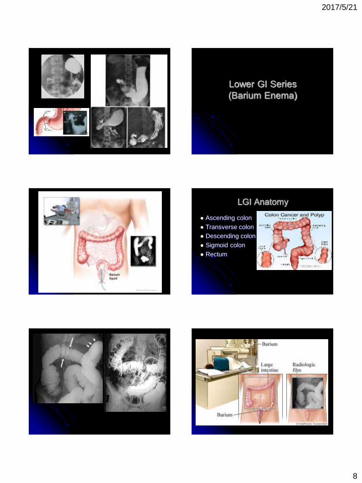
Lower GI Series
(Barium Enema)
LGI Anatomy
Ascending colon
Transverse colon
Descending colon
Sigmoid colon
Rectum

2017/5/21
9
Patient Preparation
成人 ①排定檢查時間前二天開始,儘可能食用低渣食物,少
吃肉類和纖維多的蔬菜。並於睡前服用Ducolax一顆。
②檢查前一天每小時喝水200cc直到睡眠為止。早、午、晚餐一律食用低渣或流質食物,晚餐後於晚上九點鐘服用30cc蓖麻油後喝二杯開水。服完蓖麻油後,除了茶、水之外,禁吃一切食物。
③檢查當天禁用早餐,於排定時間前半小時左右,到影像診療部登記,換好檢查服裝,等候醫師檢查,做完檢查後才能進食。
小兒: 5歲以下小兒,在檢查前3天開始餵食牛奶、果汁等流
質食物,檢查當天延早餐,送影像診療科檢查即可。
8至14歲小孩,輕瀉劑量約成人1/3~1/2量。
Overview
Contrast medium
Barium Sulfate (硫酸鋇劑):以每120mg硫酸鋇加700cc水的配法,配成70~80% W/V
barium suspansion,(Barytgen和E-Z paque各半量混合)。每一病人每次檢查大約需要200~250cc之硫酸鋇浮懸液。
低張劑
Buscopan
Glucagon
Procedures 病人檢查前5分鐘視病情可以IM注射20mg Buscopan。(若病人患有青光
眼、高血壓或前列腺肥大時,改用glucagon 0.5mg IV注射)。
檢查時病人側躺在X光檢查台,護理人員幫忙放入連接顯影劑之肛管,然後令病人俯臥,檢查台頭部稍低。
在X光透視監視下,徐徐灌入硫酸鋇劑,通常在鋇劑逆流到脾彎(splenic flexure) 時停止。先解開導管接頭,排掉直腸處之硫酸鋇,再換上打氣之導管。
讓病人翻身俯臥頭部放低,身體右側稍微抬高,在X光透視監視下由導管注入適當量空氣,當硫酸鋇劑流到橫結腸中央部位時,令病人身體左側抬高,繼續再灌入空氣。
當硫酸鋇流到結腸肝彎(hepatic flexure) 時,將病人頭端之X光檢查台適度抬高,再以空氣把硫酸鋇推送到升結腸(每一病人總共約需700~1000cc空氣)。
在透視下,適當調整病人角度,並攝影取直腸AP、PA、Lat及S狀結腸RAO位 X光像。此時,把X光檢查台豎立起來,讓硫酸鋇流到迴盲部及S狀結腸處,攝影取肝彎和脾彎之像片,最後拍攝影迴盲部AP、PA 像片。
病灶部位加照適當角度之X光像。
最後再以X光透視自直腸至迴盲部全部再察看一次,以免遺漏。
以over tube用14×17吋底片攝影取結腸之AP、PA、及水平X射線bilateral decubitus之全面像及直腸(sigmoid-rectum) 之側位像。
必要時可加照排便後的大腸X光像(post-evacuation films)。
Notice 病人若因便秘而做鋇劑X光檢查者,檢查完畢,由醫師
斟酌發給輕瀉劑(如MgO、Solven、Ducolax等)讓病人服用,以利排清鋇劑。
檢查時,原則上灌到脾彎為度。倘若S狀結腸長而扭曲時,則應斟酌減量,否則鋇劑勢必過多。
檢查時,醫師若發現結腸內有較多殘留糞便時,宜以硫酸鋇劑灌注到盲腸處;隨即讓病人在檢查室廁所排解顯影劑,此時殘留糞便多能一併解出(相當於一次清潔灌腸),之後,馬上在檢查台上,把空氣灌入結腸,即成雙重顯影劑效果(時間要快)。
臨床上懷疑阻塞或檢查當中發現結腸堵塞者,不可採用結腸雙重對比X光檢查技術,應改為單對比鋇劑灌注檢查法。鋇劑容器高度不超過X光檢查台面3英尺為宜。
務必在X光透視監視下注入空氣。
2017/5/21
10
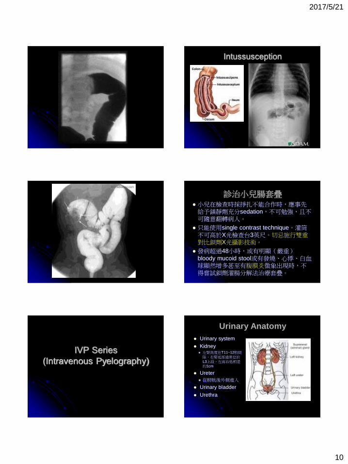
Intussusception
診治小兒腸套疊 小兒在檢查時採掙扎不能合作時,應事先給予鎮靜劑充分sedation,不可勉強,且不可隨意翻轉病人。
只能使用single contrast technique。灌筒不可高於X光檢查台3英尺。切忌施行雙重對比鋇劑X光攝影技術。
發病超過48小時,或有明顯(嚴重)bloody mucoid stool或有發燒、心悸、白血球顯然增多甚至有腹膜炎徵象出現時,不得嘗試鋇劑灌腸分解法治療套疊。
IVP Series
(Intravenous Pyelography)
Urinary system
Kidney 左腎高度在T11~12肋間
隙,右腎底部通常位於L3上段,左高右低相差約1cm
Ureter
從膀胱後外側進入
Urinary bladder
Urethra
Urinary Anatomy

2017/5/21
11
Urinary Anatomy
A. Minor calyces
B. Major calyces
C. Renal pelvis
D. Ureteropelvic
junction (UPJ)
E. Proximal ureter
F. Distal ureter
G. Urinary bladder
Urinary Anatomy
Ureteropelvic
junction :
(UP Junction )
Pelvic brim
Ureterovesical
junction : (UV
Junction)
Urinary Anatomy 1.腹部腫塊或骨盆腫塊(abdominal
mass、pelvis mass)
2.腎結石(kidney stones) 、輸尿管結石(ureteral calculi) 、膀胱結石(cystolith)
3.腎臟外傷(kidney trauma)
4.腰痛(fIank pain)
5.血尿(hematuria)
6.高血壓(hypertension)
7.腎衰竭(renal failure)
8.泌尿道感染(urinary tract infection ,UTI)
Indications
1.對含碘造影劑過敏(hypersensitivity to iodinated contrast media)
2.無尿或沒有排出尿液(anuria)
3.多發性骨髓瘤(multiple myeloma)
4.糖尿病(diabetes)
5.嚴重的肝病或腎病(hepatic disease)
6.嗜鉻細胞瘤(pheochromocytoma)
7.鐮狀血球貧血(sickle cell anemia)
Contraindications
病人需NPO(最少8小時)
詢問過敏病史(碘,海產)
前一天晚上口服輕瀉劑,清晨塞劑灌腸
更換檢查衣
排空膀胱
Check 血壓 &脈搏
Patient Preparation

2017/5/21
12
IVP Series
Routine
Plain film (KUB)
5 min
10 min
AP
RPO
LPO
30 min
+30 min
Post-voiding
Additional imaging
PA
RAO/LAO
Lateral Projection
of bladder
PV standing
Plain film
Imaging Techniques
5 min 10 min
RPO(30o~45o) LPO(30o~45o)
被觀察的該側輸尿管不能與脊椎重疊 喝5杯水,使膀胱脹大以便更明確觀察膀胱周圍是否有腫瘤或腫塊
10 min
30 min

2017/5/21
13
Post-Void & Delay 特殊照法
PA-view & RAO & LAO 病人有過多的腸氣或是輸尿管沒有顯影
特殊照法
膀胱的Lateral-view
特殊照法
PV Standing-view
逆行性尿路攝影
Retrograde Pyelography
(RP)
Ureteroscopy with Fluoroscopy
2017/5/21
14
用膀胱鏡插入導管,經輸尿管到腎盂,再注入造影劑。
優點:造影劑不需靠腎臟排泄,無過敏之風險。
缺點:做膀胱鏡大多使用局部麻醉,有些許不舒服。
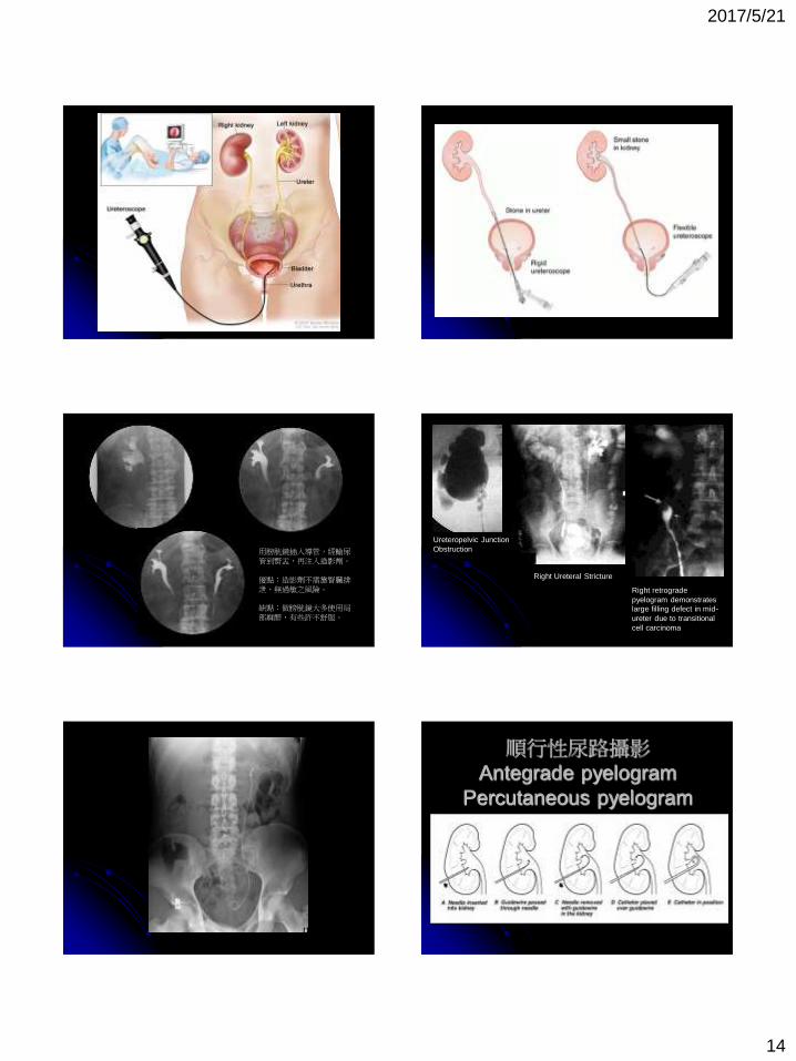
Ureteropelvic Junction
Obstruction
Right Ureteral Stricture
Right retrograde
pyelogram demonstrates
large filling defect in mid-
ureter due to transitional
cell carcinoma
順行性尿路攝影
Antegrade pyelogram
Percutaneous pyelogram
2017/5/21
15
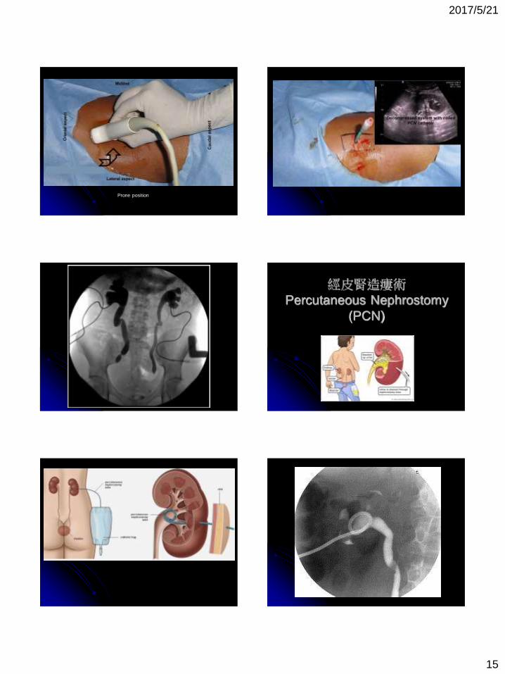
Prone position
經皮腎造瘻術
Percutaneous Nephrostomy
(PCN)

2017/5/21
16
a Image revealing high-grade
obstruction within the right upper ureter
(denoted by arrow); no contrast can be
visualized in the distal ureter
b Image demonstrating multiple
filling defects within the left ureter
(denoted by arrows)
Percutaneous nephrostomy
Mammography
-- Basics and Techniques --
2017/5/21
17
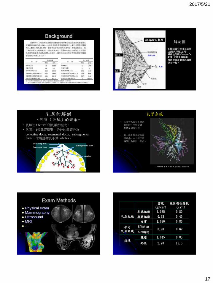
Background 解剖圖
乳腺組織介於淺在肌膜(淺層與深層)之間。
纖維性的膜(Cooper‘s 韌帶)支撐乳腺組織,將乳腺與皮膚及乳頭連結在一起。
Cooper’s 靱帯
胸大肌
肋骨
乳頭
脂肪組織
乳房的解剖 - 乳葉(區域)的概念 -
• 乳腺由15-20個乳葉所組成。
• 乳葉由1根乳管聯繫,分歧的乳管分為
collecting ducts、segmental ducts、 subsegmental
ducts,末端連結乳小葉 lobules。
Segmental duct
Lobules
Collecting duct Subsegmental duct
乳管系統
• 各乳管系統呈不規則
狀分歧,互相交織,
整體呈扇狀分布。
• 有一些乳管系統會互
相重疊,由上往下看
易誤以為在同一部位。
T. Ohtake et al. Cancer 2001;91:2263-72
Exam Methods
Physical exam
Mammography
Ultrasound
MRI
…
密度 (g/cm3)
線性吸收係數(cm-1)
乳房組織
乳腺組織 1.035 0.80
脂肪組織 0.93 0.45
皮膚 1.090 0.80
平均 乳房組織
50%乳腺 0.98 0.62
50%脂肪
病灶 腫瘤 1.045 0.85
鈣化 2.20 12.5

2017/5/21
18
Types of Mammography
Xeroradiography
Screen-film
Digital Mammography
DR
CR
Tomosynthesis
Flat-panel CT
Central ray
Background Physics Target / Filter
Mo/Mo, Mo/Rh, Rh/Rh, W/Rh, W/Al, W/Ag
Tube voltage (kVp)
20 ~ 32 (Target: Mo, Rh)
24 ~ 40 (Target: W)
Background Physics
Source-to-imager distance (SID)
60 ~ 66 cm
Field-of-view (FOV)
8” x 10”, 10” x 12”
Image Resolution
DR CR
GE
DS/2000D
Hologic
Selenia
Fujifilm
FCRm
Kodak
DirectView
Konica
Reguis 190
Pixel Size (μm) 100 70 50 47.5 43.75
Area Ratio* 4.43 2.17 1.11 1 0.85
Background Anatomy
乳房的解剖
成人女性的乳腺分布在胸廓、胸肌上,呈斜三角形。「tail of
Spence」往靠腋窩方向延伸。
末梢部分的邊界並不清楚。
深部:胸肌肌膜
淺部:
側面:覆蓋在前鋸肌上
足部方向:覆蓋在外斜肌、
上直肌肌腱上
正中側面:胸骨
胸大肌
Tail of Spence

2017/5/21
19
Routine Series (1) CC (Craniocaudal) View
(2) MLO (Mediolateral oblique) View
RCC LCC RMLO LMLO
Compression 由於散亂X光射線的減少,使得對比度及分辨率提高。
乳腺的濃度的均一化。
分離乳腺構造的重疊,使得組織間對比度加大。
乳腺組織吸收放射量減少。
由於被攝影對象-底片間距離的縮小,減少幾何學上的模糊。
對乳房的固定防止了模糊的發生。
正確的壓迫基準
1. 至少要把乳房壓迫到組織繃緊的狀態。
2. 受檢者「能夠忍受的最大限度下的壓迫」即不感到很大痛苦程度的壓迫。
Breast Positioning – CC view

2017/5/21
20
CC擺位評比標準: 1.包括所有內側組織都應被看到
2.乳頭位於Image中線位置
3.其乳頭後連線的長度不能短於在MLO
攝影中的乳頭後連線長度1公分,或以要能看到胸肌為準
4.乳房沒有皺紋。
Breast Positioning – MLO view
MLO擺位評比標準: 1. 左右對稱。
2. 乳房輪廓呈現。
3. 胸大肌要照到乳房的基準線。
4. 乳腺後方的脂肪要被描繪出來。
5. 乳房下部組織也要照出來,Inframammary fold伸長。
6. 乳房沒有皺紋。
其他特殊擺位法
較常做的有900 lateral view
Spot compression 焦點式壓迫
對於組織密度高的地方壓迫,可以提升乳房組織分離程度
RCC RMLO Spot view

2017/5/21
21
Spot view
其他特殊擺位法
Magnification 放大攝影
對於腫塊的邊緣結構性特點、辨別病灶良/惡性有很大的幫助,而放大更容易看出鈣化的數目與型態(放大1.5~2倍)
Axillary Tail
照乳房腋下的淋巴結,其需與乳房支撐平台平行,擺位時將之從胸壁外拉
Implant displacement 義乳攝影
植入物推開攝影,填充物往內或上推,上胸壁方向,輕輕拉乳房組織再壓迫
LCC
Axillary Tail
LMLO
Axillary Tail
LCC LMLO
Implant displacement
LCC

2017/5/21
22
RCC RCC-1
RCC-1
其他特殊擺位法
Tangential
切面攝影法,對於鈣化是否在皮膚表層內,而相對於週圍纖維組織密度高者,不清楚時所特別使用攝影方法
LCC LMLO Tangential View
Tangential View
Tangential View
The temporal changes of mammograms obtained from the same woman over seven
years. The top row illustrates the raw images processed to show density and the
bottom row illustrates the “For Presentation” images generated by the manufacturers
(including Hologic, Siemens and GE).
子宮輸卵管攝影Hysterosalpingography
HSG

2017/5/21
23
Introduction 將對比劑注入子宮及輸卵管顯像來評估子宮及輸卵管通暢情形,並診斷子宮及輸卵管有無異常。
Preparations
在月經過後7天左右做檢查
檢查前先排空膀胱內尿液
換上檢查服
受檢者躺在檢查台,姿勢與婦科內診相同
HSG Exam Procedures
醫師以陰道窺管張開陰道
以子宮探針測子宮方向、大小
置入注射針管
拔出陰道窺管
X光透視下以適當的速率注射對比劑,同時照相
等待30分鐘後再照一張
Complications
疼痛
骨盆腔感染
出血
子宮破裂
過敏反應
血管或淋巴管內注入顯影劑造成肺栓塞
骨盆腔沾粘發生腸阻塞
子宮內膜異位
膿性輸卵管破裂
使用含碘顯影劑時可能感覺噁心、暈眩、嘔吐、打噴涕;
具過敏體質患者可能引起全身性蕁麻疹、寒顫;
極少數特異體質患者會發生喉頭水腫、血壓降低、心肺衰竭、休克。
Informed Consent
Risk of infection
Contrast reaction
Hemorrhage
Uterine perforation
Radiation exposure
Theoretical risk of harm to an undiagnosed pregnancy
Oil embolism after extravasation
Exam Dose (mGy)
HSG 0.4 ~ 5.5
Tubal cannulation 8.5
Barium enema 6.5
Pelvic CT 1 ~ 19

2017/5/21
24
(A) normal tubal patency,
(B) endometrial polyp,
(C) submucosal leiomyomata, (D) intrauterine synechiae,
(E) hydrosalpinges,
(F) salpingitis isthmica nodosum. Arcuate uterus.
Normal hysterosalpingography
Unicornous uterus
Didelphys uterus
After Examination
檢查結束時醫師會放一塊紗布在陰道內,於當天洗澡時取出
部分醫師會給予藥物,抗生素、止痛藥之類,檢查結束後服用
Contraindication
月經剛結束或將來時
月經過期尚未確定懷孕者
子宮外孕
骨盆腔發炎或骨盆腔內有腫瘤
對顯影劑過敏者