Technetium-99m-HMPAOBrainSPECTin AnorexiaNervosajnm.snmjournals.org/content/39/2/304.full.pdf ·...
4
very useful for predicting prognosis for patients with meningi- omas. REFERENCES 1. lida H, Miura S, Kanno I, Ogawa T, Uemura K. A new PET camera for noninvasive quantitation of physiological functional parametric images: Headtome-V-Dual. In: Myers R, et al., eds. Quanlitalion of brain function using PET. San Diego, CA: Academic Press; 1996:57-61. 2. Phelps ME, Huang SC. Hoffman EJ, Selin C. Sokoloff L. KühlDE. Tomographie measurement of local cerebral metabolic rate in humans with (F-18)-2-fluoro-2-deoxy- D-glucosc. validation of method. Ann Neural 1979:6:371-388. 3. Reivich M, Alavi A, Wolf A, et al. Glucose metabolic rate kinetic model parameter determination in humans: the lumped constants and rate constants for (lxF)fluorode- oxyglucose and ("Odeoxyglucose. J Cereb Blood Flow Melab 1985;5:179-192. 4. Ardekani B. Braun M. Hutton B, Kanno I. lida H. A fully automatic multimodality image registration algorithm. J Compiti Ansisi Tomogr 1995:19:615-623. 5. Friedman CD, Costammo PD. Teitelbaum B. Berktold RE, Sisson GA Sr. Primary extracranial meningiomas of the head and neck. Lanmgoscope 1990:100:41-48. 6. Hoye SJ. Hoar CS, Murray JE. Extracranial meningioma presenting as a mass of the neck. Am J Surg 1960:100:486-490. 7. George T. Nager. Jaems H. Mark H. Meningiomas invading the temporal bone with extension to the neck. Am J Otolatyngpl 1983:4:297-324. 8. Crompton MR. Gautier-Smith PC. Prediction of recurrence in meningiomas. J Neural Neurosurg Psychialr 1970:33:80-87. 9. Jellinger K. Histological subtypes and prognostic problems in meningiomas. J Neural 1975:208:279-298. 10. Di Chiro G, Hatazawa J, Katz DA, Rizzoli HV, De Michele DJ. Glucose utilization by intracranial meningiomas as an index of tumor aggressivity and probability of recurrence: a PET study. Radiolog)- 1987:164:521-526. 11. Di Chiro G. Meningioma subtypes: MR and PET features [Letter]. Radiology 1990:172:578. 12. Suwa T. Kawano N, Kameya T, Ito H, Oka H. Vada K. Invasive meningiomas in relation to high proliferating potential. Bruin Tumor Pathal 1993:10:63-67. 13. Ericson K, Lilja A, Bergstrom M, et al. Positron emission tomography with (ll-C methyl)-L-mcthionine, ( 1l-C)-D-glucose and (68-Ga) EDTA in supratentorial tumors. J Comput Assist Tomogr 1985:9:683-689. 14. Heiss WD. Beil C. Herholz K. Pawlick G, Wagner R. Wienhard K. Measurement of glucose metabolism. In: Alias of positron emission lontographv oi the hrain. New York: Springer-Verlag; 1985:47-93. Technetium-99m-HMPAO Brain SPECT in Anorexia Nervosa Asli Çepik Kuruoglu, Özlem Kapucu, Tamer Atasever, Zehra Ankan, Erdal Isik and Mustafa Ãoenlü Departments of Psychiatry and Nuclear Medicine, Gazi University Faculty of Medicine, Ankara, Turkey Eating disorders have been redefined in recent years. Brain imaging techniques are useful in demonstrating the association between the morphologic and the functional cerebral changes in these cases. We report "Tc-HMPAO brain SPECT findings in two patients with anorexia nervosa, before and after the treatment. While the detailed neurologic and laboratory examinations, including EEG and cranial CT, were within normal limits before therapy, SPECT study revealed diffuse bilateral hypoperfusion in frontal, parietal and frontotemporal areas which was more prominent in the left hemisphere. Post- treatment SPECT studies obtained after a clinical remission period of 3 mo showed normal brain perfusion in both patients. The pre- and post-treatment SPECT studies accurately reflect the functional state of the patients, and this technique may be used to follow-up the effect of treatment and predict the clinical response to therapy in patients with eating disorders. Key Words: technetium-99m; HMPAO brain SPECT; computerized tomography; anorexia nervosa J NucÃ-Med 1998; 39:304-306 Anorexia nervosa is a behavioral disorder characterized by refusal to maintain body weight at or above a minimally normal weight for age and height. There is a significant disturbance in the individual's perception of the shape or size of the body and denial of the seriousness of the current low body weight (/). There is an increased interest in the functional and structural cerebral changes in psychiatric disorders in the last decade. Recent studies with brain imaging techniques not only investi gate the basic pathological condition in a specified disease, but also help to identify the activation phases and to discriminate the different clinical states, such as remission and exacerbation of the same disease process (2). The limited number of brain imaging studies in anorexia nervosa failed to give uniform results, the main pathological Received Dec. 18, 1996; revision accepted May 2, 1997. For correspondence or reprints contact: L ÖzlemKapucu, MD, HoçdereCad. 3/17, Yukan Ayranci, Ankara, 06540, Turkey. finding being the cerebral cortical atrophy detected in some patients using CT (3). MRI studies revealed a smaller cross- sectional area of the pituitary gland and thalamus (4,5). An early PET study in anorectic patients revealed normal cortical glucose metabolism (6), while another study showed increased metabolism of the caudate nucleus in anorexic state returning to normal levels after realimentation (7). In a recent PET study, the underweight anorectic group showed a global hypometab- olism and an absolute, as well as relative, hypometabolism of glucose in cortical regions most marked in the frontal and parietal cortices as compared to controls (8). These confusing results obtained by the neuroimaging studies are mainly due to methodological discrepancies and patient selection criteria. Moreover, evaluating the patients in remis sion will probably lead to a better understanding of eating disorders. In this study, we investigated cerebral blood flow using 99mTc-HMPAO brain SPECT of two patients with an orexia nervosa, both at the time of diagnosis and after remission of symptoms. CASE REPORTS Patient 1 A 16-yr-old woman presented with a 14-mo history of inten tional loss of weight (17% of the initial weight), refusal of eating because of morbid fear of weight gain, and amenorrhea for the previous 8 mo. She also had provocative vomiting. During the last year of her college education as a successful student, although not overweight, she began dieting with the intention of losing weight. Initially, the weight loss was not recognized by the family. Gradually, she started to drop out of classes, while spending most of her time at home. Ten months after the onset of symptoms, obvious weight loss as well as the emergence of binge and purge cycles alarmed the family and she was brought for treatment. During the initial visits, the psychiatric evaluation of the patient revealed defective self-regulatory functions, regressive pseudo- 304 THE JOURNALOFNUCLEARMEDICINE• Vol. 39 • No. 2 • February 1998 by on April 23, 2018. For personal use only. jnm.snmjournals.org Downloaded from
Transcript of Technetium-99m-HMPAOBrainSPECTin AnorexiaNervosajnm.snmjournals.org/content/39/2/304.full.pdf ·...

very useful for predicting prognosis for patients with meningi-omas.
REFERENCES
1. lida H, Miura S, Kanno I, Ogawa T, Uemura K. A new PET camera for noninvasivequantitation of physiological functional parametric images: Headtome-V-Dual. In:
Myers R, et al., eds. Quanlitalion of brain function using PET. San Diego, CA:Academic Press; 1996:57-61.
2. Phelps ME, Huang SC. Hoffman EJ, Selin C. Sokoloff L. KühlDE. Tomographiemeasurement of local cerebral metabolic rate in humans with (F-18)-2-fluoro-2-deoxy-D-glucosc. validation of method. Ann Neural 1979:6:371-388.
3. Reivich M, Alavi A, Wolf A, et al. Glucose metabolic rate kinetic model parameterdetermination in humans: the lumped constants and rate constants for (lxF)fluorode-oxyglucose and ("Odeoxyglucose. J Cereb Blood Flow Melab 1985;5:179-192.
4. Ardekani B. Braun M. Hutton B, Kanno I. lida H. A fully automatic multimodalityimage registration algorithm. J Compiti Ansisi Tomogr 1995:19:615-623.
5. Friedman CD, Costammo PD. Teitelbaum B. Berktold RE, Sisson GA Sr. Primaryextracranial meningiomas of the head and neck. Lanmgoscope 1990:100:41-48.
6. Hoye SJ. Hoar CS, Murray JE. Extracranial meningioma presenting as a mass of theneck. Am J Surg 1960:100:486-490.
7. George T. Nager. Jaems H. Mark H. Meningiomas invading the temporal bone withextension to the neck. Am J Otolatyngpl 1983:4:297-324.
8. Crompton MR. Gautier-Smith PC. Prediction of recurrence in meningiomas. J NeuralNeurosurg Psychialr 1970:33:80-87.
9. Jellinger K. Histological subtypes and prognostic problems in meningiomas. J Neural1975:208:279-298.
10. Di Chiro G, Hatazawa J, Katz DA, Rizzoli HV, De Michele DJ. Glucose utilization byintracranial meningiomas as an index of tumor aggressivity and probability ofrecurrence: a PET study. Radiolog)- 1987:164:521-526.
11. Di Chiro G. Meningioma subtypes: MR and PET features [Letter]. Radiology1990:172:578.
12. Suwa T. Kawano N, Kameya T, Ito H, Oka H. Vada K. Invasive meningiomas inrelation to high proliferating potential. Bruin Tumor Pathal 1993:10:63-67.
13. Ericson K, Lilja A, Bergstrom M, et al. Positron emission tomography with (ll-Cmethyl)-L-mcthionine, ( 1l-C)-D-glucose and (68-Ga) EDTA in supratentorial tumors.J Comput Assist Tomogr 1985:9:683-689.
14. Heiss WD. Beil C. Herholz K. Pawlick G, Wagner R. Wienhard K. Measurement ofglucose metabolism. In: Alias of positron emission lontographv oi the hrain. NewYork: Springer-Verlag; 1985:47-93.
Technetium-99m-HMPAO Brain SPECT inAnorexia NervosaAsli Çepik Kuruoglu, Özlem Kapucu, Tamer Atasever, Zehra Ankan, Erdal Isik and Mustafa ÜnlüDepartments of Psychiatry and Nuclear Medicine, Gazi University Faculty of Medicine, Ankara, Turkey
Eating disorders have been redefined in recent years. Brain imagingtechniques are useful in demonstrating the association between themorphologic and the functional cerebral changes in these cases. Wereport "Tc-HMPAO brain SPECT findings in two patients with
anorexia nervosa, before and after the treatment. While the detailedneurologic and laboratory examinations, including EEG and cranialCT, were within normal limits before therapy, SPECT study revealeddiffuse bilateral hypoperfusion in frontal, parietal and frontotemporalareas which was more prominent in the left hemisphere. Post-treatment SPECT studies obtained after a clinical remission periodof 3 mo showed normal brain perfusion in both patients. The pre-and post-treatment SPECT studies accurately reflect the functionalstate of the patients, and this technique may be used to follow-upthe effect of treatment and predict the clinical response to therapy inpatients with eating disorders.Key Words: technetium-99m; HMPAO brainSPECT; computerizedtomography; anorexia nervosa
J NucÃMed 1998; 39:304-306
Anorexia nervosa is a behavioral disorder characterized byrefusal to maintain body weight at or above a minimally normalweight for age and height. There is a significant disturbance inthe individual's perception of the shape or size of the body and
denial of the seriousness of the current low body weight (/).There is an increased interest in the functional and structural
cerebral changes in psychiatric disorders in the last decade.Recent studies with brain imaging techniques not only investigate the basic pathological condition in a specified disease, butalso help to identify the activation phases and to discriminatethe different clinical states, such as remission and exacerbationof the same disease process (2).
The limited number of brain imaging studies in anorexianervosa failed to give uniform results, the main pathological
Received Dec. 18, 1996; revision accepted May 2, 1997.For correspondence or reprints contact: L ÖzlemKapucu, MD, HoçdereCad. 3/17,
Yukan Ayranci, Ankara, 06540, Turkey.
finding being the cerebral cortical atrophy detected in somepatients using CT (3). MRI studies revealed a smaller cross-sectional area of the pituitary gland and thalamus (4,5). Anearly PET study in anorectic patients revealed normal corticalglucose metabolism (6), while another study showed increasedmetabolism of the caudate nucleus in anorexic state returning tonormal levels after realimentation (7). In a recent PET study,the underweight anorectic group showed a global hypometab-olism and an absolute, as well as relative, hypometabolism ofglucose in cortical regions most marked in the frontal andparietal cortices as compared to controls (8).
These confusing results obtained by the neuroimaging studiesare mainly due to methodological discrepancies and patientselection criteria. Moreover, evaluating the patients in remission will probably lead to a better understanding of eatingdisorders. In this study, we investigated cerebral blood flowusing 99mTc-HMPAO brain SPECT of two patients with an
orexia nervosa, both at the time of diagnosis and after remissionof symptoms.
CASE REPORTS
Patient 1A 16-yr-old woman presented with a 14-mo history of inten
tional loss of weight (17% of the initial weight), refusal of eatingbecause of morbid fear of weight gain, and amenorrhea for theprevious 8 mo. She also had provocative vomiting.
During the last year of her college education as a successfulstudent, although not overweight, she began dieting with theintention of losing weight. Initially, the weight loss was notrecognized by the family. Gradually, she started to drop out ofclasses, while spending most of her time at home. Ten months afterthe onset of symptoms, obvious weight loss as well as theemergence of binge and purge cycles alarmed the family and shewas brought for treatment.
During the initial visits, the psychiatric evaluation of the patientrevealed defective self-regulatory functions, regressive pseudo-
304 THE JOURNALOF NUCLEARMEDICINE•Vol. 39 •No. 2 •February 1998
by on April 23, 2018. For personal use only. jnm.snmjournals.org Downloaded from

oedipal conflicts and an immature ego. Psychometric examinationsruled out additional psychiatric disturbances and depression. TheEating Attitudes Test (EAT) score was 68 (9), while the HamiltonDepression Rating Scale (70) score was 14. The MinnesotaMultiphasic Personality Inventory (MMP1) (11) profile of thepatient was inconsistent with any comorbid Axis I and II disorders.Laboratory tests including the serum glucose or electrolytes,urinalysis and endocrine profile were within normal limits, and noorganic pathology consistent with the weight loss or secondaryamenorrhea was found. Cranial CT as well as EEG were withinnormal limits.
After the diagnosis of binge eating and purging type anorexianervosa was established by two independent interviewers according to Diagnostic Statistical Manual of Mental Disorders-IV(DSM-IV) diagnostic criteria (7), the patient was introduced to atherapeutic program, with dietary counseling and individual psy-chotherapeutic measures. The therapy was initially supportive,followed by both supportive and cognitive-behavioral measures.Pharmacotherapy was given as fluovoxamine 100 mg daily duringthe first 8 mo of the treatment. Furthermore, the patient participatedin outpatient group therapy.
During the second year of the treatment, there was an apparentimprovement in her behavioral pattern and interpersonal relationships. The symbiotic pattern was not evident, and the individual-ization process was more conspicuous. She had remissions inbinge/purge cycles lasting for long periods, and her weight wasnormalized. The EAT score decreased to 20.
Patient 2An 18-yr-old woman presented with a 16-mo history of weight
loss (19% of her initial weight). Although she was underweight,she had provocative vomiting in order to prevent weight gain. Shehad amenorrhea for the previous 7 mo.
Being the last child of a conservative family, she was describedas a quiet and conscientious student. The refusal of eating startedduring the adolescence period when she became an attractive girland began to think about marriage. Progressive weight losspromoted anxiety and concern within the family, and severalphysicians were consulted without reaching a definitive diagnosis.During that time, binge-purge cycles emerged so that she decidedto come to the psychiatry outpatient department without informingher family.
Psychiatric evaluation revealed impulsiveness, disturbance ofinterpersonal relationships and preoccupation with body image.She reported a decline in academic performance. The MMPIprofile revealed no comorbid Axis I or II disorder (77). TheHamilton Depression Rating Scale (70) score was 12, while theEAT (9) score was 55. The results of detailed laboratory tests,including CT and EEG, were within normal limits.
A diagnosis of binge eating and purging type anorexia nervosawas made by two independent psychiatrists and outpatient treatment with dietary counseling, as well as psychotherapeutic measures was instituted. For the initial 6 mo of treatment, 20 mgfluoxetine was given daily. One year after the patient's admission,
her eating pattern improved and her weight increased by 10 kg. Sherestarted her education and formulated long-term plans to copewith family conflicts.
MATERIALS AND METHODSThe SPECT study consisted of two steps. Pretreatment SPECT
was performed after intravenous injection of 592-666 MBq 99mTc-
HMPAO 4-6 wk after the diagnosis of anorexia nervosa wasestablished and the laboratory tests were completed. Patients wereneither dehydrated nor on drug treatment. The patients did not haveany binge-purge cycles and unprescribed drug intake before the
.wffF
PPCTRCATMCNT
FIGURE 1. Upper row: pretreatment SPECT study; four sequential transaxialconsecutive slices of Patient 1 show diffuse hypoperfusion in frontal, fronto-
parietal, parietal and frontotemporal cortices which is more prominent in theleft hemisphere. Lower row: post-treatment SPECT study; four sequential
transaxial slices of the same patient show that cortical hypoperfusion is nolonger apparent.
study. Images of the head were acquired over 60 angles through360", with each angle being collected for 30 sec on a dual-headed
rotating gamma camera equipped with a high-resolution collimator.Images were obtained in a quiet and semidark room with thepatient's eyes open. Total acquisition time was approximately 30
min. Data were stored on a computer in a 64 X 64 matrix. After thepatient remained free of symptoms for at least 3 mo and offmedication for at least 6 mo, the SPECT study was repeated withthe same dose of 99mTc-HMPAO and the same acquisition param
eters as the pretreatment study.After doing backprojection, image reconstruction was performed
using Butterworth and Ramp filters with an attenuation coefficientof 0.12, cutoff frequency of 0.39 and power factor of 10. Transaxialslices were obtained parallel to the orbito-mcatal line. Transaxial,coronal and sagittal slices were generated in the pixel size of 6 mm.Slices were analyzed visually and quantitatively. For quantitativeanalysis, the mean counts/pixel was calculated for 11 regions ofinterest (ROIs) on four sequential transaxial slices and for one ROIoutlining the cerebellum. Pairs of ROIs were mirrored from theright hemisphere to the left using a semiautomated technique.Count density was calculated for each ROI and region-to-cerebellarratios were obtained for pretreatment and post-treatment studies.From these normalized values, the asymmetry between pretreatment and post-treatment studies of the same region was expressedas follows:
% Asymmetry index =
(Region post-treatment —Region pretreatmcnt)100 X 0.5 (Region post-treatment + Region pretreatment)'
RESULTSAt the initial evaluation, the results of the EEG and CT
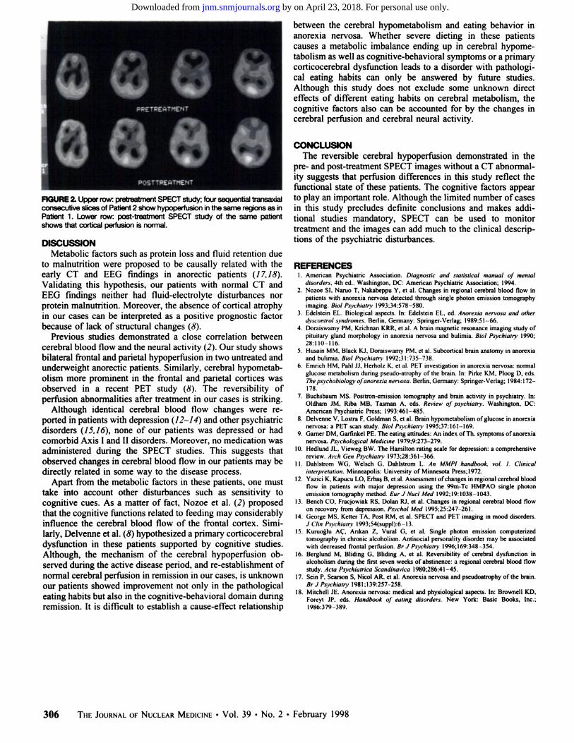
studies were within normal limits, while SPECT revealedbilateral frontal, parietal and frontotemporal hypoperfusion.The left hemispheric hypoperfusion was more prominent. At theend of 1.5 yr of treatment, with the patients having been inremission for 3 mo, a control SPECT was obtained (Fig. 1, 2).Interestingly, the previously demonstrated hypoperfusion wasno longer seen. Asymmetry indexes between post-treatment andpretreatment studies ranged from 3%-8%, compatible with thevisual analysis of both of the patients.
HMPAO BRAINSPECT INANOREXIANERVOSA•Kuruoglu et al. 305
by on April 23, 2018. For personal use only. jnm.snmjournals.org Downloaded from
/n ßkfu^U Wj1 *.
PDETPEOTMENT
POSTTPEOTMENT
FIGURE 2. Upper row: pretreatment SPECT study; four sequential transaxialconsecutive slices of Patient 2 show hypoperfusion in the same regions as inPatient 1. Lower row: post-treatment SPECT study of the same patient
shows that cortical perfusion is normal.
DISCUSSIONMetabolic factors such as protein loss and fluid retention due
to malnutrition were proposed to be causally related with theearly CT and EEG findings in anorectic patients (17,IS).Validating this hypothesis, our patients with normal CT andEEG findings neither had fluid-electrolyte disturbances norprotein malnutrition. Moreover, the absence of cortical atrophyin our cases can be interpreted as a positive prognostic factorbecause of lack of structural changes (8).
Previous studies demonstrated a close correlation betweencerebral blood flow and the neural activity (2). Our study showsbilateral frontal and parietal hypoperfusion in two untreated andunderweight anorectic patients. Similarly, cerebral hypometab-
olism more prominent in the frontal and parietal cortices wasobserved in a recent PET study (8). The reversibility ofperfusion abnormalities after treatment in our cases is striking.
Although identical cerebral blood flow changes were reported in patients with depression (12-14) and other psychiatricdisorders (15,16), none of our patients was depressed or hadcomorbid Axis I and II disorders. Moreover, no medication wasadministered during the SPECT studies. This suggests thatobserved changes in cerebral blood flow in our patients may bedirectly related in some way to the disease process.
Apart from the metabolic factors in these patients, one musttake into account other disturbances such as sensitivity tocognitive cues. As a matter of fact, Nozoe et al. (2) proposedthat the cognitive functions related to feeding may considerablyinfluence the cerebral blood flow of the frontal cortex. Similarly, Delvenne et al. (8) hypothesized a primary corticocerebraldysfunction in these patients supported by cognitive studies.Although, the mechanism of the cerebral hypoperfusion observed during the active disease period, and re-establishment ofnormal cerebral perfusion in remission in our cases, is unknownour patients showed improvement not only in the pathologicaleating habits but also in the cognitive-behavioral domain duringremission. It is difficult to establish a cause-effect relationship
between the cerebral hypometabolism and eating behavior inanorexia nervosa. Whether severe dieting in these patientscauses a metabolic imbalance ending up in cerebral hypometabolism as well as cognitive-behavioral symptoms or a primarycorticocerebral dysfunction leads to a disorder with pathological eating habits can only be answered by future studies.Although this study does not exclude some unknown directeffects of different eating habits on cerebral metabolism, thecognitive factors also can be accounted for by the changes incerebral perfusion and cerebral neural activity.
CONCLUSIONThe reversible cerebral hypoperfusion demonstrated in the
pre- and post-treatment SPECT images without a CT abnormality suggests that perfusion differences in this study reflect thefunctional state of these patients. The cognitive factors appearto play an important role. Although the limited number of casesin this study precludes definite conclusions and makes additional studies mandatory, SPECT can be used to monitortreatment and the images can add much to the clinical descriptions of the psychiatric disturbances.
REFERENCES1. American Psychiatric Association. Diagnostic and statistical manual of mental
disorders, 4th ed.. Washington, DC: American Psychiatric Association: 1994.2. Nozoe SI, Naruo T, Nakabeppu Y. et al. Changes in regional cerebral blood flow in
patients with anorexia nervosa detected through single photon emission tomographyimaging. Biol Psychiatry- 1993:34:578-580.
3. Edelstein EL. Biological aspects. In: Edelstein EL. ed. Anorexia nervosa and otherdyscontrul syndromes. Berlin. Germany: Springer-Verlag; 1989:51-66.
4. Doraiswamy PM. Krichnan KRR. et al. A brain magnetic resonance imaging study ofpituitary gland morphology in anorexia nervosa and bulimia. Biol Psychiatry 1990:28:110-116.
5. Husain MM. Black K I Doraiswamy PM. et al. Subcortical brain anatomy in anorexiaand bulimia. Biol Psychiatry 1992:31:735-738.
6. Emnch HM. Pahl JJ. Herholz K. et al. PET investigation in anorexia nervosa: normalglucose metabolism during pseudo-atrophy of the brain. In: Pirke KM. Ploog D, eds.The psvchobiology of anorexia nervosa. Berlin. Germany: Springer-Verlag; 1984:172
178.7. Buchsbaum MS. Positron-emission tomography and brain activity in psychiatry. In:
Oldham JM. Riba MB. Tasman A. eds. Review of psychiatry. Washington, DC:American Psychiatric Press; 1993:461-485.
8. Delvenne V. Lostra F. Goldman S, et al. Brain hypometabolism of glucose in anorexianervosa: a PET scan study. Biol Psychiatry 1995:37:161-169.
9. Gamer DM, Garfmkel PE. The eating attitudes: An index of Th. symptoms of anorexianervosa. Psychological Medicine 1979;9:273-279.
10. Hedlund JL. Vieweg BW. The Hamilton rating scale for depression: a comprehensivereview. Arch Gen Psychiatry 1973:28:361-366.
11. Dahlstrom WG. Welsch G, Dahlstrom L. An MMPI handbook, vol. I. Clinicalinterpretation. Minneapolis: University of Minnesota Press;1972.
12. Yazici K Kapucu LO. Erbas B. et al. Assessment of changes in regional cerebral bloodflow in patients with major depression using the 99m-Tc HMPAO single photonemission tomography method. Ear J NucÃMed 1992:19:1038-1043.
13. Bench CO, Fracjowiak RS. Dolan RJ. et al. Changes in regional cerebral blood flowon recovery from depression. Psycho/ Med 1995:25:247-261.
14. George MS. Ketter TA. Post RM, et al. SPECT and PET imaging in mood disorders.J Clin Psychiatry 1993;54(suppl):6-13.
15. Kuruoglu AC. Ankan Z. Vural G, et al. Single photon emission computerizedtomography in chronic alcoholism. Antisocial personality disorder may be associatedwith decreased frontal perfusion. Br J Psychiatry 1996:169:348-354.
16. Berglund M. Bliding G. Sliding A, et al. Reversibility of cerebral dysfunction inalcoholism during the first seven weeks of abstinence: a regional cerebral blood flowstudy. Ada Psychiatrien Scandinavia 1980:286:41-45.
17. Sein P. Searson S. Nicol AR. et al. Anorexia nervosa and pseudoatrophy of the brain.Br J Psychiatry 1981:139:257-258.
18. Mitchell JE Anorexia nervosa: medical and physiological aspects. In: Brownell KD.Foreyl JP, eds. Handbook of eating disorders. New York: Basic Books, Inc.;1986:379-389.
306 THEJOURNALOFNUCLEARMEDICINE•Vol. 39 •No. 2 •February 1998
by on April 23, 2018. For personal use only. jnm.snmjournals.org Downloaded from

1998;39:304-306.J Nucl Med. Asli Çepik Kuruoglu, Özlem Kapucu, Tamer Atasever, Zehra Arikan, Erdal Isik and Mustafa Ünlü Technetium-99m-HMPAO Brain SPECT in Anorexia Nervosa
http://jnm.snmjournals.org/content/39/2/304This article and updated information are available at:
http://jnm.snmjournals.org/site/subscriptions/online.xhtml
Information about subscriptions to JNM can be found at:
http://jnm.snmjournals.org/site/misc/permission.xhtmlInformation about reproducing figures, tables, or other portions of this article can be found online at:
(Print ISSN: 0161-5505, Online ISSN: 2159-662X)1850 Samuel Morse Drive, Reston, VA 20190.SNMMI | Society of Nuclear Medicine and Molecular Imaging
is published monthly.The Journal of Nuclear Medicine
© Copyright 1998 SNMMI; all rights reserved.
by on April 23, 2018. For personal use only. jnm.snmjournals.org Downloaded from