Taxonomic patterns in the zoonotic potential of mammalian ... · Data collection Olival et al....
17
Taxonomic patterns in the zoonotic potential of mammalian viruses Alex D. Washburne 1 , Daniel E. Crowley 1 , Daniel J. Becker 1 , Kevin J. Olival 2 , Matthew Taylor 1 , Vincent J. Munster 3 and Raina K. Plowright 1 1 Department of Microbiology and Immunology, Montana State University, Bozeman, MT, USA 2 Ecohealth Alliance, New York, NY, USA 3 National Institute of Allergy and Infectious Disease, Hamilton, MT, USA ABSTRACT Predicting and simplifying which pathogens may spill over from animals to humans is a major priority in infectious disease biology. Many efforts to determine which viruses are at risk of spillover use a subset of viral traits to find trait-based associations with spillover. We adapt a new method—phylofactorization—to identify not traits but lineages of viruses at risk of spilling over. Phylofactorization is used to partition the International Committee on Taxonomy of Viruses viral taxonomy based on non-human host range of viruses and whether there exists evidence the viruses have infected humans. We identify clades on a range of taxonomic levels with high or low propensities to spillover, thereby simplifying the classification of zoonotic potential of mammalian viruses. Phylofactorization by whether a virus is zoonotic yields many disjoint clades of viruses containing few to no representatives that have spilled over to humans. Phylofactorization by non-human host breadth yields several clades with significantly higher host breadth. We connect the phylogenetic factors above with life-histories of clades, revisit trait-based analyses, and illustrate how cladistic coarse-graining of zoonotic potential can refine trait-based analyses by illuminating clade-specific determinants of spillover risk. Subjects Evolutionary Studies, Virology, Infectious Diseases, Statistics Keywords Zoonosis, Spillover, Viruses, Phylofactorization, Epidemiology INTRODUCTION Zoonotic spillover, the transmission of pathogens from non-human vertebrate animals to humans, is a major public health challenge yet the prediction and management of which pathogens spillover is poorly understood (Plowright et al., 2017). Recent, high- profile spillover events, such as that of Nipah virus (Chua et al., 2000; Pulliam et al., 2012), Hendra virus (Murray et al., 1995; Plowright et al., 2015), avian influenza virus (Fouchier et al., 2004; Li et al., 2014), Ebola virus (Baize et al., 2014), rabies virus (Messenger, Smith & Rupprecht, 2002; Schneider et al., 2009), Chikungunya virus (Powers & Logue, 2007), Zika virus (Wikan & Smith, 2016), and West Nile virus (Petersen & Roehrig, 2001; Hayes & Gubler, 2006), among others, strongly motivate a broader understanding of the trait determinants and viral clades at risk of pathogen spillover (Rosenberg, 2015; Geoghegan et al., 2016; Plowright et al., 2016; Geoghegan & Holmes, 2017; Lloyd-Smith et al., 2009). How to cite this article Washburne AD, Crowley DE, Becker DJ, Olival KJ, Taylor M, Munster VJ, Plowright RK. 2018. Taxonomic patterns in the zoonotic potential of mammalian viruses. PeerJ 6:e5979 DOI 10.7717/peerj.5979 Submitted 19 July 2018 Accepted 22 October 2018 Published 28 November 2018 Corresponding author Alex D. Washburne, [email protected] Academic editor Elliot Lefkowitz Additional Information and Declarations can be found on page 13 DOI 10.7717/peerj.5979 Copyright 2018 Washburne et al. Distributed under Creative Commons CC-BY 4.0
Transcript of Taxonomic patterns in the zoonotic potential of mammalian ... · Data collection Olival et al....
Taxonomic patterns in the zoonoticpotential of mammalian virusesAlex D. Washburne1, Daniel E. Crowley1, Daniel J. Becker1,Kevin J. Olival2, Matthew Taylor1, Vincent J. Munster3 andRaina K. Plowright1
1Department of Microbiology and Immunology, Montana State University, Bozeman, MT, USA2 Ecohealth Alliance, New York, NY, USA3 National Institute of Allergy and Infectious Disease, Hamilton, MT, USA
ABSTRACTPredicting and simplifying which pathogens may spill over from animals to humansis a major priority in infectious disease biology. Many efforts to determine whichviruses are at risk of spillover use a subset of viral traits to find trait-basedassociations with spillover. We adapt a new method—phylofactorization—to identifynot traits but lineages of viruses at risk of spilling over. Phylofactorization is used topartition the International Committee on Taxonomy of Viruses viral taxonomybased on non-human host range of viruses and whether there exists evidence theviruses have infected humans. We identify clades on a range of taxonomic levels withhigh or low propensities to spillover, thereby simplifying the classification of zoonoticpotential of mammalian viruses. Phylofactorization by whether a virus is zoonoticyields many disjoint clades of viruses containing few to no representatives that havespilled over to humans. Phylofactorization by non-human host breadth yields severalclades with significantly higher host breadth. We connect the phylogenetic factorsabove with life-histories of clades, revisit trait-based analyses, and illustrate howcladistic coarse-graining of zoonotic potential can refine trait-based analyses byilluminating clade-specific determinants of spillover risk.
Subjects Evolutionary Studies, Virology, Infectious Diseases, StatisticsKeywords Zoonosis, Spillover, Viruses, Phylofactorization, Epidemiology
INTRODUCTIONZoonotic spillover, the transmission of pathogens from non-human vertebrate animalsto humans, is a major public health challenge yet the prediction and management ofwhich pathogens spillover is poorly understood (Plowright et al., 2017). Recent, high-profile spillover events, such as that of Nipah virus (Chua et al., 2000; Pulliam et al., 2012),Hendra virus (Murray et al., 1995; Plowright et al., 2015), avian influenza virus(Fouchier et al., 2004; Li et al., 2014), Ebola virus (Baize et al., 2014), rabies virus (Messenger,Smith & Rupprecht, 2002; Schneider et al., 2009), Chikungunya virus (Powers & Logue, 2007),Zika virus (Wikan & Smith, 2016), and West Nile virus (Petersen & Roehrig, 2001;Hayes & Gubler, 2006), among others, strongly motivate a broader understanding of the traitdeterminants and viral clades at risk of pathogen spillover (Rosenberg, 2015;Geoghegan et al.,2016; Plowright et al., 2016; Geoghegan & Holmes, 2017; Lloyd-Smith et al., 2009).
How to cite this article Washburne AD, Crowley DE, Becker DJ, Olival KJ, Taylor M, Munster VJ, Plowright RK. 2018. Taxonomicpatterns in the zoonotic potential of mammalian viruses. PeerJ 6:e5979 DOI 10.7717/peerj.5979
Submitted 19 July 2018Accepted 22 October 2018Published 28 November 2018
Corresponding authorAlex D. Washburne,[email protected]
Academic editorElliot Lefkowitz
Additional Information andDeclarations can be found onpage 13
DOI 10.7717/peerj.5979
Copyright2018 Washburne et al.
Distributed underCreative Commons CC-BY 4.0

Emerging infectious diseases arise from a pre-existing pool of viruses with traits,life-histories, and evolutionary histories, all of which interact to determine the propensityfor pathogen spillover. With an extremely large diversity of viral species in nature,understanding which clades of viruses are most prone to zoonotic spillover is critical forimproving pathogen surveillance efforts and designing public health interventions acrossscales from reservoir hosts to humans.
A recent study examined viral traits that predicted a virus’ zoonotic status; aftercontrolling for research effort they found that predictors included the phylogenetichost breadth of the virus, genome length, and whether or not the virus was vector-borne,reproduced in the cytoplasm, or was enveloped (Olival et al., 2017). Trait-based analysessuch as these are insightful and useful, but can be limited by incomplete characterizationof traits associated with zoonoses and confounded or biased by non-zoonotic clades—clades with dramatically lower rates of zoonosis—whose homologous traits can all becomenegatively associated with zoonosis. Cladistic analyses complement trait-based analysesby identifying monophyletic clades or taxonomic groups with common patterns ofzoonosis (Fig. 1).
Cladistic analyses can enhance and focus trait-based analyses to refine an understandingof the causes of viral zoonoses. Viral life-histories—the patterns and traits determiningsurvival and reproduction—vary greatly across lineages with vastly different molecularmachinery for host cell entry, viral gene expression, replication, assembly anddissemination (Jane Flint et al., 2015). General traits, such genetic material or presenceof an envelope, may miss the specific molecular mechanisms, such as promiscuousreceptor binding or immunoevasion, by which viral quasispecies from a particular cladecan become capable of infecting humans. Furthermore, general traits found across allviruses may be less actionable for management interventions than clade-specificmechanisms. For example, knowing the particular species of mosquito, Aedes aegypti,responsible for transmitting many flaviviruses (Black et al., 2002) enables ecologicalinterventions aimed at modifying A. aegypti population density, susceptibility to Flavivirusinfection, and more (Morrison et al., 2008). Identifying viral clades with unusually highor low propensity for spillover can prioritize surveillance effort and generate detailed,clade-specific hypotheses about molecular mechanisms and pathogen life-history traitsdriving spillover risk. Furthermore, identification of non-zoonotic clades can allowrefined trait-based analyses: instead of “which traits are associated with zoonosis,” onecan ask “which traits are associated with zoonosis for those pathogens at risk of spillingover?” Identifying molecular mechanisms and hypothesized life-history traits forclade-specific determinants of spillover can revise the list of traits used in trait-basedanalyses (Fig. 1).
To build off the recent study of viral traits determining spillover, we apply a newbiological machine-learning method, phylofactorization (Washburne et al., 2017a, 2017b),to identify clades of mammalian viruses with high within-clade similarity in zoonoticpotential and high between-clade differences. Phylofactorization is a greedy, graph-partitioning algorithm which partitions a tree, in our case the International Committeeon Taxonomy of Viruses (ICTV) taxonomy tree, by identifying edges with the most
Washburne et al. (2018), PeerJ, DOI 10.7717/peerj.5979 2/17

significant differences in the response variable. Using a cladistic taxonomy tree as ascaffold for identifying clades with common propensities for spillover can flexibly identifythe range of taxonomic scales of interest and implicitly identify life history traits whichenable viruses to spill over into humans. We analyze viruses from a recently publisheddataset of mammalian viruses (Olival et al., 2017) and replicate the paper’s trait-basedanalysis alongside our cladistic analysis to provide a side-by-side comparison of howa cladistic analysis informs, complements and, in some cases, changes the results oftrait-based analyses.
Figure 1 Cladistic analyses complement and refine trait-based analyses of viral zoonotic potential.Understanding which clades of viruses spillover can improve our understanding of life-history andtrait-based predictors of zoonosis. A thought experiment of predicting flight in a subset of 10 vertebrates,including reptiles, birds, and mammals reveals the importance of cladistic analyses. Trait-based analysesaim to identify explanatory traits correlated with a focal response trait, such as identifying feathers andhollow bones as associated with flight in the species observed. We propose a method outlined bythe arrows. First, cladistic analysis can identify clades with a common pattern in the response variable.The clade highlighted in green—birds—has more representatives who fly than others. Then, cladisticanalyses can allow life-history classification of clades to generate hypotheses about which traits(e.g., wings) and even clade labels (e.g., birds) should be included for amend trait-based analyses. Sometraits, such as feathers, hollow bones, wings, and living in trees are dependent upon one-another and aconsequence of life histories, and life-histories can reveal the more nuanced sets of traits, includingclade-specific traits, which may underlie pathogen spillover. In this paper, for example, we findTogaviridae to be at risk for spilling over and discuss how previously unconsidered traits, such as variantsof the E2 protein shared among Togaviridae, may be clade-specific determinants of spillover.
Full-size DOI: 10.7717/peerj.5979/fig-1
Washburne et al. (2018), PeerJ, DOI 10.7717/peerj.5979 3/17

We perform phylofactorization by whether or not a clade has infected humans, discoverseveral non-zoonotic clades, and remove the non-zoonotic clades from a replicatedtrait-based analysis. We then perform phylofactorization by non-human host breadth, as itis a related measurement of the host-tropism of viruses and a robust predictor of whetheror not a virus can infect humans. Host-breadth phylofactorization yields a set of cladeswith high host breadth and high propensity to spillover. Having identified non-zoonoticclades and clades with high non-human host breadth and a high risk of zoonosis, we finishwith a discussion of the life-histories of two somewhat opposite clades on the spectrumof spillover risk: the families Togaviridae and Herpesviridae. The life-history discussionis used to motivate a level of molecular and mechanistic detail which can enable clade-specific surveillance and management of pathogen spillover.
METHODSData collectionOlival et al. (2017) presented the most comprehensive analysis of mammalian host—virusrelationships to date, spanning 2,805 mammal—virus associations (754 mammal speciesand 586 unique viral species from 28 viral families). We obtained these data and associatedscripts for model selection from the EcoHealth Alliance HP3 GitHub repository onSeptember 20, 2017. The ICTV taxonomy tree was constructed with arbitrary branchlengths using the R package ape version 5.4 (Paradis, Claude & Strimmer, 2004) inR version 3.4. Phylofactorization was implemented with the R package phylofactorversion 0.0.1 available at https://github.com/reptalex/phylofactor.
PhylofactorizationPhylofactorization partitions the phylogeny by defining a contrast function along edges(Washburne et al., 2017a; Washburne, 2017), identifying the edge which maximizes anobjective of the contrast function, and iteratively partitions the tree along objective-maximizing edges to ensure non-overlapping contrasts. Phylofactorization is used toidentify edges (ancestral lineages) along which major changes in zoonotic propensity ornon-human host-breadth occurred, allowing us to identify clades for focused naturalhistory analysis and inclusion as categorical variables in predictions of zoonotic risk fordiscrete surveillance prioritization. While phylofactorization can be used in conjunctionwith time-reversible ancestral state reconstruction to avoid the nested dependence of otherroot-to-tip ancestral state reconstruction methods, the absence of well-defined branchlengths on the ICTV taxonomy would make dubious such methods dependent uponexplicit models of evolution, and so we use two-sample tests to identify edges alongwhich major changes occurred.
For phylofactorization of Bernoulli-distributed zoonosis data, we used Fisher’s exacttest as a contrast function and the inverse of its P-value as the objective function. Forphylofactorization of real-valued host-breadth data, we used a Wilcox test, and similarlyused its inverse-P-value as the objective function. Null simulations (n = 350) were runfor both phylofactorizations and only those factors whose objective function is larger than
Washburne et al. (2018), PeerJ, DOI 10.7717/peerj.5979 4/17
the 95% quantile from null simulations of the corresponding factor were kept. Thisresulted in 10 factors of zoonosis, and nine factors for host breadth.
To compare the effect of non-zoonotic clades on trait determinants of spillover,we removed the first five factors from zoonosis-phylofactorization, all of which hadsignificantly lower rates of zoonosis than the rest (Papillomaviridae, Herpesviridae,Orthoretrovirinae, Nidovirales, and Parvovirus). After removing non-zoonotic clades,we repeated the trait-based (Olival et al., 2017) classification of whether or not a virushas been observed as zoonotic using the best-fit, all data model from Olival et al. (2017).Similarly, to determine whether or not the taxa from host-breadth phylofactorizationpredict zoonosis, we replicated the original model selection from Olival et al., but includedthe various taxonomic levels identified as a multilevel factor.
Classifying zoonotic transmission of phylofactorization-derivedcladesClassification of host-breadth clades with zoonotic transmission traits required curationof additional trait data for the 170 zoonoses in the data. We followed a systematic processfor trait data collection (Haddaway & Watson, 2016). To determine human-to-humantransmission, we supplemented data from Geoghegan et al. (2016) using eight referencetextbooks in virology (https://github.com/BozemanDiseaseLab/virus_phylofactorization)and a targeted search for each zoonotic virus in GoogleScholar (see https://github.com/BozemanDiseaseLab/virus_phylofactorization for search strings). FollowingPlowright et al. (2017), we next determined zoonotic transmission through the threeprimary routes of pathogen release: vector-borne transmission, reservoir excretion, andreservoir host slaughter. We expanded datasets from Olival et al. (2017), Jones et al. (2008),and Plourde et al. (2017) by screening the same texts and with a similar targetedGoogleScholar search. Pathogen release was recorded as three binary covariates forwhether zoonotic transmission occurs through excretion, slaughter, or vectors; thesecategories are not mutually exclusive. For all zoonotic viruses within each host-breadthclade, we tabulated both the proportion of each trait (onward human transmission,vector transmission, transmission through excretion, and transmission through slaughter)and the proportion of records for which values were unknown.
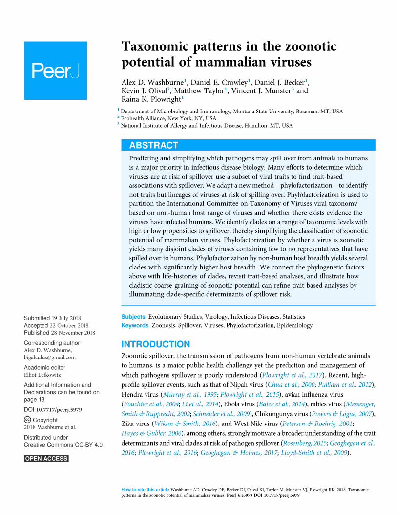
RESULTSZoonotic phylofactorization and comparative trait-based analysisPhylofactorization of whether or not a virus is zoonotic yielded 10 significant cladesof various sizes and taxonomic levels (Fig. 2). The viral family Papillomaviridae, familyHerpesviridae, subfamily Orthoretrovirinae, order Nidovirales, genus Parvovirus, andfamilies Caliciviridae, Adenoviridae, and Astroviridae all had a lower fraction ofzoonotic representatives compared to the paraphyletic remainder, which at the end ofphylofactorization had 352 viruses of which 43.5% (n = 153) were zoonotic. The generaAlphavirus and Deltaretrovirus had significantly high proportions of zoonoticrepresentatives, with 64% of the 25 Alphavirus species being zoonotic and 100% of thefour Deltaretrovirus species being zoonotic.
Washburne et al. (2018), PeerJ, DOI 10.7717/peerj.5979 5/17
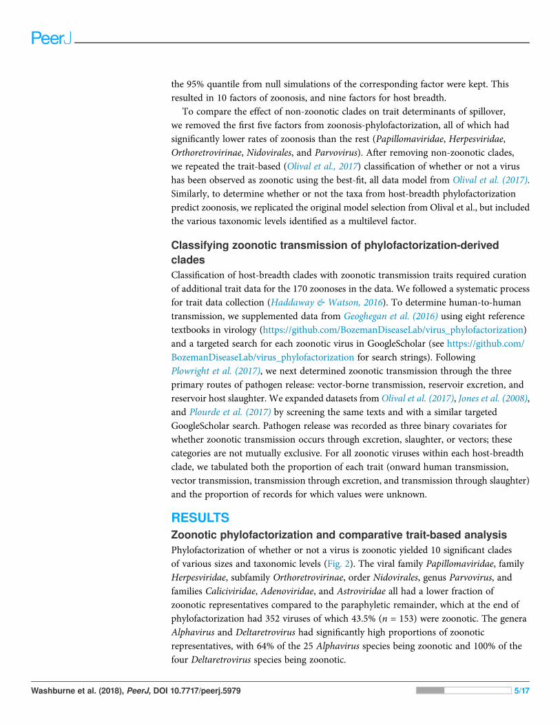
Next, we excluded the top-5 taxonomic factors found to be non-zoonotic clades—Parvoviridae, Herpesviridae, Orthoretrovirinae, Nidovirales, and Parvovirus—andreplicated the trait-based classification of zoonotic viruses from previous work(Olival et al., 2017). Excluding non-zoonotic clades affects the significance and explanatorypower of vector-borne transmission, whether or not a virus is enveloped, and whetheror not a virus replicates within the cytoplasm (Table 1). The sensitivity of resultsreported in previous trait-based analyses is due to strong associations between these traitsand non-zoonotic clades identified through phylofactorization. Non-human hostphylogenetic breadth remained a highly significant predictor of zoonosis, suggestingthat among clades of viruses likely to spillover, the ability to infect disparate mammalianhosts confers a significant ability to infect humans. To perform a similar cladisticanalysis of this stable predictor of a virus’ ability to infect humans, non-human hostbreadth was used for subsequent phylofactorization to identify any phylogeneticpatterns in host breadth.
Figure 2 Phylogenetic factors of zoonosis in mammalian viruses. Phylogenetic factorization iterativelypartitions a phylogeny along edges separating species with meaningful differences. Phylofactorization ofthe ICTV taxonomy by a Fisher test on the fraction of zoonotic viruses identified clades with differentnumbers of species (n) and different rates of zoonosis (%Zoonotic). A total of 32% of the viruses in theoriginal dataset of mammalian viruses are zoonotic, indicated as black dots on the tip of the tree. The firsteight phylogenetic factors identify clades whose distinction is a significantly low rate of zoonosis relativeto other viruses. Deltaretroviruses and Alphaviruses are then identified as having an unusually highfraction of zoonotic viruses. Cladistic structure in zoonotic potential can be used to identify previouslyignored traits and prioritize surveillance programs. Full-size DOI: 10.7717/peerj.5979/fig-2
Table 1 Effect sizes and significance of traits pre- and post-cladistic analysis.
All clades Zoonotic clades Non-zoonotic clades
Vector 1.4* 0.62 -0.16**
Envelope 0.87 1.2* -0.36**
Cytoplasmic replication 1.8** 0.88 -0.38***Notes:
The associations between zoonotic history and traits such as enveloped, vector-borne, and cytoplasmic replication aresensitive to the inclusion/exclusion of non-zoonotic clades due to the traits’ cladistic signal. Here, effect sizes (differencein linear predictors) are presented for the all viruses in the original trait-based analysis, the zoonotic clades (removal ofnon-zoonotic clades found in Fig. 2), and non-zoonotic clades. The direction of the effect of all three traits is conserved,but the magnitude and relative significance changes, as these traits are strongly associated with non-zoonotic clades.Asterisks indicate significance levels *P < 0.05, **P < 0.01, ***P < 0.001 from Chi-squared tests of the deviance fromgeneralized additive models.
Washburne et al. (2018), PeerJ, DOI 10.7717/peerj.5979 6/17
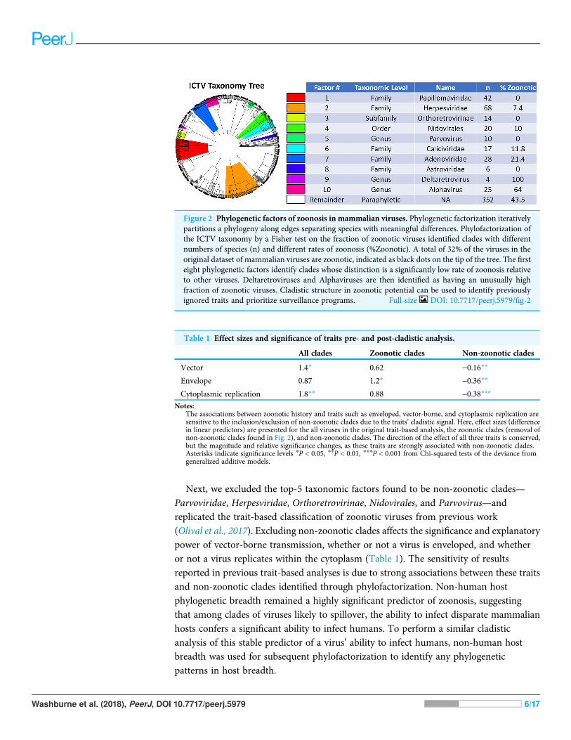
Host-breadth phylofactorizationPhylofactorization by host breadth yielded nine significant clades with a range of hostbreadth, each clade being partitioned as having a higher host breadth than the paraphyleticremainder. Specifically, the viral family Togaviridae, genus Arenavirus, genus Flavivirus,genus Orthopoxvirus, family Picornaviridae, family Bunyaviridae, order Mononegavirales,family Reoviridae and subfamily Alphaherpesvirinae all had significantly higher hostbreadth than the paraphyletic remainder (Fig. 3). The family Togaviridae contains
Figure 3 Phylogenetic factors of non-human mammalian host breadth. (A) Non-human phylogeneticdistance on a cytochrome B phylogeny is a reliable predictor of zoonotic viruses, even upon removal ofnon-zoonotic clades identified in Fig. 2. Zoonotic prediction is the linear predictor from generalizedadditive modelling, plotted here against phylogenetic breadth. (B) Phylofactorization of viruses bymammalian host breadth identifies clades with high host breadth. The family Togaviridae, which con-tains the alphaviruses, was identified as having a significantly higher host breadth than other viruses.
Full-size DOI: 10.7717/peerj.5979/fig-3
Washburne et al. (2018), PeerJ, DOI 10.7717/peerj.5979 7/17

the genus Alphavirus which was also obtained in the zoonosis-phylofactorization.The subfamily Alphaherpesvirinae, in the family Herpesviridae also identified in thezoonosis-phylofactorization, was identified as having higher host breadth than theremainder of Herpesviridae yet the lowest host breadth of the significant, monophyleticclades obtained by phylofactorization.
In order to determine if the clades partitioned by host breadth are significantdeterminants of zoonotic spillover, the clades identified through host-breadthphylofactorization were then included as a multilevel factor in a replicated trait-basedanalysis and model selection (Olival et al., 2017). Incorporating the multilevel factor ofhost-breadth clades dramatically affected the resulting model: vector-borne transmission,viral envelopes, and cytoplasmic replication—all traits identified in model selectionwhen not controlling for host-breadth clades—are all dropped during model selectionwhen host-breadth clades are included as a multilevel explanatory factor.
The linear predictors of zoonotic clades under the resulting generalized additivemodel suggest a prioritization scheme of host-breadth clades based on the log-odds ofzoonosis within-clades factored by host breadth (Fig. 4). At the top of the list is Togaviridaeand at the bottom of the list is the paraphyletic remainder. The Togaviridae, Flavivirus,and Bunyaviridae clades partitioned by phylogenetic breadth are at high risk of spilloverinto humans; the viruses in these clades are enveloped and almost exclusively transmitted
Figure 4 Phylofactorization of viruses by host range suggests a taxonomic-based surveillanceprioritization scheme. Clades identified by host-range phylofactorization vary in their fraction ofzoonotic representatives, and inclusion of clades into model selection of trait-based analysis producesupdated predictions for zoonosis in clades while controlling for other traits. Traits classifying the host-range clades suggest different pathways to spillover, from the vector-borne transmission of Togaviridae toenvironmental persistence of excreted Picornaviridae. Full-size DOI: 10.7717/peerj.5979/fig-4
Washburne et al. (2018), PeerJ, DOI 10.7717/peerj.5979 8/17

to humans through arthropod vectors. The Picornaviridae and Orthopoxvirus hadlower proportions of zoonoses but higher propensities for onward transmission inhumans. Alphaherpesvirinae was partitioned from the paraphyletic remainder as havinga slightly higher phylogenetic breadth than the remainder, which included the Beta-and Gammaherpesvirinae; however, compared to other clades partitioned by host breadth,the Alphaherpesvirinae have a low propensity to spillover into people.
DISCUSSIONPhylofactorization of mammalian virusesEmerging infectious diseases arise from a pre-existing pool of viruses with traits, life-histories, and evolutionary histories, all of which interact to determine the propensityfor spillover. Any one virus has myriad traits adapted to the virus’ life cycle, ranging frominteractions with a host cell that facilitate infection and replication to the processesthat facilitate transmission and environmental persistence. Life-histories determine theevolutionary pressure and the constellation of traits defining a virus, and using theevolutionary tree as a scaffold for identifying clades with common propensities forspillover can implicitly identify traits and life-histories which enable viruses to spillover into humans. Modeling, prediction, and management of pathogen spillover requiresa comprehensive approach that incorporates clade-specific life-histories to identifyclade-specific traits that enable pathogens to jump from wildlife to people.
In this paper, we have built on a recent study of traits determining spillover inmammalian viruses (Olival et al., 2017) by using cladistic algorithms to partition the viraltaxonomy and classify viral zoonoses. Using a modern graph-partitioning techniquebuilt for evolutionary trees (Washburne et al., 2017a; Washburne, 2017), we havepartitioned the ICTV taxonomy tree and flexibly identified 10 clades of viruses acrosstaxonomic scales with significantly extreme (high or low) propensities for zoonosis.
Removing non-zoonotic clades from the existing trait-based prediction of zoonoticviruses decreased the importance of vector-borne and cytoplasmic replication traits, butincreases the importance of enveloped viruses, due to strong, negative associations of thesetraits with non-zoonotic clades of viruses. One trait remains a robust predictor of zoonosis:the phylogenetic breadth of non-human hosts.
Phylofactorization of the viral taxonomy by non-human host breadth yields nineclades of viruses at various taxonomic levels. These nine clades classify viruses with a rangeof zoonotic potential, even when controlling for host breadth. Identifying phylogeneticfactors which can predict zoonosis enables researchers to take a fresh look at the problemof pathogen spillover by focusing on the life-histories of these nine clades.
Non-human host breadth is a particularly useful trait to use for phylofactorization.Zoonosis combines human:host exposure with the capacity of viruses to cross speciesbarriers. Clades of viruses with molecular mechanisms enabling spillover into humanpopulations, yet currently absent from humans due to infrequent exposure (i.e., virusesin remote regions or hosts, or viruses that are not shed from their hosts) will not beidentified under an analysis of “zoonosis” as a response variable but can be identified by ananalysis of host breadth. Likewise, using zoonoses as the outcome variable for trait-based
Washburne et al. (2018), PeerJ, DOI 10.7717/peerj.5979 9/17

analyses may overemphasize the importance of traits of viruses with high human:hostexposure, such as viruses that require large doses to initiate infections but manage toinfect humans due to repeated exposure increasing the chance that a person is exposed to asufficient dose to initiate infection. By performing phylofactorization on both humanzoonosis and non-human host breadth, we have identified clades with risks of spilloverin both humans and wildlife. There were converging lines of evidence on multipleclades in phylofactorization of both human zoonosis and host breadth: Togaviridae arehighly zoonotic and have a broad host-range, while Herpesviridae have low rates ofzoonosis and narrow host ranges; finer detail is observed in the alphaviruses’propensity for zoonosis and the Alphaherpesvirinae having higher phylogeneticbreadth than other Herpesviridae while, compared to other clades, still having a lowerpropensity to spillover. We examine possible life history traits of these and otherclades below.
Life history traits of zoonotic and non-zoonotic cladesThe family Togaviridae, containing the genus Alphavirus, is a family with a high hostbreadth and a high propensity for spilling over into humans. Togaviruses are vector-bornethrough mosquitoes and ticks, and such life-history may have selected generalist pathogensand produced a collection of traits shared within the lineage which enable proliferationin the blood of numerous vertebrate hosts. The Togaviruses are generalist pathogens withhigh host breadth and propensities for spillover, and their host breadth requiresproteins which enable receptor-binding and replication in cells from different species.Identifying these determinants of host breadth in Togaviruses may focus attention onthe key traits enabling pathogen spillover in this highly zoonotic clade.
It’s hypothesized that Chikungunya virus, an Alphavirus, binds receptors ubiquitouslyexpressed among species and cell types (Van Duijl-Richter et al., 2015), and that theSindbis virus binds the laminin receptor (Wang et al., 1992), heparan sulfate (Wang et al.,1992; Byrnes & Griffin, 1998), and the C-type lectins DC-SIGN and L-SIGN (Klimstraet al., 2003), all of which are found on most cell surfaces of most animal tissues. The broadrange of cellular receptors utilized by alphaviruses is attributable to a common proteinshared among alphaviruses: the E2 protein (Voss et al., 2010). The E2 protein containsresidues critical for broad host range and tissue tropism and thus may be a key traitunderlying the zoonotic potential of Togaviruses. Togaviruses all contain six genes,and sequence-based surveillance targeting all Togaviruses can improve assessments ofspillover risk among mammalian viruses. Furthermore, targeted sequencing of genesparticular to Togavirus life history—in particular, variation in the E2 protein—mayimprove assessments of spillover risk within Togaviridae (Klimstra et al., 2003).
At the opposite end of the spillover spectrum from Togaviridae is the viral familyHerpesviridae, which has a low propensity to be zoonotic. The subfamilyAlphaherpesvirinae has low non-human host breadth relative to the other clades pulledout in phylofactorization, but it was identified as having a higher host breadth than theBeta- and Gammaherpesvirinae and the paraphyletic remainder of viruses from whichit was partitioned.
Washburne et al. (2018), PeerJ, DOI 10.7717/peerj.5979 10/17

Herpesviruses have a range of modes of transmission, often establish lifelong infections,and have several methods to evade the immune system (Roizman, 1982). Cross-speciestransmission events of Herpesviruses have been documented for closely related hostspecies (Woźniakowski & Samorek-Salamonowicz, 2015), such as cases of fatal humanherpesvirus infection in wild primates (Heldstab et al., 1981) and the converse lethalinfections of humans exposed to Herpes B from macaques (Huff & Barry, 2003).However, herpesviruses generally show relatively strict species specificity and difficultywith cross-species transmission, including when non-human primates consume thetissues of other non-human primates (Murthy et al., 2013). The reasons for the species-specificity of Herpesviridae are multifactorial, but careful understanding of their lifehistory illuminates some compelling hypotheses for clade-specific barriers to spillover.
One possibility for the species specificity of Herpesviridae is tissue tropism.Herpesviridae infections are often associated with terminally-differentiated cells, likeneurons, and immune cells. Entry into these cells may require binding receptors whichare well-known to have undergone positive selection among mammalian lineages(Kosiol et al., 2008). However, the generalization that Herpesviridae are tissue-tropicand infect immune cells is not universal; it is particularly true for Gammaherpesvirinae,somewhat true for Betaherpesvirinae, and not at all true for Alphaherpesvirinae.Interestingly, the Alphaherpesvirinae were identified from phylofactorization as havinga higher host breadth than the remaining Herpesviridae, suggesting that tissue tropismmay be a clade-specific barrier to spillover in the Herpesviridae.
Immune evasion also plays a major role in Herpesviridae life history and themechanisms of immune evasion in Herpesviruses may limit their host range. For example,the ICP47 gene of herpes simplex virus type-1 is involved in immune evasion throughthe inhibition of TAP-mediated antigen transport; the ICP47 protein exhibits a 100-folddecrease in its binding affinity for mouse TAP compared to human TAP (Ahn et al., 1996).TAP-inhibition has also been documented in human cytomegalovirus (Ahn et al., 1997),equine herpesvirus 1 (Ambagala, Gopinath & Srikumaran, 2004), and bovine herpesvirus1 (Koppers-Lalic et al., 2003). TAP-inhibition is only one of many mechanisms ofimmunoevasion within the Herpesviridae. Host-specificity for each member of theHerpesviridae might be related to mechanisms for immunoevasion, like TAP-binding,which require viral proteins interfacing with the conserved pathways yet species-specificenzyme structure underlying mammalian immune systems. The immunological barriersto spillover have been documented not just in the family Herpesviridae, but also inEbola virus (Groseth et al., 2012) and Zika virus (Xia et al., 2018)—in all cases, the species-specificity of protein:protein interactions can be a barrier to spillover. For the manyHerpesviridae whose life cycles involve latent or chronic infections, unsuccessfulimmunoevasion in new hosts may be especially costly.
Similar life-history case studies of viral clades partitioned here, and the clade-specificbarriers to spillover, may greatly improve our understanding and surveillance of viralspillover. Clades such as the flaviviruses and bunyaviruses—both predominantlyvector-borne and enveloped (Fig. 4)—corroborate the notion of generalist pathogensand each clade may have genes determining spillover which are particular to that clade.
Washburne et al. (2018), PeerJ, DOI 10.7717/peerj.5979 11/17

Arenaviruses have high host breadth (Fig. 3) yet comparatively low zoonotic propensity(Fig. 4). Arenaviruses bind to a-dystroglycan protein as a receptor, enabling broad celltropism (Meyer, De La Torre & Southern, 2002), yet arenaviruses contain a low proportionof viruses which have spilled over into humans. Unlike the Togaviruses, bunyaviruses,and flaviviruses, arenaviruses are transmitted to humans primarily from direct oraerosolized exposure to rodent excreta (Emonet et al., 2007; Gonzalez et al., 2007). Onone hand, the limited frequency of such exposures makes rodent-human transmissioninefficient and less likely to result in a human-rodent zoonosis (Bausch & Mills, 2014);on the other hand, the high incidence of Lassa hemorrhagic fever and onward human–human transmission of the Lassa virus suggests such transmission-related bottlenecks maybe bypassed with appropriate adaptations. If exposure from reservoirs to humans isthe main bottleneck for the zoonosis of many arenaviruses, the sensitivity of aerosolizedarenaviruses to ultraviolet radiation, pH, and temperature may modulate the likelihoodfor cross-species transmission (Stephenson, Larson & Dominik, 1984; Gonzalez et al., 2007;Sagripanti & Lytle, 2011), and thus genetic determinants of environmental persistencemay be important for spillover surveillance in arenaviruses.
The case studies and hypothesized barriers to spillover expounded here are not exhaustivenor intended to be authoritative; they are intended to remind of the complexity of virallineages’ life-histories and illustrate pipeline from cladistic analysis of spillover, life historyclassification of identified clades, and consideration of new traits and molecular determinantsclade-specific risks of zoonosis (Fig. 1). More broadly, bottlenecks to pathogen spillover mayoccur at one or several of the stages in the viral life cycle and the contact process betweenreservoir hosts and humans. Entry, viral gene expression, replication, assembly, dissemination,and human-reservoir contact are all processes which may produce clade-specific bottlenecksto spillover. Additionally, there may be multiple stages of the viral life cycle for whichviruses contain adaptations which allow them to specialize or generalize across hosts.
CONCLUSIONSPartitioning viruses into monophyletic clades with common propensities for spillovercan simplify the problem of pathogen spillover, assist primer design for genomicsurveillance (Gardy & Loman, 2017) and increase our resolution for understandingclade-specific barriers to spillover. We have reduced a dataset of 586 viruses into nineclades with different patterns of spillover and, likely, common sequences that can be usedfor primer design in sequencing-based surveillance. The different clades have differentlife-histories, ranging from acute infections, vector-borne transmission, envelope-mediated immune evasion and promiscuous receptor-binding in Togaviridae to thepersistent-recurrent infections, non-vector-borne transmission and species-specificimmune evasion mechanisms of Herpesviridae.
Understanding life-histories in a handful of clades can focus surveillance efforts onclades most likely to spill-over and target particular genes believed to underlie spilloverrisk within the focal clade. In vitro studies of molecular mechanisms within high-riskclades, such as the Togaviruses, flaviviruses, and bunyaviruses, can determine the risk ofjump-capable pathogens arising from viral variants in reservoir hosts and identify
Washburne et al. (2018), PeerJ, DOI 10.7717/peerj.5979 12/17

clade-specific life-history bottlenecks to spillover. Future trait-based analyses canincorporate life-history details obtained from cladistic analyses. Similarly, future cladisticanalyses can focus study on viral life-histories to provide a more complete understandingof the traits correlated with pathogen spillover.
ACKNOWLEDGEMENTSThe views, opinions and/or findings expressed are those of the author and should notbe interpreted as representing the official views or policies of the Department of Defenseor the US Government. We thank members of the Plowright and Cross groups atMontana State University and the USGS for feedback.
ADDITIONAL INFORMATION AND DECLARATIONS
FundingThis research was developed with funding from the Defense Advanced Research ProjectsAgency (DARPA; D16AP00113), the National Science Foundation DEB-1716698, theNational Institute of General Medical Sciences of the National Institutes of Health underAward Number P20GM103474 and P30GM110732, and SERDP RC-2633. The fundershad no role in study design, data collection and analysis, decision to publish, orpreparation of the manuscript.
Grant DisclosuresThe following grant information was disclosed by the authors:The Defense Advanced Research Projects Agency: D16AP00113.The National Science Foundation: DEB-1716698.The National Institute of General Medical Sciences of the National Institutes of Health:P20GM103474 and P30GM110732.Strategic Environmental Research and Development Program: RC-2633.
Competing InterestsKevin J. Olival is employed by EcoHealth Alliance New York.
Author Contributions� Alex D. Washburne analyzed the data, contributed reagents/materials/analysis tools,prepared figures and/or tables, authored or reviewed drafts of the paper, approvedthe final draft.
� Daniel E. Crowley analyzed the data, contributed reagents/materials/analysis tools,prepared figures and/or tables, authored or reviewed drafts of the paper, approvedthe final draft.
� Daniel J. Becker analyzed the data, contributed reagents/materials/analysis tools, preparedfigures and/or tables, authored or reviewed drafts of the paper, approved the final draft.
� Kevin J. Olival contributed reagents/materials/analysis tools, authored or reviewed draftsof the paper, approved the final draft.
� Matthew Taylor authored or reviewed drafts of the paper, approved the final draft.
Washburne et al. (2018), PeerJ, DOI 10.7717/peerj.5979 13/17
� Vincent J. Munster authored or reviewed drafts of the paper, approved thefinal draft.
� Raina K. Plowright contributed reagents/materials/analysis tools, authored or revieweddrafts of the paper, approved the final draft.
Data AvailabilityThe following information was supplied regarding data availability:
GitHub: https://github.com/dncrwlye/BZDEL/tree/master/Mammalian_Virus_Phylofactorization
REFERENCESAhn K, Gruhler A, Galocha B, Jones TR, Wiertz EJHJ, Ploegh HL, Peterson PA, Yang Y, Früh
K. 1997. The ER-luminal domain of the HCMV glycoprotein US6 inhibits peptide translocationby TAP. Immunity 6(5):613–621. DOI 10.1016/S1074-7613(00)80349-0.
Ahn K, Meyer TH, Uebel S, Sempé P, Djaballah H, Yang Y, Peterson PA, Früh K, Tampé R.1996. Molecular mechanism and species specificity of TAP inhibition by herpes simplex virusICP47. EMBO Journal 15(13):3247–3255. DOI 10.1002/j.1460-2075.1996.tb00689.x.
Ambagala APN, Gopinath RS, Srikumaran S. 2004. Peptide transport activity of the transporterassociated with antigen processing (TAP) is inhibited by an early protein of equine herpesvirus-1. Journal of General Virology 85(2):349–353. DOI 10.1099/vir.0.19563-0.
Baize S, Pannetier D, Oestereich L, Rieger T, Koivogui L, Magassouba N, Soropogui B, SowMS,Keïta S, De Clerck H, Tiffany A, Dominguez G, Loua M, Traoré A, Kolié M, Malano ER,Heleze E, Bocquin A, Mély S, Raoul H, Caro V, Cadar D, Gabriel M, Pahlmann M, Tappe D,Schmidt-Chanasit J, Impouma B, Diallo AK, Formenty P, Van Herp M, Günther S. 2014.Emergence of Zaire ebola virus disease in Guinea. New England Journal of Medicine371(15):1418–1425 DOI 10.1056/NEJMoa1404505.
Bausch DG, Mills JN. 2014. Arenaviruses: lassa fever, lujo hemorrhagic fever,lymphocytic choriomeningitis, and the South American hemorrhagic fevers. In: Kaslow R,Stanberry L, Le Duc J, eds. Viral Infections of Humans. Boston: Springer, 147–171DOI 10.1007/978-1-4899-7448-8_8.
Black WC, Bennett KE, Gorrochótegui-Escalante N, Barillas-Mury CV, Fernández-Salas I, DeLourdes Muñoz M, Farfán-Alé JA, Olson KE, Beaty BJ. 2002. Flavivirus susceptibility in Aedesaegypti. Archives of Medical Research 33(4):379–388 DOI 10.1016/S0188-4409(02)00373-9.
Byrnes AP, Griffin DE. 1998. Binding of Sindbis virus to cell surface heparan sulfate. Journal ofVirology 72(9):7349–7356.
Chua KB, Bellini WJ, Rota PA, Harcourt BH, Tamin A, Lam SK, Ksiazek TG, Rollin PE,Zaki SR, Shieh W, Goldsmith CS, Gubler DJ, Roehrig JT, Eaton B, Gould AR, Olson J,Field H, Daniels P, Ling AE, Peters CJ, Anderson LJ, Mahy BW. 2000. Nipah virus: arecently emergent deadly paramyxovirus. Science 288(5470):1432–1435DOI 10.1126/science.288.5470.1432.
Emonet S, Retornaz K, Gonzalez JP, De Lamballerie X, Charrel RN. 2007. Mouse-to-humantransmission of variant lymphocytic choriomeningitis virus. Emerging Infectious Diseases13(3):472–475 DOI 10.3201/eid1303.061141.
Fouchier RAM, Schneeberger PM, Rozendaal FW, Broekman JM, Kemink SAG, Munster V,Kuiken T, Rimmelzwaan GF, Schutten M, Van Doornum GJJ, Koch G, Bosman A,Koopmans M, Osterhaus ADME. 2004. Avian influenza a virus (H7N7) associated with
Washburne et al. (2018), PeerJ, DOI 10.7717/peerj.5979 14/17

human conjunctivitis and a fatal case of acute respiratory distress syndrome. Proceedings ofthe National Academy of Sciences of the United States of America 101(5):1356–1361DOI 10.1073/pnas.0308352100.
Gardy JL, Loman NJ. 2017. Towards a genomics-informed, real-time, global pathogensurveillance system. Nature Reviews Genetics DOI 10.1038/nrg.2017.88.
Geoghegan JL, Holmes EC. 2017. Predicting virus emergence amid evolutionary noise.Open Biology 7(10):170189 DOI 10.1098/rsob.170189.
Geoghegan JL, Senior AM, Giallonardo FD, Holmes EC. 2016. Virological factors that increasethe transmissibility of emerging human viruses. Proceedings of the National Academy of Sciencesof the United States of America 113(15):4170–4175 DOI 10.1073/pnas.1521582113.
Gonzalez JP, Emonet S, De Lamballerie X, Charrel R. 2007. Arenaviruses. In: Childs JE,Mackenzie JS, Richt JA, eds. Wildlife and Emerging Zoonotic Diseases: The Biology,Circumstances and Consequences of Cross-Species Transmission. Berlin, Heidelberg: Springer,253–288.
Groseth A, Marzi A, Hoenen T, Herwig A, Gardner D, Becker S, Ebihara H, Feldmann H. 2012.The Ebola virus glycoprotein contributes to but is not sufficient for virulence in vivo.PLOS Pathogens 8(8):e1002847 DOI 10.1371/journal.ppat.1002847.
Haddaway NR, Watson MJ. 2016. On the benefits of systematic reviews for wildlifeparasitology. International Journal for Parasitology. Parasites and Wildlife 5(2):184–191DOI 10.1016/j.ijppaw.2016.05.002.
Hayes EB, Gubler DJ. 2006. West Nile virus: epidemiology and clinical features of anemerging epidemic in the United States. Annual Review of Medicine 57(1):181–194DOI 10.1146/annurev.med.57.121304.131418.
Heldstab A, Rüedi D, Sonnabend W, Deinhardt F. 1981. Spontaneous generalized Herpesvirushominis infection of a lowland gorilla (Gorilla gorilla gorilla). Journal of Medical Primatology10(2–3):129–135 DOI 10.1159/000460063.
Huff JL, Barry PA. 2003. B-virus (Cercopithecine herpesvirus 1) infection in humans andmacaques: potential for zoonotic disease. Emerging Infectious Diseases 9(2):246–250DOI 10.3201/eid0902.020272.
Jane Flint S, Racaniello VR, Rall GF, Enquist LW, Skalka A-M. 2015. Principles of virology: Vol. 1:molecular biology. Washington, D.C.: American Society for Microbiology.
Jones KE, Patel NG, Levy MA, Storeygard A, Balk D, Gittleman JL, Daszak P. 2008.Global trends in emerging infectious diseases. Nature 451(7181):990–993DOI 10.1038/nature06536.
Klimstra WB, Nangle EM, Smith MS, Yurochko AD, Ryman KD. 2003. DC-SIGN and L-SIGNcan act as attachment receptors for alphaviruses and distinguish between mosquito cell- andmammalian cell-derived viruses. Journal of Virology 77(22):12022–12032DOI 10.1128/JVI.77.22.12022-12032.2003.
Koppers-Lalic D, Rychlowski M, Leeuwen DV, Rijsewijk FAM, Ressing ME, Neefjes JJ,Bienkowska-Szewczyk K, Wiertz EJHJ. 2003. Bovine herpesvirus 1 interferes withTAP-dependent peptide transport and intracellular trafficking of MHC class I molecules inhuman cells. Archives of Virology 148(10):2023–2037 DOI 10.1007/s00705-003-0142-5.
Kosiol C, Vina�r T, Fonseca RRD, Hubisz MJ, Bustamante CD, Nielsen R, Siepel A. 2008.Patterns of positive selection in six mammalian genomes. PLOS Genetics 4(8):e1000144DOI 10.1371/journal.pgen.1000144.
Li Q, Zhou L, Zhou M, Chen Z, Li F, Wu H, Xiang N, Chen E, Tang F, Wang D, Meng L,Hong Z, Tu W, Cao Y, Li L, Ding F, Liu B, Wang M, Xie R, Gao R, Li X, Bai T, Zou S, He J,
Washburne et al. (2018), PeerJ, DOI 10.7717/peerj.5979 15/17

Hu J, Xu Y, Chai C, Wang S, Gao Y, Jin L, Zhang Y, Luo H, Yu H, He J, Li Q, Wang X, Gao L,Pang X, Liu G, Yan Y, Yuan H, Shu Y, Yang W, Wang Y, Wu F, Uyeki TM, Feng Z. 2014.Epidemiology of human infections with avian influenza A(H7N9) virus in China. New EnglandJournal of Medicine 370(6):520–532 DOI 10.1056/NEJMoa1304617.
Lloyd-Smith JO, George D, Pepin KM, Pitzer VE, Pulliam JRC, Dobson AP, Hudson PJ,Grenfell BT. 2009. Epidemic dynamics at the human-animal interface. Science326(5958):1362–1367 DOI 10.1126/science.1177345.
Messenger SL, Smith JS, Rupprecht CE. 2002. Emerging epidemiology of bat-associated crypticcases of rabies in humans in the United States. Clinical Infectious Diseases: An OfficialPublication of the Infectious Diseases Society of America 35(6):738–747 DOI 10.1086/342387.
Meyer BJ, De La Torre JC, Southern PJ. 2002. Arenaviruses: genomic RNAs, transcription, andreplication. In: Oldstone MBA, ed. Arenaviruses I. Current Topics in Microbiology andImmunology. Berlin, Heidelberg: Springer, 139–157.
Morrison AC, Zielinski-Gutierrez E, Scott TW, Rosenberg R. 2008. Defining challengesand proposing solutions for control of the virus vector Aedes aegypti. PLOS Medicine 5(3):e68DOI 10.1371/journal.pmed.0050068.
Murray K, Selleck P, Hooper P, Hyatt A, Gould A, Gleeson L, Westbury H, Hiley L, Selvey L,Rodwell B. 1995. A morbillivirus that caused fatal disease in horses and humans. Science268(5207):94–97 DOI 10.1126/science.7701348.
Murthy S, Couacy-Hymann E, Metzger S, Nowak K, Nys HD, Boesch C, Wittig R, Jarvis MA,Leendertz FH, Ehlers B. 2013. Absence of frequent herpesvirus transmission in anonhuman primate predator-prey system in the wild. Journal of Virology 87(19):10651–10659DOI 10.1128/JVI.01104-13.
Olival KJ, Hosseini PR, Zambrana-Torrelio C, Ross N, Bogich TL, Daszak P. 2017. Hostand viral traits predict zoonotic spillover from mammals. Nature 546(7660):646–650DOI 10.1038/nature22975.
Paradis E, Claude J, Strimmer K. 2004. APE: Analyses of phylogenetics and evolution inR language. Bioinformatics 20(2):289–290 DOI 10.1093/bioinformatics/btg412.
Petersen LR, Roehrig JT. 2001. West Nile virus: a reemerging global pathogen. EmergingInfectious Diseases 7(4):611–614 DOI 10.3201/eid0704.017401.
Plourde BT, Burgess TL, Eskew EA, Roth TM, Stephenson N, Foley JE. 2017. Are diseasereservoirs special? Taxonomic and life history characteristics. PLOS ONE 12(7):e0180716DOI 10.1371/journal.pone.0180716.
Plowright RK, Eby P, Hudson PJ, Smith IL, Westcott D, Bryden WL, Middleton D, Reid PA,McFarlane RA, Martin G, Tabor GM, Skerratt LF, Anderson DL, Crameri G, Quammen D,Jordan D, Freeman P, Wang L-F, Epstein JH, Marsh GA, Kung NY, McCallum H. 2015.Ecological dynamics of emerging bat virus spillover. Proceedings of the Royal Society B: BiologicalSciences 282(1798):20142124 DOI 10.1098/rspb.2014.2124.
Plowright RK, Parrish CR, McCallum H, Hudson PJ, Ko AI, Graham AL, Lloyd-Smith JO.2017. Pathways to zoonotic spillover. Nature Reviews. Microbiology 15(8):502–510DOI 10.1038/nrmicro.2017.45.
Plowright RK, Peel AJ, Streicker DG, Gilbert AT, McCallum H, Wood J, Baker ML,Restif O. 2016. Transmission or within-host dynamics driving pulses of zoonotic virusesin reservoir-host populations. PLOS Neglected Tropical Diseases 10(8):e0004796DOI 10.1371/journal.pntd.0004796.
Powers AM, Logue CH. 2007. Changing patterns of chikungunya virus: re-emergence of azoonotic arbovirus. Journal of General Virology 88(9):2363–2377 DOI 10.1099/vir.0.82858-0.
Washburne et al. (2018), PeerJ, DOI 10.7717/peerj.5979 16/17

Pulliam JRC, Epstein JH, Dushoff J, Rahman SA, Bunning M, Jamaluddin AA, Hyatt AD,Field HE, Dobson AP, Daszak P, The Henipavirus Ecology Research Group (HERG). 2012.Agricultural intensification, priming for persistence and the emergence of Nipah virus: alethal bat-borne zoonosis. Journal of the Royal Society Interface 9(66):89–101DOI 10.1098/rsif.2011.0223.
Roizman B. ed. 1982. The family Herpesviridae: general description, taxonomy, and classification.In: The Herpesviruses. Boston: Springer, 1–23.
Rosenberg R. 2015. Detecting the emergence of novel, zoonotic viruses pathogenic to humans.Cellular and Molecular Life Sciences 72(6):1115–1125 DOI 10.1007/s00018-014-1785-y.
Sagripanti J-L, Lytle CD. 2011. Sensitivity to ultraviolet radiation of Lassa, vaccinia, and Ebolaviruses dried on surfaces. Archives of Virology 156(3):489–494 DOI 10.1007/s00705-010-0847-1.
Schneider MC, Romijn PC, Uieda W, Tamayo H, Da Silva DF, Belotto A, Da Silva JB,Leanes LF. 2009. Rabies transmitted by vampire bats to humans: an emerging zoonoticdisease in Latin America? Pan American Journal of Public Health 25(3):260–269DOI 10.1590/s1020-49892009000300010.
Stephenson EH, Larson EW, Dominik JW. 1984. Effect of environmental factors onaerosol-induced Lassa virus infection. Journal of Medical Virology 14(4):295–303DOI 10.1002/jmv.1890140402.
Van Duijl-Richter MKS, Hoornweg TE, Rodenhuis-Zybert IA, Smit JM. 2015. Early events inchikungunya virus infection-from virus cell binding to membrane fusion. Viruses7(7):3647–3674 DOI 10.3390/v7072792.
Voss JE, Vaney M-C, Duquerroy S, Vonrhein C, Girard-Blanc C, Crublet E, Thompson A,Bricogne G, Rey FA. 2010. Glycoprotein organization of chikungunya virus particlesrevealed by X-ray crystallography. Nature 468(7324):709–712 DOI 10.1038/nature09555.
Wang KS, Kuhn RJ, Strauss EG, Ou S, Strauss JH. 1992. High-affinity laminin receptor is areceptor for Sindbis virus in mammalian cells. Journal of Virology 66(8):4992–5001.
Washburne A 2017. Phylofactorization—theory and challenges. bioRxiv preprintDOI 10.1101/196378.
Washburne AD, Silverman JD, Leff JW, Bennett DJ, Darcy JL, Mukherjee S, Fierer N,David LA. 2017a. Phylogenetic factorization of compositional data yields lineage-levelassociations in microbiome datasets. PeerJ 5(8):e2969 DOI 10.7717/peerj.2969.
Washburne AD, Silverman JD, Morton JT, Becker D, Crowley D, Mukherjee S, David LA,Plowright RK. 2017b. Phylofactorization—a graph partitioning algorithm to identifyphylogenetic scales of ecological data. bioRxiv preprint DOI 10.1101/235341.
Wikan N, Smith DR. 2016. Zika virus: history of a newly emerging arbovirus. LancetInfectious Diseases 16(7):e119–e126 DOI 10.1016/S1473-3099(16)30010-X.
Woźniakowski G, Samorek-Salamonowicz E. 2015. Animal herpesviruses and their zoonoticpotential for cross-species infection. Annals of Agricultural and Environmental Medicine22(2):191–194 DOI 10.5604/12321966.1152063.
Xia H, Luo H, Shan C, Muruato AE, Nunes BTD, Medeiros DBA, Zou J, Xie X, Giraldo MI,Vasconcelos PFC, Weaver SC, Wang T, Rajsbaum R, Shi P-Y. 2018. An evolutionaryNS1 mutation enhances Zika virus evasion of host interferon induction. NatureCommunications 9(1):414 DOI 10.1038/s41467-017-02816-2.
Washburne et al. (2018), PeerJ, DOI 10.7717/peerj.5979 17/17