Tau Protein Function in Living Cells
8
Tau Protein Function in Living Cells David G. Drubin and Marc W. Kirschner Department of Biochemistry and Biophysics, University of California, San Francisco, California 94143-0448. Dr. Drubin's present address is Department of Biology, Massachusetts Institute of Technology, Cambridge, Massachusetts 02139. Abstract. Tau protein from mammalian brain pro- motes microtubule polymerization in vitro and is in- duced during nerve cell differentiation. However, the effects of tau or any other microtubule-associated pro- tein on tubulin assembly within cells are presently un- known. We have tested tau protein activity in vivo by microinjection into a cell type that has no endogenous tau protein. Immunofluorescence shows that tau pro- tein microinjected into fibroblast cells associates specifically with microtubules. The injected tau protein increases tubulin polymerization and stabilizes microtubules against depolymerization. This increased polymerization does not, however, cause major changes in cell morphology or microtubule arrange- ment. Thus, tau protein acts in vivo primarily to in- duce tubulin assembly and stabilize microtubules, ac- tivities that may be necessary, but not sufficient, for neuronal morphogenesis. N 'EURONAL processes are densely filled with parallel arrays of microtubules that act as scaffolds neces- sary for process extension (Daniels, 1975; Yamada et al., 1976) and serve as tracks for axonal transport (Schnapp et al., 1985). Studies using clonal cell lines that extend neu- ritic processes in culture have shown that neurite extension involves net microtubule assembly, and not simply rear- rangement of existing microtubules into growing processes (Drubin et al., 1985; Gard et al., 1985; Olmstead, 1981). Microtubule assembly during neuronal process extension appears to involve the recruitment of tubulin from preexist- ing monomer pools into tubulin polymers (microtubules) (Drubin et al., 1985; Gard et al., 1985; Olmsted, 1981). It has been proposed that this assembly is driven by nontubulin protein factors that accumulate during process extension. Support for this proposal comes from the observation that extracts from mature rat brain tissue and from differentiated neuroblastoma ceils contain higher levels of protein factors that promote microtubule assembly than do extracts from their respective less-differentiated counterparts (Nunez et al., 1975; Seeds and Maccioni, 1978). Three major protein factors that promote microtubule as- sembly in vitro have been identified in brain extracts. These include the tau proteins of 50-70 kD (Weingarten et al., 1975; Cleveland et al., 1977), microtubule-associated protein 1 (MAP1) ~ of '~330 kD, and microtubule-associated protein 2 (MAP2) of "~300 kD (for a review of MAP1 and MAP2 see Vallee et al., 1984). During neurite extension in PC12 pheochromocytoma cells, levels of tau protein correlate pre- cisely with levels of assembled tubulin (Drubin et al., 1985). Furthermore, tau protein is bound to microtubules in PC12 cells (Drubin et al., 1986). These observations make tau pro- 1. Abbreviations used in this paper: MAP1 and MAP2, microtubule-associ- ated proteins 1 and 2. tein a strong candidate for a factor that promotes microtubule assembly during process extension. Still, given the complex function that microtubules serve, it is not clear which associ- ated proteins regulate polymer assembly and which regulate the interaction of microtubules with other components in the cell or serve some other function. Subtle questions about the importance of microtubule-associated proteins for polymer stability and dynamics have not been addressed in an in vivo context. To test the activity of tau protein in vivo we have microin- jected purified tau protein into living fibroblast cells (RATI) that normally contain undetectable levels of this factor. Im- munofluorescence shows that microinjected tau protein binds specifically to microtubules (Drubin et al., 1985). Two effects were demonstrated: first, tau causes a marked in- crease in the level of assembled tubulin. Even more dramati- cally tau protein increases both centrosome-nucleated and nonnucleated assembly in cells treated with low levels of microtubule depolymerizing drugs. Second, tau protein decreases the depolymerization rate of microtubules. These observations support the conclusion that tau protein pro- motes the assembly of microtubules that is required for neu- ronal process extension, and additionally stabilizes the re- suiting polymers. Materials and Methods Cell Culture and Microinjections RAT1 fibroblast cells were cultured in DME supplemented with 10% calf serum (Gibco, Grand Island, NY). For microinjection experiments, cells were trypsinized and replated on poly-D-lysine coated coverslips. To com- pare microinjected cells with control sister cells, single cells were plated at low density ,,o15 h before microinjection. This allowed many cells time to divide once, often resulting in pairs of associated sister cells with similar morphologies. Micruinjections were carried out exactly as described by Schulze and Kirschner (1986). © The Rockefeller University Press, 0021-9525/86/12/2739/8 $1.00 The Journal of Cell Biology, Volume 103 (No. 6, Pt. 2), Dec. 1986 2739-2746 2739 Downloaded from http://rupress.org/jcb/article-pdf/103/6/2739/1054324/2739.pdf by guest on 03 April 2022
Transcript of Tau Protein Function in Living Cells
Tau Protein Function in Living Cells David G. D r u b i n a n d M a r c W. K i r s c h n e r
Department of Biochemistry and Biophysics, University of California, San Francisco, California 94143-0448. Dr. Drubin's present address is Department of Biology, Massachusetts Institute of Technology, Cambridge, Massachusetts 02139.
Abstract. Tau protein from mammalian brain pro- motes microtubule polymerization in vitro and is in- duced during nerve cell differentiation. However, the effects of tau or any other microtubule-associated pro- tein on tubulin assembly within cells are presently un- known. We have tested tau protein activity in vivo by microinjection into a cell type that has no endogenous tau protein. Immunofluorescence shows that tau pro- tein microinjected into fibroblast cells associates
specifically with microtubules. The injected tau protein increases tubulin polymerization and stabilizes microtubules against depolymerization. This increased polymerization does not, however, cause major changes in cell morphology or microtubule arrange- ment. Thus, tau protein acts in vivo primarily to in- duce tubulin assembly and stabilize microtubules, ac- tivities that may be necessary, but not sufficient, for neuronal morphogenesis.
N 'EURONAL processes are densely filled with parallel
arrays of microtubules that act as scaffolds neces- sary for process extension (Daniels, 1975; Yamada
et al., 1976) and serve as tracks for axonal transport (Schnapp et al., 1985). Studies using clonal cell lines that extend neu- ritic processes in culture have shown that neurite extension involves net microtubule assembly, and not simply rear- rangement of existing microtubules into growing processes (Drubin et al., 1985; Gard et al., 1985; Olmstead, 1981).
Microtubule assembly during neuronal process extension appears to involve the recruitment of tubulin from preexist- ing monomer pools into tubulin polymers (microtubules) (Drubin et al., 1985; Gard et al., 1985; Olmsted, 1981). It has been proposed that this assembly is driven by nontubulin protein factors that accumulate during process extension. Support for this proposal comes from the observation that extracts from mature rat brain tissue and from differentiated neuroblastoma ceils contain higher levels of protein factors that promote microtubule assembly than do extracts from their respective less-differentiated counterparts (Nunez et al., 1975; Seeds and Maccioni, 1978).
Three major protein factors that promote microtubule as- sembly in vitro have been identified in brain extracts. These include the tau proteins of 50-70 kD (Weingarten et al., 1975; Cleveland et al., 1977), microtubule-associated protein 1 (MAP1) ~ of '~330 kD, and microtubule-associated protein 2 (MAP2) of "~300 kD (for a review of MAP1 and MAP2 see Vallee et al., 1984). During neurite extension in PC12 pheochromocytoma cells, levels of tau protein correlate pre- cisely with levels of assembled tubulin (Drubin et al., 1985). Furthermore, tau protein is bound to microtubules in PC12 cells (Drubin et al., 1986). These observations make tau pro-
1. Abbreviations used in this paper: MAP1 and MAP2, microtubule-associ- ated proteins 1 and 2.
tein a strong candidate for a factor that promotes microtubule assembly during process extension. Still, given the complex function that microtubules serve, it is not clear which associ- ated proteins regulate polymer assembly and which regulate the interaction of microtubules with other components in the cell or serve some other function. Subtle questions about the importance of microtubule-associated proteins for polymer stability and dynamics have not been addressed in an in vivo context.
To test the activity of tau protein in vivo we have microin- jected purified tau protein into living fibroblast cells (RATI) that normally contain undetectable levels of this factor. Im- munofluorescence shows that microinjected tau protein binds specifically to microtubules (Drubin et al., 1985). Two effects were demonstrated: first, tau causes a marked in- crease in the level of assembled tubulin. Even more dramati- cally tau protein increases both centrosome-nucleated and nonnucleated assembly in cells treated with low levels of microtubule depolymerizing drugs. Second, tau protein decreases the depolymerization rate of microtubules. These observations support the conclusion that tau protein pro- motes the assembly of microtubules that is required for neu- ronal process extension, and additionally stabilizes the re- suiting polymers.
Materials and Methods
Cell Culture and Microinjections
RAT1 fibroblast cells were cultured in DME supplemented with 10% calf serum (Gibco, Grand Island, NY). For microinjection experiments, cells were trypsinized and replated on poly-D-lysine coated coverslips. To com- pare microinjected cells with control sister cells, single cells were plated at low density ,,o15 h before microinjection. This allowed many cells time to divide once, often resulting in pairs of associated sister cells with similar morphologies. Micruinjections were carried out exactly as described by Schulze and Kirschner (1986).
© The Rockefeller University Press, 0021-9525/86/12/2739/8 $1.00 The Journal of Cell Biology, Volume 103 (No. 6, Pt. 2), Dec. 1986 2739-2746 2739
Dow
nloaded from http://rupress.org/jcb/article-pdf/103/6/2739/1054324/2739.pdf by guest on 03 April 2022
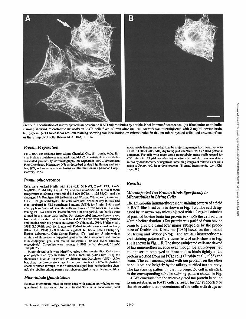
Figure 1. Localization of microinjected tau protein on RAT1 microtubules by double-label immunofluorescence. (A) Rhodamine antitubulin staining showing micmtubule networks in RATI cells fixed 40 min after one cell (arrow) was microinjected with 2 mg/ml bovine brain tau protein. (B) Fluorescein anti-tau staining showing tau localization on microtubules in the tau-micminjected cells, and absence of tan in the uninjected cells shown in A. Bar, 10 p.m.
Protein Preparation
FITC-BSA was obtained from Sigma Chemical Co., (St. Louis, MO). Bo- vine brain tau protein was separated from MAP2 in heat-stable microtubule- associated proteins by chromatography on Sepharose 4BCL (Pharmacia Fine Chemicals, Piscataway, NJ) as described in detail in Herzog and We- ber, 1978, and was concentrated using an ultrafiltration unit (Amicon Corp., Danvers, MA).
microtubule lengths were digitized by projecting images from negatives onto a GITCO (Rockville, MD) digitizing pad interfaced with an IBM personal computer. For cells with more dense microtubule arrays (cells treated for <30 min with 33 ~tM nocodazole) relative microtubule mass was deter- mined by densitometry of negatives containing images of mitotic sister cells using a Zeinet soft laser densitometer (Biomed Instruments, Inc., Chi- cago, IL).
Immunofluorescence Cells were washed briefly with PBS (0.13 M NaC1, 2 mM KCI, 8 mM NazHPO4, 2 mM KH2PO4, pH 7.2) and then immersed for 10 min at room temperature in 80 mM Pipes, pH 6.8, 5 mM EGTA, 1 mM MgC12, and the detergent 1% Empigen BB (Albright and Wilson, Whitehaven, Cumbria, UK), 0.3% glutaraldehyde. The cells were next rinsed briefly in PBS and then incubated in PBS containing 1 mg/ml NaBI-h for 7 min. Before and after each antibody addition the cells were washed five times in PBS con- raining 1% BSA and 0.1% Tween 20 over a 10-rain period. Antibodies were diluted in this same wash buffer. For double-label immunofluorescence, fixed and permeabilized cells were treated for 30 min with affinity-purified anti-bovine brain tau protein antiserum (Drubin et al., 1984; Pfeffer et al., 1983) (1:200 dilution), for 15 min with DMct-I tubulin monoclonal antibody (Blose et al., 1984) (1:2,000 dilution, a gift of Dr. Steven Blose, Cold Spring Harbor Laboratory, Cold Spring Harbor, NY), and for 15 min with a mixture of fluorescein-conjugated goat anti-rabbit antiserum and rhoda- mine-conjugated goat anti-mouse antiserum (1:50 and 1:200 dilution, respectively). Coverslips were mounted in 90% vol/vol glycerol, 20 mM Tris pH 7.9.
Microinjected cells were identified using a fluorescein filter. Cells were photographed on hypersensitized Kodak Tech-Pan (2415) film using the fluorescein filter as described by Schulze and Kirschner (1986). After bleaching the fluorescein image for several minutes to eliminate possible effects of"bleed through" of the fluorescein signal into the rhodamine chan- nel, the tubulin staining pattern was photographed using a rhodamine filter.
Microtubule Quantitation Relative microtubule mass in sister cells with similar morphologies was quantitated in two ways. For cells treated 30 min in nocodazole, total
Results
Microinjected Tau Protein Binds Specifically to Microtubules in Living Cells The antitubulin immunofluorescent staining pattern of a field of RAT1 fibroblast cells is shown in Fig. 1 A. The cell desig- nated by an arrow was microinjected with a 2 mg/ml solution of purified bovine brain tau protein to '~10 % the cell volume 40 min before fixation. (Tau protein was purified from bovine brain to give the usual four major subspecies by the proce- dure of Drubin and Kirschner [1986] based on the method of Herzog and Weber [1978]). The anti-tau immunofluores- cent staining pattern of the same field of cells shown in Fig. 1 A is shown in Fig. 1 B. The three uninjected cells are devoid of tau immunofluorescence even though the affinity-purified tau antiserum employed in these studies binds tightly to tau protein isolated from rat PC12 cells (Drubin et al., 1985) and brain. The cell microinjected with tau protein, on the other hand, is stained brightly by the affinity-purified tau antibody. The tau staining pattern in the microinjected cell is identical to the corresponding tubulin staining pattern shown in Fig. 1 A. We conclude that the microinjected tau protein is bound to microtubules in RATI cells, a result further supported by the observation that pretreatment of the cells with drugs in-
The Journal of Cell Biology, Volume 103, 1986 2740
Dow
nloaded from http://rupress.org/jcb/article-pdf/103/6/2739/1054324/2739.pdf by guest on 03 April 2022
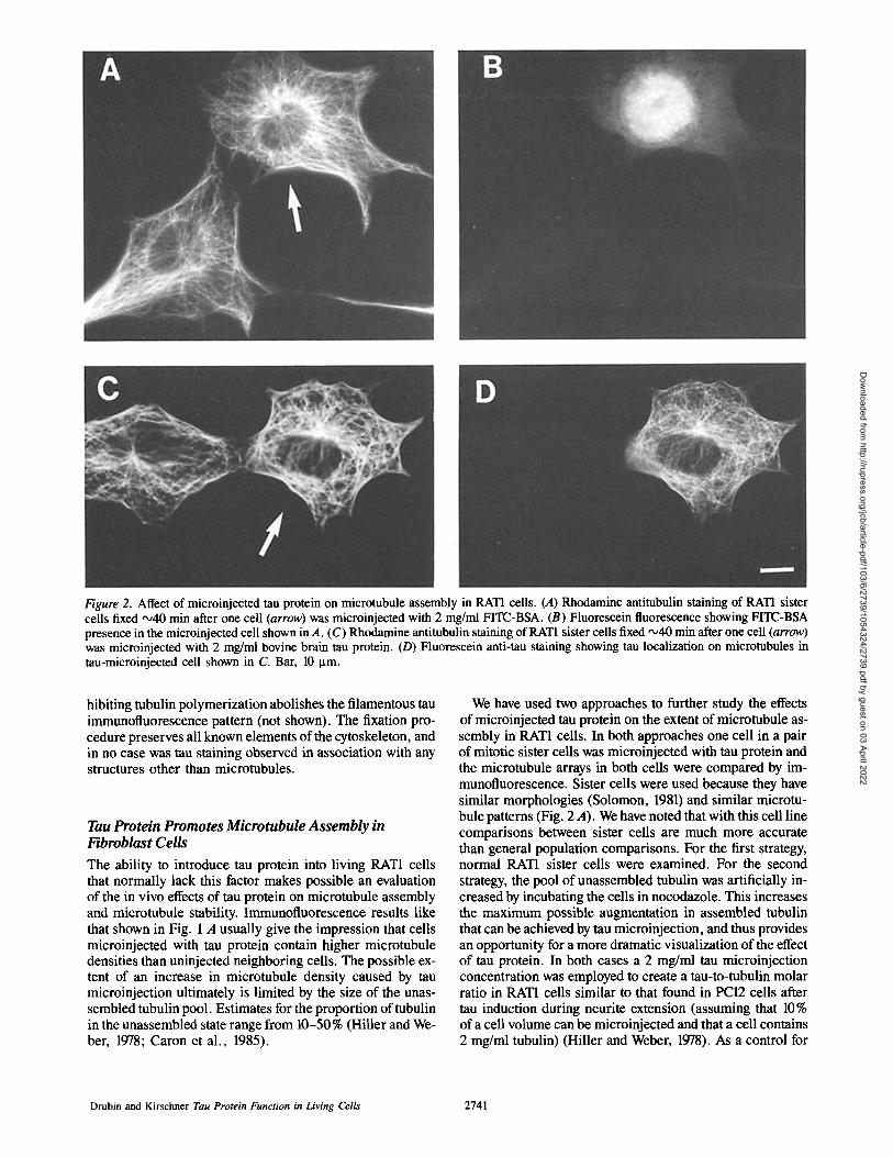
Figure 2. Affect of microinjected tau protein on microtubule assembly in RAT1 cells. (A) Rhodamine antitubulin staining of RAT1 sister cells fixed •40 min after one cell (arrow) was microinjected with 2 mg/ml FITC-BSA. (B) Fluorescein fluorescence showing FITC-BSA presence in the microinjected cell shown in A. (C) Rhodamine antitubulin staining of RAT1 sister cells fixed ,x,40 min after one cell (arrow) was microinjected with 2 mg/ml bovine brain tau protein. (D) Fluorescein anti-tau staining showing tau localization on microtubules in tau-microinjected cell shown in C. Bar, 10 I.tm.
hibiting tubulin polymerization abolishes the filamentous tau immunofluorescence pattern (not shown). The fixation pro- cedure preserves all known elements of the cytoskeleton, and in no case was tau staining observed in association with any structures other than microtubules.
Tau Protein Promotes Microtubule Assembly in Fibroblast Cells
The ability to introduce tau protein into living RAT1 cells that normally lack this factor makes possible an evaluation of the in vivo effects of tau protein on microtubule assembly and microtubule stability. Immunofluorescence results like that shown in Fig. 1 A usually give the impression that cells microinjected with tau protein contain higher microtubule densities than uninjected neighboring cells. The possible ex- tent of an increase in microtubule density caused by tau microinjection ultimately is limited by the size of the unas- sembled tubulin pool. Estimates for the proportion of tubulin in the unassembled state range from 10-50% (Hiller and We- ber, 1978; Caron et al., 1985).
We have used two approaches to further study the effects of microinjected tau protein on the extent of microtubule as- sembly in RAT1 cells. In both approaches one cell in a pair of mitotic sister ceils was microinjected with tau protein and the microtubule arrays in both cells were compared by im- munofluorescence. Sister cells were used because they have similar morphologies (Solomon, 1981) and similar microtu- bule patterns (Fig. 2 A). We have noted that with this cell line comparisons between sister cells are much more accurate than general population comparisons. For the first strategy, normal RAT1 sister cells were examined. For the second strategy, the pool of unassembled tubulin was artificially in- creased by incubating the cells in nocodazole. This increases the maximum possible augmentation in assembled tubulin that can be achieved by tau microinjection, and thus provides an opportunity for a more dramatic visualization of the effect of tau protein. In both cases a 2 mg/ml tau microinjection concentration was employed to create a tau-to-tubulin molar ratio in RAT1 cells similar to that found in PC12 cells after tau induction during neurite extension (assuming that 10% of a cell volume can be microinjected and that a cell contains 2 mg/ml tubulin) (Hiller and Weber, 1978). As a control for
Drubin and Kirschner Tau Protein Function in Living Cells 2741
Dow
nloaded from http://rupress.org/jcb/article-pdf/103/6/2739/1054324/2739.pdf by guest on 03 April 2022
No
A
i i i
l a
05 10 15
Ratio ot fluorescent intensity
N 2 . 0
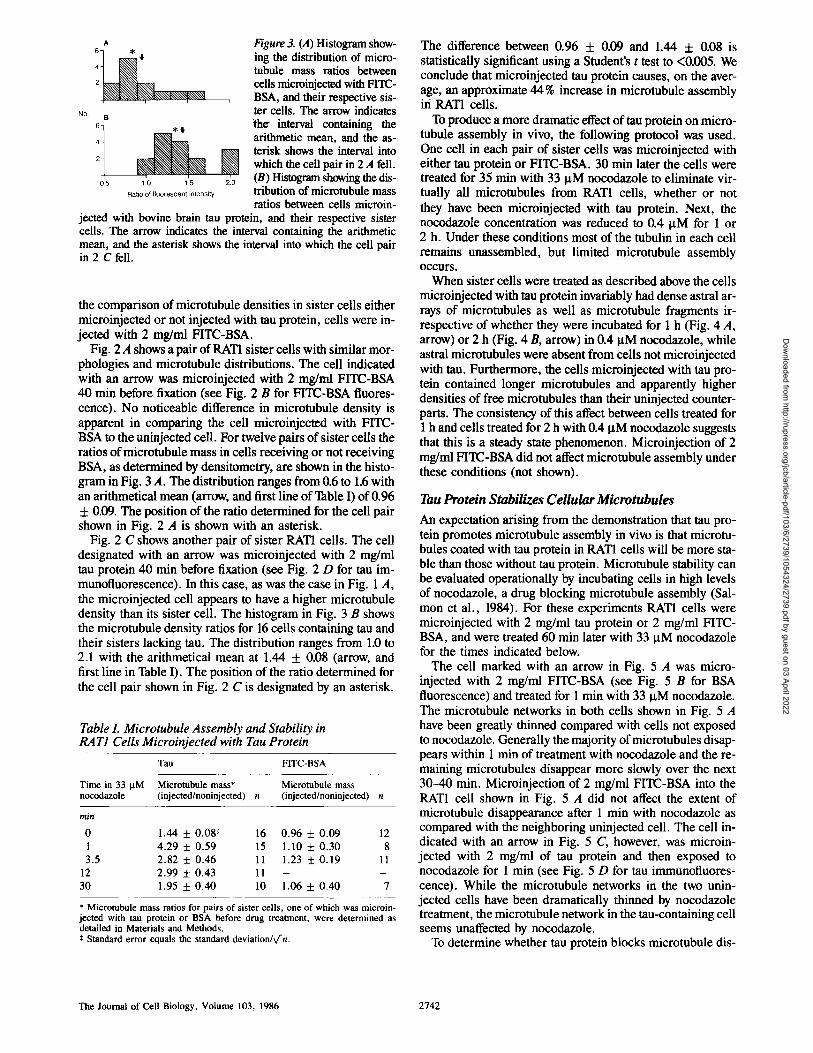
Figure 3. (A) Histogram show- ing the distribution of micro- tubule mass ratios between cells microinjected with FITC- BSA, and their respective sis- ter cells. The arrow indicates the interval containing the arithmetic mean, and the as- terisk shows the interval into which the cell pair in 2 A fell. (B) Histogram showing the dis- tribution of microtubule mass ratios between cells microin-
jected with bovine brain tan protein, and their respective sister cells. The arrow indicates the interval containing the arithmetic mean, and the asterisk shows the interval into which the cell pair in 2 C fell.
the comparison of microtubule densities in sister cells either microinjected or not injected with tau protein, cells were in- jected with 2 mg/ml FITC-BSA.
Fig. 2 A shows a pair of RAT1 sister cells with similar mor- phologies and microtubule distributions. The cell indicated with an arrow was microinjected with 2 mg/ml FITC-BSA 40 min before fixation (see Fig. 2 B for FITC-BSA fluores- cence). No noticeable difference in microtubule density is apparent in comparing the cell microinjected with FITC- BSA to the uninje~ted cell. For twelve pairs of sister cells the ratios of microtubule mass in cells receiving or not receiving BSA, as determined by densitometry, are shown in the histo- gram in Fig. 3 A. The distribution ranges from 0.6 to 1.6 with an arithmetical mean (arrow, and first line of Table I) of 0.96 + 0.09. The position of the ratio determined for the cell pair shown in Fig. 2 A is shown with an asterisk.
Fig. 2 C shows another pair of sister RATI cells. The cell designated with an arrow was microinjected with 2 mg/ml tau protein 40 min before fixation (see Fig. 2 D for tau im- munofluorescence). In this case, as was the case in Fig. 1 A, the microinjected cell appears to have a higher microtubule density than its sister cell. The histogram in Fig. 3 B shows the microtubule density ratios for 16 cells containing tau and their sisters lacking tau. The distribution ranges from 1.0 to 2.1 with the arithmetical mean at 1.44 + 0.08 (arrow, and first line in Table I). The position of the ratio determined for the cell pair shown in Fig. 2 C is designated by an asterisk.
Table L Microtubule Assembly and Stability in RAT1 Cells Microinjected with Tau Protein
Tau FITC-BSA
Time in 33 g M Microtubule mass* Microtubule mass nocodazole (injected/noninjected) n (injected/noninjected) n
rain
0 1 .44 + 0 .08¢ 16 0 . 9 6 + 0 . 0 9 12
1 4 . 2 9 + 0 . 5 9 15 1 .10 + 0 . 3 0 8
3 .5 2 . 8 2 + 0 . 4 6 11 1 .23 + 0 . 1 9 11
12 2 . 9 9 + 0 . 4 3 11 - -
3 0 1 .95 + 0 . 4 0 10 1 .06 + 0 . 4 0 7
* Microtubule mass ratios for pairs of sister cells, one of which was microin- jected with tau protein or BSA before drug treatment, were determined as detailed in Materials and Methods. ¢ Standard error equals the standard deviation/~/-n.
The difference between 0.96 + 0.09 and 1.44 + 0.08 is statistically significant using a Student's t test to <0.005. We conclude that microinjected tan protein causes, on the aver- age, an approximate 44% increase in microtubule assembly iri RAT1 cells.
To produce a more dramatic effect oftau protein on micro- tubule assembly in vivo, the following protocol was used. One cell in each pair of sister cells was microinjected with either tau protein or FITC-BSA. 30 min later the cells were treated for 35 min with 33 gM nocodazole to eliminate vir- tually all microtubules from RAT1 cells, whether or not they have been microinjected with tau protein. Next, the nocodazole concentration was reduced to 0.4 gM for 1 or 2 h. Under these conditions most of the tubulin in each cell remains unassembled, but limited microtubule assembly occurs.
When sister cells were treated as described above the cells microinjected with tau protein invariably had dense astral ar- rays of microtubules as well as microtubule fragments ir- respective of whether they were incubated for 1 h (Fig. 4 A, arrow) or 2 h (Fig. 4 B, arrow) in 0.4 gM nocodazole, while astral microtubules were absent from ceils not microinjected with tau. Furthermore, the cells microinjected with tau pro- tein contained longer microtubules and apparently higher densities of free microtubules than their uninjected counter- parts. The consistency of this affect between cells treated for 1 h and cells treated for 2 h with 0.4 I~M nocodazole suggests that this is a steady state phenomenon. Microinjection of 2 mg/ml FITC-BSA did not affect microtubule assembly under these conditions (not shown).
Tau Protein Stabilizes Cellular Microtubules
An expectation arising from the demonstration that tau pro- tein promotes microtubule assembly in vivo is that microtu- bules coated with tau protein in RAT1 cells will be more sta- ble than those without tau protein. Microtubule stability can be evaluated operationally by incubating cells in high levels of nocodazole, a drug blocking microtubule assembly (Sal- mon et al., 1984). For these experiments RAT1 cells were microinjected with 2 mg/ml tau protein or 2 mg/ml FITC- BSA, and were treated 60 min later with 33 gM nocodazole for the times indicated below.
The cell marked with an arrow in Fig. 5 A was micro- injected with 2 mg/ml FITC-BSA (see Fig. 5 B for BSA fluorescence) and treated for 1 min with 33 ~M nocodazole. The microtubule networks in both cells shown in Fig. 5 A have been greatly thinned compared with cells not exposed to nocodazole. Generally the majority of microtubules disap- pears within 1 min of treatment with nocodazole and the re- maining microtubules disappear more slowly over the next 30-40 min. Microinjection of 2 mg/ml FITC-BSA into the RAT1 cell shown in Fig. 5 A did not affect the extent of microtubule disappearance after 1 min with nocodazole as compared with the neighboring uninjected cell. The cell in- dicated with an arrow in Fig. 5 C, however, was microin- jected with 2 mg/ml of tau protein and then exposed to nocodazole for 1 min (see Fig. 5 D for tau immunofluores- cence). While the microtubule networks in the two unin- jected cells have been dramatically thinned by nocodazole treatment, the microtubule network in the tau-containing cell seems unaffected by nocodazole.
To determine whether tau protein blocks microtubule dis-
The Journal of Ceil Biology, Volume 103, 1986 2742
Dow
nloaded from http://rupress.org/jcb/article-pdf/103/6/2739/1054324/2739.pdf by guest on 03 April 2022
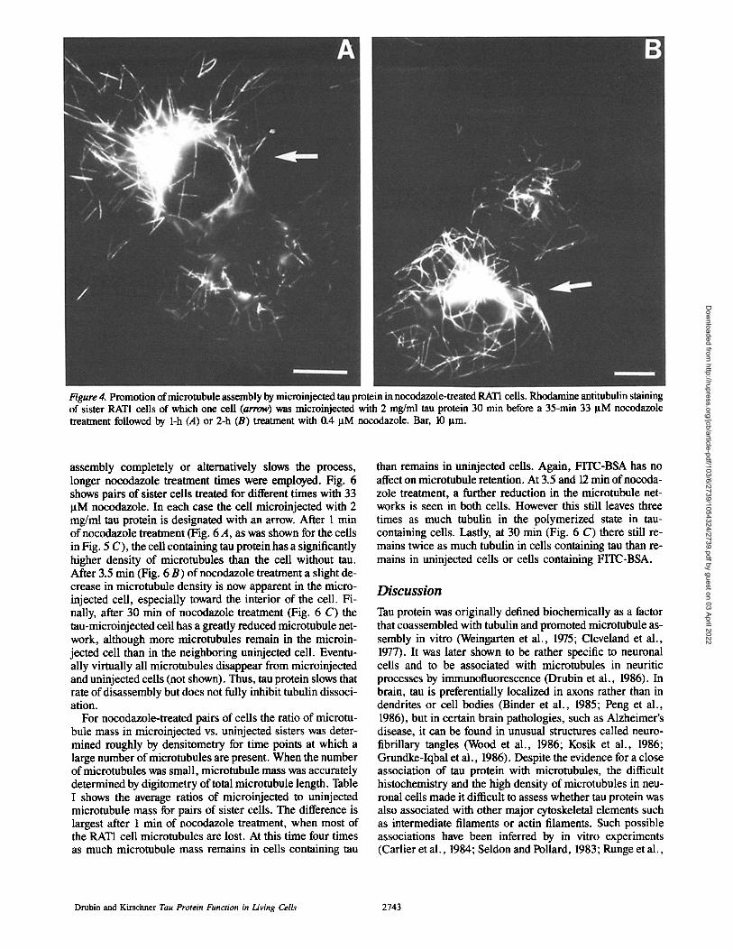
Figure 4. Promotion of microtubule assembly by microinjected tau protein in nocodazole-treated RATI cells. Rhodamine antitubulin staining of sister RAT1 cells of which one cell (arrow) was microinjected with 2 mg/ml tau protein 30 min before a 35-min 33 ltM nocodazole treatment followed by 1-h (A) or 2-h (B) treatment with 0.4 I~M nocodazole. Bar, 10 Ixm.
assembly completely or alternatively slows the process, longer nocodazole treatment times were employed. Fig. 6 shows pairs of sister cells treated for different times with 33 lxM nocodazole. In each case the cell microinjected with 2 mg/ml tau protein is designated with an arrow. After 1 rain of nocodazole treatment (Fig. 6 A, as was shown for the cells in Fig. 5 C), the cell containing tau protein has a significantly higher density of microtubules than the cell without tau. After 3.5 min (Fig. 6 B) of nocodazole treatment a slight de- crease in microtubule density is now apparent in the micro- injected cell, especially toward the interior of the cell. Fi- naUy, after 30 min of nocodazole treatment (Fig. 6 C) the tau-microinjected cell has a greatly reduced microtubule net- work, although more microtubules remain in the microin- jected cell than in the neighboring uninjected cell. Eventu- ally virtually all microtubules disappear from microinjected and uninjected cells (not shown). Thus, tau protein slows that rate of disassembly but does not fully inhibit tubulin dissoci- ation.
For nocodazole-treated pairs of cells the ratio of microtu- bule mass in microinjected vs. uninjected sisters was deter- mined roughly by densitometry for time points at which a large number of microtubules are present. When the number of microtubules was small, microtubule mass was accurately determined by digitometry of total microtubule length. Table I shows the average ratios of microinjected to uninjected microtubule mass for pairs of sister cells. The difference is largest after 1 min of nocodazole treatment, when most of the RATI cell microtubules are lost. At this time four times as much microtubule mass remains in cells containing tau
than remains in uninjected cells. Again, FITC-BSA has no affect on microtubule retention. At 3.5 and 12 min of nocoda- zole treatment, a further reduction in the microtubule net- works is seen in both ceils. However this still leaves three times as much tubulin in the polymerized state in tau- containing cells. Lastly, at 30 min (Fig. 6 C) there still re- mains twice as much tubulin in cells containing tau than re- mains in uninjected cells or cells containing FITC-BSA.
D i s c u s s i o n
Tau protein was originally defined biochemically as a factor that coassembled with tubulin and promoted microtubule as- sembly in vitro (Weingarten et al., 1975; Cleveland et al., 1977). It was later shown to be rather specific to neuronal cells and to be associated with microtubules in neuritic processes by immunofluorescence (Drubin et al., 1986). In brain, tau is preferentially localized in axons rather than in dendrites or cell bodies (Binder et al., 1985; Peng et al., 1986), but in certain brain pathologies, such as Alzheimer's disease, it can be found in unusual structures called neuro- fibrillary tangles (Wood et al., 1986; Kosik et al., 1986; Grundke-Iqbal et al., 1986). Despite the evidence for a close association of tau protein with microtubules, the difficult histochemistry and the high density of microtubules in neu- ronal cells made it difficult to assess whether tau protein was also associated with other major cytoskeletal elements such as intermediate filaments or actin filaments. Such possible associations have been inferred by in vitro experiments (Carlier et al., 1984; Seldon and Pollard, 1983; Runge et al.,
Drubin and Kirschner Tau Protein Function in Living Cells 2743
Dow
nloaded from http://rupress.org/jcb/article-pdf/103/6/2739/1054324/2739.pdf by guest on 03 April 2022
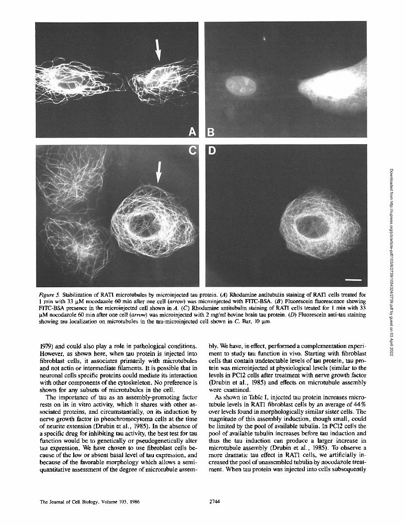
Figure 5. Stabilization of RAT1 microtubules by microinjected tau protein. (A) Rhodamine antitubulin staining of RAT1 cells treated for 1 min with 33 IxM nocodazole 60 min after one cell (arrow) was microinjected with FITC-BSA. (B) Fluorescein fluorescence showing FITC-BSA presence in the microinjected cell shown in A. (C) Rhodamine antitubulin staining of RATI cells treated for 1 min with 33 ~tM nocodazole 60 min after one cell (arrow) was microinjected with 2 mg/ml bovine brain tau protein. (D) Fluorescein anti-tau staining showing tau localization on microtubules in the tau-microinjected cell shown in C. Bar, 10 I.tm.
1979) and could also play a role in pathological conditions. However, as shown here, when tau protein is injected into fibroblast cells, it associates primarily with microtubules and not actin or intermediate filaments. It is possible that in neuronal cells specific proteins could mediate its interaction with other components of the cytoskeleton. No preference is shown for any subsets of microtubules in the cell.
The importance of tau as an assembly-promoting factor rests on its in vitro activity, which it shares with other as- sociated proteins, and circumstantially, on its induction by nerve growth factor in pheochromocytoma cells at the time of neurite extension (Drubin et al., 1985). In the absence of a specific drug for inhibiting tau activity, the best test for tau function would be to genetically or pseudogenetically alter tau expression. We have chosen to use fibroblast cells be- cause of the low or absent basal level of tau expression, and because of the favorable morphology which allows a semi- quantitative assessment of the degree of microtubule assem-
bly. We have, in effect, performed a complementation experi- ment to study tau function in vivo. Starting with fibroblast cells that contain undetectable levels of tau protein, tau pro- tein was microinjected at physiological levels (similar to the levels in PC12 cells after treatment with nerve growth factor (Drubin et al., 1985) and effects on microtubule assembly were examined.
As shown in Table I, injected tau protein increases micro- tubule levels in RAT1 fibroblast cells by an average of 44% over levels found in morphologically similar sister cells. The magnitude of this assembly induction, though small, could be limited by the pool of available tubulin. In PC12 cells the pool of available tubulin increases before tau induction and thus the tau induction can produce a larger increase in microtubule assembly (Drubin et al., 1985). To observe a more dramatic tan effect in RATI cells, we artificially in- creased the pool of unassembled tubulin by nocodazole treat- ment. When tau protein was injected into cells subsequently
The Journal of Cell Biology, Volume 103, 1986 2744
Dow
nloaded from http://rupress.org/jcb/article-pdf/103/6/2739/1054324/2739.pdf by guest on 03 April 2022
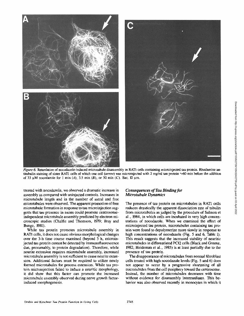
Figure 6. Retardation of nocodazole-induced microtubule disassembly in RAT1 ceils containing microinjected tau protein. Rhodamine an- titubulin staining of sister RAT1 cells of which one cell (arrow) was microinjected with 2 mg/ml tau protein ~60 rnin before the addition of 33 ~tM nocodazole for 1 min (A), 3.5 min (B), or 30 min (C). Bar, 10 Ixm.
treated with nocodazole, we observed a dramatic increase in assembly as compared with uninjected controls. Increases in microtubule length and in the number of astral and free microtubules were observed. The apparent promotion of free microtubule formation in response to tau microinjection sug- gests that tau presence in axons could promote centrosome- independent microtubule assembly predicted by electron mi- croscopic studies (Chalfie and Thomson, 1979; Bray and Bunge, 1981).
While tau protein promotes microtubule assembly in RAT1 cells, it does not cause obvious morphological changes over the 3-h time course examined (beyond 3 h, microin- jected tau protein cannot be detected by immunofluorescence due, presumably, to protein degradation). Therefore, while neurite extension requires microtubule assembly, increased microtubule assembly is not sufficient to cause neurite exten- sion. Additional factors must be required to utilize newly formed microtubules for process extension. While tau pro- tein microinjection failed to induce a neuritic morphology, it did show that this factor can promote the increased microtubule assembly observed during nerve growth factor- induced morphogenesis.
Consequences of Tau Binding for Microtubule Dynamics
The presence of tau protein on microtubules in RAT1 cells reduces drastically the apparent dissociation rate of tubulin from microtubules as judged by the procedure of Salmon et al., 1984, in which cells are incubated in very high concen- trations of nocodazole. When we examined the effect of microinjected tau protein, microtubules containing tau pro- tein were found to depolymerize more slowly in response to high concentrations of nocodazole (Fig. 5 and 6, Table I). This result suggests that the increased stability of neuritic microtubules in differentiated PCI2 cells (Black and Greene, 1982; Heideman et al., 1985) is at least partially due to the presence of tau protein.
The disappearance of microtubules from normal fibroblast cells treated with high nocodazole levels (Fig. 5 and 6) does not appear to occur by a progressive shortening of all microtubules from the cell periphery toward the centrosome. Instead, the number of microtubules decreases with time without evidence for disassembly intermediates. This be- havior was also observed recently in monocytes in which it
Drubin and Kirschner Tau Protein Function in Living Cells 2745
Dow
nloaded from http://rupress.org/jcb/article-pdf/103/6/2739/1054324/2739.pdf by guest on 03 April 2022
is possible to count and measure every microtubule in a cell (Cassimeris et al., 1986). In monocytes and in fibroblasts most microtubules are lost quickly in response to nocodazole treatment but a subset of mierotubules is lost at a signifi- cantly slower rate. The rate-limiting step appears to be initia- tion of depolymerization, not dissociation of tubulin subunlts from depolymerizing microtubules. These observations can be explained if the disappearance of microtubules in fibro- blast cells results from a transition from a stable or growing state to a rapidly shrinking state, as suggested by the model of dynamic instability (Mitchison and Kirschner, 1984).
Tan protein slows the rate of microtubule loss without al- lowing visualization of disassembly intermediates. Thus it appears that tau stabilizes microtubules primarily by de- creasing the transition rate to the rapidly disassembling state. Recent in vitro studies have in fact shown that microtubule- associated proteins can have an inhibitory effect on such tran- sitions (Horio and Hotani, 1986).
The fact that tau protein microinjection into growing RATI cells causes a significant increase in microtubule assembly and stability suggests that these cells normally lack tau-like factors at levels found in differentiated PC12 cells. Thus it may be that tau-like factors are characteristic of neuronal cells that have adopted a differentiated morphology. In sup- port of this, assembly behavior of microtubules in fibroblast cells (Schulze and Kirschner, 1986) is similar to that found in vitro with tubulin lacking associated proteins (Mitchison and Kirschner, 1984). Unfortunately we know nothing about the presence of MAP proteins in RAT-1 cells. The subset of microtubules in monocytes and fibroblasts that have in- creased stability could, however, contain selectively bound tan-like factors. General lack of tau-like factors in proliferat- ing fibroblast cells may explain why only tubulin mutants have been obtained when attempts were made to genetically identify drug resistant cells in culture.
We are indebted to Dr. David Kristofferson for statistics advice and instruc- tion on digitizing microtubules, Eric Schulze for microinjection instruction, and Kent Matlack for tau protein preparation. We are grateful to Dr. Frank Solomon for providing helpful insights on axonal microtubule assembly. We thank Dr. Gideon Dreyfuss for suggesting the use of Empigen detergent and Sumire Kobayashi for developing the fixation protocol. We thank Cynthia Cunningham-Hernandez for her assistance in preparing this manuscript.
This work was supported by grants from the National Institutes of Health and the American Cancer Society.
Received for publication 29 July 1986, and in revised form 11 September 1986.
References
Binder, L. I., A. Frankfurter, and L. I. Rebhun. 1985. The distribution of tau in the mammalian central nervous sytem. J. Cell Biol. 101:1371-1378.
Black, M. M., and L. A. Greene. 1982. Changes in the colchicine suscepti- bility of microtubules associated with neurite outgrowth: studies with nerve growth factor-responsive PC12 pheochromocytoma cells. J. Cell Biol. 95:379-386.
Blose, S. H., D. I. Meltzer, and J. R. Feramisco. 1984. 10-nm filaments are induced to collapse in living cells rnicroinjected with monoclonal and poly- clonal antibodies against tubulin. J. Cell Biol. 98:847-858.
Bray, D., and M. B. Bunge. 1981. Serial analysis of microtubules in cultured rat sensory axons. J. Neurocytol. 10:589-605.
Carlier, M.-F., C. Simon, R. Cassoly, and L.-A. Pradel. 1984. Interaction between microtubule-associated protein tau and spectrin. Biochimie (Paris). 66:305-311.
Caron, J. M., A. L. Jones, and M. W. Kirschner. 1985. Autoregulation of tubulin synthesis in hepatocytes and fibroblasts. J. Cell Biol. 101:1763-1772.
Cassimeris, L. U., P. Wadsworth, and E. D. Salmon. 1986. Dynamics of microtubule depolymerization in monocytes. J. Cell Biol. 102:2023-2032.
Chalfie, M., and J. N. Thomson. 1979. Organization in neuronal microtu- bules in the nematode Caenorhabditis elegans. J. Cell Biol. 82:278-289.
Cleveland, D. W., S. Y. Hwo, and M. W. Kirschner. 1977. Purification of tau, a microtubule-associated protein that induces assembly of microtubules from purified tubulin. J. Mol. Biol. 116:207-225.
Daniels, M. 1975. The role of micrntubules in the growth and stabilization of nerve fibers. Ann NYAcad. Sci. 253:535-544.
Drubin, D., and M. Kirschner. 1986. Purification oftau protein. Methods EnzymoL 134: In press.
Drubin, D. G., D. Caput, and M. W. Kirschner. 1984. Studies on the expres- sion of microtubule-associated protein, tau, during mouse brain development, with newly isolated complementary DNA probes. J. Cell. Biol. 98:1090-1097.
Drubin, D. G., S. C. Feinstein, E. M. Shooter, andM. W. Kirschner. 1985. Nerve growth factor-induced neurite outgrowth in PC12 cells involves the coor- dinate induction of microtubule assembly and assembly-promoting factors. J. Cell BioL 101:1799-1807.
Drubin, D., S. Kobayashi, and M. W. Kirschner. 1986. Association oftau protein with microtubules in living cells. Ann. NYAcad. Sci. 466:257-268.
Gard, D. L., and M. W. Kirschner. 1985. A polymer-dependent phosphory- lation of l~-tubulin accompanies differentiation of a mouse neuroblastoma cell line. J. Cell Biol. 100:764-774.
Grandke-Iqbal, I., K. Iqbal, Y.-C. Tung, M. Quinlan, H. M. Wisniewski, and L. I. Binder. 1986. Abnormal phosphorylation of the microtubule- associated protein x (tau) in Alzheimer cytoskeletal pathology. Proc. Natl. Acad. Sci. USA. 83:4913--4917.
Heidemann, S. R., H. C. Joshi, A. Schechter, J. R. Fletcher, and M. Both- well. 1985. Synergistic effects of cyclic AMP and nerve growth factor on neurite outgrowth and microtubule stability of PC12 cells. J. Cell Biol. 100:916-927.
Herzog, W., and K. Weber. 1978. Fractionation of brain microtubute- associated proteins. Fur. J. Biochem. 92:1-8.
Hiller, G., and K. Weber. 1978. Radioimmunoassay for tubulin: a quantita- tive comparison oftbe tubulin content of different established tissue culture cells and tissues. Cell. 147:795-804.
Horio, T., and H. Hotani. 1986. Visualization of the dynamic instability of individual microtubules by dark-field microscopy. Nature (Lond.). 321:605- 607.
Kosik, K. S., C. L. Joachim, and D. J. Selkoc. 1986. Microtubule associated protein tan (x) is a major antigenic component of paired helical filaments in Alz- heimar disease. Proc. Natl. Acad. Sci. USA. 83:4044--4048.
Mitchison, T., and M. Kirschner. 1984. Dynamic instability of microtubule growth. Nature (Lond.). 312: 237-242.
Nunez, J., A. Fellous, J. Francon, and A. M. Lcnnon. 1975. Neurotubulin and brain development. In Microtubules and Microtubule Inhibitors. M. Borgers and M. de Brabanders, editors. Elsevier/North-Holland, Amsterdam. 269-279.
Olmsted, J. B. 1981. Tubulin pools in differentiating neuroblastoma cells. J. Cell Biol. 89:418-423.
Peng, I., L. I. Binder, and M. M. Black. 1986. Biochemical and immunolog- ical analyses of cytoskeletal domains of neurons. J. Cell. Biol. 102:252-262.
Pfeffer, S. R., D. G. Drubin, and R. J. Kelly. 1983. Identification of three coated vesicle components as u- and I~-tubulin linked to a phosphorylated 50,000-dalton polypeptide. J. Cell Biol. 97:40-47.
Runge, M. S., H. W. Detrich, m , and R. C. Williams, Jr. 1979. Identifica- tion of the major 68000-dalton protein of microtubule preparations as a 10-nm filament protein and its effect on microtubute assembly in vitro. Biochemistry. 18:1689-1698.
Salmon, E. D., M. McKeel, and T. Hays. 1984. Rapid rate of tubulin dis- sociation from microtubules in the mitotic spindle in vivo measured by blocking polymerization with colchicine. J. Cell Biol. 99:1066-1075.
Schnapp, B. J., R. D. Vale, M. P. Shectz, and T. S. Reese. 1985. Single rnicrotubules from squid axoplasm support bidirectional movement of or- ganelles. Cell. 40:455--462.
Schulze, E., and M. Kirschner. 1986. Microtubule dynamics in interphase cells. J. Cell BioL 102:1020-1031.
Seeds, N. W., and R. B. Maccioni. 1978. Proteins from morphologically differentiated neuroblastoma cells promote tubulin polymerization. J. Cell Biol. 76:547-555.
Seldon, S. C., and T. D. Pollard. 1983. Phosphorylation of microtubule- associated proteins regulates their interaction with actin filaments. J. Biol. Chem. 258:7064-7071.
Solomon, F. 1981. Specification of cell morphology by endogenous deter- minants. J. Cell Biol. 90:547-553.
Vallee, R. B., G. S. Bloom, and W. E. Theurkauf. 1984. Microtubule- associated proteins: subunits of the cytomatrix. J. Cell Biol. 99(Suppl.): 38s--44s.
Weingarten, M. D.. A. H. Lockwood, S. Y. Hwo, and M. W. Kirschner. 1975. A protein factor essential for microtubule assembly. Proc. Natl. Acad. Sci. USA. 72:1858-1862.
Wood, J. G., S. S. Mirra, N. J. Pollack, and L. I. Binder. 1986. Neurofibrillary tangles of Alzheimer disease share antigenic determinants with the axonal microtubule associated protein tau (x). Proc. Natl. Acad. Sci. USA. 83:4040- 4043.
Yamada, K. M., B. S. Spooner, and N. K. Wessells. 1976. Axon growth: role of microfilaments and microtubules. Proc. Natl. Acad. Sci. USA. 66:1206- 1212.
The Journal of Cell Biology, Volume 103, 1986 2746
Dow
nloaded from http://rupress.org/jcb/article-pdf/103/6/2739/1054324/2739.pdf by guest on 03 April 2022


![Tubulin II [Kompatibilitätsmodus] - uni-marburg.de · Mikrotubuli-bindende Proteine Expression of tau protein induces process formation in SF9 cells The Sf9 cell bodies have a round](https://static.fdocuments.net/doc/165x107/5d4c601f88c99325278b9851/tubulin-ii-kompatibilitaetsmodus-uni-mikrotubuli-bindende-proteine-expression.jpg)