Targeting the PI3K/Akt/mTOR signalling pathway in Cystic Fibrosis10.1038/s41598-017... · Targeting...
12
Targeting the PI3K/Akt/mTOR signalling pathway in Cystic Fibrosis Reilly, R Mroz, MS Dempsey, E Wynne, K Keely, SJ McKone, EF Behl, C Hiebel, C Coppinger, JA Supplementary Materials All antibodies and inhibitors and their sources are listed in Supplementary Materials. All buffer compositions for CO-PIT, STAGE clean up, Immunoblotting, Immunofluorescence, LC-MS/MS and Short Circuit Analysis are also listed in table format (Tables 1.1-1.5) Supplementary Table S1 Mass spectrometry analysis of the ΔF508 CFTR Interactome. The gene accession numbers, protein names, protein classes GO functions, score, and peptide numbers are displayed. Supplementary Table S2 Mass spectrometry analysis of the WT CFTR Interactome. The gene accession numbers, protein names, protein classes GO functions, score, and peptide numbers are displayed. Supplementary Table S3 Selected inhibitors to the PI3K/Akt/mTOR are listed along with their molecular target and references. Supplementary Figure S1 RICTOR was immunoprecipitated from HBE41o- and CFBE41o- cells expressing WT and ΔF508 CFTR and the resulting RICTOR immunocomplexes were subjected to mass spectrometry analysis. The gene name, protein name and number of peptides identified of RICTOR and eight interactors are displayed in the table. Supplementary Figure S2 ΔF508 CFBE41o- cells (37°C) were treated with increasing concentrations of AZD-8055, KU-0063794, AKT-VIII and MK-2206 and protein expression for CFTR was determined by immunoblotting. Supplementary Figure S3 Subcellular localization of CFTR in ΔF508 CFBE41o- cells treated with MK-2206, KU- 0063794 was detected by confocal microscopy. Supplementary Figure S4 Co-localization of CFTR and LC3 in WT HBE41o- cells
Transcript of Targeting the PI3K/Akt/mTOR signalling pathway in Cystic Fibrosis10.1038/s41598-017... · Targeting...

Targeting the PI3K/Akt/mTOR signalling pathway in Cystic Fibrosis
Reilly, R Mroz, MS Dempsey, E Wynne, K Keely, SJ McKone, EF Behl, C Hiebel, C
Coppinger, JA
Supplementary Materials
All antibodies and inhibitors and their sources are listed in Supplementary Materials. All
buffer compositions for CO-PIT, STAGE clean up, Immunoblotting, Immunofluorescence,
LC-MS/MS and Short Circuit Analysis are also listed in table format (Tables 1.1-1.5)
Supplementary Table S1
Mass spectrometry analysis of the ΔF508 CFTR Interactome. The gene accession numbers,
protein names, protein classes GO functions, score, and peptide numbers are displayed.
Supplementary Table S2
Mass spectrometry analysis of the WT CFTR Interactome. The gene accession numbers,
protein names, protein classes GO functions, score, and peptide numbers are displayed.
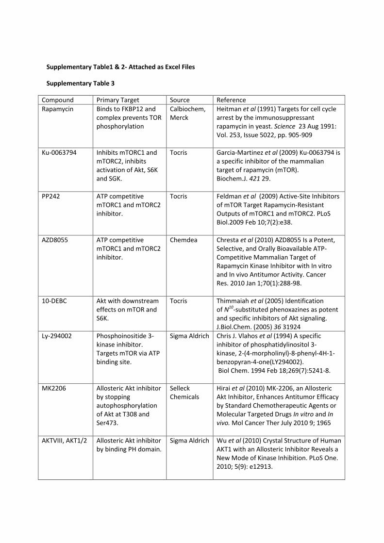
Supplementary Table S3
Selected inhibitors to the PI3K/Akt/mTOR are listed along with their molecular target and
references.
Supplementary Figure S1
RICTOR was immunoprecipitated from HBE41o- and CFBE41o- cells expressing WT and
ΔF508 CFTR and the resulting RICTOR immunocomplexes were subjected to mass
spectrometry analysis. The gene name, protein name and number of peptides identified of
RICTOR and eight interactors are displayed in the table.
Supplementary Figure S2
ΔF508 CFBE41o- cells (37°C) were treated with increasing concentrations of AZD-8055,
KU-0063794, AKT-VIII and MK-2206 and protein expression for CFTR was determined by
immunoblotting.
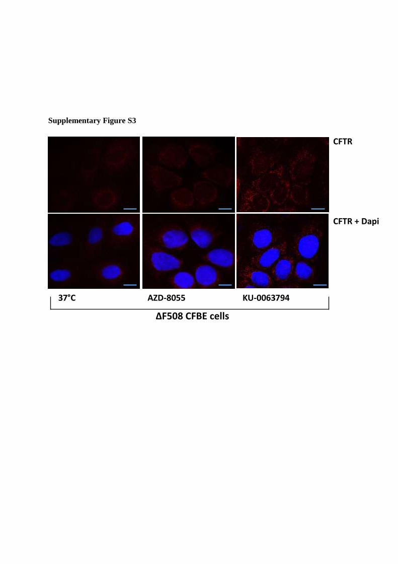
Supplementary Figure S3
Subcellular localization of CFTR in ΔF508 CFBE41o- cells treated with MK-2206, KU-
0063794 was detected by confocal microscopy.
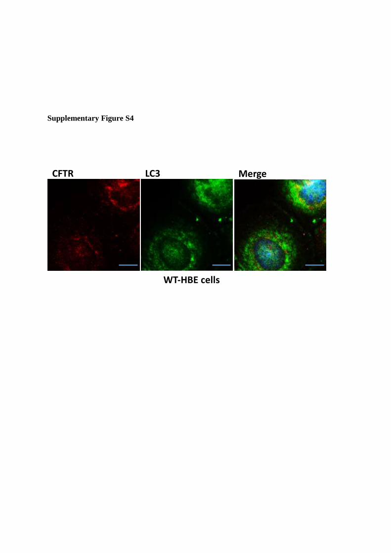
Supplementary Figure S4
Co-localization of CFTR and LC3 in WT HBE41o- cells
Supplementary Figure S5
Co-localization of CFTR and aggresomes was investigated by confocal microscopy in ΔF508
CFBE cells treated with MK-2206.
Supplementary Materials
ABCAM: BAG3 antibody (ab86298)
Cell Signalling: mTOR (2983 P), mTOR ser2448 (5536 P), mTOR ser2481 (2974 P), total
p70s6 kinase, p70s6 kinase thr389 (9234 P), total Akt, pAkt ser473 (9271 S), RICTOR (2114
P), RAPTOR (2280 P), Gβl (3274 P), Autophagy Antibody Sampler Kit #4445 containing:
ATG3, ATG5, ATG12, Beclin-1, ATG16L and ATG7. HSP90 (4877 P), HSP70 (4872 P),
Sin1 (12860 S) and anti-rabbit HRP (7074 P2).
Chemdea: AZD-8055 (CD0348)
GIBCO: Penicillin Streptomycin (15070), Foetal bovine serum (10270-106)
MERCK: Rapamycin (553210-100UG)
MERCK MILLIPORE: CFTR antibody M3A7 (05-583)
Nanotools: LC3 antibody (0231-100/LC3-5F10)
Santa Cruz: goat anti-mouse HRP (sc-2005), MAPKAP1 antibody (sc-393166)
Selleck Chemicals: MK-2206 (S1078), VX-770 (S1144), VX-809 (S1565)
Sigma Aldrich: Ly-294002 (L9908), AKT-VIII (A6730), genistein (G6649), CFTR (inh) 172
(C2992), Forskolin (F3917)
Thermo fisher scientific: Minimum essential medium with L-glutamine (31095-029), goat
anti-mouse Alexa Fluor® 594 (A-11005), anti-rabbit Alexa Fluor® 488 (A 11034)
TORCIS: KU-0063794 (3725), PP 242 (4257), 10-DEBC (2558)
UNC Chapel Hill: CFTR 660 antibody, CFTR 596 antibody

Buffer Composition
Co-PIT
Solutions Reagent Composition
Dynabeads 200 ul per IP
Crosslinking buffer 0.2 M (1.52 ml)
triethanolamine in 50 ml PBS
pH 8.2
Crosslinking buffer: Dimethyl
pimelimidate (DMP)
0.2 M (1.52 ml)
triethanolamine, 0.025 M DMP
in 50 ml PBS pH 8.2
Quenching/blocking buffer 0.2 M (610 µl) ethanolamine in
50 ml PBS, pH 8.2
Antibody Elution buffer 1 M (3.75 g) glycine in 50 ml
H2O pH 2.5
Lysis buffer 1% (500 µl) Surfact-Amps®
NP-40, protease and
phosphatase inhibitor mix in 50
ml PBS
Protein complex elution buffer 8 M (4.8 g) urea, 0.5 % (100
µl) Surfact-Amps® NP-40 in
10 ml PBS
Table 1.1: Solutions used in Immunoprecipitation.

Stop and Go protocol; protein digest clean-up (STAGE)
Solutions Reagent composition
Buffer A 0.1 % 20 µl trifluoroacetic acid
(TFA) in 19.9 ml ddH2O
Buffer B 50 % 10 ml acetonitrile, 0.1 % 20
µl trifluoroacetic acid, 50 % 9.9
ml ddH2O
Buffer C 1 % 200 µl trifluoroacetic acid in
19.98 ml ddH2O
Table 1.2 Solutions used in STAGE protocol.
LC-MS/MS Buffers
Solutions Reagent composition
Buffer A Buffer A: 97 % water, 2.5 %
acetonitrile, 0.5 % acetic acid
Buffer B Buffer B: 97 % acetonitrile,
2.5 % water, 0.5 % acetic acid.

Immunoblotting
Solutions Reagent composition
Lysis buffer 1 % SDS protease and
phosphatase inhibitor mix
Running buffer 0.025 M (3.03 g) Tris base,
0.192 M (14.04 g) glycine, 0.1
% (1 g) SDS, pH 8.5 in 1 L
H2O
Transfer buffer 0.025 M (6.06 g) Tris base,
0.192 M (28.8 g) glycine, 30
% methanol in 2 L H2O
10X Tris-buffered saline
(TBS)
0.171 M (12.1 g) Tris, 1.44 M
(42 g) NaCl in 500 ml H2O
1x Tris-buffered saline
tween®20 (TBS-T)
100 ml of 10 X TBS, 0.1 %
Tween®20 in 900 ml H2O
5x SDS sample buffer 50 mM Tris, 4 % w/v SDS, 20
% glycerol, 10 % v/v β-
mercaptoethanol, 0.02 % w/v
bromophenol blue
Protein stain 0.1 % w/v ponceau s stain in
10 % acetic acid
Table 1.3: Solutions used in immunoblotting protocol.

Immunofluorescence
Solutions Reagent composition
Fix solution 0.45 g of PFA, 200 µl 1 M
NaOH, 15 ml PBS
Blocking buffer 2 g BSA, 100 µl triton X-100,
50 ml PBS
Antibody dilution solution 0.5 g BSA, 100 µl triton X-
100, 15 ml PBS
Table 1.4: Solutions used in immunofluorescence protocol.
Short circuit current analysis
Reagent Concentration (mM) Location in chamber
NaCl 120 Basolateral
NaHCO3 25 Bilateral
KH2PO4 3.3 Bilateral
K2HPO4 0.8 Bilateral
MgCl2 1.2 Bilateral
CaCl2 1.2 Bilateral
Glucose 10 Bilateral
Na gluconate 120 Apical
Supplementary Table1 & 2- Attached as Excel Files
Supplementary Table 3
Compound Primary Target Source Reference
Rapamycin Binds to FKBP12 and complex prevents TOR phosphorylation
Calbiochem, Merck
Heitman et al (1991) Targets for cell cycle arrest by the immunosuppressant rapamycin in yeast. Science 23 Aug 1991: Vol. 253, Issue 5022, pp. 905-909
Ku-0063794 Inhibits mTORC1 and mTORC2, inhibits activation of Akt, S6K and SGK.
Tocris Garcia-Martinez et al (2009) Ku-0063794 is a specific inhibitor of the mammalian target of rapamycin (mTOR). Biochem.J. 421 29.
PP242 ATP competitive mTORC1 and mTORC2 inhibitor.
Tocris Feldman et al (2009) Active-Site Inhibitors of mTOR Target Rapamycin-Resistant Outputs of mTORC1 and mTORC2. PLoS Biol.2009 Feb 10;7(2):e38.
AZD8055 ATP competitive mTORC1 and mTORC2 inhibitor.
Chemdea Chresta et al (2010) AZD8055 Is a Potent, Selective, and Orally Bioavailable ATP-Competitive Mammalian Target of Rapamycin Kinase Inhibitor with In vitro and In vivo Antitumor Activity. Cancer Res. 2010 Jan 1;70(1):288-98.
10-DEBC Akt with downstream effects on mTOR and S6K.
Tocris Thimmaiah et al (2005) Identification of N10-substituted phenoxazines as potent and specific inhibitors of Akt signaling. J.Biol.Chem. (2005) 36 31924
Ly-294002 Phosphoinositide 3-kinase inhibitor. Targets mTOR via ATP binding site.
Sigma Aldrich Chris J. Vlahos et al (1994) A specific inhibitor of phosphatidylinositol 3-kinase, 2-(4-morpholinyl)-8-phenyl-4H-1-benzopyran-4-one(LY294002). Biol Chem. 1994 Feb 18;269(7):5241-8.
MK2206 Allosteric Akt inhibitor by stopping autophosphorylation of Akt at T308 and Ser473.
Selleck Chemicals
Hirai et al (2010) MK-2206, an Allosteric Akt Inhibitor, Enhances Antitumor Efficacy by Standard Chemotherapeutic Agents or Molecular Targeted Drugs In vitro and In vivo. Mol Cancer Ther July 2010 9; 1965
AKTVIII, AKT1/2 Allosteric Akt inhibitor by binding PH domain.
Sigma Aldrich Wu et al (2010) Crystal Structure of Human AKT1 with an Allosteric Inhibitor Reveals a New Mode of Kinase Inhibition. PLoS One. 2010; 5(9): e12913.

Supplementary Figure S1

Supplementary Figure S2
Supplementary Figure S3
37°C AZD-8055 KU-0063794
CFTR
CFTR + Dapi
∆F508 CFBE cells
Supplementary Figure S4
CFTR LC3 Merge
WT-HBE cells

Supplementary Figure S5