Targeting RNA by the Antisense Approach and a Close Look ...170860/FULLTEXT01.pdf · Comparison of...
60
ACTA UNIVERSITATIS UPSALIENSIS UPPSALA 2007 Digital Comprehensive Summaries of Uppsala Dissertations from the Faculty of Science and Technology 355 Targeting RNA by the Antisense Approach and a Close Look at RNA Cleavage Reaction JHARNA BARMAN ISSN 1651-6214 ISBN 978-91-554-6995-5 urn:nbn:se:uu:diva-8272
Transcript of Targeting RNA by the Antisense Approach and a Close Look ...170860/FULLTEXT01.pdf · Comparison of...

ACTA
UNIVERSITATIS
UPSALIENSIS
UPPSALA
2007
Digital Comprehensive Summaries of Uppsala Dissertationsfrom the Faculty of Science and Technology 355
Targeting RNA by the AntisenseApproach and a Close Look atRNA Cleavage Reaction
JHARNA BARMAN
ISSN 1651-6214ISBN 978-91-554-6995-5urn:nbn:se:uu:diva-8272
���������� �������� �� ������ �������� � �� �������� ������� � ���� ���� �� ������ ��!�"� ������� #$������� %������ &� �''� �� '()!� *� �$� ��+��� * ���� *,$����$�- #$� �������� .��� �� ������� � �+���$-
�������������� /- �''�- #��+���+ 0%1 �� �$� 1������ 1�����$ �� � ���� 23 �� 0%1������+� 0�����- 1��� ����������� ���������- ������� ��� � ���� ����� � �������� ���� ������� �� �� ������� � ��� �� ��� � ������ "��- �& ��- ������-4��% (�& (! ��5 �((� �-
#$�� �$���� �������6�� �$� ������� * ������� �. ������� * ������ �����- �$�����������*��� ������� ��+��������� 718%�9 $��� ��� ��������� .��$ ��+���� � �$�������������� *� �0%1 ���+���+ � � ������� ������$ 7��� � ���9- #$� �$�����������*��� ��������� ���� �: � �� #� �: � �8� #� ����� #� 2%1 #� �6������ #��6� �%1 #� ���������� �%1 # �� ���������� 2%1 # .��� ��������� �� !� ���18%� �� ���+���� �+���� � !� ��� 0%1 �$�� *�� �$� ���+ ��+� * �; 5' ���+� #���+�- #$� ���������� ����� �$.�� �$�� � ��+�� ���*��� �������� � �$� 18% .��$��������� ��3�� ��+�� 7����� # �� �6������ #9 �.���� �$� �**���� *� �$������������ 0%1 .$����� ���� ��3�� ��+��� 72%1 #� �6� �%1 #����������� �%1 #� �� ���������� 2%1 #9 ��+�*������ ������� �$� �**����- 1���������� 0%��� < ��+���� ����� �$.�� �$�� ���*������ * �$� ���� ���� 7������������� � ���� ����9 � ��**���� ��=����� +��� ���� � ������� ������+� �������-���������� * �$� ���$����� ���� ���������� �� 3����� ���������� �$.�� �$�� �$����*��� 18%� ������� 0%��� < �������+ � �$���� ������ ������ 7�
���9 ���$�+$ .��$
.��3�� �6��� ��������� ����+ 7!> �9 ������� � �$� ����*��� 18%- #$� ���*���
18%� .��� ��� ��������� .��$ ��+���� � ��������� �.���� ��3� ��� �$��$������������ $��� ����� � �������� �$��� ��������� �.��� ����������- #$� �6� �%1 # ������������ �%1 # ���*��� 18%� �$.�� ������� ��������� ������� � ��� �$�����*��� 18%�- 4 +������ �$� ���*��� 18%� .��$ ���� ���� ��������� 7������ 2%1 #9.��� *�� � �� ������� � �$� ��������� ���� �� �$�� �$.�� ������� ���+�� �**�������������� 0%��� < ���������� ���������� �� ������� ��������� ���������-
#$� ���� ������ ������� � �$�� �$���� �� ����� �$�����$������ ������� * �$�� 0%1�������� �����6�+ %�0 ����� �< ������� �� ��3���� $�������� ������� 7��� �� �9-#$� %�0 ����� 7!< �� "!,9 �< ������� ������� �������� �$� �**��� * +����� �*������ ����+���� ���������������� �$��+$ � ��+�� ������� �$�� � � ��=����������� ����- #$� �������� �������� �$������� * �$� ��������������$��$�������� .�� *���$�� ����*��� �� ��3���� $�������� ����������- #$� ��������������$��$��������� .$��$ .��� �*������ �� +����� � *������ .��� $�����6�� �� �*����� ���� �$� �$�� ��=����� .$��� ���$ +����� � *����� .�� �������� ���������+ ? .��$ %! �� ?-
�!���" �0%1 ���+���+� ������� ��+���������� ���+�� �**����� 0%��� <����$����� ���� 3������� �� ������� ���������� %�0� �< �������� ��3���� $��������
#����� $����% � ���� �� � $��� ����� ��� ������� �� �����% &�� ����� � �'�������� � �
@ /$��� ����� �''�
4��% !��! ��!54��% (�& (! ��5 �((� ���)�)��)��)���� &��� 7$���)>>��-3�-��>������A��B��)�)��)��)���� &���9

To my parents


List of Original Publications
This thesis is comprised of the following original publications, which will be referred in the text by their Roman numerals. I. Honcharenko, D.; Barman, J.; Varghese, O. P.; Chattopadhyaya, J.
Comparison of the RNase H cleavage kinetics and blood serum sta-bility of the north conformationally constrained and 2'-alkoxy modi-fied oligonucleotides
Biochemistry 2007, 46, 5635 – 5646 II. Varghese, O. P.; Barman, J.; Pathmasiri, W.; Plashkevych, O.; Hon-
charenko, D.; Chattopadhyaya, J. Conformationally constrained 2'-N,4'-C-ethylene-bridged thymidine (aza-ENA-T): synthesis, struc-ture, physical, and biochemical studies of aza-ENA-T modified oli-gonucleotides
J. Am. Chem. Soc. 2006, 128, 15173 – 15187 III. Srivastava, P.; Barman, J.; Pathmasiri, W.; Plashkevych, O.; Wen-
ska, M.; Chattopadhyaya J. The five- and six-membered conforma-tionally-locked 2',4'-carbocyclic ribo-thymidine: synthesis, structure and biochemical studies
J. Am. Chem. Soc. 2007, 129, 8362 – 8379 IV. Acharya, S.; Barman, J.; Cheruku, P.; Chatterjee, S.; Acharya, P.;
Isaksson, J.; Chattopadhyaya, J. Significant pKa perturbation of nu-cleobases is an intrinsic property of the sequence context in DNA and RNA
J. Am. Chem. Soc. 2004, 126, 8674 – 8681 V. Barman, J.; Acharya, S.; Zhou, C.; Chatterjee, S.; Engstroem, A.;
Chattopadhyaya, J. Non-identical electronic characters of the inter-nucleotidic phosphates in RNA modulate the chemical reactivity of the phosphodiester bonds
Org. Biomol. Chem. 2006, 4, 928 – 941 Reprints were made with permission from the publishers
List of publications, not included in this thesis VI. Honcharenko, D.; Varghese, O. P.; Plashkevych, O.; Barman, J.;
Chattopadhyaya, J. Synthesis and structure of novel conformation-ally constrained 1',2'-azetidine-fused bicyclic pyrimidine nucleo-sides: their incorporation into oligo-DNAs and thermal stability of the hetero-duplexes
J. Org. Chem. 2006, 71, 299 – 314 VII. Bogucka, M.; Naus, P.; Pathmasiri, W.; Barman, J.; Chat-
topadhyaya, J. Facile preparation of the oxetane-nucleosides Org. Biomol. Chem. 2005, 3, 4362 – 4372
VIII. Isaksson, J.; Plashkevych, O.; Pradeepkumar, P. I.; Chatterjee, S.;
Barman, J.; Pathmasiri, W.; Shrivastava, P.; Petit, C.; Chat-topadhyaya, J. Oxetane locked thymidine in the Dickerson-Drew dodecamer causes local base pairing distortions - an NMR structure and hydration study
J. Biomol. Struct. Dyn. 2005, 23, 299 – 330 IX. Isaksson, J.; Acharya, S.; Barman, J.; Cheruku, P.; Chattopadhyaya,
J. Single-stranded adenine-rich DNA and RNA retain structural characteristics of their respective double-stranded conformations and show directional differences in stacking pattern
Biochemistry 2004, 43, 15996 – 16010 X. Opalinska, J. B.; Kalota, A.; Gifford, L. K.; Lu, P.; Jen, Kuang-Yu;
Pradeepkumar, P. I.; Barman, J.; Kim, T. K.; Swider, C. R.; Chat-topadhyaya, J.; Gewirtz, A. M. Oxetane modified, conformationally constrained, antisense oligodeoxyribonucleotides function efficiently as gene silencing molecules
Nucl. Acids Res. 2004, 32, 5791 – 5799
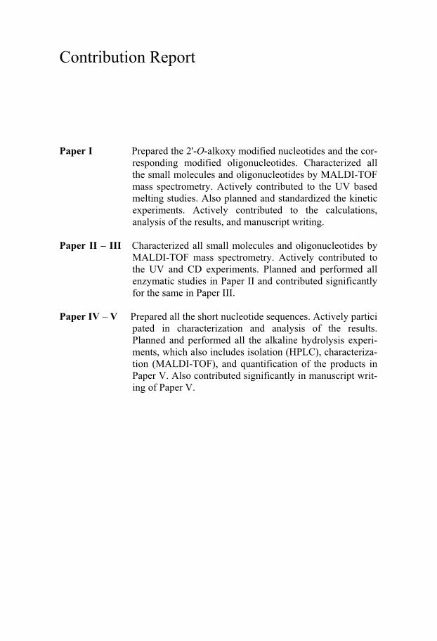
Contribution Report
Paper I Prepared the 2'-O-alkoxy modified nucleotides and the cor-responding modified oligonucleotides. Characterized all the small molecules and oligonucleotides by MALDI-TOF mass spectrometry. Actively contributed to the UV based melting studies. Also planned and standardized the kinetic experiments. Actively contributed to the calculations, analysis of the results, and manuscript writing.
Paper II – III Characterized all small molecules and oligonucleotides by
MALDI-TOF mass spectrometry. Actively contributed to the UV and CD experiments. Planned and performed all enzymatic studies in Paper II and contributed significantly for the same in Paper III.
Paper IV – V Prepared all the short nucleotide sequences. Actively partici
pated in characterization and analysis of the results. Planned and performed all the alkaline hydrolysis experi-ments, which also includes isolation (HPLC), characteriza-tion (MALDI-TOF), and quantification of the products in Paper V. Also contributed significantly in manuscript writ-ing of Paper V.


Contents
1 Introduction................................................................................................13 1.1 General introduction to nucleic acids .................................................13
1.1.1 Structure of nucleic acids............................................................14 1.1.2 Quantification and analysis.........................................................17 1.1.3 Reactivity of nucleic acids..........................................................18
1.2 Molecular recognition of nucleic acids ..............................................19 1.2.1 Interactions with metal ions and small molecules ......................19 1.2.2 Interactions with proteins ...........................................................20 1.2.3 Interactions with short oligonucleotides .....................................20
1.3 Targeting mRNA................................................................................21 1.3.1 The antisense strategy.................................................................21
1.3.1.1 Chemically modified nucleotides developed for the antisense strategy............................................................................................22
1.3.2 RNA catalysis .............................................................................24 1.4 Overview of the thesis........................................................................25
2 Evaluation of Modified AONs (Paper I-III) ..............................................26 2.1 Thermal stability ................................................................................27 2.2 RNase H recruitment properties .........................................................29
2.2.1 RNase H footprint pattern induced by AONs modified with 1',2'-locked nucleotide .................................................................................29 2.2.2 RNase H footprint pattern induced by AONs modified with 2',4'-locked nucleotides ...............................................................................30
2.3 RNase H cleavage kinetics .................................................................31 2.3.1 Pseudo first order cleavage rates ................................................31 2.3.2 Michaelis-Menten kinetics..........................................................32
2.4 Resistance to nuclease degradation ....................................................32 2.4.1 Resistance of modified AONs to snake venom phosphodiesterase degradation ..........................................................................................33 2.4.2 Stability in human serum ............................................................34
2.5 Conclusions and implications.............................................................34
3 Sequence Dependent Physicochemical Properties of ssRNA (Paper IV – V) ..................................................................................................................36
3.1 Determination of pKa values by 1H NMR ..........................................37 3.1.1 Sequence dependent pKa modulation a G� ion formation...........38
3.1.2 Differential electrostatic modulation toward the 3'- and the 5'- end .......................................................................................................41 3.1.3 Correlation of �pKa of G with �H8G and with the oligomerization shift............................................................................42
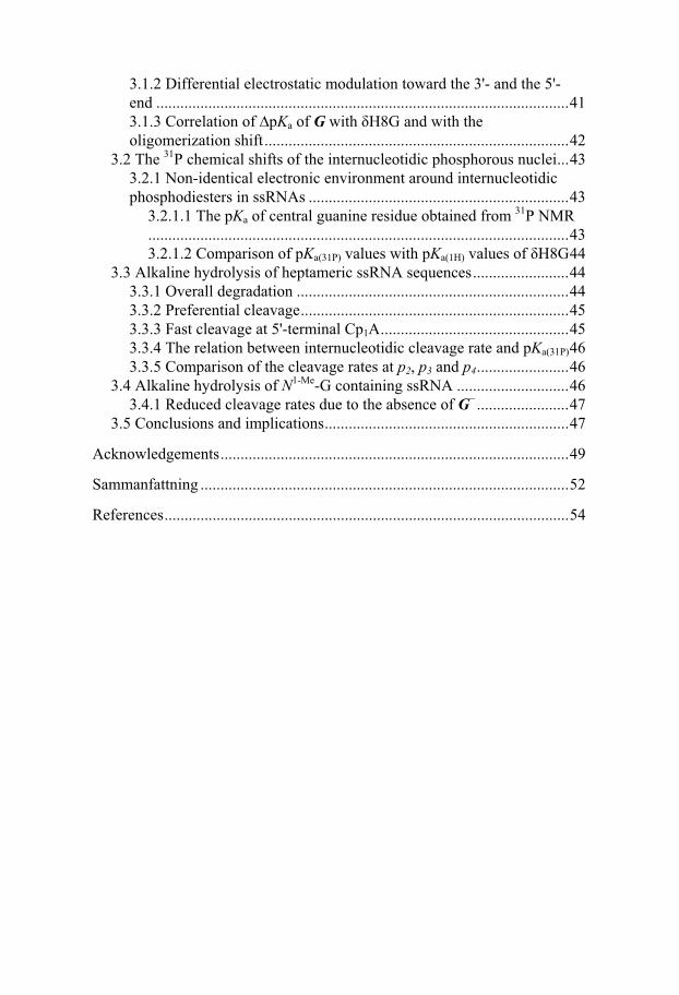
3.2 The 31P chemical shifts of the internucleotidic phosphorous nuclei...43 3.2.1 Non-identical electronic environment around internucleotidic phosphodiesters in ssRNAs .................................................................43
3.2.1.1 The pKa of central guanine residue obtained from 31P NMR.........................................................................................................43 3.2.1.2 Comparison of pKa(31P) values with pKa(1H) values of �H8G44
3.3 Alkaline hydrolysis of heptameric ssRNA sequences........................44 3.3.1 Overall degradation ....................................................................44 3.3.2 Preferential cleavage...................................................................45 3.3.3 Fast cleavage at 5'-terminal Cp1A...............................................45 3.3.4 The relation between internucleotidic cleavage rate and pKa(31P)46 3.3.5 Comparison of the cleavage rates at p2, p3 and p4.......................46
3.4 Alkaline hydrolysis of N1-Me-G containing ssRNA ............................46 3.4.1 Reduced cleavage rates due to the absence of G� .......................47
3.5 Conclusions and implications.............................................................47
Acknowledgements.......................................................................................49
Sammanfattning ............................................................................................52
References.....................................................................................................54

Abbreviations
A Adenin-9-yl or adenosine Ac Acetyl ANA Arabino nucleic acid AON Antisense oligonucleotide Aza-ENA 2'-N,4'-C-Ethylene bridged nucleoside Azetidine 1',2'-Aminoethylene bridged nucleoside B Any nucleobase Bn Benzyl BNA Bridged nucleic acid C Cytosin-1-yl or cytidine CD Circular dichroism CeNA Cyclohexene nucleic acid CMV Cytomegalovirus DNA Deoxyribonucleic acid ds Double stranded DTT Dithiothreitol EDTA Ethylenediaminetetraacetic acid ENA 2'-O,4'-C-Ethylene bridged nucleic acid FANA 2'-Deoxy-2'-fluoro-�-D-arabino nucleic acid G Guanin-9-yl or guanosine HDV Hepatitis delta virus HNA Hexitol nucleic acid HOMO-DNA Hexapyranosyl nucleic acid K Kelvin kcat Turnover number Km Michaelis constant LNA Locked nucleic acid MALDI-TOF Matrix-assisted laser desorption/ionization-time of flight MOE 2'-O-Methoxyethyl MF Morpholino mRNA Messenger RNA MS Modification site NMR Nuclear magnetic resonance ODN Oligodeoxynucleotide ON Oligonucleotide Oxetane (1',3'-O-Anhydro-�-D-psicofuranosyl) nucleosides

PAGE Polyacrylamide gel electrophoresis PNA Peptide nucleic acid PO Phosphodiester PS Phosphorothioate RNA Ribonucleic acid RNase H Ribonuclease H siRNA Small interfering ribonucleic acids ss Single strand SVPDE Snake venom phosphodiesterase T Thymin-1-yl or thymidine TFO Triplex forming oligonucleotide Tm Melting temperature U Uracil-1-yl or uridine UV Ultraviolet Vmax Maximum velocity VS Varkud satellite
13
1 Introduction
1.1 General introduction to nucleic acids
Nucleic acids are essential biopolymers in the transmission, expression, and conservation of the genetic information. Deoxyribonucleic acid (DNA) acts mainly as the carrier of genetic information whereas ribonucleic acid (RNA) transports the genetic information from the DNA to the ribosome where another essential biopolymer, the protein, is manufactured by means of translation. In this process RNA has many functions. It brings the genetic information in the form of messenger (mRNA) to the ribosome which is also largely made up of RNA (called rRNA). It delivers the amino acids accord-ing to the genetic code triplet on the mRNA in the form of tRNA. This flow of genetic information constitutes the central dogma of molecular biology.1
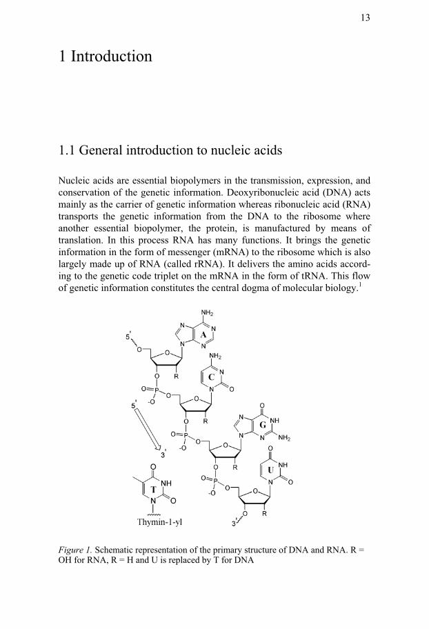
Figure 1. Schematic representation of the primary structure of DNA and RNA. R = OH for RNA, R = H and U is replaced by T for DNA

14
RNA also acts as a carrier of genetic information in RNA viruses and ret-roviruses. Apart from this, certain RNA molecules are known to act as en-zymes either individually or in combination with proteins. This diverse ac-tivity of the RNA molecule led to the ‘RNA World’2,3 hypothesis which considers RNA as the only molecule, in a primordial age, that can serve not only as a carrier of genetic information from generation to generation, but also can carry out complex catalytic activities needed for their own replica-tion and other complex reactions.4,5,6
1.1.1 Structure of nucleic acids The primary structure of nucleic acids contains three parts, a pentofuranose sugar, phosphate and nucleobases. A 2'-deoxy-�-D-ribose (in DNA) or �-D-ribose (in RNA) sugar ring attached to a heterocyclic aromatic nucleobase through an N-glycosidic bond [Figure 1] is refered to as a nucleoside. The nucleoside units are connected through phosphodiester linkages from the 3'- carbon atom of one sugar unit to the 5'- carbon of the next sugar. The nu-cleobases found in DNA are the pyrimidines cytosine (C) and thymine (T) and the purines adenine (A) and guanine (G) whereas in RNA thymine is replaced by uracil (U) [Figure 1]. The sequence of nucleobases designates the primary structure of the nucleic acids.
Figure 2. Schematic representation of a few naturally occuring modified nucleo-bases
15
Apart from the aforementioned nucleobases, a large number of modified nucleobases are also found in naturally occurring nucleic acids, mostly in RNAs7,8 and particularly in transfer RNAs (tRNA) [Figure 2]. Some are modifications of the most common natural nucleobases whereas others are hypermodified.
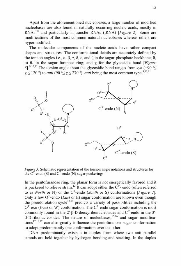
The molecular components of the nucleic acids have rather compact shapes and structures. The conformational details are accurately defined by the torsion angles i.e., �, �, �, �, �, and � in the sugar-phosphate backbone; 0 to 4 in the sugar furanose ring; and for the glycosidic bond [Figure 3].9,10,11 The torsion angle about the glycosidic bond ranges from syn (�90 º� � 120 º) to anti (90 º� � 270 º), anti being the most common type.9,10,11
Figure 3. Schematic representation of the torsion angle notations and structures for the C2'-endo (S) and C3'-endo (N) sugar puckerings
In the pentofuranose ring, the planar form is not energetically favored and it is puckered to relieve strain.12 It can adopt either the C3'- endo (often referred to as North or N) or the C2'-endo (South or S) conformations [Figure 3]. Only a few O4'-endo (East or E) sugar conformation are known even though the pseudorotation cycle13,14 predicts a variety of possibilities including the O4'-exo (West or W) conformation. The C2'-endo sugar conformation is most commonly found in the 2'-�-D-deoxyribonucleosides and C3'-endo in the 3'-�-D-ribonucleosides. The nature of nucleobases,15,16 and sugar modifica-tions17,18,19 can also greatly influence the pentofuranose sugar conformation to adopt predominantly one conformation over the other.
DNA predominantly exists a in duplex form where two anti parallel strands are held together by hydrogen bonding and stacking. In the duplex
16
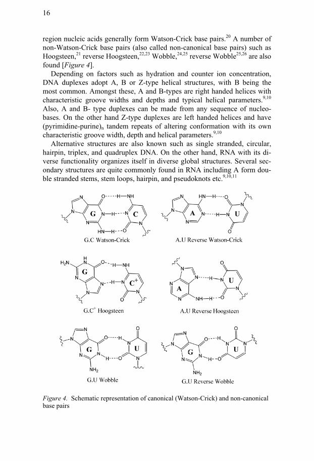
region nucleic acids generally form Watson-Crick base pairs.20 A number of non-Watson-Crick base pairs (also called non-canonical base pairs) such as Hoogsteen,21 reverse Hoogsteen,22,23 Wobble,24,25 reverse Wobble25,26 are also found [Figure 4].
Depending on factors such as hydration and counter ion concentration, DNA duplexes adopt A, B or Z-type helical structures, with B being the most common. Amongst these, A and B-types are right handed helices with characteristic groove widths and depths and typical helical parameters.9,10 Also, A and B- type duplexes can be made from any sequence of nucleo-bases. On the other hand Z-type duplexes are left handed helices and have (pyrimidine-purine)n tandem repeats of altering conformation with its own characteristic groove width, depth and helical parameters.9,10
Alternative structures are also known such as single stranded, circular, hairpin, triplex, and quadruplex DNA. On the other hand, RNA with its di-verse functionality organizes itself in diverse global structures. Several sec-ondary structures are quite commonly found in RNA including A form dou-ble stranded stems, stem loops, hairpin, and pseudoknots etc.9,10,11
Figure 4. Schematic representation of canonical (Watson-Crick) and non-canonical base pairs

17
1.1.2 Quantification and analysis The nucleobases have UV absorption maxima near 260 nm with extinction coefficients of about 10,000 M�1 cm�1. Interactions among the nucleobases such as stacking causes up to 40% reduction of the absorption for double stranded polynucleotides and 25% reduction for single stranded polynucleo-tides compared to that of the sum of the absorptions of the nucleotide units.* The extinction coefficient of a single stranded polynucleotide is estimated from the sum of its components taking into consideration the nearest neighbor approximation.27 The equation requires that the extinction coeffi-cients of the nucleotides and the component dinucleotide phosphates are known and is used routinely with an estimated error of ±10% accuracy [Equation 1].
�(ApCpGpUp...ApG) = 2[�(ApC) + �(CpG) + � (GpU) +…�(ApG)]
� [�(Cp) + �(Gp) + �(Up)… + �(Ap)] Equation 1
The extinction coefficient of a double stranded nucleic acid is estimated
with the help of a quadratic equation which involves Adenine-Thymidine (A.T) or Adenine-Uridine (A.U) base composition fraction, fA.T.9
This method of quantification had been a very useful tool for routine con-centration measurements throughout the studies presented in this thesis and was extensively used for the concentration correction of the small nucleo-tidic fragments in the alkaline hydrolysis experiments (discussed in the later part of the thesis).
Apart from the quantification of nucleic acids in solution, UV spectros-copy is often used for monitoring their melting behavior. A plot of UV ab-sorption versus temperature is known as a melting curve and is usually co-operative in nature. The inflection point of the sigmoidal melting curve is known as the melting temperature.
Though the effect of structure on the UV absorption of oligonucleotides is complex and only the basic interpretations can be made, UV spectroscopy is commonly used to study the effects of chemical modifications on the thermal stability and the higher order structures of nucleic acids.10,11 Thus UV based thermal melting studies has been very useful for the interpretation of the target affinity of an antisense oligonucleotide (AON) towards the comple-mentary RNA, discussed in this thesis.
* An absorbance in a polymer that is weaker than the sum of the absorbances of the compo-nents is known as hyperchromism.

18
Other spectroscopic techniques such as fluorescence, circular dichroism (CD), infrared, Raman, and nuclear magnetic resonance (NMR) as well as X-ray diffraction are also well used in nucleic acid structural analysis. Amongst these, fluorescence spectroscopy is increasingly being utilized in nucleic acids based detection techniques and diagnostics. On the other hand NMR spectroscopy and X-ray diffraction techniques are extensively used in nucleic acid structure determination and analysis of interactions with small molecules, proteins etc.
In the works presented in this thesis, NMR spectroscopy (1H, 13C and 31P) is extensively used not only for the characterization and analysis but also for the studies of physicochemical properties of nucleotides and small oligonu-cleotides.
1.1.3 Reactivity of nucleic acids The groups that are directly involved in chemical reactions of the nucleic acids are the nucleobase (the site for hydrogen bonding and stacking), the 2'-OH group of RNA (the site for transesterifications in the splicing reaction, and RNA catalysis, also discussed in the later part of the introduction), and the internucleotidic phosphodiesters of RNA (the site of cleavage reactions).
Figure 5. The pKa values of nucleobases
All these different reaction sites contain ionizable groups with a variety of pKa values. The wide range of pKa values for the nucleobases is shown in Figure 5. The 2'-OH group of RNA has a pKa value of around 12.0 – 14.028,29 whereas the internucleotidic phosphate group has a pKa of 1.530,31 and the terminal phosphate has pKa values of 1.5 and 6.5 [Equation 2].10

19
Equation 2 The reactivity of nucleic acids is greatly influenced by the primary struc-
ture and the environmental factors such as temperature, pH, salt concentra-tion etc. This is because the primary structure in combination with the envi-ronment, and metal ion cofactors determine the secondary and tertiary struc-tures, which in tern influences the molecular recognition as well as the reac-tivity.
1.2 Molecular recognition of nucleic acids
1.2.1 Interactions with metal ions and small molecules Many small molecules, synthetic and naturally occuring, as well as metal ions interact strongly with nucleic acids. Metal ions being positively charged interact electrostatically with the negatively charged phosphate oxygens of the backbone of nucleic acids in a nonspecific manner. In addition, atom specific, group specific, base specific, sequence specific, as well as secon-dary/tertiary structure specific binding of metal ions to nucleic acids are known. Metal ion binding is also known to be involved in nucleic acid bio-synthesis, processing and degradation, as well as genetic information transfer and gene expression, mutagenesis, chromosomal abnormalities, carcinogene-sis etc.10,11,32
A number of small organic molecules are known for reversible interac-tions with the nucleic acids such as electrostatic interactions along the exte-rior of the helix, direct interactions with the edges of the base pairs (known as groove binding), and intercalation between the base pairs. On the other hand, the intrinsic chemical nature of the building units, as discussed above, makes nucleic acids susceptible to various chemical reactions such as oxida-tion/reduction, hydrolysis, metallation, and photochemical reactions.11,32
Interactions with small molecules and metal ions can lead to inactivation or destruction of nucleic acids. Understanding interactions between nucleic acids and small molecules and metal ions can lead to improvement of exist-ing chemotherapeutic treatments by antiviral, antitumor, and anticancer agents.11,32

20
Apart from the aforementioned important aspects, this area of research fu-eled the development of artificial nucleases,33 usually acting in a combina-tion of metal ions with small organic molecules with the aim to destruct tar-geted nucleic acids.
1.2.2 Interactions with proteins Many biological processes such as transcription, replication, and recombina-tion depend on interactions between nucleic acids and proteins, both specific and non-specific. DNA sequences that are active when bound to proteins in sequence specific manner can be classified into two groups, those bound to gene regulatory proteins34,35,36 and those that are substrates for polymerase, topoisomerase, recombinase. 37,38
RNA can interact with proteins through hydrophobic stacking, hydrogen bonding, and salt bridge formation with high sequence specificity. Specific uridine rich ssRNA binding to sex-lethal protein,39 3'-mRNA poly(A) tail recognition by poly(A) binding protein (PABP),40 and the ssRNA region which binds to tRNA binding attenuation protein (TRAP)41 are examples of such sequence-specific interactions.
1.2.3 Interactions with short oligonucleotides Nucleic acids have received much attention as potential therapeutic agents. They can be used, in principle, to replace defective nonfunctioning genes and also to prevent the expression of unwanted genes or the specific mRNA which produces unwanted proteins. The specific interactions found between complementary strands, by means of Watson-Crick base pairing, suggests that oligonucleotides can be used as therapeutic agents to target DNA (anti-gene) or RNA, particularly mRNA, (antisense). With the discoversy of solid phase synthesis, together with already existing possibilities of chemical ma-nipulation, it became practically possible to screen modified AONs in a cost effective manner.
In the antigene approach, an artificial short DNA sequence, a triplex forming oligonucleotides or TFOs, is used to target double stranded genomic DNA through major groove binding by formation of a triple helix or by strand invasion.42,43,44 The TFO typically utilizes Hoogsteen or reverse Hoogsteen base pair [Figure 4] in order to bind to the DNA double strand. In spite of the apparent simplicity in the concept of controlling gene expression directly, there has been limited success until now. This is because of the difficulties in target accessibility due to the proteins associated with the ge-nome and the need for homopurine-homopyrimidine tracts for triplex forma-tion.45
21
1.3 Targeting mRNA There are mainly three types of anti-mRNA strategies known. The first one utilizes single stranded antisense oligonucleotides (AON), is the most vali-dated approach and was first discovered by Zamecnik and Stephenson.46,47 The second approach is based on catalytically active oligonucleotides re-ferred to as ribozymes, first discovered by Thomas Cech48,49 and Sidney Altman.50 RNA interference,51,52 induced by small interfering RNA (siRNA) molecules, is the third method which has caught much attention re-cently.53,54,55 Further details of the RNA interference is beyond the scope of this introduction.
Apart from the above mentioned strategies, DNAzymes56,57 (DNA mole-cules acting catalytically) represent an increasingly interesting field of re-search toward the nucleic acid based therapeutics.
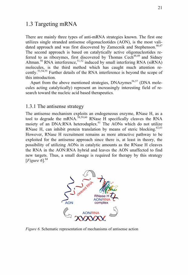
1.3.1 The antisense strategy The antisense mechanism exploits an endogeneous enzyme, RNase H, as a tool to degrade the mRNA.58,59,60 RNase H specifically cleaves the RNA moiety of an DNA:RNA heteroduplex.61 The AONs which do not utilize RNase H, can inhibit protein translation by means of steric blocking.62,63 However, RNase H recruitment remains as more attractive pathway to be exploited for the antisense approach since there is, at least in theory, the possibility of utilizing AONs in catalytic amounts as the RNase H cleaves the RNA in the AON:RNA hybrid and leaves the AON unaffected to find new targets. Thus, a small dosage is required for therapy by this strategy [Figure 6].64
Figure 6. Schematic representation of mechanisms of antisense action

22
1.3.1.1 Chemically modified nucleotides developed for the antisense strategy A synthetic short oligonucleotide to be used successfully as a therapeutic agent must fulfill a number of criterions. It must have high target affinity, a property related high target specificity. It must have high nuclease stability which is related to low toxicity and low effective dosage. It must also show good bioavailability which includes good cellular uptake and favorable pharmacokinetics.65,66
Since unmodified oligonucleotides are degraded very rapidly inside the cell, chemical modifications have been attempted to increase the resistance of the AONs to enzymatic degradation. In oligonucleotides, the nucleobase,
Figure 7. Nucleotide analogues corresponding to the first, second, and third genera-tion of AONs
the sugar and the phosphate backbone are the most common targets for chemical modifications. In spite of the increased target affinity achieved
23
with modified nucleobases67 when incorporated into AONs, their therapeutic potential in vivo have not been reported.65
The backbone modified phosphorothioate AONs (PS) where the non-bridging oxygen atom in the phosphodiester linkage was replaced by sulfer atom,68,69 showed increased resistance toward nucleases. However, lower target affinity compared to the unmodified oligonucleotide,70 non-specific interactions with proteins causing to unwanted side effects lead to the devel-opment of the second generation of modified AONs [Figure 7].
The second generation of modified AONs contains 2'-O-alkylated sugar moieties.18 The combination of a phosphorothioate (PS) backbone with a 2'-O-alkylated sugar moiety lead to increased nuclease resistance and enhanced target affinity.18 However, these modified AONs failed to recruit RNase H because of their RNA like sugar conformation that induced an A type duplex structure in the AON:RNA heteroduplex. This gave rise to the ‘gap-mer’ strategy in which the modified nucleotides are incorporated in a gap of 6 to 8 (or suitable) 2'-deoxyribonucleotide units.18,71,72
The lessons from first and second generations lead to the third generation of modified AONs with increased target affinity, high nuclease stability and good RNase H recruiting properties. Peptide nucleic acids (PNA),73,74,75 ara-bino nucleic acids (ANA and FANA),76,77,78 hexitol nucleic acids (HNA),79 hexapyranosyl nucleic acids (HOMO-DNA),80,81 cyclohexene nucleic acids (CeNA),82 conformationally constrained nucleic acids (bridged/locked nu-cleic acid, BNA83/LNA;84 ethylene bridged nucleic acids ENA85,86 etc.), morpholino modified oligonucleotides (MF),87,88,89 N3'-P5' phosphorami-dates (NP),90,91 and tricyclic DNA (tcDNA)92,93 are important examples of third generation modified AONs. In spite of their many favorable properties, the challenges still remaining are to improve the cellular uptake and identify-ing the accessible sites94 in the target mRNA. However, the delivery issue has been addressed by several strategies based on, for example, liposomes, dendrimers, nano particles, viral vectors etc.95 On the other hand; methods utilizing oligonucleotide microarrays96 and oligonucleotide libraries97 are increasingly being applied to accessibility issue.
Nucleic acid based therapeutics have yielded two FDA approved drugs so far, Vitravene®, to treat the inflammatory viral infection of the eye caused by cytomegalovirus (CMV) and Macugen®, for age-related macular degenera-tion (AMD).95,98 Another drug, Genasense®, targeting Bcl-2, a protein ex-pressed in cancer cells to protect against chemotherapy, is waiting for FDA approval.95,99 Though the "real" mechanism of action of these drugs is still debated,100 more than 30 antisense based drugs are in different phases of clinical trials.95,98

24
1.3.2 RNA catalysis The fact that RNA can act as catalyst was first discovered in the early 80’s.48,49,50 Several RNA sequences with enzymatic activities are known today, Group I and Group II introns, hepatitis delta virus (HDV), hairpin, hammerhead, Varkud satellite (VS) ribozymes are few examples.3,101 Many investigations have been undertaken in order to understand how RNA with its fewer reactive groups compared to those of proteins can act as a biocata-lyst. Ribozyme reactions can be broadly classified into two types on the ba-sis of the termini generated during self-cleavage reactions. Ribozymes that use an exogenous nucleophile leave 2',3'-cis-diol and 5'-phosphate termini [Figure 8]; the group I intron and RNase P are examples of this type. Ri-bozymes that use the vicinal 2'-hydroxyl as the nucleophile leave 2',3'-cyclic phosphate and 5'-hydroxyl termini [Figure 8].101,102
Figure 8. Mechanism of phosphodiester cleavage in large (A) and small ribozymes (B)
An increasing number of RNAs wih catalytic activities and a developing understanding of their mechanism of action have generated much interest for harnessing their catalytic activity for hydrolyzing RNA molecules.
25
1.4 Overview of the thesis
This thesis presents the biochemical evaluation of chemically modified short oligonucleotides in terms of antisense based RNA targeting. Chemical modi-fications at pentofuranose sugar have been used for this purpose. The modi-fications are 2'-O-Me-T, 2'-O-MOE-T, oxetane-T, LNA-T, azetidine-T, aza-ENA-T, carbocyclic-ENA-T, and carbocyclic-LNA-T.
The RNase H recruitment properties of these modified oligonucleotides, duplexed with the target RNA, have been evaluated along with the thermal melting studies as a measure of target affinity. Also, the modified oligonu-cleotides were subjected to studies for resistance towards snake venom phos-phodiesterase (SVPDE) and human serum.
In another study, the physicochemical properties of unmodified model short RNA sequences were addressed. It was demonstrated how changes in primary structure can modulate the intrinsic properties along a single strand. Finally, alkaline hydrolysis was used in a model chemical cleavage study, to study the impact of small structural changes on the reactivity of the internu-cleotidic phosphodiester bonds.

26
2 Evaluation of Modified AONs (Paper I-III)
The most used and validated antisense mechanism is that of RNase H re-cruitment and a synthetic AON has to show RNase H eliciting properties in addition to high affinity, good bioavailability and nuclease resistance. The PS oligonucletides were found to be toxic and also showed non specific in-teraction with proteins.72 The second generation of modified AONs contain-ing 2'-O-Me and 2'-O-MOE were attempted were found to be less toxic and also showed slightly higher target affinity towards complementary RNA.18 Pradeepkumar et al. earlier developed North-East conformationally con-strained 1',2'-oxetane modified nucleosides, which did not show increased target affinity towards complementary RNA (��m ~���to �3 °C with pyrimidine nucleotides and almost no decrease with purine nucleo-tides).103,104,105,106
Figure 9. Schematic representation of nucleoside analogues incorporated into an-tisense oligonucleotides used in the present study
27
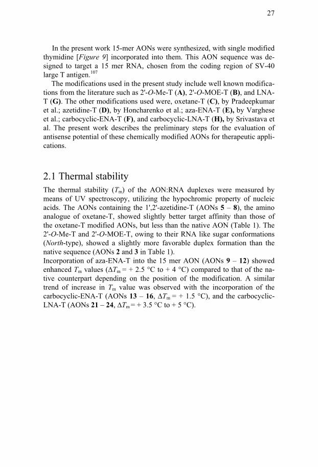
In the present work 15-mer AONs were synthesized, with single modified
thymidine [Figure 9] incorporated into them. This AON sequence was de-signed to target a 15 mer RNA, chosen from the coding region of SV-40 large T antigen.107
The modifications used in the present study include well known modifica-tions from the literature such as 2'-O-Me-T (A), 2'-O-MOE-T (B), and LNA-T (G). The other modifications used were, oxetane-T (C), by Pradeepkumar et al.; azetidine-T (D), by Honcharenko et al.; aza-ENA-T (E), by Varghese et al.; carbocyclic-ENA-T (F), and carbocyclic-LNA-T (H), by Srivastava et al. The present work describes the preliminary steps for the evaluation of antisense potential of these chemically modified AONs for therapeutic appli-cations.
2.1 Thermal stability The thermal stability (Tm) of the AON:RNA duplexes were measured by means of UV spectroscopy, utilizing the hypochromic property of nucleic acids. The AONs containing the 1',2'-azetidine-T (AONs 5 – 8), the amino analogue of oxetane-T, showed slightly better target affinity than those of the oxetane-T modified AONs, but less than the native AON (Table 1). The 2'-O-Me-T and 2'-O-MOE-T, owing to their RNA like sugar conformations (North-type), showed a slightly more favorable duplex formation than the native sequence (AONs 2 and 3 in Table 1). Incorporation of aza-ENA-T into the 15 mer AON (AONs 9 – 12) showed enhanced Tm values (�Tm = + 2.5 °C to + 4 °C) compared to that of the na-tive counterpart depending on the position of the modification. A similar trend of increase in Tm value was observed with the incorporation of the carbocyclic-ENA-T (AONs 13 – 16, �Tm = + 1.5 °C), and the carbocyclic-LNA-T (AONs 21 – 24, �Tm = + 3.5 °C to + 5 °C).

28
Table 1. Thermal denaturationa of modified and the native AONs, duplexed with the complementary RNA and DNA strands. The target RNA is 5'-r(GAAGAAAAAAUGAAG)-3', and the target DNA is 5'-r(GAAGAAAAAAUGAAG)-3'.
a Tm values were measured as the maximum of the first derivative of the melting curve (A260 vs temperature) recorded in medium salt buffer (60 mM Tris-HCl at pH 7.5, 60 mM KCl, 0.8 mM MgCl2, and 2 mM DTT) in the temperature range 20-70 C using 1 M concentrations of the two complementary strands. �Tm = Tm relative to RNA complement, and �Tm* = Tm relative to DNA complement. T-6-carbo = carbocyclic-ENA-T; T-5-carbo = carbocyclic-LNA-T.
AON
Sequence
Tm/°C with RNA
�Tm
Tm/°C with DNA
�Tm*
1 3'-d(CTTCTTTTTTACTTC)-5' 44 … 45 …
2 3'-d(CTTCTTTTTT-O-Me ACTTC)-5' 44.5 +0.5 41 �4
3 3'-d(CTTCTTTTTT-O-MOE ACTTC)-5' 44.5 +0.5 40.5 �4.5
4 3'-d(CTTCTTTTTT-oxe ACTTC)-5' 39 �5 40.5 �4.5
5 3'-d(CTT-azeCTTTTTTACTTC)-5' 38.5 �5.5 41 �4
6 3'-d(CTTCTT-azeTTTTACTTC)-5' 40 �4 39.5 �5.5
7 3'-d(CTTCTTTT-aze
TTACTTC)-5' 40 �4 39 �6
8 3'-d(CTTCTTTTTT-aze
ACTTC)-5' 40 �4 41 �4
9 3'-d(CTT-aza-ENA
CTTTTTTACTTC)-5' 48 +4 44.5 �0.5
10 3'-d(CTTCTT-aza-ENA
TTTTACTTC)-5' 46.5 +2.5 42.5 �2.5
11 3'-d(CTTCTTTT-aza-ENA
TTACTTC)-5' 47.5 +3.5 42 �3
12 3'-d(CTTCTTTTTT-aza-ENA
ACTTC)-5' 48 +4 42 �3
13 3'-d(CTT-6-carbo
CTTTTTTACTTC)-5' 45.5 +1.5 43.5 �1.5
14 3'-d(CTTCTT-6-carbo
TTTTACTTC)-5' 45.5 +1.5 39.5 �5.5
15 3'-d(CTTCTTTT-6-carbo
TTACTTC)-5' 45.5 +1.5 40.0 �5.0
16 3'-d(CTTCTTTTTT-6-carbo
ACTTC)-5' 45.5 +1.5 39.5 �5.5
17 3'-d(CTT-LNA
CTTTTTTACTTC)-5' 48 +4 47 +2
18 3'-d(CTTCT T-LNA
TTTTACTTC)-5' 49 +5 46.5 +1.5
19 3'-d(CTTCTTTT-LNA
TTACTTC)-5' 49 +5 45.0 0
20 3'-d(CTTCTTTTTT-LNA
ACTTC)-5' 49 +5 46 +1
21 3'-d(CTT-5-carbo
CTTTTTTACTTC)-5' 47.5 +3.5 45 0
22 3'-d(CTTCTT-5-carbo
TTTTACTTC)-5' 49 +5 44 �1
23 3'-d(CTTCTTTT-5-carbo
TTACTTC)-5' 48 +4 44 �1
24 3'-d(CTTCTTTTTT-5-carbo
ACTTC)-5' 47.5 +3.5 43.0 �2

29
2.2 RNase H recruitment properties As mentioned above, to function in the classical antisense mechanism,
AONs have to be able to recruit RNase H. Therefore, all the AON:RNA duplexes containing modified AONs were subjected to digestion by Es-cherichia coli RNase H1. All of the modified AONs hybridized with the complementary RNA were found to recruit the E. coli RNase H1 with vary-ing potential and patterns. The target RNA in the unmodified native AON 1:RNA duplex was cleaved rather randomly but with a slight preference after A-8 position [Figure 10 and Figure 11].
2.2.1 RNase H footprint pattern induced by AONs modified with 1',2'-locked nucleotide
The pattern of RNase H mediated cleavage of azetidine-T modified AON 5:RNA was found to be quite similar to the pattern of the
Figure 10. E. coli RNase H1 mediated cleavage footprints of the complementary RNA when duplexed with the native and the azetidine-T modified AONs
30
native AON 1:RNA duplex except that there was no cleavage at the A5 and A6 positions. In the azetidine-T modified AONs 6 – 7, duplexed with RNA, five nucleotides toward the 3'-end from the site opposite to the modification site (MS) were found not to be cleaved by RNase H. On the other hand for the AON 8/RNA duplex, six nucleotides toward the 3'-end from the site op-posite to the MS showed resistance toward RNase H mediated cleavage. Interestingly, all the azetidine-T modified AON 5 – 8 duplexed with RNA, showed similar cleavage patterns as those of the isosequential oxetane-T modified AON:RNA duplexes as reported earlier.103,104
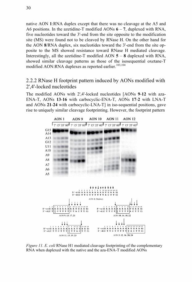
2.2.2 RNase H footprint pattern induced by AONs modified with 2',4'-locked nucleotides The modified AONs with 2',4'-locked nucleotides [AONs 9-12 with aza-ENA-T, AONs 13-16 with carbocyclic-ENA-T, AONs 17-2 with LNA-T and AONs 21-24 with carbocyclic-LNA-T] in iso-sequential positions, gave rise to uniquely similar cleavage footprinting. However, the footprint pattern
Figure 11. E. coli RNase H1 mediated cleavage footprinting of the complementary RNA when duplexed with the native and the aza-ENA-T modified AONs

31
was found to be different from those of iso-sequential AONs with 1',2'- locked nucleotides [AONs 5-8 with azetidine-T, and oxetane-T103,104]. For the AONs with modifications at position T3 from 3'-end [AONs 9, 13, 17, 21], the main cleavage site was found at A13 position of the complementary RNA unlike the iso-sequential azetidine-T modified AON 5 and iso-sequential oxetane-T103,104 modified AONs, which showed additional strong cleavage at A7 and G12 positions. In the case of modification at position T10 from 3'-end [AONs 12, 16, 20, 24], seven nucleotides toward the 3'-end from the site opposite to the modification were found to be insensitive to-ward RNase H promoted cleavage [Figure 11].
Interestingly, the RNase H enzyme could discriminate the subtle differ-ences between the flexibility of the modified sugars exhibited by the North-type 2',4'-locked system [aza-ENA, carbocyclic-ENA, LNA, and carbocyc-lic-LNA] and North-East-type 1',2'-locked system [oxetane-T, and azetidine-T]. On the other hand it could not differentiate between the hydrophobic [carbocyclic-ENA, carbocyclic-LNA] and hydrophilic [aza-ENA and LNA] character of the modified nucleotides unlike the exonucleases i.e., snake venom phophodiesterase (SVPDE) and human serum. Studies with exonu-cleases are discussed toward the end of this chapter.
The 2'-O-alkylated nucleotides [2'-O-Me-T and 2'-O-MOE-T], known to confer North-type conformation to the sugar,18 were expected to have similar effects as 2',4'-locked nucleotides, on RNase H mediated cleavage of the target RNA. The 2'-O-MOE-T modified AON 3 indeed imparted similar resistance toward the RNase H mediated cleavage of the target RNA as the iso-sequential AON counterparts containing 2',4'-locked nucleotides [AONs 12, 16, 20, and 24]. However, the RNase H cleavage pattern of 2'-O-Me-T modified AON 2 was rather like iso-sequential AON with 1',2'-locked nu-cleotide, AON 8, but with very little cleavage at A9 position. It is notewor-thy that an extra strong cleavage at A9 position was the only difference be-tween iso-sequential AONs with 2',4'-locked nucleotide [AONs 12, 16, 20, and 24] and 1',2'-locked nucleotide [AONs 4103,104 and 8].
2.3 RNase H cleavage kinetics
2.3.1 Pseudo first order cleavage rates The pseudo first order cleavage rate constants of RNase H digestion were determined by densitometric quantification of gels and subsequently plotting of the uncleaved RNA fraction as a function of time. The reaction rate con-stants were obtained by fitting the degradation curves to single-exponential decay functions.108 The relative cleavage rates were found to be very similar to that of the native counterpart irrespective of the type and the site of modi-

32
fication in the AON strand [Papers I, II and III]. However, pseudo first order cleavage kinetics is a rather crude simplification to an enzymatic reac-tion. Therefore, a detailed Michaelis-Menten kinetics study was performed for selected AONs.
2.3.2 Michaelis-Menten kinetics We chose iso-sequentially modified AONs [2, 3, 8, 12] for the detailed Michaelis-Menten kinetic studies in order to understand the impact of the modifications on the binding affinity and the catalytic activity of the RNase H [Paper I]. Kinetic parameters were subsequently compared with those of the native AON 1 and earlier reported oxetane-T modified AON 4.105
The catalytic constant (kcat), which represents the maximum number of substrate molecules converted to products per active site per unit time,109 is greater for the reaction with the modified AON:RNA duplexes [1.4 – 1.6 fold for 2'-O-Me-T (AON 2), 2'-O-MOE-T (AON 3), and azetidine-T (AON 8) and ~2.6-fold for aza-ENA-T (AON 12)] than for that with the native AON 1:RNA duplex. However, the catalytic effectivity (kcat/Km) for the hy-drolysis of all chemically modified substrates was found to be lower than that for the native substrate, without exception [~1.1 fold less for 2'-O-Me-T (AON 2), ~2.6-fold less for 2'-O-MOE-T (AON 3), ~1.4 fold less for oxetane-T (AON 4), ~2.7 fold less for azetidine-T (AON 8), and ~3.2 fold less for aza-ENA-T (AON 12)] [Table 5, Figure 7 in Paper I]. This was due to the fact that introduction of a modified moiety to the AON decreased the enzyme binding affinity (1/Km) [1.3 – 1.7 fold less for 2'-O-Me-T (AON 2) and oxetane-T (AON 4), 3.8 – 4.2 fold less for azetidine-T (AON 8) and 2'-O-MOE-T (AON 3), and ~8.3 fold less for aza-ENA-T (AON 12)].
2.4 Resistance to nuclease degradation Native DNA is degraded very fast in a cellular system. Replacement of the phosphodiester (PO) backbone with the phosphorothioates (PS) was shown to prevent the AONs from fast degradation inside the cell. However, these modifications were probably less stable than they were initially believed66 and moreover led to toxicity problems related to non-specific target bind-ing.95 Therefore, one of the major problems to be resolved in order to allow successful applications of AONs in therapeutics is their stability in living cells.
It is primarily the exonucleases, with modest contributions from endonu-cleases, which degrade the AONs.66 Therefore, the modified AONs were treated with snake venom phosphodiesterase (SVPDE) from Crotalus ada-manteus, which is a 3'-exonuclease, and human blood serum, which contains several 3'-exonucleases.

33
2.4.1 Resistance of modified AONs to snake venom phosphodiesterase degradation The unmodified native AON 1 was completely degraded within 1 h, not to our surprise. Also the 2'-O-Me-T modified AON 2, 2'-O-MOE-T modified AON 3, and all LNA-T modfied AONs 17 – 20 showed similarly poor resis-tance towards SVPDE hydrolysis. The azetidine-T containing AONs 5 – 7, showed relatively higher stability, compared to those of the aforementioned unmodified native and modified AONs. The azetidine-T containing AON 8 degraded as fast as the native AON 1 [Figure 12].
All the other modified AONs containing aza-ENA-T, carbocyclic-ENA-T, and carbocyclic-LNA-T nucleotides were stable for more than 48 h in presence of SVPDE. It is important to note that the full length modified AONs were hydrolyzed within the initial hours of treatment with SVPDE. The stable fractions were found to be the ones cleaved on the 3'-side of the modification site (MS) in the case of azetidine-T containing AONs 5 – 7, and at the next phosphodiester bond on the 3'-side of the MS in the case of aza-ENA-T, carbocyclic-ENA-T, and carbocyclic-LNA-T containing AONs 9 – 12, 13 – 16, and 21 – 24 respectively. However, aza-ENA-T modified AONs were further hydrolyzed at the phosphodiester on the 3'-side of the MS with increasing time. It can be concluded that the carbocyclic modifica-tions in general imparted high stability to the AONs against SVPDE.
Figure 12. Autoradiograms of 20% denaturing PAGE showing degradation pattern of 5'-32P-labeled AONs in SVPDE. The numbering of the lanes represents the time of incubation with enzyme (h)

34
2.4.2 Stability in human serum The native AON 1 and the 2'-O-Me-T modified AON 2 were found to be degraded completely within 4 h of treatment with human serum. The 2'-O-MOE-T modified AON 3, the azetidine-T modified AON 8 and all the LNA-T modfied AONs 17 – 20 were somewhat more stable than the native coun-terpart but completely degraded within 12 h of treatment with human serum. All other modified AONs 5 – 7, 9 – 12, and 21 – 24 showed good stability, and were not completely degraded by 48 h. The cleavage pattern was found to be comparable to those found after the treatment with SVPDE for all the stable AONs. However, presence of phosphodiesterases in the human serum cleaved the radio labeled 5'-phosphate and made the quantification impossi-ble.
2.5 Conclusions and implications As stated above, extensive comparative studies are very important in or-
der for step-by-step evaluation of the chemical modifications necessary in the development of successful therapeutic agents. The isosequential AONs with 1',2'-locked nucleotides showed lower Tm with complementary RNA. This was expected as a 1',2'-lock in the sugar residue is known to impose strain in the glycosidic torsion () and thus it disrupts the linearity required for hydrogen bonding.110 On the other hand, a 2',4'-lock in the sugar moiety arrests the sugar in the absolute North-type conformation and does not im-pose such strain in the glycosidic torsion. Therefore AONs with 2',4'-lock nucleotides showed much improvement in Tm when duplexed with comple-mentary RNA. The order of stability was found to be LNA-T carbocyclic-LNA-T aza-ENA-T > carbocyclic ENA-T > OMe-T � OMOE > native > azetidine-T > oxetane T, in the AON:RNA duplexes.
The RNase H could uniquely differentiate between different types of modification in the AON:RNA hybrid duplexes, even though the global du-plex structure remained unchanged, measured by CD spectroscopy.103,104,105 The 1',2' locked oxetane and azetidine modified AONs showed identical cleavage pattern, and the 2',4'-locked aza-ENA-T, LNA-T carbocyclic LNA-T and carbocyclic ENA-T showed similar pattern. Another interesting result was that the chemical difference between the carbocyclic-LNA-T and LNA-T, nucleotide with a 2'-carbon atom linked to an exocyclic methyl group instead of 2'-oxygen, could cause a significant difference in nuclease stabil-ity of the AONs. This can be an important achievement from the pharmacol-ogical perspective as enhanced life-time of the AONs in blood serum could mean reduced dosage.
It is evident from the above results that small structural and chemical changes can bring about significant differences in the intrinsic physico-

35
chemical properties which are well-recognized by the enzymes. Therefore it is important to understand the subtle changes at the molecular level. The changes may apparently be small but they are intriguing in the way they modulate the chemical reactivity of the nucleic acids. This aspect is ad-dressed in the next section of this thesis.

36
3 Sequence Dependent Physicochemical Properties of ssRNA (Paper IV – V)
The discovery that the RNA can act as an enzyme, a ribozyme, has been asubject of great interest to chemists. In the ribozyme action only some phosphodiester bonds in presence of several others, are hydrolyzed preferen-tially with the help of their intricate structural motifs. This can be induced by metal ion cofactors and enzymes.111,112,113 The phosphodiester bond cleavage, both in ribonuclease mediated RNA cleavage (i.e. RNase H) and in ribozyme action, takes place via a transesterification reaction (ribozyme mechanism is discussed in the introduction). Recently, the base-promoted RNA transesterification reaction and cleavage has been studied in several small model systems in many laboratories in order to understand the mecha-nistic detail of ribozyme activity at the molecular level. Studies of alkali promoted hydrolysis experiments in small model systems, such as mono-methyl and monoisopropyl esters of adenosine 2'- and 3'-monophosphates,114,115 dimeric RNAs116 were carried out previously in order to understand the cleavage mechanism. Various short 2'-O-methylated117,118,119 and 2'-deoxy120,121 chimeric oligonucleotide sequences containing only one reactive ribonucleotide have also been subjected to alka-line cleavage studies. Several mechanistic features have been identified as essential prerequisites for non enzymatic phosphodiester bond cleavage. The nucleophilicity of the 2'-OH (which is dependent on its pKa),122,120 the elec-trophilicity of the reacting phosphate,123,124 the in-line conformation of the attacking 2'-oxyanion and the departing 5'-oxyanion,125 and the leaving group capability of the 5'-oxyanion.123,124 It has also emerged that the in-tramolecular environment with the hydrogen bonding network, stacking, nucleobase composition around the scissile bond also play important role.126,127
It was known that the electronic environment around the phosphate group in RNA is vital for protein-RNA interactions.128 It was also known that the conformational change at the phosphate is important for facilitating the hy-drolysis in ribozymes.129 However, it was not known if some specific scissile phosphodiesters in RNA have variable charges in comparison with the other phosphates owing to some sequence-specific unique scaffolds or folding motifs. It was also not understood if the variable phosphate charges can po-tentially tune the chemical reactivity in the biologically ubiquitous trans-esterification reactions.

37
In the present work it has been shown in model short heptameric single stranded DNA and RNA sequences that the pKa of the 9-guanine residue that ionizes to form a 9-guaninyl anion, varies in a sequence dependent manner. It has been demonstrated that the effect of 9-guaninyl ion formation can be seen in the neighboring bases toward 3'- and 5'- ends in both the ssDNA and ssRNA. It has also emerged that the electronic environments and thus the reactivity of the neighboring phosphodiesters in the ssRNA sequences were affected in a sequence specific manner whereas in the ssDNA sequences they were not.
3.1 Determination of pKa values by 1H NMR It has been shown previously that the change in physicochemical character of a nucleobase can be followed by monitoring the pH dependent chemical shifts of the aromatic marker proton of the nucleobase itself as well as by monitoring the aromatic marker proton of neighboring nucleobases.130,131 In the present work, heptameric ssDNA and ssRNA sequences were designed
Figure 13. Schematic representation of the monomeric and trimeric sequences used in the present work
in such a way that, from among all the aromatic residues, only one gave rise to an anionic species under the alkaline pH. Only the G in the middle of the sequence was ionized. Similar studies were also carried out with reference

38
trimeric sequences [Figure 13], which were at the core of heptamers [Figure 14].
Figure 14. Schematic representation of the heptameric sequences used in the present work
3.1.1 Sequence dependent pKa modulation a G� ion formation Titrations in the pH range 6.6 – 12.5, of the model heptameric sequences, 6a – 9a and 6b – 9b as well as of their reference trimeric sequences, 2a – 5a
39
and 2b – 5b were performed. The apparent pKa1† (for detailed specification
see paper IV) of G in the group of four trimeric ssDNAs 2a – 5a varied from 9.49 ± 0.01 to 10.24 ± 0.01 (�pKa1 = 0.75), whereas in the group of four heptameric ssDNAs 6a – 9a, the apparent pKa1 of G varied from 10.39 ± 0.01 to 11.06 ± 0.01, �pKa1 = 0.67 [Table 2]. On the other hand, the apparent pKa1 of G in the four trimeric ssRNAs 2b – 5b varies from 10.03 ± 0.01 to 10.25 ± 0.01 (�pKa1 = 0.22), while in a group of four heptameric ssRNAs 6b – 9b the apparent pKa1 varied from 10.09 ± 0.02 to 10.58 ± 0.01 (�pKa1 = 0.49).
A variation in apparent pKa1 values of G was observed within the set of trimeric or heptameric ssDNA and ssRNA sequences. Though the phosphate charges and the pentose sugar units within each group of trimers or heptam-ers remain the same, the variation was observed most likely because the pseudoaromatic character of G within each group was changed due to differ-ent sequence context. On the other hand, when the apparent pKa1 variations of the G in ssDNA trimers were compared with those of the corresponding ssDNA heptamers and, the ssRNA trimers with those of the corresponding ssRNA heptamers, the effect of different microenvironments were evident. The differences in the microenvironments were due to the variable stacking owing to the respective nearest-neighbors and chain lengths.
† pKa, as is used here, is of the monomeric unit, Etp(d/rG)pEt (1a/1b). Apparent pKa1 is the pKa arising in presence of electrostatic interaction of G with additional neighboring electronic groups such as additional nucleobases, phosphates, and the pentose-sugar units across the single-stranded nucleic acids. Apparent pKa2 is the pKa which is seen from the marker protons in the neighboring nucleobases (A or C) in the trimeric and heptameric ssDNA/ssRNA se-quences.

40
Table 2. Comparison of pKaa values of G in ssDNAs 2a – 9a and in ssRNAs 2b – 9b
as well as in their monomeric counterparts EtpdGpEt (1a) and EtpGpEt (1b)
2'-Deoxy units 1H pKa 2'-Ribo units 1H pKa (1a) H8G 9.59 (± 0.01) (1b) H8G 9.29 (± 0.01)
dA5 H8A
10.33 (± 0.02) rA5
H8A H2A
10.04 (± 0.01) 9.98 (± 0.01)
dG H8G 10.24 (± 0.01) rG H8G 10.03 (± 0.01)
(2a)
dA3 H8A
10.45 (± 0.03)
(2b)
rA3 H8A H2A
9.97 (± 0.01) 10.03 (± 0.02)
dA5
H8A H2A
10.08 (± 0.01) 10.14 (± 0.06)
rA5
H8A H2A
10.08 (± 0.01) 10.04 (± 0.01)
dG H8G 10.10 (± 0.01) rG --- ------
(3a)
dC3 H5C H6C
10.07 (± 0.01) 10.11 (± 0.01)
(3b)
rC3 H5C 10.03 (± 0.01)
dC5
H5C H6C
9.91 (± 0.02) 9.95 (± 0.01)
rC5
H5C H6C
10.07 (± 0.01) 10.18 (± 0.01)
dG H8G 10.01 (± 0.01) rG H8G 10.25 (± 0.01)
(4a)
dA3 H2A 9.98 (± 0.01)
(4b)
rA3 H8A 10.19 (± 0.01) dC5 H5C
H6C9.42 (± 0.04) 9.50 (± 0.01)
rC5 H5C H6C
10.20 (± 0.01) 10.26 (± 0.02)
dG H8G 9.49 (± 0.01) rG H8G 10.13 (± 0.01)
(5a)
dC3 H5C H6C
9.37 (± 0.01) 9.37 (± 0.01)
(5b)
rC3 H5C H6C
10.11 (± 0.01) 10.16 (± 0.01)
dC5' --- ------ rC5' H6C 10.41 (± 0.01) dA5' H2A 11.19 (± 0.02) rA5' H2A 10.40 (± 0.01) dA5 H2A 11.13 (± 0.01) dG H8G 11.06 (± 0.01)
rA5
H8A H2A
10.58 (± 0.01) 10.35 (± 0.01)
rG H8G 10.58 (± 0.01) dA3
H8A H2A
10.86 (± 0.01) 11.21 (± 0.02) rA3 H2A 10.39 (± 0.01)
dA3' H8A 11.15 (± 0.01) rA3' H8A 10.46 (± 0.02)
(6a)
dC3'
H5C H6C
11.13 (± 0.02) 11.01 (± 0.01)
(6b)
rC3' --- ------
dC5' --- ------ rC5' H6C 10.53 (± 0.02) dA5' H2A 10.88 (± 0.02) rA5' H2A 10.46 (± 0.03) dA5
H8A H2A
10.67 (± 0.03) 10.76 (± 0.02)
rA5 H8A 10.80 (± 0.04)
dG H8G 10.74 (± 0.02) rG --- ------ dC3 H5C 10.80 (± 0.02) rC3 H6C 10.31 (± 0.02)
rA3' H8A 10.34 (± 0.03) dA3' H8A H2A
10.61 (± 0.03) 10.83 (± 0.02)
(7a)
dC3'
H5C H6C
10.65 (± 0.03) 10.71 (± 0.02)
(7b)
rC3' H5C H6C
10.31 (± 0.03) 10.87 (± 0.08)
rC5' H6C 10.50 (± 0.02) dC5'
H5C H6C
10.85 (± 0.04) 10.78 (± 0.03)
dA5' --- ------ rA5'
H8A H2A
10.60 (± 0.01) 10.50 (± 0.02)
rC5 --- ------ dC5
H5C H6C
10.71 (± 0.04) 10.64 (± 0.04) rG --- ------
dG H8G 10.79 (± 0.04) dA3 H8A 10.69 (± 0.04)
rA3
H8A H2A
10.56 (± 0.01) 10.59 (± 0.01)
dA3' --- ------ rA3'
H8A H2A
10.37 (± 0.02) 10.17 (± 0.03)
(8a)
dC3' --- ------
(8b)
rC3' --- ------ rC5' H6C 10.60 (± 0.02) dC5' H5C
H6C 10.41 (± 0.01) 10.55 (± 0.01)
dA5' H2A 10.37 (± 0.02) rA5'
H8A H2A
10.69 (± 0.03) 10.59 (± 0.02)
dC5
H5C H6C
10.31 (± 0.01) 10.25 (± 0.01)
rC5
H5C H6C
9.78 (± 0.02) 9.74 (± 0.02)
dG H8G 10.39 (± 0.01) rG H8G 10.09 (± 0.02) dC3
H5C H6C
10.32 (± 0.01) 10.58 (± 0.01)
rC3
H5C H6C
10.99 (± 0.03) 10.70 (± 0.04)
dA3' H8A 10.42 (± 0.02) rA3' H8A 10.47 (± 0.01)
(9a)
dC3' H5C H6C
10.36 (± 0.01) 10.45 (± 0.02)
(9b)
rC3' H5C 10.37 (± 0.01)

41
a All pKa values and the corresponding errors have been calculated from Hill plot analyses (Figure S2 in the Supporting Information of Paper IV).
3.1.2 Differential electrostatic modulation toward the 3'- and the 5'- end The cross-modulation of apparent pKa1 values of G, induced by tandem near-est-neighbor electrostatic interactions, was also evident from the variation of the apparent pKa2 values, obtained from each marker protons in the neighboring nucleobase moieties, A and C in trimers 2a – 5a and 2b – 5b and in heptamers 6a – 9a and 6b – 9b. The trimeric sequences show similar electrostatic transmission in both the 3'- and 5'- direction from G for both ssDNA 2a – 5a and ssRNA 2b – 5b. In contrast, the electrostatic effect of the G� formation was transmitted differently through the neighboring nu-cleobases across the strand in both the 3'- and 5'- directions, depending upon the sequence context in the heptameric ssDNA and ssRNA sequences. The distance up to which the electrostatic effect of G� was transmitted is shown in Figure 15. Figure 15. Propagation of the electrostatic interaction toward the 3'- and the 5'-ends as a result of G� formation
The apparent pKa2 values of G determined from the marker protons of the neighboring nucleobases in both the 3'- and 5'- ends remain almost the same as the apparent pKa1 values [Table 2] for all the trimeric ssDNA (2a – 5a) and ssRNA (2b – 5b) sequences as well as for the heptameric ssDNA (6a – 9a) and ssRNA (6b) sequences. This is because the pseudoaromatic charac-ters of A or C in those sequences are similarly modulated by the electrostatic interaction with the G� anion. In contrast, the pKa2 values of G in the hep-tameric ssRNA sequences 7b, 8b, and 9b in both the 3'- and 5'- ends, are further modulated by that of the pKa1 of G. The apparent pKa2 values meas-ured from different marker protons were found to be non uniform as A3'�� A5'�� A5 in 7b, A3'�� A3 � A5'�in 8b, and A3'�� A5'�in 9b; as well as C3'�� C3 � C5'�in 7b and C3'�� C3'�� C5' � C5 in 9b. This is due to the difference

42
in partial ionic charges within the same nucleobase as well as due to the dif-ferences in intrinsic pseudoaromatic character depending upon their non identical microenvironments.
The heptameric ssRNA sequence 6b, where G flanked by 5'-A and 3'-A showed comparatively poorer cross-modulation of pKa of G (�pKa2 = 0.23) by its neighboring nucleobases, while the sequences 7b, where G is flanked by 5'-A and 3'-C, (�pKa2 = 0.56), 8b, where G is flanked by 5'-C and 3'-A, (�pK a2 = 0.43), and 9b, where G is flanked by 5'-C and 3'-C (�pKa2 = 1.25) show larger cross modulation of pKa. [�pKa2 = �(pKa1 of G)from �H8G in ssDNA or
ssRNA��� �(pKa2 of G)from marker protons (H8A/H2A/H5C/H6C) of ssDNA or ssRNA�]. It is note-worthy that the pKa2 perturbation within the sequence was the largest when the neighboring nucleobases of G were pyrimidines, whereas it was lowest when the neighboring nucleobases of G were purines.
3.1.3 Correlation of �pKa of G with �H8G and with the oligomerization shift The apparent pKa1 of G as observed in heptameric ssDNAs 6a – 9a and ssRNAs 6b and 9b [Table 2] was always more basic by ca. 0.2 – 0.3 units from the pKa of G in their monomeric counterparts EtpdGpEt and EtpGpEt, respectively. Comparing such modulation of pKa of G [�pKa = �(pKa of G)from �H8G in ssDNA or ssRNA��� �(pKa of G)from �H8G in EtpdGpEt or EtpGpEt�] as a func-tion of either the chemical shift of G [Figure 3A in Paper IV] or the oli-gomerization shift (�monomer � �oligomer) [Figure 3B in Paper IV], within the series of heptameric ssDNA and ssRNA sequences at neutral pH, showed the effect of the sequence context in their respective chemical environments. It became evident from the above correlation that the pKa perturbation (�pKa) of G in an oligomer increases with an increased stacking interaction with the nearest-neighbors which means that the heptamers with the middle 5'- purine(A)-G-purine(A)-3'�sequences (6a and 6b) are the most stacked, hence the pKa is most modulated, and those having a middle 5'- pyrimidine(C)-G-pyrimidine(C)-3'�sequences (9a and 9b) are the least stacked, hence the pKa is least modulated.
However, in the case of trimeric ssDNA and ssRNA sequences, such cor-relations could not be obtained, as their structures are presumably more ran-dom than those of the heptamers.

43
3.2 The 31P chemical shifts of the internucleotidic phosphorous nuclei The internucleotidic phosphodiesters (pKa = 1.5) in ssDNA and ssRNA are fully ionized in the pH range (6.6 – 12.5) used in the present study (Paper V). Yet the 31P resonances for each of the internucleotidic phosphates are shifted downfield due to the formation of G� and show sigmoidal behavior giving an inflection point typical of a titration curve. The pKa of G obtained from the pH dependent 31P chemical shifts [Table 2] of various 31P reso-nances in the trimeric ssDNAs and ssRNAs, and in the heptameric ssDNAs is a result of the through-space repulsive electrostatic interaction. The central G is gradually transformed to G� upon titration which results in repulsive interaction with already negatively charged phosphates. In the case of hep-tameric ssRNA sequences 31P chemical shifts are the results of electrostatic interactions between the phosphate and the 9-guaninyl anion as well as of the interaction between the phosphate anion and the 2'-oxyanion.
It is well known that the chemical shift is dictated by the screening of a nucleus, which in turn is directly correlated to the diamagnetic shielding by the neighboring electrons. This would normally mean that the phosphate ionization would be expected to shield the phosphorus to a higher field as for protons. However, it is well known that for various types of phos-phates,132,133,134 phosphonates,135 and aminophosphonates135 the resonances are shifted downfield in alkaline pH compared to those under neutral condi-tions. The downfield shift of the 31P resonances reflects weaker screening of the 31P nucleus owing to delocalization of charge into its d orbitals.136
This is also true for some of the internucleotidic phosphorus nuclei, in our short model sequences. The 31P NMR shifts of those phosphorus atoms are shifted downfield with an increase in pH, because of the excess negative charge accumulation (charge repulsion occurs between the electron cloud in the outermost orbitals of phosphorus and the central 9-guanylate ion/2'-oxyanion) around the phosphorus nucleus leads to the delocalization of the excess negative charge into its own d orbitals through p –d orbital over-lap.
3.2.1 Non-identical electronic environment around internucleotidic phosphodiesters in ssRNAs 3.2.1.1 The pKa of central guanine residue obtained from 31P NMR It was possible to obtain the pKa(31P)
‡ values from almost all the internucleo-tidic phopsphorus nuclei in case of the trimeric sequences. However, in the
‡ The pKa values obtained from 1H NMR is mentioned as pKa(1H) and that from
31P NMR is mentioned as pKa(31P) in this thesis. They were denoted as pKa1 in paper IV and pKa2 in paper V respectively.

44
case of heptameric ssDNAs, pKa(31P) values were obtainable only from two of the neighboring intenucleotidic phosphate groups (p3 and p4 for sequences 6a, 7a, and 8a; p2 and p4 for sequence 9a) and those for heptameric ssRNAs were obtainable from three neighboring internucleotidic phosphate residues (p2, p3, and p4).
The pKa(31P) values within the trimeric ssDNA, trimeric ssRNA as well heptameric ssDNA sequences were not appreciably different [Table 1 and Table S1 in Paper V]. However, for the heptameric ssRNA sequences, the pKa(31P) values within a specific sequence were noticeably different and they varied in a sequence dependent manner. The maximum difference [between pKa(31P) values obtained from p3 and p4] was found to be 0.76 pKa unit in the case of ssRNA sequence 8b, and the minimum difference [between pKa(31P) values obtained from p3 and p4] was 0.25 pKa unit in the case of sequence 7b [Table 1, and Table S1 in Paper V].
3.2.1.2 Comparison of pKa(31P) values with pKa(1H) values of �H8G The pKa(31P) values from phosphate groups within a sequence of all trimeric ssDNA, trimeric ssRNA, all four heptameric ssDNA sequences and one hep-tameric ssRNA, 6a, were comparable to the pKa(1H) values within the ex-perimental errors. On the contrary, in the case of heptameric ssRNA se-quence 9b, the pKa(31P) value from p2, was found to be as much as 0.84 pKa unit higher than that of its pKa(1H) value, obtained from its pH dependent �H8G chemical shift. Unfortunately, similar pKa(1H) values could not be ob-tained for the heptameric ssRNA sequences 7b and 8b, therefore we can not make any comparison with the pKa(31P) values.
3.3 Alkaline hydrolysis of heptameric ssRNA sequences
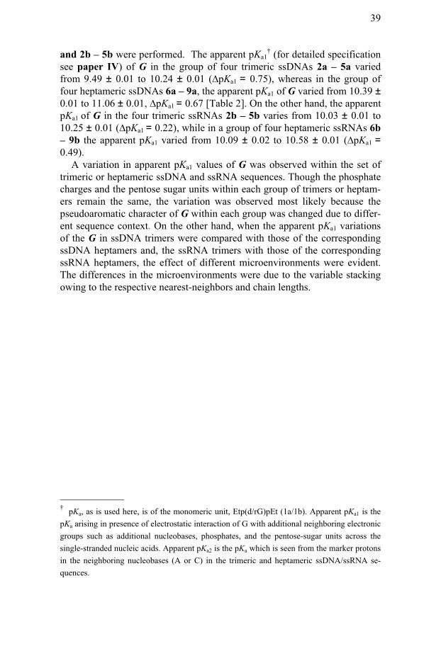
3.3.1 Overall degradation The electronic environments around the internucleotidic phosphates were found to be sequence dependent in our studies and alkaline hydrolysis ex-periments were carried out to investigate if the chemical reactivity of the internucleotidic phosphodiesters varied accordingly. All the heptamers were treated with 0.03 N aqueous NaOH, at pH 12.5 and 20 °C. It was found that the ssRNAs were degraded at different rates depending upon the sequence context. The overall degradation of the heptamers after 1 h was of 11.7% for 6b, 7.8% for 7b, 13.9% for 8b and 11.3% for 9b [Figure 16].

45
3.3.2 Preferential cleavage Even though there are six phosphodiester bonds in each of the four native heptameric ssRNAs (6b, 7b, 8b and 9b), hydrolysis was preferred at the internucleotide phosphodiesters (p2, p3 and p4, Figure 16) from which pKa(31P) values were obtained. Some other relatively minor cleavages were also observed at other internucleotide phosphodiesters, which did not show a pKa(31P), such as p1 of Cp1A- (in all ssRNAs) as well as at p5 of the Cp5A block in 7b and 9b, and in that of the Ap5A block in 6b and 8b (no cleavage was observed at p6 of any of the ssRNAs).
Figure 16. The alkaline hydrolysis at 1 h (in 0.03 N aqueous NaOH, pH 12.5, 20 �C) for the heptameric ssRNAs.
3.3.3 Fast cleavage at 5'-terminal Cp1A The cleavage rates at Cp1A were quite comparable to or lower than the cleavage rates at the phosphodiesters (p2, p3 and p4) from which the pKa(31P) values were obtainable. In contrast, the cleavage rates at Cp1A were always higher than the cleavage rates at internucleotidic p5, from which pKa(31P) value was not obtained. This higher cleavage rate at Cp1A, outside the group of phosphodiesters with pKa(31P), is consistent with earlier observations that the 5'-r(CpA)-3' or 5'-r(UpA)-3' blocks undergo cleavage at a faster rate than other internucleotide linkages in the chimeric DNA/RNA120,121or 2'-O-

46
methylated-RNA/RNA117,118,119oligonucleotides as well as in natural RNA polymers.137
3.3.4 The relation between internucleotidic cleavage rate and pKa(31P) Comparison of the cleavage rates at p2, which shows a pKa(31P) revealed [Fig-ure 16] that the Ap2C fragment in 9b was cleaved at a much faster rate (3.2%) than that in 8b (1.8%), whereas the relative cleavage rates at Ap2A in 6b (1.8%) and in 7b (ca. 2.0%) were comparable. The cleavage at Ap2C was quicker in 8b (1.8%) and in 9b (3.2%), while Ap6C blocks in all the hep-tameric ssRNAs were completely resistant to hydrolysis (also, no pKa(31P) was obtainable from these). This highly preferential cleavage at the Ap2C over the Ap6C in 8b or 9b was remarkable, assuming that the 2'-OH of the adenosine in Ap2C and Ap6C have comparable pKa values. Despite the fact that the internucleotide phosphodiester at Ap2C was capable of sampling fewer in-line cleavage configurations than that of the 3'-terminal Ap6C (be-cause of its location in the strand), the cleavage was favored at Ap2C. This suggests that the chemical character of the internucleotidic p2 in 8b or 9b, were very special, perhaps due to their enhanced electrophilic character compared to those of p6. A comparison of the chemical reactivity at the p2 [Ap2C in 8b (1.8%) and 9b (3.2%), Ap2A in 6b (1.8%) and 7b (2.0%)] also showed that there was a complex set of stereoelectronic and conformational factors that was responsible for dictating the propensity for cleavage, de-pending upon the sequence context.
3.3.5 Comparison of the cleavage rates at p2, p3 and p4 The cleavage rates at p3 and p4 were found to be faster than those of the cleavage at p2 in –Ap2Ap3Gp4A– in 6b and –Ap2Cp3Gp4A– in 8b, whereas the cleavage at p2 is preferred over p3 and p4 in –Ap2Cp3Gp4C– in 9b. In contrast, the relative cleavages at p2, p3 and p4 in –Ap2Ap3Gp4C– in 7b were comparable. This shows the importance of the sequence context in a hy-drolysis experiment with many internucleotide phosphodiester, as in the heptameric ssRNA sequences.
3.4 Alkaline hydrolysis of N1-Me-G containing ssRNA The above studies on alakaline hydrolysis of the heptameric ssRNA se-quences clearly suggested that due to the presence of a charged center G� in close proximity, some of the internucleotidic phosphodiesters (p2, p3, and p4)
47
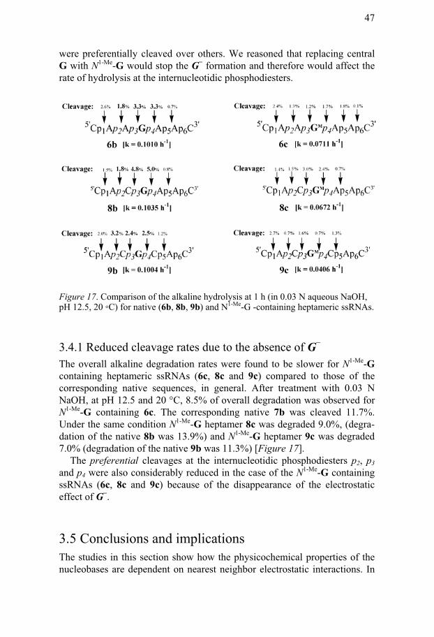
were preferentially cleaved over others. We reasoned that replacing central G with N1-Me-G would stop the G� formation and therefore would affect the rate of hydrolysis at the internucleotidic phosphodiesters.
Figure 17. Comparison of the alkaline hydrolysis at 1 h (in 0.03 N aqueous NaOH, pH 12.5, 20 �C) for native (6b, 8b, 9b) and N1-Me-G -containing heptameric ssRNAs.
3.4.1 Reduced cleavage rates due to the absence of G� The overall alkaline degradation rates were found to be slower for N1-Me-G containing heptameric ssRNAs (6c, 8c and 9c) compared to those of the corresponding native sequences, in general. After treatment with 0.03 N NaOH, at pH 12.5 and 20 °C, 8.5% of overall degradation was observed for N1-Me-G containing 6c. The corresponding native 7b was cleaved 11.7%. Under the same condition N1-Me-G heptamer 8c was degraded 9.0%, (degra-dation of the native 8b was 13.9%) and N1-Me-G heptamer 9c was degraded 7.0% (degradation of the native 9b was 11.3%) [Figure 17].
The preferential cleavages at the internucleotidic phosphodiesters p2, p3 and p4 were also considerably reduced in the case of the N1-Me-G containing ssRNAs (6c, 8c and 9c) because of the disappearance of the electrostatic effect of G�.
3.5 Conclusions and implications The studies in this section show how the physicochemical properties of the nucleobases are dependent on nearest neighbor electrostatic interactions. In

48
an RNA sequence P1Q1NQ2P2, the physicochemical integrity of N is actually dictated by the characters of both neighboring Q1 and Q2. At the same time the properties of Q1 and Q2 are further tuned by the electronic nature of P1 and P2. Thus the pseudoaromatic character of N can have a number of possi-ble variations in the nature depending on the nature of Q1 and Q2. These ine-qualities in the electronic environments brought in by the different sequence contexts can also lead to variable chemical reactivities of the internucleotidic phosphodiester bonds. Thus these studies offer insights into the mechanistic detail of enzymatic and non-enzymatic processes at a molecular level. Also, these insights can help designing new chemical modifications which can be used to tune and control the reactivity of RNA.
49
Acknowledgements
First I would like to thank Professor Jyoti Chattopadhyaya for introducing me to the fascinating frontier of Nucleic Acids Chemistry and for providing me the opportunity to learn modern techniques in his luxuriously-equipped laboratory. My sincere gratitude and thanks to Professor Lars Baltzer for being so kind and accommodative and guiding me through the thesis. Thank you for giving me the sense of freedom while communicating or discussing with you. My endless thanks to Docent Andras Földesi for being the real guide during the bench-work time and for his enormous help and support from my day one in Uppsala till the day of thesis corrections. Sincere thanks to Professor Helena Grennberg, for the last minute arrangement of my first time ever interna-tional conference in Organic Chemistry in Dublin. Thanks to Åke Engström for letting me handling the Maldi-Tof machine, the HPLC machine and for being very helpful during our collaborative works. My sincere thanks to Dr. Oleksandr Plaskevych for his helping hand in all computer related prob-lems. Dr. Dimitri Ossipov, Dr. Nariman Amirkhanov, Dr. Petr Naus, Dr. M. Mariappan, Dr. Mounir Andaloussi, Dr. Naresh Badgujar, Wenbin Hu, Mo-hitosh Maity, Dimitri Kulik, Ayan Samanta and Karthik Babu for being nice work-mates. Dr. Johan Isaksson and Chuanzheng Zhou for being nice col-laborators. Jaana Evander and Eva Pylvänen for their kind assistances and advices. Susan Bergman for being cordial, and for the nice summer parties. Zekaria Rundasa for his kind help during shifting to the new apartment and all his helps during these years. I would like to thank Dr. Klas Udekwu, Ramesh Vetukuri, and Dr. Jaydip Ghosh for their kind help during radio-labelling and related works. My special thanks to Wimal Pathmasiri for his collaboration, helping hand in any type of problem, starting from the electricity problem in my apartment to thesis printing problem. Subhrangsu Chatterjee for being good friend, good collaborator and always being enthusiastic about professional discussions. Dr. Malgorzata Wenska, for being friendly in and outside the lab, and for initiating wonderful socialization parties. These are very special people, whom I knew for they were my colleagues and collaborators but turned to be more than that in all the way, my true friends. Dmitry Honcharekno, and Puneet Srivastava; thank you for the time

50
we shared together. Pradeep Cheruku, for his friendship. I always felt you are the one who can understand my problems without much explanation. Dr. P. I. Pradeepkumar, for generous teaching and sharing knowledge in person and also long telephonic lessons about the experimental protocols and details, for being so supportive during all these years, for inspiring me to grow in general knowledge with all those ‘quote of the days’. Docent Santanu Dasgupta and Docent Lena Åslund for being so kind and affectionate, for the nice foods, wordless thanks for visiting me during my stay in isolation after the operation, for numerous rides by Lena, for taking me to the Karen Armstrong talk (its only one occasion out of many). Docent Suparna Sanyal, Docent Biplab Sanyal and their son Ruku, for all those countless memorable time of good foods and refreshments, late-night chats and movies, arguments, kidding, fishing, photography and constant encouragement and support. Dr. Sandipta Acharya and Dr. Parag Achrya for being so generous collabora-tors in the lab and being so very kind to accommodate me in their apartment during my initial days in Uppsala. Thanks for all long phone calls from other part of the globe. And of course little Ryan, for the welcome grin in the eve-nings when I came back from work, during our stay together. Dr. Santosh Thomas, Suma, Ajit and Anu for offering me homely affection. Elsa, Stefen, Hari and little Samuel for being ever-refreshing kids and offer-ing those happy moments. Arun and Diaga Levman for your constant sup-port and prayer throughout all these years. My sincere thanks to all the friends in Sweden, who made my life in Sweden better in their own way. Dr. Nelson Theethayi, came to Sweden the same month I came, been good friend all through out. Joan Sancho turned my first Swedish winter to a warm memorable one with all her enthusiastic plans for movies, Elton John live show, dances-house show and many more. Thanks to Dipanjan Roychowdhuri, Seema Sarkar, Santosh Dahl, Sonchita Bagchi, Bhupender Singh, Surajit Midya, Brinda Maity (Midya), Nimesh Bhaskaran, Smitha Sreedharan, Madhuri Cheruku, Dr. Anna Trifonova, Maria Sandberg, Donnie Phillip and many others I cannot mention. Thanks to my teachers specially Late Dr. Subir Basu Roy, and Dr. Swaraj Baran Maity, for generating my interest in Organic Chemistry. Dr. Asish De and Prof. Amit Basak, for showing me what ‘research’ really means and for offering the best laboratory atmosphere with wonderful lab seniors and for inspiring me to pursue doctoral studies. Dr. Maya Datta Gupta for her en-couragements. My lab-seniors in India, Dr. Sukanta Kamila, Dr. Chandrani Mukherjee, Dr. Kakali Rani Rudra, Dr. Subrata Madal, Dr. Subhendu Sekhar Bag, Dr. Subhas Chandra Ghosh, for their warmth, care and teachings. Their enthusiasm in research initiated my interest in team-work. Thanks to the bunch of brilliant M. Sc. classmates, Arijit, Balaji, Binita, Bishnu, Debarshi, Kishore, Manoj, Nihar, Suman, Rajarshi, Rajesh, Srinivas, and Uma, for showing me what ambitiousness, competence, and confidence mean (most of them have become ‘Dr.’ by now !!!). Special thanks to my
51
roommate Subarna for her innocence and affection during my stay at Kharagpur. My very dear friends Dr. Indira Chowdhuri, Dr. Madhuri Man-dal, Dr. Subrata Kundu, Dr. Selim Reza, Dr. Rajkumar Halder, Maumita, Rina and Bani for showing the meaning of friendship over the years with their constant love and affection. Thanks to Mridul for his unconditional love and support during the time of need and thanks for patiently waiting for me. Oommen P. Varghese, for his constant optimism, who taught me to see the other side of the coin and to survive in any situation. I would have given up a long back without his immense support both in and outside the lab. My dear parents, chhotomama, and my loving brothers Bulu and Nilu… my eyes fill with tears now… I have no words to express myself… it had been a struggle for them to let me come this far and for me to leave them. It was because of their constant love, support and encouragement that I could be what I am.

52
Sammanfattning
Avhandlingen beskriver studier av viktiga egenskaper hos nukleinsyror av-seende deras förmåga att fungera som läkemedel samt studier av grund-läggande fysikalisk-kemiska egenskaper av betydelse för dess reaktivitet. Ett antal kemiskt modifierade nukleotider har inkorporerats i syntetiska oligo-nukleotider och dess egenskaper har utvärderats och jämförts med dem som förekommer i naturen. De syntetiska nukleotiderna 2'-O-Me-T, 2'-O-MOE-T, oxetane-T, LNA-T, azetidine-T, aza-ENA-T, carbocyclic-ENA-T och carbocyclic-LNA-T har inkorporerats i en sekvens om 15 nukleotider, en så kallad 15-mer, som binder till en komplementär 15-mer tagen ur en naturligt förekommande sekvens som kodar för ett naturligt T antigen hos organismen SV-40. En jämförande studie visar att 15-mererna med oxetane-T och azeti-dine-T har lägre affinitet för målfragmentet än den naturligt förekommande sekvensen. Dessa två baser kännetecknas av att sockerresten i nukleotiden låsts i en väldefinierad konformation som inte är kompatibel med stark bind-ning. De modifierade nukleotiderna LNA-T, aza-ENA-T, carbocyclic-ENA-T och carbocyclic-LNA-T band emellertid bättre än den naturliga sekvensen. En viktig del av läkemedelsstrategin är att syntetisera icke-naturliga frag-ment som kan binda till målsekvensen i en konformation som känns igen av enzymet RNAs H, så att det bryts ned av kroppsegna komponenter. De mo-difierade sekvenserna har därför undersökts med avseende på förmåga att binda till målfragmentet och rekrytera RNAs H. Modifierade sekvenser vi-sade förmåga att rekrytera RNAs H med högre katalytisk aktivitet (kcat) men med lägre affinitet (1/Km ) än de naturliga fragmenten. Vidare har mot-ståndskraften emot enzymatisk nedbrytning av nukleaser, såsom fosfodieste-ras från ormgift samt de som förekommer i humant serum, undersökts ge-nom att nedbrytningshastigheten bestämts. De 15-merer som modifierats med aza-ENA-T and carbocyclic-ENA-T visade väsentligt förhöjd mot-ståndskraft mot enzymatisk nedbrytning av nukleaser. Sammantaget så visa-de en grupp av modifierade oligonukleotider väsentligt förbättrade egenska-per i form av förhöjd affinitet, jämförbar förmåga att rekrytera RNAs H samt förhöjd förmåga att motstå enzymatisk nedbrytning, jämfört med den natur-liga sekvensen. NMR spektroskopiska studier och studier av alkalisk hydrolys av oligonuk-leotider har utförts för att undersöka effekten av elektrostatisk växelverkan på oligonukleotiders funktionella egenskaper. Jonisering av guanin resten kan monitoreras via NMR spektroskopiska studier av pKa värden för angrän-sande grupper, t ex med hjälp av 31P NMR spektroskopi av fosforkärnan i

53
den internukleotidiska fosfodiestern. De fosfodiestrar vars pKa påverkas av guanins dissociation visade sig hydrolyseras snabbare är andra och den elek-trostatiska växelverkan har därmed visats vara en faktor som påverkar reak-tiviteten på ett sekvensberoende sätt.

54
References
1. Crick, F. Nature 1970, 227, 561. 2. Gilbert, W. Nature 1986, 319, 618. 3. Gesteland, R. F.; Cech, T. R.; Atkins, J. F. The RNA World; Cold Spring Harbor Laboratory Press, New York, 1998. 4. Woese, C. The Genetic Code; Harper & Row, New York, 1967. 5. Crick, F. H. C. J. Mol. Biol. 1968, 38, 367. 6. Orgel, L. E. J. Mol. Biol. 1968, 38, 381. 7. Hall, R. H. The Modified Nucleosides in Nucleic Acids; Columbia University Press, New York, 1971. 8. Limbach, P. A.; Crain, P. F.; McCloskey, J. A. Nucl. Acids Res. 1994, 22, 2183. 9. Saenger, W. Principles of Nucleic Acid Chemistry; Springer-Verlag, New York, 1984. 10. Bloomfield, V. A.; Crothers, D. M.; Tinoco, I. Nucleic Acids: Structures, Prop-erties and Functions; University Science Books, Sausalito, California, 1999. 11. Blackburn, G. M.; Gait, M. J. Nucleic Acids in Chemistry and Biology; Oxford University Press, New York, 1996. 12. Carreira, L. A.; Jiang, G.J.; Person, W.B.; Willis, J. N. J. J. Chem. Phys. 1972, 56, 1440. 13. Kilpatrick, J. E.; Pitzer, K. S.; Spitzer, R. J. Am. Chem. Soc. 1947, 69, 2483. 14. Strauss, H. L. Ann. Rev. Phys. Chem. 1983, 34, 301. 15. Thaibaudeau, C.; Plavec, J.; Chattopadhyaya, J. J. Org. Chem. 1996, 61, 266. 16. Miha, P.; Cmugelj, M.; Stimac, A.; Kobe, J.; Plavec, J. J. Chem. Soc. Perkin Trans. 2001, 28, 1433. 17. Herdewijn, P. Biochem. Biophys. Acta 1999, 1489, 167. 18. Manoharan, M. Biochem. Biophys. Acta 1999, 1489, 117. 19. Pattanayek, R.; Sethaphong, L.; Pan, C.; Prhavc, M.; Prakash, T. P.; Manoharan,| M.; Egli, M. J. Am. Chem. Soc. 2004, 126, 15006. 20. Watson, J. D.; Crick, F. H. C. Nature 1953, 171, 737. 21. Hoogsteen, K. Acta Cryst. 1963, 16, 907. 22. Sanchez, V.; Redfield A. G.; Johnston, P. D.; Tropp J. Proc. Natl. Acad. Sci. USA 1980, 77, 5659. 23. Mooers, B. H. M.; Eichman, B. F.; Ho, P. S. J. Mol. Biol. 1997, 269, 796. 24. Kim, S. G. Prog. Nucl. Acids Res. Mol. Biol. 1976, 17, 181. 25. Sussman, J. L.; Holbrook, S. R.; Wade Warrant, R.; Church, G. M.; Kim, S. G. J. Mol. Biol. 1978, 123, 607. 26. Boulard, Y.; Cognet, J. A.; Gabarro-Arpa, J.; Le, B. M.; Sowers, L. C.; Fazaker-ley, G. V. Nucl. Acids Res. 1992, 20, 1933. 27. Cantor, C. R.; Warshaw, M. M.; Shapiro, H. Biopolymers 1970, 9, 1059.
55
28. Velikyan, I.; Acharya, S.; Trifonova, A.; Chatopadhyaya, J. J. Am. Chem. Soc. 2001, 123, 2893. 29. Acharya S.; Földesi, A.; Chattopadhyaya, J. J. Org. Chem. 2003, 68, 1906. 30. Kulmer, W. D.; Eiler, J. J. J. Am. Chem. Soc. 1943, 65, 2355. 31. Chamberlin, S.; Merino, E. J.; Weeks, K. M. Proc. Natl. Acad. Sci. USA 2002, 99, 14688. 32. Hecht, S. M. Bioorganic Chemistry: Nucleic Acids; Oxford University Press, Oxford, New York, 1996. 33. Niittymäki, T.; Lönnberg, H. Org. Biomol. Chem. 2006, 4, 15. 34. Swaminathan, S. K.; Nambiar, A.; Guntaka, R. V. FASEB 1998, 12, 515. 35. Anderson, E. M.; Halsey, W A.; Wuttke, D. S. Biochemistry 2003, 42, 3751. 36. Bochkareva, E.; Belegu, V.; Korolev, S.; Bochkaren, A. EMBO J. 2001, 20, 612. 37. Nishinaka, T.; Ito, Y.; Yokoyama, S.; Shibata, T. Proc. Atl. Acad. Sci. USA 1997, 94, 6623. 38. Bar-Ziv, R.; Libehaber, A. Proc. Natl. Acad. Sci. USA 2001, 20, 612. 39. Handa, N.; Nureki, O.; Kurimoto, K.; Kim, I.; Sakamoto, H.; Shimura, Y.; Muto, Y.; Yokoyama, S. Nature 1999, 398, 579. 40. Deo, R.C.; Bonano, J.B.; Sonenberg, N.; Burley, S. K. Cell 1999, 98, 835. 41. Bogden, C. E.; Fass, D.; Bergman, N.; Nichols, M. D.; Berger, J. M. Mol. Cell 1999, 3, 487. 42. Hélène, C. Eur. J. Cancer 1991, 27, 1466. 43. Giovannangeli, C.; Hélène, C. Nat. Biotechnol. 2000, 18, 1245. 44. Buchini, S.; Leumann, C. J. Current Opin. Chem. Biol. 2003, 7, 717. 45. Opalinska, J. B.; Gewirtz, A. M. Nat. Rev. Drug Discov. 2002, 7, 503. 46. Zamecnik, P. C.; Stephenson, M. L. Proc. Natl. Acad. Sci. USA 1978, 75, 280. 47. Stephenson, M. L.; Zamecnik, P. C. Proc. Natl. Acad. Sci. USA 1978, 75, 285. 48. Cech, T. R.; Zaug, A. J.; Grabowski, P. J. Cell, 1981, 27, 487. 49. Kruger, K.; Grabowski, P. J.; Zuag, A. J.; Sands, J.; Gottschling, D. E.; Cech, T. R. Cell, 1982, 31, 147. 50. Guerrier-Takada, C.; Gardiner, K.; Marsh, T.; Pace, N.; Altman, S. Cell, 1983, 35, 849. 51. Rocheleau, C. E.; Downs,W. D.; Lin, R.,Wittmann, C.; Bei,Y.; Cha,Y. H.; Ali, M.; Priess, J. R.; Mello, C. C. Cell 1997, 90, 707. 52. Fire, A.; Xu, S.; Montgomery, M. K.; Kostas, S. A.; Driver, S. E.; and Mello, C. C. Nature, 1998, 391, 806. 53. Fire, A.; Xu, S.; Motegomery, M. K.; Kostas, S. A.; Driver, S. E.; Mello, C. C. Nature 1998, 391, 806. 54. Agami, R. Current. Opin. Chem. Biol. 2002, 6, 829. 55. Fougerolles, A. D.; Vornlocher, H. P.; Maraganore, J.; Lieberman, J. Nat. Rev. Drug Discov. 2007, 6, 443. 56. Breaker, R. R.; Joyce, G. F.; Santoro, S. W. Chem. Biol. 1994, 1, 223. 57. Coppins, R. L.; Silverman, S. K. Nat. Struct. Mol. Biol. 2004, 11, 270. 58. Haeuptle, M. T.; Frank, R.; Dobberstein, B. Nucleic Acids Res. 1986, 14, 1427. 59. Lee, L. K.; Roth, C. M. Current Opin. Biotechnol. 2003, 14, 505. 60. Phillips, M. I. Antisense Therapeutics, Humana Press, NJ, USA, 2004. 61. Lima, W. F.; Crooke, S. T. Biochemistry 1997, 36, 390. 62. Blacke, K. R.; Murakami, A.; Spitz, S. A. Biochemistry 1985, 24, 6139.

56
63. Johansson, H. E.; Belsham, G. J.; Sproat, B. S. Nucleic Acids Res. 1994, 22, 4591. 64. Zamaratski, E.; Pradeepkumar, P. I.; Chattopadhyaya, J. Biochem. Biophys. Methods 2001, 48, 189. 65. Kurreck, J. Eur. J. Biochem. 2003, 270, 1628. 66. Crooke, S. T. Annu. Rev. Med. 2004, 55, 61. 67. Herdewijn, P. Antisense Nucleic Acids Drug Dev. 2000, 10, 297. 68. De Clerq, E.; Eckstein, F.; Merigan, T. C. Science 1969, 165, 1137. 69. Eckstein, F. Antisense Nucleic Acids Drug Dev. 2000, 10, 117. 70. Eckstein, F. Ann. Rev. Biochem. 1985, 54, 367. 71. Monia, B. P.; Lesnik, E. A.; Gonzalez, C.; Lima, W. F.; McGee, D.; Guinosso, C. J.; Kawasaki, A. M.; Cook, P. D.; Freier, S. M. J. Biol. Chem. 1993, 268, 14514. 72. Lebedeva, I.; Stein, C. A. Annu. Rev. Pharmacol. Toxicol. 2001, 41, 403. 73. Nielsen, P. E.; Egholm, M.; Berg, R. H.; Buchardt, O. Science 1991, 254, 1497. 74. De, M. A.; Altmann, K.; Waldner, A.; Wendeborn, S. Curr. Opin. Struct. Biol. 1995, 5, 343. 75. Braasch, D. A.; Corey, D. R. Biochemistry, 2002, 41, 4503. 76. Damha, M. J.; Wilds, C. J.; Noronha, A.; Brukner, I.; Borkow, G.; Arion, D.; Parniak, M. A. J. Am. Chem. Soc. 1998, 120, 12976. 77. Noronha, A. M.; Wilds, C. J.; Lok, C-N.; Viazovkina, K.; Arion, D.; Parniak, M. A.; Damha, M. J. Biochemistry; 2000, 39, 7050. 78. Mangos, M. M.; Damha, M. J. Curr. Top. Med. Chem. 2002. 79. Hendrix, C.; Rosemeyer, H.; Verheggen, I.; Aerschot, A. V.; Seela, F.; Herde-wijn P. Chem. Eur. J. 1997, 3, 110. 80. Eschenmoser, A.; Dobler, M. Helv. Chim. Acta 1992, 75, 218. 81. Eschenmoser, A. Science 1999, 284, 2118. 82. Wang, J.; Verbeure, B.; Luyten, I.; Lescrinier, E.; Froeyen, M.; Hendrix, C.; Rosemeyer, H.; Seela, F.; Aerschot, A. V.; Herdewijn P. J. Am. Chem. Soc. 2000, 122, 8595. 83. Obika, S.; Nanbu, D.; Hari, Y.; Morio, K.-I.; In, Y.; Ishida, T.; Imanishi, T. Tetrahedron Lett. 1997, 38, 8735. 84. Koshkin, A. A.; Singh, S. K.; Nielson, P.; Rajwanshi, V. K.; Kumar, R.; Meld-gaard, M.; Wengel, J. Tetrahedron 1998, 54, 3607. 85. Morita, K.; Hasegawa, C.; Kaneko, M.; Tsutsumi, S.; Sone, J.; Ishikawa, T.; Imanishi, T.; Koizumi, M. Bioorg. Med. Chem. Lett. 2002, 12, 73. 86. Morita, K.; Yamate, K.; Kurakata, S.; Abe, K.; Imanishi, T.; Koizumi, M. Nu-cleic Acids Res. Sup. 2002, 2, 99. 87. Summerton, J. Weller, D. Antisense Nucleic Acid Drug Dev. 1997, 7, 187. 88. Summerton J. Biochim. Biophys. Acta 1999, 1489, 141. 89. Amantana, A.; Iversen, P. L. Current Opin. Pharm. 2005, 5, 550. 90. Gryaznov, S. M.; Lloyd, D. H.; Chen, J. K.; Schultz, R. G.; DeDionisio, L. A.; Ratmeyer, L.; Wilson, W. D. Proc. Natl. Acad. Sci. USA 1995, 92, 5798. 91. Gryaznov, S. M. Biochem. Biophys. Acta. 1999, 1489, 131. 92. Steffens, R.; Leumann, C. J. J. Am. Chem. Soc. 1997, 119, 11548. 93. Renneberg, D.; Bouliong, E.; Reber, U.; Schumperli, D.; Leumann, C. J. Nucleic Acids Res. 2002, 30, 2751. 94. Sohail, M. Drug Discov. Today 2001, 2, 1260. 95. Pan, W. H.; Clawson, G. A. J. Cell. Biochem. 2006, 98, 14.
57
96. Sohail, M.; Hochegger H.; Klotzbücher, A.; Guellec, R. L.; Hunt, T.; Southern, E. M. Nucl. Acids Res. 2001, 29, 2041. 97. Ho, S. P.; Bao, Y.; Lesher, T.; Malhotra, R; Ma, L. Y.; Fluharty, S. J.; Sakai R. R. Nat. Biotech. 1998, 16, 53. 98. Fougerolles, A. D.; Vornlocher, H. P.; Maraganore, J.; Lieberman, J. Nat. Rev. Drug Discov. 2007, 6, 443. 99. Dias, N.; Stein C. A. Eur. J. Pharm. Biopharm. 2002; 54, 263. 100. Dove, A. Nature. Biotechnol. 2002, 20, 121. 101. Bevilacqua, P. C.; Yajima, R. Curr. Opin. Chem. Biol. 2006, 10, 455. 102. Takagi, Y.; Ikeda, Y.; Taira, K. Top. Curr. Chem. 2004, 232, 213. 103. Pradeepkumar, P. I.; Chattopadhyaya, J. J. Chem. Soc. Perkin Trans. 2 2001, 2074. 104. Pradeepkumar, P. I.; Zamaratski, E.; Földesi, A.; and Chattopadhyaya, J. J. Chem. Soc. Perkin Trans. 2 2001, 402. 105. Pradeepkumar, P. I.; Amirkhanov, N. V.; and Chattopadhyaya, J. Org. Biomol. Chem. 2003, 1, 81. 106. Pradeepkumar, P. I.; Cheruku, P.; Plashkevych, O.; Acharya, P.; Gohil, S.; Chattopadhyaya, J. J. Am. Chem. Soc., 2004, 126, 11484. 107. Wanger, R. W.; Matteucci, M. D.; Lewis, J. G.; Gutierrez, A. J.; Moulds, C; Froehler, B. C. Science 1993, 260, 1510. 108. Kurreck, J.; Wyszko, E.; Gillen, C.; Erdmann, V. A. Nucl. Acids Res. 2002, 30, 1911. 109. Fersht, A. The Basic Equations of Enzyme Kinetics, in Structure and Mecha-nism in Protein Science: A Guide to Enzyme Catalysis and Protein Folding, W. H. Freeman and Company, New York, 2000. 110. Isaksson, J.; Plashkevych, O.; Pradeepkumar, P. I.; Pathmasiri, W.; Shrivastava, P.; Chatterjee, S.; Barman, J.; Petit C.; Chattopadhyaya, J. J. Biomol. Struct. Dyn. 2005, 23, 233. 111. Holbrook, S. R. Curr. Opin. Struct. Biol. 2005, 15, 302. 112. Lilley, D. M. Curr. Opin. Struct. Biol. 2005, 15, 313. 113. Fedor, M. J. Curr. Opin. Struct. Biol. 2002, 12, 289. 114. Brown, D. M.; Magrath, D. I.; Neilson, A. H. Nature, 1956, 177, 1124. 115. Oivanen, M.; Schnell, R.; Pfleiderer, W.; Lönnberg, H. J.Org. Chem. 1991, 56, 3623. 116. Jarvinen, P.; Oivanen, M.; Lonnberg, H. J. Org. Chem. 1991, 56, 5396. 117. Kaukinen, U.; Lyytikäinen, S.; Mikkola, S.; Lönnberg, H. Nucl. Acids Res. 2002, 30, 468. 118. Kaukinen, U.; Venäläinen, T.; Lönnberg, H.; Peräkylä, M. Org. Biomol. Chem. 2003, 1, 2439. 119. Kaukinen, U.; Lönnberg, H.; Peräkylä, M. Org. Biomol. Chem. 2004, 2, 66. 120. Li, Y.; Breaker, R. R. J. Am. Chem. Soc. 1999, 121, 5364. 121. Kawamura, K. Bull. Chem. Soc. Jpn., 2003, 76, 153. 122. Lyne, P. D.; Karplus, M. J. Am. Chem. Soc. 2000, 122 166. 123. Perreault, D. M.; Anslyn, E. V. Angew. Chem. Int. Ed. Engl. 1997, 36, 432. 124. Oivanen, M.; Kuusela, S.; Lönnberg, H. Chem. Rev. 1998, 98, 961. 125. Soukup, G. A.; Breaker, R. R. RNA 1999, 5, 1308. 126. Kierzek, R. Methods in Enzymology 2001, 541, 657. 127. Lönnberg, T.; Lönnberg, H. Curr. Opin. Chem. Biol. 2005, 9, 665.
58
128. Antson, A. A. Curr. Opin. Struct. Biol. 2000, 10, 87. 129. Lilley, D. M. RNA, 2004, 10, 151. 130. Acharya, S.; Acharya, P; Földesi, A.; Chattopadhyaya, J. J. Am. Chem. Soc. 2002, 124, 13722. 131. Acharya, P.; Acharya, S; Földesi, A.; Chattopadhyaya, J. J. Am. Chem. Soc. 2003, 125, 2094. 132. Cohn, M.; Hughes, T. R. Jr. J. Biol. Chem. 1960, 237, 3250. 133. Gorenstein, D. G. Phosphorous-31 NMR: Principles and Applications, Aca-demic Press, Orlando, FL, 1984. 134. Pietri, S.; Miollan, M.; Martel, S.; Moigne, F. L.; Blaive, B.; Culcasi, M. J. Biol. Chem. 2000, 275, 19505. 135. Moedritzer, K. Inorg. Chem. 1967, 6, 936. 136. Porubcan, M. A.; Westler, W. M.; Ibanez, I. B.; Markley, J. L. Biochemistry, 1979, 18, 4108. 137. Williams, K.; Ciafre, P.; Tocchini-valentine, G. P. EMBO J. 1995, 14, 4551.

Acta Universitatis UpsaliensisDigital Comprehensive Summaries of Uppsala Dissertationsfrom the Faculty of Science and Technology 355
Editor: The Dean of the Faculty of Science and Technology
A doctoral dissertation from the Faculty of Science andTechnology, Uppsala University, is usually a summary of anumber of papers. A few copies of the complete dissertationare kept at major Swedish research libraries, while thesummary alone is distributed internationally through theseries Digital Comprehensive Summaries of UppsalaDissertations from the Faculty of Science and Technology.(Prior to January, 2005, the series was published under thetitle “Comprehensive Summaries of Uppsala Dissertationsfrom the Faculty of Science and Technology”.)
Distribution: publications.uu.seurn:nbn:se:uu:diva-8272
ACTA
UNIVERSITATIS
UPSALIENSIS
UPPSALA
2007