Targeted deletion of a cis-regulatory region reveals...
15
Targeted deletion of a cis-regulatory region reveals differential gene dosage requirements for Pdx1 in foregut organ differentiation and pancreas formation Yoshio Fujitani, 1 Shuko Fujitani, 1 Daniel F. Boyer, 1 Maureen Gannon, 2,3 Yoshiya Kawaguchi, 4 Michael Ray, 1 Masakazu Shiota, 2 Roland W. Stein, 1,3 Mark A. Magnuson, 1,3 and Christopher V.E. Wright 1,5 1 Vanderbilt Program in Developmental Biology and Department of Cell and Developmental Biology, 2 Department of Medicine, and 3 Department of Molecular Physiology and Biophysics, Vanderbilt University School of Medicine, Nashville, Tennessee 37232-2175, USA; 4 Department of Surgery and Surgical Basic Science, Kyoto University Graduate School of Medicine, Kyoto 606-8507, Japan Pdx1 (IPF-1 in humans, which is altered in MODY-4) is essential for pancreas development and mature -cell function. Pdx1 is expressed dynamically within the developing foregut, but how its expression characteristics are linked to the various steps of organ specification, differentiation, and function is unknown. Deletion of a conserved enhancer region (Area I-II-III) from Pdx1 produced a hypomorphic allele (Pdx1 I-II-III ) with altered timing and level of expression, which was studied in combination with wild-type and protein-null alleles. Lineage labeling in homozygous Area I-II-III deletion mutants (Pdx1 I-II-III/I-II-III ) revealed lack of ventral pancreatic bud specification and early-onset hypoplasia in the dorsal bud. Acinar tissue formed in the hypoplastic dorsal bud, but endocrine maturation was greatly impaired. While Pdx1 -/- (protein-null) mice have nonpancreatic abnormalities (e.g., distorted pylorus, absent Brunner’s glands), these structures formed normally in Pdx1 I-II-III/I-II-III and Pdx1 I-II-III/- mice. Surprisingly, heterozygous (Pdx1 +/I-II-III ) mice had abnormal islets and a more severe prediabetic condition than Pdx1 +/- mice. These findings provide in vivo evidence of the differential requirements for the level of Pdx1 gene activity in the specification and differentiation of the various organs of the posterior foregut, as well as in pancreas and gut endocrine cell differentiation. [Keywords: Organogenesis; pancreatic cells; Pdx1; MODY; Enhancer; cis-element; foregut differentiation] Supplemental material is available at http://www.genesdev.org. Received July 28, 2005; revised version accepted November 30, 2005. Studies in several vertebrate species have started to de- fine conserved developmental pathways controlled by extrinsic and intrinsic factors that are essential for the development of the endocrine and exocrine pancreas (Kim and Hebrok 2001; Wilson et al. 2003; Jensen 2004). It is generally accepted that signals from mesodermal tissues control the early stage of pancreas development. Dorsal bud outgrowth depends on inductive signals from the nearby notochord (Kim et al. 1997; Hebrok et al. 1998), and its subsequent expansion and differentiation require signals from embryonic blood vessel cells (Lam- mert et al. 2001; Yoshitomi and Zaret 2004) and other signaling pathways, including Activin, FGFs, and Notch (Kim and Hebrok 2001; Wilson et al. 2003; Jensen 2004). These signals affect the expression of transcription fac- tors that execute the pancreatic developmental program. Among the transcription factors identified as essential for proper pancreas differentiation, the homeodomain protein Pdx1 (also known as IPF-1, STF-1, or IDX-1) (Wright et al. 1989; Leonard et al. 1993; Ohlsson et al. 1993; Miller et al. 1994) is both expressed in precursors of the endocrine and exocrine compartments of the pan- creas (Gu et al. 2002), and is essential for their develop- ment and maintenance, especially the insulin-producing cells (Jonsson et al. 1994; Offield et al. 1996; Ahlgren et al. 1998; Holland et al. 2002). In addition to pancreatic agenesis, Pdx1 -/- mouse mutants have defects through- out the posterior foregut region, including distorted gas- tro-duodenal junction, loss of Brunner’s glands, and de- ficiency of enteroendocrine differentiation in the stom- ach and duodenum (Larsson et al. 1996; Offield et al. 1996; Jepeal et al. 2005). Loss of IPF-1 function also results in pancreatic agenesis in humans (Stoffers et al. 1997). 5 Corresponding author. E-MAIL [email protected]; FAX (615) 322-1917. Article and publication are at http://www.genesdev.org/cgi/doi/10.1101/ gad.1360106. GENES & DEVELOPMENT 20:253–266 © 2006 by Cold Spring Harbor Laboratory Press ISSN 0890-9369/06; www.genesdev.org 253 Cold Spring Harbor Laboratory Press on December 26, 2019 - Published by genesdev.cshlp.org Downloaded from
Transcript of Targeted deletion of a cis-regulatory region reveals...
Targeted deletion of a cis-regulatoryregion reveals differential gene dosagerequirements for Pdx1 in foregut organdifferentiation and pancreas formationYoshio Fujitani,1 Shuko Fujitani,1 Daniel F. Boyer,1 Maureen Gannon,2,3 Yoshiya Kawaguchi,4
Michael Ray,1 Masakazu Shiota,2 Roland W. Stein,1,3 Mark A. Magnuson,1,3 andChristopher V.E. Wright1,5
1Vanderbilt Program in Developmental Biology and Department of Cell and Developmental Biology, 2Department ofMedicine, and 3Department of Molecular Physiology and Biophysics, Vanderbilt University School of Medicine, Nashville,Tennessee 37232-2175, USA; 4Department of Surgery and Surgical Basic Science, Kyoto University Graduate School ofMedicine, Kyoto 606-8507, Japan
Pdx1 (IPF-1 in humans, which is altered in MODY-4) is essential for pancreas development and mature �-cellfunction. Pdx1 is expressed dynamically within the developing foregut, but how its expression characteristicsare linked to the various steps of organ specification, differentiation, and function is unknown. Deletion of aconserved enhancer region (Area I-II-III) from Pdx1 produced a hypomorphic allele (Pdx1�I-II-III) with alteredtiming and level of expression, which was studied in combination with wild-type and protein-null alleles.Lineage labeling in homozygous Area I-II-III deletion mutants (Pdx1�I-II-III/�I-II-III) revealed lack of ventralpancreatic bud specification and early-onset hypoplasia in the dorsal bud. Acinar tissue formed in thehypoplastic dorsal bud, but endocrine maturation was greatly impaired. While Pdx1−/− (protein-null) mice havenonpancreatic abnormalities (e.g., distorted pylorus, absent Brunner’s glands), these structures formednormally in Pdx1�I-II-III/�I-II-III and Pdx1�I-II-III/− mice. Surprisingly, heterozygous (Pdx1+/�I-II-III) mice hadabnormal islets and a more severe prediabetic condition than Pdx1+/− mice. These findings provide in vivoevidence of the differential requirements for the level of Pdx1 gene activity in the specification anddifferentiation of the various organs of the posterior foregut, as well as in pancreas and gut endocrine celldifferentiation.
[Keywords: Organogenesis; pancreatic � cells; Pdx1; MODY; Enhancer; cis-element; foregut differentiation]
Supplemental material is available at http://www.genesdev.org.
Received July 28, 2005; revised version accepted November 30, 2005.
Studies in several vertebrate species have started to de-fine conserved developmental pathways controlled byextrinsic and intrinsic factors that are essential for thedevelopment of the endocrine and exocrine pancreas(Kim and Hebrok 2001; Wilson et al. 2003; Jensen 2004).It is generally accepted that signals from mesodermaltissues control the early stage of pancreas development.Dorsal bud outgrowth depends on inductive signals fromthe nearby notochord (Kim et al. 1997; Hebrok et al.1998), and its subsequent expansion and differentiationrequire signals from embryonic blood vessel cells (Lam-mert et al. 2001; Yoshitomi and Zaret 2004) and othersignaling pathways, including Activin, FGFs, and Notch(Kim and Hebrok 2001; Wilson et al. 2003; Jensen 2004).
These signals affect the expression of transcription fac-tors that execute the pancreatic developmental program.
Among the transcription factors identified as essentialfor proper pancreas differentiation, the homeodomainprotein Pdx1 (also known as IPF-1, STF-1, or IDX-1)(Wright et al. 1989; Leonard et al. 1993; Ohlsson et al.1993; Miller et al. 1994) is both expressed in precursorsof the endocrine and exocrine compartments of the pan-creas (Gu et al. 2002), and is essential for their develop-ment and maintenance, especially the insulin-producing� cells (Jonsson et al. 1994; Offield et al. 1996; Ahlgren etal. 1998; Holland et al. 2002). In addition to pancreaticagenesis, Pdx1−/− mouse mutants have defects through-out the posterior foregut region, including distorted gas-tro-duodenal junction, loss of Brunner’s glands, and de-ficiency of enteroendocrine differentiation in the stom-ach and duodenum (Larsson et al. 1996; Offield et al.1996; Jepeal et al. 2005). Loss of IPF-1 function also resultsin pancreatic agenesis in humans (Stoffers et al. 1997).
5Corresponding author.E-MAIL [email protected]; FAX (615) 322-1917.Article and publication are at http://www.genesdev.org/cgi/doi/10.1101/gad.1360106.
GENES & DEVELOPMENT 20:253–266 © 2006 by Cold Spring Harbor Laboratory Press ISSN 0890-9369/06; www.genesdev.org 253
Cold Spring Harbor Laboratory Press on December 26, 2019 - Published by genesdev.cshlp.orgDownloaded from
In mature � cells, Pdx1 transactivates the Insulin geneand other genes involved in glucose sensing and metabo-lism, such as Glut2 and Glucokinase (Waeber et al. 1996;Watada et al. 1996). Pdx1+/− mice are glucose intolerant,indicating that gene dosage for Pdx1/IPF-1 is crucial fornormal glucose homeostasis (Ahlgren et al. 1998; Duttaet al. 1998; Brissova et al. 2002; Johnson et al. 2003).These findings are concordant with the prior discoverythat humans heterozygous for an inactivating mutationof IPF-1 are at elevated risk for adult-onset type II diabe-tes and are linked to a dominant heritable condition,maturity onset diabetes of the young type 4 (MODY4)(Stoffers et al. 1997, 1998).
The regulatory mechanisms that orchestrate the com-plex developmental transitions involved in cellular dif-ferentiation and function during pancreatic organogen-esis are far from elucidated. Normal endocrine pancreasdevelopment and function depends on a highly inte-grated transcription-factor network, and even subtle ab-normalities in islet tissue caused by heterozygosity orreduced gene dosage of MODY susceptibility genes cansubstantially predispose toward severe diabetes in hu-mans (Bell and Polonsky 2001). Recent promoter analy-ses of genes involved in islet differentiation and functionsuggest complex genetic interactions among these fac-tors (Shih et al. 2002; Servitja and Ferrer 2004). Most ofthis evidence, however, is inferred from in vitro studiesusing immortalized �-cell lines or other cells such as EScells, which are perhaps of limited relevance to normalontogeny and organogenesis.
As one key entry point into understanding the opera-tion of these gene regulatory networks in vivo, we haveexamined the cis- and trans-regulation of Pdx1. Align-ment of the mouse and human Pdx1 gene sequences re-vealed three phylogenetically conserved regions referredto collectively as Area I-II-III (Gerrish et al. 2000), whichare also characterized as PH-1, PH-2, and PH-3 in human(Melloul et al. 2002). The Area I-II-III region harborsbinding sites for MODY transcription factors such asHNF-1� and Pdx1 itself (Marshak et al. 2000; Gerrish etal. 2001), as well as other endodermal and islet-enrichedtranscriptional regulators, including Foxa2/HNF3� (Ger-rish et al. 2000; Ben-Shushan et al. 2001), Pax6 (Samaraset al. 2002), Maf/RIPE3b1 (Samaras et al. 2003), andHNF-6/OC-1 (Jacquemin et al. 2003). We have reportedon sufficiency tests in transgenic mice and cultured celllines that identified islet-specific (PstI–BstEII fragment:containing Area I-II) and �-cell-specific (XhoI–BglII frag-ment: containing Area III and an adjacent 3� region) cis-regulatory regions that overlap with Area I-II-III, suggest-ing that Area I-II-III functions specifically in differentia-tion and maintenance of pancreatic islets (Wu et al.1997; Gerrish et al. 2000, 2001; Gannon et al. 2001; Sa-maras et al. 2002). A slightly more distal Area IV, whichcan act in vitro to drive �-cell-selective and glucocorti-coid-regulated transcription of rat Pdx1 (STF-1) (Sharmaet al. 1997), may also potentiate Area I/II-mediated ac-tivity in � cells (Gerrish et al. 2004).
Here we demonstrate that removal of conserved se-quences from the endogenous Pdx1 locus results in a
decreased level and abnormal spatiotemporal expressionof Pdx1 protein. Our studies overcome a main caveat oftransgenic studies, which do not elucidate the functionof transcriptional elements in a native chromosomalcontext. This study lays the foundation for further invivo dissection of the networks that control pancreasorganogenesis via the direct and precise manipulation ofPdx1 and its trans-regulatory genes.
Results
Targeting of the Pdx1 locus with an Area I-II-III floxedsequence
Gene targeting in ES cells was used to delete Area I-II-IIIfrom the endogenous Pdx1 locus (Fig. 1). The targetingvector contained the 1.2-kb Area I-II-III flanked by loxPsites and FRT-flanked PGKneopA (neo−) cassette in the3� adjacent region. Vector electroporation into ES cells,positive-negative selection, and Southern blot analysis(data not shown) gave rise to frt-neor heterozygous EScell clones. These cells were injected into C57BL/6 blas-tocysts, and chimeric mutant mice derived. The neor
cassette was excised in vivo by crossing chimeras tomice expressing the Flpe recombinase (ACTB:FLPe),leading to Pdx1floxI-II-III/+ offspring.
After neor cassette removal, the remaining FRT siteand adjacent loxP site accounted for ∼200 base pairs (bp)of exogenous bystander sequence in the Pdx1floxI-II-III al-lele. We ascertained that this extra sequence does notdisrupt the function of the floxed allele by examininglitters derived from mating Pdx1floxI-II-III/+ mice toPdx1+/− mice (Offield et al. 1996). All genotypes arose atthe expected Mendelian frequency. No significant differ-ences in appearance or body weight were observed atbirth or 4 wk of age between Pdx1floxI-II-III/− mice andtheir controls: Pdx1floxI-II-III/+, Pdx1+/−, and Pdx1+/+ (datanot shown). Immunohistochemical analyses revealedthat Pdx1floxI-II-III/− mice have normal islets with all fourmajor endocrine cell types (�, �, �, and PP; producingglucagon, insulin, somastatin, or pancreatic polypeptide,respectively) arranged indistinguishably from those inPdx1+/− mice (data not shown). Pdx1floxI-II-III/floxI-II-III
mice also showed normal islet morphology and architec-ture. Intraperitoneal glucose tolerance test (IP-GTT)demonstrated that glucose clearance in Pdx1floxI-II-III/+
mice was comparable to wild-type mice (data notshown). We therefore conclude that the floxI-II-III alleleis functionally equivalent to the wild-type allele.
Pancreatic agenesis in Pdx1�I-II-III/− mutants
Germline deletion of Area I-II-III was accomplished bycrossing Pdx1floxI-II-III/+ mice to EIIaCre transgenicmice. Because EIIaCre produces a mosaic pattern of re-combination by paternal transmission, candidate F1 mu-tants were screened by Southern blot and PCR (data notshown) to confirm deletion of Area I-II-III, yielding thePdx1�I-II-III allele (Fig. 1). Pdx1�I-II-III/+ and Pdx1+/− micewere mated to generate Pdx1�I-II-III/− progeny. NeonatalPdx1�I-II-III/− mice were outwardly indistinguishable
Fujitani et al.
254 GENES & DEVELOPMENT
Cold Spring Harbor Laboratory Press on December 26, 2019 - Published by genesdev.cshlp.orgDownloaded from
from Pdx1+/+ or Pdx1+/− littermates. By postnatal day 2(P2), however, Pdx1�I-II-III/− animals showed growth re-tardation and dehydration. Most Pdx1�I-II-III/− pups diedby P9, and none survived past 17 d. Internal examinationof P1 Pdx1�I-II-III/− pups revealed that the liver, gall blad-der, spleen, and stomach were present and grossly nor-mal. Also, the common bile duct was of similar diameterand length to that of wild-type littermates (data notshown). In all Pdx1�I-II-III/− pups, the pancreas was re-placed by a small rudiment protruding from the duode-nal wall representing a stunted dorsal pancreatic out-growth (see below). The dorsal outgrowth contained nu-merous small clusters of glucagon-producing cells at thetips of a ductular tree that displayed limited branching(Fig. 2I). No other endocrine hormones (insulin, somato-statin, PP), nor exocrine markers (e.g., amylase), weredetected in the dorsal rudiment at birth, and Pdx1 pro-tein was not detected in its epithelium at this stage (datanot shown). Comparison of several animals of each geno-type showed that the block to pancreas development inPdx1�I-II-III/− mice was indistinguishable from that inhomozygous protein-null mice (Fig. 2H; Jonsson et al.1994; Offield et al. 1996).
The early failure of ventral pancreatic bud formationwas confirmed by using the Pdx1lacZko allele (Offield etal. 1996), in which Pdx1 exon 2 is replaced by nuclear-targeted �-galactosidase, to analyze the ontogeny of pan-creas malformation in Pdx1�I-II-III/− embryos. As previ-ously reported (Offield et al. 1996), in Pdx1 heterozygousembryonic day 12.0 (E12.0) embryos (Pdx1+/lacZko), �-ga-lactosidase labels the endogenous Pdx1 expression do-main, including the dorsal and ventral pancreatic buds,caudal stomach, rostral duodenum, and common bileduct (Fig. 2M). In Pdx1�I-II-III/lacZko mutants, the dorsalpancreatic bud was markedly smaller, with no sign ofbranching morphogenesis at this stage; a separate ventralpancreatic bud was not present (Fig. 2N).
Pdx1�I-II-III/�I-II-III animals show dorsal pancreashypoplasia and loss of ventral pancreas
Intercrossing of Pdx1�I-II-III/+ mice generatedPdx1�I-II-III/�I-II-III mice. The Pdx1�I-II-III/�I-II-III pups
lived at least 7 d, but none survived beyond weaning(n = 17). While the dorsal pancreas of Pdx1�I-II-III/�I-II-III
mice was hypoplastic (Fig. 3A,B), it developed substan-tially more than that of Pdx1�I-II-III/− mutants (Fig. 2C).Proximal to the duodenum, the pancreatic remnant con-tained a relatively expansive region of zymogen granule-laden acinar cells connected by a well-formed ductal net-work (Fig. 4C,D). Pancreatic endocrine cell differentiationin this zone was highly defective, with no well-differenti-ated islets observed (Fig. 4E–J). While glucagon-producingcells were abundant, they were less clustered than in wild-type tissue and did not surround a core of insulin-produc-ing cells. All four major endocrine cell types (�, �, �, andPP) were detected, but only as small cell clusters, or singlecells tightly apposed to ductal structures (arrows in Fig.4F,H,J). The number of insulin-producing cells was dra-matically decreased in Pdx1�I-II-III/�I-II-III mutants (Fig.4G,H), and these cells had a uniformly much lower in-sulin level. The portion of the dorsal pancreatic remnantdistal to the duodenum (bracket in Fig. 3B) developed asa cyst-like structure lined by a simple cuboidal epithe-lium (Fig. 4B); no pancreatic endocrine or acinar markerswere detected in or near it. The cystic epithelium pro-duced cytokeratins typical of pancreatic ductal epithe-lium and lacked expression of Nkx6.1, a marker of theearly, progenitor-stage pancreatic epithelium (data notshown). These findings provide evidence for regional(distal vs. proximal) differences in the differentiationprogram of the dorsal pancreatic bud as related to thegene dosage or spatial and temporal expression charac-teristics of the Pdx1�I-II-III allele.
To gain insight into the etiology of pancreatic hypo-plasia in Pdx1�I-II-III/�I-II-III mice, we investigated em-bryonic stages. Analysis at E12.0 revealed progressivepancreas-specific reduction in Pdx1 protein immunore-activity in the Pdx1�I-II-III/�I-II-III and Pdx1�I-II-III/− mu-tants (cf. signal in caudal stomach vs. dorsal pancreas;Fig. 5A–C). The relative signal intensities were consis-tent in careful side-by-side processing of several serialsections of the pancreatic region across three embryos ofeach genotype. Examination of gut-proximal dorsal pan-creatic tissue at E16.5 (Fig. 5D–F) revealed less intensePdx1 signal in the developing acini, the epithelial cords,
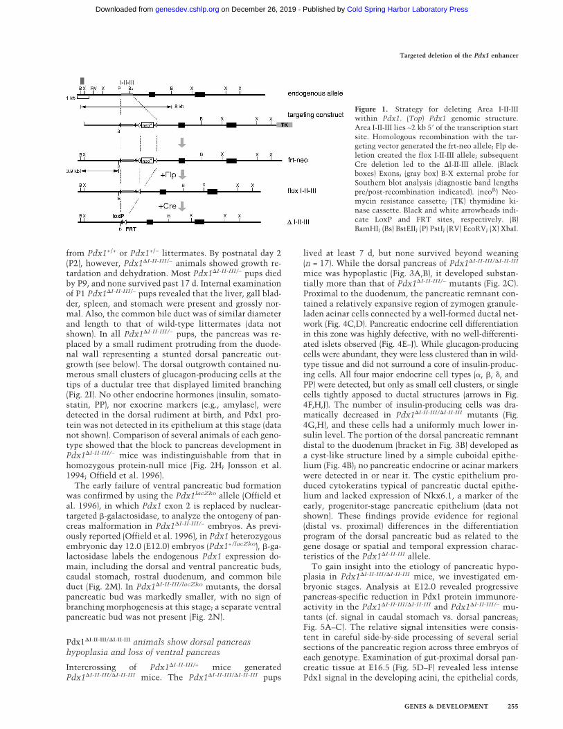
Figure 1. Strategy for deleting Area I-II-IIIwithin Pdx1. (Top) Pdx1 genomic structure.Area I-II-III lies ∼2 kb 5� of the transcription startsite. Homologous recombination with the tar-geting vector generated the frt-neo allele; Flp de-letion created the flox I-II-III allele; subsequentCre deletion led to the �I-II-III allele. (Blackboxes) Exons; (gray box) B-X external probe forSouthern blot analysis (diagnostic band lengthspre/post-recombination indicated). (neoR) Neo-mycin resistance cassette; (TK) thymidine ki-nase cassette. Black and white arrowheads indi-cate LoxP and FRT sites, respectively. (B)BamHI; (Bs) BstEII; (P) PstI; (RV) EcoRV; (X) XbaI.
Targeted deletion of the Pdx1 enhancer
GENES & DEVELOPMENT 255
Cold Spring Harbor Laboratory Press on December 26, 2019 - Published by genesdev.cshlp.orgDownloaded from
and ductal regions. In addition, there was a notable ab-sence of focal high-level expressing cells, which couldrepresent the precursors of endocrine islet cells based ontheir number and location tightly abutting the epithelialcords. The reduced level of Pdx1 throughout gestationmay account for the reduced mass of acinar tissue andfailed �-cell differentiation in Pdx1�I-II-III/�I-II-III mu-tants. While it has been reported recently that matureislets in Pdx1+/− animals are susceptible to apoptosis(Johnson et al. 2003), the phenomenon was observed inold mice (∼5 mo to 1 yr of age). Together with the findingof an early stage hypoplasia of the Pdx1�I-II-III/�I-II-III
pancreatic bud (Figs. 3, 5), we therefore consider it un-likely that apoptosis is a major contributor to the re-duced pancreas mass in Pdx1�I-II-III/�I-II-III animals. Fur-thermore, immunodetection of the proapoptotic cleavedform of caspase-3 revealed no increase in apoptosis inE16.5 or E18.5 Pdx1�I-II-III/�I-II-III tissue (data not shown).
Differential requirement for Area I-II-III for dorsalversus ventral pancreas specification
Since newborn Pdx1�I-II-III/�I-II-III animals appear to lackthe ventral pancreas lobe (Fig. 3B), we used the genetic
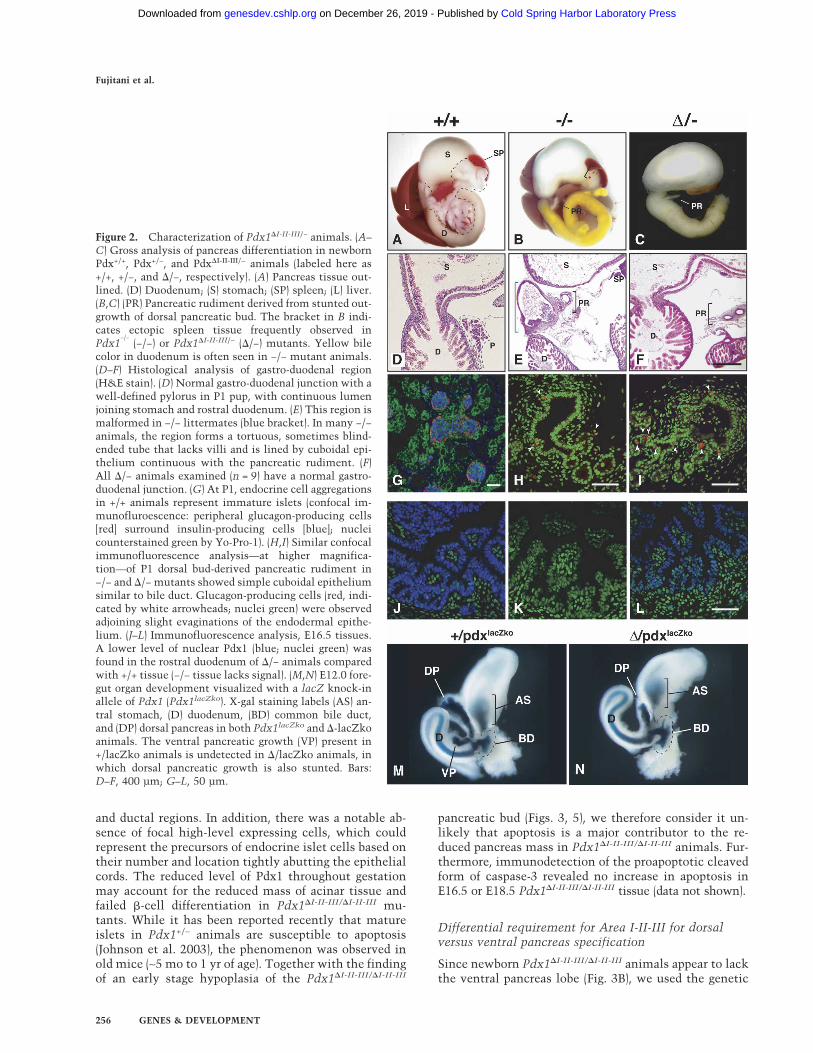
Figure 2. Characterization of Pdx1�I-II-III/− animals. (A–C) Gross analysis of pancreas differentiation in newbornPdx+/+, Pdx+/−, and Pdx�I-II-III/− animals (labeled here as+/+, +/−, and �/−, respectively). (A) Pancreas tissue out-lined. (D) Duodenum; (S) stomach; (SP) spleen; (L) liver.(B,C) (PR) Pancreatic rudiment derived from stunted out-growth of dorsal pancreatic bud. The bracket in B indi-cates ectopic spleen tissue frequently observed inPdx1
−/−(−/−) or Pdx1�I-II-III/− (�/−) mutants. Yellow bile
color in duodenum is often seen in −/− mutant animals.(D–F) Histological analysis of gastro-duodenal region(H&E stain). (D) Normal gastro-duodenal junction with awell-defined pylorus in P1 pup, with continuous lumenjoining stomach and rostral duodenum. (E) This region ismalformed in −/− littermates (blue bracket). In many −/−animals, the region forms a tortuous, sometimes blind-ended tube that lacks villi and is lined by cuboidal epi-thelium continuous with the pancreatic rudiment. (F)All �/− animals examined (n = 9) have a normal gastro-duodenal junction. (G) At P1, endocrine cell aggregationsin +/+ animals represent immature islets (confocal im-munofluroescence: peripheral glucagon-producing cells[red] surround insulin-producing cells [blue]; nucleicounterstained green by Yo-Pro-1). (H,I) Similar confocalimmunofluorescence analysis—at higher magnifica-tion—of P1 dorsal bud-derived pancreatic rudiment in−/− and �/− mutants showed simple cuboidal epitheliumsimilar to bile duct. Glucagon-producing cells (red, indi-cated by white arrowheads; nuclei green) were observedadjoining slight evaginations of the endodermal epithe-lium. (J–L) Immunofluorescence analysis, E16.5 tissues.A lower level of nuclear Pdx1 (blue; nuclei green) wasfound in the rostral duodenum of �/− animals comparedwith +/+ tissue (−/− tissue lacks signal). (M,N) E12.0 fore-gut organ development visualized with a lacZ knock-inallele of Pdx1 (Pdx1lacZko). X-gal staining labels (AS) an-tral stomach, (D) duodenum, (BD) common bile duct,and (DP) dorsal pancreas in both Pdx1lacZko and �-lacZkoanimals. The ventral pancreatic growth (VP) present in+/lacZko animals is undetected in �/lacZko animals, inwhich dorsal pancreatic growth is also stunted. Bars:D–F, 400 µm; G–L, 50 µm.
Fujitani et al.
256 GENES & DEVELOPMENT
Cold Spring Harbor Laboratory Press on December 26, 2019 - Published by genesdev.cshlp.orgDownloaded from
lineage-tracing approach of Kawaguchi et al. (2002) totest whether any ventral pancreatic tissue was specifiedin these mutants. This approach is powerful because itallows the progeny of cells that activate Ptf1a—an earlymarker that, within endoderm, is exquisitely specific foronly the region that acquires the pancreatic fate—to befollowed even if their abnormal genetic constitutioncauses them to move toward nonpancreatic fates, for ex-ample, taking up residence in and differentiating as duo-denal cell types (Kawaguchi et al. 2002). We crossedPtf1aCRE/+;Pdx1�I-II-III/+ with R26R;Pdx1�I-II-III/+ miceto obtain Ptf1aCRE/+;R26R and littermate Ptf1aCRE/+;R26R;Pdx1�I-II-III/�I-II-III mice. In these animals, Ptf1a-driven Cre expression excises a floxed stop cassette andactivates �-galactosidase expression in a heritable cell-
type-independent manner. In normal animals, Ptf1aCRE/
+; R26R labeling traces pancreatic progenitors and theirdescendants in the duct, acinar, and endocrine lineages
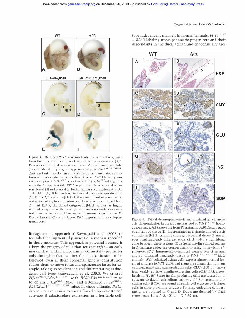
Figure 4. Distal dysmorphogenesis and proximal quasipancre-atic differentiation in dorsal pancreas bud of Pdx1�I-II-III homo-zygous mice. All tissues are from P1 animals. (A,B) Distal regionof dorsal bud tissue (D) differentiates as a simple dilated cysticepithelium (H&E staining), while gut-proximal tissue (P) under-goes quasipancreatic differentiation (cf. A), with a transitionalzone between these regions. Blue hematoxylin-stained regionsin A indicate endocrine compartment forming in newborn +/+pancreas. (C–J) Immunohistochemical comparison of normaland gut-proximal pancreatic tissue of Pdx1�I-II-III/�I-II-III (�/�)animals. Well-polarized acinar cells express almost normal lev-els of amylase (AMY) (C,D), and there are substantial numbersof disorganized glucagon-producing cells (GLU) (E,F), but only afew, weakly positive insulin-expressing cells (G,H; INS, arrow-heads in H). (H) Some insulin-producing cells are located in oradjacent to ductal epithelium (arrows). (I,J) Somatostatin-pro-ducing cells (SOM) are found as small cell clusters or isolatedcells in close proximity to ducts. Forming endocrine compart-ments are outlined in C and D. Ducts are denoted by blackarrowheads. Bars: A–B, 400 µm; C–J, 50 µm.
Figure 3. Reduced Pdx1 function leads to dysmorphic growthfrom the dorsal bud and loss of ventral bud specification. (A,B)Pancreas is outlined in newborn pups. Ventral pancreatic lobe(intraduodenal loop region) appears absent in Pdx1�I-II-III/�I-II-III
(�/�) mutants. Bracket in B indicates cystic pancreatic epithe-lium with associated ectopic splenic tissue. (C–F) Heterozygousmice carrying a Ptf1aCRE knock-in allele (Ptf1aCRE/+) togetherwith the Cre-activatable R26R reporter allele were used to as-sess dorsal (d) and ventral (v) bud pancreas specification at E10.5and E14.5. (C,D) In contrast to normal pancreas specification(C), E10.5 �/� mutants (D) lack the ventral bud region-specificactivation of Ptf1a expression and have a reduced dorsal bud.(E,F) At E14.5, the dorsal outgrowth (black arrows) is highlystunted compared with normal, and there is no evidence of ven-tral lobe-derived cells (blue arrow in normal situation in E).Dotted lines in C and D denote Ptf1a expression in developingspinal cord.
Targeted deletion of the Pdx1 enhancer
GENES & DEVELOPMENT 257
Cold Spring Harbor Laboratory Press on December 26, 2019 - Published by genesdev.cshlp.orgDownloaded from
(Kawaguchi et al. 2002). Therefore, both dorsal and ven-tral pancreatic buds at E10.5 and both pancreatic lobes atE14.5 were labeled robustly in Ptf1aCRE/+;R26R mice(Fig. 3C,E). In contrast, at both the early and midgesta-tion time points, there was no formation of putative ven-tral pancreas or its progenitors in Pdx1�I-II-III/�I-II-III mu-tants. Importantly, no ventral pancreas-specified pro-genitors led to progeny that differentiated as eitherpancreatic or intestinal cell types in the gut tube. Thislabeling strategy also showed that the dorsal bud wasalready stunted at the earliest time point, and was muchsmaller than normal at midgestation (Fig. 3D,F). The fail-ure to activate Ptf1a expression shows that a specificlevel and/or timing of Pdx1 expression is required for thespecification of the ventral pancreas; i.e., the productionor survival of Ptf1a-expressing progenitor cells.
Gastro-duodenal defects limited to enteroendocrinecells in Pdx1�I-II-III/− mutants
It was previously shown that Pdx1−/− (global protein-null) animals display various defects in addition to pan-creas agenesis, including a distorted pylorus, absence ofBrunner’s glands, and localized reductions in enteroen-docrine cell numbers (Larsson et al. 1996; Offield et al.
1996; Jepeal et al. 2005). Many Pdx1−/− animals had py-loric atresia and the rostral-most duodenal cavity had anundulating appearance, lacking villi, that was lined by asimple cuboidal epithelium continuous with the com-mon bile duct and dorsal pancreatic ductule (Fig. 2E;Offield et al. 1996). At P1, a substantial proportion ofPdx1−/− pups showed stomach distension caused by poorgastric emptying likely associated with the malformedgastro-duodenal junction (Offield et al. 1996). In contrast, adistended stomach was not observed in Pdx1�I-II-III/− mu-tants (n = 12). The gastro-duodenal junction developednormally in Pdx1�I-II-III/− mice, and the smooth musclebands of the pyloric ring, columnar epithelium of therostral duodenal villi (Fig. 2F), and Brunner’s glands(Supplementary Fig. 1) were all well formed. Pdx1 pro-tein was detected at E16.5 in the rostral duodenum (Fig.2J–L) and antral stomach epithelium (data not shown) ofPdx1�I-II-III/− animals, although the level was lower thanin Pdx1+/+ animals (Fig. 2J,L); similar findings were madeat E18.5 (data not shown). Therefore, while Pdx1 expres-sion in the Pdx1�I-II-III/− animals is sufficient for normalmorphological formation of the gastro-duodenal junctionand Brunner’s gland differentiation, the reduced level, orabnormalities in the spatiotemporal expression profile,cannot support pancreatic development over and abovethe level seen in Pdx1−/− mutants. The very low level of
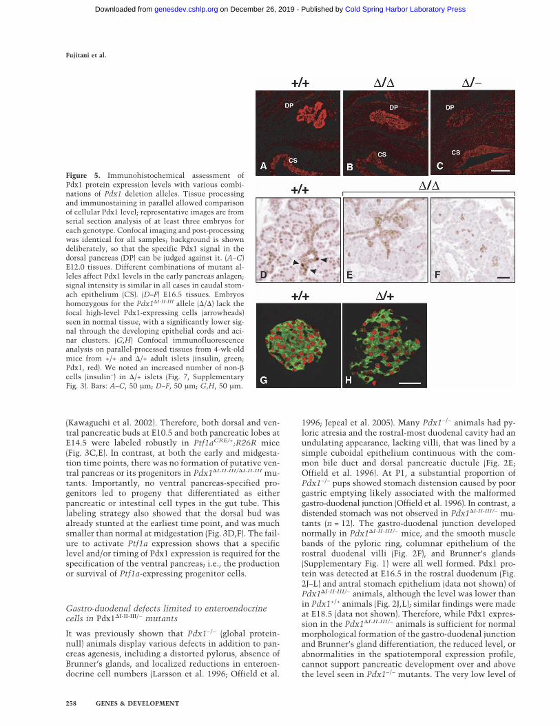
Figure 5. Immunohistochemical assessment ofPdx1 protein expression levels with various combi-nations of Pdx1 deletion alleles. Tissue processingand immunostaining in parallel allowed comparisonof cellular Pdx1 level; representative images are fromserial section analysis of at least three embryos foreach genotype. Confocal imaging and post-processingwas identical for all samples; background is showndeliberately, so that the specific Pdx1 signal in thedorsal pancreas (DP) can be judged against it. (A–C)E12.0 tissues. Different combinations of mutant al-leles affect Pdx1 levels in the early pancreas anlagen;signal intensity is similar in all cases in caudal stom-ach epithelium (CS). (D–F) E16.5 tissues. Embryoshomozygous for the Pdx1�I-II-III allele (�/�) lack thefocal high-level Pdx1-expressing cells (arrowheads)seen in normal tissue, with a significantly lower sig-nal through the developing epithelial cords and aci-nar clusters. (G,H) Confocal immunofluorescenceanalysis on parallel-processed tissues from 4-wk-oldmice from +/+ and �/+ adult islets (insulin, green;Pdx1, red). We noted an increased number of non-�cells (insulin−) in �/+ islets (Fig. 7, SupplementaryFig. 3). Bars: A–C, 50 µm; D–F, 50 µm; G,H, 50 µm.
Fujitani et al.
258 GENES & DEVELOPMENT
Cold Spring Harbor Laboratory Press on December 26, 2019 - Published by genesdev.cshlp.orgDownloaded from
Pdx1 in the Pdx1�I-II-III/− dorsal pancreas detected atE12.0 (Fig. 5) was transient, not being detected at lategestation or in neonates (data not shown).
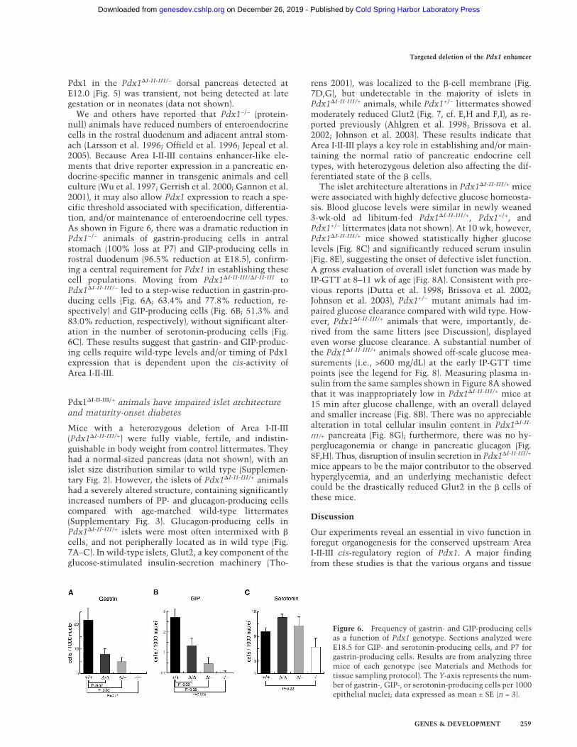
We and others have reported that Pdx1−/− (protein-null) animals have reduced numbers of enteroendocrinecells in the rostral duodenum and adjacent antral stom-ach (Larsson et al. 1996; Offield et al. 1996; Jepeal et al.2005). Because Area I-II-III contains enhancer-like ele-ments that drive reporter expression in a pancreatic en-docrine-specific manner in transgenic animals and cellculture (Wu et al. 1997; Gerrish et al. 2000; Gannon et al.2001), it may also allow Pdx1 expression to reach a spe-cific threshold associated with specification, differentia-tion, and/or maintenance of enteroendocrine cell types.As shown in Figure 6, there was a dramatic reduction inPdx1−/− animals of gastrin-producing cells in antralstomach (100% loss at P7) and GIP-producing cells inrostral duodenum (96.5% reduction at E18.5), confirm-ing a central requirement for Pdx1 in establishing thesecell populations. Moving from Pdx1�I-II-III/�I-II-III toPdx1�I-II-III/− led to a step-wise reduction in gastrin-pro-ducing cells (Fig. 6A; 63.4% and 77.8% reduction, re-spectively) and GIP-producing cells (Fig. 6B; 51.3% and83.0% reduction, respectively), without significant alter-ation in the number of serotonin-producing cells (Fig.6C). These results suggest that gastrin- and GIP-produc-ing cells require wild-type levels and/or timing of Pdx1expression that is dependent upon the cis-activity ofArea I-II-III.
Pdx1�I-II-III/+ animals have impaired islet architectureand maturity-onset diabetes
Mice with a heterozygous deletion of Area I-II-III(Pdx1�I-II-III/+) were fully viable, fertile, and indistin-guishable in body weight from control littermates. Theyhad a normal-sized pancreas (data not shown), with anislet size distribution similar to wild type (Supplemen-tary Fig. 2). However, the islets of Pdx1�I-II-III/+ animalshad a severely altered structure, containing significantlyincreased numbers of PP- and glucagon-producing cellscompared with age-matched wild-type littermates(Supplementary Fig. 3). Glucagon-producing cells inPdx1�I-II-III/+ islets were most often intermixed with �cells, and not peripherally located as in wild type (Fig.7A–C). In wild-type islets, Glut2, a key component of theglucose-stimulated insulin-secretion machinery (Tho-
rens 2001), was localized to the �-cell membrane (Fig.7D,G), but undetectable in the majority of islets inPdx1�I-II-III/+ animals, while Pdx1+/− littermates showedmoderately reduced Glut2 (Fig. 7, cf. E,H and F,I), as re-ported previously (Ahlgren et al. 1998; Brissova et al.2002; Johnson et al. 2003). These results indicate thatArea I-II-III plays a key role in establishing and/or main-taining the normal ratio of pancreatic endocrine celltypes, with heterozygous deletion also affecting the dif-ferentiated state of the � cells.
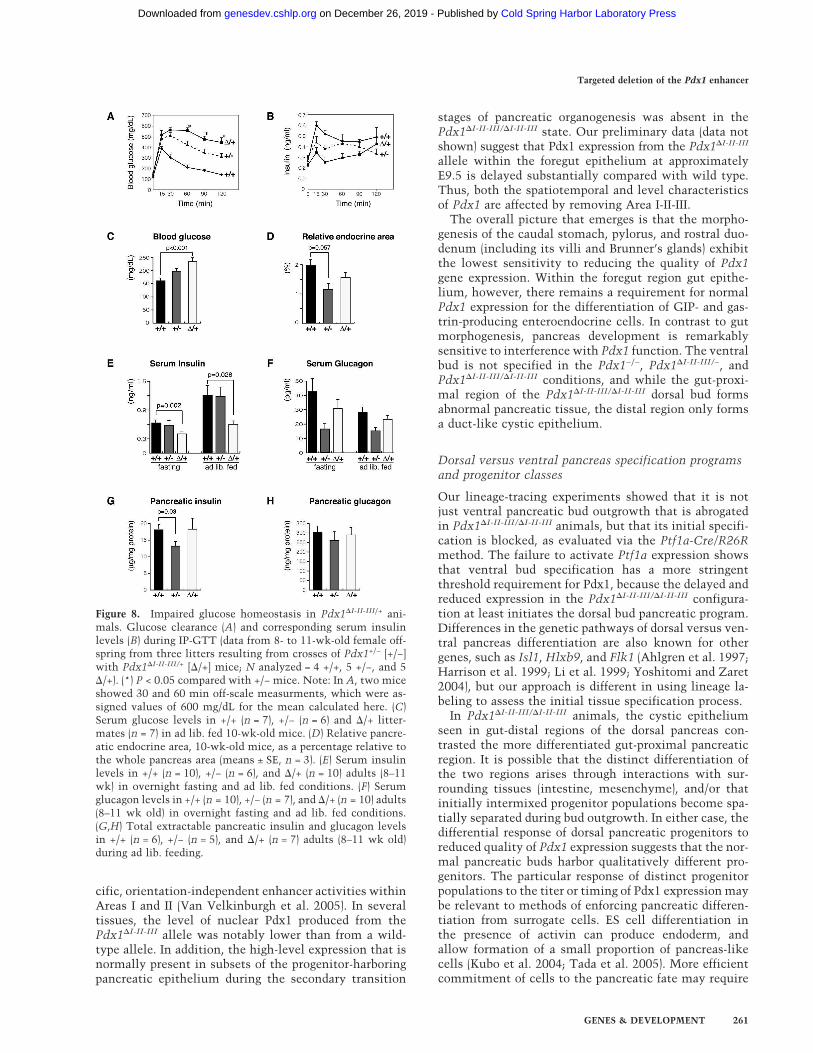
The islet architecture alterations in Pdx1�I-II-III/+ micewere associated with highly defective glucose homeosta-sis. Blood glucose levels were similar in newly weaned3-wk-old ad libitum-fed Pdx1�I-II-III/+, Pdx1+/+, andPdx1+/− littermates (data not shown). At 10 wk, however,Pdx1�I-II-III/+ mice showed statistically higher glucoselevels (Fig. 8C) and significantly reduced serum insulin(Fig. 8E), suggesting the onset of defective islet function.A gross evaluation of overall islet function was made byIP-GTT at 8–11 wk of age (Fig. 8A). Consistent with pre-vious reports (Dutta et al. 1998; Brissova et al. 2002;Johnson et al. 2003), Pdx1+/− mutant animals had im-paired glucose clearance compared with wild type. How-ever, Pdx1�I-II-III/+ animals that were, importantly, de-rived from the same litters (see Discussion), displayedeven worse glucose clearance. A substantial number ofthe Pdx1�I-II-III/+ animals showed off-scale glucose mea-surements (i.e., >600 mg/dL) at the early IP-GTT timepoints (see the legend for Fig. 8). Measuring plasma in-sulin from the same samples shown in Figure 8A showedthat it was inappropriately low in Pdx1�I-II-III/+ mice at15 min after glucose challenge, with an overall delayedand smaller increase (Fig. 8B). There was no appreciablealteration in total cellular insulin content in Pdx1�I-II-
III/+ pancreata (Fig. 8G); furthermore, there was no hy-perglucagonemia or change in pancreatic glucagon (Fig.8F,H). Thus, disruption of insulin secretion in Pdx1�I-II-III/+
mice appears to be the major contributor to the observedhyperglycemia, and an underlying mechanistic defectcould be the drastically reduced Glut2 in the � cells ofthese mice.
Discussion
Our experiments reveal an essential in vivo function inforegut organogenesis for the conserved upstream AreaI-II-III cis-regulatory region of Pdx1. A major findingfrom these studies is that the various organs and tissue
Figure 6. Frequency of gastrin- and GIP-producing cellsas a function of Pdx1 genotype. Sections analyzed wereE18.5 for GIP- and serotonin-producing cells, and P7 forgastrin-producing cells. Results are from analyzing threemice of each genotype (see Materials and Methods fortissue sampling protocol). The Y-axis represents the num-ber of gastrin-, GIP-, or serotonin-producing cells per 1000epithelial nuclei; data expressed as mean ± SE (n = 3).
Targeted deletion of the Pdx1 enhancer
GENES & DEVELOPMENT 259
Cold Spring Harbor Laboratory Press on December 26, 2019 - Published by genesdev.cshlp.orgDownloaded from
derivatives of the posterior foregut (caudal stomach, py-lorus, rostral duodenum, and pancreas) are differentiallysensitive to the quality of Pdx1 expression—i.e., the spa-tiotemporal and level characteristics—which is substan-tially altered when Area I-II-III is deleted. Moreover,there is a differential requirement for Pdx1 in specifyingthe dorsal or ventral pancreatic bud, and our results sug-gest that the dorsal bud contains different classes of pro-genitors with respect to their ability to move through thepancreatic differentiation program. These findings setthe stage for the future iterative dissection in vivo ofspecific subregions and selected motifs within this ∼1-kbregion, which contains highly clustered binding sites fora variety of well-known endoderm and pancreas tran-scription factors. Based upon the level of analysis com-pared with other pancreas genes, Pdx1 could be consid-ered an important reference gene, as it may represent anexus for the complex signaling and transcriptional in-puts that orchestrate proliferation, outgrowth, and dif-ferentiation of this organ. It will be particularly relevantto determine how regionalized intercellular signals leadto the dynamic level and timing of Pdx1 expression in
specific cell types through the assembly of transcrip-tional complexes at the various motifs in Area I-II-III,and how these control lineage diversification decisionsor, later, affect the maintenance of the mature cell state.Clearly, a substantial hurdle to overcome in such studieswill be the current paucity of markers that define criticalprogenitor subtypes in the pancreatic endocrine differen-tiation program. Information on the networks orchestrat-ing endodermal progenitor cell differentiation could have alarge impact on protocols for directed tissue differentiationleading toward cell-based therapies for diabetes.
Dosage effects of Pdx1 on foregut developmentand pancreas organogenesis
Figure 9 summarizes how foregut development is af-fected by the various combinations of Pdx1�I-II-III,Pdx1−, and wild-type alleles. Notably, removing Area I-II-III does not inactivate the gene, but instead generates ahypomorphic allele in terms of its level and spatiotem-poral expression characteristics. This finding is consis-tent with the recent characterization of cell-type-spe-
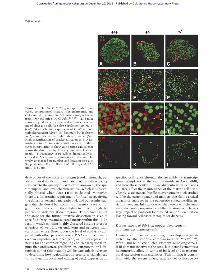
Figure 7. The Pdx1�I-II-III/+ genotype leads to se-verely compromised mature islet architecture andendocrine differentiation. All tissues analyzed werefrom 4-wk-old mice. (A–C) Pdx1�I-II-III/+ (�/+) miceshow a reproducible increase and intra-islet scatter-ing of glucagon cells (see also Supplementary Fig. 3)(D–F) �-Cell-selective expression of Glut2 is mod-estly decreased in Pdx1+/− (+/−) animals, but is absentin �/+ animals (arrowheads indicate ducts). (G–I)High magnification of bracketed region in D–F (ar-rowheads in H,I indicate autofluorescent erythro-cytes in capillaries to show gain setting equivalenceacross the three panels; three erythrocytes clusteredin H). (J–L) Frequency of PP cells is dramatically in-creased in �/+ animals; somatostatin cells are rela-tively unchanged in number and location (see alsoSupplementary Fig. 3). Bars: A–F, 50 µm; G–I, 12.5µm; J–L, 50 µm.
Fujitani et al.
260 GENES & DEVELOPMENT
Cold Spring Harbor Laboratory Press on December 26, 2019 - Published by genesdev.cshlp.orgDownloaded from
cific, orientation-independent enhancer activities withinAreas I and II (Van Velkinburgh et al. 2005). In severaltissues, the level of nuclear Pdx1 produced from thePdx1�I-II-III allele was notably lower than from a wild-type allele. In addition, the high-level expression that isnormally present in subsets of the progenitor-harboringpancreatic epithelium during the secondary transition
stages of pancreatic organogenesis was absent in thePdx1�I-II-III/�I-II-III state. Our preliminary data (data notshown) suggest that Pdx1 expression from the Pdx1�I-II-III
allele within the foregut epithelium at approximatelyE9.5 is delayed substantially compared with wild type.Thus, both the spatiotemporal and level characteristicsof Pdx1 are affected by removing Area I-II-III.
The overall picture that emerges is that the morpho-genesis of the caudal stomach, pylorus, and rostral duo-denum (including its villi and Brunner’s glands) exhibitthe lowest sensitivity to reducing the quality of Pdx1gene expression. Within the foregut region gut epithe-lium, however, there remains a requirement for normalPdx1 expression for the differentiation of GIP- and gas-trin-producing enteroendocrine cells. In contrast to gutmorphogenesis, pancreas development is remarkablysensitive to interference with Pdx1 function. The ventralbud is not specified in the Pdx1−/−, Pdx1�I-II-III/−, andPdx1�I-II-III/�I-II-III conditions, and while the gut-proxi-mal region of the Pdx1�I-II-III/�I-II-III dorsal bud formsabnormal pancreatic tissue, the distal region only formsa duct-like cystic epithelium.
Dorsal versus ventral pancreas specification programsand progenitor classes
Our lineage-tracing experiments showed that it is notjust ventral pancreatic bud outgrowth that is abrogatedin Pdx1�I-II-III/�I-II-III animals, but that its initial specifi-cation is blocked, as evaluated via the Ptf1a-Cre/R26Rmethod. The failure to activate Ptf1a expression showsthat ventral bud specification has a more stringentthreshold requirement for Pdx1, because the delayed andreduced expression in the Pdx1�I-II-III/�I-II-III configura-tion at least initiates the dorsal bud pancreatic program.Differences in the genetic pathways of dorsal versus ven-tral pancreas differentiation are also known for othergenes, such as Isl1, Hlxb9, and Flk1 (Ahlgren et al. 1997;Harrison et al. 1999; Li et al. 1999; Yoshitomi and Zaret2004), but our approach is different in using lineage la-beling to assess the initial tissue specification process.
In Pdx1�I-II-III/�I-II-III animals, the cystic epitheliumseen in gut-distal regions of the dorsal pancreas con-trasted the more differentiated gut-proximal pancreaticregion. It is possible that the distinct differentiation ofthe two regions arises through interactions with sur-rounding tissues (intestine, mesenchyme), and/or thatinitially intermixed progenitor populations become spa-tially separated during bud outgrowth. In either case, thedifferential response of dorsal pancreatic progenitors toreduced quality of Pdx1 expression suggests that the nor-mal pancreatic buds harbor qualitatively different pro-genitors. The particular response of distinct progenitorpopulations to the titer or timing of Pdx1 expression maybe relevant to methods of enforcing pancreatic differen-tiation from surrogate cells. ES cell differentiation inthe presence of activin can produce endoderm, andallow formation of a small proportion of pancreas-likecells (Kubo et al. 2004; Tada et al. 2005). More efficientcommitment of cells to the pancreatic fate may require
Figure 8. Impaired glucose homeostasis in Pdx1�I-II-III/+ ani-mals. Glucose clearance (A) and corresponding serum insulinlevels (B) during IP-GTT (data from 8- to 11-wk-old female off-spring from three litters resulting from crosses of Pdx1+/− [+/−]with Pdx1�I-II-III/+ [�/+] mice; N analyzed = 4 +/+, 5 +/−, and 5�/+). (*) P < 0.05 compared with +/− mice. Note: In A, two miceshowed 30 and 60 min off-scale measurments, which were as-signed values of 600 mg/dL for the mean calculated here. (C)Serum glucose levels in +/+ (n = 7), +/− (n = 6) and �/+ litter-mates (n = 7) in ad lib. fed 10-wk-old mice. (D) Relative pancre-atic endocrine area, 10-wk-old mice, as a percentage relative tothe whole pancreas area (means ± SE, n = 3). (E) Serum insulinlevels in +/+ (n = 10), +/− (n = 6), and �/+ (n = 10) adults (8–11wk) in overnight fasting and ad lib. fed conditions. (F) Serumglucagon levels in +/+ (n = 10), +/− (n = 7), and �/+ (n = 10) adults(8–11 wk old) in overnight fasting and ad lib. fed conditions.(G,H) Total extractable pancreatic insulin and glucagon levelsin +/+ (n = 6), +/− (n = 5), and �/+ (n = 7) adults (8–11 wk old)during ad lib. feeding.
Targeted deletion of the Pdx1 enhancer
GENES & DEVELOPMENT 261
Cold Spring Harbor Laboratory Press on December 26, 2019 - Published by genesdev.cshlp.orgDownloaded from
careful control of the level of Pdx1 expression at precisestages of differentiation.
Pdx1 regulation in enteroendocrine and gut epithelialcell differentiation
Our previous studies (Wu et al. 1997; Gerrish et al. 2000,2001; Gannon et al. 2001) in transgenic mice and in vitrocell culture focused on pancreatic enhancer-like activi-ties of Area I-II-III and not whether this region regulatesPdx1 in gut enteroendocrine cells. Gastrin-producingcells in the antral stomach-pyloric mucosa and GIP-pro-ducing cells in the rostral duodenum strictly depend onthe presence of Area I-II-III, and are thus similar to pan-creatic � cells. This result strongly suggests that over-lapping sets of upstream transcriptional regulators forPdx1 drive endocrine differentiation in the pancreas andgut. There is evidence that transcriptional regulators ofendocrine pancreas development, such as Ngn3, Neu-roD, Nkx6.1, Pax6, and Pax4, are essential for enteroen-docrine differentiation (Naya et al. 1997; Larsson et al.1998; Larsson 2000; Jenny et al. 2002; Lee et al. 2002). Ofthese factors, null mutants for Pax6 have a characteristicphenotype in the enteroendocrine lineages—eliminationof duodenal GIP cells and gastrin cells in the antral stom-ach—that is reminiscent of Pdx1-null mutants (Larssonet al. 1998). Pax6 can bind to Area II in the Pdx1 pro-moter (Samaras et al. 2002), suggesting a fundamentalrole in the endocrine-specific regulation of the Pdx1 genein both pancreas and gut.
The enterocytes and Brunner’s gland cells, which areaffected in Pdx1-null mice, underwent normal differen-tiation in Pdx1�I-II-III/�I-II-III or Pdx1�I-II-III/− animals.Other elements must therefore regulate the basal tran-scription of Pdx1 or generate enough cis-activation forthe normal differentiation of the caudal stomach, pylo-rus, bile duct, and duodenum. Currently, we hypothesizethat distal 5� regions, such as the phylogenetically con-served Area IV (Sharma et al. 1997; Gerrish et al. 2004),or more distributed cis-regulatory motifs, are involved in
driving normal expression in the duodenum and stom-ach (D. Boyer and C. Wright, unpubl.). Future discoveriesmay reveal that potentiation of Area I-II-III activity bythese elements (Gerrish et al. 2004) fine-tunes Pdx1 ex-pression in the pancreatic endocrine lineage.
Etiology of the diabetic phenotype in Pdx1�I-II-III/+
mutants
By several measurements—serum insulin and glucoselevels under ad lib. feeding or glucose challenge, abnor-mal islet architecture—Pdx1�I-II-III/+ mice are more se-verely affected than their Pdx1+/− littermates, despitesimilar pancreatic insulin content. One major contribu-tor to the phenotype may be the lack of Glut2 on the �cells of Pdx1�I-II-III/+ islets compared with the moderatereduction in Pdx1+/− mice (Fig. 7). Glut2 is required forglucose-stimulated insulin secretion in mice (Guillam etal. 1997) and is a �-cell gene target of Pdx1 (Waeberet al. 1996). Quantitative Western blot analysis (datanot shown) detected a similar overall Pdx1 level inPdx1�I-II-III/+ and Pdx1+/− pancreata, and Pdx1�I-II-III/+ is-lets faithfully expressed Pdx1 in � cells (Fig. 5G,H), sug-gesting that it is not lower Pdx1 levels or inconsistentexpression that lead to the lack of Glut2. We hypothesizethat an underlying defect in islet genesis and organiza-tion is responsible for a profound disruption of the glu-cose-sensing machinery. Other mouse models in whichperipheral islet cells mix into the islet core, such as en-forced expression of HNF6 or dominant-negative HNF1�(Gannon et al. 2000; Yamagata et al. 2002), also showabsent or largely reduced Glut2 expression. Mixing ofperipheral cell types with � cells may disrupt the gapjunctional or other coupling that is involved in a con-certed, efficient insulin release (Caton et al. 2002).
Heterozygous enhancer deletion and the mechanismof disruption of islet morphology
The mixed islet phenotype and abnormally high �, �, andPP cell numbers in Pdx1�I-II-III/+ mice compared with
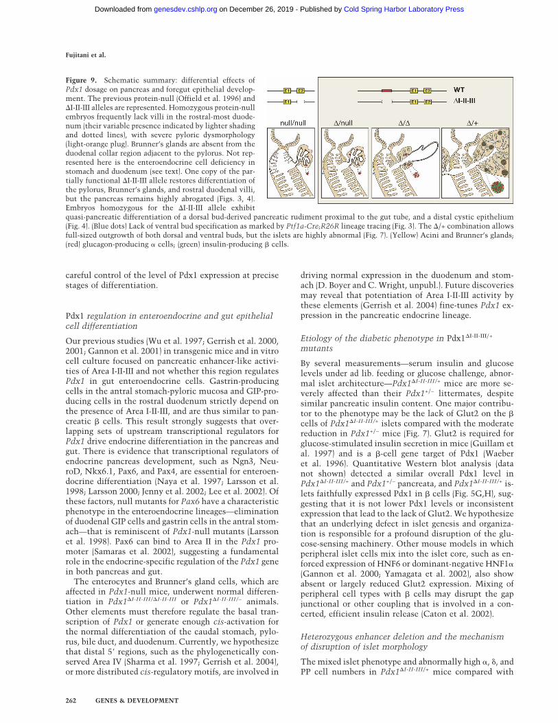
Figure 9. Schematic summary: differential effects ofPdx1 dosage on pancreas and foregut epithelial develop-ment. The previous protein-null (Offield et al. 1996) and�I-II-III alleles are represented. Homozygous protein-nullembryos frequently lack villi in the rostral-most duode-num (their variable presence indicated by lighter shadingand dotted lines), with severe pyloric dysmorphology(light-orange plug). Brunner’s glands are absent from theduodenal collar region adjacent to the pylorus. Not rep-resented here is the enteroendocrine cell deficiency instomach and duodenum (see text). One copy of the par-tially functional �I-II-III allele restores differentiation ofthe pylorus, Brunner’s glands, and rostral duodenal villi,but the pancreas remains highly abrogated (Figs. 3, 4).Embryos homozygous for the �I-II-III allele exhibitquasi-pancreatic differentiation of a dorsal bud-derived pancreatic rudiment proximal to the gut tube, and a distal cystic epithelium(Fig. 4). (Blue dots) Lack of ventral bud specification as marked by Ptf1a-Cre;R26R lineage tracing (Fig. 3). The �/+ combination allowsfull-sized outgrowth of both dorsal and ventral buds, but the islets are highly abnormal (Fig. 7). (Yellow) Acini and Brunner’s glands;(red) glucagon-producing � cells; (green) insulin-producing � cells.
Fujitani et al.
262 GENES & DEVELOPMENT
Cold Spring Harbor Laboratory Press on December 26, 2019 - Published by genesdev.cshlp.orgDownloaded from
wild-type or Pdx1+/− mice (Supplementary Fig. 3) sug-gests that loss of one copy of the Area I-II-III region leadsto defective lineage allocation from the endocrine pro-genitors. At the current time, mostly related to a lack ofknowledge on how to mark specific progenitor classes,this result cannot be explained mechanistically. How-ever, we consider it unlikely that the difference is attrib-utable to genetic background differences between thevarious mutant combinations. All comparisons betweenwild-type, Pdx1�I-II-III/+, and Pdx1+/− were between sib-lings, and the results were consistent among multiplelitters. Furthermore, analysis of mice bearing the floxedallele from which the Pdx1�I-II-III allele was derivedshowed no defects in islet architecture in Pdx1floxI-II-III/+,Pdx1floxI-II-III/floxI-II-III, or Pdx1floxI-II-III/− pancreata (datanot shown). One potential reason that heterozygosity forArea I-II-III is more severe than heterozygosity for theprotein-coding region could be that its enhancer activityalso affects the expression of another gene involved inislet progenitor differentiation. Candidate genes are thehomeobox genes flanking Pdx1 in the “Parahox cluster”,Gsh1 and Cdx2. Both genes may be expressed in thedeveloping pancreas and mature islets (Rosanas-Urgell etal. 2005), and Cdx2 can transcriptionally activate Gluca-gon in pancreatic cell lines (Andersen et al. 1999). It isalso possible that deletion of Area I-II-III eliminates arepressor element resulting in ectopic expression ofPdx1; however, no ectopic expression was observed inany tissues at any of the stages examined. Another pos-sibility is that Area I-II-III heterozygosity blunts a large-scale fold increase in Pdx1 expression that is linked tothe channeling of cells from a common endocrine pre-cursor pool toward the �-cell fate. Lineage-tracing ex-periments show that all pancreatic endocrine precursorsexpress Pdx1 (Gu et al. 2002), and in the mature organPdx1 is found at the highest levels in � cells (Guz et al.1995). The Pdx1�I-II-III allele produces a substantial base-line amount of Pdx1 in immature endoderm (Figs. 2L,5B), but is deficient for high-level expression (Fig. 5D–F),consistent with the idea (see above) that Area I-II-IIItrans-activators promote high-level �-cell-selective ex-pression. It is therefore plausible that the fold increasebetween baseline and maximum expression levels wouldbe reduced in Pdx1�I-II-III/+ mice compared with wildtype, while the active allele in +/− mice would stillachieve a fold-increase similar to wild type. If the effi-ciency of converting common endocrine precursors tocommitted �-cell progenitors depends on the degree ofup-regulation of Pdx1 expression at a specific lineagecommitment point, then the selective impairment tohigh-level expression could lead to cells defaulting to theother endocrine fates.
Conserved Pdx1 enhancers as targets for MODYsyndrome
Various transcriptional activators implicated in the hu-man MODY syndrome, or as major regulators of endo-derm development, have been shown capable of bindingwithin Area I-II-III: HNF1�, Foxa2/HNF3�, Pdx1 itself,
Maf, Pax6, and HNF6 (Gerrish et al. 2000, 2001; Marshaket al. 2000; Ben-Shushan et al. 2001; Samaras et al. 2002,2003; Jacquemin et al. 2003). In addition, MODY factorshave been reported to synergistically regulate Pdx1 ex-pression in � cells in vivo (Shih et al. 2002). These find-ings suggest that Area I-II-III may be a major site of ac-tion for transcription factors implicated in MODY. Con-sistent with this hypothesis, we found that heterozygousdeletion of Area I-II-III results in post-weaning diabetesas early as 4 wk of age. Although mutations in theequivalent region of human IPF1 have not yet been iden-tified, it is possible that even heterozygous Area I-II-IIImutations/deletions, or reductions in the level or activ-ity of trans-acting MODY factors that operate throughArea I-II-III, could be a significant underlying influencein MODY and type II diabetes.
Among previously characterized mutants for the fac-tors that can bind within Area I-II-III, the Hnf6-null mice(Jacquemin et al. 2003) exhibit delayed and reduced Pdx1expression similar to that of Pdx1�I-II-III/�I-II-III mutants.Although the pancreatic phenotype of Hnf6−/− seems lesssevere overall, both types of mutant have delayed pan-creas specification, impaired endocrine differentiation,and blocked ventral bud outgrowth. The phenotypicsimilarities raise the possibility that HNF6 may be a keyregulator of Pdx1 via Area I-II-III during the early phasesof pancreas formation. A future goal will be to test rig-orously the contribution of individual trans-acting fac-tors to the Pdx1�I-II-III/�I-II-III phenotype by altering spe-cific binding motifs in Area I-II-III.
Materials and methods
PdxArea I-II-III flox mice
All animal experiments were performed in accordance with theguidelines of the Vanderbilt University Animal Care and UseCommittee. Details of the targeting vector construction areavailable upon request from Y. Fujitani. In brief, a 5�-located3.4-kb XbaI–PstI fragment (converted to a SalI–ClaI fragment; 5�
arm), a 1.2-kb PCR fragment with added HindIII ends encom-passing Area I-II-III, and an ∼10-kb fragment encompassing thePdx1 gene from a NotI site added by PCR immediately 3� ofArea III to the SacI site ∼3 kb 3� of the stop codon (3� arm) weresubcloned into pFRT.loxP (vector from M.A. Magnuson), whichcontains a thymidine kinase cassette, FRT-flanked neomycinresistance cassette, and loxP-flanked multiple cloning site. Lin-earized targeting vector was electroporated into ES cells (TL1/129S6 strain) and clones selected for homologous integration.Southern analysis of BamHI-digested DNA from doubly resis-tant ES cell clones revealed a targeting rate of 5.5% (10/182clones). ES cells were injected into C57BL/6 blastocysts thatwere transferred into pseudopregnant ICR female mice; malechimeras were bred to Black Swiss females and agouti offspringgenotyped by Southern analysis. Heterozygous offspring werebred to ACTB:FLPe mice (see below) to remove the neomycin-resistance cassette.
Other genetically engineered animals
EIIA-cre (H. Westphal, National Institutes of Health, Bethesda,MD) and ACTB:FLPe mice (S.M. Dymecki, Harvard Medical
Targeted deletion of the Pdx1 enhancer
GENES & DEVELOPMENT 263
Cold Spring Harbor Laboratory Press on December 26, 2019 - Published by genesdev.cshlp.orgDownloaded from
School, Boston, MA) were provided by B. Hogan (Duke Univer-sity, Durham, NC). PdxlacZko and Ptf1aCRE/+ mice were de-scribed (Offield et al. 1996; Kawaguchi et al. 2002). All micewere kept on a C57BL/6 and DBA mixed-inbred background.Genotyping was by PCR or Southern blot with tail DNA fromanimals staged E16.5 through adult. DNA for genotyping wasisolated from heads of E14.5 or earlier embryos. Primer se-quences for PCR genotyping are available upon request to Y.Fujitani.
X-gal staining
Embryos or dissected tissues were fixed (ice-cold 4% paraform-aldehyde/PBS, 4°C, 60 min) then washed twice in permeabili-zation solution (2 mM MgCl2, 0.01% sodium deoxycholate,0.02% Nonidet P-40 in PBS). �-Galactosidase was detected us-ing X-gal (Wu et al. 1997). Samples were post-fixed and dehy-drated for embedding as described below. Photographs weretaken on an SZX12 microscope (Olympus) and Spot digital cam-era.
Histology
Dissected tissues were fixed (ice-cold 4% paraformaldehyde,4°C, 60 min), dehydrated in an ethanol series, washed intoHisto-Clear (National Diagnostics), infiltrated in Histo-Clear/paraffin (1:1 v/v) and two changes of paraffin under vacuum at56°C, embedded, and cut into 5-µm sections.
Immunohistochemistry and confocal image analysis
Hematoxylin-eosin (H&E) staining and periodic acid-Schiffstaining were as described (Offield et al. 1996). For immunoper-oxidase, Vectastain ABC kit (Vector Labs) was used. Primaryantibodies: guinea pig anti-bovine insulin (Linco), 1:10,000;anti-glucagon (Linco), 1:10,000; rabbit anti-somatostatin(Dako), 1:1000; guinea pig anti-pancreatic polypeptide (Linco),1:2000; rabbit anti-amylase (Sigma), 1:20,000; rabbit anti-Pdx1(Peshavaria et al. 1994; Kawaguchi et al. 2002), 1:5000. Antibod-ies for immunofluorescence: guinea pig anti-Pdx1 (Hingorani etal. 2003), 1:5000; rabbit anti-Pdx1, 1:250; rabbit anti-rat Glut2(Alpha Diagnostic), 1:200. Secondary antibodies: CY2-conju-gated donkey anti-guinea pig IgG (Jackson ImmunoReaerchlaboratories, for insulin), CY3-conjugated donkey anti-rabbitIgG (for glucagon and Glut2) and CY5-conjugated anti-guineapig IgG (Pdx1 staining in Fig. 2). Some samples were counter-stained with the nuclear dye, YO-PRO-1 (Molecular Probes; 1:1000 in PBS). Immunofluorescence was imaged on a Zeiss LSM510 confocal microscope. TIFF images were processed in AdobePhotoshop.
Quantification of gastrin-, GIP-, and serotonin-positive cells
E18.5 foregut sections were immunostained with rabbit GIPantiserum (Research Diagnostics) or mouse serotonin mono-clonal antibody (Dako). P7 distal stomach was immunostainedwith rabbit gastrin antiserum (Research Diagnostics). Sectionswere subjected to immunoperoxidase staining and light hema-toxylin counterstain to reveal nuclei and general morphology.For each animal, cell counting was performed on five nonadja-cent longitudinal sections, to avoid double scoring, within anarea bounded by a 2:1 ratio rectangle whose short side was simi-lar to the gut diameter (∼900 µm), short side adjacent to thepylorus. For Pdx1−/− mutants, the rectangle was shifted ∼0.3mm distal to the pylorus to avoid the abnormal cuboidal epi-thelium in the rostral duodenum. Eight fields (two rows of four
nonoverlapping fields [340 µm × 450 µm] running down eachside of the rectangle, with the gut wall included at one edge) ofeach section were photographed (SPOT camera) and printed at450× magnification to count gastrin-, GIP-, or serotonin-posi-tive cells, and the total number of epithelial cells.
Glucose tolerance tests (IP-GTT)
Following a 16- to 18-h fast, baseline blood glucose levels (mil-ligrams per deciliter) were measured in tail-vein blood by BDLogic glucose monitor (Becton Dickinson). Dextrose (2 mg/gbody weight) was injected intraperitoneally and blood glucosemeasured at 15, 30, 60, 90, and 120 min after injection. Forsimultaneous measurement of plasma insulin, blood was ob-tained via the saphenous vein.
Radioimmunoassay
Plasma insulin and glucagon were assayed by radioimmunoas-say (RIA; Linco) according to the manufacturer’s protocol. Pan-creatic insulin and glucagon content was assessed by RIA inacid-ethanol extracts of whole pancreas.
Islet morphometric analysis
Relative endocrine area was measured using NIH Image 1.41software. For each experimental group, 10-wk-old mice wereused. There was no statistical difference in pancreas weightamong these mice. A total of nine sections were prepared fromthree pancreata (three sections per mouse). Each section wasseparated enough to avoid double scoring the same islet. Thecomplete tissue area over all three sections was determined andpercentage represented by islet cells calculated.
Statistics
Data are expressed as mean ± SE. A two-way ANOVA for re-peated measures was used to analyze time course differencesbetween groups. When significant changes were obtained overtime, post hoc comparisons were made by paired t-test. Pairwisecomparison for the number of enteroendocrine cells, blood glu-cose levels, plasma insulin and glucagon, pancreatic insulin andglucagon, and relative endocrine area was made by unpairedt-test. P values <0.05 were considered statistically significant.
Acknowledgments
We thank A. Means, K. Gerrish, S. Samaras, D. Melton, G. Gu,and members of the Wright, Stein, and Magnuson labs for dis-cussions, and P. Soriano for R26R mice. Support from the Trans-genic Mouse/ES Cell Shared Resource, Cell Imaging Core, Se-quencing Core, and Histology Core at Vanderbilt University isacknowledged. This work was funded in part by Research Fel-lowships of the Japan Society for the Promotion of Science andJuvenile Diabetes Research Foundation International (to Y.F.)and NIH Grant U19 DK 042502 (to C.V.E.W.).
References
Ahlgren, U., Pfaff, S.L., Jessell, T.M., Edlund, T., and Edlund, H.1997. Independent requirement for ISL1 in formation of pan-creatic mesenchyme and islet cells. Nature 385: 257–260.
Ahlgren, U., Jonsson, J., Jonsson, L., Simu, K., and Edlund, H.1998. �-Cell-specific inactivation of the mouse Ipf1/Pdx1gene results in loss of the �-cell phenotype and maturity
Fujitani et al.
264 GENES & DEVELOPMENT
Cold Spring Harbor Laboratory Press on December 26, 2019 - Published by genesdev.cshlp.orgDownloaded from
onset diabetes. Genes & Dev. 12: 1763–1768.Andersen, F.G., Heller, R.S., Petersen, H.V., Jensen, J., Madsen,
O.D., and Serup, P. 1999. Pax6 and Cdx2/3 form a functionalcomplex on the rat glucagon gene promoter G1-element.FEBS Lett. 445: 306–310.
Bell, G.I. and Polonsky, K.S. 2001. Diabetes mellitus and geneti-cally programmed defects in �-cell function. Nature414: 788–791.
Ben-Shushan, E., Marshak, S., Shoshkes, M., Cerasi, E., and Mel-loul, D. 2001. A pancreatic �-cell-specific enhancer in thehuman PDX-1 gene is regulated by hepatocyte nuclear factor3� (HNF-3�), HNF-1�, and SPs transcription factors. J. Biol.Chem. 276: 17533–17540.
Brissova, M., Shiota, M., Nicholson, W.E., Gannon, M., Knobel,S.M., Piston, D.W., Wright, C.V., and Powers, A.C. 2002.Reduction in pancreatic transcription factor PDX-1 impairsglucose-stimulated insulin secretion. J. Biol. Chem.277: 11225–11232.
Caton, D., Calabrese, A., Mas, C., Serre-Beinier, V., Wonkam,A., and Meda, P. 2002. �-cell crosstalk: A further dimensionin the stimulus–secretion coupling of glucose-induced insu-lin release. Diabetes Metab. 28: 3S45–3S53; discussion3S108–3S112.
Dutta, S., Bonner-Weir, S., Montminy, M., and Wright, C. 1998.Regulatory factor linked to late-onset diabetes? Nature392: 560.
Gannon, M., Ray, M.K., Van Zee, K., Rausa, F., Costa, R.H., andWright, C.V. 2000. Persistent expression of HNF6 in isletendocrine cells causes disrupted islet architecture and loss of� cell function. Development 127: 2883–2895.
Gannon, M., Gamer, L.W., and Wright, C.V. 2001. Regulatoryregions driving developmental and tissue-specific expressionof the essential pancreatic gene pdx1. Dev. Biol. 238: 185–201.
Gerrish, K., Gannon, M., Shih, D., Henderson, E., Stoffel, M.,Wright, C.V., and Stein, R. 2000. Pancreatic � cell-specifictranscription of the pdx-1 gene. The role of conserved up-stream control regions and their hepatic nuclear factor 3�
sites. J. Biol. Chem. 275: 3485–3492.Gerrish, K., Cissell, M.A., and Stein, R. 2001. The role of hepatic
nuclear factor 1� and PDX-1 in transcriptional regulation ofthe pdx-1 gene. J. Biol. Chem. 276: 47775–47784.
Gerrish, K., Van Velkinburg, J.C., and Stein, R. 2004. Conservedtranscriptional regulatory domains of the pdx-1 gene. Mol.Endocrinol. 18: 533–548.
Gu, G., Dubauskaite, J., and Melton, D.A. 2002. Direct evidencefor the pancreatic lineage: NGN3+ cells are islet progenitorsand are distinct from duct progenitors. Development129: 2447–2457.
Guillam, M.T., Hummler, E., Schaerer, E., Yeh, J.I., Birnbaum,M.J., Beermann, F., Schmidt, A., Deriaz, N., and Thorens, B.1997. Early diabetes and abnormal postnatal pancreatic isletdevelopment in mice lacking Glut-2. Nat. Genet. 17: 327–330.
Guz, Y., Montminy, M.R., Stein, R., Leonard, J., Gamer, L.W.,Wright, C.V., and Teitelman, G. 1995. Expression of murineSTF-1, a putative insulin gene transcription factor, in � cellsof pancreas, duodenal epithelium and pancreatic exocrineand endocrine progenitors during ontogeny. Development121: 11–18.
Harrison, K.A., Thaler, J., Pfaff, S.L., Gu, H., and Kehrl, J.H.1999. Pancreas dorsal lobe agenesis and abnormal islets ofLangerhans in Hlxb9-deficient mice. Nat. Genet. 23: 71–75.
Hebrok, M., Kim, S.K., and Melton, D.A. 1998. Notochord re-pression of endodermal Sonic hedgehog permits pancreas de-velopment. Genes & Dev. 12: 1705–1713.
Hingorani, S.R., Petricoin, E.F., Maitra, A., Rajapakse, V., King,
C., Jacobetz, M.A., Ross, S., Conrads, T.P., Veenstra, T.D.,Hitt, B.A., et al. 2003. Preinvasive and invasive ductal pan-creatic cancer and its early detection in the mouse. CancerCell 4: 437–450.
Holland, A.M., Hale, M.A., Kagami, H., Hammer, R.E., andMacDonald, R.J. 2002. Experimental control of pancreaticdevelopment and maintenance. Proc. Natl. Acad. Sci.99: 12236–12241.
Jacquemin, P., Lemaigre, F.P., and Rousseau, G.G. 2003. TheOnecut transcription factor HNF-6 (OC-1) is required fortimely specification of the pancreas and acts upstream ofPdx-1 in the specification cascade. Dev. Biol. 258: 105–116.
Jenny, M., Uhl, C., Roche, C., Duluc, I., Guillermin, V., Guil-lemot, F., Jensen, J., Kedinger, M., and Gradwohl, G. 2002.Neurogenin3 is differentially required for endocrine cell fatespecification in the intestinal and gastric epithelium. EMBOJ. 21: 6338–6347.
Jensen, J. 2004. Gene regulatory factors in pancreatic develop-ment. Dev. Dyn. 229: 176–200.
Jepeal, L.I., Fujitani, Y., Boylan, M.O., Wilson, C.N., Wright,C.V., and Wolfe, M.M. 2005. Cell-specific expression of glu-cose-dependent-insulinotropic polypeptide is regulated bythe transcription factor PDX-1. Endocrinology 146: 383–391.
Johnson, J.D., Ahmed, N.T., Luciani, D.S., Han, Z., Tran, H.,Fujita, J., Misler, S., Edlund, H., and Polonsky, K.S. 2003.Increased islet apoptosis in Pdx1+/− mice. J. Clin. Invest.111: 1147–1160.
Jonsson, J., Carlsson, L., Edlund, T., and Edlund, H. 1994. Insu-lin-promoter-factor 1 is required for pancreas developmentin mice. Nature 371: 606–609.
Kawaguchi, Y., Cooper, B., Gannon, M., Ray, M., MacDonald,R.J., and Wright, C.V. 2002. The role of the transcriptionalregulator Ptf1a in converting intestinal to pancreatic pro-genitors. Nat. Genet. 32: 128–134.
Kim, S.K. and Hebrok, M. 2001. Intercellular signals regulatingpancreas development and function. Genes & Dev. 15: 111–127.
Kim, S.K., Hebrok, M., and Melton, D.A. 1997. Notochord toendoderm signaling is required for pancreas development.Development 124: 4243–4252.
Kubo, A., Shinozaki, K., Shannon, J.M., Kouskoff, V., Kennedy,M., Woo, S., Fehling, H.J., and Keller, G. 2004. Developmentof definitive endoderm from embryonic stem cells in cul-ture. Development 131: 1651–1662.
Lammert, E., Cleaver, O., and Melton, D. 2001. Induction ofpancreatic differentiation by signals from blood vessels. Sci-ence 294: 564–567.
Larsson, L.I. 2000. Developmental biology of gastrin and so-matostatin cells in the antropyloric mucosa of the stomach.Microsc. Res. Tech. 48: 272–281.
Larsson, L.I., Madsen, O.D., Serup, P., Jonsson, J., and Edlund,H. 1996. Pancreatic-duodenal homeobox 1-role in gastric en-docrine patterning. Mech. Dev. 60: 175–184.
Larsson, L.I., St-Onge, L., Hougaard, D.M., Sosa-Pineda, B., andGruss, P. 1998. Pax 4 and 6 regulate gastrointestinal endo-crine cell development. Mech. Dev. 79: 153–159.
Lee, C.S., Perreault, N., Brestelli, J.E., and Kaestner, K.H. 2002.Neurogenin 3 is essential for the proper specification of gas-tric enteroendocrine cells and the maintenance of gastricepithelial cell identity. Genes & Dev. 16: 1488–1497.
Leonard, J., Peers, B., Johnson, T., Ferreri, K., Lee, S., and Mont-miny, M.R. 1993. Characterization of somatostatin transac-tivating factor-1, a novel homeobox factor that stimulatessomatostatin expression in pancreatic islet cells. Mol. Endo-crinol. 7: 1275–1283.
Li, H., Arber, S., Jessell, T.M., and Edlund, H. 1999. Selective
Targeted deletion of the Pdx1 enhancer
GENES & DEVELOPMENT 265
Cold Spring Harbor Laboratory Press on December 26, 2019 - Published by genesdev.cshlp.orgDownloaded from
agenesis of the dorsal pancreas in mice lacking homeoboxgene Hlxb9. Nat. Genet. 23: 67–70.
Marshak, S., Benshushan, E., Shoshkes, M., Havin, L., Cerasi, E.,and Melloul, D. 2000. Functional conservation of regulatoryelements in the pdx-1 gene: PDX-1 and hepatocyte nuclearfactor 3� transcription factors mediate �-cell-specific expres-sion. Mol. Cell. Biol. 20: 7583–7590.
Melloul, D., Marshak, S., and Cerasi, E. 2002. Regulation ofpdx-1 gene expression. Diabetes 51: S320–S325.
Miller, C.P., McGehee Jr., R.E., and Habener, J.F. 1994. IDX-1: Anew homeodomain transcription factor expressed in rat pan-creatic islets and duodenum that transactivates the somato-statin gene. EMBO J. 13: 1145–1156.
Naya, F.J., Huang, H.P., Qiu, Y., Mutoh, H., DeMayo, F.J., Lei-ter, A.B., and Tsai, M.J. 1997. Diabetes, defective pancreaticmorphogenesis, and abnormal enteroendocrine differentia-tion in BETA2/NeuroD-deficient mice. Genes & Dev.11: 2323–2334.
Offield, M.F., Jetton, T.L., Labosky, P.A., Ray, M., Stein, R.W.,Magnuson, M.A., Hogan, B.L. and Wright, C.V. 1996. PDX-1is required for pancreatic outgrowth and differentiation ofthe rostral duodenum. Development 122: 983–995.
Ohlsson, H., Karlsson, K., and Edlund, T. 1993. IPF1, a ho-meodomain-containing transactivator of the insulin gene.EMBO J. 12: 4251–4259.
Peshavaria, M., Gamer, L., Henderson, E., Teitelman, G.,Wright, C.V., and Stein, R. 1994. XIHbox 8, an endoderm-specific Xenopus homeodomain protein, is closely related toa mammalian insulin gene transcription factor. Mol. Endo-crinol. 8: 806–816.
Rosanas-Urgell, A., Marfany, G., and Garcia-Fernandez, J. 2005.Pdx1-related homeodomain transcription factors are dis-tinctly expressed in mouse adult pancreatic islets. Mol. Cell.Endocrinol. 237: 59–66.
Samaras, S.E., Cissell, M.A., Gerrish, K., Wright, C.V., Gannon,M., and Stein, R. 2002. Conserved sequences in a tissue-specific regulatory region of the pdx-1 gene mediate tran-scription in pancreatic � cells: Role for hepatocyte nuclearfactor 3� and Pax6. Mol. Cell. Biol. 22: 4702–4713.
Samaras, S.E., Zhao, L., Means, A., Henderson, E., Matsuoka,T.A., and Stein, R. 2003. The islet � cell-enriched RIPE3b1/Maf transcription factor regulates pdx-1 expression. J. Biol.Chem. 278: 12263–12270.
Servitja, J.M. and Ferrer, J. 2004. Transcriptional networks con-trolling pancreatic development and � cell function. Diabe-tologia 47: 597–613.
Sharma, S., Jhala, U.S., Johnson, T., Ferreri, K., Leonard, J., andMontminy, M. 1997. Hormonal regulation of an islet-spe-cific enhancer in the pancreatic homeobox gene STF-1. Mol.Cell. Biol. 17: 2598–2604.
Shih, D.Q., Heimesaat, M., Kuwajima, S., Stein, R., Wright,C.V., and Stoffel, M. 2002. Profound defects in pancreatic�-cell function in mice with combined heterozygous muta-tions in Pdx-1, Hnf-1�, and Hnf-3�. Proc. Natl. Acad. Sci.99: 3818–3823.
Stoffers, D.A., Zinkin, N.T., Stanojevic, V., Clarke, W.L., andHabener, J.F. 1997. Pancreatic agenesis attributable to asingle nucleotide deletion in the human IPF1 gene codingsequence. Nat. Genet. 15: 106–110.
Stoffers, D.A., Stanojevic, V., and Habener, J.F. 1998. Insulinpromoter factor-1 gene mutation linked to early-onset type 2diabetes mellitus directs expression of a dominant negativeisoprotein. J. Clin. Invest. 102: 232–241.
Tada, S., Era, T., Furusawa, C., Sakurai, H., Nishikawa, S., Ki-noshita, M., Nakao, K., and Chiba, T. 2005. Characterizationof mesendoderm: A diverging point of the definitive endo-
derm and mesoderm in embryonic stem cell differentiationculture. Development 132: 4363–4374.
Thorens, B. 2001. GLUT2 in pancreatic and extra-pancreaticgluco-detection (review). Mol. Membr. Biol. 18: 265–273.
Van Velkinburgh, J.C., Samaras, S.E., Gerrish, K., Artner, I., andStein, R. 2005. Interactions between areas I and II directpdx-1 expression specifically to islet cell types of the matureand developing pancreas. J. Biol. Chem. 280: 38438–38444.
Waeber, G., Thompson, N., Nicod, P., and Bonny, C. 1996.Transcriptional activation of the GLUT2 gene by the IPF-1/STF-1/IDX-1 homeobox factor. Mol. Endocrinol. 10: 1327–1334.
Watada, H., Kajimoto, Y., Umayahara, Y., Matsuoka, T.,Kaneto, H., Fujitani, Y., Kamada, T., Kawamori, R., and Ya-masaki, Y. 1996. The human glucokinase gene �-cell-typepromoter: An essential role of insulin promoter factor1/PDX-1 in its activation in HIT-T15 cells. Diabetes45: 1478–1488.
Wilson, M.E., Scheel, D., and German, M.S. 2003. Gene expres-sion cascades in pancreatic development. Mech. Dev.120: 65–80.
Wright, C.V., Schnegelsberg, P., and De Robertis, E.M. 1989.XlHbox 8: A novel Xenopus homeo protein restricted to anarrow band of endoderm. Development 105: 787–794.
Wu, K.L., Gannon, M., Peshavaria, M., Offield, M.F., Hender-son, E., Ray, M., Marks, A., Gamer, L.W., Wright, C.V., andStein, R. 1997. Hepatocyte nuclear factor 3� is involved inpancreatic �-cell-specific transcription of the pdx-1 gene.Mol. Cell. Biol. 17: 6002–6013.
Yamagata, K., Nammo, T., Moriwaki, M., Ihara, A., Iizuka, K.,Yang, Q., Satoh, T., Li, M., Uenaka, R., Okita, K., et al. 2002.Overexpression of dominant-negative mutant hepatocytenuclear factor-1� in pancreatic �-cells causes abnormal isletarchitecture with decreased expression of E-cadherin, re-duced �-cell proliferation, and diabetes. Diabetes 51: 114–123.
Yoshitomi, H. and Zaret, K.S. 2004. Endothelial cell interac-tions initiate dorsal pancreas development by selectively in-ducing the transcription factor Ptf1a. Development131: 807–817.
Fujitani et al.
266 GENES & DEVELOPMENT
Cold Spring Harbor Laboratory Press on December 26, 2019 - Published by genesdev.cshlp.orgDownloaded from
10.1101/gad.1360106Access the most recent version at doi: 20:2006, Genes Dev.
Yoshio Fujitani, Shuko Fujitani, Daniel F. Boyer, et al. pancreas formation
in foregut organ differentiation andPdx1dosage requirements for -regulatory region reveals differential genecisTargeted deletion of a
Material
Supplemental
http://genesdev.cshlp.org/content/suppl/2006/01/18/20.2.253.DC1
References
http://genesdev.cshlp.org/content/20/2/253.full.html#ref-list-1
This article cites 62 articles, 30 of which can be accessed free at:
License
ServiceEmail Alerting
click here.right corner of the article or
Receive free email alerts when new articles cite this article - sign up in the box at the top
Cold Spring Harbor Laboratory Press
Cold Spring Harbor Laboratory Press on December 26, 2019 - Published by genesdev.cshlp.orgDownloaded from