Tara Renton Trigeminal Nerve Injuries Related to ...
13
OralSurgery 522 DentalUpdate June 2018 Trigeminal Nerve Injuries Related to Restorative Treatment Tara Renton, BDS, MDSc, PhD, FDS RCS, FRACDS(OMS) FHEA, Professor of Oral Surgery, King’s College London, King’s College London Dental Institute, King’s College Hospital, Bessemer Road, London SE5 9RS, UK. Trigeminal nerve injury is the most problematic consequence of dental surgical procedures with major medico-legal implications. 1 The incidence of lingual nerve injury has remained static in the UK over the last 30 years, however, the incidence of inferior alveolar nerve injury has increased; the latter being due to implant surgery and endodontic therapy. 2 Causes of trigeminal nerve injury (TNI) are varied but many occur in relation to restorative dentistry. These include TNIs caused by local anaesthetic injections, implants and endodontics. The inferior alveolar nerve (IAN) neuropathy related to third molar surgery or inferior alveolar block injections is arises in the trigeminal nerve area that the patient is neuro-physiologically wired to ‘run for the hills’ from the dental chair. Iatrogenic (caused by surgery or medicine) trigeminal nerve injuries (TNI) result in pain in 70% of patients seen seeking treatment in the author's clinic. 7 The ongoing or evoked pain results in interference with speaking, eating, kissing, shaving, applying make-up, toothbrushing and drinking; in fact just about every social interaction we take for granted. Thus, these injuries have a significant negative effect on the patient’s self-image, quality of life and psychology; in addition most are permanent. 8 Persistence of any peripheral sensory nerve injury depends on the mechanisms, severity of the injury, increased age of the patient, the time elapsed since the injury and the proximity of the injury to the cell body (the more proximal lesions have a worse prognosis). Most sensory nerve injuries related to dentistry are permanent. Only LA nerve injuries have a 75% likelihood of usually temporary but can persist and become permanent (by definition at 3 months). There are rare reports of resolution of implant-related IAN neuropathies at over 4 years, 3 but these do not comply with normal reports of peripheral sensory nerve injuries. 4 Many authors recommend referral of injuries after 6 months, 5 but this may be too late for many peripheral sensory nerve injuries related to restorative care. We now understand that many dentally induced nerve injuries require intervention ideally immediately, within 30 hours, or within 3 months, dependent upon the mechanism of injury, to optimize resolution from injury and prevent the permanent central and peripheral changes within the nervous system. 6 The trigeminal nerve is the largest sensory nerve represented in the sensory cortex, reflecting the disproportionate large sensory input from the orofacial region. It protects vital structures that underpin our very survival, providing sensory supply to the eyes, airway, brain, mouth and ears. It is no ‘wonder’ that when the threat or actual pain Tara Renton Enhanced CPD DO C Abstract: Restorative dentistry provides many opportunities to cause trigeminal nerve damage. Chronic post-surgical pain, resulting from nerve damage, is rarely associated with dentistry as a result of local anaesthetic (LA) infiltration injections but is more commonly associated with injuries to the nerve trunks of division two and three caused by LA blocks, implants and endodontics. In dentistry, the term paraesthesia is often used inappropriately to mean neuropathy. Paraesthesia is only a descriptive term of symptoms, meaning altered sensation, and not a diagnosis. When sensory nerves are injured, a neuropathy (malfunction) may arise and this may be painful or non-painful. Fortunately, painful post-traumatic neuropathy, caused by injury to nerves, is rare in dentistry compared with other common general surgical procedures, where up to 20−40% of patients experience chronic post-surgical pain after limb amputation, thoracotomy and breast surgery. This article aims to highlight how to prevent nerve injuries using strategies for risk assessment, appropriate surgical techniques and suitable follow-up protocols to allow urgent management to optimize resolution of the nerve injuries when they occur. CPD/Clinical Relevance: Prevention of rare nerve injuries arising from common dental procedures is key, as many high risk procedures can cause lifelong neuropathic pain, functional and immense psychological impact for the patients involved, for which there is no simple remedy. Dent Update 2018; 45: 522–540
Transcript of Tara Renton Trigeminal Nerve Injuries Related to ...
OralSurgery
522 DentalUpdate June 2018
Trigeminal Nerve Injuries Related to Restorative Treatment
Tara Renton, BDS, MDSc, PhD, FDS RCS, FRACDS(OMS) FHEA, Professor of Oral Surgery, King’s College London, King’s College London Dental Institute, King’s College Hospital, Bessemer Road,London SE5 9RS, UK.
Trigeminal nerve injury is the most problematic consequence of dental surgical procedures with major medico-legal implications.1 The incidence of lingual nerve injury has remained static in the UK over the last 30 years, however, the incidence of inferior alveolar nerve injury has increased; the latter being due to implant surgery and endodontic therapy.2 Causes of trigeminal nerve injury (TNI) are varied but many occur in relation to restorative dentistry. These include TNIs caused by local anaesthetic injections, implants and endodontics. The inferior alveolar nerve (IAN) neuropathy related to third molar surgery or inferior alveolar block injections is
arises in the trigeminal nerve area that the patient is neuro-physiologically wired to ‘run for the hills’ from the dental chair. Iatrogenic (caused by surgery or medicine) trigeminal nerve injuries (TNI) result in pain in 70% of patients seen seeking treatment in the author's clinic.7 The ongoing or evoked pain results in interference with speaking, eating, kissing, shaving, applying make-up, toothbrushing and drinking; in fact just about every social interaction we take for granted. Thus, these injuries have a significant negative effect on the patient’s self-image, quality of life and psychology; in addition most are permanent.8
Persistence of any peripheral sensory nerve injury depends on the mechanisms, severity of the injury, increased age of the patient, the time elapsed since the injury and the proximity of the injury to the cell body (the more proximal lesions have a worse prognosis).
Most sensory nerve injuries related to dentistry are permanent. Only LA nerve injuries have a 75% likelihood of
usually temporary but can persist and become permanent (by definition at 3 months). There are rare reports of resolution of implant-related IAN neuropathies at over 4 years,3 but these do not comply with normal reports of peripheral sensory nerve injuries.4 Many authors recommend referral of injuries after 6 months,5 but this may be too late for many peripheral sensory nerve injuries related to restorative care. We now understand that many dentally induced nerve injuries require intervention ideally immediately, within 30 hours, or within 3 months, dependent upon the mechanism of injury, to optimize resolution from injury and prevent the permanent central and peripheral changes within the nervous system.6
The trigeminal nerve is the largest sensory nerve represented in the sensory cortex, reflecting the disproportionate large sensory input from the orofacial region. It protects vital structures that underpin our very survival, providing sensory supply to the eyes, airway, brain, mouth and ears. It is no ‘wonder’ that when the threat or actual pain
Tara Renton
Enhanced CPD DO C
Abstract: Restorative dentistry provides many opportunities to cause trigeminal nerve damage. Chronic post-surgical pain, resulting from nerve damage, is rarely associated with dentistry as a result of local anaesthetic (LA) infiltration injections but is more commonly associated with injuries to the nerve trunks of division two and three caused by LA blocks, implants and endodontics. In dentistry, the term paraesthesia is often used inappropriately to mean neuropathy. Paraesthesia is only a descriptive term of symptoms, meaning altered sensation, and not a diagnosis. When sensory nerves are injured, a neuropathy (malfunction) may arise and this may be painful or non-painful. Fortunately, painful post-traumatic neuropathy, caused by injury to nerves, is rare in dentistry compared with other common general surgical procedures, where up to 20−40% of patients experience chronic post-surgical pain after limb amputation, thoracotomy and breast surgery. This article aims to highlight how to prevent nerve injuries using strategies for risk assessment, appropriate surgical techniques and suitable follow-up protocols to allow urgent management to optimize resolution of the nerve injuries when they occur.CPD/Clinical Relevance: Prevention of rare nerve injuries arising from common dental procedures is key, as many high risk procedures can cause lifelong neuropathic pain, functional and immense psychological impact for the patients involved, for which there is no simple remedy.Dent Update 2018; 45: 522–540

June 2018 DentalUpdate 523
OralSurgery
the prevalence of IDB-related nerve injuries in UK general dental practice is 1:14,000 blocks, or 1:56,000 IDBs patients experience during permanent lingual or inferior alveolar nerve injury, 25% of which nerve injuries are permanent.7 It is estimated that every practising dentist will experience causing 4−6 temporary nerve injuries and one permanent nerve injury related to IDBs during his/her working life, based upon current practice.
Nerve injury due to LA is complex. The nerve injury may be physical (needle, compression due to epineural or perineural haemorrhage) or chemical (haemorrhage or LA contents). Thus the resultant nerve injury may be a combination of peri-, epi- and intra-neural trauma causing subsequent haemorrhage, inflammation and scarring resulting in demyelination (loss of nerve lining).15 Only 1.3−8.6% of patients get an ‘electric shock’ type sensation on application of an IAN block and 57% of patients suffer from prolonged neuropathy having not experienced the discomfort on injection, thus this is not a specific sign.16 Routine practice in Germany includes warning patients of potential nerve injury in relation to dental block injections. Risk factors for persistent local anaesthesia nerve injuries is summarized in Table 1.
A higher incidence of permanent damage of the lingual nerve (LN) compared with the IAN during local anaesthesia is reported. This may be related to the reduced number of fascicles in the LN compared with the IAN,17 but is possibly due to the direct access techniques where the LN is potentially ‘in the way’ of the needle when aiming for the IAN.
There is increasing evidence that higher concentration agents are more neurotoxic, therefore more likely to cause persistent inferior dental block (IDB) related neuropathy.18-21 Irrefutably, Schwann cell death is related to increased concentration and time exposure to LA.22 Articaine is provided in 4% concentration and Lidocaine in 2% solution in most countries. A recent prospective randomized study reports that there is no benefit using 4% Articaine IDBs compared with 2% Lidocaine.23 Thus logically why would anyone use a higher concentration agent for an IDB when there is no increased efficacy and higher risk of nerve injury?
Intra-operatively all clinicians should document unusual patient reactions (pain) occurring during application of an IDB
abnormal sensation (neuropathy with the exception in trigeminal neuralgia) and the patient’s maximum pain is associated with the area of sensory deficit (ie suffering from a mixture of pain, numbness and altered sensation). This is an important diagnostic feature for sensory nerve neuropathy. Pain discomfort, altered sensation, numbness (anaesthesia). Neuropathic pain is commonly experienced by 50−70% of patients, either spontaneous ongoing pain, which often has a burning character, and spontaneous shooting, electric shock-like sensations (neuralgia).7 Patients experiencing evoked pain due to touch or cold often have difficulties with daily function, such as kissing, socializing, speech, eating and drinking. Consequently, patients may often be anxious, tearful and have psychological repercussions of surgery. These symptoms may often be compounded by the lack of informed consent (given by only 30% of patients who were not specifically warned about potential nerve injury7). Functional implications (eating, speaking, drinking, kissing, toothbrushing and avoidance). Psychological (personality traits, anxiety, stress, post-traumatic stress disorder, anger, etc).
Prevention and management of trigeminal nerve injuries
Prevention and management of trigeminal nerve injuries related to: Local anaesthesia; Endodontics; Implants.
Prevention of local anaesthetic-related nerve injuries
Neuropathy caused by local block injections is a well-recognized complication throughout medicine anaesthesia and dentistry. Dentistry is the only specialty that still trains clinicians to aim for nerves rather than avoid neural contact (often using ultrasound), which probably explains the continued prevalence of LA-related nerve injuries in dentistry. Some American anaesthesiologists will state that dental IDBs are the last remaining blind injections and potentially negligent. All other block injections are undertaken using ultrasound in order to avoid nerve injury. One report highlights that
recovery and lingual access third molar surgery has 90% potential for recovery.9 Many authors recommend referral of injuries before 4 months,2 but this may be too late for many peripheral sensory nerve injuries as inferior alveolar nerve injuries related to dentistry require immediate attention, with permanency likely after 24−30 hours. We now understand that, after 3 months, permanent central and peripheral changes occur within the nervous system subsequent to injury that are unlikely to respond to surgical intervention.10
Nerve damage is likely to result from a combination of poor risk assessment, poor technique, lack of recognition and the acute management of intra-operative and post-operative signs of neuropathy. Risk assessment involves the patient selection, pre-operative planning, both clinical and radiographic, appropriate selection of implant site and type (width and length) and suitable treatment protocol and follow-up.
It is important that the clinician is familiar with the nerve injury risk factors, specific for each of the types of invasive procedures. For example, in the case of protrusion through the IDC and resultant direct IAN mechanical injury by implant drill, a ‘sudden give’ or an ‘electric shock’ type feeling is reported by most of the patients seen on the author's clinic with post-traumatic neuropathy. This should result in the clinician stopping surgery, not reaching for another LA injection, and reassessing the surgical position with regards to the IAN.
The most significant issue with implant-related nerve injuries is that they are entirely avoidable as this is elective surgery, thus negligent, and likely to be permanent and painful for the patient.11 In addition, persistent nerve injuries cannot be resolved. Surgical intervention for hypoaesthetic nerve injuries does not return the patient to normality12 and surgery for patients with pain and hyperaesthesia is not appropriate as the pain is not abated and patients are faced with long-term antiepileptics or antidepressants for chronic pain.13
When assessing patients with iatrogenic nerve injury this study highlights the need for a more holistic approach in assessing patients with nerve injury.14 Features of iatrogenic trigeminal nerve injury worthy of assessment include: Focal sensory neuropathy (not always present). There is almost always an area of

OralSurgery
524 DentalUpdate June 2018
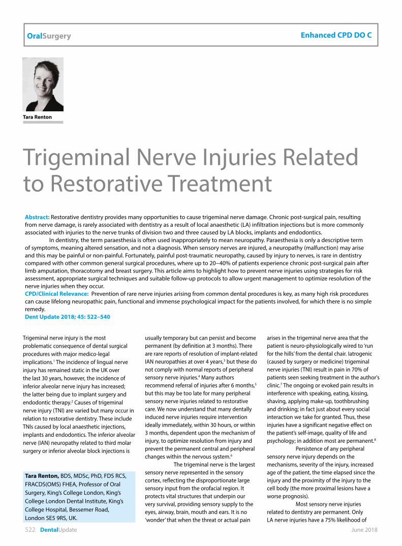
(such as sharp pain or an electrical shock–like sensation) and multiple blocks may also increase the risk of local anaesthetic-related nerve injuries. Neuralgia during injections is associated with increased persistence of nerve injury.16 Thus, it is imperative that the clinician uses an appropriate LA protocol to allow the patient to indicate proximity of the surgical instruments to the IDC, for example infiltration anaesthesia for implant surgery.
Avoiding block anaesthesia by using infiltration dentistry
Daubländer et al reported that, in a 2014 survey of German dental LA practice, 74% were using infiltration dentistry routinely and rarely giving IDBs (pers comm).24 Improved patient comfort is reported by patients with a
In order to minimize complications related to dental LA you need to consider modifying the following risks:
Block anaesthesia: Nerve block injections should be undertaken without intent on direct ‘hit’ of the nerve. 60% of patients who experience the ‘funny bone’ neuralgia due to the IDB needle being placed too close to the lingual or inferior alveolar nerves experience persistent neuropathy.20
Lingual nerve > IAN: Is this technique related or anatomically related (fewer fascicles in LN lower capacity for recovery). Perhaps the direct IDB approach may place the lingual nerve at increased risk compared with the indirect technique.14
Concentration of LA: Any increased concentration of any agent leads to increased neural neurotoxicity.21
Volume of LA: There is no evidence to support this suggestion but all chemicals are neurotoxic, dependent upon the proximity, LA concentration, neural damage additional volume would add to potential neurotoxicity.
Multiple injections: Second or subsequent injections that impede directly on or in neural tissue may not be associated with the usual ‘funny bone’ neuralgic pain. Thus the patient does not self-protect as effectively possibly rendering the nerves more at risk of direct damage.
Severe pain on injection: 60% increased occurrence of persistent neuropathy after IDBs.21
Type of LA Agent: Bupivicaine most neurotoxic of all LA agents.
Type of vasoconstrictor?: The role of vasoconstrictor in nerve damage is unknown.
Sedated or anaesthetized patients?: There is no evidence to support unresponsive patients are less likely to protect themselves when neuralgia (funny bone reaction) occurs as the IDB needle encroaches too close to the nerve.
Lack of LA aspiration?: Again there is no evidence to support that aspiration during IDB results in lower persistent neuropathies but a pragmatic view may infer less chemical injected intra-neurally will cause less chemical nerve injury.
.Table 1. Risk factors for persistent neuropathy related to IDBs.
preference for having full lingual sensation and shorter duration LA anaesthesia after dental treatment.
There is further evidence to support the notion that infiltration dentistry can be successful in many aspects of dentistry: Maxillary infiltration anaesthesia: Some studies have found 4% Articaine to be more effective than 2% Lidocaine for lateral incisors but not molars,25 while others reported no clinical superiority for this injection.26,27 However, a recent randomized controlled trial found a statistically significant difference supporting use of 4% Articaine in place of 2% Lidocaine for buccal infiltration in patients experiencing irreversible pulpitis in maxillary posterior teeth.28
Pulpal anaesthesia in the anterior mandible
compared with inferior dental block (IDBs):29 Meechan provides evidence supporting the significantly increased rates of pulpal anaesthesia using infiltration anaesthesia when compared with IDB anaesthesia, particularly for premolar and incisor teeth. Pulpitic mandibular molars in adults:30,31 A recent systematic review reports that Articaine is 3.4 times more effective for pulpitic mandibular molars when compared with Lidocaine but there is no difference between Articaine and Lidocaine maxillary infiltrations or IDBs.32
For exodontia in adults and children:33 Paedodontic extractions do not require IDBs as the bone is very porous and susceptible to absorption of infiltrative anaesthesia. Is ideal for implant surgery: Several reports of supra-periosteal infiltration anaesthesia claim it is not only sufficient for posterior mandible implant surgery but it may be protective of the IAN.34
Is suitable for periodontal surgery: The standard care for periodontal and implant surgery is infiltration LA.29
Intraligamental anaesthesia for extractions and avoiding IDBs
Intraligamental anaesthesia for extractions and avoiding IDBs is also gaining popularity.35
Thus, prevention of LA nerve injuries is possible and some simple steps may minimize LA-related nerve injuries: Avoid high concentration LA (Articaine 4%) for block injections; for ID blocks (use 2% Lidocaine as standard) as the efficacy is equal; Avoid multiple blocks where possible; Avoid nerve contact during block injections; Avoid block anaesthesia by using infiltration dentistry, thus preventing LA-related nerve injury, for which there is no cure.
Management of LA nerve injuriesThere is no evidence base for
managing dental LA-related nerve injuries, it is only known that 25% are permanent and there is no ‘magic bullet’ to fix them. A sit and wait approach has to be adopted with the reassurance of the patient. HOMECHECK - If you cause pain during an IDB injection to the patient do follow them up the next day and check that they are OK. If the patient reports numbness, altered sensation and/or pain, reassure them. Continue to support and reassure your patient and advise them to visit to confirm
June 2018 DentalUpdate 527
OralSurgery
the presence of neuropathy. If the neuropathy affects most of the dermatome, +/- associated with severe neuropathic pain, nerve injury must be suspected. Reassure your patient that 75% of these injuries resolve. Say SORRY: this is NOT an admission of guilt. Initiate medical management (recommended for other peripheral sensory nerve injuries). High dose oral NSAIDs (600–800 mgs Ibuprofen PO QDS). GMP prescription for Prednisolone 5 day step down dose 50-40-30-20-10 mg PO (not for patients with contraindications for steroids or NSAIDs). Vitamn B complex. Arrange a review of the patient. All advice is summarized on the Trigeminalnerve.org.uk website. Long-term management of patients with
non-resolving LA nerve injuries. The reality for these patients is that, if they have persistent neuropathic pain, they have to be treated as such with psychological and medical management. Psychological interventions play a significant role in managing these patients and recommendations for treatment of trigeminal neuropathic pain are also well described by Renton & Zakzrewska.36
Recommendations for LA nerve injuriesThere are two main issues
currently for LA. Changing practice by using tailored LA techniques rather than always reaching for the IDB, and consenting patients regarding potential nerve injury.1. Tailored LA/Technique specific infiltration dentistry (Figure 1): Infiltration dentistry avoids the use of IDBs, thus preventing LA-related nerve injury, for which there is no cure and which is occurring more frequently than we were taught at dental school (1 in 14,000 blocks causes persistent neuropathy of which 25% are permanent).37 Another study reports the incidence of nerve injury in relation to IDBs has been calculated as 1:609,000 but with a significant increase in injury rate with 4% agents.38 By avoiding IDBs there is less risk of injury to the lingual and inferior alveolar nerves which, though rare, is debilitating to the patients and has
no cure. This technique requires less skill, less discomfort for the patient during the injection and avoids unnecessary lingual anaesthesia after dental treatment. 2. Consent for LA: Patients are warned of a risk of nerve injury when routinely undergoing epidural or spinal injections.39 Reports have estimated that nerve injury resulting from neuroaxial blocks (epidurals, spinals and combined epidural with spinals) resulted in sensory or motor nerve injuries in 1 in 24–54K patients (and paraplegia or death in 1 in 50–140K patients) – The National Royal College of Anaesthetists RCA Audit 2012.39 Already in Germany there is a legal precedent to warn all patients of the risk24 and this was suggested originally in the US.40 With Montgomery consent principles, based upon what is material to the patient, it is difficult to argue that a permanently numb and/or painful tongue for life will not be immaterial to any patient attending for routine dentistry.
Prevention of endodontic nerve injuriesEndodontic treatment of premolar
and mandibular teeth has the potential to damage the inferior alveolar nerve via direct trauma, pressure or neurotoxicity.
IAN is at risk from a variety of dental procedures, in that the IAN is contained within a bony canal predisposing
Table 2. Case series of endodontic nerve injuries.41-45
Pogrel41 61 cases
Knowles et al42 8 cases
Scolozzi et al43 4 cases
Renton et al45 28 cases
Figure 1. Infiltration dentistry is dependant upon the site and procedure. Illustrations modified from figure courtesy of Andrew Mason University Dundee.

OralSurgery
528 DentalUpdate June 2018
it to ischaemia, trauma and subsequent mechanical and chemical injury (Table 2). 41-45 There are relatively few case series reports on endodontic nerve injuries which may not be limited to those teeth proximal to the IAN canal but may occur in maxillary teeth as well. Persistent pain after endodontics has been reported to occur in 3–13% of patients,41-44 whilst surgical endodontics resulted in chronic neuropathic pain in 5% of patients.45 In one study the significant factors associated with persistent post endodontic pain included prolonged preoperative pain, female gender and previous chronic pain symptoms.44 The largest series of endodontic–related trigeminal nerve injuries included 61 patients over an 8 year period,41 with most of these patients presenting with persistent pain. Onset of neuropathy may be delayed in some cases.45 There relatively few reports of persistent pain subsequent to dental procedures, however, in a recent study of 221 patients with trigeminal nerve injuries related to dentistry, 70% are reported to have chronic neuropathic post traumatic pain.46
It is important to exclude the onset of neuropathy prior to endodontic procedure is ascertained, as periapical lesions and pathology can cause neuropathy.47,48
Risk factors for endodontic nerve injuryEndodontic nerve injuries
are likely due to a combination of factors; over instrumentation or over filling direct mechanical trauma will be a main factor, however, the chemicals used in endodontic procedures are chemically neurotoxic and, if the apical plug is breached during treatment, this is the likely cause of the severe nerve injuries seen in relation to endodontics.
Intra-operatively all clinicians should document unusual patient reactions (pain) occurring during the endo procedure (such as sharp pain or an electrical shock–like sensation) Any loss of apical seal and subsequent breech of the IDC with instruments, caustic chemicals or materials is likely to result in acute and often severe neuralgic type pain intra-operatively.49-51 These patients should be followed up the following day and subsequent 3−4 days as chemical leakage can cause a delayed
Chemicals 52-55
- Neurotoxic effects of root filling materials on rat phrenic nerve in vitro- Neurotoxic and analgesic effects of root canal cements and pulp-protecting dental materials- Comparative neurototoxic effects of root canal materials on rat sciatic nerve- The effect of eugenol on nerve transmission Na Hypochlorite Thermal gutta percha 56
Mechanical injury 57due to: - Crush and resultant ischaemia by over fill; - Mechanical and haemorrhagic injury by over instrumentation
Table 3. Possible mechanisms of causation of nerve injury caused by endodontics 52-57
A. Inadequate preoperative assessment and planning due to: A reduced level of knowledge GDP (80% of referrals) GDP endodontic success rates are significantly lower than that of specialist endodontists (15% vs 85%) 58
Inability to read the radiographs or CBCT Inadequate informed consent – all options provided and related risk benefit for each Lack of identification of existing pre-surgical neuropathy (periapical lesions).
B. Proximity of tooth apex to IDC – 90% of the mandibular teeth in this series were close to the IAN canal or premolars adjacent to the mental foramenProximity to the apex to the IAN/ breach apical seal and over chemical or instrumentation In a prospective study, Tantanapornkul et al59 reported the specificity and sensitivity of CBCT versus panorals in identifying the proximity of the IAN to the tooth roots in 161 mandibular third molars; for it was CBCT 93% and 77%, respectively, and for panoramic 70% and 63% which were not significantly different. Findings supported by Tilotta-Yasukawa et al,61 a previous study, report that a significant correlation between the tooth location and the suggested cause of nerve injury was found.62 A classic study of the relationship between mandibular premolar apices and the mental foramen has reported close proximity with the first premolar apex in 15.4% of patients and with the second premolar apex in 13.9% of patient.63 More precisely, each mental foramen was found to be located, on average, anywhere between 3.8 mm mesial, 2.7 mm distal, 3.4 mm above or 3.5 mm below the apex of the respective second.64 In contrast, the apex of each second premolar was between 0 and 4.7 mm from the respective mental foramen in various cadaveric studies.65
C. Poor technique - Breach of apex causing pain during surgery on irrigation or during instrumentation and damage to periapical tissues- Apical extrusion of sodium hypochlorite activated with two laser systems and ultrasonics: a spectrophotometric analysis.66-70 - Over instrumentation- Detectable overfill occurred in 60% of cases and over instrumentation during preparation.D. Early recognition and intervention for endodontic-related nerve injuries ALWAYS undertake HOMECHECK , review patient and confirm neuropathy Neuropathy related to endodontics can be delayed and the patient must be encouraged to report any change in sensation up to 3-4 days post treatment (Renton et al unpubl). If nerve injury is suspected, you will already be aware of the proximity of the tooth apex to the IDC and whether there was likely breach of apex, over instrumentation or deposition of endodontic material into the IAN canal. If suspected, the material, the apex and/or tooth must be removed within 48 hours of placement in order to maximize recovery from nerve injury.42 If the patient is insistent on keeping the tooth, urgent referral of the patient may be indicated for mandibular decompression and saline irrigation of the IDC.42
Table 4. Risk factors for endodontic-related neuropathy.42, 58-70

June 2018 DentalUpdate 529
OralSurgery
presentation of endo-related nerve injuries.45
Risk factors for endodontic nerve injury are summarized in Table 3.52-57
Radiographic factors may lead to an increased risk of chemical leakage from the dental canal system and include proximity of the apex to the IDC, root fractures, accessory canals and resorption. These factors are
summarized in Table 4.42,58-70
Neurological injuries resulting from untreated periapical infections
Apical periodontitis of endodontic origin rarely leads to sensory impairment of the inferior alveolar or mental nerve. These cases are likely to present with teeth apices and/or the apical
area is superimposed on the IDC. There are few reports of neurological disorder where paraesthesia and hyperaesthesia of the mental nerve resulted as a sequel of apical periodontitis of a mandibular second premolar.47,48,71,72
Motor nerve palsy due to endodontic treatment
There is one report of facial motor nerve deficit as a result of endodontic treatment due to periapical leakage of 3% hydrogen peroxide and 90% ethanol rinses.73
Local anaesthesia related neuropathy during endodontic procedures
Local anaesthesia is may be complicated in endodontics as the patients often experience difficulty in achieving analgesia. Thus due to multiple injections, endodontic patients may be more at risk of local anaesthetic-related nerve injuries.17
Consequences of endodontic nerve injury Table 541,
45, 51, 76, 77
Neuropathic pain (NP) syndromes are chronic pain disorders that develop after a lesion of neuropathic pain is seen in most patients presenting with endodontic-related neuropathy. The peripheral or central nervous structures are normally involved in signalling pain. The characteristics of NP differ substantially from those of other chronic pain states, ie chronic nociceptive pain, which develops while the nervous system that is involved in pain processing
Chronic post-surgical pain
Campbell et al51 Whilst surgical endodontics resulted in chronic neuropathic pain in 5% of patients
Oshima et al76 In a previous study of 135 patients with inferior alveolar nerve injuries caused by dental treatment or malignancy, 22% presented with dysaesthesia which was significantly associated with the female gender and previous chronic pain
Pogrel41 11 of 61 patients with Endo NI presented with significant pain, only 40% responded to surgery
Klasser et al77 The prevalence of persistent pain of neuropathic origin among respondents after receiving single-tooth non-surgical root canal treatment was 7% with the average pain reported as 7.2 on a visual analogue scale. Neuropathic pain was more common in middle-aged (mean 50.6 years of age) individuals with no sex predilection and occurred more frequently in the mandibular arch without any differences attributable to the number of canals treated.
Devine et al45 In a cohort of 28 patients presenting with post endodontic neuropathy over 90% presented with neuropathic pain.
Table 5. Consequences of endo nerve injury.41, 45, 51, 76, 77
Predisposing tooth factor that may result in an adverse incident during root canal treatment
Potential adverse incident if tooth factor not recognized
Resorption defects where extent is not identified such as internal /external communicating with root canal and external surface of the root
Extrusion of endo filler/Hypochlorite accident
Suspicion of a perforation communicating with the external root surface Extrusion of endo filler/Hypochlorite accident
Root fracture where there could be a potential communication of the root canal with external root surface
Extrusion of endo filler/Hypochlorite accident
Sclerosed root canal Possible perforation with subsequent hypochlorite accident
Dens invaginatus Possible perforation with subsequent hypochlorite accident
Periapical lesions and other pathology (cysts) Neurological injury (may occur if lesion close to IDC
Lower molar teeth where root apices are in close proximity to the inferior dental canal and/or mental foramen
Neurological injury (over instrumentation, overfilling with obturation materials or sealer)
.Table 6. Radiographic risk factors for extrusion of endodontic chemicals into inferior dental canal.
OralSurgery
530 DentalUpdate June 2018
is intact. As well as the existence of negative somatosensory signs (deficit in function), there are other features that are characteristic of neuropathic conditions (allodynia, hyperalgesia and hyperpathia).78 Paraesthesia is a symptom typically described by patients that are bothersome but not painful. Furthermore, NP states require different therapeutic approaches, such as anticonvulsants, which are not effective in nociceptive pain.79
The evoked pain due to touch or cold significantly impacts on a patient’s quality of life including; eating, speaking, kissing, sleeping or socializing.47 Thus, when these iatrogenic injuries occur, a patient’s quality of life may significantly diminish and lead to significant psychological problems.80 Increasingly, complaints received by the GDC and ADA are nerve injury related.
Thus, symptoms experienced by patients with post-traumatic neuropathy of the trigeminal nerve can range from next to no symptoms, such as minimal anaesthesia in a small area, to devastating effects on a patient’s quality of life.47,80
Recommendations based on current evidence: practitioners undertaking endodontic treatment should: Pre-operatively: – Identify any teeth apices proximal
to the IAN (radiographic risk factors − Table 6) and take special care in preventing apical breech or chemical leak. GDPs may want to refer patients with teeth close to IDC to refer for specialist care
– Screen out neuropathic pain pre RCT. Operatively prevent overfill or extrusion (Figures 2 and 3) consider: – Creating an apical stop or
dentine apical plug; – Make sure the preparation has taper
and hence resistance form;
– Obturating shorter; – Using cold lateral condensation to
gain apical control; – Do not use resin-based sealers such
as AH plus sealer.– Avoid over instrumentation (Figure 4) – Care with instrumentation and
patency filing may have to work shorter;
– Care using intracanal medicament
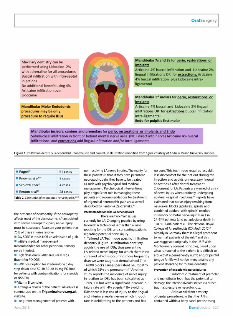
Figure 3. A post endo panoral demonstrating overfill in the IDC from the extracted LL5.
Figure 4. A post endo panoral film demonstrating over instrumentation of the distal canal of the LL6.
Figure 2. Post endodontic CBCT illustrating overfill in the inferior dental canal (IDC) from the distal canal of the LL7.
June 2018 DentalUpdate 533
OralSurgery
(eg calcium hydroxide) do not syringe down to full working length;
– Deliver more coronally; – Use a file to deliver the calcium
hydroxide toward the apical part of the canal.
– Record any events that may indicate
operative nerve injury, including: – Extreme pain during LA IDB,
canal instrumentation, irrigation, medication or filling;
– Sudden and profuse haemorrhage arising from the apex of the tooth.
ManagementPost-operatively, before
discharge in accordance with Endodontic Guidelines, an appropriate post-operative procedural periapical radiograph must be undertaken to check for any extrusion of dressing or filling materials into the inferior dental canal or around the mental foramen before patient discharge. HOMECHECK − The treating clinician must contact the patient between 6 and 24 hours after surgery (Homecheck) to establish any persistent neuropathy after LA has resolved. (This builds on the relationship of the clinician with the patient that will be premised upon good consent process). Neuropathy related to endodontics can be delayed and the patient must be encouraged to report any change in sensation up to 3−4 days post-treatment. Continue to support and reassure your patient and advise him/her to visit to confirm the presence of neuropathy. If the neuropathy affects most of the dermatome, +/- associated with severe neuropathic pain, nerve injury must be suspected. Say SORRY: this is NOT an admission of guilt. Additional scanning or radiography may not be essential. Post-traumatic neuropathy is a clinical diagnosis. You will already be aware of the proximity of the tooth apex to the IDC and whether there was likely to be a breach of apex, over instrumentation or deposition of endodontic material into the IAN canal. Nerve injury may be possible, even with no radiographic evidence of overfill or over instrumentation present possibly due to: – apical inflammation (neuritis)
confirmed by prescription of antibiotics;
– chemical nerve injury from irrigant or filler material;
– thermal damage.\ If overfill or over instrumentation issuspected, the material, the apex and/ortooth must be removed within 48 hoursof placement in order to maximize recoveryfrom nerve injury. If the patient is insistent onkeeping the tooth, urgent referral of thepatient may be indicated for mandibulardecompression and saline irrigation of the IDCunder general anaesthetic.41
Initiate medical management (recommended for other peripheral
A. Inadequate preoperative assessment and planning due to:
- Surgeon lack of knowledge/inexperience/training
- Inadequate informed consent – all options provided and related risk benefit for each option of treatment. Implants are elective treatment sublingual haematoma can require the need for tracheostomy, post-implant treatment and rare events of death86
- Lack of identification of existing pre-surgical neuropathy (especially important in edentulous patients)
- Poor planning in risk assessment and positioning the implant. A sectional DPT is recommended as a minimum for mandibular implant planning. If there is limited bone depth, a CBCT may be used to quantify and qualify bone density and volume. The clinician must be able to read and analyse the CBCT, dependence upon technicians, software or radiologist specialist (who are not present with you intra-operatively)
- Bone assessment quality and quantity87
- Not knowing where the nerve is. Nerve localization, risk factors when assessing IAN position (Mental loop, characteristics of IAN position in various sites of mandible). Parasymphyseal zone high risk. The accuracy of estimating the position of the IDC based on plain films or CT scans is highlighted in the radiographic assessment section.88
- Safety zone – the recommendation is 2 mm (by ITI and ADI) may be insufficient considering that most implant drills are 1.5 mm longer than implants. This increases the risk perforation of a canal surrounding IDC, or even direct perforation and damage to the nerve.89
- Selection of implants 10 mm+ (short implants <8 mm to simplify procedure and minimize morbidity)90
B. Surgical procedure should include the execution of:
- Local Anaesthesia (use infiltration LA techniques to allow patients to notify the surgeon or intra-operative neuralgia, if pain is reported intra-operatively stop surgery and reassess preparation depth and width)34, 91
- Flap design- Use surgical guides to minimize morbidity92, 93
- Surgical stents94
- Using intra-operative radiographs ITI recommend stopping drilling after 60% of planned depth and reassess with bed marker and LCPA)95
- Drill stops96
C. Post-operative care should attend to:
- Early post-operative recognition of neuropathy (HOMECHECK)
- Prompt management of neuropathy (removal of implant if indicated)96
- Acute phase
- Late phase- Early or late post-operative infection
Table 7. Risk factors for implant-related nerve injury.86-96
OralSurgery
534 DentalUpdate June 2018
sensory nerve injuries) – High dose oral NSAIDs (600−800
mg Ibuprofen PO QDS); – GMP prescription for Prednisolone
5 day step down dose 50-40-30-20-10mg PO (not for patients with contra-indications for steroids or NSAIDs);
– Vitamin B complex. Arrange a review of the patient. All advice is summarized on the Trigeminalnerve.org.uk website
Long-term management of endodontic-related nerve injuries
The reality for these patients is that, if they have persistent neuropathic pain and have to be treated using psychological and medical management. Oshima et al76 reported that of 16 patients of 271 patients presenting with chronic pain after endodontics, 70% of the patients responded to tricyclic antidepressant
therapy. Other medications commonly used for chronic neuropathic pain include Pregabalin and topical 5% Versatis patches (12 hours on nocte and 12 hours off daily) for debilitating mechanical allodynia in the extra-oral dermatome causing functional problems.81 Psychological interventions play a significant role in managing these patients and recommendations for treatment of trigeminal neuropathic pain are also well described by Renton & Zakzrewska.36
Prevention of implant-related nerve injuriesThe incidence of implant-
related IANIs varies from 0−40%.82 More recently, two studies have raised the issue of persistent neuropathic pain due to implant-related nerve injuries.83,84
Based upon literature prevention, diagnosis and management of nerve injuries in relation to dental implants, the cause of permanent implant-related nerve injury can be attributed to direct
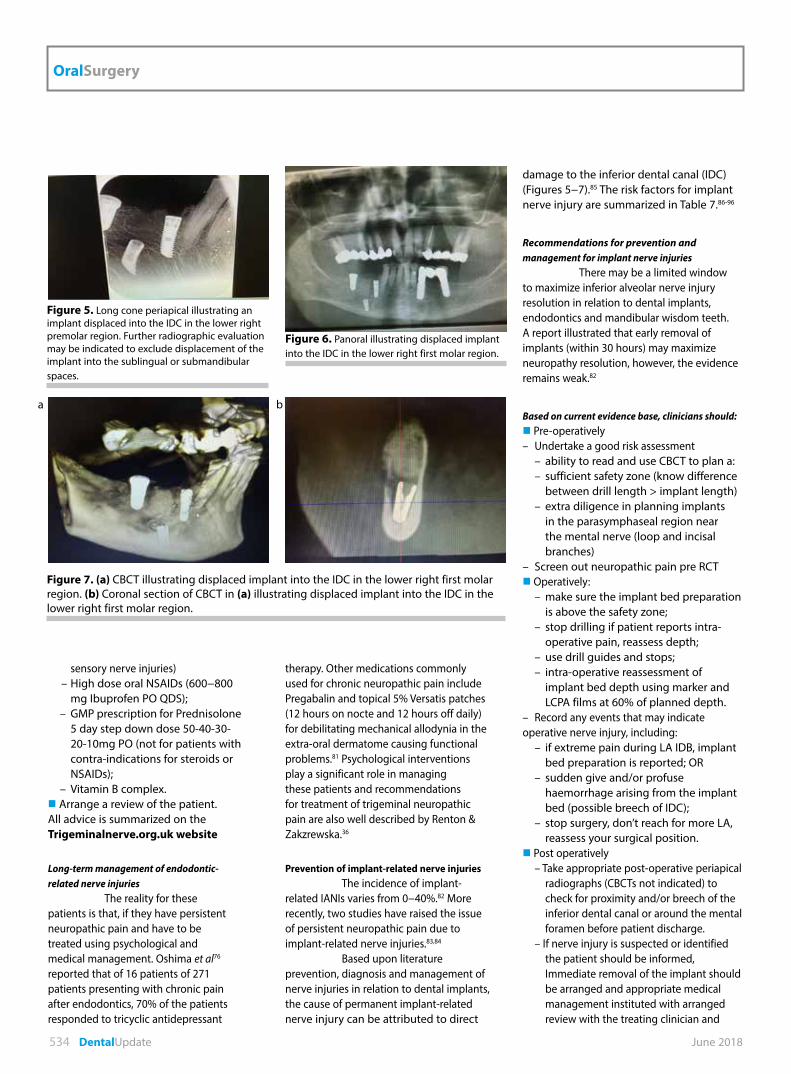
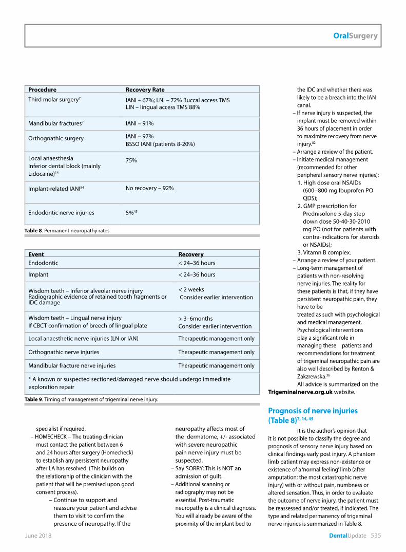
damage to the inferior dental canal (IDC) (Figures 5−7).85 The risk factors for implant nerve injury are summarized in Table 7.86-96
Recommendations for prevention and management for implant nerve injuries
There may be a limited window to maximize inferior alveolar nerve injury resolution in relation to dental implants, endodontics and mandibular wisdom teeth. A report illustrated that early removal of implants (within 30 hours) may maximize neuropathy resolution, however, the evidence remains weak.82
Based on current evidence base, clinicians should: Pre-operatively– Undertake a good risk assessment – ability to read and use CBCT to plan a: – sufficient safety zone (know difference
between drill length > implant length) – extra diligence in planning implants
in the parasymphaseal region near the mental nerve (loop and incisal branches)
– Screen out neuropathic pain pre RCT Operatively: – make sure the implant bed preparation
is above the safety zone; – stop drilling if patient reports intra-
operative pain, reassess depth; – use drill guides and stops; – intra-operative reassessment of
implant bed depth using marker and LCPA films at 60% of planned depth.
– Record any events that may indicate operative nerve injury, including: – if extreme pain during LA IDB, implant
bed preparation is reported; OR – sudden give and/or profuse
haemorrhage arising from the implant bed (possible breech of IDC);
– stop surgery, don’t reach for more LA, reassess your surgical position.
Post operatively – Take appropriate post-operative periapical
radiographs (CBCTs not indicated) to check for proximity and/or breech of the inferior dental canal or around the mental foramen before patient discharge.
– If nerve injury is suspected or identified the patient should be informed, Immediate removal of the implant should be arranged and appropriate medical management instituted with arranged review with the treating clinician and
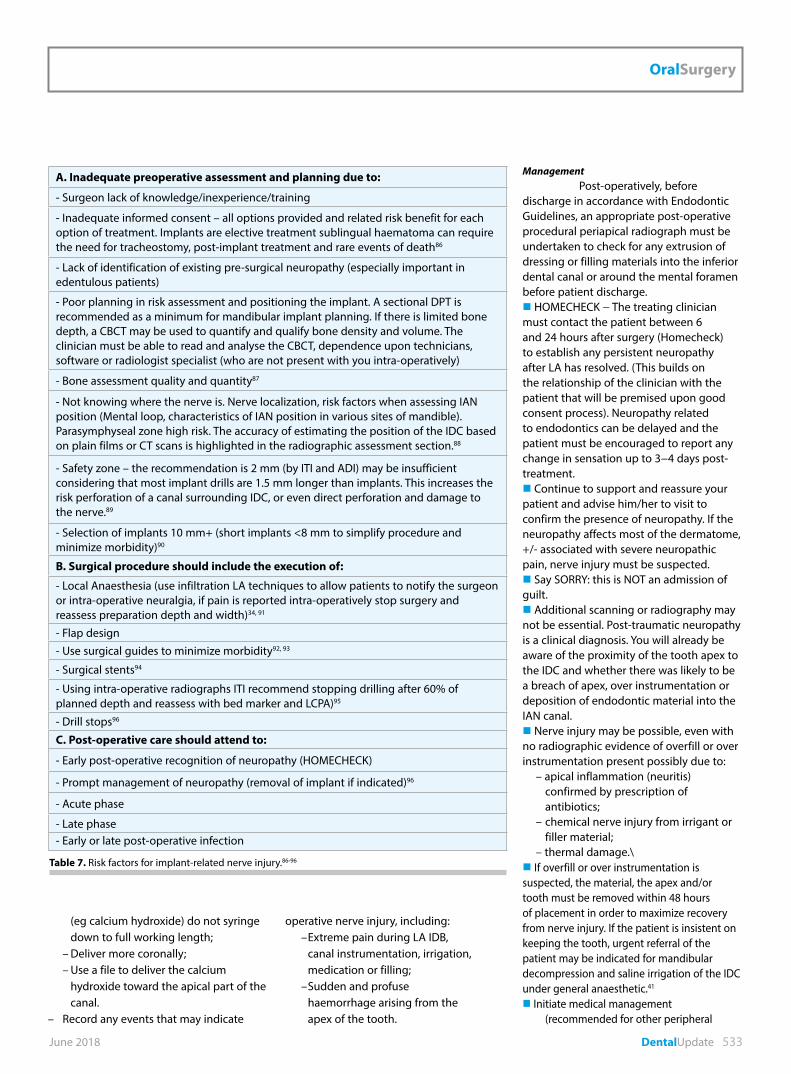
Figure 5. Long cone periapical illustrating an implant displaced into the IDC in the lower right premolar region. Further radiographic evaluation may be indicated to exclude displacement of the implant into the sublingual or submandibular spaces.
Figure 7. (a) CBCT illustrating displaced implant into the IDC in the lower right first molar region. (b) Coronal section of CBCT in (a) illustrating displaced implant into the IDC in the lower right first molar region.
Figure 6. Panoral illustrating displaced implant into the IDC in the lower right first molar region.
ba
June 2018 DentalUpdate 535
OralSurgery
specialist if required. – HOMECHECK − The treating clinician
must contact the patient between 6 and 24 hours after surgery (Homecheck) to establish any persistent neuropathy after LA has resolved. (This builds on the relationship of the clinician with the patient that will be premised upon good consent process).
– Continue to support and reassure your patient and advise them to visit to confirm the presence of neuropathy. If the
neuropathy affects most of the dermatome, +/- associated with severe neuropathic pain nerve injury must be suspected.
– Say SORRY: This is NOT an admission of guilt.
– Additional scanning or radiography may not be essential. Post-traumatic neuropathy is a clinical diagnosis. You will already be aware of the proximity of the implant bed to
the IDC and whether there was likely to be a breach into the IAN canal.
– If nerve injury is suspected, the implant must be removed within 36 hours of placement in order to maximize recovery from nerve injury.82
– Arrange a review of the patient. – Initiate medical management
(recommended for other peripheral sensory nerve injuries):
1. High dose oral NSAIDs (600−800 mg Ibuprofen PO QDS);
2. GMP prescription for Prednisolone 5-day step down dose 50-40-30-2010 mg PO (not for patients with contra-indications for steroids or NSAIDs);
3. Vitamn B complex. – Arrange a review of your patient. – Long-term management of
patients with non-resolving nerve injuries. The reality for these patients is that, if they have persistent neuropathic pain, they have to be treated as such with psychological and medical management. Psychological interventions play a significant role in managing these patients and recommendations for treatment of trigeminal neuropathic pain are also well described by Renton & Zakzrewska.36
All advice is summarized on the Trigeminalnerve.org.uk website.
Prognosis of nerve injuries (Table 8)7, 14, 45
It is the author’s opinion that it is not possible to classify the degree and prognosis of sensory nerve injury based on clinical findings early post injury. A phantom limb patient may express non-existence or existence of a ‘normal feeling’ limb (after amputation; the most catastrophic nerve injury) with or without pain, numbness or altered sensation. Thus, in order to evaluate the outcome of nerve injury, the patient must be reassessed and/or treated, if indicated. The type and related permanency of trigeminal nerve injuries is summarized in Table 8.
Event RecoveryEndodontic < 24–36 hours
Implant < 24–36 hours
Wisdom teeth – Inferior alveolar nerve injuryRadiographic evidence of retained tooth fragments or IDC damage
< 2 weeks Consider earlier intervention
Wisdom teeth – Lingual nerve injuryIf CBCT confirmation of breech of lingual plate
> 3–6monthsConsider earlier intervention
Local anaesthetic nerve injuries (LN or IAN) Therapeutic management only
Orthognathic nerve injuries Therapeutic management only
Mandibular fracture nerve injuries Therapeutic management only
* A known or suspected sectioned/damaged nerve should undergo immediate exploration repair
Table 9. Timing of management of trigeminal nerve injury.
Procedure Recovery Rate
Third molar surgery7 IANI – 67%; LNI – 72% Buccal access TMSLIN – lingual access TMS 88%
Mandibular fractures7 IANI – 91%
Orthognathic surgery IANI – 97%BSSO IANI (patients 8-20%)
Local anaesthesia Inferior dental block (mainly Lidocaine)14
75%
Implant-related IANI84 No recovery – 92%
Endodontic nerve injuries 5%45
Table 8. Permanent neuropathy rates.
OralSurgery
536 DentalUpdate June 2018
Summary of type and timing of management (Table 9)1. Timing of intervention and mechanism of injury are paramount in decision-making in treatment of trigeminal nerve injuries. Surgical intervention is indicated for acute nerve injuries (known or suspected) or those that become evident immediately after treatment (implants and endodontics) and later at 3 days (if there is a delay in presentation after endo treatment).2. Counselling is the most useful tool for managing patients with permanent sensory problematic nerve injuries.3. Medical symptomatic therapy is indicated for patients with pain or discomfort and for patients with anxiety and/or depression in relation to chronic pain. However, due to the extensive side-effects of chronic pain medication, less than 8% of patients remain on medication: i. acute (medical); ii. late (chronic pain management with psychological interventions).4. Surgical exploration: Acute -Immediate repair if nerve section is known; -Remove implant or endodontic overfill, over instrumentation within 24 hours (ideally); -Clinical presentation of persistent neuropathy is paramount and radiographic follow-up is not necessary, however, if there is CBCT evidence of breech of lingual plate or IDC, consider immediate action − nerve exploration +/- repair. Exploratory surgery for lingual or inferior alveolar nerve injuries within 3 months post injury.97 Surgical intervention is not effective for neuropathic pain and if this is the driving force behind seeking surgery it should be reconsidered.98,99
There is reported exciting results of allografting lingual and inferior alveolar nerve injuries.100 In contrast to the USA, these human nerve allografts cannot be used in many parts of the world, including the UK, due to the perceived risk of Creutzfeld Jacob disease.101
ConclusionsUnfortunately, none of these
interventions ‘fixes’ the patient, but the aim is to manage his/her symptoms in the best way possible, often not very satisfactorily.
This article was intended to highlight some key issues around implant related nerve injuries and key take away messages include:
Iatrogenic nerve injury has a significant and unpleasant effect on the patient due to most experiencing neuropathic pain as well as altered sensation and numbness (improve your consent!). All nerve injuries are avoidable. Most IAN injuries in relation to implant and endodontic dentistry are permanent and ‘unfixable’. Improvement pre-operative planning, operative execution ad post-operative care will minimize and hopefully prevent these injuries. Some useful and pragmatic strategies are presented here to prevent nerve injuries. Owing to the significant problems following nerve injury, pre-operative strategies for minimizing this risk of nerve damage need to be considered carefully. There is a need for a consensus and standardization of risk assessment and management, and a holistic approach in managing the pain-related effect on functionality and psychological implications caused to the patients affected by iatrogenic nerve injury.
References1. Caissie R, Goulet J, Fortin M, Morielle D.
Iatrogenic paresthesia in the third division of the trigeminal nerve: 12 years of clinical experience. J Can Dent Assoc 2005; 71: 185−190.
2. Hillerup S. Iatrogenic injury to oral branches of the trigeminal nerve: records of 449 cases. Clin Oral Investig 2007; 11: 133−142. Epub 2006 Dec 22.
3. Elian N, Mitsias M, Eskow R, Jalbout ZN, Cho SC, Froum S, Tarnow DP. Unexpected return of sensation following 4.5 years of paresthesia: case report. Implant Dent 2005; 14: 364−367.
4. Loescher AR, Robinson PP. The effect of surgical medicaments on peripheral nerve function. Br J Oral Maxillofac Surg 1998; 36: 327−332.
5. Hegedus F, Diecidue RJ. Trigeminal nerve injuries after mandibular implant placement − practical knowledge for clinicians. Int J Oral Maxillofac Implants 2006; 21: 111−116.
6. Ziccardi VB, Assael LA. Mechanisms of trigeminal nerve injuries. Atlas Oral Maxillofac Surg Clin North Am 2001; 9: 1−11.
7. Renton T, Yilmaz Z. Profiling of patients presenting with posttraumatic neuropathy of the trigeminal nerve. J Orofac Pain 2011; 25: 333−344.
8. Renton T, Yilmaz Z. Managing iatrogenic trigeminal nerve injury: a case series and review of the literature. Int J Oral Maxillofac Surg 2012; 41: 629−637. doi: 10.1016/j.ijom.2011.11.002.
9. Mason DA. Lingual nerve damage following lower third molar surgery. Int J Oral Maxillofac Surg 1988; 17: 290−294.
10. Leung YY, Cheung LK. Longitudinal treatment outcomes of microsurgical treatment of neurosensory deficit after lower third molar
surgery: a prospective case series. PLoS One 2016; 11: e0150149.
11. Barrowman RA, Grubor D, Chandu A. Dental implant tourism. Aust Dent J 2010; 55: 441−445. doi: 10.1111/j.1834-7819.2010.01267.
12. Zuniga JR, Yates DM, Phillips CL. The presence of neuropathic pain predicts postoperative neuropathic pain following trigeminal nerve repair. J Oral Maxillofac Surg 2014; 72: 2422−2427.
13. Zuniga JR, Yates DM. Factors determining outcome after trigeminal nerve surgery for neuropathic pain. J Oral Maxillofac Surg 2016; 74: 1323−1329.
14. Renton T, Janjua H, Gallagher JE, Dalgleish M, Yilmaz Z. UK dentists’ experience of iatrogenic trigeminal nerve injuries in relation to routine dental procedures: why, when and how often? Br Dent J 2013; 214: 633−642. doi: 10.1038/sj.bdj.2013.583.
15. Pogrel MA, Schmidt BL, Sambajon V, Jordan RC. Lingual nerve damage due to inferior alveolar nerve blocks: a possible explanation. J Am Dent Assoc 2003; 134: 195−199.
16. Smith MH, Lung KE. Nerve injuries after dental injection: a review of the literature. J Can Dent Assoc 2006; 72: 559−564.
17. Pogrel MA, Thamby S. Permanent nerve involvement resulting from inferior alveolar nerve blocks. J Am Dent Assoc 2000; 131: 901−907.
18. Haas DA, Lennon D. A 21 year retrospective study of reports of paresthesia following local anesthetic administration. J Can Dent Assoc 1995; 61: 319−330.
19. Hillerup S. Iatrogenic injury to oral branches of the trigeminal nerve: records of 449 cases. Clin Oral Investig 2007; 11: 133−142. doi: 10.1007/s00784-006-0089-5.
20. Hillerup S, Jensen R. Nerve injury caused by mandibular block analgesia. Int J Oral Maxillofac Surg 2006; 35: 437−443. doi: 10.1016/j.ijom.2005.10.004.
21. Pedlar J. Prolonged paraesthesia. Br Dent J 2003; 195: 119. doi: 10.1038/sj.bdj.4810421.
22. Perez-Castro R, Patel S, Garavito-Aguilar ZV, Rosenberg A, Recio-Pinto E, Zhang J, Blanck TJ, Xu F. Cytotoxicity of local anesthetics in human neuronal cells. Anesth Analg 2009; 108: 997−1007.
23. Kämmerer PW, Schneider D, Palarie V, Schiegnitz E, Daubländer M. Comparison of anesthetic efficacy of 2 and 4% articaine in inferior alveolar nerve block for tooth extraction − a double-blinded randomized clinical trial. Clin Oral Investig 2017; 21: 397−403.
24. Daubländer M, Kämmerer PW, Willershausen B, Leckel M, Lauer HC, Buff S, Rösl B. Clinical use of an epinephrine-reduced (1/400,000) articaine solution in short-time dental routine treatments − a multicenter study. Clin Oral Investig 2012; 16: 1289−1295.
25. Evans G, Nusstein J, Drum M, Reader A, Beck M. A prospective, randomized, double-blind comparison of articaine and lidocaine for maxillary infiltrations. J Endod 2008; 34: 389−393.
26. Oliveira PC, Volpato MC, Ramacciato JC, Ranali J. Articaine and lignocaine efficiency in infiltration anaesthesia: a pilot study. Br Dent J 2004; 197: 45−46.
27. Vähätalo K, Antila H, Lehtinen R. Articaine and lidocaine for maxillary infiltration anesthesia. Anesth Prog 1993; 40: 114−116.

OralSurgery
538 DentalUpdate June 2018
28. Srinivasan N, Kavitha M, Loganathan CS, Padmini G. Comparison of anesthetic efficacy of 4% articaine and 2% lidocaine for maxillary buccal infiltration in patients with irreversible pulpitis. Oral Surg Oral Med Oral Pathol Oral Radiol Endod 2009; 107: 133−136.
29. Meechan JG. The use of the mandibular infiltration anesthetic technique in adults. J Am Dent Assoc 2011; 142(Suppl 3): 19S−24S.
30. Zain M, Rehman Khattak SU, Sikandar H, Shah SA, Fayyaz. Comparison of anaesthetic efficacy of 4% articaine primary buccal infiltration versus 2% lidocaine inferior alveolar nerve block in symptomatic mandibular first molar teeth. J Coll Physicians Surg Pak 2016; 26: 4−8.
31. Poorni S, Veniashok B, Senthikumar AD, Indira R, Ramachandran S. Anesthetic efficacy of four percent articaine for pulpal anesthesia by using inferior alveolar nerve block and buccal infiltration techniques in patients with irreversible pulpitis: a prospective randomized double-blind clinical trial. J Endod 2011; 37: 1603−1607.
32. Kung J, McDonagh M, Sedgley CM. Does articaine provide an advantage over lidocaine in patients with symptomatic irreversible pulpitis? A systematic review and meta-analysis. J Endod 2015; 41: 1784−1794.
33. Thakare A, Bhate K, Kathariya R. Comparison of 4% articaine and 0.5% bupivacaine anesthetic efficacy in orthodontic extractions: prospective, randomized crossover study. Acta Anaesthesiol Taiwan 2014; 52: 59−63.
34. Etoz OA, Er N, Demirbas AE. Is supraperiosteal infiltration anesthesia safe enough to prevent inferior alveolar nerve during posterior mandibular implant surgery? Med Oral Patol Oral Cir Bucal 2011; 16: e386−e389.
35. Dumbrigue HB, Lim MV, Rudman RA, Serraon A. A comparative study of anesthetic techniques for mandibular dental extraction. Am J Dent1997; 10: 275−278.
36. Renton T, Zakrzewska JM. Chapter 22: Orofacial Pain. In: Oxford Textbook of Anaesthesia for Oral and Maxillofacial Surgery. Shaw I, Kumar C, Dodds C, eds. Oxford: Oxford University Press, 2010 ISBN 978-0-19-956421-7: pp283−298.
37. Renton T, Adey-Viscuso D, Meechan JG, Yilmaz Z. Trigeminal nerve injuries in relation to the local anaesthesia in mandibular injections. Br Dent J 2010; 209: E15. doi: 10.1038/sj.bdj.2010.978.
38. Haas DA, Lennon D. A 21 year restrospective study of reports of paraesthetsia following local anaesthetic administration. J Can Dent Assoc 1995; 61: 319−330.
39. The National Royal College of Anaesthetists. RCA Audit 2012.
40. Orr DL, Curtis WJ. Obtaining written informed consent for the administration of local anesthetic in dentistry. J Am Dent Assoc 1939; 136(11): 1568−1571. (Impact Factor: 1.82). 12/2005.
41. Pogrel MA. Damage to the inferior alveolar nerve as the result of root canal therapy. J Am Dent Assoc 2007; 138: 65−69.
42. Knowles KI, Jergenson MA, Howard JH. Paresthesia associated with endodontic treatment of mandibular premolars. J Endod 2003; 29: 768−770.
43. Scolozzi P, Lombardi T, Jaques B. Successful inferior alveolar nerve decompression for dysesthesia following endodontic treatment: report of 4 cases treated by mandibular sagittal osteotomy. Oral Surg Oral Med Oral Pathol Oral Radiol Endod 2004; 97: 625−631.
44. Grötz KA, Al-Nawas B, de Aguiar EG, Schulz A, Wagner W. Treatment of injuries to the inferior alveolar nerve after endodontic procedures. Clin Oral Investig 1998; 2: 73−76.
45. Devine M, Modgill O, Renton T. Mandibular division trigeminal nerve injuries following primary endodontic treatment. A case series. Aust Endod J 2017; 43: 56−65. doi: 10.1111/aej.12209. [Epub ahead of print] PMID: 28685976.
46. Renton T, Yilmaz Z. Profiling of patients presenting with posttraumatic neuropathy of the trigeminal nerve. J Orofac Pain 2011; 25: 333−344.
47. Giuliani M, Lajolo C, Deli G, Silveri C. Inferior alveolar nerve paresthesia caused by endodontic pathosis: a case report and review of the literature. Oral Surg Oral Med Oral Pathol Oral Radiol Endod 2001; 92: 670−674.
48. Devine M, Yilmaz Z, Hirani M, Renton T. A case series of trigeminal nerve injuries caused by periapical lesions of mandibular teeth. Br Dent J 2017; 222: 447−455. doi: 10.1038/sj.bdj.2017.268.
49. Oshima K, Ishii T, Ogura Y, Aoyama Y, Katsuumi I. Clinical investigation of patients who develop neuropathic tooth pain after endodontics procedures. J Endod 2009; 35: 958−961.
50. Polycarpou N, Ng YL, Canavan D, Moles DR, Gulabivala K. Prevalence of persistent pain after endodontic treatment and factors affecting its occurrence in cases with complete radiographic healing. Int Endod J 2005; 38: 169−178.
51. Campbell RL, Parks KW, Dodds RN. Chronic facial pain associated with endodontic therapy. Oral Surg Oral Med Oral Pathol 1990; 69: 287−290.
52. Brodin P, Roed A, Aars H, Orstravik D. Neurotoxic effects of root filling materials on rat phrenic nerve in vitro. J Dent Res 1982; 61: 1020−1023.
53. Brodin P. Neurotoxic and analgesic effects of root canal cements and pulp-protecting dental materials. Endod Dent Traumatol 1988; 2: 1−11.
54. Serper A, Ucer O, Onur R, Etikan I. Comparative neurotoxic effects of root canal materials on rat sciatic nerve. J Endod 1998; 24: 592−594.
55. Kozam G, Newark NJ. The effect of eugenol on nerve transmission. Oral Surg Oral Med Oral Pathol Oral Radiol Endod 1977; 44: 799−805.
56. Fanibunda K, Whitworth J, Steele J. The management of thermomechanically compacted gutta percha extrusion in the inferior dental canal. Br Dent J 1998; 184: 330−332.
57. Leckel M, Kress B, Schmitter M. Neuropathic pain resulting from implant placement: Case report and diagnostic conclusions. J Oral Rehabil 2009; 36: 543−546. doi: 10.1111/j.1365-2842.2009.01950.
58. Patel S, Durack C, Abella F, Roig M, Shemesh H, Lambrechts P, Lemberg K. European Society of Endodontology position statement: the use of CBCT in endodontics. Int Endod J 2014; 47: 502−504.
59. Tantanapornkul W, Okouchi K, Fujiwara Y, Yamashiro M, Maruoka Y, Ohbayashi N, Kurabayashi T. A comparative study of cone-beam computed tomography and conventional panoramic radiography in assessing the topographic relationship between the mandibular canal and impacted third molars. Oral Surg Oral Med Oral Pathol
Oral Radiol Endod 2007; 103: 253−259.60. Patel S, Dawood A, Ford TP, Whaites E.
The potential applications of cone beam computed tomography in the management of endodontic problems. Int Endod J 2007; 40: 818−830. Epub 2007 Aug 14.
61. Tilotta-Yasukawa F, Millot S, El Haddioui A, Bravetti P, Gaudy JF. Labiomandibular paresthesia caused by endodontic treatment: an anatomic and clinical study. Oral Surg Oral Med Oral Pathol Oral Radiol Endod 2006; 102: e47−59.
62. Chikvashvili J. Overcoming unforeseen incidents: what to do when an unlikely event occurs. Compend Contin Educ Dent 2011; 32: 44−48.
63. Fishel D, Buchner A, Hershkowith A, Kaffe I. Roentgenologic study of the mental foramen. Oral Surg Oral Med Oral Pathol 1976; 41: 682−686.
64. Phillips JL, Weller RN, Kulild JC. The mental foramen: 2. Radiographic position in relation to the mandibular second premolar. J Endod 1992; 18: 271−274.
65. Denio D, Torabinejad M, Bakland LK. Anatomical relationship of the mandibular canal to its surrounding structures in mature mandibles. J Endod 1992; 18: 161−165.
66. Helvacıoğlu Kıvanç B, Deniz Arısu H, Yanar NÖ, Silah HM, İnam R, Görgül G. Apical extrusion of sodium hypochlorite activated with two laser systems and ultrasonics: a spectrophotometric analysis. BMC Oral Health 2015; 15: 71.
67. Scarano A, Di Carlo F, Quaranta A, Piattelli A. Injury of the inferior alveolar nerve after overfilling of the root canal with endodontic cement: a case report. Oral Surg Oral Med Oral Pathol Oral Radiol Endod 2007; 104: e56−59.
68. Escoda-Francoli J, Canalda-Sahli C, Soler A, Figueiredo R, Gay-Escoda C. Inferior alveolar nerve damage because of overextended endodontic material: a problem of sealer cement biocompatibility? J Endod 2007; 33: 1484−1489.
69. Blanas N, Kienle F, Sándor GK. Inferior alveolar nerve injury caused by thermoplastic gutta-percha overextension. J Can Dent Assoc 2004; 70: 384−387.
70. Köseoğlu BG, Tanrikulu S, Sübay RK, Sencer S. Anesthesia following overfilling of a root canal sealer into the mandibular canal: a case report. Oral Surg Oral Med Oral Pathol Oral Radiol Endod 2006; 101: 803−806.
71. von Ohle C, ElAyouti A. Neurosensory impairment of the mental nerve as a sequel of periapical periodontitis: case report and review. Oral Surg Oral Med Oral Pathol Oral Radiol Endod. 2010; 110: e84−89.
72. Shadmehr E, Shekarchizade N. Endodontic periapical lesion-induced mental nerve paresthesia. Dent Res J (Isfahan) 2015; 12: 192−196.
73. Kruse A, Hellmich N, Luebbers HT, Grätz KW. Neurological deficit of the facial nerve after root canal treatment. Oral Surg Oral Med Oral Pathol Oral Radiol Endod 2009; 108: e46−48.
74. Mohammadi Z. Endodontics-related paresthesia of the mental and inferior alveolar nerves: an updated review. J Can Dent Assoc 2010; 76: a117.
75. Campbell RL, Parks KW, Dodds RN. Chronic facial pain associated with endodontic therapy. Oral Surg Oral Med Oral Pathol 1990; 69: 287−290.
76. Oshima K, Ishii T, Ogura Y, Aoyama Y, Katsuumi I. Clinical investigation of patients who
OralSurgery
540 DentalUpdate June 2018
develop neuropathic tooth pain after endodontics procedures. J Endod 35; 7: 958−961.
77. Klasser GD, Kugelmann AM, Villines D, Johnson BR. The prevalence of persistent pain after nonsurgical root canal treatment. Quintessence Int 2011; 42: 259−269.
78. Costigan M, Scholz J, Woolf CJ. Neuropathic pain: a maladaptive response of the nervous system to damage. Ann Rev Neurosci 2009; 32: 1−32.
79. Truelove E. Management issues of neuropathic trigeminal pain from a dental perspective. J Orofac Pain 2004; 18: 374−380.
80. Kiyak HA, Beach BH, Worthington P, Taylor T, Bolender C, Evans J. Psychological impact of osseointegrated dental implants. Int J Oral Maxillofac Implants 1990; 5: 61−69. PMID: 2202671.
81. Khawaja N, Yilmaz, Z, Renton T. Case studies illustrating the management of trigeminal neuropathic pain using topical 5% lidocaine plasters. Br J Pain 2013; 7: 107−113.
82. Khawaja N, Renton T. Case studies on implant removal influencing the resolution of inferior alveolar nerve injury. Br Dent J 2009; 206: 365−370. doi: 10.1038/sj.bdj.2009.258.
83. Fukuda K, Ichinohe T, Kaneko Y. Pain management for nerve injury following dental implant surgery at Tokyo Dental College Hospital. Int J Dent 2012; 209474. doi: 10.1155/2012/209474.
84. Renton T, Dawood A, Shah A, Searson L, Yilmaz Z. Post-implant neuropathy of the trigeminal nerve. A case series. Br Dent J 2012; 212: E17. doi: 10.1038/sj.bdj.2012.497.
85. Alhassani AA, AlGhamdi AS. Inferior alveolar nerve injury in implant dentistry: diagnosis, causes, prevention, and management. J Oral Implantol 2010; 36: 401−407. doi: 10.1563/aaid-joi-d-09-00059.
86. Isaacson TJ. Sublingual hematoma formation during immediate placement of mandibular endosseous implants. J Am Dent Assoc 2004; 135: 168−172.
87. Juodzbalys G, Kubilius M. Clinical and radiological classification of the jawbone anatomy in endosseous dental implant treatment. J Oral Maxillofac Res 2013; 4: e2. doi: 10.5037/jomr.2013.4202.N.
88. Harris D, Horner K, Gröndahl K, Jacobs R, Helmrot E, Benic GI, Bornstein MM, Dawood A, Quirynen M. EAO guidelines for the use of diagnostic imaging in implant dentistry 2011. A consensus workshop organized by the European Association for Osseointegration at the Medical University of Warsaw. Clin Oral Implants Res 2012; 23: 1243−1253.
89. Greenstein G, Tarnow D. The mental foramen and nerve: clinical and anatomical factors related to dental implant placement: a literature review. J Periodontol 2006; 77: 1933−1943.
90. Thoma DS, Cha JK, Jung UW. Treatment concepts for the posterior maxilla and mandible: short implants versus long implants in augmented bone. J Periodontal Implant Sci 2017; 47: 2−12.
91. Bartlett G, Mansoor J. Articaine buccal infiltration vs lidocaine inferior dental block − a review of the literature. Br Dent J 2016; 220: 117−120.
92. Al-Ani O, Nambiar P, Ha KO, Ngeow WC. Safe zone for bone harvesting from the interforaminal region of the mandible. Clin Oral Implants Res 2013; 24(Suppl A100): 115−121.
93. Van Assche N, van Steenberghe D, Guerrero ME, Hirsch E, Schutyser F, Quirynen M, Jacobs R. Accuracy of implant placement based on pre-surgical planning of three-dimensional cone-beam images: a pilot study. J Clin Periodontol 2007; 34: 816−821. doi: 10.1111/j.1600-051X.2007.01110.
94. De Kok IJ, Thalji G, Bryington M, Cooper LF. Radiographic stents: integrating treatment planning and implant placement. Dent Clin North Am 2014; 58: 181−192. doi: 10.1016/j.cden.2013.09.008.
95. Burstein J, Mastin C, Le B. Avoiding injury to the inferior alveolar nerve by routine use of intraoperative radiographs during implant placement. J Oral Implantol 2008; 34: 34−38.
96. Greenstein G, Greenstein B, Desai RN. Using drill stops on twist drills to promote safety and efficiency when creating osteotomies for dental implants. J Am Dent Assoc 2014; 145: 371−375.
97. Strauss ER, Ziccardi VB, Janal MN. Outcome assessment of inferior alveolar nerve microsurgery: A retrospective review. J Oral Maxillofac Surg 2006; 64(12), 1767−1770. doi: 10.1016/j.joms.2005.11.111.
98. Susarla SM, Kaban LB, Donoff RB, Dodson TB. Does early repair of lingual nerve injuries improve functional sensory recovery? J Oral Maxillofac Surg 2007; 65: 1070−1076. doi: 10.1016/j.joms.2006.10.010.
99. Zuniga JR, Yates DM, Phillips CL. The presence of neuropathic pain predicts postoperative neuropathic pain following trigeminal nerve repair. J Oral Maxillofac Surg 2014; 72: 2422−2427. doi: 10.1016/j.joms.2014.08.003. Epub 2014 Aug 11.PMID:25308410.
100. Ziccardi VB, Zuniga JR. Nerve injuries after third molar removal. Oral Maxillofac Surg Clin North Am 2007; 19: 105−115.
101. Zuniga JR, Renton T. Managing post-traumatic trigeminal neuropathic pain: is surgery enough? J Neurol Neuromed 2016; 1: 10−14.
www.shofu.co.uk
ZiLMasterAdjustment Kits
Time-efficient adjusting, finishing and polishing kits for zirconia and lithium disilicate
CA kit for intra-oral use
HP kit for extra-oral use
Shapes available separately.
ZiLMaster_Anz_E-UK_89x274.indd 1 05.02.18 14:49