Take home message… Muscles > Fasicle (Bundles of muscle...
10
Functions of the Muscular System 1. movement 2. heat generation (shivering) 3. maintenance of posture 4. stabilization of joints Common properties of the different muscle types: --All contain myofilaments (thick, thin, and elastic) --All function in movement Three major types of muscle: 1. Skeletal Muscles --Voluntary --Striated --Made of muscle cells, CT wrappings, blood vessels, nerves *muscles are wrapped in a CT covering called the epimysium *bundles of skeletal muscle cells (fasicles) are wrapped in a CT covering called the perimysium *individual skeletal muscle cells (muscle fibers) are wrapped in the CT covering called the endomysium Take home message… Muscles > Fasicle (Bundles of muscle cells) > Muscle fiber (A muscle cell) > Myofbril (Complex muscle cell organelles that are made up of myofilaments) Attachments Direct - epimysium fused to bone or cartilage Indirect - epimysium forms a tendon (rope-like) or aponeurosis (sheet-like) which then attaches to bone, cartilage or another muscle Insertion - attachment site of the moveable bone Origin - attachment at the immovable or less movable bone
Transcript of Take home message… Muscles > Fasicle (Bundles of muscle...

Functions of the Muscular System1. movement2. heat generation (shivering)3. maintenance of posture4. stabilization of joints
Common properties of the different muscle types: --All contain myofilaments (thick, thin, and elastic)--All function in movement
Three major types of muscle: 1. Skeletal Muscles --Voluntary--Striated--Made of muscle cells, CT wrappings, blood vessels, nerves *muscles are wrapped in a CT covering called the epimysium *bundles of skeletal muscle cells (fasicles) are wrapped in a CT covering called the perimysium *individual skeletal muscle cells (muscle fibers) are wrapped in the CT covering called the endomysium
Take home message…Muscles > Fasicle (Bundles of muscle cells) > Muscle fiber (A muscle cell) > Myofbril (Complex muscle cell organelles that are made up of myofilaments)
Attachments Direct - epimysium fused to bone or cartilageIndirect - epimysium forms a tendon (rope-like) or aponeurosis (sheet-like) which then attaches to bone, cartilage or another muscle Insertion - attachment site of the moveable bone Origin - attachment at the immovable or less movable bone
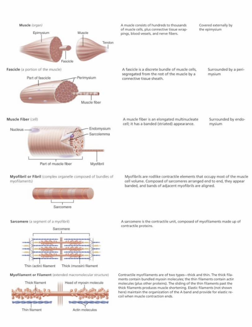

Microscopic anatomy of skeletal muscle Figs 9.2, 9.3--skeletal muscle cells (muscle fibers)-long cylindrical cells with multiple nuclei formed by fusion of embryonic cells; contain myofibrils--sarcolemma - specialized plasma membrane of skeletal muscle cells; invaginates into T-tubules at the A-band-I band junction--sarcoplasm – cytosol (liquid portion) of a muscle cell--myofibrils - rod-like structures contained within skeletal muscles cells that are the contractile elements (specialized cytoskeletal elements/organelles) composed of myofilaments (3 types) and associated proteins: 1. thick filaments - comprised of myosin (protein), with “head” and “tail” regions 2. thin filaments- comprised mostly of actin; also contain the regulatory proteins *tropomyosin – blocks myosin binding sites on actin to prevent myosin binding *troponin – 3 subunit protein that binds actin, tropomysin, and Ca2+ --TnI – inhibitory; binds actin --TnT – binds tropomyosin --TnC – binds Ca2+ 3. elastic filaments comprised of titin

Banding pattern on the myofibrils responsible for striationsA band - darker region containing thick filaments (myosin filaments)I band – lighter region lacking thick filamentsZ disc - center region of I band containing the protein nebulin that anchors the thin filaments (actin filaments)H zone- lighter region in the center of each A band lacking thin filaments when the muscle fiber is relaxed muscle.
**These zones do NOT ned to be idnetified on the exam**
sarcoplasmic reticulum--highly specialized of muscle cells that forms a network of tubules around each myofibril within the muscle cell --regulates the intracellular levels of Ca2+ by storing Ca2+ and releasing it when needed for muscle contraction--Terminal cisternae of the SR are the “end sacs” that abut the T-tubules
sarcomere - a myofibril segment from Z disc to Z disc

Skeletal muscle contraction --sliding filament model - proposed mechanism of contraction that states that during contraction, the thin filaments (actin) slide past the thick filaments (myosin) so that they come closer together and then overlap; the thick filaments remain essentially in the same position
--I bands shorten and the H zones disappear during contraction due to the sliding movement of the thin filaments--Thin filament sliding is mediated by “cross bridges” (myosin heads) attaching, ratcheting, and detaching from actin filaments
-ATP is required for crossbridge attachment and detachment-Ca2+ is also required:When intracellular Ca2+ levels are low→tropomyosin blocks the myosin binding site on actin, so the cross bridges are inactive-Ca2+ released from the sarcoplasmic reticulum upon stimulation of the muscle binds to troponinC, which undergoes a conformational change that allows the myosin cross bridges to attach to actin and undergo the sliding filament mechanism.

--Neuromuscular junction-: Junction (comprised of synapses) of a motor neuron with its branched axonl terminal and a skeletal muscle cell or cardiac muscle cell site where release of neurotransmitter (Acetacholine) by a motor neuron stimulates muscle fiber contraction. -nervous system input always stimulates (excites) the muscle to contract
--motor end plate-specialized, folded area of the muscle fiber sarcolemma at the nm junction*contains ACh receptors that Ach binds to--synaptic cleft- space between the motor neuron and the motor end plate into which Ach is released--synaptic vesicles-present in the motor neuron axonal ending; contain the excitatory neurotransmitter Ach which is release into the synaptic cleft by exocytosis--acetylcholine esterase- enzyme present in the nm junction synaptic cleft that cleaves Achthis prevents continued muscle contractionEvents at the NM junction that lead to generation of an action potential in the sarcolemma and skeletal muscle fiber
contraction
1. Action potential in motor neuron travels to the axon terminal→2. Ca2+ enters the axon terminal3. stimulates Ach release from synaptic vesicles 4. Ach diffuses through synaptic cleft, binds to Ach receptors on motor endplate of the sarcolemma® 5. Ach binding to Ach receptors allows Na+ entry

into the skeletal muscle fiber(and K+ out) which causes a change in sarcolemma membrane potential (depolarization; called the end plate potential)6. voltage-sensitive Na+ channels open →7. leads to action potential along sarcolemma of muscle cell → muscle contraction
Action Potential: Fig 9.10An electrical impulse generated by a rapid influx of Na+ into a cell, resulting in a depolarization of the membrane above a threshold level.
1) electrical signal of the motor neuron which triggers release of Ach from the presynaptic neuron terminal
2) Electrical signal on the sarrcolemma that triggers muscle cell contraction.
Excitation -contraction coupling- the sequence of events by which transmission of an action potential along the sarcolemms leads to the sliding of myofilamentswhich results in muscle contraction
Motor unit –motor neuron and all of the muscle fibers it innervates may be comprised of a few or many muscle fibers depending on the muscle type

Muscle tone-slightly contracted state of muscles even when not in usespinal reflex response to activation of stretch receptors that activate a motor unit; keeps muscle firm, ready to respondhelps stabilize joints, maintain posture
Disuse atrophy-loss of muscle tone due to lack of muscle activity resulting in decreased muscle mass
Cardiac Muscle-muscle of the heart
Characteristics- striated, but different banding pattern than skeletal muscle due to branching of cellsinvoluntarycardiac muscle cells (also called cardiomyocytes) contain large mitochondriauni or binucleatemyofibrils present but less uniform Intercalated discs-junctions between cardiac muscle fibers containing desmosomes and gap junctions; help coordinate cardiac muscle cell contraction
Cardiac Muscle contraction --mechanism similar to skeletal muscle contraction (SR Ca2+ release, sliding filament mechanism, troponinC regulation, etc…)--nervous system signal from autonomic nervous system (ANS) may cause excitation or inhibition --speed of contraction is slower than skeletal muscle --cardiomyocytes display “autorhythmicity”

Smooth Muscle-makes up the walls of hollow organs
Characteristics-small, spindle-shaped cells with single nuclei; also called ‘muscle fibers’non-striated smooth muscle cells have actin and myosin filaments attached to structures called dense bodies; no myofibrils presentmost hollow organs have 2 sheets of smooth musclediffuse junctions- junctions where axons from axon terminals from the
ANS--make contact on smooth muscle; --no motor end plateneurotransmitter released from an ANS neuron terminal into awide synaptic cleft →diffuses to receptors on the smooth muscle cells
--Neurotransmitter may be: ; Ach, Epineph. Norepineph
--Smooth Muscle Contraction myofilaments pull together during contraction in a sliding filament mechanism
--Ca2+- regulates contraction through binding to calmodulin, rather than troponinC --Pacemaker cells present that can regulate contraction cells which have autorhymicity, smooth cells have gap junstions to coordinate contraction.