T cell Receptor - Northern Arizona Universityfpm/immunology/lectures/Chapter09.pdf · 1 Chapter 9 T...
6
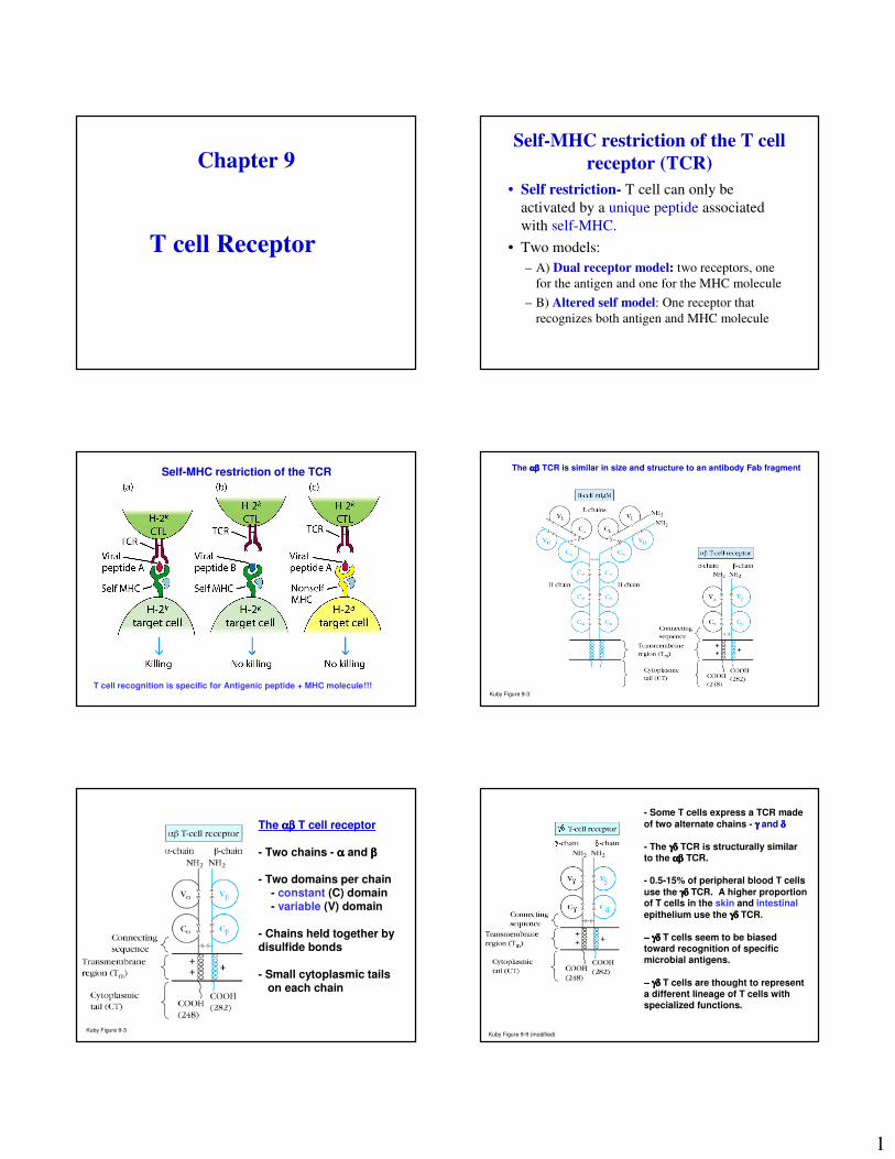
1 Chapter 9 T cell Receptor Self-MHC restriction of the T cell receptor (TCR) • Self restriction- T cell can only be activated by a unique peptide associated with self-MHC. • Two models: – A) Dual receptor model: two receptors, one for the antigen and one for the MHC molecule – B) Altered self model: One receptor that recognizes both antigen and MHC molecule Self-MHC restriction of the TCR T cell recognition is specific for Antigenic peptide + MHC molecule!!! Kuby Figure 9-3 The αβ αβ αβ αβ TCR is similar in size and structure to an antibody Fab fragment Kuby Figure 9-3 The αβ αβ αβ αβ T cell receptor - Two chains - α and β - Two domains per chain - constant (C) domain - variable (V) domain - Chains held together by disulfide bonds - Small cytoplasmic tails on each chain Kuby Figure 9-9 (modified) - Some T cells express a TCR made of two alternate chains - γ and δ - The γδ γδ γδ γδ TCR is structurally similar to the αβ αβ αβ αβ TCR. - 0.5-15% of peripheral blood T cells use the γδ γδ γδ γδ TCR. A higher proportion of T cells in the skin and intestinal epithelium use the γδ γδ γδ γδ TCR. - γδ γδ γδ γδ T cells seem to be biased toward recognition of specific microbial antigens. - γδ γδ γδ γδ T cells are thought to represent a different lineage of T cells with specialized functions.
-
Upload
truongkhue -
Category
Documents
-
view
218 -
download
0
Transcript of T cell Receptor - Northern Arizona Universityfpm/immunology/lectures/Chapter09.pdf · 1 Chapter 9 T...
1
Chapter 9
T cell Receptor
Self-MHC restriction of the T cell
receptor (TCR)
• Self restriction- T cell can only be
activated by a unique peptide associated
with self-MHC.
• Two models:
– A) Dual receptor model: two receptors, one
for the antigen and one for the MHC molecule
– B) Altered self model: One receptor that
recognizes both antigen and MHC molecule
Self-MHC restriction of the TCR
T cell recognition is specific for Antigenic peptide + MHC molecule!!!Kuby Figure 9-3
The αβαβαβαβ TCR is similar in size and structure to an antibody Fab fragment
Kuby Figure 9-3
The αβαβαβαβ T cell receptor
- Two chains - αααα and ββββ
- Two domains per chain - constant (C) domain- variable (V) domain
- Chains held together by disulfide bonds
- Small cytoplasmic tails on each chain
Kuby Figure 9-9 (modified)
- Some T cells express a TCR made
of two alternate chains - γγγγ and δδδδ
- The γδγδγδγδ TCR is structurally similar to the αβαβαβαβ TCR.
- 0.5-15% of peripheral blood T cells
use the γδγδγδγδ TCR. A higher proportion of T cells in the skin and intestinal
epithelium use the γδγδγδγδ TCR.
−−−− γδγδγδγδ T cells seem to be biased toward recognition of specific microbial antigens.
−−−− γδγδγδγδ T cells are thought to represent a different lineage of T cells with specialized functions.

2
Table 9.1 Comparison of TCR
αβ T cells γδγδγδγδ T cells
• % CD3+ 90-99% 1-10%
• TCR V gene Large Small
in germline
• CD4/CD8
CD4 60% <1%**
CD8 30% 30%
CD4-CD8- <1% 60%**
• MHC restriction Yes No**
• Ligands Peptide+ MHC Phospholipid antigen,
intact antigen Kuby Figure 9-9
VββββVαααα
CββββCαααα
VDJVD
CC
So, Which one is the “light”chain?
Which one is the “heavy” chain?
Organization and rearrangement
of TCR genes
1
2
- Rearrangement of TCRαααα removes genes for TCRδδδδ
1
2

3
1, rearrangement
6, Post-translational modifications
5, polypeptide
4, mature mRNA
1, rearrangement
3, Post-transcriptional modifications
2, RNA transcript
Rearrangement of TCR genes• TCR Genes also composed of V, D, J and C gene
segments
• Genes are located in different chromosomes
• The β and δ chains contain D segments (like Ig Heavy chains!) while the α and γγγγ chains do not.
• α and γγγγ chains - VJ rearrangement only
• β and δ chains - DJ and then V-DJ rearrangement
• Segments of the δ chain are embedded within the segments encoding the α chain
• When the α chain rearranges, δ segments are deleted
• T cells express only αβ or γγγγδ TCR
• Rearrangement involves RAG-1 and RAG-2 and TdT
• Rearrangement is governed by the one turn-two turn rule
Generation of antibody diversity
1. Multiple germline V, D and J gene segments
2. Combinatorial V-J and V-D-J joining
3. Somatic hypermutation
4. Junctional flexibility
5. P-nucleotide addition
6. N-nucleotide addition
Generation of TCR diversity
-Varying number of D segments in the delta (and beta) chain, why?(arrangement of RSS sequences differs from that in Ig loci to allow
this)
Antibody
12 bp
23 bp
Generation of TCR diversity
2) N-region nucleotide addition, occurs in all chains
Note: Increased diversity in TCR!
1.6 x 10¹¹ VS 3 x 10⁷

4
MAJOR DIFFERENCES
BETWEEN TCR AND Ig GENES• Somatic hyper-mutation (affinity maturation)
- During an antibody response, mutations accumulate at a rapid rate in the VDJ gene segments encoding the BCR.
- Thus, as an immune response proceeds, the affinity of the antibody produced (i.e. its ability to bind to the antigen) increases.
• Alternative joining of D segments (β, δ)
• N-nucleotide addition to both chains
*
*
*
1.6 x 10¹¹ VS 3 x 10⁷
Properties of Ig and TCR Genes
Ig TCR
Many VDJs, few Cs yes yes
VDJ rearrangement yes yes
V-regions form antigen yes yes
recognition site
Somatic hypermutation yes no
Properties of Ig and TCR
ProteinsIg TCR
Transmembrane forms yes yes
Secreted forms yes no
Isotypes with different yes no*
functions
Valency 2 1
The TCR complex includes CD3 - 3 heterodimers: γεγεγεγε, εδεδεδεδ and ζζζζζζζζ
- 1) TCR is not expressed without CD3. It is required to bring TCR to surface
- 2) All chains of CD3 possess ITAM motifs. (Immunoreceptortyrosine-based activation motif) ���� Signal Transduction
RECOGNITION
SIGNAL TRANSDUCTION
TCR Receptor Complex- CD3

5
WHY ACCESSORY MOLECULES?
1) Due to low affinity of TCR with peptide MHCcomplex
2) Provide:
- Adhesion, Activation and Co-stimulation
- Some show increased expression in response tocytokines
*
RECAP:
-The BCR consists of IgM or IgD plus Ig-αααα/Ig-ββββheterodimers. The Ig binds the antigen while the Ig-αααα/Ig-ββββ heterodimers are involved in activation of the B cell.
- The TCR consists of either the αααα////ββββ chains or the γγγγ////δδδδchains plus CD3. The αβαβαβαβ or γδγδγδγδ chains bind the antigen while CD3 is involved in activation of the T cell.
Accessory Molecules Involved
in Cell-Cell Interactions
Interactions of Th Cell and APC
LFA-3
CD-2 LFA-1 TCR
CD4
ICAM-1 Class IIMHC
B7-1/B7-2(CD80/CD86
CD28
IL-1
IL-6TNF-alphaIL-12
IL-15
TNF-betaIFN-gamma
GM-CSFIL-4
CD4+ T cell
APC
peptide
CD45R
CD22
Interactions of Tc and Target Cell
LFA-1 TCR
CD8
ICAM-1 Class IMHC
LFA-3
CD2CD8+ T cell
Target
cell
peptide
CD45R
CD22
T-cell Accessory molecules
• CD4 and CD8 are co-receptors because they
recognize the peptide-MHC complex
• CD8 recognizes the α3 MHC-I domain; while
CD4 interacts with α2 MHC-II domain
• Both CD4 and CD8 act in signal transduction
• OTHER

6
Accessory Molecules Involved
in Cell-Cell InteractionsCell Adhesion:
T Cell Ligand on APC
CD2- (LFA-2) LFA-3
LFA-1 ICAM-1, ICAM-2
LFA = Leukocyte Function-associated
Antigen
ICAM = InterCellular Adhesion Molecule
Costimulatory Molecules• Molecules on T cell and 2nd cell that engage
to deliver 2nd signal required for activation of
T cell
• Most important co-stimulatory molecules:
T cell Ligand on 2nd cell
CD28 B7-1 (CD80), B7-2 (CD86)
CTLA-4 B7-1 (CD80), B7-2 (CD86)
CD45R CD22
CD4/CD8 MHC-I/II
T cell APC
Alloreactive T cells
• Allogeneic – genetically different individual of the
same species (humans!!)
• MHC molecules are alloantigens: CD4 T cells are
alloreactive to MHC-II alloantigens and CD8 T
cells are alloreactive to MHC-I alloantigens
• Direct and Indirect recognition of alloantigens: a)
nonself MHC moleucle on foreign cell recognized
in its native form; b) peptides from allogeneic
MHC after processing are prpresented by self-
MHC molecules
• The End


![IMMUNOGLOBULINE E T CELL RECEPTOR T. Strachan e A.P. … · B cell antigen receptor tetramero [ IgH 2 + IgL 2 (Ig oppure Ig )] T cell receptor (TCR) eterodimero TCR /TCR TCR /TCR](https://static.fdocuments.net/doc/165x107/5c017b5c09d3f26f1e8cc6a0/immunoglobuline-e-t-cell-receptor-t-strachan-e-ap-b-cell-antigen-receptor.jpg)