
Systems/Circuits ...2010; Chen et al., 2011) to two of these transgenic mouse lines, which...
15
Systems/Circuits Characteristic Patterns of Dendritic Remodeling in Early-Stage Glaucoma: Evidence from Genetically Identified Retinal Ganglion Cell Types Rana N. El-Danaf 1,2,3 and Andrew D. Huberman 1,2,3,4 1 Department of Neurosciences, 2 Neurobiology Section in the Division of Biological Sciences, and 3 Department of Ophthalmology, University of California, San Diego, La Jolla, California 92093, and 4 Salk Institute for Biological Studies, La Jolla, California 92093 Retinal ganglion cell (RGC) loss is a hallmark of glaucoma and the second leading cause of blindness worldwide. The type and timing of cellular changes leading to RGC loss in glaucoma remain incompletely understood, including whether specific RGC subtypes are prefer- entially impacted at early stages of this disease. Here we applied the microbead occlusion model of glaucoma to different transgenic mouse lines, each expressing green fluorescent protein in 1–2 specific RGC subtypes. Targeted filling, reconstruction, and subsequent comparison of the genetically identified RGCs in control and bead-injected eyes revealed that some subtypes undergo significant den- dritic rearrangements as early as 7 d following induction of elevated intraocular pressure (IOP). By comparing specific On-type, On-Off- type and Off-type RGCs, we found that RGCs that target the majority of their dendritic arbors to the scleral half or “Off” sublamina of the inner plexiform layer (IPL) undergo the greatest changes, whereas RGCs with the majority of their dendrites in the On sublamina did not alter their structure at this time point. Moreover, M1 intrinsically photosensitive RGCs, which functionally are On RGCs but structurally stratify their dendrites in the Off sublamina of the IPL, also underwent significant changes in dendritic structure 1 week after elevated IOP. Thus, our findings reveal that certain RGC subtypes manifest significant changes in dendritic structure after very brief exposure to elevated IOP. The observation that RGCs stratifying most of their dendrites in the Off sublamina are first to alter their structure may inform the development of new strategies to detect, monitor, and treat glaucoma in humans. Key words: dendrites; glaucoma; retinal ganglion cells Introduction Glaucoma is a common CNS neurodegenerative disease and the second leading cause of blindness (Quigley and Broman, 2006; Pascolini and Mariotti, 2012). The hallmark feature of glaucoma is damage and loss of retinal ganglion cells (RGCs), the neurons that connect the eyes to the brain (Quigley, 1999; Weinreb and Khaw, 2004; Casson et al., 2012). Glaucoma is often associated with elevated intraocular pressure (IOP; Sommer, 1989; Peters et al., 2014) and pressure-lowering agents can be effective in slow- ing RGC loss (Morrison et al., 1998; Heijl et al., 2002; Kass et al., 2002; Lichter, 2002; Kim et al., 2013). Even with pressure- lowering drugs, however, RGCs eventually die. Understanding the timing and pattern of cellular changes leading to RGC death in glaucoma should facilitate the development of tools to detect and impede those cellular changes and ultimately to preserve vision. Several experimental models (Bouhenni et al., 2012; Vidal- Sanz et al., 2012) indicate that RGC death is a relatively late event in glaucoma, and is preceded by axonal transport defects, which can occur even within days of IOP elevation and before axonal loss (Buckingham et al., 2008; Crish et al., 2010; Chen et al., 2011; Calkins, 2012; Ward et al., 2014). Also, Wong and coworkers recently reported that dendritic and/or electrophysiological changes occur in subsets of RGCs as early as 2 weeks after IOP elevation (Della Santina et al., 2013). Whether changes in RGC dendritic structure occur during the period immediately follow- ing the induction of elevated IOP, however, remains unresolved. A persistent barrier to understanding the pathogenesis of glaucoma is the wide diversity of RGC subtypes (Berson, 2008; Dhande and Huberman, 2014). In recent years, we discovered and characterized various transgenic mouse lines, each with 1–2 distinct RGC subtypes selectively expressing green fluorescent protein (GFP; Huberman et al., 2008, 2009; Osterhout et al., 2011; Rivlin-Etzion et al., 2011; Dhande et al., 2013). These mice raise the opportunity to study the detailed morphological and functional changes occurring in defined RGC populations in re- sponse to various experimental perturbations. Here we applied the “microbead occlusion” model of glaucoma (Sappington et al., Received April 4, 2014; revised Nov. 13, 2014; accepted Dec. 15, 2014. Author contributions: R.N.E.-D. and A.D.H. designed research; R.N.E.-D. and A.D.H. performed research; R.N.E.-D. analyzed data; R.N.E.-D. and A.D.H. wrote the paper. This work was supported by a grant from The Glaucoma Research Foundation Catalyst for a Cure (A.D.H. and R.N.E.-D.) and the E. Matilda Ziegler Foundation for the Blind (A.D.H.). We thank Drs. David Berson, Rachel Wong, Jeff Goldberg, Alf Dubra, and Vivek Srinivasan, as well as members of the Huberman laboratory for their helpful comments and suggestion on an earlier version of this manuscript. We also thank J. Bradley Segal for collecting preliminary data for this project, and Ann Phan for assistance with cell counts. The authors declare no competing financial interests. Correspondence should be addressed to Andrew D. Huberman at the above address. E-mail: [email protected]. DOI:10.1523/JNEUROSCI.1419-14.2015 Copyright © 2015 the authors 0270-6474/15/352329-15$15.00/0 The Journal of Neuroscience, February 11, 2015 • 35(6):2329 –2343 • 2329
Transcript of Systems/Circuits ...2010; Chen et al., 2011) to two of these transgenic mouse lines, which...
Systems/Circuits
Characteristic Patterns of Dendritic Remodeling inEarly-Stage Glaucoma: Evidence from Genetically IdentifiedRetinal Ganglion Cell Types
Rana N. El-Danaf1,2,3 and Andrew D. Huberman1,2,3,4
1Department of Neurosciences, 2Neurobiology Section in the Division of Biological Sciences, and 3Department of Ophthalmology, University of California,San Diego, La Jolla, California 92093, and 4Salk Institute for Biological Studies, La Jolla, California 92093
Retinal ganglion cell (RGC) loss is a hallmark of glaucoma and the second leading cause of blindness worldwide. The type and timing ofcellular changes leading to RGC loss in glaucoma remain incompletely understood, including whether specific RGC subtypes are prefer-entially impacted at early stages of this disease. Here we applied the microbead occlusion model of glaucoma to different transgenicmouse lines, each expressing green fluorescent protein in 1–2 specific RGC subtypes. Targeted filling, reconstruction, and subsequentcomparison of the genetically identified RGCs in control and bead-injected eyes revealed that some subtypes undergo significant den-dritic rearrangements as early as 7 d following induction of elevated intraocular pressure (IOP). By comparing specific On-type, On-Off-type and Off-type RGCs, we found that RGCs that target the majority of their dendritic arbors to the scleral half or “Off” sublamina of theinner plexiform layer (IPL) undergo the greatest changes, whereas RGCs with the majority of their dendrites in the On sublamina did notalter their structure at this time point. Moreover, M1 intrinsically photosensitive RGCs, which functionally are On RGCs but structurallystratify their dendrites in the Off sublamina of the IPL, also underwent significant changes in dendritic structure 1 week after elevated IOP.Thus, our findings reveal that certain RGC subtypes manifest significant changes in dendritic structure after very brief exposure toelevated IOP. The observation that RGCs stratifying most of their dendrites in the Off sublamina are first to alter their structure mayinform the development of new strategies to detect, monitor, and treat glaucoma in humans.
Key words: dendrites; glaucoma; retinal ganglion cells
IntroductionGlaucoma is a common CNS neurodegenerative disease and thesecond leading cause of blindness (Quigley and Broman, 2006;Pascolini and Mariotti, 2012). The hallmark feature of glaucomais damage and loss of retinal ganglion cells (RGCs), the neuronsthat connect the eyes to the brain (Quigley, 1999; Weinreb andKhaw, 2004; Casson et al., 2012). Glaucoma is often associatedwith elevated intraocular pressure (IOP; Sommer, 1989; Peters etal., 2014) and pressure-lowering agents can be effective in slow-ing RGC loss (Morrison et al., 1998; Heijl et al., 2002; Kass et al.,2002; Lichter, 2002; Kim et al., 2013). Even with pressure-lowering drugs, however, RGCs eventually die. Understandingthe timing and pattern of cellular changes leading to RGC death
in glaucoma should facilitate the development of tools to detectand impede those cellular changes and ultimately to preservevision.
Several experimental models (Bouhenni et al., 2012; Vidal-Sanz et al., 2012) indicate that RGC death is a relatively late eventin glaucoma, and is preceded by axonal transport defects, whichcan occur even within days of IOP elevation and before axonalloss (Buckingham et al., 2008; Crish et al., 2010; Chen et al., 2011;Calkins, 2012; Ward et al., 2014). Also, Wong and coworkersrecently reported that dendritic and/or electrophysiologicalchanges occur in subsets of RGCs as early as 2 weeks after IOPelevation (Della Santina et al., 2013). Whether changes in RGCdendritic structure occur during the period immediately follow-ing the induction of elevated IOP, however, remains unresolved.
A persistent barrier to understanding the pathogenesis ofglaucoma is the wide diversity of RGC subtypes (Berson, 2008;Dhande and Huberman, 2014). In recent years, we discoveredand characterized various transgenic mouse lines, each with 1–2distinct RGC subtypes selectively expressing green fluorescentprotein (GFP; Huberman et al., 2008, 2009; Osterhout et al.,2011; Rivlin-Etzion et al., 2011; Dhande et al., 2013). These miceraise the opportunity to study the detailed morphological andfunctional changes occurring in defined RGC populations in re-sponse to various experimental perturbations. Here we appliedthe “microbead occlusion” model of glaucoma (Sappington et al.,
Received April 4, 2014; revised Nov. 13, 2014; accepted Dec. 15, 2014.Author contributions: R.N.E.-D. and A.D.H. designed research; R.N.E.-D. and A.D.H. performed research; R.N.E.-D.
analyzed data; R.N.E.-D. and A.D.H. wrote the paper.This work was supported by a grant from The Glaucoma Research Foundation Catalyst for a Cure (A.D.H. and
R.N.E.-D.) and the E. Matilda Ziegler Foundation for the Blind (A.D.H.). We thank Drs. David Berson, Rachel Wong,Jeff Goldberg, Alf Dubra, and Vivek Srinivasan, as well as members of the Huberman laboratory for their helpfulcomments and suggestion on an earlier version of this manuscript. We also thank J. Bradley Segal for collectingpreliminary data for this project, and Ann Phan for assistance with cell counts.
The authors declare no competing financial interests.Correspondence should be addressed to Andrew D. Huberman at the above address. E-mail:
[email protected]:10.1523/JNEUROSCI.1419-14.2015
Copyright © 2015 the authors 0270-6474/15/352329-15$15.00/0
The Journal of Neuroscience, February 11, 2015 • 35(6):2329 –2343 • 2329
2010; Chen et al., 2011) to two of these transgenic mouse lines,which collectively label three RGC subtypes: (1) an Off-� RGCsubtype (Huberman et al., 2008), (2) an On-direction selectiveRGC subtype, and (3) an On-Off-direction selective RGC sub-type (Dhande et al., 2013). We also used the antibody againstmelanopsin protein to label and examine type 1 intrinsically pho-tosensitive RGCs (M1 ipRGCs; Berson et al., 2010; Table 1). Wetested whether there are dendritic morphological changes thatoccur in any of the labeled RGC populations 1 week followinginduction of elevated IOP, an early stage that, to our knowledge,has not been examined. Our goal was also to determine whetherthe dendrites and somas of some RGC subtypes are impactedmore than others at this stage and to determine whether any suchchanges predicted rates of RGC death at later time points. Theidea that some RGC subtypes are more vulnerable to aging and/orglaucoma has existed for some time, but has been met with con-tradictory evidence (Quigley et al., 1988; Glovinsky et al., 1991;Weber et al., 1998; Morgan et al., 2000; Pavlidis et al., 2003; Shouet al., 2003; Jakobs et al., 2005; Li et al., 2006; Samuel et al., 2011;Della Santina et al., 2013; Feng et al., 2013). Our results indicatethat dendritic rearrangements of RGCs that stratify primarilyin the Off sublamina of the inner retina are among the earlieststructural changes in glaucoma. Moreover, the RGCs that un-dergo these early stages are among the first to die. These findingspoint to the specific structural features and cell types in the retinathat may act as key factors involved in the progression of glaucoma.
Materials and MethodsAnimals. All experimental procedures were approved by the InstitutionalAnimal Care and Use Committee at the University California, San Diego.All mice used in this study were 1 month old or older [postnatal day (P)31–P73]. Both male and female mice were used in this study in equalnumbers. The following bacterial artificial chromosome (BAC) trans-genic mouse lines were used: Calretinin-EGFP (CB2-GFP; Huberman elal., 2008) and Homeobox d10-EGFP (Hoxd10-GFP; Dhande et al., 2013;Table 1). Founder mice were backcrossed onto C57BL/6 for �4 genera-tions and their offspring used in this study.
Bead injections. Elevation of IOP was induced using the microbeadocclusion model described previously (Sappington et al., 2010; Chen etal., 2011; Fig. 1). Mice from one group were anesthetized with an isoflu-rane– oxygen mixture (2– 4% v/v) at a flow rate of 1.21 L/min, and a smallhole was made in the cornea using a 30 gauge needle. A 2–3 �l volume ofpolyesterene beads (10 �m; Invitrogen F-8834) was injected into theanterior chamber using a Hamilton syringe with 33 gauge needle. The needlealso contained a small volume (0.1–0.5 �l) of sodium hyaluronate (AlconLaboratories) to keep the beads from exiting the eye after the injection(Cone et al., 2010; Frankfort et al., 2013). For every animal, one eyereceived the bead injection and the other eye served as an internal controlin which a hole was made in the cornea but no beads were injected. Asecond group of mice were anesthetized with a mixture of ketamine/xylazine and a hole was made in the cornea using a glass micropipetteattached to a picospritzer. One microliter of the 10 �m microbeads wasdelivered into one eye, while the other eye received a 1 �l injection ofsaline. For both groups, IOPs were measured using a rebound tonometer(TonoLab, Colonial Medical Supply). Six to 10 consecutive IOP mea-surements were made daily, and the IOP reported for that day was theaverage of all the readings. Pressures were monitored at the same time
every day (�60 min) for 2– 4 d before bead injections to establish “base-line” IOP readings and then for the 7 d following the bead injections (atwhich time the animal was killed for analysis). Mice that showed noelevation in IOP after bead injection (likely due to leakage of the beads)were excluded from the study. No differences were noted in IOP readingsaccording to the age and/or strain of mice used.
Cell fills. Intracellular dye filling of single ganglion cells was performedusing methods described in detail previously (Beier et al., 2013; Dhandeet al., 2013; Cruz-Martín et al., 2014; Fig. 2). Briefly, mice were anesthe-tized with isoflurane and the eyes were enucleated. The retinas weredissected and kept in an oxygenated (95% O2/5% CO2) solution of Ames’
Table 1. Summary of transgenic mice and antibody labeling used for labeling RGCsubtypes
Mouse line/stain Labeled RGC subtype Reference
CB2-GFP tOff-� Huberman et al., 2008Hoxd10-GFP On DSGC Dhande et al., 2013
aOn-Off DSGCAnti-melanopsin M1 ipRGC Berson et al., 2010
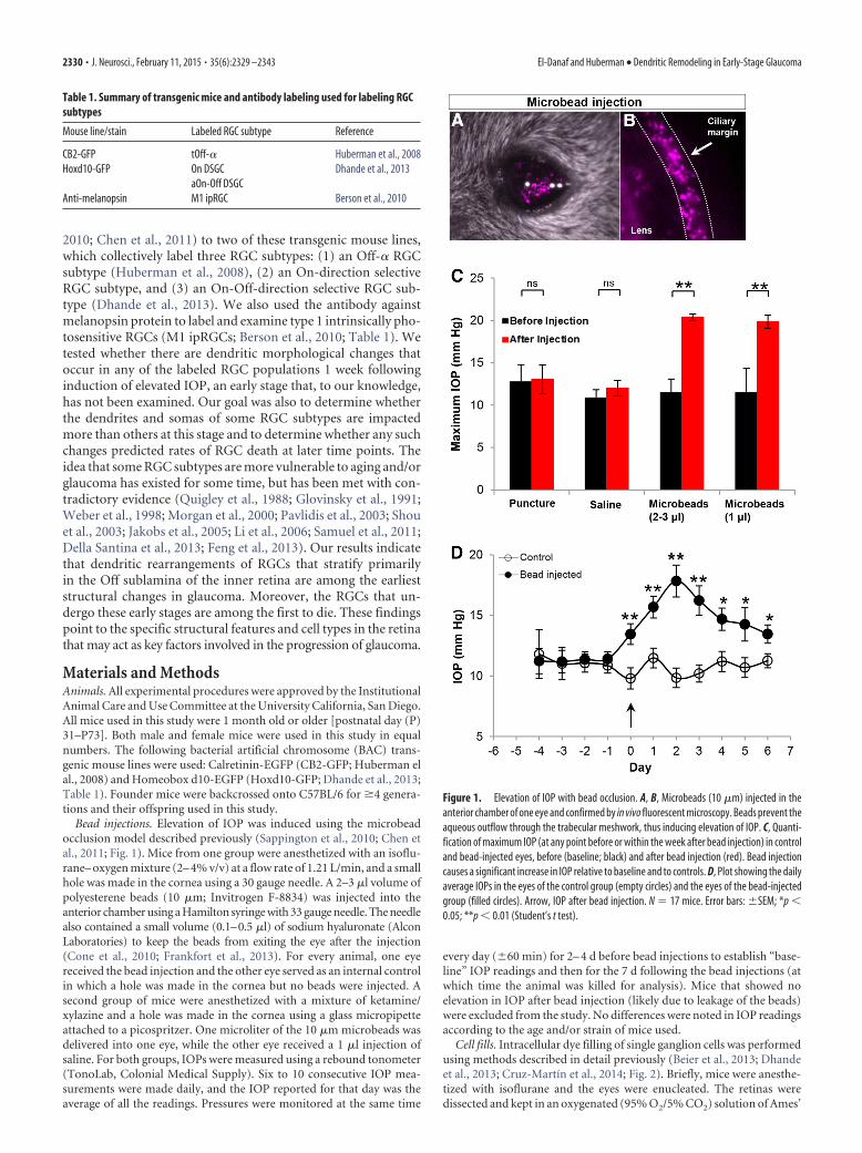
Figure 1. Elevation of IOP with bead occlusion. A, B, Microbeads (10 �m) injected in theanterior chamber of one eye and confirmed by in vivo fluorescent microscopy. Beads prevent theaqueous outflow through the trabecular meshwork, thus inducing elevation of IOP. C, Quanti-fication of maximum IOP (at any point before or within the week after bead injection) in controland bead-injected eyes, before (baseline; black) and after bead injection (red). Bead injectioncauses a significant increase in IOP relative to baseline and to controls. D, Plot showing the dailyaverage IOPs in the eyes of the control group (empty circles) and the eyes of the bead-injectedgroup (filled circles). Arrow, IOP after bead injection. N � 17 mice. Error bars: �SEM; *p �0.05; **p � 0.01 (Student’s t test).
2330 • J. Neurosci., February 11, 2015 • 35(6):2329 –2343 El-Danaf and Huberman • Dendritic Remodeling in Early-Stage Glaucoma
medium (Sigma-Aldrich, catalog #A1420) supplemented with 23 mM
NaHCO3. GFP� RGCs were visualized under epifluorescence, and thentargeted and filled under differential interference contrast (DIC) withelectrodes made with borosilicate glass (Sutter Instruments; 15–20 M�)containing a 10 mM solution of Alexa Fluor 555 hydrizide (Invitrogen,catalog #A20501MP) in 200 mM KCl. Hyperpolarizing current pulsesranging between 0.1 and 0.9 nA were applied for 1–5 min to obtain acomplete cell fill, which was assessed by visualization of the axon, soma,and filling of fine distal dendritic processes. The central-peripheral loca-tion of each filled neuron was tracked. To control for possible eccentricityeffects, we normalized the height and width of each retina as a percentagevalue, and the location of each individual filled neuron was recorded as acoordinate value relative to the optic nerve head (e.g., the nerve head:0,0). Only the neurons that were located within the central 30 –70% of theretina were included in this study.
Retinal histology. After the completion of cell filling, the retinas werefixed for 1 h in 4% paraformaldehyde (PFA), then washed with 1� PBSand incubated for 1 h in a blocking solution consisting of 10% goat serum
or 10% donkey serum, with 0.25% Triton-X, at room temperature. Theretinas were then incubated for 1–3 d at 4°C with the following pri-mary antibodies diluted in blocking solution: rabbit anti-GFP [1:1000; Invitrogen, catalog #A6455; Research Resource Identifier(RRID): AB_221570], guinea pig anti-VAChT (1:1000; Millipore, catalog#AB1588; RRID: AB_2187981), and goat anti-ChAT (1:100; Millipore,catalog #AB144P; RRID: AB_2079751). To reveal the morphology of theM1 ipRGCs (Berson et al., 2010), some retinas were stained for melanop-sin (1:1000 rabbit anti-melanopsin; Advanced Targeting Systems, catalog#AB-N39; RRID: AB_1608076). Retinas were rinsed with PBS (3�, 30min each), and incubated for 2 h at room temperature with the followingsecondary antibodies: Alexa Fluor 488 goat anti-rabbit (1:1000; LifeTechnologies, catalog #A11034; RRID: AB_10562715), Alexa Fluor 647goat anti-guinea pig (1:1000; Invitrogen, catalog #A21450; RRID:AB_141882), Alexa Fluor 594 goat anti-rabbit (1:1000; Life Technolo-gies, catalog #A11037; RRID: AB_10561549), Alexa Fluor 647 donkeyanti-goat (1:1000; Life Technologies, catalog #A21447; RRID:AB_10584487), Alexa Fluor 488 donkey anti-rabbit (1:1000; Invitrogen,
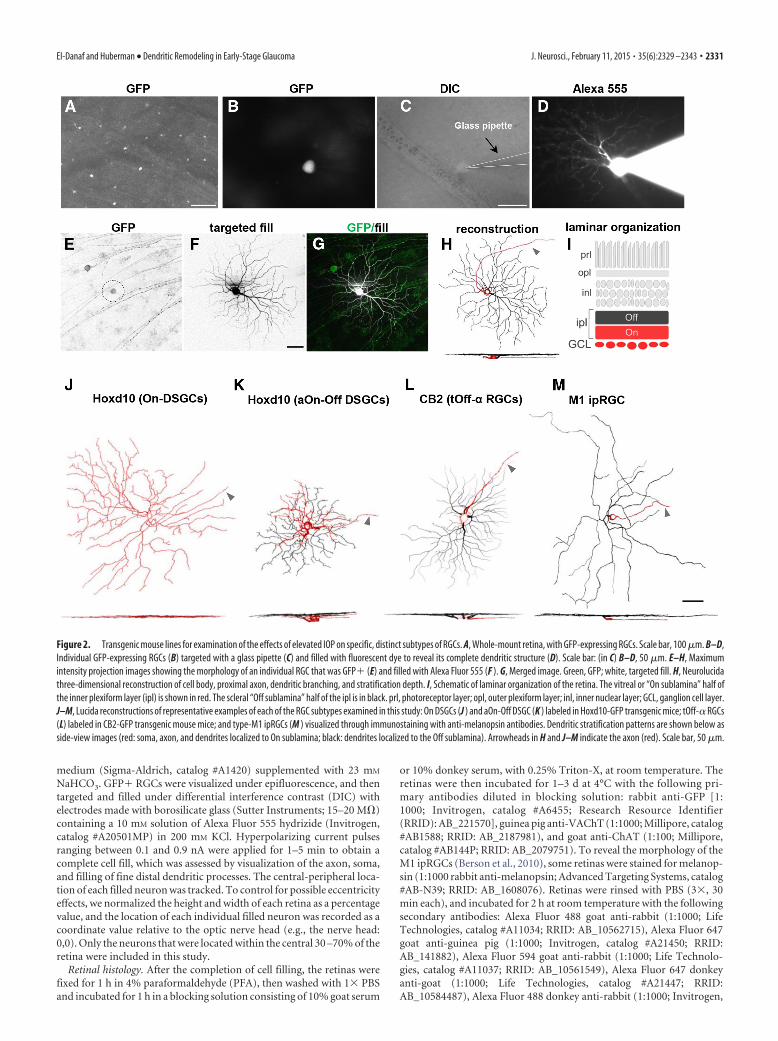
Figure 2. Transgenic mouse lines for examination of the effects of elevated IOP on specific, distinct subtypes of RGCs. A, Whole-mount retina, with GFP-expressing RGCs. Scale bar, 100 �m. B–D,Individual GFP-expressing RGCs (B) targeted with a glass pipette (C) and filled with fluorescent dye to reveal its complete dendritic structure (D). Scale bar: (in C) B–D, 50 �m. E–H, Maximumintensity projection images showing the morphology of an individual RGC that was GFP� (E) and filled with Alexa Fluor 555 (F ). G, Merged image. Green, GFP; white, targeted fill. H, Neurolucidathree-dimensional reconstruction of cell body, proximal axon, dendritic branching, and stratification depth. I, Schematic of laminar organization of the retina. The vitreal or “On sublamina” half ofthe inner plexiform layer (ipl) is shown in red. The scleral “Off sublamina” half of the ipl is in black. prl, photoreceptor layer; opl, outer plexiform layer; inl, inner nuclear layer; GCL, ganglion cell layer.J–M, Lucida reconstructions of representative examples of each of the RGC subtypes examined in this study: On DSGCs (J ) and aOn-Off DSGC (K ) labeled in Hoxd10-GFP transgenic mice; tOff-� RGCs(L) labeled in CB2-GFP transgenic mouse mice; and type-M1 ipRGCs (M ) visualized through immunostaining with anti-melanopsin antibodies. Dendritic stratification patterns are shown below asside-view images (red: soma, axon, and dendrites localized to On sublamina; black: dendrites localized to the Off sublamina). Arrowheads in H and J–M indicate the axon (red). Scale bar, 50 �m.
El-Danaf and Huberman • Dendritic Remodeling in Early-Stage Glaucoma J. Neurosci., February 11, 2015 • 35(6):2329 –2343 • 2331
catalog #A21206; RRID: AB_141708), and Alexa Fluor 647 donkey anti-guinea pig (1:1000; Jackson ImmunoResearch, catalog #706-606-148).Sections were rinsed with PBS (3�, 30 min each) and mounted onto glassslides and coverslipped with either Prolong Gold containing DAPI (In-vitrogen, P36931) or Vectashield containing DAPI (Vector Laboratories,catalog #H-1200).
Brain histology to analyze RGC axonal projections. Brains were dis-sected, fixed overnight with 4% PFA, and then transferred to a 30% (w/v)sucrose solution. Using a freezing sliding microtome, brain slices werecollected at 40 – 45 �m in the coronal plane, and immunostained for GFPusing the methods described above. Sections were mounted onto glassslides and coverslipped with Vectashield containing DAPI.
Imaging. All RGCs were imaged with a laser scanning confocal micro-scope (Zeiss LSM 710 or 780). Image stacks were collected using a LDC-Apochromat 40�/1.1 water-immersion objective lens, with a Z-stepincrement size of 0.48 – 0.5 �m, a scanning resolution of 1024 � 1024pixels, and a Kalman averaging of 2– 4.
For counting GFP� and melanopsin� cells, whole-mount images ofentire retinas were acquired at 5� or 10� using a Zeiss Axio Imager 2epifluorescence microscope equipped with an Axiocam HR camera. Us-ing Adobe Photoshop software, the images were stitched together toinclude the entire retina. Brain sections containing central visual targetswere acquired using a 10� objective lens.
Analysis. A total of 152 RGCs from 17 mice were analyzed for this study(72 RGCs from control eyes; 80 RGCs from eyes with elevated IOP).Complete morphological reconstructions were obtained using Neurolu-cida software (10.42.1, MBF Bioscience; RRID: nif-0000-10294), andanalyses were performed using either Neurolucida explorer (10.42.1,MBF Bioscience) or Fiji software. A range of morphological parameterswere examined, including soma diameter, total number of dendriticbranches, dendritic field area (calculated by measuring the area enclosedby the outermost segments of the distal dendritic branches), dendriticlength (sum of the length of the total dendritic branches), and 10 � 10�m Sholl ring analysis. Dendritic orientation index (DOi) was analyzedusing methods described by Krahe et al. (2011). The dendritic field ofeach RGC was divided into five equidistant concentric regions centeredon the soma. These rings were then divided into axial planes (horizontaland vertical planes) by passing two perpendicular lines through thesoma. The sum of dendritic processes crossing each of the five rings wascounted within the two axial planes, and the DOi was calculated as theratio of the minimum/maximum number of dendritic crossings in thetwo planes. A DOi value of 1 indicates a RGC with a radial symmetricprofile.
For analysis of GFP� cell numbers, the total number of GFP-expressing RGCs in each whole-mount retina was measured, and for eachanimal the normalized fraction of GFP� RGCs between the controlretina and the IOP-elevated retina from the same animal was calculatedand averaged. Since the total number of GFP� cells was not statisticallydifferent between the two eyes of nonbead-injected mice, this approachcontrolled for any variation in GFP� cell numbers that might occurbetween different transgenic mice. Counts were performed by three in-dependent observers.
The dendritic change index (DCI) quantified changes in the dendriticlength of the Off dendrites. We selected this parameter because it wasmost affected by elevated IOP. The DCI was defined as the ratio of thetotal dendritic length of the Off arbor in the elevated IOP condition for acell of a given subtype [e.g., transient Off � (tOff-�) RGCs] to the pop-ulation mean dendritic length of the Off arbor under control conditionsfor the same subtype. Since, the On direction-selective ganglion cells(DSGCs) showed no changes in their dendritic parameters after IOPelevation, they were excluded from this analysis (see Results).
Statistical significance was assessed using two-tailed unpaired Stu-dent’s t test. Statistical significance of p � 0.05 was chosen. Error barsshown in graphs represent the SEM.
ResultsApplication of the bead occlusion model of glaucoma in miceHere we applied the microbead occlusion animal model of glau-coma, which mimics many of the key features of elevated tension
glaucoma in humans (Sappington et al., 2010; Chen et al., 2011).We injected 1–3 �l of 10-�m-diameter microbeads into the an-terior chamber of the eye, where they accumulated along theciliary margin (Fig. 1A,B). This created a physical blockage of thenormal outflow of the aqueous humor through the trabecularmeshwork, and caused IOP to increase in the period following thebead injection (Fig. 1C,D; Sappington et al., 2010; Chen et al.,2011). One eye was injected with microbeads. The other eyeserved as an internal control by receiving either no injection (cor-neal puncture alone) or an injection of saline (Fig. 1A,B; seeMaterials and Methods). Pressures in both eyes of each animalwere monitored daily using a hand-held tonometer (see Materi-als and Methods). Figure 1C displays the maximum IOP valuesfrom the control and two bead-injected groups. Across the pop-ulation of mice studied (N � 17 mice) the maximum IOPs of thebead-injected eyes were significantly greater than both the base-line (preinjected) values and the values for the control eyes (Fig.1C; p � 0.001). These results are in agreement with earlier reportsusing this model that microbead injection is a reliable way toelevate IOPs in mice (Sappington et al., 2010; Chen et al., 2011).
We found that the mean maximum IOP for the control retinasthat had corneal puncture alone versus the control retinas thatreceived injections of 1 �l saline eyes was not significantly differ-ent before or after injection (Fig. 1C; baseline maximum IOP:corneal puncture, 12.74 � 2.04 mmHg; saline injected, 10.86 �0.96 mmHg; p � 0.12; postinjection maximum IOP: cornealpuncture, 13.04 � 1.7 mmHg; saline injected, 12.00 � 0.90mmHg; p � 0.06). Thus data from these groups were combinedinto a single control group for all subsequent analysis. In addi-tion, the mean IOP of the eyes injected with 2–3 �l beads and theeyes injected with 1 �l beads were also not significantly differentfrom one another and thus data from these groups were com-bined and referred to as the “IOP-elevated” group in subsequentanalyses (preinjection maximum IOP: 2–3 �l bead-injected eyes,11.48 � 1.6 mmHg; 1 �l bead-injected eyes, 11.5 � 2.83 mmHg;p � 0.26; postinjection maximum IOP: 2–3 �l bead-injectedeyes, 20.37 � 0.37 mmHg; 1 �l bead-injected eyes, 19.85 � 0.75mmHg; p � 0.18).
We also analyzed the time course of IOPs in the control versusbead-injected (IOP-elevated) groups (Fig. 1D). Before bead in-jection, IOPs were comparable between the two eyes (Fig. 1D) butby 1 d after bead injection (Fig. 1D, arrow at time point “0”),there was a steady increase in the IOP of the bead-injected eyes.The mean pressure of the bead-injected eyes over the first weekwas 15.09 � 0.42 mmHg, which is significantly greater than theaverage value at baseline for either group (p � 0.0001; Student’st test; Fig. 1D). By contrast, the IOP of the control eye did not varysignificantly from prepuncture/saline injection baseline (meanpressure, 10.64 � 0.26 mmHg; p � 0.41; Fig. 1D).
Targeting of transgenically labeled RGC subtypesWe next applied the bead occlusion model to two different trans-genic mouse lines, each of which expresses GFP in 1–2 distinctRGC subtypes (Table 1). The GFP� somas of these RGCs arereadily visible in live explanted, nonimmunostained retinas (Fig.2A,B), allowing them to be targeted in live retinal explants forintracellular filling to reveal their complete somatic and dendriticmorphology (Fig. 2C–G). In some animals, we also used mel-anopsin immunostaining, which selectively labels M1-type andM2-type ipRGCs (Table 1; Berson et al., 2010). However, we onlyfocused on M1 ipRGCs because their complete somatic and den-dritic morphologies can be revealed by melanopsin staining (Ber-
2332 • J. Neurosci., February 11, 2015 • 35(6):2329 –2343 El-Danaf and Huberman • Dendritic Remodeling in Early-Stage Glaucoma
son et al., 2010). For all the experiments described below, retinaswere harvested 7 d after microbead injection.
After filling and staining, we imaged the RGCs using confocalmicroscopy and generated three-dimensional reconstructions oftheir dendritic arbors using Neurolucida software (Fig. 2E–H).Sample en face morphologies and side views of each of the RGCsubtypes examined in this study are shown in Figure 2J–M. Thesomas, axons, and the portion of the dendritic arbors stratifyingin the inner (vitreal) half of the inner plexiform layer (IPL) areshown in red, and dendrites stratifying in the outer (scleral) por-tion of the IPL is shown in black (Fig. 2H–M; see Materials andMethods). Notably, the subtypes examined here cover a widerange of sizes, shapes, and arbor complexities (Fig. 2J–M). Somemouse RGC subtypes are reported to display size differences as afunction of eccentricity or retinal quadrant (Bleckert et al., 2014).Therefore, we restricted our analysis to RGCs filled from themiddle portions (30 –70% eccentricity) of the retina (see Materi-als and Methods).
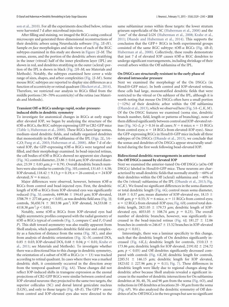
Transient Off-� RGCs undergo rapid, ocular-pressure-induced shifts in dendritic symmetryTo investigate for anatomical changes in RGCs at early stagesafter elevated IOP, we began by analyzing the structure of thetOff-� RGCs, the RGC subtype expressing GFP in CB2-GFP mice(Table 1; Huberman et al., 2008). These RGCs have large somas,medium-sized dendritic fields, and radially organized dendritesthat monostratify in the Off sublamina of the IPL (Figs. 2L, 3A–C,O; Pang et al., 2003; Huberman et al., 2008). After 7 d of ele-vated IOP, the GFP-expressing tOff-� RGCs were targeted andfilled, and their morphology examined. In bead-injected retinas,the cell bodies of tOff-� RGCs showed no apparent size changes(Fig. 3G; control diameter, 23.86 � 0.64 �m; IOP-elevated diam-eter, 23.59 � 0.83 �m; p � 0.79). Overall dendritic branch num-bers were also similar to controls (Fig. 3H; control, 131.65 � 4.58;IOP elevated, 118.42 � 9.13; p � 0.19; n � 26 control; n � 24 IOPelevated; N � 6 mice).
Major differences were observed, however, between tOff-�RGCs from control and bead-injected eyes. First, the dendriticlength of tOff-� RGCs from IOP-elevated eyes was significantlyreduced (Fig. 3I; controls, 4499.86 � 194.65 �m; IOP elevated,3708.79 � 277.66 �m; p � 0.05), as was dendritic field area (Fig. 3J;controls, 50,450.74 � 3813.98 �m2; IOP elevated, 34,533.08 �4118.38 �m2; p � 0.01).
Notably, some tOff-� RGCs from IOP-elevated eyes hadhighly asymmetric profiles compared with the radial geometry oftOff-� RGCs typical of controls (Fig. 3, compare C and F, O andP). This shift toward asymmetry was also readily apparent fromSholl analysis, which quantifies dendritic field size and complex-ity as a function of distance from the soma (Fig. 3K), and alsofrom analysis of dendritic orientation (Fig. 3L–N; control DOi,0.85 � 0.03; IOP-elevated DOi, 0.68 � 0.04; p � 0.01; Krahe etal., 2011; see Materials and Methods). To investigate whetherthere was a directional bias to this change in dendritic symmetry,the orientation of a subset of tOff-� RGCs (n � 13) was trackedaccording to retinal quadrant. In cases where there was a markeddendritic shift, it consistently occurred in the direction awayfrom the temporal quadrant (Fig. 4A). These changes did notreflect IOP-induced shifts in transgene expression as the axonalprojections of CB2-GFP RGCs were similar between control andIOP-elevated neurons: they projected to the same two targets, thesuperior colliculus (SC) and dorsal lateral geniculate nucleus(LGN), and only to those targets (Fig. 4B–E). The GFP� axonsfrom control and IOP-elevated eyes also were directed to the
same sublaminar zones within those targets: the lower stratumgriseum superficialis of the SC (Huberman et al., 2008) and the“core” of the dorsal LGN (Huberman et al., 2008; Krahe et al.,2011; Dhande and Huberman et al., 2014). This supports theconclusion that the GFP� RGCs in both experimental groupsconsisted of the same RGC subtype: tOff-� RGCs (Fig. 4B–E;Huberman at al., 2008). Collectively, these results demonstratethat just 7 d of elevated IOP causes tOff-� RGC dendrites toundergo significant rearrangements, including shrinkage of theiroverall arbors within the Off sublamina of the IPL.
On DSGCs are structurally resistant to the early phase ofelevated intraocular pressureNext we examined the morphology of the On DSGCs (inHoxd10-GFP mice). In both control and IOP-elevated retinas,these cells had large, monostratified dendritic fields that wererestricted to the vitreal or On sublayer of the IPL, although it isworth noting that mouse On DSGCs often have a small portion(�12%) of their dendritic arbor within the Off sublamina(Dhande et al., 2013), which we observed here (Fig. 5A–C, K, M).Of the On-DSGC features we examined (soma size, dendriticbranch number, field, length or patterns of branching), none ofthem differed significantly between control and IOP-elevated ret-inas (Fig. 5G–L; p 0.16 in all cases; N � 6 mice; n � 15 RGCsfrom control eyes; n � 18 RGCs from elevated-IOP eyes). Sincethe GFP-expressing RGCs in Hoxd10-GFP mice include all threesubtypes of On DSGCs (Dhande et al., 2013), we conclude thatthe somas and dendrites of On DSGCs appear structurally unaf-fected during the first week following bead-elevated IOP.
Bidirectional dendritic rearrangements in anterior-tunedOn-Off DSGCs caused by elevated IOPNext we examined the anterior-tuned On-Off DSGCs (aOn-OffDSGCs) labeled in Hoxd10-GFP mice. These neurons are char-acterized by small dendritic fields that normally stratify �60% oftheir dendrites within the Off (scleral) sublamina and �40% inthe On (vitreal) sublamina of the IPL (Dhande et al., 2013; Fig.6C,K). We found no significant differences in the soma diameteror total dendritic length (Fig. 6G; control mean soma diameter,18.69 � 0.57 �m; mean diameter of IOP-elevated eye, 18.15 �0.68 �m; p � 0.55; N � 6 mice, n � 11 RGCs from control eyes;n � 12 RGCs from elevated-IOP eyes; Fig. 6H; control total den-dritic length, 2821.05 � 179.73 �m; dendritic length of IOP-elevated eye, 4053.05 � 108.74 �m; p � 0.23). The overallnumber of dendritic branches, however, was significantly in-creased in the bead-injected retinas (Fig. 6J; 181.27 � 12.06branches in controls vs 246.67 � 15.52 branches in IOP-elevatedeyes; p � 0.01).
Interestingly, there was a laminar specificity to this change,such that the dendritic length of On dendrites significantly in-creased (Fig. 6K,L; dendritic length for controls, 1510.13 �173.86 �m; dendritic length for IOP elevated, 2191.02 � 234.72�m; p � 0.05) and Off dendrites significantly decreased com-pared with controls (Fig. 6K,M; dendritic length for controls,2285.51 � 146.15 �m; dendritic length for IOP elevated,1823.02 � 227.96 �m; p � 0.11; Fig. 7). These differences indendritic length were likely due to regional changes along thedendritic arbor because Sholl analysis revealed a significant in-crease in the number of dendritic intersections for On sublaminadendrites at 30, 40, 70, and 90 �m from the soma (Fig. 6O), andreductions in Off dendrites at locations 20 –30 �m from the soma(Fig. 6P). We also analyzed the dendritic symmetry of Off den-drites of aOn-Off DSGCs in the two groups but saw no significant
El-Danaf and Huberman • Dendritic Remodeling in Early-Stage Glaucoma J. Neurosci., February 11, 2015 • 35(6):2329 –2343 • 2333
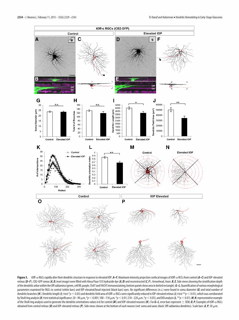
Figure 3. tOff-� RGCs rapidly alter their dendritic structure in response to elevated IOP. A–F, Maximum intensity projection confocal images of tOff-� RCCs from control (A–C) and IOP-elevatedretinas (D–F ). CB2-GFP somas (A, D, inset image) were filled with Alexa Fluor 555 hydrazide dye (A, D) and reconstructed (C, F ). Arrowhead, Axon. B, E, Side views showing the stratification depthof the dendritic arbor within the Off sublamina (green, cell fill; purple, ChAT and VAChT immunostaining; bottom panels show area in dotted rectangle). G–L, Quantification of various morphologicalparameters examined for RGCs in control (white bars) and IOP-elevated/bead-injected (black bars) eyes. No significant differences (n.s.) were found in soma diameter (G) and total number ofdendritic branches (H ). Dendritic length (I; t test *p � 0.05) and dendritic field area of tOff-� RGCs were significantly reduced in IOP-elevated retinas (J; t test **p � 0.01), which was corroboratedby Sholl ring analysis (K; t test statistical significance: 20 –90 �m, *p � 0.001; 100 –110 �m, *p � 0.01; 210 –220 �m, *p � 0.05), and DOi analysis (L, **p � 0.01). M, N, representative exampleof the Sholl ring analysis used to generate the dendritic orientations values in L for control (M ) and IOP-elevated neurons (N ). For G–L, error bars represent � SEM; O, P, Examples of tOff-� RGCsobtained from control retinas (O) and IOP-elevated retinas (P). Side views shown at the bottom of each neuron (red: soma and axon; black: Off sublamina dendrites). Scale bars: A, P, 50 �m.
2334 • J. Neurosci., February 11, 2015 • 35(6):2329 –2343 El-Danaf and Huberman • Dendritic Remodeling in Early-Stage Glaucoma

differences (DOi for controls, 0.69 � 0.07; DOi for IOP elevated,0.67 � 0.07; p � 0.86).
To address the possibility that the apparent increase in Ondendrites was because bead injections somehow reduced On ar-bors in the control eye (between-eye effects). We compared thedendritic length of On arbors and field area in aOn-Off DSGCs in
naive untreated retinas versus control ret-inas and saw no differences between thesegroups. These values were consistent withthose reported previously for the propor-tion of On-arbor size in these cells (Dhandeet al., 2013). Thus, the increase in On arborsin bead-injected eyes reflects changes in-duced by elevated IOP (dendritic length ofOn arbors: naive, 1355 � 244.77 �m; p �0.62 compared with controls; p � 0.05 com-pared with IOP elevated; Fig. 6I; dendriticfield: naive, 25,892.04 � 2345.4 �m2; con-trols, 26,286.89 � 2141.16 �m2; IOP ele-vated, 29,616.57 � 2649.28 �m2; p 0.34for all cases).
Together, these data indicate that, afterbrief exposure to elevated IOP, aOn-OffDSGCs undergo significant dendritic re-modeling involving a reduction in thedendrites targeted to the Off sublaminaand an increase in the length of dendritestargeted to the On sublamina of the IPL(Fig. 7).
M1 ipRGCs: On-type RGCs with Off-stratifying dendrites that rearrangein glaucomaThus far, we observed a pattern in whichOff dendrites of tOff-� RGCs and aOn-Off DSGCs undergo a reduction in areaand/or length, whereas On dendrites ex-hibit either no change (On DSGCs) or anoverall expansion (aOn-Off DSGCs). Isthere something structurally importantabout the Off sublayer that makes den-drites stratifying at this IPL depth morevulnerable to loss under conditions of el-evated IOP or is it the functional attri-butes of Off-type connections that occurat this IPL depth that are relevant? M1ipRGCs provide a unique way to addressthis question because, functionally, M1sare pure On-type RGCs that respond toincrements in light, but their dendrites ex-clusively stratify in the “Off” sublayer andreceive their On-bipolar input from enpassant synapses (Dumitrescu et al., 2009;Zhao et al., 2014).
M1 ipRGCs have large dendritic fieldsthat stratify near the boundary of the in-ner nuclear layer and IPL (Figs. 2M, 8A–C,L; Berson et al., 2010). Neither somasize nor dendritic field area were differentfor M1 ipRGCs in control versus IOP-elevated retinas (Fig. 8G,H; control n � 20RGCs; IOP-elevated n � 26 RGCs, N � 7mice; p 0.31). We did, however, observe
a significant overall reduction in the amount of dendritic branch-ing and length in the elevated-IOP condition (Fig. 8I; total branchnumber: controls, 27.25 � 1.89; IOP elevated, 20.62 � 1.7; p �0.05; Fig. 8J; dendritic length: controls, 2102.9 � 145.51 �m; IOPelevated, 1656.51 � 117.55 �m; p � 0.05). Such reductions ap-peared regionally biased along the dendritic arbor: Sholl analysis
Figure 4. Change in symmetry of tOff-� RGCs is not due to changes in GFP expression patterns. A, Neurolucida reconstructionsof tOff-� RGCs in CB2-GFP mice showing asymmetric morphologies. For most cells, the dendrites are pointed away from thetemporal axis. D, dorsal; N, nasal; T, temporal; V, ventral. Scale bar, 50 �m. B–E, Axonal projections patterns of tOff-� RGCs aremaintained to and within central visual targets after IOP elevation, indicating that there is no change in the GFP expression patternscaused by the bead injections. dLGN, dorsal LGN; vLGN, ventral LGN; SO, stratum optimum; lSGS, lower stratum griseum superfi-cialis; uSGS, upper stratum griseum superficialis; SZ, stratum zonale. D, E, dashed line delineates the border of the dLGN. Asterisksindicate the shell of the dLGN, which is devoid of CB2-GFP axonal projections (Huberman et al., 2008, 2009). Scale bar, 100 �m.
El-Danaf and Huberman • Dendritic Remodeling in Early-Stage Glaucoma J. Neurosci., February 11, 2015 • 35(6):2329 –2343 • 2335

Figure 5. On DSGCs are structurally resistant to the early phase of elevated IOP. A–F, Same format as for Figure 3 but GFP cells are On DSGCs from Hoxd10-GFP mice. G–K, Quantification of variousmorphological parameters examined for RGCs in control (white bars) and IOP-elevated (black bars) eyes. No significant differences (n.s.) were found for any of the morphological parameters studied, includingsoma diameter (G), total number of branches (H ), dendritic field area (I ), dendritic length (J ), as well as the percentage dendritic length in the On versus Off sublaminae (K ). L, Moreover, no changes were notedwhen examining the overall dendritic architecture using Sholl ring analysis. For G–L, error bars represent�SEM. M, N, Representative examples of On DSGCs in control (M ) and IOP-elevated retinas (N ). For C,F, M, and N, red represents the soma, axon (arrowhead), and dendrites found in the On sublamina, while dendrites present in the Off sublamina are depicted in black. Scale bars: A, N, 50 �m.
2336 • J. Neurosci., February 11, 2015 • 35(6):2329 –2343 El-Danaf and Huberman • Dendritic Remodeling in Early-Stage Glaucoma
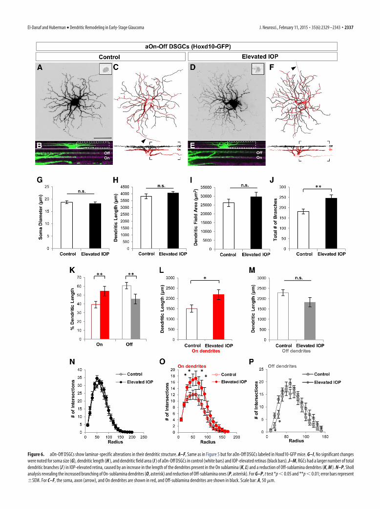
Figure 6. aOn-Off DSGCs show laminar-specific alterations in their dendritic structure. A–F, Same as in Figure 5 but for aOn-Off DSGCs labeled in Hoxd10-GFP mice. G–I, No significant changeswere noted for soma size (G), dendritic length (H ), and dendritic field area (I ) of aOn-Off DSGCs in control (white bars) and IOP-elevated retinas (black bars). J–M, RGCs had a larger number of totaldendritic branches (J ) in IOP-elevated retina, caused by an increase in the length of the dendrites present in the On sublamina (K, L) and a reduction of Off-sublamina dendrites (K, M ). N–P, Shollanalysis revealing the increased branching of On-sublamina dendrites (O, asterisk) and reduction of Off-sublamina ones (P, asterisk). For G–P, t test *p � 0.05 and **p � 0.01; error bars represent�SEM. For C–F, the soma, axon (arrow), and On dendrites are shown in red, and Off-sublamina dendrites are shown in black. Scale bar: A, 50 �m.
El-Danaf and Huberman • Dendritic Remodeling in Early-Stage Glaucoma J. Neurosci., February 11, 2015 • 35(6):2329 –2343 • 2337

revealed that elevated IOP caused significant reductions in den-dritic branching at the proximal-most (50 –120, 150 �m from thesoma) locations along the arbors of M1 ipRGCs (Fig. 8K). Thus,elevated IOP causes rapid loss of dendritic arbors for yet anothercell type stratifying in the Off sublamina, even though function-ally, these are On RGCs.
RGC subtype-specific loss occurs in a manner predicted bydendritic changesPrevious studies have shown using the bead occlusion model inmice that �20% of RGCs are lost after 2 weeks of IOP elevation(Chen et al., 2011). Next we sought to determine whether there isany subtype specificity to the RGC loss during the early period ofIOP elevation and, if so, whether that loss was predicted by theRGC subtype-specific dendritic changes discussed above. Foreach transgenic line, the total number of GFP-expressing RGCsper retina was compared between control and IOP-elevated eyesat 1 or 2 weeks after IOP elevation (Fig. 9A). CB2-GFP RGCsshowed the greatest reduction compared with other RGC sub-types examined; after 1 week of IOP elevation, the fraction ofCB2-GFP RGCs in IOP-elevated eyes was 0.91 � 0.05 of that incontrol CB2-GFP eyes (N � 5 mice; p � 0.09) and, at 2 weeksafter IOP elevation, this value was reduced to 0.77 � 0.08 com-pared with controls (N � 4 mice; p � 0.01). Hoxd10-GFP RGCsalso showed a significant and stable reduction in cell number at 1and 2 weeks after IOP elevation (1 week: 0.94 � 0.02, N � 4 mice,p � 0.01; 2 weeks: 0.95 � 0.02, N � 4 mice, p � 0.01). Interest-ingly, no significant change was observed for the fraction of M1ipRGCs at 1 or 2 weeks after IOP elevation, which is consistentwith previous reports showing that melanopsin-expressing RGCsare less susceptible to glaucoma-induced death (1 week: 1.01 �0.08, p � 0.9, N � 4 mice; 2 weeks: 0.99 � 0.01, p � 0.31, N � 3mice; Li et al., 2006). Comparison between transgenic lines re-vealed that CB2-RGCs displayed the most significant reduction
compared with all the other RGC subtypes (p � 0.05 comparedwith Hoxd10-GFP RGCs; p � 0.05 compared with M1 ipRGCs).In our experience, even within the same strain or litter, the num-ber of GFP-expressing cells can vary between animals. We note,however, that all the analysis of RGC number reported here re-flects the fraction of GFP cells in the control versus the IOP-elevated eyes of the same animal and in untreated animals thefraction of RGCs between the two eyes is not significantly differ-ent (Fig. 9B–D). Thus, we can reliably conclude that tOff-� RGCsare among the early cohorts of RGCs that display dendriticchanges and are lost in response to IOP elevation.
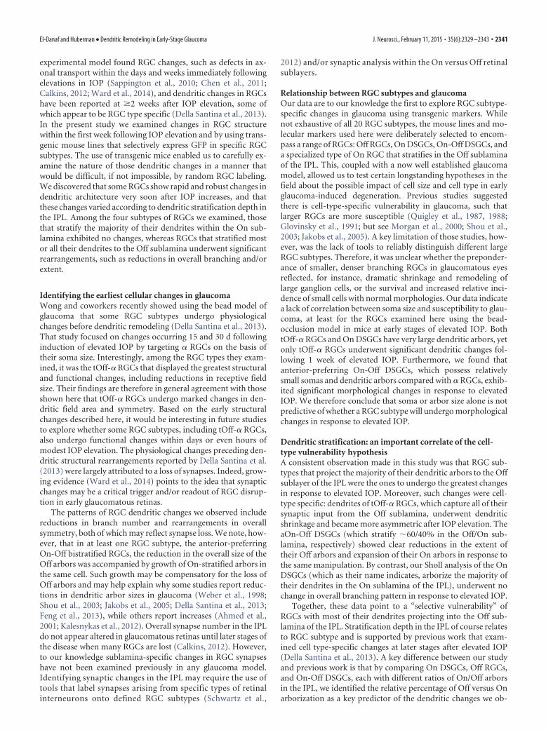
Lack of correlation between soma size and dendriticvulnerability in early response to IOP elevationBecause transgenic labeling allows for quantitative analysis ofvarious RGC characteristics related to both somatic and dendriticmorphology, we asked whether there are any correlations be-tween soma size and dendritic vulnerability to IOP elevation. Todo this, we generated a DCI that takes into account the degree ofchange in Off dendritic length for RGCs in IOP-elevated versuscontrol retinas (see Materials and Methods) and plotted that rel-ative to RGC soma size (Fig. 10). We observed no significantcorrelation between the DCI and soma size across the RGC sub-types that altered their dendrites in response to IOP elevation(Fig. 10; linear regression: r 2 � 0.004, p � 0.23), nor was there asignificant correlation within tOff-� RGC and aOn-Off DSGCsubtypes (linear regression: tOff-� RGCs, r 2 � 0.11, p � 0.11;aOn-Off DSGCs, r 2 � 0.2, p � 0.15). M1 ipRGCs with smallersomas were weakly correlated with increased alterations in den-dritic length (r 2 � 0.2, p � 0.02).
DiscussionHere we investigated the early structural changes that occur inRGCs exposed to elevated IOP. Previous work using the same
Figure 7. Additional examples of aOn-Off DSGCs in Hoxd10-GFP retinas. A, B, RGCs from control retinas (A) and IOP-elevated retinas (B). The soma, axon, and dendrites found in the On sublaminaare shown in red, while dendrites present in the Off sublamina are depicted in black. Note the increase in On-sublamina dendrites (red arrows) and the reduction in Off-sublamina dendrites (blackarrows; Fig. 6). Scale bar, 50 �m.
2338 • J. Neurosci., February 11, 2015 • 35(6):2329 –2343 El-Danaf and Huberman • Dendritic Remodeling in Early-Stage Glaucoma
Figure 8. M1 ipRGCs have reduced dendritic branching in response to elevated IOP. A–E, Maximum-intensity projection images showing the morphologies of M1 ipRGCs obtained by immuno-staining for melanopsin in control (A–C) and IOP-elevated retinas (D–F ). Reconstructed M1 cells (B, E; arrow, axon). G–K, Quantitative morphological comparison of control (white bars) andIOP-elevated (black bars) RGCs showed no significant differences (n.s.) for soma diameter (G) and dendritic field area (H ). Reduction was significant for total number of branches (I ) and dendriticlength (J ). K, Sholl analysis showing reduction of branches along distinct locations of the dendritic arbor denoted by asterisks. For G–K, error bars represent �SEM; t test *p � 0.05. L, M,Representative examples of M1 ipRGCs are shown from control (L) and IOP-elevated (M ) retinas. Scale bars: A, M, 50 �m.
El-Danaf and Huberman • Dendritic Remodeling in Early-Stage Glaucoma J. Neurosci., February 11, 2015 • 35(6):2329 –2343 • 2339
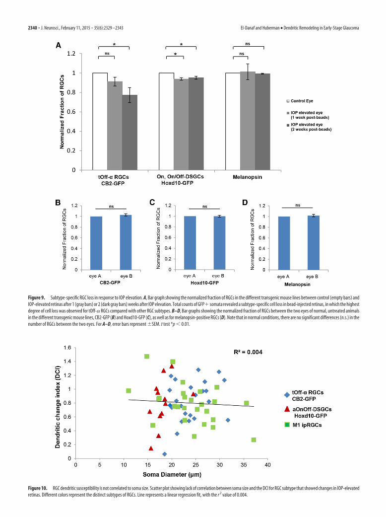
Figure 10. RGC dendritic susceptibility is not correlated to soma size. Scatter plot showing lack of correlation between soma size and the DCI for RGC subtype that showed changes in IOP-elevatedretinas. Different colors represent the distinct subtypes of RGCs. Line represents a linear regression fit, with the r 2 value of 0.004.
Figure 9. Subtype-specific RGC loss in response to IOP elevation. A, Bar graph showing the normalized fraction of RGCs in the different transgenic mouse lines between control (empty bars) andIOP-elevated retinas after 1 (gray bars) or 2 (dark gray bars) weeks after IOP elevation. Total counts of GFP� somata revealed a subtype-specific cell loss in bead-injected retinas, in which the highestdegree of cell loss was observed for tOff-� RGCs compared with other RGC subtypes. B–D, Bar graphs showing the normalized fraction of RGCs between the two eyes of normal, untreated animalsin the different transgenic mouse lines, CB2-GFP (B) and Hoxd10-GFP (C), as well as for melanopsin-positive RGCs (D). Note that in normal conditions, there are no significant differences (n.s.) in thenumber of RGCs between the two eyes. For A–D, error bars represent �SEM. t test *p � 0.01.
2340 • J. Neurosci., February 11, 2015 • 35(6):2329 –2343 El-Danaf and Huberman • Dendritic Remodeling in Early-Stage Glaucoma
experimental model found RGC changes, such as defects in ax-onal transport within the days and weeks immediately followingelevations in IOP (Sappington et al., 2010; Chen et al., 2011;Calkins, 2012; Ward et al., 2014), and dendritic changes in RGCshave been reported at �2 weeks after IOP elevation, some ofwhich appear to be RGC type specific (Della Santina et al., 2013).In the present study we examined changes in RGC structurewithin the first week following IOP elevation and by using trans-genic mouse lines that selectively express GFP in specific RGCsubtypes. The use of transgenic mice enabled us to carefully ex-amine the nature of those dendritic changes in a manner thatwould be difficult, if not impossible, by random RGC labeling.We discovered that some RGCs show rapid and robust changes indendritic architecture very soon after IOP increases, and thatthese changes varied according to dendritic stratification depth inthe IPL. Among the four subtypes of RGCs we examined, thosethat stratify the majority of their dendrites within the On sub-lamina exhibited no changes, whereas RGCs that stratified mostor all their dendrites to the Off sublamina underwent significantrearrangements, such as reductions in overall branching and/orextent.
Identifying the earliest cellular changes in glaucomaWong and coworkers recently showed using the bead model ofglaucoma that some RGC subtypes undergo physiologicalchanges before dendritic remodeling (Della Santina et al., 2013).That study focused on changes occurring 15 and 30 d followinginduction of elevated IOP by targeting � RGCs on the basis oftheir soma size. Interestingly, among the RGC types they exam-ined, it was the tOff-� RGCs that displayed the greatest structuraland functional changes, including reductions in receptive fieldsize. Their findings are therefore in general agreement with thoseshown here that tOff-� RGCs undergo marked changes in den-dritic field area and symmetry. Based on the early structuralchanges described here, it would be interesting in future studiesto explore whether some RGC subtypes, including tOff-� RGCs,also undergo functional changes within days or even hours ofmodest IOP elevation. The physiological changes preceding den-dritic structural rearrangements reported by Della Santina et al.(2013) were largely attributed to a loss of synapses. Indeed, grow-ing evidence (Ward et al., 2014) points to the idea that synapticchanges may be a critical trigger and/or readout of RGC disrup-tion in early glaucomatous retinas.
The patterns of RGC dendritic changes we observed includereductions in branch number and rearrangements in overallsymmetry, both of which may reflect synapse loss. We note, how-ever, that in at least one RGC subtype, the anterior-preferringOn-Off bistratified RGCs, the reduction in the overall size of theOff arbors was accompanied by growth of On-stratified arbors inthe same cell. Such growth may be compensatory for the loss ofOff arbors and may help explain why some studies report reduc-tions in dendritic arbor sizes in glaucoma (Weber et al., 1998;Shou et al., 2003; Jakobs et al., 2005; Della Santina et al., 2013;Feng et al., 2013), while others report increases (Ahmed et al.,2001; Kalesnykas et al., 2012). Overall synapse number in the IPLdo not appear altered in glaucomatous retinas until later stages ofthe disease when many RGCs are lost (Calkins, 2012). However,to our knowledge sublamina-specific changes in RGC synapseshave not been examined previously in any glaucoma model.Identifying synaptic changes in the IPL may require the use oftools that label synapses arising from specific types of retinalinterneurons onto defined RGC subtypes (Schwartz et al.,
2012) and/or synaptic analysis within the On versus Off retinalsublayers.
Relationship between RGC subtypes and glaucomaOur data are to our knowledge the first to explore RGC subtype-specific changes in glaucoma using transgenic markers. Whilenot exhaustive of all 20 RGC subtypes, the mouse lines and mo-lecular markers used here were deliberately selected to encom-pass a range of RGCs: Off RGCs, On DSGCs, On-Off DSGCs, anda specialized type of On RGC that stratifies in the Off sublaminaof the IPL. This, coupled with a now well established glaucomamodel, allowed us to test certain longstanding hypotheses in thefield about the possible impact of cell size and cell type in earlyglaucoma-induced degeneration. Previous studies suggestedthere is cell-type-specific vulnerability in glaucoma, such thatlarger RGCs are more susceptible (Quigley et al., 1987, 1988;Glovinsky et al., 1991; but see Morgan et al., 2000; Shou et al.,2003; Jakobs et al., 2005). A key limitation of those studies, how-ever, was the lack of tools to reliably distinguish different largeRGC subtypes. Therefore, it was unclear whether the preponder-ance of smaller, denser branching RGCs in glaucomatous eyesreflected, for instance, dramatic shrinkage and remodeling oflarge ganglion cells, or the survival and increased relative inci-dence of small cells with normal morphologies. Our data indicatea lack of correlation between soma size and susceptibility to glau-coma, at least for the RGCs examined here using the bead-occlusion model in mice at early stages of elevated IOP. BothtOff-� RGCs and On DSGCs have very large dendritic arbors, yetonly tOff-� RGCs underwent significant dendritic changes fol-lowing 1 week of elevated IOP. Furthermore, we found thatanterior-preferring On-Off DSGCs, which possess relativelysmall somas and dendritic arbors compared with � RGCs, exhib-ited significant morphological changes in response to elevatedIOP. We therefore conclude that soma or arbor size alone is notpredictive of whether a RGC subtype will undergo morphologicalchanges in response to elevated IOP.
Dendritic stratification: an important correlate of the cell-type vulnerability hypothesisA consistent observation made in this study was that RGC sub-types that project the majority of their dendritic arbors to the Offsublayer of the IPL were the ones to undergo the greatest changesin response to elevated IOP. Moreover, such changes were cell-type specific: dendrites of tOff-� RGCs, which capture all of theirsynaptic input from the Off sublamina, underwent dendriticshrinkage and became more asymmetric after IOP elevation. TheaOn-Off DSGCs (which stratify �60/40% in the Off/On sub-lamina, respectively) showed clear reductions in the extent oftheir Off arbors and expansion of their On arbors in response tothe same manipulation. By contrast, our Sholl analysis of the OnDSGCs (which as their name indicates, arborize the majority oftheir dendrites in the On sublamina of the IPL), underwent nochange in overall branching pattern in response to elevated IOP.
Together, these data point to a “selective vulnerability” ofRGCs with most of their dendrites projecting into the Off sub-lamina of the IPL. Stratification depth in the IPL of course relatesto RGC subtype and is supported by previous work that exam-ined cell type-specific changes at later stages after elevated IOP(Della Santina et al., 2013). A key difference between our studyand previous work is that by comparing On DSGCs, Off RGCs,and On-Off DSGCs, each with different ratios of On/Off arborsin the IPL, we identified the relative percentage of Off versus Onarborization as a key predictor of the dendritic changes we ob-
El-Danaf and Huberman • Dendritic Remodeling in Early-Stage Glaucoma J. Neurosci., February 11, 2015 • 35(6):2329 –2343 • 2341

served in the bead model of glaucoma. Moreover, the M1ipRGCs, which functionally are On-type cells, but that stratify themajority of their dendrites in the scleral-most portion of the“Off” sublamina, also displayed significant changes shortly afterelevated IOP. M1 ipRGC dendrites became sparser and branchedsignificantly less, particularly at locations close to their somas.Together these data suggest that RGCs stratifying in the scleralhalf of the IPL are especially susceptible in early stages of glau-coma, although there is cell-type specificity in the patterns ofdendritic changes that Off-stratifying RGCs undergo in responseto IOP elevation.
Off-sublamina vulnerability: possible sources and relevanceto clinical/translational studiesA key question that arises from our findings is as follows: whatmakes stratification in the Off sublamina (scleral half of the IPL)a predictor of whether RGCs are affected during early stages ofelevated IOP? One idea is that the scleral half of the IPL, unlike thevitreal half of the IPL (On sublamina), is highly vascularized withcapillaries (Cuthbertson and Mandel, 1986). Previous studieshave shown that the vasculature is highly compromised in glau-comatous retinas in both humans and rodent models (Venkat-araman et al., 2010; Almasieh et al., 2013). In addition, a recentstudy in rats showed that there is a significant reduction in thenumber of capillaries, starting as soon as 3–7 d after elevation ofIOP (Almasieh et al., 2013). Given the intimate relationship be-tween vasculature, glial processes, and synapses (Zhang andStone, 1997; Pournaras et al., 2008), we speculate that vasculardamage may be a primary insult leading to the dendritic changesobserved here and the synaptic changes reported in previousstudies. As such, future explorations of RGC survival in glauco-matous retinas, including both animal models and human retinalimaging studies, may benefit from monitoring stratificationdepth within the IPL. In the longer term, therapeutic strategiesthat target endothelial cells, as well as direct neuroprotection,may be of benefit for those suffering from glaucoma, an idea thathas already started to gain support from studies of other CNSneurodegenerative diseases, such as Alzheimer’s dementia (Hal-liday et al., 2000; de la Torre, 2004).
ReferencesAhmed FA, Chaudhary P, Sharma SC (2001) Effects of increased intraocular
pressure on rat retinal ganglion cells. Int J Dev Neurosci 19:209 –218.CrossRef Medline
Almasieh M, MacIntyre JN, Pouliot M, Casanova C, Vaucher E, Kelly ME, DiPolo A (2013) Acetylcholinesterase inhibition promotes retinal vaso-protection and increases ocular blood flow in experimental glaucoma.Invest Ophthalmol Vis Sci 54:3171–3183. CrossRef Medline
Beier KT, Borghuis BG, El-Danaf RN, Huberman AD, Demb JB, Cepko CL(2013) Transsynaptic tracing with vesicular stomatitis virus reveals novelretinal circuitry. J Neurosci 33:35–51. CrossRef Medline
Berson DM (2008) Retinal ganglion cell types and their central projection.In: The senses: a comprehensive reference (Masland R, ed). Oxford:Elsevier.
Berson DM, Castrucci AM, Provencio I (2010) Morphology and mosaics ofmelanopsin-expressing retinal ganglion cell types in mice. J Comp Neurol518:2405–2422. CrossRef Medline
Bleckert A, Schwartz GW, Turner MH, Rieke F, Wong RO (2014) Visualspace is represented by nonmatching topographies of distinct mouse ret-inal ganglion cell types. Curr Biol 24:310 –315. CrossRef Medline
Bouhenni RA, Dunmire J, Sewell A, Edward DP (2012) Animal models ofglaucoma. J Biomed Biotechnol 2012:692609. CrossRef Medline
Buckingham BP, Inman DM, Lambert W, Oglesby E, Calkins DJ, Steele MR,Vetter ML, Marsh-Armstrong N, Horner PJ (2008) Progressive gan-glion cell degeneration precedes neuronal loss in a mouse model of glau-coma. J Neurosci 28:2735–2744. CrossRef Medline
Calkins DJ (2012) Critical pathogenic events underlying progression ofneurodegeneration in glaucoma. Prog Retin Eye Res 31:702–719.CrossRef Medline
Casson RJ, Chidlow G, Wood JP, Crowston JG, Goldberg I (2012) Defini-tion of glaucoma: clinical and experimental concepts. Clin ExperimentOphthalmol 40:341–349. CrossRef Medline
Chen H, Wei X, Cho KS, Chen G, Sappington R, Calkins DJ, Chen DF (2011)Optic neuropathy due to microbead-induced elevated intraocular pres-sure in the mouse. Invest Ophthalmol Vis Sci 52:36 – 44. CrossRefMedline
Cone FE, Gelman SE, Son JL, Pease ME, Quigley HA (2010) Differentialsusceptibility to experimental glaucoma among 3 mouse strains usingbead and viscoelastic injection. Exp Eye Res 91:415– 424. CrossRefMedline
Crish SD, Sappington RM, Inman DM, Horner PJ, Calkins DJ (2010) Distalaxonopathy with structural persistence in glaucomatous neurodegenera-tion. Proc Natl Acad Sci U S A 107:5196 –5201. CrossRef Medline
Cruz-Martín A, El-Danaf RN, Osakada F, Sriram B, Dhande OS, Nguyen PL,Callaway EM, Ghosh A, Huberman AD (2014) A dedicated circuit linksdirection-selective retinal ganglion cells to the primary visual cortex. Na-ture 507:358 –361. CrossRef Medline
Cuthbertson RA, Mandel TE (1986) Anatomy of the mouse retina. Endo-thelial cell-pericyte and capillary distribution. Invest Ophthalmol Vis Sci27:1659 –1664. Medline
de la Torre JC (2004) Is Alzheimer’s disease a neurodegenerative or a vascu-lar disorder? Data, dogma and dialectics. Lancet Neurol 3:184 –190.CrossRef Medline
Della Santina L, Inman DM, Lupien CB, Horner PJ, Wong RO (2013) Dif-ferential progression of structural and functional alterations in distinctretinal ganglion cell types in a mouse model of glaucoma. J Neurosci33:17444 –17457. CrossRef Medline
Dhande OS, Huberman AD (2014) Retinal ganglion cell maps in the brain:implications for visual processing. Curr Opin Neurobiol 24:133–142.CrossRef Medline
Dhande OS, Estevez ME, Quattrochi LE, El-Danaf RN, Nguyen PL, BersonDM, Huberman AD (2013) Genetic dissection of retinal inputs to brain-stem nuclei controlling image stabilization. J Neurosci 33:17797–17813.CrossRef Medline
Dumitrescu ON, Pucci FG, Wong KY, Berson DM (2009) Ectopic retinalON bipolar cell synapses in the OFF inner plexiform layer: contacts withdopaminergic amacrine cells and melanopsin ganglion cells. J Comp Neu-rol 517:226 –244. CrossRef Medline
Feng L, Zhao Y, Yoshida M, Chen H, Yang JF, Kim TS, Cang J, Troy JB, Liu X(2013) Sustained ocular hypertension induces dendritic degeneration ofmouse retinal ganglion cells that depends on cell type and location. InvestOphthalmol Vis Sci 54:1106 –1117. CrossRef Medline
Frankfort BJ, Khan AK, Tse DY, Chung I, Pang JJ, Yang Z, Gross RL, Wu SM(2013) Elevated intraocular pressure causes inner retinal dysfunction be-fore cell loss in a mouse model of experimental glaucoma. Invest Oph-thalmol Vis Sci 54:762–770. CrossRef Medline
Glovinsky Y, Quigley HA, Dunkelberger GR (1991) Retinal ganglion cellloss is size dependent in experimental glaucoma. Invest Ophthalmol VisSci 32:484 – 491. Medline
Halliday G, Robinson SR, Shepherd C, Kril J (2000) Alzheimer’s disease andinflammation: a review of cellular and therapeutic mechanisms. Clin ExpPharmacol Physiol 27:1– 8. Medline
Heijl A, Leske MC, Bengtsson B, Hyman L, Bengtsson B, Hussein M, HusseinM (2002) Reduction of intraocular pressure and glaucoma progression:results from the Early Manifest Glaucoma Trial. Arch Ophthalmol 120:1268 –1279. CrossRef Medline
Huberman AD, Manu M, Koch SM, Susman MW, Lutz AB, Ullian EM,Baccus SA, Barres BA (2008) Architecture and activity-mediated refine-ment of axonal projections from a mosaic of genetically identified retinalganglion cells. Neuron 59:425– 438. CrossRef Medline
Huberman AD, Wei W, Elstrott J, Stafford BK, Feller MB, Barres BA (2009)Genetic identification of an On-Off direction-selective retinal ganglioncell subtype reveals a layer-specific subcortical map of posterior motion.Neuron 62:327–334. CrossRef Medline
Jakobs TC, Libby RT, Ben Y, John SW, Masland RH (2005) Retinal ganglioncell degeneration is topological but not cell type-specific in DBA/2J mice.J Cell Biol 171:313–325. CrossRef Medline
Kalesnykas G, Oglesby EN, Zack DJ, Cone FE, Steinhart MR, Tian J, Pease
2342 • J. Neurosci., February 11, 2015 • 35(6):2329 –2343 El-Danaf and Huberman • Dendritic Remodeling in Early-Stage Glaucoma
ME, Quigley HA (2012) Retinal ganglion cell morphology after opticnerve crush and experimental glaucoma. Invest Ophthalmol Vis Sci 53:3847–3857. CrossRef Medline
Kass MA, Heuer DK, Higginbotham EJ, Johnson CA, Keltner JL, Miller JP,Parrish RK 2nd, Wilson MR, Gordon MO (2002) The Ocular Hyperten-sion Treatment Study: a randomized trial determines that topical ocularhypotensive medication delays or prevents the onset of primary open-angle glaucoma. Arch Ophthalmol 120:701–713; discussion 829 – 830.CrossRef Medline
Kim M, Kim DM, Park KH, Kim TW, Jeoung JW, Kim SH (2013) Intraoc-ular pressure reduction with topical medications and progression ofnormal-tension glaucoma: a 12-year mean follow-up study. Acta Oph-thalmol 91:e270 –5. CrossRef Medline
Krahe TE, El-Danaf RE, Dilger EK, Henderson SC, Guido W (2011) Mor-phologically distinct classes of relay cells exhibit regional preferences inthe dorsal lateral geniculate nucleus of the mouse. J Neurosci 31:17437–17448. CrossRef Medline
Li RS, Chen BY, Tay DK, Chan HH, Pu ML, So KF (2006) Melanopsin-expressing retinal ganglion cells are more injury-resistant in a chronicocular hypertension model. Invest Ophthalmol Vis Sci 47:2951–2958.CrossRef Medline
Lichter PR (2002) Impact of intraocular pressure reduction on glaucomaprogression. JAMA 288:2607–2608. CrossRef Medline
Morgan JE, Uchida H, Caprioli J (2000) Retinal ganglion cell death in ex-perimental glaucoma. Br J Ophthalmol 84:303–310. CrossRef Medline
Morrison JC, Nylander KB, Lauer AK, Cepurna WO, Johnson E (1998)Glaucoma drops control intraocular pressure and protect optic nerves ina rat model of glaucoma. Invest Ophthalmol Vis Sci 39:526 –531. Medline
Osterhout JA, Josten N, Yamada J, Pan F, Wu SW, Nguyen PL, PanagiotakosG, Inoue YU, Egusa SF, Volgyi B, Inoue T, Bloomfield SA, Barres BA,Berson DM, Feldheim DA, Huberman AD (2011) Cadherin-6 mediatesaxon-target matching in a non-image-forming visual circuit. Neuron 71:632– 639. CrossRef Medline
Pang JJ, Gao F, Wu SM (2003) Light-evoked excitatory and inhibitory syn-aptic inputs to ON and OFF alpha ganglion cells in the mouse retina.J Neurosci 23:6063– 6073. Medline
Pascolini D, Mariotti SP (2012) Global estimates of visual impairment:2010. Br J Ophthalmol 96:614 – 618. CrossRef Medline
Pavlidis M, Stupp T, Naskar R, Cengiz C, Thanos S (2003) Retinal ganglioncells resistant to advanced glaucoma: a postmortem study of human ret-inas with the carbocyanine dye DiI. Invest Ophthalmol Vis Sci 44:5196 –5205. CrossRef Medline
Peters D, Bengtsson B, Heijl A (2014) Factors associated with lifetime risk ofopen-angle glaucoma blindness. Acta Ophthalmol 92:421– 425. CrossRefMedline
Pournaras CJ, Rungger-Brandle E, Riva CE, Hardarson SH, Stefansson E(2008) Regulation of retinal blood flow in health and disease. Prog RetinEye Res 27:284 –330. CrossRef Medline
Quigley HA (1999) Neuronal death in glaucoma. Prog Retin Eye Res 18:39–57.CrossRef Medline
Quigley HA, Broman AT (2006) The number of people with glaucoma in2010 and 2020. Br J Ophthalmol 90:262–267. CrossRef Medline
Quigley HA, Sanchez RM, Dunkelberger GR, L’Hernault NL, Baginski TA(1987) Chronic glaucoma selectively damages large optic nerve fibers.Invest Ophthalmol Vis Sci 28:913–920. Medline
Quigley HA, Dunkelberger GR, Green WR (1988) Chronic human glau-coma causing selectively greater loss of large optic nerve fibers. Ophthal-mology 95:357–363. CrossRef Medline
Rivlin-Etzion M, Zhou K, Wei W, Elstrott J, Nguyen PL, Barres BA, Huber-man AD, Feller MB (2011) Transgenic mice reveal unexpected diversityof On-Off direction-selective retinal ganglion cell subtypes and brainstructures involved in motion processing. J Neurosci 31:8760 – 8769.CrossRef Medline
Samuel MA, Zhang Y, Meister M, Sanes JR (2011) Age-related alterations inneurons of the mouse retina. J Neurosci 31:16033–16044. CrossRefMedline
Sappington RM, Carlson BJ, Crish SD, Calkins DJ (2010) The microbeadocclusion model: a paradigm for induced ocular hypertension in rats andmice. Invest Ophthalmol Vis Sci 51:207–216. CrossRef Medline
Schwartz GW, Okawa H, Dunn FA, Morgan JL, Kerschensteiner D, WongRO, Rieke F 2012 The spatial structure of a nonlinear receptive field. NatNeurosci 15:1572–1580. CrossRef Medline
Shou T, Liu J, Wang W, Zhou Y, Zhao K (2003) Differential dendriticshrinkage of alpha and beta retinal ganglion cells in cats with chronicglaucoma. Invest Ophthalmol Vis Sci 44:3005–3010. CrossRef Medline
Sommer A (1989) Intraocular pressure and glaucoma. Am J Ophthalmol107:186 –188. CrossRef Medline
Venkataraman ST, Flanagan JG, Hudson C (2010) Vascular reactivity ofoptic nerve head and retinal blood vessels in glaucoma—a review. Micro-circulation 17:568 –581. CrossRef Medline
Vidal-Sanz M, Salinas-Navarro M, Nadal-Nicolas FM, Alarcon-Martínez L,Valiente-Soriano FJ, de Imperial JM, Aviles-Trigueros M, Agudo-Barriuso M, Villegas-Perez MP (2012) Understanding glaucomatousdamage: anatomical and functional data from ocular hypertensive rodentretinas. Prog Retin Eye Res 31:1–27. CrossRef Medline
Ward NJ, Ho KW, Lambert WS, Weitlauf C, Calkins DJ (2014) Absence oftransient receptor potential vanilloid-1 accelerates stress-induced ax-onopathy in the optic projection. J Neurosci 34:3161–3170. CrossRefMedline
Weber AJ, Kaufman PL, Hubbard WC (1998) Morphology of single gan-glion cells in the glaucomatous primate retina. Invest Ophthalmol Vis Sci39:2304 –2320. Medline
Weinreb RN, Khaw PT (2004) Primary open-angle glaucoma. Lancet 363:1711–1720. CrossRef Medline
Zhang Y, Stone J (1997) Role of astrocytes in the control of developingretinal vessels. Invest Ophthalmol Vis Sci 38:1653–1666. Medline
Zhao X, Stafford BK, Godin AL, King WM, Wong KY (2014) Photoresponsediversity among the five types of intrinsically photosensitive retinal gan-glion cells. J Physiol 592:1619 –1636. CrossRef Medline
El-Danaf and Huberman • Dendritic Remodeling in Early-Stage Glaucoma J. Neurosci., February 11, 2015 • 35(6):2329 –2343 • 2343


