Profiling Hospital-Acquired Pathogens and Antibiotic Resistance Genes Webinar
The Plant Cell, Vol. 8, 1809-1819, October 1996 O 1996 American Society of Plant Physiologists
Systemic Acquired Resistance
John A. Ryals,' Urs H. Neuenschwander, Michael G. Willits, Antonio Molina, Henry-York Steiner, and Michelle D. Hunt Agricultura1 Biotechnology Research Unit, Ciba-Geigy Corporation, P.O. Box 12257, Research Triangle Park, North Carolina 27709-2257
INTRODUCTION
Systemic acquired resistance (SAR) refers to a distinct signal transduction pathway that plays an important role in the abil- ity of plants to defend themselves against pathogens. After the formation of a necrotic lesion, either as a part of the hyper- sensitive response (HR) or as a symptom of disease, the SAR pathway is activated. SAR activation results in the develop- ment of a broad-spectrum, systemic resistance (Hunt and Ryals, 1996; Neuenschwander et al., 1996). Although SAR is interesting as a paradigm for signal transduction, it may have practical value as well. An understanding of the biochemical changes leading to the resistance state could enable the de- velopment of either genetically engineered plants with enhanced disease resistance or novel mode-of-action plant protection chemicals that act by stimulating the plant's inher- ent disease resistance mechanisms.
SAR can be distinguished from other disease resistance re- sponses by both the spectrum of pathogen protection and the associated changes in gene expression. In tobacco, SAR ac- tivation results in a significant reduction of disease symptoms caused by the fungi Phytophthora parasifica, Cercospora nico- tianae, and Peronospora tabacina, the viruses tobacco mosaic virus (TMV) and tobacco necrosis virus (TNV), and the bacte- ria Pseudomonas syringae pv tabaci and Erwinia carotovora (Vernooij et al., 1995). However, the protection is not effective against all pathogens. For example, there is no significant pro- tection against either Botrytis cinerea or Alternaria alternata. Thus, SAR provides resistance against seven of nine tobacco pathogens, establishing a distinctive fingerprint of protection.
Associated with SAR is the expression of a set of genes called SAR genes (Ward et al., 1991). However, not all defense- related genes are expressed during SAR, and the particular spectrum of gene expression therefore distinguishes the SAR response from other resistance responses in plants. Tobacco is perhaps the best characterized model for SAR, but other plants respond similarly. For example, in Arabidopsis, SAR is effective against F! parasitica, F! syringae pv tomato DC3000, and turnip crinkle virus, and the associated SAR genes are a subset of those expressed in tobacco (Uknes et al., 1992).
To whom correspondence should be addressed.
The SAR signal transduction pathway appears to function as a potentiator or modulator of other disease resistance mech- anisms. When SAR is activated, a normally compatible plant-pathogen interaction (i.e., one in which disease is the normal outcome) can be converted into an incompatible one (Uknes et al., 1992; Mauch-Mani and Slusarenko, 1996). Con- versely, when the SAR pathway is incapacitated, a normally incompatible interaction becomes compatible (Delaney et al., 1994; Mauch-Mani and Slusarenko, 1996). The mechanism by which this modulation occurs is not understood; however, at least part of the resistance response could be due to ex- pression of the SAR genes.
Severa1 comprehensive literature reviews have been pub- lished recently (Chen et al., 1995; Hunt and Ryals, 1996; Neuenschwander et al., 1996; Shirasu et al., 1996), so in this article we review recent findings that relate to specific steps in the SAR signal transduction pathway. In particular, we ad- dress progress in the identification of biochemical markers for SAR, the role of salicylic acid (SA) in SAR, chemical activa- tors of SAR, and progress in establishing genetic systems to further elucidate steps in the SAR signaling cascade.
MOLECULAR MARKERS FOR SAR
SAR has been recognized as a plant response to pathogen infection for almost 100 years (see Chester, 1933). However, most of the early studies were mainly descriptive and lacked quantitative tools to analyze the response. Thus, considerable effort has been devoted to identifying and isolating biochemi- cal markers for SAR that could be used to distinguish it from other inducible plant resistance responses. A number of bio- chemical and physiological changes have been associated with pathogen infection. These include cell death and the oxida- tive burst (Low and Merida, 1996), deposition of callose and lignin (Vance et al., 1980; Kauss, 1987), and the synthesis of phytoalexins (Dixon, 1986) and novel proteins (BOI et al., 1990; Bowles, 1990; Linthorst, 1991; see also Dangl et al., 1996; Hammond-Kosack and Jones, 1996, in this issue). Recently, however, marker genes termed SAR genes have been identified
1810 The Plant Cell
whose induction is tightly correlated with the onset of SAR in uninfected tissue (Métraux et al., 1989; Ward et al., 1991; Uknes et al., 1992), and these are described in more detail below.
A protein is classified as a SAR protein when its presence or activity correlates tightly with maintenance of the resistance state (Neuenschwander et al., 1996). Analysis of SAR proteins showed that many belong to the class of pathogenesis-related (PR) proteins, which originally were identified as novel pro- teins accumulating after TMV infection of tobacco leaves (Gianinazzi et al., 1970; Van Loon and Van Kammen, 1970; Van Loon, 1985). In tobacco, the set of SAR markers consists of at least nine families comprising acidic forms of PR-1 (PR- la, PR-lb, and PR-lc), p-1,3-glucanase (PR-2a, PR-2b, and PR-~c), class II chitinase (PR-3a and PR-3b, also called PR- a), hevein-like protein (PR-4a and PR-4b), thaumatin-like pro- tein (PR-5a and PRBb), acidic and basic isoforms of class 111 chitinase, an extracellular p-l,3-glucanase (PR-o'), and the ba- sic isoform of PR-1 (Ward et al., 1991). A basic protein family called SAR 8.2 that is induced during the onset of SAR but which shows a pattern of gene expression distinct from that of the other SAR genes has also been described (Ward et al., 1991; Alexander et al., 1992). In Arabidopsis, the SAR marker genes are PR-1, PR-2, and PR-5 (Uknes et al., 1992). The genes encoding these SAR marker proteins have been cloned and characterized and have been used extensively to evaluate the onset of SAR (Ward et al., 1991; Uknes et al., 1992).
Both the expression of marker genes for SAR and the acti- vation of SAR can be triggered by a number of viral, bacterial, and fungal pathogens in a variety of dicotyledonous plants (Neuenschwander et al., 1996); however, the identity and rel- ative expression levels of SAR genes vary between different plant species. For example, in cucumber, acidic PR-1 is weakly expressed (Ryals et al., 1992), whereas in tobacco and Arabidop sis, acidic PR-1 is the predominant SAR-related protein. Such species-specific differences may reflect different evolutionary or breeding constraints that have selected for the most effec- tive SAR response against the particular suite of pathogens to which an individual species is subject (Kessmann et al., 1994).
A number of genes homologous with SAR genes from dicots have been identified in monocot species. Homologs of the PR-1 family have been characterized in maize and barley, and ad- ditional PR proteins have been identified in maize (Nassuth and Sanger, 1986; White et al., 1987; Nasser et al., 1988). How- ever, it has not been determined whether the induction of these genes correlates with the onset of SAR in these species. Re- cently, markers for chemically activated SAR (see below) have been described in wheat (Gorlach et al., 1996). These wheat ghemically jnduced (WCI) genes encode a novel lipoxygenase, a cysteine proteinase, and three other proteins whose func- tions are unknown. The WCI genes are coordinately expressed in response to chemical activators of resistance, and the ex- pression pattern of these genes is similar to those of chemically induced SAR genes in dicot species. However, because a bi- ological model for SAR does not yet exist in wheat, it cannot be confirmed that the WCI genes are bona fide SAR genes (Gorlach et al., 1996).
Because the SAR genes are strongly expressed when re- sistance is maintained, the encoded proteins could contribute to resistance. In support of this idea, in vitro antimicrobial ac- tivity has been described for tobacco PR-la (Sandoz, 1991), chitinases (PR-3; Schlumbaum et al., 1986), p-1,3-glucanases (PR-2), PR-4 (Ponstein et al., 1994), and osmotin (PR-5; Woloshuk et al., 1991). Furthermore, synergistic activity has been found for chitinases and P-1,3-glucanases (Mauch et al., 1988). In vivo studies involving overexpression of PR-la in tobacco have demonstrated a significant increase in resistance to infection by the two Oomycete pathogens, F! fabacina-and P parasifica var nicofianae (Alexander et al., 1993b). In other experiments, resistance to f! parasifica was enhanced in tobacco overexpressing SAR 8.2 (Alexander et al., 1993a), and overexpression of tobacco osmotin partially inhibited growth of F! infesfans in potato but not in tobacco (Liu et al., 1994). Also, synergistic activity of chitinases and p-1,3-glucanases has been demonstrated in transgenic plants (Zhu et al., 1994; Jach et al., 1995). This evidence suggests that the proteins encoded by the SAR genes are causally associated with dis- ease resistance.
,
ACCUMULATION OF SA IS REQUIRED FOR SAR SIGNAL TRANSDUCTION
A large body of evidence suggests that SA plays a key role in both SAR signaling and disease resistance. Initially, the leve1 of SA was found to increase by severa1 hundred-fold in tobacco or cucumber after pathogen infection, and this increase was shown to correlate with SAR (Malamy et al., 1990; Métraux et al., 1990; Rasmussen et al., 1991). Since these reports, a considerable amount of data has established a correlation be- tween the concentration of SA and the establishment of enhanced disease resistance not only in tobacco and cucum- ber but in other plants as well (Malamy et al., 1990; Métraux et al., 1990; Rasmussen et al., 1991; Dempsey et al., 1993; Uknes et al., 1993; Yalpani et al., 1993b; Cameron et al., 1994). These data, coupled with the finding that exogenous SA can induce SAR (White, 1979; Ward et al., 1991; Vernooij et al., 1995) and SAR gene expression (Ward et al., 1991; Uknes et al., 1992), led to the suggestion that SA was involved in SAR signaling.
Compelling evidence supporting this idea comes from the analysis of transgenic plants expressing the bacterial nahG gene encoding salicylate hydroxylase, an enzyme that cata- lyzes the conversion of SA to catechol. These plants are not only unable to accumulate free SA, but they are incapable of mounting a SAR response to viral, fungal, or bacterial patho- gens (Gaffney et al., 1993; Bi et al., 1995; Friedrich et al., 1995; Lawton et al., 1995), indicating that SA accumulation is required for SAR induction.
Interestingly, in Arabidopsis, depletion of SA causes a break- down of both SAR and gene-for-gene resistance. lnoculation
Systemic Acquired Resistance 181 1
of nahG-transformed (NahG) Arabidopsis with incompatible races of I? parasitica or with strains of I? syríngae DC3000 car- rying an avirulence gene led to the development of severe disease symptoms, which is in contrast to the absence of patho- gen growth on isogenic wild-type plants (Delaney et al., 1994). Recently, Mauch-Mani and Slusarenko (1996) used 2-amino- indan-2-phosphonic acid (AIP), an inhibitor of phenylalanine ammonia-lyase (PAL) activity, to block general phenylpropa- noid metabolism, which is thought to include the biosynthetic pathway of SA. Pretreatment of Arabidopsis ecotype COLO with AIP converts the interaction with I? parasitica isolate EMWA from incompatible to compatible. Interestingly, the AIP effect is suppressed by the exogenous application of SA (Mauch- Mani and Slusarenko, 1996). These inhibitor experiments in- dicate that an important function of PAL activity in plant disease resistance is to provide precursors for the production of SA. Both the phenotype of NahG plants and the AIP experiments suggest that SA is a signal in resistance (R) gene-mediated resistance responses and that the ability of the plant to rap- idly produce high levels of SA modulates disease resistance.
of SA in the infected leaf (M.G. Willits and J.A. Ryals, unpub- lished data). Also, Beffa et al. (1995) have found that transgenic tobacco plants producing cholera toxin form spontaneous le- sions, accumulate high levels of SA, and display enhanced disease resistance. In grafting experiments in which these plants are used as rootstocks, the wild-type scions are not in- duced for SAR, again providing evidence that SA is not the translocated signal.
Even though SA is not likely to be the translocated signal that triggers SAR in dista1 plant organs, it is essential for SAR signal transduction. lnoculation of wild-type rootstocks with TMV leads to the induction of SAR in wild-type scions but not in NahG scions, demonstrating that the induction of SAR in systemic tissues is SA dependent (Vernooij et al., 1994). These findings indicate that SA is an essential signal in SAR and that it is required downstream of the long distance signal.
B~OSYNTHES~S OF SA
IS SA THE TRANSLOCATED SIGNAL?
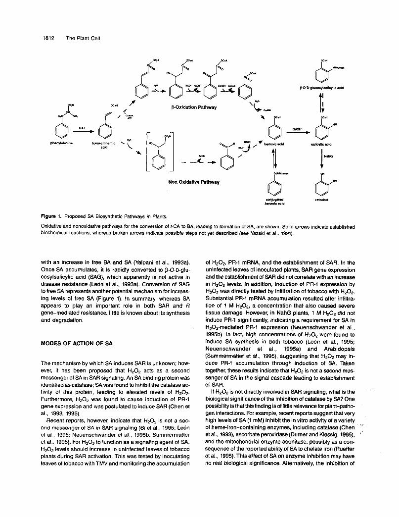
Given the importance of SA in disease resistance, the path- way of SA biosynthesis may represent a major control point in plant defense responses. The biosynthetic pathway of SA appears to begin with the conversion of phenylalanine to trans- cinnamic acid (t-CA) catalyzed by PAL, as shown in Figure 1. The conversion of t-CA into SA has been proposed to proceed via chain shortening to produce benzoic acid (BA), followed by hydroxylation at the C-2 position to derive SA (Yalpani et al., 1993a). The latter step is likely catalyzed by a cytochrome P450 monooxygenase, called benzoic acid 2-hydroxylase (BA2H), the activity of which is induced by either pathogen infection or exogenous BA application (León et al., 1993b). Be- cause exogenous BA causes SA accumulation but f-CA does not (Yalpani et al., 1993a), it seems plausible that the rate- limiting step in SA biosynthesis is the conversion of CCA to BA, although other possibilities exist.
The mechanism of BA production from t-CA is unknown, but it may occur in a manner similar to the Paxidation of fatty acids. Evidence for P-oxidation of t-CA to BA comes from studies on Quercuspedunculara, showing that acetyl-COA and ATPstimu- late the formation of SA from t-CA in cell-free extracts (Alibert and Ranjeva, 1971). However, other studies have suggested a nonoxidative mechanism; in suspension cultures of Lithosper- mum erythrorhizon and Daucus cama, the conversion of p-coumarate (differing from t-CA by an additional 4-hydroxyl group) to p-hydroxybenzoic acid appears to be independent of acetyl-COA as a cofactor (Yazaki et al., 1991; Schnitzler et al., 1992). In this case, p-hydroxybenzaldehyde is formed as an intermediate, which is also inconsistent with P-oxidation. Both possibilities are illustrated in Figure 1.
Interestingly, both BA and SA can be conjugated to glucose, and regulation of SA levels through SA or BA conjugation may be important. In healthy tobacco plants, there is a large pool of conjugated BA that decreases transiently in size after patho- gen infection. The decrease in conjugated BA levels correlates
Pathogen infection results in significant amounts of SA in the phloem sap of both cucumber and tobacco (Métraux et al., 1990; Yalpani et al., 1991). Additionally, in vivo SA-labeling studies provide evidence that SA produced in the leaves of TMV-infected tobacco or TNV-infected cucumber is transported throughout the plant and accumulates in uninfected tissues (Shulaev et al., 1995; Molders et al., 1996). In fact, as much as 70% (tobacco) and 50% (cucumber) of the increase in SA in uninfected tissue of pathogen-inoculated plants results from SA translocation from infected leaves to uninfected leaves.
These data support the contention that SA may be the sig- na1 that translocates from an infection site to activate SAR elsewhere in the plant. However, two lines of evidence sug- gest that SA is not the long distance signal. First, in cucumber, primary leaves infected with I? syringae can be removed 6 hr after inoculation, which is before SA accumulates in the phloem, without affecting the systemic increase of SA or SAR gene expression (Rasmussen et al., 1991). Second, in grafted tobacco plants, TMV inoculation of NahG rootstocks resulted in very little SA accumulation in infected tissue, compared with a 185-fold increase for wild-type (Xanthi) plants. However, trans- mission of the systemic signal out of the NahG rootstocks appeared to be unaffected because the grafted wild-typescions had elevated levels of both SAR gene expression and induced resistance equivalent to those seen in ungrafted wild-type plants (Vernooij et al., 1994).
These results suggest that either SA is not the long distance signal or very small amounts of SA in infected leaves are suf- ficient for full SAR induction. We have recently found that maximal induction of SAR occurs only at high concentrations
.
1812 The Plant Cell
Wdl I
W L I
mdi f
acid \ Y d:
\ " b- I: P-Oxidation Pathway
chn N y* I
/
Non Oxidative Pathway o Figure 1. Proposed SA Biosynthetic Pathways in Plants.
Oxidative and nonoxidative pathways for the conversion of CCA to BA, leading to formation of SA, are shown. Solid arrows indicate established biochemical reactions, whereas broken arrows indicate possible steps not yet described (see Yazaki et al., 1991).
with an increase in free BA and SA (Yalpani et al., 1993a). Once SA accumulates, it is rapidly converted to B-O-D-glu- cosylsalicylic acid (SAG), which apparently is not active in disease resistance (Lebn et al., 1993a). Conversion of SAG to free SA represents another potential mechanism for increas- ing levels of free SA (Figure 1). In summary, whereas SA appears to play an important role in both SAR and R gene-mediated resistance, little is known about its synthesis and degradation.
MODES OF ACTION OF SA
The mechanism by which SA induces SAR is unknown; how- ever, it has been proposed that H202 acts as a second messenger of SA in SAR signaling. An SA binding protein was identified as catalase; SA was found to inhibit the catalase ac- tivity of this protein, leading to elevated levels of H202. Furthermore, H202 was found to cause induction of PR-1 gene expression and was postulated to induce SAR (Chen et al., 1993, 1995).
Recent reports, however, indicate that H 2 0 2 is not a sec- ond messenger of SA in SAR signaling (Bi et al., 1995; LeÓn et al., 1995; Neuenschwander et al., 1995b; Summermatter et al., 1995). For H202 to function as a signaling agent of SA, H 2 0 2 levels should increase in uninfected leaves of tobacco plants during SAR activation. This was tested by inoculating leaves of tobacco with TMV and monitoring the accumulation
of H202, PR-1 mRNA, and the establishment of SAR. In the uninfected leaves of inoculated plants, SAR gene expression and the establishment of SAR did not correlate with an increase in H202 levels. In addition, induction of PR-1 expression by H202 was directly tested by infiltration of tobacco with H202. Substantial PR-1 mRNA accumulation resulted after infiltra- tion of 1 M H202, a concentration that also caused severe tissue damage. However, in NahG plants, 1 M H202 did not induce PR-1 significantly, indicating a requirement for SA in H202-mediated PR-1 expression (Neuenschwander et al., 1995b). In fact, high concentrations of H202 were found to induce SA synthesis in both tobacco (Lebn et al., 1995; Neuenschwander et al., 1995a) and Arabidopsis (Summermatter et al., 1995), suggesting that H202 may in- duce PR-1 accumulation through induction of SA. Taken together, these results indicate that H202 is not a second mes- senger of SA in the signal cascade leading to establishment of SAR.
If H202 is not directly involved in SAR signaling, what is the biological significance of the inhibition of catalase by SA? One possibility is that this finding is of little relevance for plant-patho- gen interactions. For example, recent reports suggest that very high levels of SA (1 mM) inhibit the in vitro activity of a variety of heme-iron-containing enzymes, including catalase (Chen et al., 1993), ascorbate peroxidase (Durner and Klessig, 1995), and the mitochondrial enzyme aconitase, possibly as a con- sequence of the reported ability of SA to chelate iron (Rueffer et al., 1995). This effect of SA on enzyme inhibition may have no real biological significance. Alternatively, the inhibition of
Systemic Acquired Resistance 1813
catalase and peroxidase could be very important for lesion formation. The Kd for SA binding to catalase and ascorbate peroxidase was reported as 14 pM (Chen and Klessig, 1991) and 78 pM (Durner and Klessig, 1995), respectively. This concentration of SA occurs immediately adjacent to a pathogen- induced lesion but not in uninfected leaves in which SA con- centrations are 10- to 100-fold lower (Enyedi et al., 1992; Neuenschwander et al., 1995b). Therefore, the biological sig- nificance of SA-mediated inhibition of oxidoreductases may be restricted to local responses in infected tissue.
Interestingly, in parsley cell cultures and cucumber cot- yledons, SA pretreatment was recently found to increase dramatically the competence of the tissue to trigger a burst of H202 in response to subsequent elicitor treatment. This conditioning of cells by SA was dependent on protein synthe- sis and correlated with enhanced resistance of cucumber cotyledons to the funga1 pathogen Colletotrichum lagenarium. Moreover, the increase in H202 levels was not due to a de- crease in the rate of degradation but to an increase in H202 synthesis (Kauss and Jeblick, 1995; Fauth et al., 1996).
Thus, the data now available suggest the existence of more than one mode of action of SA in resistance responses. In unin- fected leaves, a high-affinity receptor for SA could mediate the induction of SAR gene expression. After establishment of SAR, the tissue becomes competent for rapid elicitation of an ox- idative burst at the site of pathogen attack, as opposed to a slower response in tissues in which SAR has not been estab- lished. In infected leaves, high concentrations of SA around the site of infection may inhibit catalase and other oxidoreduc- tases. lnhibition of catalase activity could prolong the half-life of H202 and lead to an amplification of the oxidative burst. The oxidative burst may trigger a variety of local defense re- sponses (Mehdy, 1994; Levine et al., 1996; see also Hammond- Kosack and Jones, 1996, in this issue), including programmed cell death during the HR as well as defense gene expression and synthesis of SA in adjacent cells. This would create a runa- way cycle leading to high levels of both SA and H202 at the site of pathogen attack. In this model, inhibition of oxidoreduc- tases represents a low-affinity perception mechanism that transduces high local SA levels into local defense responses.
CHEMICAL ACTIVATORS OF SAR
SAR was first described as a response to pathogen infection. Subsequently, it has been found that treatment of plants with low molecular weight molecules can also induce SAR. The use of chemicals to activate SAR provides nove1 alternatives for disease control in agronomic systems as well as tools for the elucidation of the SAR signal transduction cascade (Neuenschwander et al., 1995a). To be considered an activa- tor of SAR, a chemical should exhibit three characteristics (Kessmann et al., 1994): first, the compound or its significant metabolites should not exhibit direct antimicrobial activity; sec- ond, it should induce resistance against the same spectrum of pathogens as in biologically activated SAR; and third, it
should induce the expression of the same marker genes as evident in pathogen-activated SAR.
Severa1 chemicals or extracts, including silicon, phosphate, 2-thiouracil, polyacrylic acid, nucleic acids, and fosethylAl, have been reported as potential activators of resistance but have failed to fulfill the above criteria (Kessmann et al., 1994). Other compounds, such as DL-8aminobutanoic acid or probenazole, have been shown to slightly induce either PR-1 gene expression or resistance against one or two pathogens, but activation of bona fide SAR has not been demonstrated (Iwata et al., 1980; Asselin et al., 1985; Cohen et al., 1993).
To date, SA is the only plant-derived substance that has been demonstrated to be an inducer of SAR (White, 1979; Antoniw and White, 1980; Ward et al., 1991). The chemicals 2,6- dichloroisonicotinic acid and its methyl ester (both referred to as INA) were the first synthetic compounds shown to activate SAR, thus ,providing broad-spectrum disease resistance (Metraux et al., 1991; Vernooij et al., 1995). However, both SA and INA were insufficiently tolerated by some crop plants to warrant practical use as plant protection compounds. Recently, the synthetic chemical benzo(l,2,3)thiadiazole-7-carbothioic acid S-methyl ester (BTH) was demonstrated to be a potent SAR activator (Friedrich et al., 1996; Gorlach et al., 1996; Lawton et al., 1996) that supplies protection in the field against a broad spectrum of diseases in a variety of crops. Thus, BTH is an attractive compound for practical agronomic use. The resistance observed in the plants after treatment with INA or BTH is not due to a direct action of the compounds on the patho- gen, because neither the compounds nor their significant metabolites exhibit in vitro antibiotic activity (Métraux et al., 1991; Friedrich et al., 1996). Moreover, in tobacco, Arabidop- sis, and wheat, INA and BTH induce the same set of SAR genes that is induced by SA (Ward et al., 1991; Uknes et al., 1992; Friedrich et al., 1996; Gorlach et al., 1996; Lawton et al., 1996).
Neither INA nor BTH treatment causes elevated levels of SA in the plant, and both compounds activate SAR when ap- plied to NahG plants, suggesting that both INA and BTH act independently or downstream of SA in SAR signaling (Vernooij et al., 1995; Friedrich et al., 1996). INA, BTH, and SA are un- able to activate SAR gene expression in the niml mutant (noninducible hmunity) of Arabidopsis (see below), suggesting that all three compounds activate the SAR signal transduc- tion pathway through the same signaling cascade (Delaney et al., 1995; Lawton et al., 1996). Furthermore, the structural similarities of the three compounds (Gorlach et al., 1996) sug- gest that they may all bind to the same receptor, although direct evidence for this is lacking.
CONSTITUTIVE SAR MÚTANTS
In an effort to identify steps in the SAR signal transduction pathway, severa1 groups have taken a genetic approach. Arabidopsis was chosen as the model plant because it is an established plant system for mutant analysis (Redei and Koncz, 1992) and gene isolation (Meyerowitz, 1992) as well as a facile
1814 The Plant Cell
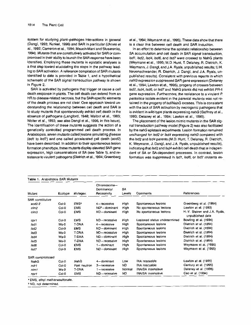
system for studying plant-pathogen interactions in general (Dangl, 1993; Kunkel, 1996) and SAR in particular (Uknes et al., 1992; Cameron et al., 1994; Mauch-Mani and Slusarenko, 1994). Mutants that are constitutively activated for SAR or com- promised in their ability to launch the SAR response have been identified. Employing these mutants in epistatic analyses is a first step toward elucidating the steps in the pathway lead- ing to SAR activation. A reference compilation of SAR mutants identified to date is provided in Table 1, and a hypothetical schematic of the SAR signal transduction pathway is shown in Figure 2.
SAR is activated by pathogens that trigger or cause a cell death response in plants. The cell death can extend from an HR to disease-related necrosis, but the SAR-specific elements of the death process are not clear. One approach toward un- derstanding the relationship between cell death and SAR is to study mutants that spontaneously exhibit cell. death in the absence of pathogens (Langford, 1948; Walbot et al., 1983; Wolter et al., 1993; see also Dangl et al., 1996, in this issue). The identification of these mutants suggests the action of a genetically controlled programmed cell death process. In Arabidopsis, seven mutants called jesions simulating disease (lsd7 to lsd7) and one called Sccelerated pell death (acd2) have been described. In addition to their spontaneous lesion formation phenotype, these mutants display elevated SAR gene expression, high concentrations of SA (see Table l), and re- sistance to virulent pathogens (Dietrich et al., 1994; Greenberg
et al., 1994; Weymann et al., 1995). These data show that there is a clear link between cell death and SAR induction.
In an effort to determine the epistatic relationship between SA accumulation and cell death in SAR signal transduction, lsdl, lsd2, lsd4, lsd6, and lsd7 were crossed to NahG plants (Weymann et al., 1995; M.D. Hunt, T. Delaney, R. Dietrich, K. Weymann, J. Dangl, and J.A. Ryals, unpublished results; U.H. Neuenschwander, R. Dietrich, J. Dangl, and J.A. Ryals, un- published results). Consistent with previous reports in which nahG expression suppressed SAR gene expression (Delaney et al., 1994; Lawton et al., 1995), progeny of crosses between lsd2,lsd4,lsd6, or lsd7 and NahG plants did not exhibit PR-1 gene expression. Furthermore, the resistance to a virulent P parasitica isolate evident in the parenta1 mutants was not re- tained in the progeny of IsdlNahG crosses. This is consistent with the lack of SAR activation by necrogenic pathogens that is evident in wild-type plants expressing nahG (Gaffney et al., 1993; Delaney et al., 1994; Lawton et al., 1995).
The placement of the lesion mimic mutants in the SAR sig- na1 transduction pathway model (Figure 2) was also facilitated by the nahG epistasis experiments. Lesion formation remained unchanged for lsd2 or lsd4 expressing nahG compared with the lsd2 and lsd4 parents (M.D. Hunt, T. Delaney, R. Dietrich, K. Weymann, J. Dangl, and J.A. Ryals, unpublished results), indicating that lsd2 and lsd4 exhibit cell death that is indepen- dent of SA or SA-dependent processes. In contrast, lesion formation was suppressed in Isdl, Isd6, or lsd7 mutants ex-
Table 1. Arabidopsis SAR Mutants
Chromosome- Dominancyf SA
Mutant Ecotype Mutagen Recessivity Levels Commants References
SAR constitutive acd2-2 cim2 cima
cpr 1 lsdl lsd2 lsd3 lsd4 lsd5 lsd6 lsd7
SAR compromised NahG ndrl nim 1
COl-o COl-o COLO
COLO ws-o COLO ws-o ws-o ws-o COLO COLO
COLO C O L O ws-o
EMSa 4-recessive EMS NDb-dominant EMS ND-dominant
EMS
EMS T-DNA
T-DNA T-DNA T-DNA EMS EMS
ND- recessive 4-recessive ND-dominant ND-recessive ND-dominant ND- recessive 1 -dominant ND-dominant
NahG 4-dominant Fast neutron 3-recessive T-DNA 1 - recessive
High High High
High High High High High High High High
LOW ND Normal
Spontaneous lesions No spontaneous lesions No spontaneous lesions
Lesioned status undetermined Spontaneous lesions Spontaneous lesions Spontaneous lesions Spontaneous lesions Spontaneous lesions Spontaneous lesions Spontaneous lesions
INA rescuable INA rescuable INNSA insensitive
Greenberg et al. (1994) Lawton et al. (1993) H.-Y. Steiner and J.A. Ryals,
unpublished data Bowling et al. (1994) Dietrich et al. (1994) Dietrich et al. (1994) Dietrich et al. (1994) Dietrich et al. (1994) Dietrich et al. (1994) Weymann et al. (1995) Weymann et al. (1995)
Lawton et al. (1995) Century et al. (1995) Delaney et al. (1995) Cao et al. (1994) nprl Col-O EMS ND-recessive ND INNSA insensitive . .
a EMS, ethyl methanesulfonate. b ND, not determined.
Systemic Acquired Resistance 1815
ndrl 1
cim3 SAR Gene Local Acquired Expression --b - Resistame
cprl ? lsd2 lsdl lsd4
Pathogen + + Plant -
Interaction
I \ niml
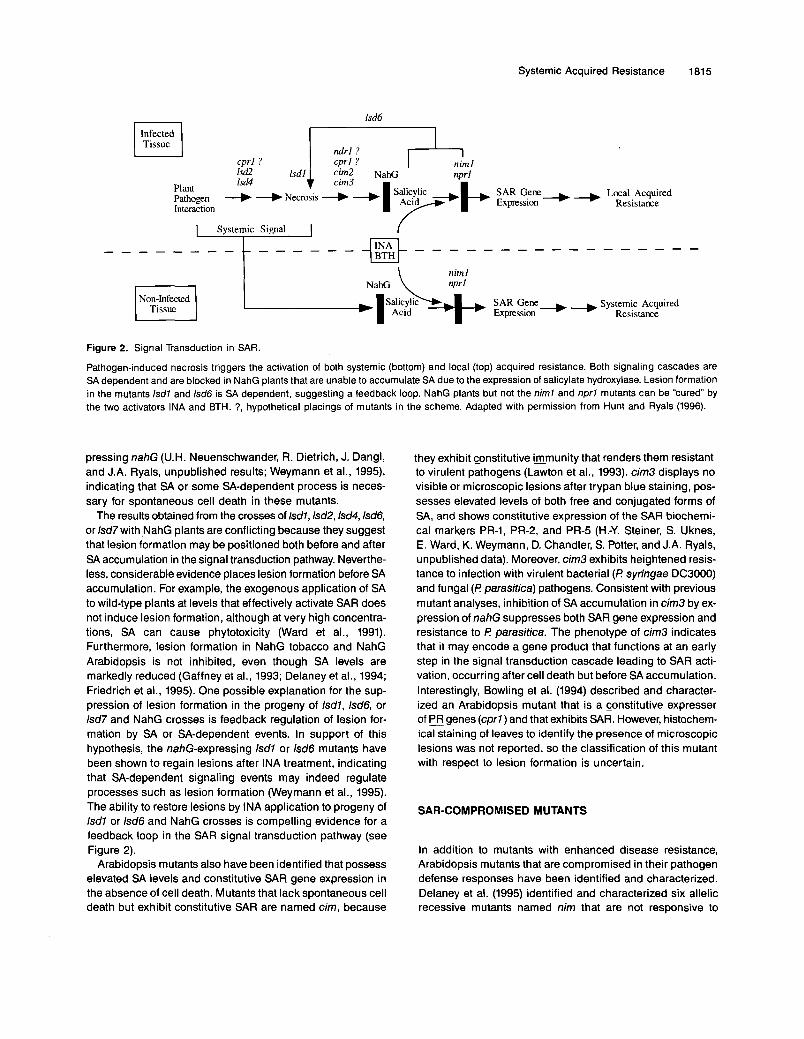
Figure 2. Signal Transduction in SAR.
Pathogen-induced necrosis triggers the activation of both systemic (bottom) and local (top) acquired resistance. 60th signaling cascades are SA dependent and are blocked in NahG plants that are unable to accumulate SA dueto the expression of salicylate hydroxylase. Lesion formation in the mutants Isdl and lsd6 is SA dependent, suggesting a feedback loop. NahG plants but not the niml and nprl mutants can be "cured" by the two activators INA and BTH. ?, hypothetical placings of mutants in the scheme. Adapted with permission from Hunt and Ryals (1996).
pressing nahG (U.H. Neuenschwander, R. Dietrich, J. Dangl, and J.A. Ryals, unpublished results; Weymann et al., 1995), indicating that SA or some SA-dependent process is neces- sary for spontaneous cell death in these mutants.
The results obtained from the crosses of lsdl, lsd2,lsd4,lsd6, or lsd7 with NahG plants are conflicting because they suggest that lesion formation may be positioned both before and after SA accumulation in the signal transduction pathway. Neverthe- less, considerable evidence places lesion formation before SA accumulation. For example, the exogenous application of SA to wild-type plants at levels that effectively activate SAR does not induce lesion formation, although at very high concentra- tions, SA can cause phytotoxicity (Ward et al., 1991). Furthermore, lesion formation in NahG tobacco and NahG Arabidopsis is not inhibited, even though SA levels are markedly reduced (Gaffney et al., 1993; Delaney et al., 1994; Friedrich et al., 1995). One possible explanation for the sup- pression of lesion formation in the progeny of Isdl, lsd6, or lsd7 and NahG crosses is feedback regulation of lesion for- mation by SA or SA-dependent events. In support of this hypothesis, the nahG-expressing lsdl or lsd6 mutants have been shown to regain lesions after INA treatment, indicating that SA-dependent signaling events may indeed regulate processes such as lesion formation (Weymann et al., 1995). The ability to restore lesions by INA application to progeny of lsdl or lsd6 and NahG crosses is compelling evidence for a feedback loop in the SAR signal transduction pathway (see Figure 2).
Arabidopsis mutants also have been identified that possess elevated SA levels and constitutive SAR gene expression in the absence of cell death. Mutants that lack spontaneous cell death but exhibit constitutive SAR are named cim, because
they exhibit Gonstitutive immunity that renders them resistant to virulent pathogens (Lawton et al., 1993). cim3 displays no visible or microscopic lesions after trypan blue staining, pos- sesses elevated levels of both free and conjugated forms of SA, and shows constitutive expression of the SAR biochemi- cal markers PR-1, PR-2, and PR-5 (H.-Y. Steiner, S. Uknes, E. Ward, K. Weymann, D. Chandler, S. Potter, and J.A. Ryals, unpublished data). Moreover, cim3 exhibits heightened resis- tance to infection with virulent bacterial (i? syringae DC3000) and funga1 (i? parasitica) pathogens. Consistent with previous mutant analyses, inhibition of SA accumulation in cim3 by ex- pression of nahG suppresses both SAR gene expression and resistance to i? pafasitica. The phenotype of cim3 indicates that it may encode a gene product that functions at an early step in the signal transduction cascade leading to SAR acti- vation, occurring after cell death but before SA accumulation. Interestingly, Bowling et al. (1994) described and character- ized an Arabidopsis mutant that is a constitutive expresser of fWgenes (cprl) and that exhibits SAR. However, histochem- ical staining of leaves to identify the presence of microscopic lesions was not reported, so the classification of this mutant with respect to lesion formation is uncertain.
SAR-COMPROMISED MUTANTS
In addition to mutants with enhanced disease resistance, Arabidopsis mutants that are compromised in their pathogen defense responses have been identified and characterized. Delaney et al. (1995) identified and characterized six allelic recessive mutants named nim that are not responsive to
1816 The Plant Cell
exogenous application of SA or synthetic SAR activators such as INA. Similarly, Cao et al. (1994) have also isolated and described a recessive Arabidopsis mutant called nprl (Eonex- - presser of PR genes), which exhibits compromised activation of SAR. nprl may be allelic to niml.
The phenotypes of niml and nprl indicate that their block in SAR signaling occurs before SAR gene expression but sub- sequent to SA accumulation. Evidence for this placement was obtained by analysis of niml plants infected with the avirulent pathogen F! syringae DC3000 harboring the cloned avrRpt2 gene (Whalen et al., 1991). niml plants were shown to accumu- late both free and glucose-conjugated SA levels in excess of those in wild-type plants. Therefore, niml plants are able to accumulate SA in response to pathogen infection but appear to be defective in SA perception or in subsequent SA-sensing events. That the niml and nprl mutations define genes that act before SAR gene expression was substantiated by the lack of PR-1 induction evident in these plants after INA or SA treat- ment. Furthermore, niml plants showed reduced and delayed levels of PR-1 mRNA accumulation after infection with the viru- lent pathogen F! parasitica isolate EMWA. Taken together, these results indicate that the niml and nprl mutants are com- promised in both pathogen-associated and chemical activation of SAR.
In contrast to niml and nprl, Century et al. (1995) identified a partia1 resistance-compromised mutant ndrl-7 (non-race- specific disease iesistance) that is susceptible to severa1 F! parasitica isolates (virulent and avirulent) as well as to F! syrin- gae DC3000 carrying any one of the cloned bacterial avirulence genes avrB, avrRpml, avrRpt2, and avrPph3. Interestingly, de- spite its susceptibility to normally avirulent pathovars, the HR of the ndrl-7 mutant was not substantially different from that in the wild type in response to infection with F! syringae har- boring avrB, avrRpml, or avrPph3. However, an HR was not evident in ndrl-7 after infection with F! syringae harboring avrRpf2. These results are particularly interesting because of the apparent uncoupling of the HR and disease resistance. ndrl-7 differs from niml and nprl in that resistance is restored upon INA treatment (K. Century and 6. Staskawicz, personal communication). Therefore, the ndrl-7 mutation probably pre- cedes SA accumulation in the SAR signal transduction pathway, whereas niml and nprl presumably act subsequent to SA accumulation.
CONCLUSION
When a pathogen is perceived by a host plant, a series of re- sponses can be activated. Some of these responses, such as the synthesis and release of ethylene, may predispose the plant to further infection and thus contribute to disease susceptibil- ity (Ecker, 1995). Other responses may contribute to the active defense of the host against the pathogen. One of these resis- tance responses is the SAR signal transduction pathway. Evidence that this response is important for resistance is that
when the pathway is blocked through design (e.g., in NahG transgenic plants) or mutation (e.g., niml, nprl, ndrl), the plant’s defense is compromised. Furthermore, when the pathway is stimulated by exogenous compounds such as BTH or INA, or by mutation (e.g., Isd, acd2, cim3, cprl), the host’s defense is strengthened.
Although it is clear that the SAR signal transduction path- way is central to disease resistance, there are still many unanswered questions. What is the identity of the translocated signal? How is SA synthesized after pathogen infection? What is the receptor for SA, INA, and BTH? A detailed understand- ing of this pathway is important for both practical and theoretical reasons.
ACKNOWLEDGMENTS
We greatly appreciate that Dr. Jeff Dangl, Dr. Diane Horvath, and Dr. Jean-Pierre Métraux shared their research results before publication. U.H.N. was supported by a postdoctoral fellowship (No. 5002-38012) from the Swiss National Science Foundation, and A.M. was supported by a postdoctoral fellowship from the Spanish Ministry of Education.
REFERENCES
Alexander, D., Stinson, J., Pear, J., Glascock, C., Ward, E., Goodman, R.M., and Ryals, J. (1992). A new multigene family in- ducible by tobacco mosaic virus or salicylic acid in tobacco. MOI. Plant-Microbe Interact. 5, 513-515.
Alexander, D., Glascock, C., Pear, J., Stinson, J., Ahl-Goy, P., Gut- Rella, M., Goodman, R.M., and Ryals, J. (1993a). Systemic ac- quired resistance in tobacco: Use of transgenic expression to study the functions of pathogenesis-related proteins. In Advances in Mo- lecular Genetics of Plant-Microbe Interactions, Vol. 3, E.W. Nester and D.P.S. Verma, eds (Dordrecht, The Netherlands: Kluwer Aca- demic Publishers), pp. 527-533.
Alexander, D., Goodman, R.M., Gut-Rella, M., Glascock, C., Weymann, K., Friedrich, L., Maddox, D., Ahl-Goy, P., Luntz, T., Wad, E., and Ryals, J. (1993b). lncreased tolerance to hvo Oomycete pathogens in transgenic tobacco expressing pathogenesis-related protein Ia. Proc. Natl. Acad. Sci. USA 90, 7327-7331.
Alibert, G., and Ranjeva, R. (1971). Research on the enzymes catalyz- ing the biosynthesis of phenolic acids in Quercus pedunculata. I. Formation of the first members of the cinnamic series and benzoic series. FEBS Lett. 19, 11-14.
Antoniw, J., and White, R. (1980). The effects of aspirin and poly- acrylic acid on soluble leaf proteins and resistance to virus infection in five cultivars of tobacco. Phytopathol. 2. 98, 331-341.
Asselin, A., Grenier, J., and Cote, F. (1985). Light-influenced extracel- lular accumulation of b (pathogenesis-related) proteins in Nicotiana green tissue induced by various chemicals or prolonged floating on water. Can. J. Bot. 63, 1276-1283.
Beffa, R., Szell, M., Meuwly, P., Pay, A., Vogeli-Lange, R., MBtraux, J.-P., Meins, F., and Nagy, F. (1995). Cholera toxin elevates patho- gen resistance and induces defense reactions in transgenic tobacco plants. EMBO J. 14, 5753-5761.
Systemic Acquired Resistance 1817
Bi, Y.-M., Kenton, P., Mur, L., Darby, R., and Draper, J. (1995). Hydro- gen peroxide does not function downstream of salicylic acid in the induction of PR protein expression. Plant J. 8, 235-245.
BOI, J.F., Linthorst, H.J.M., and Cornelissen, B.J.C. (1990). Plant pathogenesis-related proteins induced by virus infection. Annu. Rev. Phytopathol. 28, 113-138.
Bowles, D.J. (1990). Defense-related proteins in higher plants. Annu. Rev. Biochem. 59, 873-907.
Bowling, S.A., Guo, A., Cao, H., Gordon, A.S., Klessig, D.F., and Dong, X. (1994). A mutation in Arabidopsis that leads to constitu- tive expression of systemic acquired resistance. Plant Cell 6,
Camemn, R.K., Dixon, R., and Lamb, C. (1994). Biologically induced systemic acquired resistance in Arabidopsis thaliana. Plant J. 5,
Cao, H., Bowling, S.A., Gordon, A.S., and Dong, X. (1994). Char- acterization of an Arabidopsis mutant that is nonresponsive to inducers of systemic acquired resistance. Plant Cell6, 1583-1592.
Century, K.S., Holub, E.B., and Staskawicz, B.J. (1995). NDRI, a locus of Arabidopsis thaliana that is required for disease resistance to both a bacterial and a fungal pathogen. Proc. Natl. Acad. Sci.
Chen, Z., and Klessig, D.F. (1991). ldentification of a soluble salicylic acid-binding protein that may function in signal transduction in the plant disease-resistance response. Proc. Natl. Acad. Sci. USA 88,
Chen, Z., Silva, H., and Klessig, D.F. (1993). lnvolvement of reactive oxygen species in the induction of systemic acquired resistance by salicylic acid in plants. Science 242, 883-886.
Chen, Z.X., Malamy, J., Henning, J., Conrath, U., Sanchezcasas, P., Silva, H., Ricigllano, J., and Klessig, D.F. (1995). Induction, modification, and transduction of the salicylic acid signal in plant defense responses. Proc. Natl. Acad. Sci. USA 92, 4134-4137.
Chester, K.S. (1933). The problem of acquired physiological immu- nity in plants. Q. Rev. Biol. 8, 275-324.
Cohen, Y., Gisi, U., and Niderman, T. (1993). Local and systemic pro- tection against Phytopbthora infestam induced in potato and tomato plants by jasmonic acid and jasmonic acid methyl ester. Phytopathol-
Dangl, J.L. (1993). Applications of Arabidopsis thaliana to outstand- ing issues in plant-pathogen interactions. Int. Rev. Cytol. 144,53-83.
Dangl, J.L., Dietrich, R.A., and Richberg, M.H. (1996). Death don’t have no mercy: Cell death programs in plant-microbe interactions. Plant Cell 8, 1793-1807.
Delaney, T., Uknes, S., Vernooij, B., Friedrich, L., Weymann, K., Negmtto, D., Gaftney, T., Gut-Rella, M., Kessmann, H., Ward, E., and Ryals, J. (1994). A central role of salicylic acid in plant dis- ease resistance. Science 266, 1247-1250.
Delaney, T., Friedrich, L., and Ryals, J. (1995). Arabidopsis signal transduction mutant defective in chemically and biologically induced disease resistance. Proc. Natl. Acad. Sci. USA 92, 6602-6606.
Dempsey, D.M., Wobbe, K., and Klessig, D.F. (1993). Resistance and susceptible responses of Arabidopsis thaliana to turnip crinkle vi- rus. Phytopathology 83, 1021-1029.
Dietrich, R.A., Delany, T.P., Uknes, S.J., Ward, E.R., Ryals, J.A., and Dangl, J.L. (1994). Arabidopsis mutants simulating disease re- sistance response. Cell 77, 565-577.
1845-1857.
715-725.
USA 92, 6597-6601.
8179-8183.
Ogy 83, 1054-1062.
Dixon, R.A. (1986). The phytoalexin response: Elicitation, signaling, and control of host gene expression. Biol. Rev. Camb. Philos. SOC.
Durner, J., and Klessig, D.F. (1995). lnhibition of ascorbate peroxi- dase by salicylic acid and 2.6-dichloroisonicdinic acid, two inducers of plant defense responses. Proc. Natl. Acad. Sci. USA 92, 1 131 2-1 1316.
Ecker, J.R. (1995). The ethylene signal transduction pathway in plants. Science 268, 667-675.
Enyedi, A.J., Yalpani, N., Silverman, P., and Raskin, 1. (1992). Lo- calization, conjugation and function of salicylic acid in tobacco during the hypersensitive reaction to tobacco mosaic virus. Proc. Natl. Acad. Sci. USA 89, 2480-2484.
Fauth, M., Merten, A., Hahn, M.G., Jeblick, W., and Kauss, H. (1996). Competence for elicitation of H202 in hypocotyls of cucumber is in- duced by breaching the cuticle and is enhanced by salicylic acid. Plant Physiol. 110, 347-354.
Friedrich, L., Vernooij, E., Gaffney, T., Mo=, A., and Ryals, J. (1995). Characterization of tobacco plants expressing a bacterial salicylate hydroxylase gene. Plant MOI. Biol. 29, 959-968.
Friedrich, L., Lawton, K., Dincher, S., Winter, A., Staub, T., Uknes, S., Kessmann, H., and Ryals, J. (1996). Benzothiadiazole induces systemic acquired resistance in tobacco. Plant J. 10, 61-70.
Gaffney, T., Friedrich, L., Vernooij, B., Negmtto, D., Nye, G., Uknes, S., Ward, E., Kessmann, H., and Ryals, J. (1993). Requirement of salicylic acid for the induction of systemic acquired resistance. Science 261, 754-756.
Gianinaui, S., Martin, C., and Vallbe, J.C. (1970). Hypersensibilite aux virus, tempbrature et protéines solubles chez le Nicotiana Xan- thi n.c. Apparition de nouvelles macromolécules lors de Ia rbpression de Ia synthbse virale. C. R. Acad. Sci. Ser. 111 Sci. Vie 270,2382-2386.
Garlach, J., Volrath, S., Knauf-Beiter, G., Hengy, G., Beckhove, U., Kogel, K.-H., Oostendorp, M., Staub, T., Ward, E., Kessmann, H., and Ryals, J. (1996). Benzothiadiazole, a nove1 class of inducers of systemic acquired resistance, activates gene expression and dis- ease resistance in wheat. Plant C411 8, 629-643.
Greenberg, J.T., Guo, A., Klessig, D.F., and Ausubel, F.M. (1994). Programmed cell death in plants-A pathogen-triggered response activated coordinately with multiple defense functions. Cell 77,
Hammond-Kosack, K.E., and Jones, J.D.G. (1996). Resistance gene-dependent plant defense responses. Plant Cell 8, 1773-1791.
Hunt, M., and Ryals, J. (1996). Systemic acquired resistance signal transduction. Crit. Rev. Plant Sci. 15, 583-606.
Iwata, M., Suzuki, Y., Watanabe, T., Mase, S., and Sekizawa, Y. (1980). Effect of probenzole on the activities related to the resistant reac- tion in rice plant. Ann. Phytopathol. SOC. Jpn. 46, 297-306.
Jach, G., Gornhardt, B., Mundy, J., Logemann, J., Pinsdorf, P., Leah, R., Schell, J., and Maas, C. (1995). Enhanced quantitative resis- tance against fungal disease by combinatorial expression of different barley antifungal proteins in transgenic tobacco. Plant J. 8,97-109.
Kauss, H. (1987). Some aspects of calcium-dependent regulation in plant metabolism. Annu. Rev. Plant Physiol. 38, 47-72.
Kauss, H., and Jeblick, W. (1995). Pretreatment of parsley suspen- sion cultures with salicylic acid enhances spontaneous and elicited production of HZOZ. Plant Physiol. 108, 1171-1178.
61, 239-292.
551-563.
1818 The Plant Cell
Kessmann, H., Staub, T., Hofmann, C., Maetzke, T., Herzog, J., Ward, E., Uknes, S., and Ryals, J. (1994). lnduction of systemic acquired resistance in plants by chemicals. Annu. Rev. Phytopathol.
Kunkel, B.N. (1996). A useful weed put to work: Genetic analysis of disease resistance in Arabidopsis thaliana. Trends Genet. 12, 63-69.
Langford, A.N. (1948). Autogenous necrosis in tomatoes immune from Cladosporíum fulvum Cooke. Can. J. Res. 26, 35-64.
Lawton, K., Uknes, S., Friedrich, L., Gaftney, T., Alexander, D., Goodman, R., Métraux, J.-P., Kessmann, H., Ahl-Goy, P., Gut- Rella, M., Ward, E., and Ryals, J. (1993). The molecular biology of systemic aquired resistance. In Mechanisms of Defence Re- sponses in Plants, B. Fritig and M. Legrand, eds (Dordrecht, The Netherlands: Kluwer Academic Publishers), pp. 422-432.
Lawton, K., Weymann, K., Friedrich, L., Vernooij, B., Uknes, S., and Ryals, J. (1995). Systemic acquired resistance in Arabidopsis requires salicylic acid but not ethylene. MOI. Plant-Microbe Inter- act. 8, 863-870.
Lawton, K., Friedrich, L., Hunt, M., Weymann, K., Staub, T., Kessmann, H., and Ryals, J. (1996). Benzothiadiazole induces dis- ease resistance in Arabidopsis by activation of the systemic acquired resistance signal transduction pathway. Plant J. 10, 71-82.
LeÓn, J., Yalpani, N., Lawton, M.A., and Raskin, I. (1993a). Sali- cylic acid biosynthesis in healthy and virus-inoculated tobacco. In Plant Signals in lnteractions with Other Organisms, J. Schultz and I . Raskin, eds (Rockville, MD: American Society of Plant Physiolo- gists), pp. 262-265.
LeÓn, J., Yalpani, N., Raskin, I., and Lawton, M.A. (1993b). Induc- tion of benzoic acid 2-hydroxylase in virus-inoculated tobacco. Plant Physiol. 103, 323-328.
LeÓn, J., Lawton, M.A., and Raskin, 1. (1995). Hydrogen peroxide stimulates salicylic acid biosynthesis in tobacco. Plant Physiol. 108,
Levine, A., Pennell, R.I., Alvarez, M.A., Palmer, R., and Lamb, C. (1996). Calcium-mediated apoptosis in a plant hypersensitive resis- tance response. Curr. Biol. 6, 427-437.
Linthorst, H.J.M. (1991). Pathogenesis-related proteins of plants. Crit. Rev. Plant Sci. 10, 123-150.
Liu, D., Kashchandra, G., Raghothama, P., Hasegawa, P.M., and Bressan, R.A. (1994). Osmotin overexpression in potato delays de- velopment of disease symptoms. Proc. Natl. Acad. sci. USA 91,
Low, P.S., and Merida, J.R. (1996). The oxidative burst in plant de- fense: Function and signal transduction. Physiol. Plant. 96,533-542.
Malamy, J., Carr, J.P., Klessig, D.F., and Raskin, 1. (1990). Salicylic acid: A likely endogenous signal in the resistance response of tobacco to vira1 infection. Science 250, 1002-1004.
Mauch, F., Mauch-Mani, B., and Boller, T. (1988). Antifungal hydro- {ases in pea tissue. II. lnhibition of funga1 growth by combinations of chitinase and fi-1,3-glucanase. Plant Physiol. 88, 936-942.
Mauch-Mani, B., and Slusarenko, A.J. (1994). Systemic acquired re- sistance in Arabidopsis thaliana induced by prediposing infection with a pathogenic isolate of Fusarium oxysporum. MOI. Plant-Microbe Interact. 7, 378-383.
Mauch-Mani, B., and Slusarenko, A.J. (1996). Production of salicylic acid precursors is a major function of phenylalanine ammonia-lyase in the resistance of Arabidopsis to Peronospofa parasitica. Plant Cell
32, 439-459.
1673-1678.
1888-1892.
8, 203-212.
Mehdy, M.C. (1994). Active oxygen species in plant defense against pathogens. Plant Physiol. 105, 467-472.
Métraux, J.-P., Burkhart, W., Moyer, M., Dincher, S., Middlesteadt, W., Williams, S., Payne, G., Carnes, M., and Ryals, J. (1989). Iso- lation of a complementary DNA encoding a chitinase with structural homology to a bifunctional lysozymelchitinase. Proc. Natl. Acad. Sci. USA 86, 896-900.
Métraux, J.-P., Signer, H., Ryals, J., Ward, E., Wyss-Bem, M., Gaudin, J., Raschdorf, K., Schmid, E., Blum, W., and Inverardi, B. (1990). lncrease in salicylic acid at the onset of systemic acquired resistance in cucumber. Science 250, 1004-1006.
Métraux, J.-P., Ahl-Goy, P., Staub, T., Speich, J., Steinemann, A., Ryals, J., and Ward, E. (1991). lnduced resistance in cucumber in response to 2,8dichloroisonicotinic acid and pathogens. In Advances in Molecular Genetics of Plant-Microbe Interactions, Vol. 1, H. Hennecke and D.P.S. Verma, eds (Dordrecht, The Netherlands: Kluwer Academic Publishers), pp. 432-439.
Meyerowitz, E.M. (1992). lntroduction to the Arabidopsis genome. In Methods in Arabidopsis Research, C. Koncz, N.-H. Chua, and J. Schell, eds (Riveredge, NJ: World Scientific Publishing), pp. 100-118.
Molders, W., Buchala, A., and Métraux, J.-P. (1996). Transport of sali- cylic acid in tobacco necrosis virus-infected cucumber plants. Plant Physiol. 112, 787-792.
Nasser, W., De Tapia, M., Kauffmann, S., Montasser-Kouhsari, S., and Burkard, G. (1988). ldentification and characterization of maize pathogenesis-related proteins. Four maize PR proteins are chitinases. Plant MOI. Biol. 11, 529-538.
Neuenschwander, U., Friedrich, L., Delaney, T., Vernooij, B., Kessmann, H., and Ryals, J. (1995a). Activation of plant disease resistance. Aspects Appl. Biol. 42, 217-225.
Neuenschwander, U., Vernooij, B., Friedrich, L., Uknes, S., Kessmann, H., and Ryals, J. (1995b). 1s hydrogen peroxide a sec- ond messenger of salicylic acid in systemic acquired resistance? Plant J. 8, 227-233.
Neuenschwander, U., Lawton, K., and Ryals, J. (1996). Systemic acquired resistance. In Plant-Microbe Interactions, Vol. 1, G. Stacey and N.T. Keen, eds (New York: Chapman and Hall), pp. 81-106.
Ponstein, AS., Bres-Vloemans, S.A., Sela-Buurlage, M.B., van den Elzen, P.J.M., Melchers, L.S., and Cornelissen, B.J.C. (1994). A nove1 pathogen- and wound-inducible tobacco (Nicotiana tabacum) protein with antifungal activity. Plant Physiol. 104, 109-118.
Rasmussen, J.B., Hammerschmidt, R., and Zook, M.N. (1991). Sys- temic induction of salicylic acid accumulation in cucumber after inoculation with Pseudomonas syfingae pv. syringae. Plant Phys- iol. 97, 1342-1347.
Redei, G.P., and Koncz, C. (1992). Classical mutagenesis. In Methods in Arabidopsis Research, C. Koncz, N.-H. Chua, and J. Schell, eds (Riveredge, NJ: World Scientific Publishing), pp. 17-82.
Rueffer, M., Steipe, B., and Zenk, M.H. (1995). Evidence against spe- cific binding of salicylic acid to plant catalase. FEBS Lett. 377,
Ryals, J., Ward, E., and Métraux, J.-P. (1992). Systemic acquired re- sistance: An inducible defense mechanism in plants. In lnducible Plant Proteins: Their Biochemistry and Molecular Biology, J.L. Wray, ed (Cambridge, UK: Cambridge University Press), pp. 205-229.
Sandoz, L. (1991). Plant pathogenesis-related proteins. lnternational application published in the Patent Cooperation Treaty, No. WO 92/20800.
175-180.
Systemic Acquired Resistance 1819
Schlumbaum, A., Mauch, F., Vogeli, U., and Boller, T. (1986). Plant chitinases are potent inhibitors of fungal growth. Nature 324,365-36Z
Schnitzler, J.-P., Madlung, I . , Rose, A., and Seitz, H.U. (1992). Bio- synthesis of p-hydroxybenzoic acid in elicitor-treated carrot cell cultures. Planta 188, 594-600.
Shirasu, K., Dixon, R.A., and Lamb, C. (1996). Signal transduction in plant immunity. Curr. Opin. Immunol. 8, in press.
Shulaev, V., León, J., and Raskin, 1. (1995). 1s salicylic acid a trans- located signal of systemic acquired resistance in tobacco? Plant Cell
Summermatter, K., Sticher, L., and Métraux, J.-P. (1995). Systemic responses in Arabidopsis thaliana infected and challenged with Pseu- domonas syringae pv. syringae. Plant Physiol. 108, 1379-1385.
Uknes, S., Mauch-Mani, B., Moyer, M., Potter, S., Williams, S., Dincher, S., Chandler, D., Slusarenko, A., Ward, E., and Ryals, J. (1992). Acquired resistance in Arabidopsis. Plant Cell4,645-656.
Uknes, S., Winter, A., Delaney, T., Vernooij, B., Friedrich, L., Morse, A., Potter, S., Ward, E., and Ryals, J. (1993). Biological induction of systemic acquired resistance in Arabidopsis. MOI. Plant-Microbe Interact. 6, 692-698.
Vance, C.P., Kirk, T.K., and Sherwood, R.T. (1980). Lignification as a mechanism of disease resistance. Annu. Rev. Phytopathol. 18,
Van Loon, L.C. (1985). Pathogenesis-related proteins. Plant MOI. Biol.
Van Loon, L.C., and Van Kammen, A. (1970). Polyacrylamide disc electrophoresis of the soluble proteins from Nicotiana tabacum var. 'Samsun' and 'Samsun NN II. Changes in protein constitution after infection with tobacco mosaic virus. Virology 40, 199-211.
Vernooij, B., Friedrich, L., Morse, A., Reist, R., Kolditz-Jawhar, R., Ward, E., Uknes, S., Kessmann, H., and Ryals, J. (1994). Sali- cylic acid is not the translocated signal responsible for inducing systemic acquired resistance but is required in signal transduction. Plant Cell 6, 959-965.
Vernooij, B., Friedrich, L., Ahl-Goy, P., Staub, T., Kessmann, H., and Ryals, J. (1995). 2,6-Dichloroisonicotinic acid-induced resis- tance to pathogens does not require the accumulation of salicylic acid. MOI. Plant-Microbe Interact. 8, 228-234.
Walbot, V., Hoisington, D.A., and Neuffer, M.G. (1983). Disease le- sion mimics in maize. In Genetic Engineering of Plants, Vol. 3, T. Kosuge, C. Meredith, and A. Hollaender, eds (New York: Plenum Publishing), pp. 431-442.
7, 1691-1701.
259-288.
4, 111-116.
Ward, E.R., Uknes, S.J., Williams, S.C., Dincher, S.S., Wiederhold, D.L., Alexander, D.C., Ahl-Goy, P., Métraux, J.-P., and Ryals, J.A. (1991). Coordinate gene activity in response to agents that induce systemic acquired resistance. Plant Cell 3, 1085-1094.
Weymann, K., Hunt, M., Uknes, S., Neuenschwander, U., Lawton, K., Steiner, H.-Y., and Ryals, J. (1995). Suppression and restora- tion of lesion formation in Arabidopsis Isd mutants. Plant Cell 7, 2013-2022.
Whalen, M.C., Innes, R.W., Bent, A.F., and Staskawicz, B.J. (1991). ldentification of Pseudomonas syringae pathogens of Arabidopsis and a bacterial locus determining avirulence on both Arabidopsis and soybean. Plant Cell 3, 49-59.
White, R.F. (1979). Acetylsalicylic acid (aspirin) induces resistance to tobacco mosaic virus in tobacco. Virology 99, 410-412.
White, R.F., Rybicki, E.P., Von Wechmar, M.B., Dekker, J.L., and BOI, J.F. (1987). Detection of PR-1 type proteins in Amaranthaceae, Chenopodiaceae, Gramineae and Solanaceae by immunoblotting. J. Gen. Virol. 68, 2043-2048.
Woloshuk, C.P., Meulenhoff, J.S., Sela-Buurlage, M., van den Elzen, P.J.M., and Cornelissen, B.J.C. (1991). Pathogen-induced proteins with inhibitory activity toward Pbyfophthora infestam Plant Cell 3,
Wolter, M., Hollricher, K., Salamini, F., and Schulze-Lefert, P. (1993). The mlo resistance alleles to powdery mildew infection in barley trig- ger a developmentally controlled defence mimic phenotype. MOI. Gen. Genet. 239, 122-128.
Yalpani, N., Silverman, P., Wilson, T.M.A., Kleier, D.A., and Raskin, I. (1991). Salicylic acid is a systemic signal and an inducer of pathogenesis-related proteins in virus-infected tobacco. Plant Cell
Yalpani, N., LeÓn, J., Lawton, M., and Raskin, I. (1993a). Pathway of salicylic acid biosynthesis in healthy and virus-inoculated tobacco. Plant Physiol. 103, 315-321.
Yalpani, N., Shulaev, V., and Raskin, 1. (1993b). Endogenous sali- cylic acid levels correlate with accumulation of pathogenesis-related proteins and virus resistance in tobacco. Phytopathology 83,702-708.
Yazaki, K., Heide, L., and Tabata, M. (1991). Formation of p-hydrox- ybenzoic acid from pcoumaric acid by cell free extract of Lithospemum erythrorhizon cell cultures. Phytochemistry 7, 2233-2236.
Zhu, Q., Maher, E.A., Masoud, S., Dixon, R.A., and Lamb, C.J. (1994). Enhanced protection against fungal attack by constitutive co- expression of chitinase and glucanase genes in transgenic tobacco. Bio/lechnology 12, 807-812.
619-628.
3, 809-818.
DOI 10.1105/tpc.8.10.1809 1996;8;1809-1819Plant Cell
J. A. Ryals, U. H. Neuenschwander, M. G. Willits, A. Molina, H. Y. Steiner and M. D. HuntSystemic Acquired Resistance.
This information is current as of July 7, 2018
Permissions https://www.copyright.com/ccc/openurl.do?sid=pd_hw1532298X&issn=1532298X&WT.mc_id=pd_hw1532298X
eTOCs http://www.plantcell.org/cgi/alerts/ctmain
Sign up for eTOCs at:
CiteTrack Alerts http://www.plantcell.org/cgi/alerts/ctmain
Sign up for CiteTrack Alerts at:
Subscription Information http://www.aspb.org/publications/subscriptions.cfm
is available at:Plant Physiology and The Plant CellSubscription Information for
ADVANCING THE SCIENCE OF PLANT BIOLOGY © American Society of Plant Biologists