Systematic proteomic analysis of LRRK2- mediated Rab...
22
*For correspondence: d.r.alessi@ dundee.ac.uk (DRA); mmann@ biochem.mpg.de (MM) † These authors contributed equally to this work Competing interest: See page 18 Funding: See page 18 Received: 04 August 2017 Accepted: 09 November 2017 Published: 10 November 2017 Reviewing editor: Ivan Dikic, Goethe University Frankfurt, Germany Copyright Steger et al. This article is distributed under the terms of the Creative Commons Attribution License, which permits unrestricted use and redistribution provided that the original author and source are credited. Systematic proteomic analysis of LRRK2- mediated Rab GTPase phosphorylation establishes a connection to ciliogenesis Martin Steger 1† , Federico Diez 2† , Herschel S Dhekne 3 , Pawel Lis 2 , Raja S Nirujogi 2 , Ozge Karayel 1 , Francesca Tonelli 2 , Terina N Martinez 4 , Esben Lorentzen 5 , Suzanne R Pfeffer 3 , Dario R Alessi 2 *, Matthias Mann 1 * 1 Department of Proteomics and Signal Transduction, Max-Planck-Institute of Biochemistry, Martinsried, Germany; 2 Medical Research Council Protein Phosphorylation and Ubiquitylation Unit, School of Life Sciences, University of Dundee, Dundee, United Kingdom; 3 Department of Biochemistry, Stanford University School of Medicine, Stanford, United States; 4 The Michael J. Fox Foundation for Parkinson’s Research, New York, United States; 5 Department of Molecular Biology and Genetics, Aarhus University, Aarhus, Denmark Abstract We previously reported that Parkinson’s disease (PD) kinase LRRK2 phosphorylates a subset of Rab GTPases on a conserved residue in their switch-II domains (Steger et al., 2016) (PMID: 26824392). Here, we systematically analyzed the Rab protein family and found 14 of them (Rab3A/B/C/D, Rab5A/B/C, Rab8A/B, Rab10, Rab12, Rab29, Rab35 and Rab43) to be specifically phosphorylated by LRRK2, with evidence for endogenous phosphorylation for ten of them (Rab3A/ B/C/D, Rab8A/B, Rab10, Rab12, Rab35 and Rab43). Affinity enrichment mass spectrometry revealed that the primary ciliogenesis regulator, RILPL1 specifically interacts with the LRRK2- phosphorylated forms of Rab8A and Rab10, whereas RILPL2 binds to phosphorylated Rab8A, Rab10, and Rab12. Induction of primary cilia formation by serum starvation led to a two-fold reduction in ciliogenesis in fibroblasts derived from pathogenic LRRK2-R1441G knock-in mice. These results implicate LRRK2 in primary ciliogenesis and suggest that Rab-mediated protein transport and/or signaling defects at cilia may contribute to LRRK2-dependent pathologies. DOI: https://doi.org/10.7554/eLife.31012.001 Introduction LRRK2 encodes a large protein kinase and a number of LRRK2 mutations cause autosomal dominant Parkinson’s disease (PD) (Bardien et al., 2011; Funayama et al., 2005; Paisa ´n-Ruı´z et al., 2004; Zimprich et al., 2004). Furthermore, genome-wide association studies (GWAS) have pinpointed LRRK2 as a risk factor for idiopathic PD, indicating that it is a master regulator of the molecular path- ways controlling both hereditary and sporadic forms of PD (Nalls et al., 2014). LRRK2 is expressed at low levels in brain neurons and at higher levels in lung, kidney, pancreas and immune cells, there- fore multiple cell types in different tissues might act in concert during PD pathogenesis (Giesert et al., 2013; The ´venet et al., 2011). The most frequent LRRK2 mutations that segregate with familial PD (R1441C, R1441G, Y1699C, G2019S and I2020T) all map to its catalytic domains, namely the ROC-COR (GTPase activity) and kinase domains (Cookson, 2010). The kinase domain mutations (G2019S and I2020T) increase kinase activity both in vitro and in vivo, and interestingly, R1441C, R1441G and Y1699C enhance this activity only in vivo (Sheng et al., 2012; Steger et al., 2016). This indicates that (i) the kinase and the GTPase domains communicate with each other and Steger et al. eLife 2017;6:e31012. DOI: https://doi.org/10.7554/eLife.31012 1 of 22 RESEARCH ADVANCE
Transcript of Systematic proteomic analysis of LRRK2- mediated Rab...
*For correspondence: d.r.alessi@
dundee.ac.uk (DRA); mmann@
biochem.mpg.de (MM)
†These authors contributed
equally to this work
Competing interest: See
page 18
Funding: See page 18
Received: 04 August 2017
Accepted: 09 November 2017
Published: 10 November 2017
Reviewing editor: Ivan Dikic,
Goethe University Frankfurt,
Germany
Copyright Steger et al. This
article is distributed under the
terms of the Creative Commons
Attribution License, which
permits unrestricted use and
redistribution provided that the
original author and source are
credited.
Systematic proteomic analysis of LRRK2-mediated Rab GTPase phosphorylationestablishes a connection to ciliogenesisMartin Steger1†, Federico Diez2†, Herschel S Dhekne3, Pawel Lis2,Raja S Nirujogi2, Ozge Karayel1, Francesca Tonelli2, Terina N Martinez4,Esben Lorentzen5, Suzanne R Pfeffer3, Dario R Alessi2*, Matthias Mann1*
1Department of Proteomics and Signal Transduction, Max-Planck-Institute ofBiochemistry, Martinsried, Germany; 2Medical Research Council ProteinPhosphorylation and Ubiquitylation Unit, School of Life Sciences, University ofDundee, Dundee, United Kingdom; 3Department of Biochemistry, StanfordUniversity School of Medicine, Stanford, United States; 4The Michael J. FoxFoundation for Parkinson’s Research, New York, United States; 5Department ofMolecular Biology and Genetics, Aarhus University, Aarhus, Denmark
Abstract We previously reported that Parkinson’s disease (PD) kinase LRRK2 phosphorylates a
subset of Rab GTPases on a conserved residue in their switch-II domains (Steger et al., 2016)
(PMID: 26824392). Here, we systematically analyzed the Rab protein family and found 14 of them
(Rab3A/B/C/D, Rab5A/B/C, Rab8A/B, Rab10, Rab12, Rab29, Rab35 and Rab43) to be specifically
phosphorylated by LRRK2, with evidence for endogenous phosphorylation for ten of them (Rab3A/
B/C/D, Rab8A/B, Rab10, Rab12, Rab35 and Rab43). Affinity enrichment mass spectrometry
revealed that the primary ciliogenesis regulator, RILPL1 specifically interacts with the LRRK2-
phosphorylated forms of Rab8A and Rab10, whereas RILPL2 binds to phosphorylated Rab8A,
Rab10, and Rab12. Induction of primary cilia formation by serum starvation led to a two-fold
reduction in ciliogenesis in fibroblasts derived from pathogenic LRRK2-R1441G knock-in mice.
These results implicate LRRK2 in primary ciliogenesis and suggest that Rab-mediated protein
transport and/or signaling defects at cilia may contribute to LRRK2-dependent pathologies.
DOI: https://doi.org/10.7554/eLife.31012.001
IntroductionLRRK2 encodes a large protein kinase and a number of LRRK2 mutations cause autosomal dominant
Parkinson’s disease (PD) (Bardien et al., 2011; Funayama et al., 2005; Paisan-Ruız et al., 2004;
Zimprich et al., 2004). Furthermore, genome-wide association studies (GWAS) have pinpointed
LRRK2 as a risk factor for idiopathic PD, indicating that it is a master regulator of the molecular path-
ways controlling both hereditary and sporadic forms of PD (Nalls et al., 2014). LRRK2 is expressed
at low levels in brain neurons and at higher levels in lung, kidney, pancreas and immune cells, there-
fore multiple cell types in different tissues might act in concert during PD pathogenesis
(Giesert et al., 2013; Thevenet et al., 2011). The most frequent LRRK2 mutations that segregate
with familial PD (R1441C, R1441G, Y1699C, G2019S and I2020T) all map to its catalytic domains,
namely the ROC-COR (GTPase activity) and kinase domains (Cookson, 2010). The kinase domain
mutations (G2019S and I2020T) increase kinase activity both in vitro and in vivo, and interestingly,
R1441C, R1441G and Y1699C enhance this activity only in vivo (Sheng et al., 2012; Steger et al.,
2016). This indicates that (i) the kinase and the GTPase domains communicate with each other and
Steger et al. eLife 2017;6:e31012. DOI: https://doi.org/10.7554/eLife.31012 1 of 22
RESEARCH ADVANCE
(ii) LRRK2-dependent pathogenicity is triggered by an increase in kinase activity, regardless of the
mutation position.
Multiple lines of evidence suggest that LRRK2 regulates intracellular vesicular traffic and that
mutant LRRK2-associated trafficking defects contribute to PD pathogenesis (Cookson, 2016). First,
LRRK2 translocates to membranes following Toll-like receptor stimulation of immune cells
(Schapansky et al., 2014). Second, LRRK2 is reported to regulate the autophagosome/lysosome sys-
tem, which is particularly interesting as defects in proteins coordinating this pathway are found in
multiple forms of PD (Beilina and Cookson, 2016; Giaime et al., 2017; Tang, 2017). Third, LRRK2
phosphorylates at least three Rab GTPases (Rab8A, Rab10 and Rab12) in cultured human and murine
cells as well as in mice, and this reduces their binding affinity for different Rab regulatory proteins
(Ito et al., 2016; Steger et al., 2016; Thirstrup et al., 2017).
Rab GTPases are master regulators of intracellular vesicle trafficking and about 70 family mem-
bers are expressed in humans (Wandinger-Ness and Zerial, 2014). In addition to the above-men-
tioned three Rabs that can be phosphorylated in a LRRK2-dependent manner, Rab29 (also known as
Rab7L1) and Rab39B have been implicated in PD. Missense mutations in Rab39B lead to intellectual
disability and PD-like clinical features (Wilson et al., 2014). Rab29 localizes to the trans-Golgi net-
work (TGN) and Rab29 knockout mice mimic a phenotype of LRRK2 knockouts – an age-associated
lysosomal defect in the kidney (Baptista et al., 2013; International Parkinson’s Disease Genomics
Consortium et al., 2014; Kuwahara et al., 2016). Our previous work showed that LRRK2 phosphor-
ylates Rab8A, Rab10 and Rab12 at a conserved site in the switch II domain (Rab8A-T72, Rab10-T73
and Rab12-S106), which is conserved in 50 Rabs (Steger et al., 2016). We also found that LRRK2
specifically phosphorylates overexpressed Rab3A, but not Rab7A, despite their high-sequence
homology adjacent to the site of phosphorylation. A prediction of whether or not a given Rab pro-
tein can be phosphorylated by LRRK2 in cells is thus not possible, based solely on the primary amino
acid sequence. Although this work suggests that Rabs are involved in PD pathogenesis, the molecu-
lar mechanisms remain to be determined. Here, we systematically investigate whether Rabs other
than 8A, 10 and 12 are LRRK2 targets. Subsequent modification-specific interaction experiments led
us to establish an unsuspected effect of mutant LRRK2 on ciliogenesis via phosphorylated Rab
GTPases.
Results and discussion
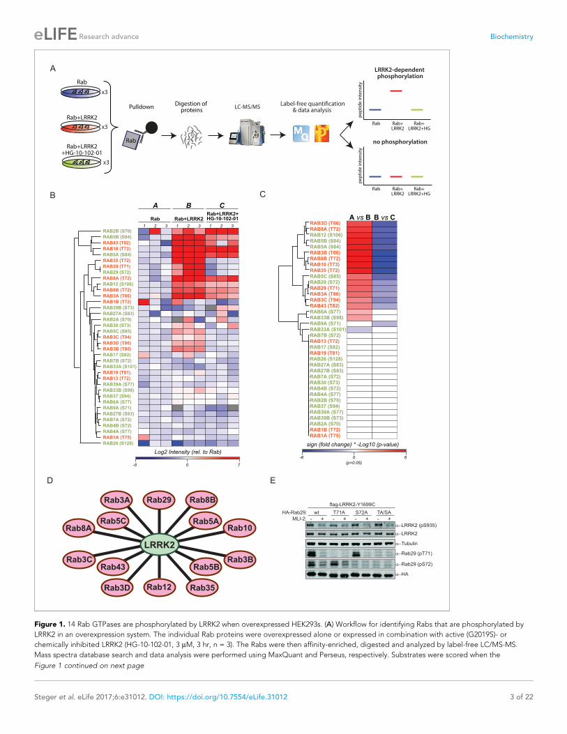
14 overexpressed rab GTPases are phosphorylated by LRRK2We set out to determine which of the 50 Rab proteins that contain a conserved, predicted LRRK2
target site are indeed phosphorylated by LRRK2. For this purpose, we overexpressed each of the 50
Rabs in triplicate in HEK293 cells, either alone or in combination with LRRK2 harboring the activating
G2019S mutation (Figure 1A and Figure 1—figure supplement 1). To determine phosphorylation
specificity, we included a third condition in which both kinase and substrate were expressed in the
presence of the selective LRRK2 inhibitor, HG-10-102-01 (Choi et al., 2012). Although Rab32 and
Rab38 do not contain the conserved phosphorylation site (Figure 1—figure supplement 2A), we
included them because they have previously been reported to interact with LRRK2
(Waschbusch et al., 2014).
Expression of all 52 constructs and inhibitor efficacy was demonstrated by immunoblotting (Fig-
ure 1—figure supplement 2B). To identify LRRK2 target candidates, we enriched the individual epi-
tope-tagged Rab family members by immunoprecipitation before digestion, peptide purification
and high-resolution, label-free LC-MS/MS analysis (Figure 1A). To generate peptides containing the
phosphorylation site with an optimal length for MS analysis, we selected the most appropriate prote-
ase among trypsin, chymotrypsin, Asp-N and Glu-C (Supplementary file 1). This generated 468 LC-
MS/MS files (52 Rabs under three conditions with triplicate analysis), which we stringently processed
using the MaxQuant software for peptide identification and quantification (Cox and Mann, 2008).
For each Rab, several phosphorylated peptides were generally detected, but only those containing
the predicted site showed significant, quantitative differences as a result of LRRK2 overexpression
and inhibition. In total, we quantified this site in 37 different Rab proteins. For 12 Rabs, the
Steger et al. eLife 2017;6:e31012. DOI: https://doi.org/10.7554/eLife.31012 2 of 22
Research advance Biochemistry
B C
D E
sign (fold change) * -Log10 (p-value)
-6 0
(p>0.05)
6
A vs B B vs CRAB3D (T86)
RAB8A (T72)
RAB12 (S106)
RAB5B (S84)
RAB5A (S84)
RAB3B (T86)
RAB8B (T72)
RAB10 (T73)
RAB35 (T72)
RAB5C (S85)
RAB29 (S72)
RAB29 (T71)
RAB3A (T86)
RAB3C (T94)
RAB43 (T82)
RAB6A (S77)
RAB33B (S98)
RAB9A (S71)
RAB33A (S101)
RAB7B (S72)
RAB13 (T72)
RAB17 (S82)
RAB19 (T81)
RAB26 (S128)
RAB27A (S83)
RAB27B (S83)
RAB7A (S72)
RAB30 (S73)
RAB4B (S72)
RAB4A (S77)
RAB2B (S70)
RAB37 (S94)
RAB39A (S77)
RAB39B (S73)
RAB2A (S70)
RAB1B (T72)
RAB1A (T75)
Tubulin
Rab29 (pT71)
HA
Rab29 (pS72)
HA-Rab29:
MLI-2: - -+ + + +wt
- -T71A S72A TA/SA
flag-LRRK2-Y1699C
LRRK2 (pS935)
LRRK2
A
Rab35
Rab5BRab3B
Rab12
Rab3C
Rab3D
Rab43
Rab8ARab5C
Rab3A Rab29
Rab10Rab5A
Rab8B
LRRK2
Rab
Rab+LRRK2
Rab+LRRK2+HG-10-102-01
PulldownDigestion of
proteins LC-MS/MS
Label-free quanti!cation& data analysis
Rab Rab+LRRK2
Rab+LRRK2+HG
x3
x3
x3
LRRK2-dependent phosphorylation
no phosphorylation
pe
pti
de
inte
nsi
ty
Rab
pe
pti
de
inte
nsi
ty
Rab Rab+LRRK2
Rab+LRRK2+HG
Log2 Intensity (rel. to Rab)
-5 0 7
Rab Rab+LRRK2Rab+LRRK2+HG-10-102-01
1 2 3 1 2 3 1 2 3
A B C
RAB2B (S70)
RAB12 (S106)
RAB8B (T72)
RAB3A (T86)
RAB35 (T72)
RAB5B (S84)
RAB43 (T82)
RAB10 (T73)
RAB5A (S84)
RAB29 (T71)
RAB29 (S72)
RAB8A (T72)
RAB26 (S128)
RAB1B (T72)
RAB39B (S73)
RAB27A (S83)
RAB2A (S70)
RAB30 (S73)
RAB5C (S85)
RAB3C (T94)
RAB3D (T86)
RAB3B (T86)
RAB17 (S82)
RAB7B (S72)
RAB33A (S101)
RAB19 (T81)
RAB13 (T72)
RAB39A (S77)
RAB33B (S98)
RAB37 (S94)
RAB6A (S77)
RAB9A (S71)
RAB27B (S83)
RAB7A (S72)
RAB4B (S72)
RAB4A (S77)
RAB1A (T75)
Figure 1. 14 Rab GTPases are phosphorylated by LRRK2 when overexpressed HEK293s. (A) Workflow for identifying Rabs that are phosphorylated by
LRRK2 in an overexpression system. The individual Rab proteins were overexpressed alone or expressed in combination with active (G2019S)- or
chemically inhibited LRRK2 (HG-10-102-01, 3 mM, 3 hr, n = 3). The Rabs were then affinity-enriched, digested and analyzed by label-free LC/MS-MS.
Mass spectra database search and data analysis were performed using MaxQuant and Perseus, respectively. Substrates were scored when the
Figure 1 continued on next page
Steger et al. eLife 2017;6:e31012. DOI: https://doi.org/10.7554/eLife.31012 3 of 22
Research advance Biochemistry
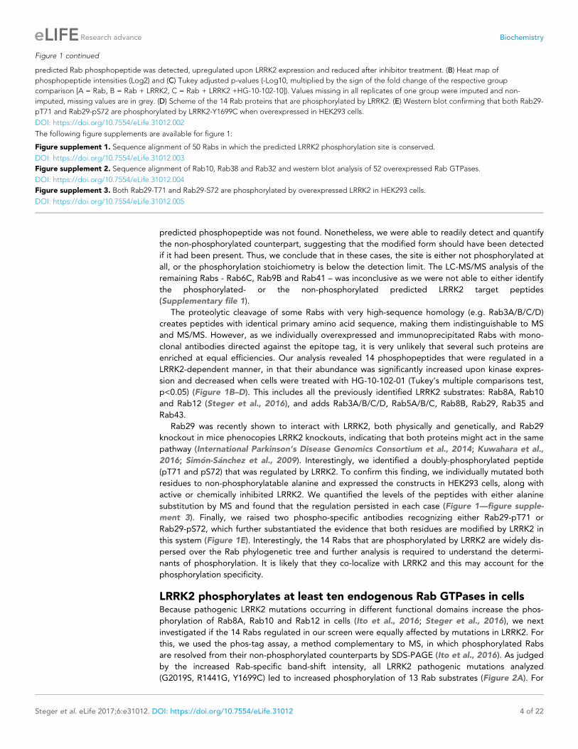
predicted phosphopeptide was not found. Nonetheless, we were able to readily detect and quantify
the non-phosphorylated counterpart, suggesting that the modified form should have been detected
if it had been present. Thus, we conclude that in these cases, the site is either not phosphorylated at
all, or the phosphorylation stoichiometry is below the detection limit. The LC-MS/MS analysis of the
remaining Rabs - Rab6C, Rab9B and Rab41 – was inconclusive as we were not able to either identify
the phosphorylated- or the non-phosphorylated predicted LRRK2 target peptides
(Supplementary file 1).
The proteolytic cleavage of some Rabs with very high-sequence homology (e.g. Rab3A/B/C/D)
creates peptides with identical primary amino acid sequence, making them indistinguishable to MS
and MS/MS. However, as we individually overexpressed and immunoprecipitated Rabs with mono-
clonal antibodies directed against the epitope tag, it is very unlikely that several such proteins are
enriched at equal efficiencies. Our analysis revealed 14 phosphopeptides that were regulated in a
LRRK2-dependent manner, in that their abundance was significantly increased upon kinase expres-
sion and decreased when cells were treated with HG-10-102-01 (Tukey’s multiple comparisons test,
p<0.05) (Figure 1B–D). This includes all the previously identified LRRK2 substrates: Rab8A, Rab10
and Rab12 (Steger et al., 2016), and adds Rab3A/B/C/D, Rab5A/B/C, Rab8B, Rab29, Rab35 and
Rab43.
Rab29 was recently shown to interact with LRRK2, both physically and genetically, and Rab29
knockout in mice phenocopies LRRK2 knockouts, indicating that both proteins might act in the same
pathway (International Parkinson’s Disease Genomics Consortium et al., 2014; Kuwahara et al.,
2016; Simon-Sanchez et al., 2009). Interestingly, we identified a doubly-phosphorylated peptide
(pT71 and pS72) that was regulated by LRRK2. To confirm this finding, we individually mutated both
residues to non-phosphorylatable alanine and expressed the constructs in HEK293 cells, along with
active or chemically inhibited LRRK2. We quantified the levels of the peptides with either alanine
substitution by MS and found that the regulation persisted in each case (Figure 1—figure supple-
ment 3). Finally, we raised two phospho-specific antibodies recognizing either Rab29-pT71 or
Rab29-pS72, which further substantiated the evidence that both residues are modified by LRRK2 in
this system (Figure 1E). Interestingly, the 14 Rabs that are phosphorylated by LRRK2 are widely dis-
persed over the Rab phylogenetic tree and further analysis is required to understand the determi-
nants of phosphorylation. It is likely that they co-localize with LRRK2 and this may account for the
phosphorylation specificity.
LRRK2 phosphorylates at least ten endogenous Rab GTPases in cellsBecause pathogenic LRRK2 mutations occurring in different functional domains increase the phos-
phorylation of Rab8A, Rab10 and Rab12 in cells (Ito et al., 2016; Steger et al., 2016), we next
investigated if the 14 Rabs regulated in our screen were equally affected by mutations in LRRK2. For
this, we used the phos-tag assay, a method complementary to MS, in which phosphorylated Rabs
are resolved from their non-phosphorylated counterparts by SDS-PAGE (Ito et al., 2016). As judged
by the increased Rab-specific band-shift intensity, all LRRK2 pathogenic mutations analyzed
(G2019S, R1441G, Y1699C) led to increased phosphorylation of 13 Rab substrates (Figure 2A). For
Figure 1 continued
predicted Rab phosphopeptide was detected, upregulated upon LRRK2 expression and reduced after inhibitor treatment. (B) Heat map of
phosphopeptide intensities (Log2) and (C) Tukey adjusted p-values (-Log10, multiplied by the sign of the fold change of the respective group
comparison [A = Rab, B = Rab + LRRK2, C = Rab + LRRK2 +HG-10-102-10]). Values missing in all replicates of one group were imputed and non-
imputed, missing values are in grey. (D) Scheme of the 14 Rab proteins that are phosphorylated by LRRK2. (E) Western blot confirming that both Rab29-
pT71 and Rab29-pS72 are phosphorylated by LRRK2-Y1699C when overexpressed in HEK293 cells.
DOI: https://doi.org/10.7554/eLife.31012.002
The following figure supplements are available for figure 1:
Figure supplement 1. Sequence alignment of 50 Rabs in which the predicted LRRK2 phosphorylation site is conserved.
DOI: https://doi.org/10.7554/eLife.31012.003
Figure supplement 2. Sequence alignment of Rab10, Rab38 and Rab32 and western blot analysis of 52 overexpressed Rab GTPases.
DOI: https://doi.org/10.7554/eLife.31012.004
Figure supplement 3. Both Rab29-T71 and Rab29-S72 are phosphorylated by overexpressed LRRK2 in HEK293 cells.
DOI: https://doi.org/10.7554/eLife.31012.005
Steger et al. eLife 2017;6:e31012. DOI: https://doi.org/10.7554/eLife.31012 4 of 22
Research advance Biochemistry
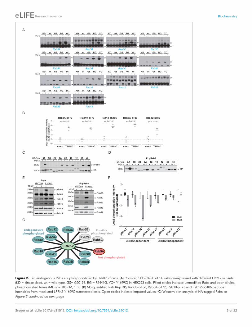
Figure 2
A
B
Rab3A Rab3DRab3B Rab3C
- - - - -+ + + + + - - - - -+ + + + + - - - - -+ + + + +MLi-2:
KD wt GS RG YC KD wt GS RG YC KD wt GS RG YC KD wt GS RG YC- - - - -+ + + + +
Rab5A Rab8ARab5B Rab5C
- - - - -+ + + + + - - - - -+ + + + + - - - - -+ + + + +MLi-2:
KD wt GS RG YC KD wt GS RG YC KD wt GS RG YC KD wt GS RG YC- - - - -+ + + + +
Rab8B Rab10 Rab12 Rab29
- - - - -+ + + + + - - - - -+ + + + + - - - - -+ + + + +MLi-2:
KD wt GS RG YC KD wt GS RG YC KD wt GS RG YC KD wt GS RG YC- - - - -+ + + + +
Rab35 Rab43
- - - - -+ + + + +MLi-2:
KD wt GS RG YC KD wt GS RG YC- - - - -+ + + + +
C
E
8A 1029 128B
25kDa pRab8
25kDa HA
5C3A 35 43+ - + - + - + - + - + - + - + - + -
HA-Rab:MLi-2:
D
pRab8
MLi-2: +-
25kDa
25kDa
25kDa
25kDa
wild type R1441C
25kDa
25kDa
Rab8A
Rab10
Rab35
Rab43
Rab1A
+-
Input
25kDa
25kDa
25kDa
25kDa
25kDa
MLi-2: +-
wild type R1441C
+-
IP: pRab8
Rab8A
Rab10
Rab35
Rab43
Rab1A
25kDa
IP: pRab8
HA
8A 1029 128B5C3A 35 43HA-Rab:
+ -MLi-2: + - + - + - + - + - + - + - + -
- MLi2
Lo
g2 p
ho
sp
ho
pep
tid
e in
ten
sit
y
(rel. t
o u
ntr
eate
d)
+ MLi2
-6
-4
-2
0
2
pRab10
pRab8
pRab35
pRab3
pRab43
pRab1
pRab7
pRab13
LRRK2 dependent LRRK2 independent
F
G
mock Y1699C
p= 2.88*10-5
Rab3A-pT86Rab10-pT73
p= 8.65*10-6
mock Y1699C
-2
0
2
4
6
8
Rab8A-pT72
mock Y1699C
p= 1.09*10-4
Lo
g2 p
ho
sp
ho
pep
tid
e in
ten
sit
y
(rel. t
o u
ntr
an
sfe
cte
d)
Rab12-pS106
mock Y1699C
p= 2.67*10-5
mock Y1699C
p= 2.33*10-3
Rab3B-pT86
Rab3D
Rab8BRab3B
Rab10
Rab35
Rab43
Rab3C
Rab8ARab3A
Rab12
Rab5A
Rab5CRab29
Rab5B
LRRK2
Endogenously
phosphorylated
Not phosphorylated
Possibly phosphorylated
Figure 2. Ten endogenous Rabs are phosphorylated by LRRK2 in cells. (A) Phos-tag SDS-PAGE of 14 Rabs co-expressed with different LRRK2 variants
(KD = kinase dead, wt = wild type, GS= G2019S, RG = R1441G, YC= Y1699C) in HEK293 cells. Filled circles indicate unmodified Rabs and open circles,
phosphorylated forms (MLi-2 = 100 nM, 1 hr). (B) MS-quantified Rab3A-pT86, Rab3B-pT86, Rab8A-pT72, Rab10-pT73 and Rab12-pS106 peptide
intensities from mock and LRRK2-Y1699C transfected cells. Open circles indicate imputed values. (C) Western blot analysis of HA-tagged Rabs co-
Figure 2 continued on next page
Steger et al. eLife 2017;6:e31012. DOI: https://doi.org/10.7554/eLife.31012 5 of 22
Research advance Biochemistry
Rab12, the assay resulted in multiple bands and no meaningful interpretation of the migration pat-
tern was possible; however, our previous work had already established this protein as a LRRK2 sub-
strate. In this context, we also investigated Rab6C, Rab9B and Rab41 by the phos-tag method, for
which the MS analysis was inconclusive, and found no evidence of LRRK2-induced mobility shifts.
We next investigated which of these 14 Rab proteins are phosphorylated by LRRK2 endoge-
nously. For this, we overexpressed the pathogenic LRRK2-Y1699C form in HEK293 cells, immunopre-
cipitated endogenous Rab proteins using commercially available- (Rab3A, Rab3B, Rab5A, Rab8A,
Rab10) or in-house raised antibodies (Rab12) and quantified LRRK2-mediated phosphorylation by
LC-MS/MS. As expected from our previous results, Rab8A-pT72, Rab10-pT73 and Rab12-pS106
phosphopeptide levels were strongly upregulated (~16 fold) in cells expressing LRRK2-Y1699C
(Figure 2B). Rab3A-pT86 was also clearly regulated by LRRK2 in this system, but we were not able
to detect any phosphopeptides mapping to either Rab3B-pT86 or Rab5A-pS84, despite the fact that
the corresponding unmodified peptides were sequenced multiple times and our initial overexpres-
sion screen had established detectability of these phosphopeptides. Therefore, to further increase
mass spectrometric sensitivity, we used stable isotope labeled (SIL) peptides equivalent to the
endogenous Rab peptides of interest to guide targeted quantification. In contrast to the absolute
quantification (AQUA) strategy (Gerber et al., 2003) that relies on fragment ions for quantification,
we recorded both light and heavy counterparts in a multiplexed selected ion monitoring (SIM) scan
mode on a quadrupole Orbitrap analyzer and used the heavy form to indicate the retention time
(heavy) and the light form for quantification. This combination of the spike-in SIL peptide together
with a very sensitive MS method should allow quantification of very low abundant endogenous Rab
phosphorylation (Materials and methods). With this increased sensitivity, we found LRRK2-specific
modification of Rab3B, but still failed to observe clear LRRK2-mediated phosphorylation of Rab5A-
S84 (Figure 2B and Figure 2—figure supplement 1). Thus, Rab5A phosphorylation by LRRK2 in
HEK293 cells occurs only when both the Rab and kinase are overexpressed exogenously. It is possi-
ble that accessory factors in other cell types are required for efficient LRRK2 phosphorylation of this
protein.
For the remaining Rab proteins for which no suitable antibodies for total-Rab protein enrichment
were available, we raised a rabbit polyclonal Rab antibody against the LRRK2-phosphorylated Rab8
switch-II motif. This efficiently recognized and immunoprecipitated LRRK2 phosphorylated Rabs con-
taining a Thr site: namely, Rab3, Rab8, Rab10, Rab35 and Rab43 (Figure 2C,D). As an exception,
the antibody failed to recognize pRab29 or to immunoprecipitate LRRK2-phosphorylated Rab29 pro-
tein. This could be either due to the particular linear sequence surrounding T71 (�1 position com-
pared to the equivalent site on other Rabs) or due to double phosphorylation (T71 and S72) by
LRRK2. Next, we treated wild type and LRRK2-R1441C knock-in mouse embryonic fibroblasts (MEFs)
with the selective LRRK2 inhibitor MLi-2 (Fell et al., 2015), subjected the cell lysates to immunopre-
cipitation with the phospho-Rab8 antibody and analyzed the immunoprecipitates by immunoblotting
or LC-MS/MS. Immunoblotting of the input cell extracts confirmed that compared to wild type, the
signal detected by the newly developed anti-phospho-Rab8-antibody strongly increased in LRRK2-
R1441C MEFs, and this was blocked by MLi-2 treatment (Figure 2E). For detecting immunoprecipi-
tates by western blotting, we used CRISPR/CAS9 knock-out-validated antibodies (Figure 2—figure
Figure 2 continued
expressed with LRRK2-Y1699C in HEK293 cells (-/+100 nM MLi-2, 2 hr) using polyclonal anti-phospho-Rab8 and HA antibodies. (D) Same as (C) but Rabs
were immunoprecipitated using the anti-pRab8 antibody and HA antibody was used for detection. (E) Western blot of endogenous Rabs in wild type
and LRRK2-R1441C MEFs (-/+100 nM MLi-2, 1 hr) using the indicated antibodies (left) and immunoprecipitation of Rabs using anti-pRab8 antibody
(right). (F) MS-quantified Rab phosphosite intensities of pRab8 immunoprecipitation in R1441C MEFs (-/+100 nM MLi-2, 2 hr). Error bars are mean ±SEM
(n = 3). (G) 10 Rab proteins at endogenous expression levels are phosphorylated by LRRK2 in cells (green). Rab5A is not (red) and Rab5B, Rab5C and
Rab29 are possibly phosphorylated (grey).
DOI: https://doi.org/10.7554/eLife.31012.006
The following figure supplements are available for figure 2:
Figure supplement 1. Rab5A was immunoprecipitated from mock or LRRK2-Y1699C-transfected HEK293 cells.
DOI: https://doi.org/10.7554/eLife.31012.007
Figure supplement 2. Nine Rab GTPases containing a Thr site in their switch-II domain are phosphorylated by LRRK2.
DOI: https://doi.org/10.7554/eLife.31012.008
Steger et al. eLife 2017;6:e31012. DOI: https://doi.org/10.7554/eLife.31012 6 of 22
Research advance Biochemistry
supplement 2A). This revealed specific enrichment of phospho-Rab8A, -Rab10, -Rab35 and -Rab43,
which increased in LRRK2-R1441C MEFs and was completely prevented by MLi-2 treatment
(Figure 2E). In contrast, Rab1A phosphorylation was not sensitive to MLi-2 in this system, confirming
the data from our initial overexpression screen.
Using the anti-phospho-Rab8 antibody in LRRK2-R1441C fibroblasts, we analyzed immunoprecipi-
tates by LC-MS/MS. Quantification of 32 Rab protein intensities showed that Rab3A, Rab8A, Rab8B,
Rab10, Rab35 and Rab43 were enriched more than twofold in a LRRK2-dependent manner. Rab3B
and Rab3D were also detected, however, compared to MLi-2 treatment, their enrichment in
untreated cells was less pronounced (about 1.5-fold), probably due to lower protein expression lev-
els (Figure 2—figure supplement 2B,C). In concordance with this, quantification of the switch-II
phosphorylated peptides of Rab3, Rab8, Rab10, Rab35 and Rab43 in the same experiment revealed
a more than two-fold decrease upon MLi-2 treatment (Figure 2F). For Rab1A, Rab7 and Rab13,
however, there was no pronounced difference in the corresponding protein and phospho-peptide
levels when comparing untreated with MLi-2-treated samples, suggesting that these Rabs are phos-
phorylated by a kinase other than LRRK2. Finally, we immunoprecipitated phosphorylated Rabs from
HEK293 cells expressing LRRK2-Y1699C using the same antibody. Again, we confirmed LRRK2-spe-
cific enrichment of nine threonine phosphorylated Rab proteins. These were Rab3A/B/C/D, Rab8A/
B, Rab10, Rab35 and Rab43 and the protein sequence coverage ranged from 33% (Rab3B) to 90%
(Rab10). Compared to the other family members, this indicates that they are recognized by the anti-
body in a direct manner (Figure 2—figure supplement 2D).
In summary, our study demonstrates that 14 Rab proteins are phosphorylated by LRRK2 at the
switch-II site in an overexpression system (Rab3A/B/C/D, Rab5A/B/C, Rab8A/B, Rab10, Rab12,
Rab29, Rab35 and Rab43). For ten of these (Rab3A/B/C/D, Rab8A/B, Rab10, Rab12, Rab35 and
Rab43), endogenous LRRK2-dependent phosphorylation is clearly established; for one (Rab5A), we
observed phosphorylation in an overexpression but not an endogenous setting. In vivo analysis of
the remaining three Rabs (Rab5B, Rab5C and Rab29) was hampered by the unavailability of suitable
antibodies for protein enrichment or detection (Figure 2G).
Phosphorylation-dependent binding of regulatory proteins to RabsDifferent residues in the switch-II domain of Rabs can be modified post-translationally, and this inter-
feres with binding to partner proteins such as Rab GDP dissociation inhibitor alpha/beta (GDI1/2).
For example, Rab1B is AMPylated on Y77 (Y80 in Rab1A) by the Legionnaires’ disease protein, DrrA,
resulting in a constitutively active Rab protein that fails to interact with GTPase-activating proteins
(GAPs) and GDIs (Muller et al., 2010; Oesterlin et al., 2012). Similarly, the switch-II domain of
Rab1A can be phosphocholinated on S79 during Legionella infection, which results in strongly
decreased GDI binding (Goody et al., 2012; Mukherjee et al., 2011). We showed previously that
phosphorylation of Rab8A-T72 and the equivalent sites, Rab10-T73 and Rab12-S106 by LRRK2
decreases GDI binding (Steger et al., 2016). Substitution of Rab7A-T72 with a phosphomimetic glu-
tamic acid has been shown to abrogate GDI interaction (Satpathy et al., 2015). Here, we set out to
test systematically, the effect of replacing the predicted LRRK2 target site with a negatively charged,
phosphomimetic glutamic acid residue on partner protein binding. For this, we mutated these sites
to either non-phosphorylatable alanine or glutamic acid in all 14 Rabs (both T71 and S72 for Rab29)
and expressed them in HEK293 cells (n = 3). Following affinity-enrichment, we digested bound pro-
teins and analyzed the resulting peptides by label-free LC-MS/MS. As expected, the S/TfiE muta-
tions strongly reduced partner binding for all tested Rabs. The S/TfiA Rab mutants instead stably
bound to GDI1/2 and the Rab escort proteins, CHM and CHML, except for both tested Rab29 con-
structs (Figure 3—figure supplement 1A,B). In that case, neither the T71A/E nor the S72A/E con-
structs interacted with GDI, indicating either low binding affinities, an alteration in nucleotide-
binding properties, or protein misfolding.
To investigate more specifically the effect of LRRK2-induced Rab phosphorylation on protein
interactions, we expressed LRRK2 in HEK293 cells and determined the interactomes of both HA-
tagged and endogenous Rab8A by AE-MS, before and after chemical LRRK2 inhibition. To increase
phosphosite occupancy at T72, we used the pathogenic ROC-COR domain LRRK2-R1441G mutant,
which confers strong intracellular kinase activity (Sheng et al., 2012; Steger et al., 2016). Upon
LRRK2 inhibition, Rab8A-pT72 levels decreased about eight fold as shown by quantitative MS, estab-
lishing inhibitor efficacy (Figure 3—figure supplement 2). As we had reported previously, GDIs
Steger et al. eLife 2017;6:e31012. DOI: https://doi.org/10.7554/eLife.31012 7 of 22
Research advance Biochemistry
A B
D E
01
23
45
6
-4 -2 0 2 4 6
ACAP2
WDR91
INPP5B
TBC1D8B
SPAG9
GDI2
OCRL
RAB39A
WDR81
TBC1D9B
MAP4K3
MICALL2MICALL1
EHBP1
RILPL2
WDR91
WDR81
TBC1
Log2 (R1441G/R1441G+HG-10-102-01)
-Lo
g1
0 (
p-v
alu
e)
IP-HA-Rab8A
01
23
45
MAP4K3
TBC1D8B
MICAL3
TBC1D9B
MICALL2
EHBP1L1
MICALL1
RILPL2
-4 -3 -2 -1 0 1 2 3 4
Log2 (R1441G/R1441G+HG-10-102-01)
-Lo
g1
0 (
p-v
alu
e)
IP-Rab8A (endogenous)
F
RILPL1-GFP
KD YC
HA-Rab8A
- +
GFP
-
YC
LysatesRILPL2-GFP
+
RILP-GFP
- - +
α−HA (Phostag)
MLi-2:
α−HA
α−LRRK2 (pS1292)
α−LRRK2
25kDa
IP: α−G
FP
Ly
sa
tes
25kDa
75kDa
50kDa
37kDa
25kDa
25kDa
250kDa
250kDa
75kDa
50kDa
37kDa
α−pRab8
α−GFP
α−HA
α−pRab8
α−GFP
LRRK2: KD YC YCKD KD
25kDa
-
YCKD
C
Mock MLi-2-1.5
-1
-0.5
0
0.5
1RILPL1
p= 0.0124
Lo
g2
Pro
tein
In
ten
sit
y
Mock MLi-2-1.5
-1
-0.5
0
0.5
1RILPL2p= 0.0544
Lo
g2
Pro
tein
In
ten
sit
y
235 PSEAGQCRFSREEFEQILQERNELKAKVFLLKEELAYFQRELLTDHRVPG 284
286 LKDPNRPRFTLQELRDVLHERNELKSKVFLLQEELAYYKSEEMEEENRIP 331
125 LTDPNRPRFTLQELRDVLQERNKLKSQLLVVQEELQCYKSGLIPPREGPG 174
RILP
RILPL1
RILPL2
291293
300310
324
G
-10
-50
51
01
5
RAB8A
RAB10
RAB12
-10 -5 0 5 10 15
Lo
g2
Te
st
sta
tis
tic
(w
t/R
13
0E
)
Log2 Test statistic (wt/wt+MLi-2)
RILPL2RILPL1
Lo
g2
Te
st
sta
tis
tic
(w
t/R
29
1E
)
Log2 Test statistic (wt/wt+MLi-2)
-10
-50
51
01
52
0
RAB8A
RAB10
RAB12
-10 -5 0 5 10 15 20
H
α-GFP
IP: α−G
FP
Ly
sa
tes
α−HA (Phostag)
α−HA
α−GFP
α−LRRK2 (pS1292)
α−LRRK2
α−GFP
α−HA (Phostag)
- + - + - + - + - +MLi-2:
α−pRab8
RILPL2-GFPRILPL1-GFP
Lysatewt R130E R132E K149E
LRRK2-Y1699C + HA-Rab8a
K310EGFP wt R291E R293E
- + - + - + - + - +
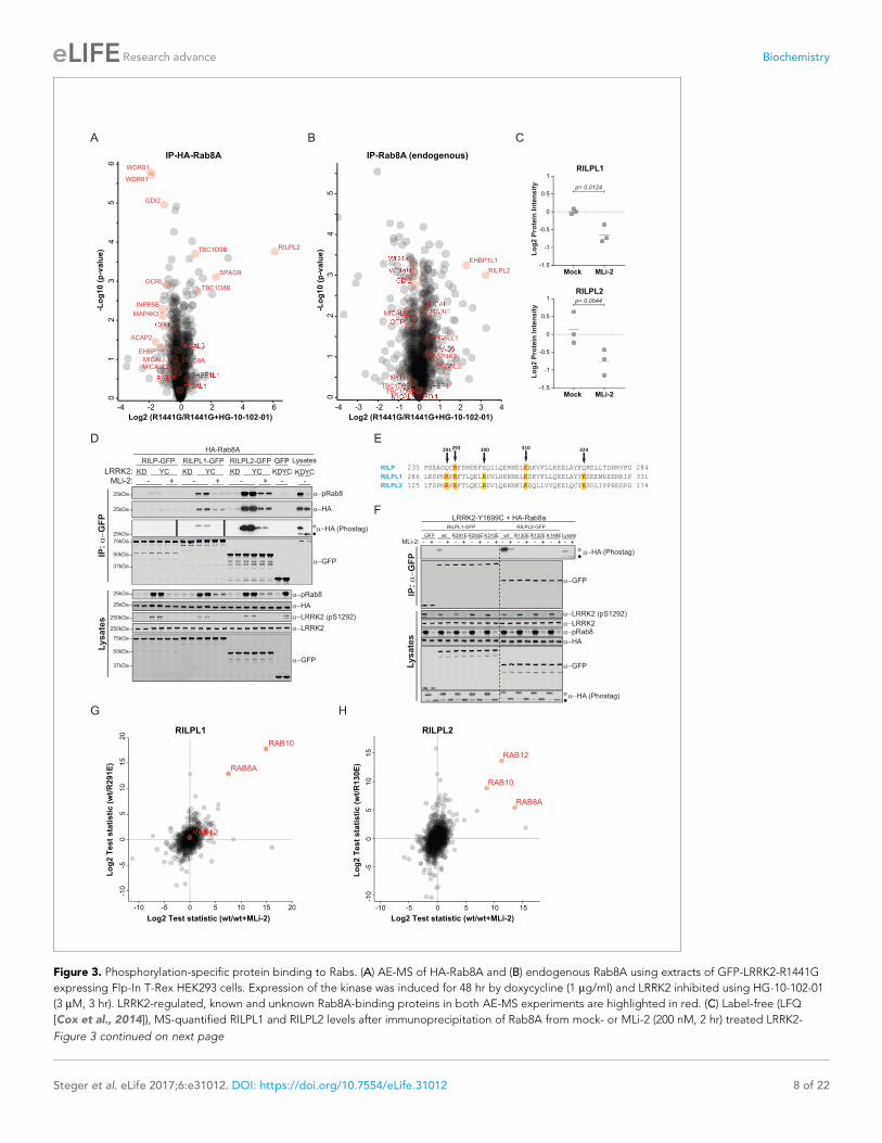
Figure 3. Phosphorylation-specific protein binding to Rabs. (A) AE-MS of HA-Rab8A and (B) endogenous Rab8A using extracts of GFP-LRRK2-R1441G
expressing Flp-In T-Rex HEK293 cells. Expression of the kinase was induced for 48 hr by doxycycline (1 mg/ml) and LRRK2 inhibited using HG-10-102-01
(3 mM, 3 hr). LRRK2-regulated, known and unknown Rab8A-binding proteins in both AE-MS experiments are highlighted in red. (C) Label-free (LFQ
[Cox et al., 2014]), MS-quantified RILPL1 and RILPL2 levels after immunoprecipitation of Rab8A from mock- or MLi-2 (200 nM, 2 hr) treated LRRK2-
Figure 3 continued on next page
Steger et al. eLife 2017;6:e31012. DOI: https://doi.org/10.7554/eLife.31012 8 of 22
Research advance Biochemistry
associated preferentially with the non-phospho forms of both HA-Rab8A and endogenous Rab8A
(Figure 3A,B). However, this effect was more pronounced in the case of overexpressed Rab8A,
probably due to the higher fraction of phosphorylated protein.
Several, to-date undescribed interaction partners that showed preferential binding to the non-
phospho-forms in both the endogenous and HA-tagged systems (Figure 3A,B). These include the
poorly characterized, WD repeat domain (WDR)-containing proteins, WDR81 and WDR91, which
have recently been linked to regulation of endosomal phosphatidylinositol 3-phosphate levels in C.
elegans and mammalian cells (Liu et al., 2016). The inositol-5-phosphatase, OCRL is a known Rab8A
effector, and our experiments revealed that phosphorylation abolishes its binding to Rab8A. Muta-
tions in the human OCRL gene cause the oculocerebrorenal syndrome of Lowe and it would be inter-
esting to investigate whether the phosphorylation of Rabs is relevant in this context (Hou et al.,
2011).
Strikingly, we found that Rab interacting lysosomal protein like 2 (RILPL2) binds preferentially to
the phosphorylated forms of both HA-Rab8A and Rab8A. RILPL2 contains a C-terminal, coiled-coil
Rab-binding domain, which is known as RILP homology (RH) domain, and this is conserved in four
other members of this protein family (RILP, RILPL1 and JIP3/4) (Matsui et al., 2012; Wu et al.,
2005). RILPL1 and RILPL2 are poorly characterized proteins; however, a recent report links both to
the regulation of protein localization in primary cilia (Schaub and Stearns, 2013). RILP is much bet-
ter studied, and interacts with Rab7 to control lysosomal motility in cells (Jordens et al., 2001).
To investigate Rab8A-RILPL2 interaction in an endogenous setting, we immunoprecipitated
Rab8A from mock- or MLi-2 treated, knock-in MEFs expressing the hyperactive LRRK2 variant,
R1441C. This confirmed our initial LRRK2-specific interaction of Rab8A and RILPL2 and also identi-
fied RILPL1 as phospho-dependent Rab8A interactor (Figure 3C).
To further verify the pRab8A interaction with RILPL1 and RILPL2 and to test for RILP interaction,
we co-expressed GFP-tagged, full length RILP, RILPL1 and RILPL2 with HA-Rab8A and LRRK2-
Y1699C or kinase-dead LRRK2-Y1699C/D2017A. Both RILPL1 and RILPL2, but not RILP, specifically
bound to LRRK2-generated phospho-Rab8A, and this interaction was strongly reduced by MLi-2
(Figure 3D). In this side by side comparison, RILPL2-bound phosphorylated Rab8A much stronger
than RILPL1, indicating that RILPL2 might be a key effector specific for pRab8A. Phos-tag analysis
independently confirmed that only the LRRK2-phosphorylated HA-Rab8A protein was co-immuno-
precipitated with either RILPL2 or RILPL1 (Figure 3D).
There are five highly conserved basic residues in the RH domain on RILPL1 and RILPL2 that could
potentially mediate the interaction with LRRK2-phosphorylated Rab8A (R291, R293, R300, K310 and
K324 in RILPL1) (Figure 3E). Individual mutation of R291, R293 or K310, but not R300 or K324, abol-
ished the interaction of N-terminally truncated RILPL1 with LRRK2-phosphorylated Rab8A (Figure 3—
figure supplement 3A). Accordingly, the identified mutations abrogated pRab8 interaction with
both full-length RILPL1 and RILPL2 (Figure 3F).
Figure 3 continued
R1441C MEFs. (D) Pulldown of GFP-tagged RILP, RILPL1 or RILPL2, transiently expressed with HA-Rab8A and the indicated LRRK2 variants (KD=
Y1699C/D2017A) in HEK293 cells. Western blot after Phos-tag SDS-PAGE was used to detect interacting proteins using the indicated antibodies. (MLi-
2 = 150 nM, 2 hr). (E) Sequence alignment of the RILP homology (RH) domains of RILP, RILPL1 and RILPL2 showing five conserved basic residues, which
are highlighted. (F) Same as (D) but using different RILPL1 and RILPL2 mutants. For phos-tag blots, filled circles indicate non phosphorylated proteins
and open circles phosphorylated proteins. (G) AE-MS of GFP-RILPL1 (wt or R291E) and (H) GFP-RILPL2 (wt or R130E), expressed with LRRK2-Y1699C in
HEK293 cells, and treated or not with MLi-2 (for wt, 200 nM, 2 hr). The student’s two sample test statistic (Log2) of the indicated comparisons was used
for plotting.
DOI: https://doi.org/10.7554/eLife.31012.009
The following figure supplements are available for figure 3:
Figure supplement 1. Phosphomimetic S/T->E mutation of the LRRK2 phosphorylation site abrogates GDI1/2 and CHM/CHML binding in 13 Rab
GTPases.
DOI: https://doi.org/10.7554/eLife.31012.010
Figure supplement 2. MS-quantified pT72-Rab8A peptide levels in HEK293 cells expressing LRRK2-R1441G.
DOI: https://doi.org/10.7554/eLife.31012.011
Figure supplement 3. Phospho-dependent interaction of Rab8A, Rab10 and Rab12 with RILPL1/2.
DOI: https://doi.org/10.7554/eLife.31012.012
Steger et al. eLife 2017;6:e31012. DOI: https://doi.org/10.7554/eLife.31012 9 of 22
Research advance Biochemistry
To extend our RILPL1/2-Rab interaction analysis reciprocally, we expressed GFP-RILPL1 (wt or
R291E) and GFP-RILPL2 (wt or R130E) with LRRK2-Y1699C in HEK293 cells, in the presence or
absence of MLi-2, and processed the samples for LC-MS/MS analysis after affinity enrichment. Strik-
ingly, in this setup, RILPL1-wt interacted not only with Rab8A, but also with Rab10, and this was
strongly reduced by MLi-2 and the R291E Rab interaction mutant (Figure 3G). RILPL2 instead bound
all three of our previously identified bona-fide LRRK2 substrates, Rab8A, Rab10 and Rab12 in a
LRRK2-dependent manner (Steger et al., 2016) (Figure 3H). We further confirmed the phospho-
specific Rab8A, Rab10 and Rab12 interactions with RILPL1/2 by immunoprecipitation-phos-tag and
phospho-Rab directed antibodies (Figure 3—figure supplement 3B–D). Together these results
unambiguously establish that both RILPL1/2 are phospho-specific Rab interactors and that this inter-
action is mediated by the RH domain of RILPL1 and RILPL2. Based on these findings, it will be inter-
esting to investigate possible genetic association of RILPL1/2 mutations with PD.
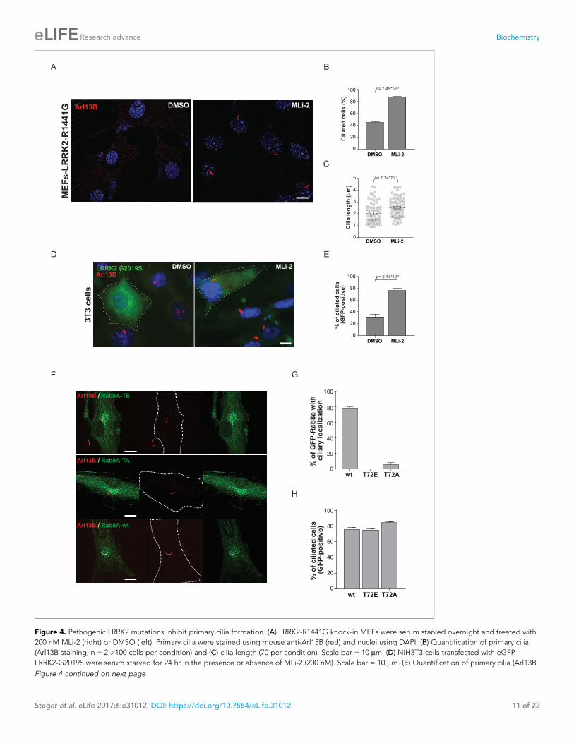
Ciliogenesis is attenuated in cells expressing hyperactive LRRK2Primary cilia are hair-like structures extending from the cell body and are found on nearly all human
cells (Malicki and Johnson, 2017). Ciliary defects lead to a variety of human diseases known as cilio-
pathies, some more severe than others, and more than 200 genes have been associated with these
disorders (Reiter and Leroux, 2017). Rab or Rab-regulatory protein dysfunctions play important
roles in a number of ciliopathies. For instance, Rab28 is localized at the periciliary membrane of cili-
ated neurons and mutations in its gene are associated with cone-rod dystrophy, an inherited ocular
disorder (Jensen et al., 2016; Roosing et al., 2013). Similarly, Rab23 is involved in ciliary protein
transport and Rab23 mutations are associated with a rare congenital disorder known as Carpenter
syndrome (Boehlke et al., 2010; Jenkins et al., 2007). Rab8A, Rab10, Rab11, Rab17 and Rab29 are
involved in ciliogenesis; however, no specific disorders are associated with mutations in these genes
(Knodler et al., 2010; Nachury et al., 2007; Onnis et al., 2015; Sato et al., 2014;
Yoshimura et al., 2007). Since LRRK2 phosphorylation of Rab8A, Rab10 and Rab12 results in
increased RILPL1/2 interaction (Figure 3) and Rab8A, Rab10 and RILPL1/2 are reported to regulate
primary ciliogenesis (Nachury et al., 2007; Sato et al., 2014; Schaub and Stearns, 2013), we rea-
soned that LRRK2 itself might be a regulator of primary cilia formation. To test this, we induced cilia
formation by serum starvation in LRRK2-R1441G knock-in MEFs that harbor increased kinase activity,
and cultured them with or without MLi-2. Strikingly, as judged by anti-Arl13B staining, DMSO-
treated LRRK2-R1441G expressing cells displayed a twofold decrease in the number of ciliated cells
and a reduction in cilia length compared with overnight-MLi-2-treated cells (Figure 4A–C). To con-
firm this finding using another cell system, we analyzed cilia formation in 3T3 fibroblasts transfected
with GFP-LRRK2-G2019S. In this case, about 30% of serum-starved, LRRK2-expressing cells (GFP
positive) were ciliated (Arl13B positive). Overnight LRRK2 inhibitor treatment (MLi-2) increased this
by twofold, demonstrating kinase dependency of the ciliation defect (Figure 4D,E). Thus, two differ-
ent cellular assays support our finding that LRRK2 regulation is important for ciliogenesis. To deter-
mine whether the observed defect in ciliogenesis seen in hyperactive LRRK2 mutant-expressing cells
is due to Rab8A phosphorylation, we expressed GFP-Rab8A-T72A or GFP-Rab8A-T72E in serum-
starved RPE-1 cells and assessed both primary cilia formation and exogenous Rab8A protein localiza-
tion by immunofluorescence microscopy. While GFP-Rab8A-wt was localized at cilia in about 80% of
Arl13B-positive cells, this was not true for the T72A nor the T72E mutants, and total numbers of cili-
ated cells did not change in any of the conditions analyzed (Figure 4F–H). Thus, exogenous expres-
sion of the T72A or T72E mutants alone was not sufficient to block cilia formation. This suggests that
other LRRK2 targets might play a role in this process or that endogenous Rab8A is sufficient to sus-
tain cilia formation in this experimental setup.
Together, our data demonstrate that LRRK2 activity influences ciliogenesis and kinase hyperactiv-
ity interferes with this process. The precise details of how LRRK2-mediated Rab8A phosphorylation
controls this process and influences RILPL1 and RILPL2 function will be important areas for future
work. Whether phosphorylation of Rabs other than Rab8A by LRRK2 influences cilia formation will
also require further analysis.
Steger et al. eLife 2017;6:e31012. DOI: https://doi.org/10.7554/eLife.31012 10 of 22
Research advance Biochemistry
D E
DMSO MLi-2
0
20
40
60
80
100
% o
f cilia
ted
cells
(GF
P-p
osit
ive)
p= 6.14*10-4
3T
3 c
ells
LRRK2 G2019SArl13B
MLi-2DMSO
A B
DMSO MLi-2
0
20
40
60
80
100
Cilia
ted
cells (
%)
p= 1.45*10-3
C
ME
Fs
-LR
RK
2-R
14
41
G Arl13B MLi-2DMSO
Cilia
len
gth
(m
)
p= 1.24*10-3
DMSO MLi-20
1
2
3
4
5
F
Arl13B / Rab8A-TE
Arl13B / Rab8A-TA
Arl13B / Rab8A-wt
G
H
wt T72E T72A0
20
40
60
80
100
% o
f cilia
ted
cells
(GF
P-p
osit
ive)
wt T72E T72A0
20
40
60
80
100
% o
f G
FP
-Rab
8a w
ith
cilia
ry lo
calizati
on
Figure 4. Pathogenic LRRK2 mutations inhibit primary cilia formation. (A) LRRK2-R1441G knock-in MEFs were serum starved overnight and treated with
200 nM MLi-2 (right) or DMSO (left). Primary cilia were stained using mouse anti-Arl13B (red) and nuclei using DAPI. (B) Quantification of primary cilia
(Arl13B staining, n = 2,>100 cells per condition) and (C) cilia length (70 per condition). Scale bar = 10 mm. (D) NIH3T3 cells transfected with eGFP-
LRRK2-G2019S were serum starved for 24 hr in the presence or absence of MLi-2 (200 nM). Scale bar = 10 mm. (E) Quantification of primary cilia (Arl13B
Figure 4 continued on next page
Steger et al. eLife 2017;6:e31012. DOI: https://doi.org/10.7554/eLife.31012 11 of 22
Research advance Biochemistry
ConclusionOur systematic analysis of LRRK2-dependent Rab phosphorylation revealed that a distinct subset of
the Rab family is subject to regulation by this kinase. When overexpressed a total of 14 Rabs are
phosphorylated by LRRK2, and at least ten are phosphorylated when they are present in cells at
endogenous levels. New tools such as specific antibodies or sensitive targeted MS methods, as well
as more in depth analyses of tissues expressing LRRK2, are needed to clarify whether the remaining
three Rabs (Rab5B/C and Rab29) are true endogenous LRRK2 substrates. Phosphomimetic substitu-
tion of the identified LRRK2 phosphosites abrogated the binding of Rabs to GDI1/2 and CHM/
CHML, demonstrating that the phosphorylation of the switch-II domain is a general regulatory mech-
anism of the Rab cycle. Our data suggest that kinases other than LRRK2 phosphorylate the switch-II
domain of some Rabs in different contexts. In fact, we found 37 overexpressed Rabs to be phos-
phorylated within this domain, but only 14 of these are catalyzed by LRRK2. Recent reports that
Rab7A is phosphorylated on T72 during B cell signaling and that Rab1A-T75 is a TAK1 target
(Levin et al., 2016; Satpathy et al., 2015) support this proposal.
Interactome analysis of all LRRK2-regulated Rabs confirmed GDI-binding as a general feature of
the non-phospho forms. Unexpectedly, our experiments revealed different binding partners when
the phosphomimetic S/TfiE substitution was compared with LRRK2-induced phosphorylation. This
suggests that the S/TfiE mutation does not accurately substitute for phosphorylation in this context.
It will therefore be necessary to analyze systematically, phosphorylated, endogenous Rabs to identify
proteins that bind specifically to their phosphorylated forms. The fact that Rabs are only phosphory-
lated to a low extent in cells may pose challenges for the detection of modification-specific
interactors.
Our previous work suggested that LRRK2 phosphorylation of Rab proteins leads to their func-
tional inactivation due to interference with binding to specific effectors and their regulating proteins
(Steger et al., 2016). Here, we have discovered a dominant interaction whereby phosphorylated
Rabs bind preferentially to RILPL1 and RILPL2, proteins that are key for ciliogenesis (Schaub and
Stearns, 2013). We verified this phosphorylation-dependent interaction in a variety of assays and
showed that RILPL1 and RILPL2 are important binding partners of LRRK2 phosphorylated-Rab8A,
Rab10 and Rab12.
For the first time, our findings establish a connection between LRRK2 and cilia formation. Indeed,
expression of activated LRRK2 mutant proteins interferes with ciliogenesis, a process that is influ-
enced by both Rab8A and Rab10 proteins. Future studies are needed to shed light on the mechanis-
tic details of how LRRK2 regulates the Rab-mediated transport of vesicles during ciliogenesis. In
particular, it will be interesting to investigate whether the defective dopamine signaling in neurons
of PD patients is at least partially due to LRRK2-mediated protein trafficking alterations in primary
cilia. Although PD is not classified as a ciliopathy, subtle changes in ciliation may have profound sig-
naling consequences in particular cell types that are critical for PD pathogenesis.
Materials and methods
ReagentsMLi-2 was synthesized by Natalia Shpiro (University of Dundee) as described previously (Fell et al.,
2015). HG-10-102-01 was from Calbiochem. Doxycycline, g-S-GTP, HA-agarose and trypsin from
Sigma and LysC from Wako. GluC, AspN and Chymotrypsin from Promega. GFP-agarose beads
were from Chromotek. Complete protease and phosphatase inhibitor tablets were from Roche.
Figure 4 continued
positive) in GFP-positive cells;>50 cells per replicate were counted (n = 3). Error bars represent SEM and p-values were determined using unpaired,
two-tailed Student’s t-tests. (F) RPE-1 cells were infected with a lentivirus encoding GFP-Rab8A (T72E, T72A or wt) and 48 hr later, serum starved
overnight to induce ciliation; primary cilia were detected with mouse anti-Arl13B (red). Dotted lines indicate cell outlines. Scale bars = 10 mm. (G)
Quantification of cells with ciliary (Arl13B positive) GFP-Rab8A localization; (H) Quantitation of primary cilia (Arl13B staining) in GFP-positive cells. Error
bars represent SEM from three experiments with >75 cells counted per condition.
DOI: https://doi.org/10.7554/eLife.31012.013
Steger et al. eLife 2017;6:e31012. DOI: https://doi.org/10.7554/eLife.31012 12 of 22
Research advance Biochemistry

AntibodiesAnti-GFP (#2555), anti-GAPDH (#5174), anti-HA (#3724), anti-Rab1A (#13075), anti-Rab8A (#6975),
and anti-Rab10 (#8127) were from Cell Signaling Technologies. Rab3A (PA1-770) and Rab5A (PA5-
29022) from Thermo. Rab3B from Abnova (H00005865-M02). Rabbit monoclonal antibodies for total
LRRK2 and pS935-LRRK2 were purified at the University of Dundee (Dzamko et al., 2012). Anti-
Arl13B from Neuromab, goat anti-mouse Alexa 555 from Thermo Scientific. Rabbit monoclonal anti-
Rab8A was from Abcam (ab188574). Sheep polyclonal antibodies against Rab29 phospho-Thr71
(S877D), Rab29 phospho-Ser72 (SA136) and Rab12 phospho-Ser106 (S876D) were generated by
injection of KLH (keyhole limpet hemocyanin) and bovine serum albumin-conjugated phospho-pepti-
des CIAGQERFT*SMTRLYYR (Rab29 pT71), CDIAGQERFTS*MTRLYYRSS (Rab29 pS72) or CAG-
QERFNS*ITSAYYR (Rab12 pS106) into sheep and affinity-purified from serum using the same
phosphopeptides. Antibodies were used for immunoblotting at final concentrations of 1 mg/ml, in
the presence of 10 mg/ml of non-phosphorylated peptide. Sheep polyclonal antibodies against total
Rab12 (SA227), Rab35 (SA314) and Rab43 (SA135) were generated by injection of recombinant full-
length Rab12 (no epitope tag), GST-Rab35 or GST-Rab43 proteins into sheep and affinity-purified
using the proteins with a different tag (6HIS-SUMO-Rab12, MBP-Rab35, HIS-SUMO-Rab43). Anti-
bodies were used for immunoblotting at the final concentration of 1 mg/ml.
The polyclonal phospho-Rab8 antibody that efficiently immunoprecipitated multiple LRRK2 phos-
phorylated Rab proteins with a Thr site was from Abcam (Burlingame, California). The antibody was
raised against two phospho-T72-Rab8A/Rab8B peptides (C-Ahx-AGQERFRT*ITTAYYR-amide and
Ac-AGQERFRT*ITTAYYR-Ahx-C-amide, corresponding to residues 65–79 of human Rab8). The anti-
body was affinity purified employing the phospho peptide as a positive selection and the non-phos-
phorylated C-Ahx-AGQERFRTITTAYYR as a negative selection step. Rabbit monoclonal antibodies
for phospho-T72 Rab8A/Rab8B and phospho-T73 Rab10 were custom-made by Abcam in collabora-
tion with the Michael J Fox Foundation (Burlingame, California) (Lis et al., 2017). The phospho-
Rab10 antibody was raised against two peptides (C-Ahx-AGQERFHT*ITTSYYR-amide and Ac-
AGQERFHT*ITTSYYR-Ahx-C-amide, corresponding to residues 66–80 of human Rab10). The phos-
pho-Rab8 antibody was raised against two peptides (C-Ahx-AGQERFRT*ITTAYYR-amide and Ac-
AGQERFRT*ITTAYYR-Ahx-C-amide, corresponding to residues 65–79 of human Rab8). For immuni-
zation, the peptides were coupled to KLH via the Cys residue. *demark the phosphorylated
residues.
PlasmidsGFP-Rab7A (Addgene plasmid #12605) and GFP-Rab9A (Addgene plasmid #12663) were from
Richard Pagano. GFP-Rab6A (Addgene plasmid #49469), GFP-Rab27A (Addgene plasmid #49605),
GFP-Rab2A (Addgene plasmid #49541), GFP-Rab4A (Addgene plasmid #49434), GFP-Rab18 (Addg-
ene plasmid #49550), GFP-Rab30 (Addgene plasmid #49607), GFP-Rab3A (Addgene plasmid
#49542), GFP-Rab35 (Addgene plasmid #49552), GFP-Rab1A (Addgene plasmid #49467), GFP-
Rab13 (Addgene plasmid #49548), GFP-Rab5A (Addgene plasmid #49888) and GFP-Rab10 (Addg-
ene plasmid #49472) from Marci Scidmore. The following Rab constructs were from the University of
Dundee: HA-Rab1B (DU25771), HA-Rab1C (DU55042), HA-Rab2B (DU51392), HA-Rab3A (DU51539),
HA-Rab3B (DU55007), HA-Rab3C (DU55048), HA-Rab3D (DU26388), HA-Rab4B (DU51607), HA-
Rab5A (DU26389), HA-Rab5B (DU26106), HA-Rab5C (DU47956), HA-Rab6B (DU47954), HA-Rab7B
(DU54134), HA-Rab8A (DU35414), HA-Rab8B (DU39856), HA-Rab9B (DU54135), HA-Rab10
(DU44250), HA-Rab12 (DU48963), HA-RAB12-S106A (DU48966), GFP-Rab15 (DU26357), HA-Rab17
(DU54205), GFP-Rab26 (DU47921, DU47922), HA-Rab27B (DU53133), HA-Rab29 (DU50222), GFP-
Rab31 (DU28238), HA-Rab32 (DU52622), HA-Rab33A (DU28313), HA-Rab33B (DU28314), HA-Rab35
(DU26478), GFP-Rab37 (DU53072), HA-Rab38 (DU52517), GFP-Rab39A (DU51554), GFP-Rab39B
(DU54141), HA-Rab40A (DU53132), HA-Rab40B (DU28315), HA-Rab40C (DU53203), HA-Rab40AL
(DU53222), GFP-Rab41 (DU28239), GFP-Rab43 (DU53044), HA-Rab43 (DU26392), GFP-Rab44
(DU53224), GFP-Rab45 (DU47924), HA-Rab8A-T72A/E (DU47360/DU47355), HA-Rab10-T73A/E
(DU51006/DU51007), HA-Rab12-S106A/E (DU48966/DU48967), Flag-LRRK2-WT (DU6841), Flag-
LRRK2-D2017A (DU52725), Flag-LRRK2-G2019S (DU10129), Flag-LRRK2-R1441G (DU13077), Flag-
LRRK2-Y1699C (DU13165), Flag-LRRK2-Y1699C/D2017A (DU52703), RILP-GFP (DU27496), RILPL1-
GFP (DU27305), RILPL2-GFP (DU27481), RILPL1[E280-END]-GFP (DU27317), RILPL1[E280-END,
Steger et al. eLife 2017;6:e31012. DOI: https://doi.org/10.7554/eLife.31012 13 of 22
Research advance Biochemistry
R291L]-GFP (DU27478), RILPL1[E280-END, R291E]-GFP (DU27479), RILPL1[E280-END, R293L]-GFP
(DU27471), RILPL1[E280-END, R293E]-GFP (DU27472), RILPL1[E280-END, R300L]-GFP (DU27473),
RILPL1[E280-END, R300E]-GFP (DU27480), RILPL1[E280-END, K310L]-GFP (DU27474), RILPL1[E280-
END, K310E]-GFP (DU27475, RILPL1[E280-END, K324L]-GFP (DU27476), RILPL1[E280-END, K324E]-
GFP (DU27477), RILPL1[R291E] (DU27525), RILPL1[R293E] (DU27526), RILPL1[K310E] (DU27527),
RILPL2[R130E] (DU27520), RILPL2[R132E] (DU27522), RILPL2[K149E] (DU27523) Full datasheets and
reagents are available on https://mrcppureagents.dundee.ac.uk/. Mutagenesis on Rab3A/B/C/D,
Rab5A/B/C, Rab8B, Rab29, Rab35 and Rab43 was carried out using Q5 site-directed mutagenesis
kit (NEB, #E0554S).
Culture and transfection of cellsHEK293, MEFs and NIH3T3 cells were cultured in Dulbecco’s modified Eagle medium (Glutamax,
Gibco) supplemented with 10% fetal calf serum (FCS), 100 U/ml penicillin and 100 mg/ml streptomy-
cin. A549 cells were cultured in DMEM supplemented with 10% FBS, 2 mM L-glutamine, 100 units/
ml penicillin and 100 mg/ml streptomycin. The HEK293-t-rex-flpIn stable cell lines with doxycycline-
inducible mutant forms of LRRK2 have been described previously (Nichols et al., 2010). LRRK2
expression in HEK293-t-rex-flpIn was induced by doxycycline (1 mg/ml). NIH3T3 cells were trans-
fected with eGFP-LRRK2-G2019S at 40% confluency and cilia formation induced by serum starvation
after 24 hr. At the same time, 200 nM MLI-2 (or DMSO as control) was added and cells incubated
for an additional 24 hr before fixation. LRRK2-R1441G MEFs were plated at 40% confluency and
serum starved for 24 hr after 1 day. MLi-2 was added at 100 nM or 200 nM over night before fixation
of the cells. Transient transfections were performed 24–48 hr prior to cell lysis using either FuGene
6, Fugene HD (Promega) or polyethylenimine PEI (Polysciences). hTERT-RPE cells were cultured in
DMEM + F12 medium with 10% FBS. GFP-Rab8 (wt, T72A and T72E) were expressed using lentivirus
vector pSLQ1371. After 48 hr of infection, cells were plated onto collagen coated coverslips and 48
hr later treated with serum-free medium to initiate ciliation. Cells were fixed with PFA and stained
using mouse anti-Arl13B (Neuromab, 1:1000). All cells were tested for mycoplasma contamination
and overexpressing lines were verified by western blot analysis.
Generation of CRISPR-CAS9 knock-outsThe A549 Rab10 knockout cell line has been described previously (Ito et al., 2016). Rab8A, Rab35
and Rab43 knockout cell lines were generated using the following constructs (available at https://
mrcppureagents.dundee.ac.uk): DU48928/DU48942 (Rab8A), DU54332/DU54340 (Rab35),
DU54344/DU54350 (Rab43). Cells at about 80% confluency were co-transfected in a six-well plate
with the pairs of corresponding vectors using Lipofectamine LTX, with a final amount of 9 ml Lipo-
fectamine LTX and 2.5 mg DNA per well. 24 hr after transfection, the medium was replaced and fresh
medium supplemented with puromycin (2 mg/ml). After 24 hr selection, the medium was replaced
with medium without puromycin and the cells were left to recover for 48 hr before performing sin-
gle-cell sorting using an Influx cell sorter (Becton Dickinson). Single cells were placed in individual
wells of a 96-well plate containing DMEM supplemented with 10% FBS, 2 mM L-glutamine, 100
units/ml penicillin and 100 mg/ml streptomycin and 100 mg/ml normocin (InvivoGen). At about 80%
confluency individual clones were transferred into six-well plates and screened for Rab8A, Rab35 or
Rab43 knock-out by western blotting.
Generation of mouse embryonic fibroblasts (MEFs)LRRK2-R1441C knock-in mice were obtained from The Jackson Laboratory and maintained on a
C57BL/6J background. Littermate matched wild-type and homozygous LRRK2-R1441C mouse
embryonic fibroblasts (MEFs) were isolated from mouse embryos at day E12.5 resulting from crosses
between heterozygous LRRK2-R1441C/WT mice as described (Wiggin et al., 2002). Cells were
spontaneously immortalized by prolonged passaging. Genotyping of mice and MEFs was performed
by PCR using genomic DNA. Primer 1 (5’ -CTGCAGGCTACTAGATGGTCAAGGT �3’) and Primer 2
(5’ –CTAGATAGGACCGAGTGTCGCAGAG- 3’) were used to detect the wild-type and knock-in
alleles. The PCR program consisted of 5 min at 95˚C, then 35 cycles of 30 s at 95˚C, 30 s at 60˚C and
30 s at 72˚C, and 5 min at 72˚C.
Steger et al. eLife 2017;6:e31012. DOI: https://doi.org/10.7554/eLife.31012 14 of 22
Research advance Biochemistry
Immunofluorescence microscopyCells were fixed (3% paraformaldehyde, 15 min room temperature), permeabilized using Triton
X-100 (0.1%), blocked with 2% BSA (in PBS) and stained with mouse anti-Arl13B and goat anti-
mouse Alexa 555 antibodies (Both 1:1000). Nuclei were stained with DAPI (Sigma). Coverslips were
mounted with Mowiol and NIH3T3 cilia visualized using an Olympus IX70 microscope with 60 � 1.4
NA Plan-Apochromat oil immersion objective (Olympus, Center Valley, PA) and a charge-coupled
device camera (CoolSNAP HQ, Photometrics, Tucson, AZ). MEF-R1441G cells were visualized using
an SP8 laser scanning confocal microscope (Leica Microsystems, Germany) using a 63 � 1.4 NA oil
immersion objective. When needed, maximum intensity projections were made and images analyzed
using ImageJ.
Phos-tag SDS-PAGE and western blottingCells were lysed in ice-cold lysis buffer containing 50 mM Tris/HCl, pH 7.4, 1% (v/v) Triton X-100,
10% (v/v) Glycerol, 1 mM sodium orthovanadate, 50 mM NaF, 10 mM 2-glycerophosphate, 5 mM
sodium pyrophosphate, 0.1 mg/ml mycrocystin-LR (Enzo Life Sciences, Switzerland) and complete
EDTA-free protease inhibitor cocktail (Roche). Lysates were clarified by centrifugation at 20,800 g
and then mixed with 4 � SDS/PAGE sample buffer (250 mM Tris/HCl, pH 6.8, 8% (w/v) SDS, 40% (v/
v) glycerol, 0.02% (w/v) Bromophenol Blue and 4% (v/v) 2-mercaptoethanol) and heated at 95˚C for
5 min. For SDS/PAGE, 4–20 mg sample was loaded on to NuPAGE Bis-Tris 4–12% gel (Life Technolo-
gies) and electrophoresed at 150 V. For Phos-tag SDS-PAGE, samples were supplemented with 10
mM MnCl2 before loading gels. Phos-tag SDS-Page was performed following the protocol described
previously with slight modifications (Ito et al., 2016). Gels for Phos-tag SDS/PAGE consisted of a
stacking gel [4% (w/v) acrylamide, 125 mM Tris/HCl, pH 6.8, 0.1% (w/v) SDS, 0.2% (v/v) N,N,N0,N0-
tetramethylethylenediamine (TEMED) and 0.08% (w/v) ammonium persulfate (APS) and a separating
gel (12% (w/v) acrylamide, 375 mM Tris/HCl, pH 8.8, 0.1% (w/v) SDS, 100 mM Phos-tag acrylamide,
200 mM MnCl2, 0.1% (v/v) TEMED and 0.05% (w/v) APS]. 4–12 mg of lysate or pull-down samples
were electrophoresed at 70 V for the stacking gel and at 135 V for the separating gel with the run-
ning buffer [25 mM Tris/HCl, 192 mM glycine and 0.1% (w/v) SDS]. After SDS-PAGE, gels were
washed three times with transfer buffer [48 mM Tris/HCl, 39 mM glycine and 20% (v/v) methanol]
containing 10 mM EDTA and 0.05% (w/v) SDS, followed by a 10-min wash with transfer buffer con-
taining 0.05% SDS. Blotting to nitrocellulose membranes was carried out at 90 V for 180 min on ice
in the transfer buffer without SDS/EDTA. Transferred membranes were blocked with 5% (w/v) non-
fat dry milk (NFDM) dissolved in TBS-T [20 mM Tris/HCl, pH 7.5, 150 mM NaCl and 0.1% (v/v) Tween
20] at room temperature for 45 min. Membranes were incubated with primary antibodies in 5%
NFDM/TBS-T overnight at 4˚C. After washing in TBS-T, membranes were incubated at room temper-
ature for 1 hr with either horseradish peroxidase-labelled secondary antibody or with near-infrared
fluorescent antibodies labeled with different fluorophores (680 and 800 nm, both diluted in 5%
NFDM/TBS-T). After washing in TBS-T, protein bands were detected by exposing films [Medical Film
(Konica Minolta)] to the membranes using an ECL solution [Amersham ECL Western Blotting Detec-
tion Reagents (GE Healthcare)] or by LI-COR scanning.
Pull-downsCells were lysed either in ice cold NP-40 buffer (50 mM Tris-HCl, pH 7.5, 120 mM NaCl, 1 mM
EDTA, 6 mM EGTA, 15 mM sodium pyrophosphate and 1% NP-40 supplemented with protease and
phosphatase inhibitors [Roche]), in buffer containing g–S-GTP (50 mM Tris-HCl, pH 7.5, 150 mM
NaCl, 5 mM MgCl2, 1 mM EDTA, 1 mM DTT, 10 mM sodium-glycerophosphate, 10 mM sodium
pyrophosphate, 100 mM g-S-GTP and 1% NP-40 supplemented with protease and phosphatase
inhibitors) in case of endogenous Rab IPs or in 50 mM Tris/HCl, pH 7.4, 1% (v/v) Triton X-100, 10%
(v/v) Glycerol, 1 mM sodium orthovanadate, 50 mM NaF, 10 mM 2-glycerophosphate, 5 mM sodium
pyrophosphate, 0.1 mg/ml mycrocystin-LR (Enzo Life Sciences) and complete EDTA-free protease
inhibitor cocktail.
Lysates were clarified by centrifugation at 14,000 rpm after a liquid nitrogen freeze-thaw cycle.
For a-GFP and a-HA pulldowns, lysates were incubated with GFP/HA-agarose resin for 2 hr (20–30
ml beads/pulldown). Bound complexes were recovered by washing the beads twice with NP-40
buffer and twice with 50 mM Tris-HCl (pH 7.5) before overnight on-bead digestion at 370C using
Steger et al. eLife 2017;6:e31012. DOI: https://doi.org/10.7554/eLife.31012 15 of 22
Research advance Biochemistry
trypsin (~250 ng/pulldown in 2 M urea [dissolved in 50 mM Tris-HCl pH 7.5]). Digestion with enzymes
other than trypsin (chymotrypsin, GluC, AspN) were performed according to manufacturer’s instruc-
tions. The resulting peptides were processed as described in ‘LC-MS-MS sample preparation’.
GFP-tagged RILP, RILPL1 and RILPL2 were affinity-purified from lysates (1–2 mg of protein) by
incubation with 5 ml GFP-agarose resin (GFP-Trap_A, Chromotek) for 1 hr at 4˚C. Immunoprecipi-
tates were washed twice with lysis buffer and samples eluted from the beads by addition of
2 � SDS/PAGE sample buffer. The mixture was then incubated at 100˚C for 10 min and the eluent
collected by centrifugation through a 0.22 mm pore-size Spinex column before adding 2-Mercaptoe-
thanol (1%). Samples were incubated for 5 min at 70˚C prior to normal SDS-PAGE or Phos-tag
analysis.
For immunoprecipitation of phospho-Rab from MEFs, the antibody was cross-linked to Protein-G-
agarose beads at a ratio of 1 mg of antibody per 1 ml of beads using dimethyl pimelimidate as the
cross-linking reagent. Lysates were incubated with the antibody-bead complexes for 2 hr at 4˚C (20
ml of beads/2 mg of cell extract). The immunocomplexes were washed three times with PBS and
samples eluted from the beads using 2 � SDS/PAGE sample buffer. The mixture was incubated at
100˚C for 10 min and the eluent was collected by centrifugation through a 0.22 mm pore-size Spinex
column before addition of 2-Mercaptoethanol (1%). Samples were incubated for 5 min at 70˚C prior
to western blotting.
LC-MS/MS sample preparationPeptides were purified using in-house prepared SDB-RPS (Empore) stage tips (Rappsilber et al.,
2003) before LC-MS/MS analysis as described previously (Kulak et al., 2014). Briefly, stage tips
were prepared by inserting two layers of SDB-RPS matrix into a 200 ml pipette tip using an in-house
prepared syringe device. Stage-tips were first activated with 100 ml of 30% Methanol/1% Trifluorace-
tic acid (TFA) and then washed with 100 ml 2% Acetonitrile (ACN)/0.2% TFA before loading of the
acidified peptides (1% TFA v/v). After centrifugation, the stage-tips were washed trice (200 ml each)
with 2% ACN/0.2% TFA. Elution was performed using 60 ml of 60% ACN/1.25% NH4OH. Eluates
were collected in 200 ml PCR tubes and dried using a SpeedVac centrifuge (Eppendorf, Concentrator
plus) at 600C. Peptides were resuspended in buffer A* (2% ACN/0.1% TFA) and briefly sonicated
(Branson Ultrasonics) before LC/MS-MS analysis.
LC-MS/MS measurementsPeptides were loaded on a 20 or 50 cm reversed phase column (75 mm inner diameter, packed in-
house with ReproSil-Pur C18-AQ 1.9 mm resin [Dr. Maisch GmbH]). Column temperature was main-
tained at 60˚C using a homemade column oven. An EASY-nLC 1200 system (Thermo Fisher Scientific)
was directly coupled online with the mass spectrometer (Q Exactive HF, Q Excative HF-X, LTQ Orbi-
trap, Thermo Fisher Scientific) via a nano-electrospray source, and peptides were separated with a
binary buffer system of buffer A (0.1% formic acid [FA]) and buffer B (80% acetonitrile plus 0.1% FA),
at a flow rate of 250 or 350 nl/min. Peptides were eluted with a nonlinear 45–180 min gradient of 5–
60% buffer B (0.1% (v/v) formic acid, 80% (v/v) ACN). After each gradient, the column was washed
with 95% buffer B for 5 min. The mass spectrometer was programmed to acquire in a data-depen-
dent mode (Top10–Top15) using a fixed ion injection time strategy. Full scans were acquired in the
Orbitrap mass analyzer with resolution 60,000 at 200 m/z (3E6 ions were accumulated with a maxi-
mum injection time of 25 ms). The top intense ions (N for TopN) with charge states � 2 were
sequentially isolated to a target value of 1E5 (maximum injection time of 120 ms, 20% underfill),
fragmented by HCD (NCE 27%, Q Exactive) or CID (NCE 35%, LTQ Orbitrap) and detected in the
Orbitrap (Q Exactive, R = 15,000 at m/z 200) or the Ion trap detector (LTQ Orbitrap).
Figure 2 (Rab3B) and Figure 2—figure supplement 1 (Rab5A): Xcalibur (v4.0.27.19) was used to
build a method in which every full scan with resolution 120,000 at 200 m/z (3E6 ions accumulated
with a maximum injection time of 20 ms) was followed by five data-dependent MS/MS scans with
resolution 15,000 at 200 m/z (the target value of 1E5 with a maximum injection time of 32 ms, 1.4
m/z isolation window and NCE 27%) and a multiplexed SIM scan employed using the multiple ion
injection method in which both light (endogenous) and heavy counterpart phosphopeptides of Rab5
and Rab3B were recorded simultaneously (amount of spike-in = 300 fmol per injection). Each SIM
scan covered a range of m/z 150–2,000 with resolution 120,000 (1E5 ions accumulated with a
Steger et al. eLife 2017;6:e31012. DOI: https://doi.org/10.7554/eLife.31012 16 of 22
Research advance Biochemistry
maximum injection time of 240 ms for both light and heavy counterparts, 1.4 m/z isolation window
and 0.4 m/z isolation offset). For Rab5, m/z values of doubly (z = 2)- and triply (z = 3)-charged oxi-
dized or non-oxidized YHpSLAPMYYR ions were defined as follows: 699.891, 691.894, 467.297,
461.965. For Rab3b, only the doubly (z = 2)-charged non-oxidized YRpTITTAYYR was targeted with
an m/z value of 695.424.
LC-MS/MS analysis of pRab8 IPs (Figure 2—figure supplement 2B): Samples were analyzed
using an Orbitrap Fusion Tribrid mass spectrometer coupled to Dionex Ultimate 3000 RSLCnano sys-
tem (Thermo Scientific). Peptides were loaded on a pre-column (C18, 100Ao, 2 cm PN: 164564) and
resolved on 50 cm analytical column (C18, 100Ao, 75mX 50 cm PN: ES803, Easy -Spray column,
Thermo Scientific). Peptides were separated with a binary buffer system of buffer A (0.1% formic
acid [FA]) and buffer B (0.1% (v/v) FA, 90% Acetonitrile [v/v]) by applying a non-linear gradient of 5%
to 35% for 65 min and a total run of 85 min. Mass spectrometry data were acquired in a data-depen-
dent mode targeting top 10 precursor peptides from the survey scan for MS2 fragmentation using
HCD (32% NCE). Full scans were acquired in the Orbitrap mass analyzer with 120,000 resolution at
200 m/z and the MS/MS was acquired with Orbitrap with 30,000 resolution at 200 m/z. AGC was set
at 2E5 and 5E4 for MS and MS/MS, respectively.
Data processing and analysisRaw mass spectrometry data were processed using MaxQuant version 1.5.3.15 or 1.5.8.3 (pRab8 IP
of Figure 2—figure supplement 2B) (Cox and Mann, 2008; Cox et al., 2011) with an FDR < 0.01
at the level of proteins, peptides and modifications. Searches were performed against the Mouse or
Human UniProt FASTA database (September 2015). Enzyme specificity was set to trypsin, GluC,
AspN, LysC or chymotrypsin depending on the employed enzyme. The search included cysteine car-
bamidomethylation as a fixed modification and N-acetylation of protein, oxidation of methionine,
and/or phosphorylation of Ser, Thr, Tyr residue (PhosphoSTY) as variable modifications. Up to two
missed cleavages were allowed for protease digestion. Protein quantification was performed by
MaxQuant (Label free quantification MaxLFQ [Cox et al., 2014]), ‘match between runs’ was enabled,
with a matching time window of 0.7 min. Ion chromatograms of the SIM-MS files were extracted
using Skyline (version 3.7.0.10940) (MacLean et al., 2010). Bioinformatic analyses were performed
with Perseus (www.perseus-framework.org) and Microsoft Excel and data visualized using Graph
Prism (GraphPad Software Inc) or Perseus (Tyanova et al., 2016). Significance was assessed using
one sample t-test, two-sample student’s t-test and ANOVA analysis, for which replicates were
grouped, and statistical tests performed with permutation-based FDR correction for multiple
hypothesis testing. Were indicated, missing data points were replaced by data imputation after fil-
tering for valid values (all valid values in at least one experimental group). Error bars are mean ±SEM
or mean ±SD.
AcknowledgementsThis work was supported The Michael J. Fox Foundation for Parkinson’s Research (grant ID 6986.05),
the Max-Planck Society for the Advancement of Science and the Medical Research Council (grant
numbers MC_UU_12016/2 (to DRA). We thank S Uebel, M Spitaler and S Pettera from the MPIB Bio-
chemistry Core Facility and S Kroiss, G Sowa, K Mayr and I Paron from the department of Proteomics
and Signal Transduction for technical assistance. Members of the Dundee MRC-PPU Cloning, DNA
Sequencing, antibody, protein production, tissue culture and mass spectrometry teams for technical
assistance. Sandy Chou at Abcam for generation of the phospho-Rab8 polyclonal antibody. LRRK2
R1441G MEFs were kindly provided by Dr Shu-Leong Ho (Division of Neurology, Department of
Medicine, University of Hong Kong) and have been described previously (Ito et al., 2016).
Additional information
Competing interests
Suzanne R Pfeffer: Reviewing editor, eLife. The other authors declare that no competing interests
exist.
Steger et al. eLife 2017;6:e31012. DOI: https://doi.org/10.7554/eLife.31012 17 of 22
Research advance Biochemistry
Funding
Funder Grant reference number Author
Max-Planck-Gesellschaft Martin StegerOzge KarayelMatthias Mann
Michael J. Fox Foundation forParkinson’s Research
6986.05 Martin StegerFederico DiezHerschel S DheknePawel LisRaja S NirujogiOzge KarayelFrancesca TonelliTerina N MartinezSuzanne R PfefferDario R AlessiMatthias Mann
Medical Research Council 357811350 R60 Federico DiezPawel LisRaja S NirujogiFrancesca TonelliDario R Alessi
The funders had no role in study design, data collection and interpretation, or the
decision to submit the work for publication.
Author contributions
Martin Steger, Conceptualization, Validation, Investigation, Visualization, Methodology, Writing—
original draft, Writing—review and editing; Federico Diez, Herschel S Dhekne, Investigation, Visuali-
zation, Methodology; Pawel Lis, Raja S Nirujogi, Ozge Karayel, Investigation, Visualization; Francesca
Tonelli, Validation, Investigation; Terina N Martinez, Resources; Esben Lorentzen, Conceptualization,
Writing—original draft; Suzanne R Pfeffer, Conceptualization, Supervision, Funding acquisition,
Investigation, Visualization, Writing—review and editing; Dario R Alessi, Conceptualization, Supervi-
sion, Funding acquisition, Writing—original draft, Project administration, Writing—review and edit-
ing; Matthias Mann, Conceptualization, Supervision, Funding acquisition, Investigation,
Methodology, Writing—original draft, Project administration, Writing—review and editing
Author ORCIDs
Martin Steger, http://orcid.org/0000-0003-1637-8190
Esben Lorentzen, http://orcid.org/0000-0001-6493-7220
Suzanne R Pfeffer, http://orcid.org/0000-0002-6462-984X
Dario R Alessi, http://orcid.org/0000-0002-2140-9185
Matthias Mann, http://orcid.org/0000-0003-1292-4799
Decision letter and Author response
Decision letter https://doi.org/10.7554/eLife.31012.017
Author response https://doi.org/10.7554/eLife.31012.018
Additional files
Supplementary files. Supplementary file 1 Summary of used proteases and identified peptides for the proteomic analysis
of 52 Rab GTPases. Proteomics raw data have been deposited to the ProteomeXchange Consortium
(Vizcaıno et al., 2014) via the PRIDE partner repository with the data set identifier PXD007214.
DOI: https://doi.org/10.7554/eLife.31012.014
. Transparent reporting form
DOI: https://doi.org/10.7554/eLife.31012.015
Steger et al. eLife 2017;6:e31012. DOI: https://doi.org/10.7554/eLife.31012 18 of 22
Research advance Biochemistry
ReferencesBaptista MA, Dave KD, Frasier MA, Sherer TB, Greeley M, Beck MJ, Varsho JS, Parker GA, Moore C, ChurchillMJ, Meshul CK, Fiske BK. 2013. Loss of leucine-rich repeat kinase 2 (LRRK2) in rats leads to progressiveabnormal phenotypes in peripheral organs. PLoS ONE 8:e80705. DOI: https://doi.org/10.1371/journal.pone.0080705, PMID: 24244710
Bardien S, Lesage S, Brice A, Carr J. 2011. Genetic characteristics of leucine-rich repeat kinase 2 (LRRK2)associated Parkinson’s disease. Parkinsonism & Related Disorders 17:501–508. DOI: https://doi.org/10.1016/j.parkreldis.2010.11.008, PMID: 21641266
Beilina A, Cookson MR. 2016. Genes associated with Parkinson’s disease: regulation of autophagy and beyond.Journal of Neurochemistry 139:91–107. DOI: https://doi.org/10.1111/jnc.13266, PMID: 26223426
Beilina A, Rudenko IN, Kaganovich A, Civiero L, Chau H, Kalia SK, Kalia LV, Lobbestael E, Chia R, Ndukwe K,Ding J, Nalls MA, International Parkinson’s Disease Genomics Consortium, North American Brain ExpressionConsortium, Olszewski M, Hauser DN, Kumaran R, Lozano AM, Baekelandt V, Greene LE, Taymans JM,Greggio E, et al. 2014. Unbiased screen for interactors of leucine-rich repeat kinase 2 supports a commonpathway for sporadic and familial Parkinson disease. PNAS 111:2626–2631. DOI: https://doi.org/10.1073/pnas.1318306111, PMID: 24510904
Boehlke C, Bashkurov M, Buescher A, Krick T, John AK, Nitschke R, Walz G, Kuehn EW. 2010. Differential role ofRab proteins in ciliary trafficking: Rab23 regulates smoothened levels. Journal of Cell Science 123:1460–1467.DOI: https://doi.org/10.1242/jcs.058883, PMID: 20375059
Choi HG, Zhang J, Deng X, Hatcher JM, Patricelli MP, Zhao Z, Alessi DR, Gray NS. 2012. Brain penetrant LRRK2inhibitor. ACS Medicinal Chemistry Letters 3:658–662. DOI: https://doi.org/10.1021/ml300123a,PMID: 23066449
Cookson MR. 2010. The role of leucine-rich repeat kinase 2 (LRRK2) in Parkinson’s disease. Nature ReviewsNeuroscience 11:791–797. DOI: https://doi.org/10.1038/nrn2935, PMID: 21088684
Cookson MR. 2016. Cellular functions of LRRK2 implicate vesicular trafficking pathways in Parkinson’s disease.Biochemical Society Transactions 44:1603–1610. DOI: https://doi.org/10.1042/BST20160228, PMID: 27913668
Cox J, Hein MY, Luber CA, Paron I, Nagaraj N, Mann M. 2014. Accurate proteome-wide label-free quantificationby delayed normalization and maximal peptide ratio extraction, termed MaxLFQ. Molecular & CellularProteomics 13:2513–2526. DOI: https://doi.org/10.1074/mcp.M113.031591, PMID: 24942700
Cox J, Mann M. 2008. MaxQuant enables high peptide identification rates, individualized p.p.b.-range massaccuracies and proteome-wide protein quantification. Nature Biotechnology 26:1367–1372. DOI: https://doi.org/10.1038/nbt.1511, PMID: 19029910
Cox J, Neuhauser N, Michalski A, Scheltema RA, Olsen JV, Mann M. 2011. Andromeda: a peptide search engineintegrated into the MaxQuant environment. Journal of Proteome Research 10:1794–1805. DOI: https://doi.org/10.1021/pr101065j, PMID: 21254760
Dzamko N, Inesta-Vaquera F, Zhang J, Xie C, Cai H, Arthur S, Tan L, Choi H, Gray N, Cohen P, Pedrioli P, ClarkK, Alessi DR. 2012. The IkappaB kinase family phosphorylates the Parkinson’s disease kinase LRRK2 at Ser935and Ser910 during toll-like receptor signaling. PLoS One 7:e39132. DOI: https://doi.org/10.1371/journal.pone.0039132, PMID: 22723946
Fell MJ, Mirescu C, Basu K, Cheewatrakoolpong B, DeMong DE, Ellis JM, Hyde LA, Lin Y, Markgraf CG, Mei H,Miller M, Poulet FM, Scott JD, Smith MD, Yin Z, Zhou X, Parker EM, Kennedy ME, Morrow JA. 2015. MLi-2, apotent, selective, and centrally active compound for exploring the therapeutic potential and safety of LRRK2kinase inhibition. Journal of Pharmacology and Experimental Therapeutics 355:397–409. DOI: https://doi.org/10.1124/jpet.115.227587, PMID: 26407721
Funayama M, Hasegawa K, Ohta E, Kawashima N, Komiyama M, Kowa H, Tsuji S, Obata F. 2005. An LRRK2mutation as a cause for the parkinsonism in the original PARK8 family. Annals of Neurology 57:918–921.DOI: https://doi.org/10.1002/ana.20484, PMID: 15880653
Gerber SA, Rush J, Stemman O, Kirschner MW, Gygi SP. 2003. Absolute quantification of proteins andphosphoproteins from cell lysates by tandem MS. PNAS 100:6940–6945. DOI: https://doi.org/10.1073/pnas.0832254100, PMID: 12771378
Giaime E, Tong Y, Wagner LK, Yuan Y, Huang G, Shen J. 2017. Age-dependent dopaminergicneurodegeneration and impairment of the autophagy-lysosomal pathway in lrrk-deficient mice. Neuron.DOI: https://doi.org/10.1016/j.neuron.2017.09.036, PMID: 29056298
Giesert F, Hofmann A, Burger A, Zerle J, Kloos K, Hafen U, Ernst L, Zhang J, Vogt-Weisenhorn DM, Wurst W.2013. Expression analysis of Lrrk1, Lrrk2 and Lrrk2 splice variants in mice. PLoS ONE 8:e63778. DOI: https://doi.org/10.1371/journal.pone.0063778, PMID: 23675505
Goody PR, Heller K, Oesterlin LK, Muller MP, Itzen A, Goody RS. 2012. Reversible phosphocholination of Rabproteins by Legionella pneumophila effector proteins. The EMBO Journal 31:1774–1784. DOI: https://doi.org/10.1038/emboj.2012.16, PMID: 22307087
Hou X, Hagemann N, Schoebel S, Blankenfeldt W, Goody RS, Erdmann KS, Itzen A. 2011. A structural basis forLowe syndrome caused by mutations in the Rab-binding domain of OCRL1. The EMBO Journal 30:1659–1670.DOI: https://doi.org/10.1038/emboj.2011.60, PMID: 21378754
Ito G, Katsemonova K, Tonelli F, Lis P, Baptista MA, Shpiro N, Duddy G, Wilson S, Ho PW, Ho SL, Reith AD,Alessi DR. 2016. Phos-tag analysis of Rab10 phosphorylation by LRRK2: a powerful assay for assessing kinase
Steger et al. eLife 2017;6:e31012. DOI: https://doi.org/10.7554/eLife.31012 19 of 22
Research advance Biochemistry

function and inhibitors. Biochemical Journal 473:2671–2685. DOI: https://doi.org/10.1042/BCJ20160557,PMID: 27474410
Jenkins D, Seelow D, Jehee FS, Perlyn CA, Alonso LG, Bueno DF, Donnai D, Josifova D, Josifiova D, MathijssenIM, Morton JE, Orstavik KH, Sweeney E, Wall SA, Marsh JL, Nurnberg P, Passos-Bueno MR, Wilkie AO. 2007.RAB23 mutations in Carpenter syndrome imply an unexpected role for hedgehog signaling in cranial-suturedevelopment and obesity. The American Journal of Human Genetics 80:1162–1170. DOI: https://doi.org/10.1086/518047, PMID: 17503333
Jensen VL, Carter S, Sanders AA, Li C, Kennedy J, Timbers TA, Cai J, Scheidel N, Kennedy BN, Morin RD,Leroux MR, Blacque OE. 2016. Whole-organism developmental expression profiling identifies rab-28 as a novelciliary gtpase associated with the bbsome and intraflagellar transport. PLOS Genetics 12:e1006469.DOI: https://doi.org/10.1371/journal.pgen.1006469, PMID: 27930654
Jordens I, Fernandez-Borja M, Marsman M, Dusseljee S, Janssen L, Calafat J, Janssen H, Wubbolts R, Neefjes J.2001. The Rab7 effector protein RILP controls lysosomal transport by inducing the recruitment of dynein-dynactin motors. Current Biology 11:1680–1685. DOI: https://doi.org/10.1016/S0960-9822(01)00531-0,PMID: 11696325
Knodler A, Feng S, Zhang J, Zhang X, Das A, Peranen J, Guo W. 2010. Coordination of Rab8 and Rab11 inprimary ciliogenesis. PNAS 107:6346–6351. DOI: https://doi.org/10.1073/pnas.1002401107, PMID: 20308558
Kulak NA, Pichler G, Paron I, Nagaraj N, Mann M. 2014. Minimal, encapsulated proteomic-sample processingapplied to copy-number estimation in eukaryotic cells. Nature Methods 11:319–324. DOI: https://doi.org/10.1038/nmeth.2834, PMID: 24487582
Kuwahara T, Inoue K, D’Agati VD, Fujimoto T, Eguchi T, Saha S, Wolozin B, Iwatsubo T, Abeliovich A. 2016.LRRK2 and RAB7L1 coordinately regulate axonal morphology and lysosome integrity in diverse cellularcontexts. Scientific Reports 6:29945. DOI: https://doi.org/10.1038/srep29945, PMID: 27424887
Levin RS, Hertz NT, Burlingame AL, Shokat KM, Mukherjee S. 2016. Innate immunity kinase TAK1 phosphorylatesRab1 on a hotspot for posttranslational modifications by host and pathogen. PNAS 113:E4776–E4783.DOI: https://doi.org/10.1073/pnas.1608355113, PMID: 27482120
Lis P, Burel S, Steger M, Mann M, Brown F, Diez F, Tonelli F, Holton JL, Ho PW, Ho S-L, Chou M-Y, Polinski NK,Martinez TN, Davies P, Alessi DR. 2017. Development of phospho-specific Rab protein antibodies to monitor invivo activity of the LRRK2 Parkinson’s disease kinase. Biochemical Journal:BCJ20170802. DOI: https://doi.org/10.1042/BCJ20170802
Liu K, Jian Y, Sun X, Yang C, Gao Z, Zhang Z, Liu X, Li Y, Xu J, Jing Y, Mitani S, He S, Yang C. 2016. Negativeregulation of phosphatidylinositol 3-phosphate levels in early-to-late endosome conversion. The Journal of CellBiology 212:181–198. DOI: https://doi.org/10.1083/jcb.201506081, PMID: 26783301
MacLean B, Tomazela DM, Shulman N, Chambers M, Finney GL, Frewen B, Kern R, Tabb DL, Liebler DC,MacCoss MJ. 2010. Skyline: an open source document editor for creating and analyzing targeted proteomicsexperiments. Bioinformatics 26:966–968. DOI: https://doi.org/10.1093/bioinformatics/btq054, PMID: 20147306
Malicki JJ, Johnson CA. 2017. The Cilium: Cellular Antenna and Central Processing Unit. Trends in Cell Biology27:126–140. DOI: https://doi.org/10.1016/j.tcb.2016.08.002, PMID: 27634431
Matsui T, Ohbayashi N, Fukuda M. 2012. The Rab interacting lysosomal protein (RILP) homology domainfunctions as a novel effector domain for small GTPase Rab36: Rab36 regulates retrograde melanosometransport in melanocytes. The Journal of biological chemistry 287:28619–28631. DOI: https://doi.org/10.1074/jbc.M112.370544, PMID: 22740695
Mukherjee S, Liu X, Arasaki K, McDonough J, Galan JE, Roy CR. 2011. Modulation of Rab GTPase function by aprotein phosphocholine transferase. Nature 477:103–106. DOI: https://doi.org/10.1038/nature10335, PMID: 21822290
Muller MP, Peters H, Blumer J, Blankenfeldt W, Goody RS, Itzen A. 2010. The Legionella effector protein DrrAAMPylates the membrane traffic regulator Rab1b. Science 329:946–949. DOI: https://doi.org/10.1126/science.1192276, PMID: 20651120
Nachury MV, Loktev AV, Zhang Q, Westlake CJ, Peranen J, Merdes A, Slusarski DC, Scheller RH, Bazan JF,Sheffield VC, Jackson PK. 2007. A core complex of BBS proteins cooperates with the GTPase Rab8 to promoteciliary membrane biogenesis. Cell 129:1201–1213. DOI: https://doi.org/10.1016/j.cell.2007.03.053,PMID: 17574030
Nalls MA, Pankratz N, Lill CM, Do CB, Hernandez DG, Saad M, DeStefano AL, Kara E, Bras J, Sharma M, SchulteC, Keller MF, Arepalli S, Letson C, Edsall C, Stefansson H, Liu X, Pliner H, Lee JH, Cheng R, et al. 2014. Large-scale meta-analysis of genome-wide association data identifies six new risk loci for Parkinson’s disease. NatureGenetics 46:989–993. DOI: https://doi.org/10.1038/ng.3043, PMID: 25064009
Nichols RJ, Dzamko N, Morrice NA, Campbell DG, Deak M, Ordureau A, Macartney T, Tong Y, Shen J, PrescottAR, Alessi DR. 2010. 14-3-3 binding to LRRK2 is disrupted by multiple Parkinson’s disease-associated mutationsand regulates cytoplasmic localization. Biochemical Journal 430:393–404. DOI: https://doi.org/10.1042/BJ20100483, PMID: 20642453
Oesterlin LK, Goody RS, Itzen A. 2012. Posttranslational modifications of Rab proteins cause effectivedisplacement of GDP dissociation inhibitor. PNAS 109:5621–5626. DOI: https://doi.org/10.1073/pnas.1121161109, PMID: 22411835
Onnis A, Finetti F, Patrussi L, Gottardo M, Cassioli C, Spano S, Baldari CT. 2015. The small GTPase Rab29 is acommon regulator of immune synapse assembly and ciliogenesis. Cell Death and Differentiation 22:1687–1699.DOI: https://doi.org/10.1038/cdd.2015.17, PMID: 26021297
Steger et al. eLife 2017;6:e31012. DOI: https://doi.org/10.7554/eLife.31012 20 of 22
Research advance Biochemistry
Paisan-Ruız C, Jain S, Evans EW, Gilks WP, Simon J, van der Brug M, Lopez de Munain A, Aparicio S, Gil AM,Khan N, Johnson J, Martinez JR, Nicholl D, Carrera IM, Pena AS, de Silva R, Lees A, Martı-Masso JF, Perez-TurJ, Wood NW, et al. 2004. Cloning of the gene containing mutations that cause PARK8-linked Parkinson’sdisease. Neuron 44:595–600. DOI: https://doi.org/10.1016/j.neuron.2004.10.023, PMID: 15541308
Rappsilber J, Ishihama Y, Mann M. 2003. Stop and go extraction tips for matrix-assisted laser desorption/ionization, nanoelectrospray, and LC/MS sample pretreatment in proteomics. Analytical Chemistry 75:663–670.DOI: https://doi.org/10.1021/ac026117i, PMID: 12585499
Reiter JF, Leroux MR. 2017. Genes and molecular pathways underpinning ciliopathies. Nature Reviews MolecularCell Biology 18:533–547. DOI: https://doi.org/10.1038/nrm.2017.60, PMID: 28698599
Roosing S, Rohrschneider K, Beryozkin A, Sharon D, Weisschuh N, Staller J, Kohl S, Zelinger L, Peters TA,Neveling K, Strom TM, van den Born LI, Hoyng CB, Klaver CC, Roepman R, Wissinger B, Banin E, Cremers FP,den Hollander AI, European Retinal Disease Consortium. 2013. Mutations in RAB28, encoding a farnesylatedsmall GTPase, are associated with autosomal-recessive cone-rod dystrophy. The American Journal of HumanGenetics 93:110–117. DOI: https://doi.org/10.1016/j.ajhg.2013.05.005, PMID: 23746546
Sato T, Iwano T, Kunii M, Matsuda S, Mizuguchi R, Jung Y, Hagiwara H, Yoshihara Y, Yuzaki M, Harada R, HaradaA. 2014. Rab8a and Rab8b are essential for several apical transport pathways but insufficient for ciliogenesis.Journal of Cell Science 127:422–431. DOI: https://doi.org/10.1242/jcs.136903, PMID: 24213529
Satpathy S, Wagner SA, Beli P, Gupta R, Kristiansen TA, Malinova D, Francavilla C, Tolar P, Bishop GA, HostagerBS, Choudhary C. 2015. Systems-wide analysis of BCR signalosomes and downstream phosphorylation andubiquitylation. Molecular Systems Biology 11:810. DOI: https://doi.org/10.15252/msb.20145880, PMID: 26038114
Schapansky J, Nardozzi JD, Felizia F, LaVoie MJ. 2014. Membrane recruitment of endogenous LRRK2 precedesits potent regulation of autophagy. Human Molecular Genetics 23:4201–4214. DOI: https://doi.org/10.1093/hmg/ddu138, PMID: 24682598
Schaub JR, Stearns T. 2013. The Rilp-like proteins Rilpl1 and Rilpl2 regulate ciliary membrane content. MolecularBiology of the Cell 24:453–464. DOI: https://doi.org/10.1091/mbc.E12-08-0598, PMID: 23264467
Sheng Z, Zhang S, Bustos D, Kleinheinz T, Le Pichon CE, Dominguez SL, Solanoy HO, Drummond J, Zhang X,Ding X, Cai F, Song Q, Li X, Yue Z, van der Brug MP, Burdick DJ, Gunzner-Toste J, Chen H, Liu X, Estrada AA,et al. 2012. Ser1292 autophosphorylation is an indicator of lrrk2 kinase activity and contributes to the cellulareffects of pd mutations. Science Translational Medicine 4:3004485. DOI: https://doi.org/10.1126/scitranslmed.3004485
Simon-Sanchez J, Schulte C, Bras JM, Sharma M, Gibbs JR, Berg D, Paisan-Ruiz C, Lichtner P, Scholz SW,Hernandez DG, Kruger R, Federoff M, Klein C, Goate A, Perlmutter J, Bonin M, Nalls MA, Illig T, Gieger C,Houlden H, et al. 2009. Genome-wide association study reveals genetic risk underlying Parkinson’s disease.Nature Genetics 41:1308–1312. DOI: https://doi.org/10.1038/ng.487, PMID: 19915575
Steger M, Tonelli F, Ito G, Davies P, Trost M, Vetter M, Wachter S, Lorentzen E, Duddy G, Wilson S, BaptistaMA, Fiske BK, Fell MJ, Morrow JA, Reith AD, Alessi DR, Mann M. 2016. Phosphoproteomics reveals thatParkinson’s disease kinase LRRK2 regulates a subset of Rab GTPases. eLife 5:e12813 . DOI: https://doi.org/10.7554/eLife.12813, PMID: 26824392
Tang BL. 2017. Rabs, membrane dynamics, and parkinson’s disease. Journal of Cellular Physiology 232:1626–1633. DOI: https://doi.org/10.1002/jcp.25713, PMID: 27925204
Thirstrup K, Dachsel JC, Oppermann FS, Williamson DS, Smith GP, Fog K, Christensen KV. 2017. SelectiveLRRK2 kinase inhibition reduces phosphorylation of endogenous Rab10 and Rab12 in human peripheralmononuclear blood cells. Scientific Reports 7:10300. DOI: https://doi.org/10.1038/s41598-017-10501-z,PMID: 28860483
Thevenet J, Pescini Gobert R, Hooft van Huijsduijnen R, Wiessner C, Sagot YJ. 2011. Regulation of LRRK2expression points to a functional role in human monocyte maturation. PLoS One 6:e21519. DOI: https://doi.org/10.1371/journal.pone.0021519, PMID: 21738687
Tyanova S, Temu T, Sinitcyn P, Carlson A, Hein MY, Geiger T, Mann M, Cox J. 2016. The Perseus computationalplatform for comprehensive analysis of (prote)omics data. Nature Methods 13:731–740. DOI: https://doi.org/10.1038/nmeth.3901, PMID: 27348712
Vizcaıno JA, Deutsch EW, Wang R, Csordas A, Reisinger F, Rıos D, Dianes JA, Sun Z, Farrah T, Bandeira N, BinzPA, Xenarios I, Eisenacher M, Mayer G, Gatto L, Campos A, Chalkley RJ, Kraus HJ, Albar JP, Martinez-Bartolome S, et al. 2014. ProteomeXchange provides globally coordinated proteomics data submission anddissemination. Nature Biotechnology 32:223–226. DOI: https://doi.org/10.1038/nbt.2839, PMID: 24727771
Wandinger-Ness A, Zerial M. 2014. Rab proteins and the compartmentalization of the endosomal system. ColdSpring Harbor Perspectives in Biology 6:a022616. DOI: https://doi.org/10.1101/cshperspect.a022616,PMID: 25341920
Waschbusch D, Michels H, Strassheim S, Ossendorf E, Kessler D, Gloeckner CJ, Barnekow A. 2014. LRRK2transport is regulated by its novel interacting partner Rab32. PLoS One 9:e111632. DOI: https://doi.org/10.1371/journal.pone.0111632, PMID: 25360523
Wiggin GR, Soloaga A, Foster JM, Murray-Tait V, Cohen P, Arthur JS. 2002. MSK1 and MSK2 are required forthe mitogen- and stress-induced phosphorylation of CREB and ATF1 in fibroblasts. Molecular and CellularBiology 22:2871–2881. DOI: https://doi.org/10.1128/MCB.22.8.2871-2881.2002, PMID: 11909979
Wilson GR, Sim JC, McLean C, Giannandrea M, Galea CA, Riseley JR, Stephenson SE, Fitzpatrick E, Haas SA,Pope K, Hogan KJ, Gregg RG, Bromhead CJ, Wargowski DS, Lawrence CH, James PA, Churchyard A, Gao Y,Phelan DG, Gillies G, et al. 2014. Mutations in RAB39B cause X-linked intellectual disability and early-onset
Steger et al. eLife 2017;6:e31012. DOI: https://doi.org/10.7554/eLife.31012 21 of 22
Research advance Biochemistry
Parkinson disease with a-synuclein pathology. The American Journal of Human Genetics 95:729–735.DOI: https://doi.org/10.1016/j.ajhg.2014.10.015, PMID: 25434005
Wu M, Wang T, Loh E, Hong W, Song H. 2005. Structural basis for recruitment of RILP by small GTPase Rab7.The EMBO Journal 24:1491–1501. DOI: https://doi.org/10.1038/sj.emboj.7600643, PMID: 15933719
Yoshimura S, Egerer J, Fuchs E, Haas AK, Barr FA. 2007. Functional dissection of Rab GTPases involved inprimary cilium formation. The Journal of Cell Biology 178:363–369. DOI: https://doi.org/10.1083/jcb.200703047, PMID: 17646400
Zimprich A, Biskup S, Leitner P, Lichtner P, Farrer M, Lincoln S, Kachergus J, Hulihan M, Uitti RJ, Calne DB,Stoessl AJ, Pfeiffer RF, Patenge N, Carbajal IC, Vieregge P, Asmus F, Muller-Myhsok B, Dickson DW, MeitingerT, Strom TM, et al. 2004. Mutations in LRRK2 cause autosomal-dominant parkinsonism with pleomorphicpathology. Neuron 44:601–607. DOI: https://doi.org/10.1016/j.neuron.2004.11.005, PMID: 15541309
Steger et al. eLife 2017;6:e31012. DOI: https://doi.org/10.7554/eLife.31012 22 of 22
Research advance Biochemistry
![extcea LRRK2変異を伴うパーキンソン病のた めの尿中バイオ ......2018/12/06 · bis(monoacylglycerol) phosphate) の増加が認められます[3][4]。LRRK2ノックアウトマウスやLRRK2阻害剤で処理](https://static.fdocuments.net/doc/165x107/60bc1919a9319234b31faf13/extcea-lrrk2cffffc-f-.jpg)