
Systematic identification of anti-interferon function on ... · Systematic identification of...
6
Systematic identification of anti-interferon function on hepatitis C virus genome reveals p7 as an immune evasion protein Hangfei Qi a , Virginia Chu b , Nicholas C. Wu c , Zugen Chen d , Shawna Truong b , Gurpreet Brar a , Sheng-Yao Su e,f,g , Yushen Du a,h , Vaithilingaraja Arumugaswami a , C. Anders Olson a , Shu-Hua Chen g , Chung-Yen Lin g , Ting-Ting Wu a , and Ren Sun a,c,h,1 a Department of Molecular and Medical Pharmacology, University of California, Los Angeles, CA 90095; b Department of Molecular, Cell and Developmental Biology, University of California, Los Angeles, CA 90095; c Molecular Biology Institute, University of California, Los Angeles, CA 90095; d Department of Human Genetics, University of California, Los Angeles, CA 90095; e Institute of Biomedical Informatics, National Yang-Ming University, Taipei, 112 Taiwan; f Bioinformatics Program, Taiwan International Graduate Program, Institute of Information Science, Academia Sinica, Taipei, 115 Taiwan; g Institute of Information Science, Academia Sinica, Taipei, 115 Taiwan; and h Cancer Institute, Collaborative Innovation Center for Diagnosis and Treatment of Infectious Diseases, UCLA-ZJU Joint Center for Medical Education and Research, Zhejiang University School of Medicine, Hangzhou 310003, China Edited by Michael B. A. Oldstone, The Scripps Research Institute, La Jolla, CA, and approved December 30, 2016 (received for review August 31, 2016) Hepatitis C virus (HCV) encodes mechanisms to evade the multilay- ered antiviral actions of the host immune system. Great progress has been made in elucidating the strategies HCV employs to down- regulate interferon (IFN) production, impede IFN signaling trans- duction, and impair IFN-stimulated gene (ISG) expression. However, there is a limited understanding of the mechanisms governing how viral proteins counteract the antiviral functions of downstream IFN effectors due to the lack of an efficient approach to identify such interactions systematically. To study the mechanisms by which HCV antagonizes the IFN responses, we have developed a high-through- put profiling platform that enables mapping of HCV sequences critical for anti-IFN function at high resolution. Genome-wide pro- filing performed with a 15-nt insertion mutant library of HCV showed that mutations in the p7 region conferred high levels of IFN sensitivity, which could be alleviated by the expression of WT p7 protein. This finding suggests that p7 protein of HCV has an immune evasion function. By screening a liver-specific ISG library, we iden- tified that IFI6-16 significantly inhibits the replication of p7 mutant viruses without affecting WT virus replication. In contrast, knockout of IFI6-16 reversed the IFN hypersensitivity of p7 mutant virus. In addition, p7 was found to be coimmunoprecipitated with IFI6-16 and to counteract the function of IFI6-16 by depolarizing the mi- tochondria potential. Our data suggest that p7 is a critical im- mune evasion protein that suppresses the antiviral IFN function by counteracting the function of IFI6-16. HCV | innate immune evasion mechanism | IF6-16 antiviral function | high-throughput mutagenesis | p7 ion channel protein W ith an estimated 170 million people persistently infected worldwide, hepatitis C virus (HCV) has emerged as a major cause of human liver diseases, including chronic hepatitis, cir- rhosis, and hepatocellular carcinoma (1, 2). Despite the recent breakthroughs in the development of HCV direct antiviral agents (DAAs) aiming to cure chronic HCV infection, emerging resistant mutations and drug-resistant polymorphisms at the baseline of treatments remain major challenges to eradicate HCV (3–8). In addition, the high cost of these DAAs limits their accessibility to the majority of patients worldwide. Therefore, HCV eradication is still heavily dependent on the development of an effective pre- ventative vaccine (9). Understanding how the virus evades the immune system, which results in a poor immune response of the infected host against the virus, will provide important information for immune therapy and vaccine development. HCV is an enveloped positive-strand RNA virus that encodes a polyprotein of around 3,000 amino acids. The genome is composed of two untranslated regions (5′ UTR and 3′ UTR), three structural proteins (core, E1, and E2), and seven nonstructural proteins (p7, NS2, NS3, NS4A, NS4B, NS5A, and NS5B) (10). Due to the limited genome space, viral proteins have evolved multiple functions for viral survival within the host. For example, in addition to their roles in viral replication (11), core, E2, NS3/4A, and NS5A proteins encode im- mune evasion functions (12) to help the virus establish persistent infection in the host. Virus–host interactions, such as the virus-IFN response, are very complex and involve a diverse range of mechanisms (13–15). Type I IFNs are critical components of the innate immune defense against viruses by controlling viral replication at multiple steps (15). De- tection of viral infection triggers type I IFN expression, which then leads secreted IFNs to bind to their receptors on the targeted cell surface. The IFN-receptor binding results in the activation of the Jak/STAT pathway, where signal transducer and activator of tran- scription (STAT) proteins are phosphorylated, dimerized, and as- sociated with interferon regulatory factor 9 (IRF-9). The complex translocates to the nucleus and binds to IFN-stimulated response elements (ISREs) within the promoter region of IFN-stimulated genes (ISGs), inducing many antiviral effectors. However, viruses have evolved to circumvent the IFN response via different strate- gies, which dampens the antiviral efficacy of IFN-α therapy (13). Previous studies have discovered several viral mechanisms (12), mainly through avoiding the induction of an IFN-mediated antiviral Significance Understanding how viruses interact with their hosts, especially the mechanisms that restrict virus replication, will provide a molecular basis for vaccine development. However, the search for restriction factors is oftentimes difficult if the virus has al- ready evolved to counteract the restriction. Here, we describe a systematic approach to identify such restriction and counter- restriction mechanisms. We constructed a library of mutant hepatitis C viruses, where each mutant has a 15-nt stretch ran- domly inserted on the genome. We aimed to identify mutations that lose the anti-IFN function, but maintain replication capacity. We have identified p7 as an immune evasion protein and further characterize the antiviral function of IFI6-16 against hepatitus C virus (HCV) replication. Author contributions: H.Q., V.C., and R.S. designed research; H.Q., V.C., Z.C., S.T., G.B., and V.A. performed research; H.Q., Z.C., and V.A. contributed new reagents/analytic tools; H.Q., V.C., N.C.W., G.B., S.-Y.S., Y.D., C.A.O., S.-H.C., C.-Y.L., T.-T.W., and R.S. analyzed data; and H.Q. and R.S. wrote the paper. The authors declare no conflict of interest. This article is a PNAS Direct Submission. 1 To whom correspondence should be addressed. Email: [email protected]. This article contains supporting information online at www.pnas.org/lookup/suppl/doi:10. 1073/pnas.1614623114/-/DCSupplemental. 2018–2023 | PNAS | February 21, 2017 | vol. 114 | no. 8 www.pnas.org/cgi/doi/10.1073/pnas.1614623114 Downloaded by guest on May 27, 2020
Transcript of Systematic identification of anti-interferon function on ... · Systematic identification of...

Systematic identification of anti-interferon functionon hepatitis C virus genome reveals p7 as animmune evasion proteinHangfei Qia, Virginia Chub, Nicholas C. Wuc, Zugen Chend, Shawna Truongb, Gurpreet Brara, Sheng-Yao Sue,f,g,Yushen Dua,h, Vaithilingaraja Arumugaswamia, C. Anders Olsona, Shu-Hua Cheng, Chung-Yen Ling, Ting-Ting Wua,and Ren Suna,c,h,1
aDepartment of Molecular and Medical Pharmacology, University of California, Los Angeles, CA 90095; bDepartment of Molecular, Cell and DevelopmentalBiology, University of California, Los Angeles, CA 90095; cMolecular Biology Institute, University of California, Los Angeles, CA 90095; dDepartment ofHuman Genetics, University of California, Los Angeles, CA 90095; eInstitute of Biomedical Informatics, National Yang-Ming University, Taipei, 112 Taiwan;fBioinformatics Program, Taiwan International Graduate Program, Institute of Information Science, Academia Sinica, Taipei, 115 Taiwan; gInstitute ofInformation Science, Academia Sinica, Taipei, 115 Taiwan; and hCancer Institute, Collaborative Innovation Center for Diagnosis and Treatment of InfectiousDiseases, UCLA-ZJU Joint Center for Medical Education and Research, Zhejiang University School of Medicine, Hangzhou 310003, China
Edited by Michael B. A. Oldstone, The Scripps Research Institute, La Jolla, CA, and approved December 30, 2016 (received for review August 31, 2016)
Hepatitis C virus (HCV) encodes mechanisms to evade the multilay-ered antiviral actions of the host immune system. Great progresshas been made in elucidating the strategies HCV employs to down-regulate interferon (IFN) production, impede IFN signaling trans-duction, and impair IFN-stimulated gene (ISG) expression. However,there is a limited understanding of the mechanisms governing howviral proteins counteract the antiviral functions of downstream IFNeffectors due to the lack of an efficient approach to identify suchinteractions systematically. To study the mechanisms by which HCVantagonizes the IFN responses, we have developed a high-through-put profiling platform that enables mapping of HCV sequencescritical for anti-IFN function at high resolution. Genome-wide pro-filing performed with a 15-nt insertion mutant library of HCVshowed that mutations in the p7 region conferred high levels ofIFN sensitivity, which could be alleviated by the expression ofWT p7protein. This finding suggests that p7 protein of HCV has an immuneevasion function. By screening a liver-specific ISG library, we iden-tified that IFI6-16 significantly inhibits the replication of p7 mutantviruses without affecting WT virus replication. In contrast, knockoutof IFI6-16 reversed the IFN hypersensitivity of p7 mutant virus. Inaddition, p7 was found to be coimmunoprecipitated with IFI6-16and to counteract the function of IFI6-16 by depolarizing the mi-tochondria potential. Our data suggest that p7 is a critical im-mune evasion protein that suppresses the antiviral IFN functionby counteracting the function of IFI6-16.
HCV | innate immune evasion mechanism | IF6-16 antiviral function |high-throughput mutagenesis | p7 ion channel protein
With an estimated 170 million people persistently infectedworldwide, hepatitis C virus (HCV) has emerged as a major
cause of human liver diseases, including chronic hepatitis, cir-rhosis, and hepatocellular carcinoma (1, 2). Despite the recentbreakthroughs in the development of HCV direct antiviral agents(DAAs) aiming to cure chronic HCV infection, emerging resistantmutations and drug-resistant polymorphisms at the baseline oftreatments remain major challenges to eradicate HCV (3–8). Inaddition, the high cost of these DAAs limits their accessibility tothe majority of patients worldwide. Therefore, HCV eradication isstill heavily dependent on the development of an effective pre-ventative vaccine (9). Understanding how the virus evades theimmune system, which results in a poor immune response of theinfected host against the virus, will provide important informationfor immune therapy and vaccine development.HCV is an enveloped positive-strand RNA virus that encodes a
polyprotein of around 3,000 amino acids. The genome is composedof two untranslated regions (5′UTR and 3′UTR), three structuralproteins (core, E1, and E2), and seven nonstructural proteins (p7,
NS2, NS3, NS4A, NS4B, NS5A, and NS5B) (10). Due to the limitedgenome space, viral proteins have evolved multiple functions for viralsurvival within the host. For example, in addition to their roles in viralreplication (11), core, E2, NS3/4A, and NS5A proteins encode im-mune evasion functions (12) to help the virus establish persistentinfection in the host.Virus–host interactions, such as the virus-IFN response, are very
complex and involve a diverse range of mechanisms (13–15). Type IIFNs are critical components of the innate immune defense againstviruses by controlling viral replication at multiple steps (15). De-tection of viral infection triggers type I IFN expression, which thenleads secreted IFNs to bind to their receptors on the targeted cellsurface. The IFN-receptor binding results in the activation of theJak/STAT pathway, where signal transducer and activator of tran-scription (STAT) proteins are phosphorylated, dimerized, and as-sociated with interferon regulatory factor 9 (IRF-9). The complextranslocates to the nucleus and binds to IFN-stimulated responseelements (ISREs) within the promoter region of IFN-stimulatedgenes (ISGs), inducing many antiviral effectors. However, viruseshave evolved to circumvent the IFN response via different strate-gies, which dampens the antiviral efficacy of IFN-α therapy (13).Previous studies have discovered several viral mechanisms (12),mainly through avoiding the induction of an IFN-mediated antiviral
Significance
Understanding how viruses interact with their hosts, especiallythe mechanisms that restrict virus replication, will provide amolecular basis for vaccine development. However, the searchfor restriction factors is oftentimes difficult if the virus has al-ready evolved to counteract the restriction. Here, we describe asystematic approach to identify such restriction and counter-restriction mechanisms. We constructed a library of mutanthepatitis C viruses, where each mutant has a 15-nt stretch ran-domly inserted on the genome. We aimed to identify mutationsthat lose the anti-IFN function, but maintain replication capacity.We have identified p7 as an immune evasion protein and furthercharacterize the antiviral function of IFI6-16 against hepatitusC virus (HCV) replication.
Author contributions: H.Q., V.C., and R.S. designed research; H.Q., V.C., Z.C., S.T., G.B., andV.A. performed research; H.Q., Z.C., and V.A. contributed new reagents/analytic tools;H.Q., V.C., N.C.W., G.B., S.-Y.S., Y.D., C.A.O., S.-H.C., C.-Y.L., T.-T.W., and R.S. analyzeddata; and H.Q. and R.S. wrote the paper.
The authors declare no conflict of interest.
This article is a PNAS Direct Submission.1To whom correspondence should be addressed. Email: [email protected].
This article contains supporting information online at www.pnas.org/lookup/suppl/doi:10.1073/pnas.1614623114/-/DCSupplemental.
2018–2023 | PNAS | February 21, 2017 | vol. 114 | no. 8 www.pnas.org/cgi/doi/10.1073/pnas.1614623114
Dow
nloa
ded
by g
uest
on
May
27,
202
0

state (16–23). Several studies on the interactions between HCV anddownstream IFN effectors have led to the identification of ISGswith inhibitory activity on HCV replication (12, 24–30). In a recentstudy, a comprehensive library of human ISGs was cloned andoverexpressed individually to test their ability in controlling thereplication of several human viruses (31). A subset of ISGs wasfound to inhibit HCV replication at different levels, but most ISGswere ineffective when overexpressed in virus-infected cells due tounknown mechanisms.In vivo studies of experimentally infected chimpanzees have
demonstrated that HCV infection strongly induces the expressionof ISGs in the liver (32, 33). ISG induction has also been observedin patients upon viral infection (34). HCV persistence in the liverdespite the apparent induction of an antiviral state raises thepossibility that the virus encodes mechanisms to counteract theantiviral functions executed by ISGs. However, the cDNA ectopicexpression screens are not optimal for identifying such interac-tions. To interrogate the anti-IFN functions of HCV systemati-cally, we carried out genome-wide mutagenesis of HCV anddetermined the replication rate of each mutant in the presenceand absence of IFN-α. We have identified p7 as an immuneevasion viral protein. Measuring the impact of each liver-specificISG (29, 30, 34, 35) on WT and p7 mutant virus replicationrevealed that IFI6-16 preferentially inhibits replication of p7mutants, but does not affect the WT. Furthermore, we showedthat p7 coimmunoprecipitates with IFI6-16 and that the over-expression of p7 causes depolarization of mitochondrial mem-brane potential, which inhibits the function of IFI6-16. Inconclusion, these findings suggest that p7 antagonizes the antiviralresponses of IFN by inhibiting the antiviral function of IFI6-16.
ResultsHigh-Resolution Profiling of HCV Genome Revealed Four IFN-HypersensitiveDomains. To profile the HCV genome systematically in an unbiasedmanner, we constructed a mutant library by in vitro Mu transposon-mediated random insertional mutagenesis on a plasmid carrying theHCV genome (pFNX-HCV; a genome that we chemically synthe-sized based on the chimeric genotype 2a clone, J6/JFH1) (36) (Fig.1A). We introduced seven silent mutations to distinguish the virusfrom the J6/JFH1 clone, which include a mutation to eliminate theendogenous NotI site. After digestion with NotI enzyme and ligationto remove the coding sequence in the transposon, a 15-nt insertionconsisting of a NotI site and a 5-nt duplication from the targeted virus
sequence remained, and was randomly distributed throughout thevirus genome as described in our previous paper (37) (Table S1).After reconstitution of the virus library, we passaged it in Huh-7.5.1cells for two rounds under IFN-α treatment at 1 unit (U)/mL (IC50;Fig. 1A and Fig. S1). The frequency of each mutant in each round ofselection was determined by next-generation sequencing (Fig. S2).After two passages, the effect of IFN-α on each mutant was evaluatedby calculating the ratio of mutant virus abundance in IFN-α–treatedlibrary (p2 + IFN) to the control (p2 − IFN). By the binomial exacttest, the P value was also determined for each mutant virus using anull hypothesis of 0.125. With a cutoff for IFN-α hypersensitivity of aratio <0.5 and P value <10−5, mutations conferring increased IFNsensitivity were found to be clustered in four regions in the genome:the N terminus of core protein, the N terminus of p7, domains II andIII in NS5A protein, and the 3′UTR (Fig. 1B).
Validation of the Phenotype of Mutants Identified by IFN-α Screen.To verify the screen results, we constructed eight putative IFN-sensitive mutant viruses and one WT-like mutant virus (insertionat nucleotide position 7,351) by inserting 15 nt at the positionsidentified in the screen. The nucleotide/amino acid sequencesinserted in the virus genome are shown in Table S2. Infectiousvirus production of the mutants at 48 and 96 h posttransfectionindicated that viral replication was not significantly affected bythese insertions (Fig. 2A). More importantly, consistent with ourscreen data, their replication was inhibited by IFN-α treatmentquantitatively more than the WT-like control virus (Fig. 2B). Thephenotype was also observed by assaying the replication of viralgenome by quantitative PCR (Fig. S3). Furthermore, we found thatinsertions in NS5A domain II and domain III were located withinregions previously identified as IFN sensitivity-determining regions(38, 39), protein kinase R-binding domain (21, 22), variable region 3,and IFN/ribavirin resistance-determining region (40–42) (Fig. S4).Collectively, the data demonstrate the reliability of the IFN screenresults and, more generally, the utility of our profiling platform.
Mutations in p7 Confer Hypersensitivity of the Virus to IFN-α Treatment.Our screen reveals that p7 carries a previously uncharacterized im-mune evasion function. Disruption of this function causes significant
Fig. 1. Genomic screen of mutant virus library with IFN-α treatment.(A) Schematic diagram of selection to identify viral sequences critical forcounteracting IFN-α responses. A 15-nt insertional mutant HCV library wassubjected to infection with Huh-7.5.1 cells in the presence or absence of IFN-αtreatment at the IC50 concentration (1 U/mL; Fig. S1) for two rounds, and thesupernatant was collected (p2). (B) IFN-sensitive mutations are clustered atfour regions on the virus genome: the N terminus of core, the N terminus ofp7, NS5A domains II and III, and the 3′UTR. The x axis indicates the positions ofthe 15-nt insertion on the genome. The y axis shows the ratio of mutant fre-quency with IFN-α treatment to mutant frequency without IFN-α treatment.The schematic picture above the histogram shows the FNX-HCV virus genomecomposition. Blue is from the J6 strain, and yellow is from the JFH1 strain.
Fig. 2. Validation of the IFN screen with individual mutant viruses. (A) Eightputative IFN-sensitive mutants and one WT-like mutant (insertion at aminoacid 7,351) were constructed individually to characterize their sensitivity toIFN-α. The infectious virus particle production was measured at 48 h post-transfection (hpt; gray) and 96 hpt (white). (B) Replication of the eightmutants in IFN-α treatment (gray), compared with the screen data (white).The y axis is the ratio of virus production in IFN-α treatment to control.
Qi et al. PNAS | February 21, 2017 | vol. 114 | no. 8 | 2019
MICRO
BIOLO
GY
Dow
nloa
ded
by g
uest
on
May
27,
202
0

inhibition of viral replication by IFN treatment (Fig. 3A). This phe-notype was validated with individually constructed p7 mutant virusescarrying 15-nt insertions at positions 2,586, 2,598, and 2,636 (Fig. 3B).To examine the specificity of the IFN-α inhibition on p7 mutantsfurther, we tested whether overexpression of WT p7 protein couldalleviate the inhibitory effect of IFN-α on p7 mutants. An Huh-7.5.1cell line constitutively expressing p7 protein (Cp7) was thus estab-lished. A cell line harboring the proteins core and E1 (CE1) served asthe control (Fig. 3C). We found that replication of p7 mutant viruseswas inhibited by ∼2-log with IFN-α treatment (Fig. 3D), whereas thedefective replication of p7 mutants in response to IFN treatment wassignificantly rescued in Cp7 cells (Fig. 3E). The rescuing effect wasalso observed on the viral genome replication in Cp7 cells, suggestingthat p7 suppresses the antiviral effect activated by IFN-α treatmentand mutations in p7 result in the loss of immune evasion function andhypersensitivity to IFN-α.
Identify Cellular Factor(s) Interacting with p7. After confirming theregulatory function of p7 on the IFN antiviral effects, we examinedwhether p7 protein expression affected the ISRE promoter activity.HEK293T cells were transfected with plasmids carrying WT ormutant p7, along with a luciferase reporter under the control ofISRE, which is responsive to IFN-α induction. The transfected cellswere subsequently stimulated with 5 U/mL IFN-α to induce theactivation of ISRE promoter. The luciferase activities were mea-sured at 20 h after treatment, and induction was calculated incomparison to the untreated sample as the readout of ISRE acti-vation. As shown in Fig. 4A, neither WT nor mutant p7 proteininhibited the activation of ISRE promoter induced by IFN-α, sug-gesting that p7 functions downstream of ISRE promoter activation.To interrogate the molecular basis of p7 counteracting innate
immune responses further, we searched for ISGs that preferen-tially inhibit replication of p7 mutant, but not the WT virus, byscreening a cDNA expression library of ISGs that are expressedin liver. The hypothesis is that KO of immune evasion functionencoded in p7 restores the antiviral effect of the ISG, which isotherwise suppressed by WT p7 protein. We analyzed the pub-lished microarray data from IFN-treated liver hepatoma cells orfetal liver cells (26), and compiled a list of 107 ISGs that areexpressed in liver cells upon IFN induction. To give a clean
background, we chose the Huh-7.5.1 cell for the screen, whichcarries a mutation in the RIG-I gene that results in impaired IFNsignaling (43). Cotransfecting ISG constructs and a puromycin-resistant vector allowed for selecting ISG-delivered Huh-7.5.1cells, which were then challenged with WT or p7 mutant viruscarrying a monocistronic Renilla luciferase reporter (Fig. 4B).Viral replication in ISG-transfected cells was evaluated at 72 hpostinfection by measuring Renilla luciferase activity. We com-pared the antiviral effect of each ISG on p7 mutant and WT viralreplication, and the ratio was calculated (Fig. S5).
P7 Forms Complex with IFI6-16. Upon carrying out statisticalanalyses on the screen results, we found that 13 ISGs (Fig. S6)preferentially inhibited the replication of p7 mutant over WT.We then performed protein–protein interaction analysis throughcoimmunoprecipitation between p7 and the 13 ISGs to identifypotential physical interactions, and found that IFI6-16 was theonly one that formed a protein complex with p7 protein. P7 andIFI6-16 were constructed in mammalian expression vectors asfusions to the epitope tags HA or Flag. Flag-tagged IFI6-16 wascotransfected with the HA-tagged p7 construct into Huh-7.5.1cells. Communoprecipitation results showed that IFI6-16 couldform a complex with p7. The interaction can be detected both in
Fig. 3. Mutations in p7 confer hypersensitivity of the virus to IFN-α and arerescued by WT p7 protein. (A) Fifteen-nucleotide insertion mutagenesis pro-filing screen shows that mutations causing higher IFN sensitivity are clusteredin the N terminus of p7. (B) Individual mutant viruses with 15-nt insertions inp7 confirmed the screen results. The three p7 mutant viruses are 2,586, 2,598,and 2,636. and 7,351 is a control mutant virus with a 15-nt insertion in NS5A.The Huh-7.5.1 cells were pretreated with 1 U/mL IFN-α for 18 h before beinginfected with the mutant viruses as indicated. At 72 h postinfection, the virusproduction in the supernatant was measured as the fold of change upon IFN-αtreatment. (C) Two Huh-7.5.1 cell lines constitutively expressing core and E1(CE1) or core, E1, E2, and p7 (Cp7) proteins. (D and E) Infectious virus particleproduction in the supernatant of the CE1 cell line or Cp7 cell line in thepresence or absence of IFN-α treatment. FFU, focus forming units.
Fig. 4. Liver-specific ISG library screen to identify ISGs that inhibit p7 mu-tant replication. (A) Effect of WT or mutant p7 protein overexpression onISRE promoter activity upon IFN-α stimuli. HEK293T were transfected withISRE-driving luciferase reporter plasmids, along with the indicated HCVprotein-expressing plasmids, respectively. The fold of activation wasdetermined upon IFN treatment in comparison to untreated control.(B) Schematic of genome constructions of WT and p7 mutant (p7-2,598)Renilla reporter viruses and overall scheme of the liver-specific ISG libraryscreen. Luciferase activities in the Huh-7.5.1 cells were measured as thereadout of viral replication at 72 h postinfection. (C) Inhibition of IFI6-16 on p7mutant virus replication was identified and validated. The replication of p7mutant virus in ISG-expressed cells was normalized to control vector andcompared with the normalized activity of WT. A relative activity equals to 1suggests that the effect of ISG is the same on p7mutant andWT. A larger than1 relative activity implies a weaker inhibition of ISG on p7 mutant, whereas asmaller than 1 ratio means a stronger inhibition of ISG on p7 mutant.
2020 | www.pnas.org/cgi/doi/10.1073/pnas.1614623114 Qi et al.
Dow
nloa
ded
by g
uest
on
May
27,
202
0
Flag-tagged IFI6-16 immunoprecipitated complex (Fig. S7A)and HA-tagged p7 immunoprecipitated complex (Fig. S7B).
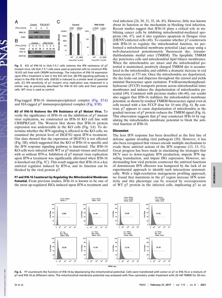
KO of IFI6-16 Restores the IFN Resistance of p7 Mutant Virus. Toverify the significance of IFI6-16 on the inhibition of p7 mutantvirus replication, we constructed an IFI6-16 KO cell line withCRISPR/Cas9. The Western blot shows that IFI6-16 proteinexpression was undetectable in the KO cells (Fig. 5A). To de-termine whether the IFN signaling is affected in the KO cells, weexamined the protein level of ISGF3G upon IFN-α treatment.Our data showed that the expression of ISGF3G is not affected(Fig. 5B), which suggested that the KO of IFI6-16 is specific andthe IFN response signaling pathway is functional. The IFI6-16KO cells were infected with WT or p7 mutant viruses and treatedwith or without IFN-α. Inhibition of p7 mutant virus replicationupon IFN-α treatment was significantly alleviated when IFI6-16is knocked out (Fig. 5C). This result suggests that IFI6-16 is a keyantiviral regulator induced by IFN-α, and its function can beblocked by the viral protein p7.
P7 and IFI6-16 Counteract by Regulating the Mitochondrial MembranePotential. From previous studies, IFI6-16 is known to be one ofthe most up-regulated ISGs induced upon IFN-α treatment and
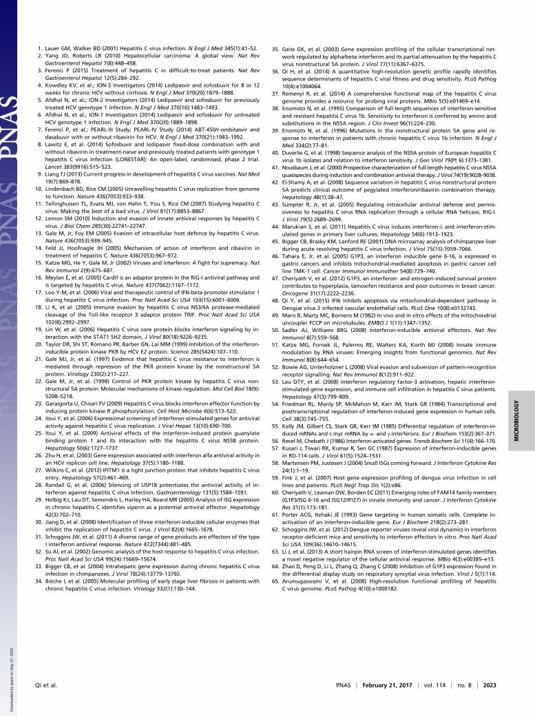
viral infection (24, 30, 31, 35, 44, 45). However, little was knownabout its function or the mechanism in blocking viral infection.Recent studies suggest that IFI6-16 plays a critical role in sta-bilizing cancer cells by inhibiting mitochondrial-mediated apo-ptosis (46, 47), and it also regulates apoptosis in Dengue virus(DENV)-infected cells (48). To examine whether p7 counteractswith IFI6-16 to regulate the mitochondrial function, we per-formed a mitochondrial membrane potential (Δψ) assay using awell-characterized potentiometric fluorescent dye, tetrame-thylrhodamine methyl ester (TMRM). The lipophilic TMRMdye penetrates cells and mitochondrial lipid bilayer membranes.When the mitochondria are intact and the mitochondrial po-tential is maintained, positively charged TMRM dye is accumu-lated in the mitochondria and exhibits a dramatic increase in redfluorescence at 573 nm. Once the mitochondria are depolarized,the dye leaks out and disperses throughout the cytosol and yieldsminimal fluorescence upon excitation. P-triflouromethoxyphenyl-hydrazone (FCCP) transports protons across mitochondrial innermembranes and induces the depolarization of mitochondria po-tential (49). Consistent with previous studies (46–48), our resultsalso suggest that IFI6-16 stabilizes the mitochondrial membranepotential, as shown by residual TMRM fluorescence signal even incells treated with a low FCCP dose for 10 min (Fig. 6). By con-trast, p7 appears to cause depolarization of mitochondria as thegradual increase of p7 protein reduces the TMRM signal (Fig. 6).This observation suggests that p7 may counteract IFI6-16 by reg-ulating the mitochondria membrane potential to block the anti-viral function of IFI6-16.
DiscussionThe host IFN response has been described as the first line ofdefense against invading viral pathogens (50). However, it hasalso been recognized that viruses encode multiple mechanisms toevade these antiviral actions of the IFN response (13, 15, 51).Great progress has been made in elucidating the strategies thatHCV uses to down-regulate IFN production, impede IFN sig-naling transduction, and impair ISG expression. However, un-derstanding how viral proteins counteract the antiviral functionsof downstream IFN effectors was hampered by the lack of anexperimental approach to identify such interactions systemati-cally. With a high-resolution mutagenesis profiling approach,we found that mutations in the p7 region increase IFN sensi-tivity and this phenotype can be rescued by overexpressionof WT p7 protein in the infected cells, implicating p7 as an
Fig. 5. KO of IFI6-16 in Huh-7.5.1 cells restores the IFN resistance of p7mutant virus. (A) Huh-7.5.1 cells were used as parental cells to construct IFI6-16 KO cell lines with CRISPR technology. The induction of IFI6-16 expressionupon IFN-α treatment is lost in the KO cell line. (B) IFN signaling pathway isintact in the IFI6-16 KO cells. ISGF3G is induced to a similar level of parentalcells. (C) IFN sensitivity of p7 mutant virus replication was measured in asimilar way as previously described for IFI6-16 KO cells and their parentalcells. WT virus is used as control.
Fig. 6. P7 counteracts the function of IFI6-16 by depolarizing the mitochondrial potential. Cells were transfected with vector or p7 or IFI6-16 or a mixture ofp7 and IFI6-16 at different ratios. The mitochondrial membrane potential was analyzed with flow cytometry under treatment with 20 nM TMRM for 30 min.
Qi et al. PNAS | February 21, 2017 | vol. 114 | no. 8 | 2021
MICRO
BIOLO
GY
Dow
nloa
ded
by g
uest
on
May
27,
202
0

immune evasion protein of HCV. We found that p7 does notdiminish the ISRE activity induced by IFN-α, which suggeststhat p7 might interfere with the antiviral functions downstreamof IFN effectors. A liver-specific ISG library screen was thenconducted, and it identified genetic and physical interactionbetween p7 and an ISG, IFI6-16.Previous studies suggest that HCV proteins antagonize the
innate immune response through inhibiting the production oftype I IFN and suppression of JAK/STAT signal transductionto avoid the induction of an IFN-mediated antiviral state (52).However, in vivo studies of experimentally infected chimpan-zees or human patient biopsy samples have demonstrated thatHCV infection strongly induces the expression of ISGs in theliver (33, 53). Despite the apparent induction of an antiviralstate, HCV persists in the liver, raising the possibility of thevirus encoding mechanisms to counteract the antiviral func-tions executed by ISGs. All of these studies lead us to hypoth-esize that p7 could inhibit the function of ISG(s) to facilitaterobust viral replication despite the induction of antiviral stageby IFN.IFI6-16, also known as G1P3, was first identified as an ISG
whose mRNA was highly inducible in multiple cell lines upontype I IFN stimulation (54–57). The expression of IFI6-16 isresponsive to viral infections, including vesicular stomatitis virus,HCV, cytomegalovirus, and DENV (58, 59). It can also beinduced by poly(I):poly(C) treatment and other immune regu-lators, namely, lipopolysaccharide and TNF-related apoptosis-induced ligand (58, 60). Despite the early identification of IFI6-16as an ISG and implications that it mediates innate immunity, theantiviral mechanism of the protein still remains obscure andelusive. Early studies attempting to evaluate the antiviral func-tion of IFI6-16 showed that introduction of IFI6-16 in a KO cellline (HT1080_IFI6−/−) does not affect the replication of en-cephalomyocarditis virus, Semliki forest virus, or coccal virus,suggesting that IFI6-16 is not required to control these viralreplications (61). In contrast, IFI6-16 was identified as a negativeregulator that markedly inhibited the replication of yellow fevervirus (31), DENV (62), and West Nile virus (63). The expressionof the gene was also found to suppress respiratory syncytial virusreplication and was down-regulated by the virus (64). The effectof IFI6-16 on HCV replication, however, seems a bit contra-dictory. In the replicon cells harboring HCV subgenomic RNA,overexpression of IFI6-16 inhibited HCV replication, and ex-pression of viral proteins, whereas knockdown of IFI6-16 in-creased the level of RNA replication. Interestingly, IFI6-16 didnot activate the IFN activation pathway, suggesting that itfunctions directly against viral replication without going throughthe IFN activity, which may amplify antiviral actions (24, 25). Incontrast, a comprehensive ISG cDNA screen using an infectionsystem demonstrated that IFI6-16 shows moderate or no signif-icant suppression on HCV replication in either Huh-7 or Huh-7.5 cell lines (31). HCV persists in chimpanzee livers regardlessof the up-regulation of IFI6-16, suggesting that either IFI6-16does not regulate viral replication (64) or the virus has developedstrategies to overcome the antiviral functions of IFI6-16, asproposed in this study.Our data suggest that p7 functions as an immune evasion
protein, most likely by counteracting the antiviral function ofIFI6-16. On one hand, IFI6-16 is one of the earliest ISGs in-duced upon IFN treatment according to the previous studies.Studies showed that overexpression of IFI6-16 can delay theapoptosis of the cells through stabilizing the mitochondria, andtherefore may extend the production of IFN in the infectedcells, which may sustain and extend the antiviral effects of theIFN system (60). On the other hand, HCV replication has beenknown to induce mitochondrial dysfunction and mitophagy.This observation is very likely attributable to the ion channelfunction of p7 because the mitochondrial dysfunction can be
blocked by amantadine, an ion channel inhibitor that interactswith p7. Therefore, a plausible explanation will be that p7 maybreak the balance that IFI6-16 confers on mitochondria throughdepolarizing mitochondria and induces mitochondrial dys-function to interfere with the antiviral state of the infected cell.Our data explain the discrepant observations that expression ofIFI6-16 protein presents a substantial level of antiviral effect inthe HCV replicon system, but not in the infectious system (25,31) (Fig. S8). This interaction could not have been identifiedwithout identification of the mutant viruses through the genome-wide mutagenesis study.To determine the mechanism of p7 counteracting IFN sig-
naling, we took two independent approaches to determine thecellular protein(s) functionally and physically interacting withp7. In the ISG screen, we have also noticed that there areseveral ISGs that inhibit p7 mutant virus replication over WT,but do not form a protein complex with p7 protein. Those ISGsmay display an indirect effect on p7 mutant virus replication.We noticed that some of these ISGs are involved in the IFNsignaling pathway, which may amplify the effect of IFN orIFI6-16 on HCV replication when p7 is mutated. For ex-ample, IFIT5 is an IFN-induced RNA-binding protein thatrecognizes single-stranded RNA and initiates IFN productionupon recognizing single-stranded 5-triphosphate RNAs, whichfurther reinforces the antiviral effect of the system.Although we have identified IFI6-16 as a direct counteracting
protein of p7, it does not rule out possibilities that other ISGsalso have an impact on p7 mutant virus replication. It will beinteresting to carry out a counterscreen as an orthogonal ap-proach to eliminate errors from the high-throughput screen assayand to characterize the antiviral function of the ISG(s).Because the HCV genome does not tolerate the 15-nt in-
sertion very well, which leaves a large portion of the virus ge-nome unexplored in the IFN screen, we anticipate that we wouldidentify more immune evasion functions on the virus genome ata much higher resolution, and possibly novel antiviral ISGs witha complex single-amino acid mutant library.Collectively, these multilayered systematic approaches offer
comprehensive insights into HCV and host interactions, whichwill provide a basis for understanding innate immune evasionmechanisms. In addition, systematic screening of a viral genometo identify immune evasion functions, including anti-IFN func-tions, will enable the construction of recombinant viruses withdesired biological properties. Multiple immune evasion functionscan be knocked out to generate recombinant viruses that arereplication-competent in immune-deficient hosts, such as IFN-deficient cells, but defective in healthy hosts. It can be expectedthat they will generate strong innate and adaptive immune re-sponses and provide protection against WT virus challenge. Thus,our work also presents an approach for vaccine developmentbased on rational design, enabled by systematic understanding ofthe viral genome.
Materials and MethodsThe mutant plasmid library was linearized and transcribed into RNA in vitro,followed by electroporation into Huh-7.5.1 cells to reconstitute the mutantvirus library. The virus library underwent two rounds of selection in Huh-7.5.1cells. A detailed description of reagents and protocols used in this study canbe found in SI Materials and Methods.
ACKNOWLEDGMENTS. We thank Dr. Francis Chisari (The Scripps ResearchInstitute) for kindly providing the Huh-7.5.1 cell line. We thank Dr. HidetoshiTahara (Hiroshima University, Japan) for the rabbit polyclonal anti-body against IFI6-16. We also thank Yong-Hoon Kim and Dr. AsimDasgupta for their comments and suggestions on the manuscript. Thiswork was supported by the following grants: National Natural Sci-ence Foundation of China Grant 81172314 and NIH Grants AI078133,P30CA016042, and P30AI028697. G.B. was supported, in part, by anInterdisciplinary Training in Virology and Gene Therapy Training Grant(NIH Grant T32 AI 060567).
2022 | www.pnas.org/cgi/doi/10.1073/pnas.1614623114 Qi et al.
Dow
nloa
ded
by g
uest
on
May
27,
202
0
1. Lauer GM, Walker BD (2001) Hepatitis C virus infection. N Engl J Med 345(1):41–52.2. Yang JD, Roberts LR (2010) Hepatocellular carcinoma: A global view. Nat Rev
Gastroenterol Hepatol 7(8):448–458.3. Ferenci P (2015) Treatment of hepatitis C in difficult-to-treat patients. Nat Rev
Gastroenterol Hepatol 12(5):284–292.4. Kowdley KV, et al.; ION-3 Investigators (2014) Ledipasvir and sofosbuvir for 8 or 12
weeks for chronic HCV without cirrhosis. N Engl J Med 370(20):1879–1888.5. Afdhal N, et al.; ION-2 Investigators (2014) Ledipasvir and sofosbuvir for previously
treated HCV genotype 1 infection. N Engl J Med 370(16):1483–1493.6. Afdhal N, et al.; ION-1 Investigators (2014) Ledipasvir and sofosbuvir for untreated
HCV genotype 1 infection. N Engl J Med 370(20):1889–1898.7. Ferenci P, et al.; PEARL-III Study; PEARL-IV Study (2014) ABT-450/r-ombitasvir and
dasabuvir with or without ribavirin for HCV. N Engl J Med 370(21):1983–1992.8. Lawitz E, et al. (2014) Sofosbuvir and ledipasvir fixed-dose combination with and
without ribavirin in treatment-naive and previously treated patients with genotype 1hepatitis C virus infection (LONESTAR): An open-label, randomised, phase 2 trial.Lancet 383(9916):515–523.
9. Liang TJ (2013) Current progress in development of hepatitis C virus vaccines. Nat Med19(7):869–878.
10. Lindenbach BD, Rice CM (2005) Unravelling hepatitis C virus replication from genometo function. Nature 436(7053):933–938.
11. Tellinghuisen TL, Evans MJ, von Hahn T, You S, Rice CM (2007) Studying hepatitis Cvirus: Making the best of a bad virus. J Virol 81(17):8853–8867.
12. Lemon SM (2010) Induction and evasion of innate antiviral responses by hepatitis Cvirus. J Biol Chem 285(30):22741–22747.
13. Gale M, Jr, Foy EM (2005) Evasion of intracellular host defence by hepatitis C virus.Nature 436(7053):939–945.
14. Feld JJ, Hoofnagle JH (2005) Mechanism of action of interferon and ribavirin intreatment of hepatitis C. Nature 436(7053):967–972.
15. Katze MG, He Y, Gale M, Jr (2002) Viruses and interferon: A fight for supremacy. NatRev Immunol 2(9):675–687.
16. Meylan E, et al. (2005) Cardif is an adaptor protein in the RIG-I antiviral pathway andis targeted by hepatitis C virus. Nature 437(7062):1167–1172.
17. Loo Y-M, et al. (2006) Viral and therapeutic control of IFN-beta promoter stimulator 1during hepatitis C virus infection. Proc Natl Acad Sci USA 103(15):6001–6006.
18. Li K, et al. (2005) Immune evasion by hepatitis C virus NS3/4A protease-mediatedcleavage of the Toll-like receptor 3 adaptor protein TRIF. Proc Natl Acad Sci USA102(8):2992–2997.
19. Lin W, et al. (2006) Hepatitis C virus core protein blocks interferon signaling by in-teraction with the STAT1 SH2 domain. J Virol 80(18):9226–9235.
20. Taylor DR, Shi ST, Romano PR, Barber GN, Lai MM (1999) Inhibition of the interferon-inducible protein kinase PKR by HCV E2 protein. Science 285(5424):107–110.
21. Gale MJ, Jr, et al. (1997) Evidence that hepatitis C virus resistance to interferon ismediated through repression of the PKR protein kinase by the nonstructural 5Aprotein. Virology 230(2):217–227.
22. Gale M, Jr, et al. (1998) Control of PKR protein kinase by hepatitis C virus non-structural 5A protein: Molecular mechanisms of kinase regulation. Mol Cell Biol 18(9):5208–5218.
23. Garaigorta U, Chisari FV (2009) Hepatitis C virus blocks interferon effector function byinducing protein kinase R phosphorylation. Cell Host Microbe 6(6):513–522.
24. Itsui Y, et al. (2006) Expressional screening of interferon-stimulated genes for antiviralactivity against hepatitis C virus replication. J Viral Hepat 13(10):690–700.
25. Itsui Y, et al. (2009) Antiviral effects of the interferon-induced protein guanylatebinding protein 1 and its interaction with the hepatitis C virus NS5B protein.Hepatology 50(6):1727–1737.
26. Zhu H, et al. (2003) Gene expression associated with interferon alfa antiviral activity inan HCV replicon cell line. Hepatology 37(5):1180–1188.
27. Wilkins C, et al. (2012) IFITM1 is a tight junction protein that inhibits hepatitis C virusentry. Hepatology 57(2):461–469.
28. Randall G, et al. (2006) Silencing of USP18 potentiates the antiviral activity of in-terferon against hepatitis C virus infection. Gastroenterology 131(5):1584–1591.
29. Helbig KJ, Lau DT, Semendric L, Harley HA, Beard MR (2005) Analysis of ISG expressionin chronic hepatitis C identifies viperin as a potential antiviral effector. Hepatology42(3):702–710.
30. Jiang D, et al. (2008) Identification of three interferon-inducible cellular enzymes thatinhibit the replication of hepatitis C virus. J Virol 82(4):1665–1678.
31. Schoggins JW, et al. (2011) A diverse range of gene products are effectors of the typeI interferon antiviral response. Nature 472(7344):481–485.
32. Su AI, et al. (2002) Genomic analysis of the host response to hepatitis C virus infection.Proc Natl Acad Sci USA 99(24):15669–15674.
33. Bigger CB, et al. (2004) Intrahepatic gene expression during chronic hepatitis C virusinfection in chimpanzees. J Virol 78(24):13779–13792.
34. Bièche I, et al. (2005) Molecular profiling of early stage liver fibrosis in patients withchronic hepatitis C virus infection. Virology 332(1):130–144.
35. Geiss GK, et al. (2003) Gene expression profiling of the cellular transcriptional net-work regulated by alpha/beta interferon and its partial attenuation by the hepatitis Cvirus nonstructural 5A protein. J Virol 77(11):6367–6375.
36. Qi H, et al. (2014) A quantitative high-resolution genetic profile rapidly identifiessequence determinants of hepatitis C viral fitness and drug sensitivity. PLoS Pathog10(4):e1004064.
37. Remenyi R, et al. (2014) A comprehensive functional map of the hepatitis C virusgenome provides a resource for probing viral proteins. MBio 5(5):e01469–e14.
38. Enomoto N, et al. (1995) Comparison of full-length sequences of interferon-sensitiveand resistant hepatitis C virus 1b. Sensitivity to interferon is conferred by amino acidsubstitutions in the NS5A region. J Clin Invest 96(1):224–230.
39. Enomoto N, et al. (1996) Mutations in the nonstructural protein 5A gene and re-sponse to interferon in patients with chronic hepatitis C virus 1b infection. N Engl JMed 334(2):77–81.
40. Duverlie G, et al. (1998) Sequence analysis of the NS5A protein of European hepatitis Cvirus 1b isolates and relation to interferon sensitivity. J Gen Virol 79(Pt 6):1373–1381.
41. Nousbaum J, et al. (2000) Prospective characterization of full-length hepatitis C virus NS5Aquasispecies during induction and combination antiviral therapy. J Virol 74(19):9028–9038.
42. El-Shamy A, et al. (2008) Sequence variation in hepatitis C virus nonstructural protein5A predicts clinical outcome of pegylated interferon/ribavirin combination therapy.Hepatology 48(1):38–47.
43. Sumpter R, Jr, et al. (2005) Regulating intracellular antiviral defense and permis-siveness to hepatitis C virus RNA replication through a cellular RNA helicase, RIG-I.J Virol 79(5):2689–2699.
44. Marukian S, et al. (2011) Hepatitis C virus induces interferon-λ and interferon-stim-ulated genes in primary liver cultures. Hepatology 54(6):1913–1923.
45. Bigger CB, Brasky KM, Lanford RE (2001) DNA microarray analysis of chimpanzee liverduring acute resolving hepatitis C virus infection. J Virol 75(15):7059–7066.
46. Tahara E, Jr, et al. (2005) G1P3, an interferon inducible gene 6-16, is expressed ingastric cancers and inhibits mitochondrial-mediated apoptosis in gastric cancer cellline TMK-1 cell. Cancer Immunol Immunother 54(8):729–740.
47. Cheriyath V, et al. (2012) G1P3, an interferon- and estrogen-induced survival proteincontributes to hyperplasia, tamoxifen resistance and poor outcomes in breast cancer.Oncogene 31(17):2222–2236.
48. Qi Y, et al. (2015) IFI6 inhibits apoptosis via mitochondrial-dependent pathway inDengue virus 2 infected vascular endothelial cells. PLoS One 10(8):e0132743.
49. Maro B, Marty MC, Bornens M (1982) In vivo and in vitro effects of the mitochondrialuncoupler FCCP on microtubules. EMBO J 1(11):1347–1352.
50. Sadler AJ, Williams BRG (2008) Interferon-inducible antiviral effectors. Nat RevImmunol 8(7):559–568.
51. Katze MG, Fornek JL, Palermo RE, Walters KA, Korth MJ (2008) Innate immunemodulation by RNA viruses: Emerging insights from functional genomics. Nat RevImmunol 8(8):644–654.
52. Bowie AG, Unterholzner L (2008) Viral evasion and subversion of pattern-recognitionreceptor signalling. Nat Rev Immunol 8(12):911–922.
53. Lau DTY, et al. (2008) Interferon regulatory factor-3 activation, hepatic interferon-stimulated gene expression, and immune cell infiltration in hepatitis C virus patients.Hepatology 47(3):799–809.
54. Friedman RL, Manly SP, McMahon M, Kerr IM, Stark GR (1984) Transcriptional andposttranscriptional regulation of interferon-induced gene expression in human cells.Cell 38(3):745–755.
55. Kelly JM, Gilbert CS, Stark GR, Kerr IM (1985) Differential regulation of interferon-in-duced mRNAs and c-myc mRNA by α- and γ-interferons. Eur J Biochem 153(2):367–371.
56. Revel M, Chebath J (1986) Interferon-activated genes. Trends Biochem Sci 11(4):166–170.57. Kusari J, Tiwari RK, Kumar R, Sen GC (1987) Expression of interferon-inducible genes
in RD-114 cells. J Virol 61(5):1524–1531.58. Martensen PM, Justesen J (2004) Small ISGs coming forward. J Interferon Cytokine Res
24(1):1–19.59. Fink J, et al. (2007) Host gene expression profiling of dengue virus infection in cell
lines and patients. PLoS Negl Trop Dis 1(2):e86.60. Cheriyath V, Leaman DW, Borden EC (2011) Emerging roles of FAM14 family members
(G1P3/ISG 6-16 and ISG12/IFI27) in innate immunity and cancer. J Interferon CytokineRes 31(1):173–181.
61. Porter ACG, Itzhaki JE (1993) Gene targeting in human somatic cells. Complete in-activation of an interferon-inducible gene. Eur J Biochem 218(2):273–281.
62. Schoggins JW, et al. (2012) Dengue reporter viruses reveal viral dynamics in interferonreceptor-deficient mice and sensitivity to interferon effectors in vitro. Proc Natl AcadSci USA 109(36):14610–14615.
63. Li J, et al. (2013) A short hairpin RNA screen of interferon-stimulated genes identifiesa novel negative regulator of the cellular antiviral response. MBio 4(3):e00385–e13.
64. Zhao D, Peng D, Li L, Zhang Q, Zhang C (2008) Inhibition of G1P3 expression found inthe differential display study on respiratory syncytial virus infection. Virol J 5(1):114.
65. Arumugaswami V, et al. (2008) High-resolution functional profiling of hepatitisC virus genome. PLoS Pathog 4(10):e1000182.
Qi et al. PNAS | February 21, 2017 | vol. 114 | no. 8 | 2023
MICRO
BIOLO
GY
Dow
nloa
ded
by g
uest
on
May
27,
202
0


