Synthetic mRNA capping - Journals · Synthetic mRNA capping Fabian€Muttach‡1,...
14
2819 Synthetic mRNA capping Fabian Muttach ‡1 , Nils Muthmann ‡1 and Andrea Rentmeister *1,2 Review Open Access Address: 1 University of Münster, Department of Chemistry, Institute of Biochemistry, Wilhelm-Klemm-Str. 2, 48149 Münster, Germany and 2 Cells-in-Motion Cluster of Excellence (EXC1003-CiM), University of Münster, Germany Email: Andrea Rentmeister * - [email protected] * Corresponding author ‡ Equal contributors Keywords: cap analogue; cap synthesis; click chemistry; enzymatic capping; methyltransferase; RNA Beilstein J. Org. Chem. 2017, 13, 2819–2832. doi:10.3762/bjoc.13.274 Received: 14 September 2017 Accepted: 04 December 2017 Published: 20 December 2017 This article is part of the Thematic Series "Nucleic acid chemistry II". Guest Editor: H.-A. Wagenknecht © 2017 Muttach et al.; licensee Beilstein-Institut. License and terms: see end of document. Abstract Eukaryotic mRNA with its 5′-cap is of central importance for the cell. Many studies involving mRNA require reliable preparation and modification of 5′-capped RNAs. Depending on the length of the desired capped RNA, chemical or enzymatic preparation – or a combination of both – can be advantageous. We review state-of-the art methods and give directions for choosing the appropriate approach. We also discuss the preparation and properties of mRNAs with non-natural caps providing novel features such as im- proved stability or enhanced translational efficiency. 2819 Introduction The 5′-cap is a hallmark of eukaryotic mRNA and involved in numerous interactions required for cellular functions. Chemical- ly, the 5′ -cap consists of an inverted 7-methylguanosine connected to the rest of the eukaryotic mRNA via a 5′ –5′ triphosphate bridge. This so-called cap0 serves as quality control for correct mRNA processing and contributes to stabi- lization of eukaryotic mRNA [1,2], splicing [3,4], nuclear export [5], initiation of translation [6,7] and mRNA decay [8]. The most important direct interaction partners of the 5′-cap are the cap binding complex (CBC) [9,10] in the nucleus required for nuclear export and the eukaryotic translation initiation factor 4E (eIF4E) [11] in the cytoplasm which is indispensable for cap-dependent translation. Additionally, capped RNA serves as a marker for the innate immune system to distinguish triphos- phorylated viral RNAs from cellular RNAs [12]. The antiviral response is among others mediated by the cytosolic receptor RIG-I which is activated by short single and double-stranded triphosphorylated RNAs and MDA-5. MDA-5 recognizes long triphosphorylated RNAs and RNAs lacking the 2′-OH methyla- tion at the first nucleotide (cap1), a modification which is com- monly observed in eukaryotes [13-15]. Besides cap0 and cap1, cap structures with further modifica- tions exist. Additional methyl groups are often found at the second nucleotide (cap2) while in trypanosomes up to four methylated nucleotides are observed (termed cap4) [16,17]. Owing to the importance of different cap structures for recogni- tion processes in the cell, it becomes clear that an uncapped transcript does not adequately represent a eukaryotic mRNA
Transcript of Synthetic mRNA capping - Journals · Synthetic mRNA capping Fabian€Muttach‡1,...
2819
Synthetic mRNA cappingFabian Muttach‡1, Nils Muthmann‡1 and Andrea Rentmeister*1,2
Review Open Access
Address:1University of Münster, Department of Chemistry, Institute ofBiochemistry, Wilhelm-Klemm-Str. 2, 48149 Münster, Germany and2Cells-in-Motion Cluster of Excellence (EXC1003-CiM), University ofMünster, Germany
Email:Andrea Rentmeister* - [email protected]
* Corresponding author ‡ Equal contributors
Keywords:cap analogue; cap synthesis; click chemistry; enzymatic capping;methyltransferase; RNA
Beilstein J. Org. Chem. 2017, 13, 2819–2832.doi:10.3762/bjoc.13.274
Received: 14 September 2017Accepted: 04 December 2017Published: 20 December 2017
This article is part of the Thematic Series "Nucleic acid chemistry II".
Guest Editor: H.-A. Wagenknecht
© 2017 Muttach et al.; licensee Beilstein-Institut.License and terms: see end of document.
AbstractEukaryotic mRNA with its 5′-cap is of central importance for the cell. Many studies involving mRNA require reliable preparation
and modification of 5′-capped RNAs. Depending on the length of the desired capped RNA, chemical or enzymatic preparation – or
a combination of both – can be advantageous. We review state-of-the art methods and give directions for choosing the appropriate
approach. We also discuss the preparation and properties of mRNAs with non-natural caps providing novel features such as im-
proved stability or enhanced translational efficiency.
2819
IntroductionThe 5′-cap is a hallmark of eukaryotic mRNA and involved in
numerous interactions required for cellular functions. Chemical-
ly, the 5′-cap consists of an inverted 7-methylguanosine
connected to the rest of the eukaryotic mRNA via a 5′–5′
triphosphate bridge. This so-called cap0 serves as quality
control for correct mRNA processing and contributes to stabi-
lization of eukaryotic mRNA [1,2], splicing [3,4], nuclear
export [5], initiation of translation [6,7] and mRNA decay [8].
The most important direct interaction partners of the 5′-cap are
the cap binding complex (CBC) [9,10] in the nucleus required
for nuclear export and the eukaryotic translation initiation factor
4E (eIF4E) [11] in the cytoplasm which is indispensable for
cap-dependent translation. Additionally, capped RNA serves as
a marker for the innate immune system to distinguish triphos-
phorylated viral RNAs from cellular RNAs [12]. The antiviral
response is among others mediated by the cytosolic receptor
RIG-I which is activated by short single and double-stranded
triphosphorylated RNAs and MDA-5. MDA-5 recognizes long
triphosphorylated RNAs and RNAs lacking the 2′-OH methyla-
tion at the first nucleotide (cap1), a modification which is com-
monly observed in eukaryotes [13-15].
Besides cap0 and cap1, cap structures with further modifica-
tions exist. Additional methyl groups are often found at the
second nucleotide (cap2) while in trypanosomes up to four
methylated nucleotides are observed (termed cap4) [16,17].
Owing to the importance of different cap structures for recogni-
tion processes in the cell, it becomes clear that an uncapped
transcript does not adequately represent a eukaryotic mRNA

Beilstein J. Org. Chem. 2017, 13, 2819–2832.
2820
Figure 1: Schematic representation of enzymatic 5′-cap formation in eukaryotic mRNA. The 5′-triphosphate-end of the pre-mRNA is hydrolyzed to adiphosphate by an RNA 5′-triphosphatase. A guanylyltransferase transfers GMP onto the β-phosphate of the 5′-diphosphate to form a 5′ to 5′-triphos-phate linkage. The guanine is methylated at the N7-position by an RNA (guanine-N7)methyltransferase, yielding the cap0 structure. Further methyla-tion at the 2′-OH position of the first nucleotide results in formation of the cap1 structure.
and that preparation of correctly capped RNAs is essential to
assess the function of mRNAs in the cellular context. Further-
more, altering the cap structure bears potential to increase
mRNA stability and translational efficiency – two properties
which may provide the key to therapeutic applications of
mRNA in the near future [18-20]. Finally, investigations of
structure and mechanism of 5′-cap/protein interactions are still
hampered by the difficulty of producing large quantities of
homogenously capped RNA.
In this review article, we present different synthetic routes to
5′-capped mRNAs based on enzymatic, chemical or chemo-
enzymatic methods. We will point out the difficulties and limi-
tations of the different strategies and – if available – will show
ways to circumvent them. This review focuses strictly on
mRNA cap analogues (and some non-natural modifications);
for preparation of other capped biomolecules such as capped
siRNAs [21], peptidyl capped oligonucleotides [22], NAD-
capped RNAs [23,24], 3'-dephospho-CoA linked RNA [25] or
methylphosphate capping [26,27] we refer to the indicated arti-
cles.
ReviewEnzymatic preparation of capped mRNAEnzymatic preparation of capped mRNA is based on in vitro
transcription (IVT) of a DNA template. While RNA synthe-
sized via solid-phase synthesis is limited in its maximum length,
RNAs with a length of several thousand nucleotides can easily
be prepared through IVT. On the other hand, enzymatically pro-
duced RNA is often inhomogeneous in length and for short
RNAs the yields obtained after purification may be low. This
impedes the enzymatic production of short RNAs of a defined
length for applications requiring defined and homogeneous
RNA species. IVT produces uncapped, 5′-triphosphorylated
RNA but there are two strategies to obtain mRNA with a cap,
which will be discussed in detail in the following chapters.
Post-transcriptional cappingIn post-transcriptional capping, the RNA from IVT is subjected
to a dedicated enzymatic capping reaction. The enzymes used in
vitro originate from capping apparatuses of different eukaryotic
organisms or DNA viruses and can be produced recombinantly
in E. coli [28,29]. Enzymatic formation of cap0 comprises three
consecutive reactions targeted to nascent 5′-triphosphorylated
pre-mRNAs (Figure 1). First, a 5′-triphosphatase (TPase)
hydrolyzes the γ-phosphate of pre-mRNA. Next, the β-phos-
phate of the resulting 5′-diphosphate end is coupled to GMP to
form 5′–5′-linked Gppp-RNA. The responsible guanylyltrans-
ferase uses GTP as substrate and forms a covalent enzyme-
(lysyl-N)-GMP intermediate, reminiscent of DNA ligase-AMP
intermediates [30,31]. Finally, the cap structure is methylated at
the N7-position by an RNA(guanine-N7)methyltransferase
using S-adenoysl-L-methionine (AdoMet) as a cosubstrate [31].
In nature, these capping enzymes act co-transcriptionally once
the transcript has reached a length of 20–30 nucleotides [32],
which is enabled by their recruitment to the C-terminal domain
of the RNA polymerase II [33]. In higher eukaryotes, cap1 and
cap2 structures are generated by subsequent methylation of the
2′-hydroxy group of the adjacent second and third ribose, re-
spectively [34].
These capping enzymes – e.g., from Vaccinia virus – can be
harnessed for the production of capped RNA in vitro by adding
them and their respective cosubstrates to the IVT reaction, as
described by pioneering work of the Rosenberg group [35]. To
date, the capping enzymes from the Vaccinia virus are commer-
cially available and most widely used for post-transcriptional in
vitro capping. They consist of two viral proteins D1 and D12.
The triphosphatase and guanylyltransferase activity are located
in the N-terminal half and the methyltransferase in the C-termi-
nal half of the large D1 protein, whereas the small D12 protein
has no catalytic activity but activates D1 [36-38].

Beilstein J. Org. Chem. 2017, 13, 2819–2832.
2821
Originally, the RNA capping with the Vaccinia capping appa-
ratus was reported to be inefficient [35,37,39,40]. To date, the
enzyme is commercially available, however, the amount of en-
zyme needed for the production of capped RNA in µmol scale
prevents its general applicability [41]. For the application of the
Vaccinia capping enzyme in the production of large-scale
5′-capped RNA, Fuchs et al. have recently reported an expres-
sion and purification protocol for the Vaccinia enzyme, allow-
ing for capping in large quantities in a more cost-efficient man-
ner compared to commercially available capping methods [41].
Post-transcriptional capping to obtain mRNA with a cap1 struc-
ture can be achieved using the Vaccinia mRNA cap 2′-O-
methyltransferase which is commercially available [42,43]. Ad-
ditionally, authentic mRNAs can be produced with the commer-
cially available mScriptTM system which combines a T7 RNA
polymerase, a trifunctional capping enzyme, a 2′-O-methyl-
transferase and a poly(A) polymerase. Albeit expensive, this
system allows for production of mRNAs in one pot with
claimed quantitative yields and high translational activity.
Post-transcriptional preparation of non-natural cap analogues
was achieved by capping enzymes with relaxed substrate speci-
ficity. For example, ribavirin is used as a substrate by the
Vaccinia capping enzyme and can be transferred onto the
diphosphate end of an RNA transcript to form a ribavirin-pppN
structure. RNA transcripts blocked with ribavirin showed little
translational efficiency, which might explain the antiviral activi-
ty of ribavirin [44]. Enzymatic formation of cap analogues from
GTP analogues was achieved with the capping enzyme of the
model organism Paramecium bursaria Chlorella virus-1
(PBCV-1) by Bisaillon and co-workers [45]. Out of 22
nucleotide analogues tested in this study, 13 were found to form
a covalent complex with the PBCV-1 guanylyltransferase
(GTase) while 11 were actually transferred onto a 5′-diphos-
phate RNA (Figure 2). Moreover, RNAs capped with those
nucleotide analogues were translated even in the absence of the
N7-methyl group when alternative modifications enabled
binding to eIF4E [45].
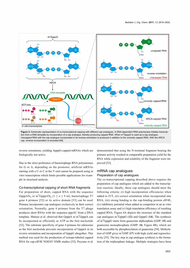
Co-transcriptional cappingIn co-transcriptional capping, cap analogues are added directly
to the IVT. Their incorporation at the 5′-end by RNA poly-
merases with relaxed substrate specificity (e.g., T3, T7 or SP6
RNA polymerases) directly yields the respective 5′-capped
mRNA (Figure 3). Internal incorporation of cap analogues
during IVT does not occur, because cap analogues lack a free
5′-triphosphate.
The most commonly used cap analogue is m7GpppG but several
modified or alternative cap analogues are also accepted by RNA
Figure 2: Nucleotide analogues 1–11 were converted by Parameciumbursaria Chlorella virus-1 capping enzyme instead of GTP to generateRNAs with the respective caps [45].
polymerases. Therefore, this route can be used to install non-
natural dinucleotides at the 5′-end that are accessible for a
further chemical reaction [46].
One often overlooked limitation of co-transcriptional capping is
that not all mRNA obtained from IVT is capped, simply
because the cap analogue competes with GTP as initiator
nucleotide. Importantly, the ratio of capped/uncapped mRNA is
usually not visible on a gel. This issue can be mitigated by
lowering the GTP concentration or by digesting uncapped (i.e.,
triphosphorylated) RNA with a 5′-polyphosphatase which
produces monophosphorylated RNA followed by 5′-phosphate-
dependent exonuclease digestion.
Another problem encountered with m7GpppG as initiator is
elongation into the “wrong” direction, namely at the 3′-OH of
m7G, yielding mRNA with the cap in reverse orientation
(Figure 3). Up to one half of the mRNA can contain the cap in
its reverse orientation and will not be translated [47]. This prob-
lem was solved by developing anti-reverse cap analogues
(ARCA) that are methylated or deoxygenated at the 3′-OH of
the N7-methylguanosine r ibose (m27 ,3 ′ -OGpppG or
m7,3′-dGpppG). This prevents elongation at the “wrong” 3′-OH
and hence ARCA caps are exclusively incorporated in the
correct orientation [48,49]. Interestingly, modifications at the
2′-position of m7G also prevented reverse incorporation of the
cap analogue [50]. The problem of orientation is circumvented
when GpppA cap analogues are used in combination with the
T7 class II promotor phi2.5 which allows initiating RNA syn-
thesis with ATP. Hence, GpppA- or m7GpppA-capped RNAs
can be produced [51]. When the common GTP-initiating T7
class III promoter phi6.5 is used, GpppA is incorporated in its
Beilstein J. Org. Chem. 2017, 13, 2819–2832.
2822
Figure 3: Schematic representation of co-transcriptional capping with different cap analogues. A DNA-dependent RNA polymerase initiates transcrip-tion from a DNA template by incorporation of a cap analogue, thereby producing capped RNA. When m7GpppG is used as a cap analogue,miscapped RNA with the cap analogue incorporated in its reverse orientation is produced in addition to the correctly capped RNA. With the ARCAcap, reverse incorporation is excluded [48].
reverse orientation, yielding ApppG-capped mRNAs which are
biologically not active.
Due to the strict preference of bacteriophage RNA polymerases
for G or A, depending on the promotor, artificial mRNAs
starting with a U or C at the 5′-end cannot be prepared using in
vitro transcription which limits possible applications for exam-
ple in structural analysis.
Co-transcriptional capping of short RNA fragmentsFor preparation of short, capped RNA with the sequence
GpppANn or m7GpppANn (1 ≤ n ≤ 9 nt), bacteriophage T7
gene 4 primase [52] or its active domain [53] can be used.
Primase incorporates cap analogues exclusively in their correct
orientation. Normally, gene 4 primase from the T7 phage
produces short RNAs with the sequence pppAC from a DNA
template. Matsuo et al. observed that GpppA or m7GpppA can
be incorporated as efficiently as ATP as the first nucleotide
[52]. The substrate specificity of gene 4 primase for adenosine
as the first nucleotide prevents incorporation of GpppA in its
reverse orientation and incorporation of GpppG altogether. This
method was used for the production of isotope-labeled capped
RNA for cap-eIF4E NOESY-NMR studies [52]. Peyrane et al.
demonstrated that using the N-terminal fragment bearing the
primase activity resulted in comparable preparation yield for the
RNA while expression and solubility of the fragment were im-
proved [53].
mRNA cap analoguesPreparation of cap analoguesThe co-transcriptional capping described above requires the
preparation of cap analogues which are added to the transcrip-
tion reaction. Ideally, these cap analogues should meet the
following criteria: (i) high incorporation efficiencies when
added to IVT, (ii) correct orientation when incorporated into
RNA, (iii) strong binding to the cap-binding protein eIF4E,
(iv) inhibitory potential when added as competitor in an in vitro
translation assay and (v) high translation efficiency of resulting
capped RNA. Figure 4A depicts the structure of the standard
cap analogues m7GpppG (12) and GpppG (14). The synthesis
of m7GpppG starts from guanosine diphosphate (GDP, 15) and
guanosine monophosphate (GMP, 16, Figure 4B), which are
both accessible by phosphorylation of guanosine [54]. Methyla-
tion of GDP gives m7GDP (17) with high yield and regioselec-
tivity [55]. The key step in cap analogue synthesis is the forma-
tion of the triphosphate linkage. Multiple strategies have been

Beilstein J. Org. Chem. 2017, 13, 2819–2832.
2823
Figure 4: (A) Structures of commercially available mRNA cap analogues. (B) Synthetic route to cap analogues as exemplified by the synthesis of them7GpppG cap analogue. 2,2′-DTDP: 2,2′-dithiodipyridine.
reported which mostly rely on the same principle: One of the
two nucleotides (typically the monophosphorylated nucleotide)
is equipped with a good leaving group while the other one acts
as a nucleophile. Different leaving groups have been exploited
for the synthesis of cap analogues, comprising phenylthio [56],
5-chloro-8-quinolyl [57], morpholidate [48] and imidazolide
moieties [58,59]. Imidazole activation in DMF with ZnCl2 was
first reported by Sekine et al. [60] and is the most often used
method for the formation of triphosphates. P-Imidazoles are
known to react with numerous nucleophiles such as nucleoside
mono-, -di- or -triphosphates and are typically reacted in an-
hydrous DMF in the presence of zinc chloride. The GMP imida-
zolide (18) is reacted with m7GDP (17) in the presence of
ZnCl2 as catalyst to yield m7GpppG (12) [49].
In the past years, variations of this general synthetic strategy
were used to obtain numerous cap analogues. Among the most
interesting modifications is the above-mentioned anti-reverse
cap analogue (ARCA) and GpppG which are commercially
available. In addition, cap analogues with improved properties –
namely binding to eIF4E, translational efficiency, and nuclease
resistance – have been developed. Furthermore, cap analogues
have therapeutic potential as demonstrated by a number of cap-
derived translation inhibitors [61-63].
Applications of novel cap analoguesThe search for novel – non-natural or modified – caps with im-
proved properties has already yielded promising results. RNA
capped with a locked nucleic acid (LNA)-modified dinu-
cleotide cap analogue was translated 3-times more efficiently
than regular m7G-capped RNA [64]. Additionally, RNA capped
with the LNA cap analogue was found to be ≈1.6-fold more
stable in a luciferase assay in cultured cells than the respective
RNA with the standard cap. However, in this study it was not

Beilstein J. Org. Chem. 2017, 13, 2819–2832.
2824
assessed how this LNA cap analogue performs in comparison to
the established ARCA cap. Interestingly, a 3′-O-propargyl con-
taining m7GpppG cap analogue also showed more than 3-fold
higher translational efficiency compared to the standard cap.
The cap analogue is exclusively incorporated in the correct
(forward) orientation and molecular modelling studies pointed
to a stronger binding of the propargyl-modified cap to eIF4E
compared to the standard cap [65].
With regard to translational activity, several dinucleotide cap
analogues containing a tetraphosphate were shown to be superi-
or to the regular triphosphate in in vitro studies [66]. RNAs
capped with m7Gp4m7G were translated with more than 3-fold
higher efficiency. Interestingly, also benzyl-modified tetraphos-
phate cap analogues showed more than 2-fold higher transla-
tion in in vitro translation experiments. In a further step,
tetraphosphates with methylene(bisphosphonate) moieties were
prepared which improved binding to eIF4E and in some cases
conferred enzymatic resistance against DcpS degradation [67].
N2-Triazole-containing monophosphate cap analogues were
shown to be as efficient as m7GpppG in translation inhibition
assays [68].
Further modifications can be placed in the phosphate moieties.
ARCA-capped RNAs substituted with a sulphur atom at the
β-position were shown to be resistant to the Dcp1/2 decapping
complex from S. pombe while at the same time displaying high
affinity to eIF4E and being translationally active when incorpo-
rated into RNA [69,70]. These properties were further im-
proved with a range of 1,2-dithiodiphosphate cap analogues,
some of which showed significantly improved stability when in-
corporated in an mRNA and applied in dendritic cells [71].
Furthermore, cap analogues providing additional functions were
synthesized. A photo-crosslinking cap analogue containing a
6-thioguanosine was prepared which allowed for selective
crosslinking [72]. Successful crosslinking was exemplified by
the intrastrand crosslinking of histone H4 mRNA capped with a
6-thioguanosine cap analogue. Synthesis of biotin-labeled caps
was achieved with a 2′-NH2-modified cap analogue which was
reacted with an N-hydroxysuccinimide biotin active ester [73].
The biotin-labeled cap analogue could be incorporated into
mRNA during IVT and retained binding to eIF4E and transla-
tional activity in an in vitro translation assay.
Besides their use in the preparation of cap-modified RNAs via
IVT, cap analogues have found alternative applications. Since
cap-binding proteins (e.g., eIF4E and DcpS) have high affinity
to cap analogues, resins functionalized with the cap analogue
m7GTP can be used to purify binding proteins from fraction-
ated cell lysates [74-76]. Using m7G-modified sepharose resins,
novel cap-binding proteins such as gemin-5 could be identified
[77]. The affinity resins can be stabilized via methylene
moieties, preventing enzymatic degradation of the cap ana-
logue [78].
In recent years, cap analogues started to be recognized as inhib-
itors of translation by interfering with the eIF4E-RNA cap inter-
action. In tumorigenesis, oncogenic activity of eIF4E was attri-
buted to its ability to activate translation [79]. Besides standard
cap analogues which have long been used for eIF4E inhibition
in vitro [80], the pro-drug 4Ei-1 bearing an N7-benzyl moiety
was shown to be a potent inhibitor of cap-dependent translation
in zebrafish [81]. Poor cellular uptake of cap analogues could
be circumvented by coupling to an adenovirus-like particle, re-
sulting in inhibition of hepatocellular carcinoma growth in a rat
model [82]. Recently, an artificially capped RNA was prepared
bearing an orthosteric eIF4E inhibitor at its 5′-end [83,84].
RNA with this cap surrogate retained binding to eIF4E as
measured by surface plasmon resonance (SPR). This work
provides the basis for introduction of other artificial cap ana-
logues at the 5′-end aiming to modulate biological activity of
the resulting RNAs.
Enzymatic modification of chemically synthesizedcap analoguesAn alternative to the complete chemical synthesis of cap ana-
logues is the use of enzymes to functionalize standard cap ana-
logues. This approach benefits from the specificity of enzymes,
hence the functional moieties are directly introduced at defined
positions of the mRNA cap. In the past years, our group de-
veloped chemoenzymatic strategies for modification and func-
tionalization at the N7- and N2-position. Enzymatic modifica-
tion is based on methyltransferases which naturally transfer a
methyl group from their cosubstrate S-adenosyl-L-methionine
(AdoMet) to the target molecule [85]. Functionalized side
chains can be transferred from AdoMet analogues if an appro-
priate promiscuous methyltransferase is available [86]. Impor-
tantly, an unsaturated bond has to be present in β-position of the
sulphonium center which stabilizes the transition state in the
enzymatic transfer from the AdoMet analogue [87].
Engineering of the trimethylguanosine synthase GlaTgs2 from
the protozoan Giardia lamblia resulted in a variant (V34A)
which accommodated AdoMet analogues with bulkier side-
chains and transferred various functional moieties including
propargyl, pentenynyl, azidobut-2-enyl and 4-vinylbenzyl to the
N2-position of capped RNA or mRNA cap analogues such as
m7GpppA, m7GpppG or m7GTP (Figure 5A) [88-91]. Recently,
we revealed that the N7-cap methyltransferase Ecm1 from
Encephalitozoon cuniculi is highly promiscuous. Sterically very
demanding AdoMet analogues bearing for example a

Beilstein J. Org. Chem. 2017, 13, 2819–2832.
2825
Figure 5: Enzymatic modification of cap analogues at their N2- or N7-position or a combination of both. (A) Functional moieties such as alkynes andazides can be enzymatically transferred to the N2-position using GlaTgs-Var. or N7-position using Ecm1 [88-91]. While transfer efficiencies decreasewith increasing sterical demand when using GlaTgs-Var., transfer efficiencies are largely independent of size with Ecm1. (B) Both enzymes can becombined to yield dual or double modified cap analogues. The AdoMet analogue can also be prepared enzymatically starting from a (seleno)-methio-nine analogue and a MAT-Var [95].
norbornene or 4-vinylbenzyl moiety were efficiently converted
[92,93]. The pronounced promiscuity can be attributed to the
structure of Ecm1 which forms a substrate binding cleft rather
than a pocket [94].
Vinylbenzyl-modified cap analogues (bearing the modification
at either the N7 or N2-position) provided a platform for inverse
electron-demand Diels–Alder reactions with tetrazine conju-
gates and for photo-click reactions using tetrazoles. Even photo-
crosslinking moieties were enzymatically transferred to the
N7-position of the mRNA cap from suitable AdoMet analogues.
Notably, quantitative modification at the N7-position was
achieved [96]. Diazirine and aryl–azide photo-crosslinker
moieties were functional showing cross-linking to the cap-
binding protein eIF4E. Microscale thermophoresis revealed that
these crosslinker-modified caps still bound to eIF4E, albeit with

Beilstein J. Org. Chem. 2017, 13, 2819–2832.
2826
strongly decreased affinity. Translation was highly susceptible
to modifications at the N7-position of the mRNA cap. While for
N7-allyl or N7-azidobutenyl modifications no translational ac-
tivity was observed in vitro, the N7-benzyl modification showed
residual activity. This may be attributed to a stacking of the
benzyl-moiety between tryptophans in the eIF4E binding pocket
[63,66,97].
Enzymatic modification at the N2- and N7-position can also be
combined to yield double and dual-modified cap analogues.
Modification of the N2-position by Tgs-enzymes is dependent
on methylation at the guanine N7-position, which results in a
positive charge. However, GlaTgs activity relies on the positive
charge rather than the methyl group itself, as exemplified by
studies showing that N7-ethyl and N7-benzyl-modified cap ana-
logues are still substrates for GlaTgs [98]. This allowed us to
enzymatically prepare cap analogues with different combina-
tions of functional moieties (Figure 5B) [95]. A 4-vinylbenzyl/
azido dual modification allowed appending two different fluo-
rescent dyes which could be applied as FRET pair. In this case,
labeling was achieved in two bioorthogonal reactions, an
iEDDA and a SPAAC reaction. Furthermore, dual modification
with an azido and an alkyne function enabled fluorophore/biotin
labeling using a combination of SPAAC and CuAAC reaction.
Efficient double labeling of the mRNA cap with alkyne
moieties could also be achieved based on a recently reported
enzymatic cascade reaction [99]. In this system a Se-propargyl-
modified AdoMet analogue (SeAdoYn [100]) was prepared
enzymatically from the respective methionine analogue and
ATP by a methionine adenosyltransferase variant (MAT-Var.).
The AdoMet analogue was directly converted by the methyl-
transferases, resulting in double alkyne modified cap analogues
[95].
Chemical synthesis of capped mRNASolid-phase synthesis of capped RNAChemical synthesis of capped RNA is based on the solid-phase
synthesis of RNA followed by chemical or enzymatic installa-
tion of the 5′-cap. The general principle of solid-phase RNA
synthesis is beyond the scope of this review and has been de-
scribed in excellent review articles [101-104]. The longest RNA
synthesized via solid-phase chemistry to date has a length of
170 nucleotides and was prepared with the 2-cyanoethoxy-
methyl (CEM) as the 2′-OH protection group [105].
Chemical synthesis of 5′-capped RNA in solution was origi-
nally reported to be low yielding, slow (reaction times of
6–10 days), and not suitable for large-scale preparations
[58,106-110]. Since then, several groups improved the chemi-
cal synthesis of capped RNA via solid-phase synthesis. The
highly base-labile m7G moiety turned out to be a limiting factor
because it is not compatible with standard solid-phase deprotec-
tion protocols. Due to its positive charge, the m7G moiety is
hydrolytically less stable than other purine nucleosides. Under
basic conditions which are commonly used for RNA deprotec-
tion and cleavage from the solid support, opening of the imida-
zole ring of the 7-methylguanine would occur [111]. Thus, for
synthesis of the cap structure on the solid support, standard
deprotection with ammonia is not possible.
An early example of capped RNA prepared by solid-phase syn-
thesis was reported by the group of Sekine in 2001 [112]. A
2,2,7-trimethylguanosine (TMG)-capped trinucleotide block of
U1 snRNA with the structure m32,2,7G5′pppAm2′Um2′A was
prepared, starting from a 5′-phosphorylated trimer synthesized
by standard phosphoramidite chemistry. To address the prob-
lem of m7G instability under basic conditions, the TMG-
capping reaction was carried out upon deprotection of all base-
labile groups. Utilization of a novel, acid labile linker to the
solid support allowed for subsequent release of the RNA. How-
ever, due to overall low coupling efficiencies and isolated yields
(the compound was isolated in 20% overall yield after anion-
exchange chromatography), this method was not used for large
scale synthesis of capped RNA (Figure 6A). As the low reac-
tion yields are mainly caused by the multistep preparation of the
triphosphate bridge, the Sekine group presented a synthetic
route to RNA bearing a 5′-terminal TMG-capped pyrophos-
phate linkage on solid support. Since pyrophosphate formation
is easier than triphosphate formation, this route resulted in
higher coupling yields. Whether this RNA is still biologically
active remains to be demonstrated [113]. Furthermore, these
capping approaches can be used to produce biologically rele-
vant RNA. U1snRNA was prepared via enzymatic ligation of a
short RNA (10 nt long) containing a trimethylated m32,2,7G cap
moiety to a 154 nt long RNA produced via IVT. The respective
U1snRNAs with a pyrophosphate bridged TMG cap and a TMG
cap containing an ethylene glycol linkage were also produced
[114].
Unlike IVT, solid-phase synthesis offers the flexibility to intro-
duce modified nucleotides at specific positions. Chemical syn-
thesis of the intricate trypanosomatid cap4 structure, character-
ized by 2′-O-methylation of the first four nucleotides and addi-
tional methylation at the first adenosine and the fourth uridine,
was reported in 2004 by the group of Darzynkiewicz. The prep-
aration was achieved by reacting an imidazole activated m7GDP
with the 5′-phosphorylated tetramer [115]. This cap was suc-
cessfully used for affinity purification of trypanosomatid cap4
interacting proteins [116,117].
Nagata et al. reported on the first preparation of mature mRNA
based on a chemically synthesized RNA strand which was

Beilstein J. Org. Chem. 2017, 13, 2819–2832.
2827
Figure 6: Synthesis of cap-containing RNA by solid-phase synthesis. (A) A TMG-capped mRNA was synthesized starting from an RNA tetramerwhich was subjected to 5′-terminal pyrophosphorylation followed by reaction with a 2,2,7-trimethylguanosine 5′-phosphorimidazolide derivative [112].Subsequent cleavage from the solid support was achieved using 80% AcOH (rt, 24 h) and TBDMS protecting groups were removed with HCl (pH 2, rt,12 h). (B) Large-scale production of RNAs with cap0 or cap1 by a combination of solid-phase synthesis and enzymatic methylation [111]. Deprotec-tion conditions: DBU (1,8-diazadicyclo[5,4,0]undec-7-ene) in acetonitrile (rt, 3 min) followed by treatment with aqueous ammonia (rt, 3 h).
shown to be biologically active in cells [105]. This was
achieved by combining solid-phase synthesis and enzymatic
modification. Specifically, 5′-diphosphorylated RNAs (up to
170 nt long) were chemically synthesized, cleaved from the
solid support, deprotected and purified. This was followed by
enzymatic capping, 2′-O-methylation and polyadenylation.
A combination of chemical synthesis and enzymatic modifica-
tion was also used by Thillier et al. for the large scale synthesis
of capped RNA. Herein, to circumvent the problem of m7G
instability, non-methylated capped RNAs were first synthe-
sized using the phosphoramidite 2′-O-pivaloyloxymethyl
method, followed by enzymatic N7 methylation using the
human (guanine-N7)-methyltransferase (Figure 6B). A cap1
structure could also be obtained via 2′-OH methylation of the
terminal nucleotide [111]. This approach was applied in collab-
oration with other groups for the production and investigation
of capped RNA [118].
In summary novel chemical capping strategies enable prepara-
tion of capped RNAs in high yield and independent of the se-
quence, providing access to RNAs that could not be prepared
via IVT. However, preparation of biologically relevant mRNAs
that are typically thousands of nucleotides long is not directly
feasible, as the longest chemically prepared RNA to date
comprises 170 nt. Methods combining chemical and enzymatic
preparation of capped RNA bear potential to resolve these limi-
tations and will be described in the following.
Combining chemical and enzymatic methods:primer extensionEngineering of the replicative DNA polymerase from Thermo-
coccus gorgonarius (Tgo) into a DNA-dependent RNA poly-
merase (termed TGK) enabled production of up to 1,700 nt long
RNAs from a ssDNA template and an RNA primer [119]. The
primer-dependent RNA synthesis obviates the need to initiate
RNA synthesis with pppG in contrast to most other RNA poly-
merases used for conventional IVT. TGK turned out to accept a
number of variations at the 5′-end including an oligoribonucleo-
tide primer containing the desired cap. This approach unites the
flexibility of RNA synthesis and processivity of RNA poly-
merases for the preparation of long and cap modified RNAs.

Beilstein J. Org. Chem. 2017, 13, 2819–2832.
2828
Figure 7: Click chemistry for the preparation of capped RNA and cap analogues. (A) Preparation of capped RNA via a copper-catalyzed azide–alkynecycloaddition (CuAAC) of an azido-modified cap analogue with a 5'-alkyne bearing RNA [120]. (B) An alkyne-modified triphosphorylated RNA isreacted with 5'-azido-methylguanosine in a CuAAC [121]. (C) Alkyne- and azido-containing nucleotide building blocks are reacted in a CuAAC to givea functional cap analogue [121].
Using this system, several biologically relevant RNAs such as
GFP RNA, firefly luciferase RNA and m7Gpppm6Am-RNA
were produced [118,119].
Click chemistry for the preparation of capped RNAsand cap analoguesAs an alternative to preparation of longer RNA via IVT, differ-
ent hypermethylated cap analogues with a 2′-azido moiety
allowed for reaction with an alkyne-modified RNA in a CuAAC
reaction to yield cap modified RNA – albeit with a non-natural
linkage (Figure 7A) [120]. This capping strategy also worked
with an alkyne-modified triphosphorylated RNA and 5′-azido
modified methylguanosine resulting in a capped RNA contain-
ing a triazole linkage after CuAAC reaction (Figure 7B) [121].
In a similar approach a 5′-azido-modified RNA was prepared by
solid-phase synthesis and reacted with an alkyne-functionalized
m7G-cap analogue in a CuAAC reaction [122]. Besides its
utility on long RNA, this click chemistry approach was also

Beilstein J. Org. Chem. 2017, 13, 2819–2832.
2829
applied to the chemical synthesis of cap analogues, simplifying
the typically laborious and time-consuming synthesis [121]. A
plethora of cap analogues was synthesized replacing one phos-
phate bridge with a triazole linkage. Depending on their struc-
ture and the exact positioning of the triazole linkage, modified
cap analogues varied largely with regard to their functionality in
in vitro translational assays, binding affinity to eIF4E and resis-
tance to the decapping enzyme DcpS. Best translational effi-
ciencies similar to the standard cap were achieved with a
tetraphosphate cap analogue containing a triazole bridge
(Figure 7C).
ConclusionThe 5′-cap is the key modification of eukaryotic mRNAs and
provides an interaction platform for proteins involved in funda-
mental processes like nuclear export and translation. Therefore,
preparation of mRNAs with the canonical cap structure is indis-
pensable for a comprehensive understanding of mRNA func-
tions that go beyond the genetically encoded information, e.g.,
studies elucidating RNA-protein interactions [123] or structure
analysis [124]. Moreover, artificially capped RNAs or RNAs
with modified 5′-caps may provide a means to control or selec-
tively block some of these functions, resulting in improved
translational efficiency or higher stability.
Depending on the desired length of the capped RNA fully syn-
thetic, enzymatic or a combination of both strategies is feasible
and allows production of differently capped RNAs with a length
ranging from several nucleotides to authentic mRNAs
(>1000 nt). Novel strategies for the synthesis of cap analogues
have led to the development of 5′-caps with tailored functionali-
ties which are, for instance, resistant to enzymatic degradation
or bear functional moieties for additional bioconjugation
reactions. A combination of chemical 5′-cap analogue synthesis
followed by enzymatic modifications has further allowed
conferring novel functionalities (e.g., photo-crosslinking
moieties) which were previously not easily accessible.
Combining enzymatic modification at different positions
(e.g., N2 and N7-position) renders dual and double modifica-
tions possible, further diversifying mRNA 5′-cap modifications
and leading to the highly regiospecific introduction of
two different functionalities. Most recent developments
focused on the development of completely artificial mRNA
caps which conferred specific properties such as eIF4E binding
and turned non-modified RNAs into strongly eIF4E-binding
RNAs.
AcknowledgementsA.R. gratefully acknowledges financial support by the DFG
(RE2796/2-1, RE2796/6-1) and the Fonds der Chemischen
Industrie (Dozentenpreis).
ORCID® iDsNils Muthmann - https://orcid.org/0000-0003-0313-4242Andrea Rentmeister - https://orcid.org/0000-0002-3107-4147
References1. Furuichi, Y.; LaFiandra, A.; Shatkin, A. J. Nature 1977, 266, 235–239.
doi:10.1038/266235a02. Shimotohno, K.; Kodama, Y.; Hashimoto, J.; Miura, K. I.
Proc. Natl. Acad. Sci. U. S. A. 1977, 74, 2734–2738.doi:10.1073/pnas.74.7.2734
3. Konarska, M. M.; Padgett, R. A.; Sharp, P. A. Cell 1984, 38, 731–736.doi:10.1016/0092-8674(84)90268-X
4. Edery, I.; Sonenberg, N. Proc. Natl. Acad. Sci. U. S. A. 1985, 82,7590–7594. doi:10.1073/pnas.82.22.7590
5. Köhler, A.; Hurt, E. Nat. Rev. Mol. Cell Biol. 2007, 8, 761–773.doi:10.1038/nrm2255
6. Sonenberg, N.; Hinnebusch, A. G. Cell 2009, 136, 731–745.doi:10.1016/j.cell.2009.01.042
7. Müller-McNicoll, M.; Neugebauer, K. M. Nat. Rev. Genet. 2013, 14,275–287. doi:10.1038/nrg3434
8. Wang, Z.; Jiao, X.; Carr-Schmid, A.; Kiledjian, M.Proc. Natl. Acad. Sci. U. S. A. 2002, 99, 12663–12668.doi:10.1073/pnas.192445599
9. Izaurralde, E.; Lewis, J.; McGuigan, C.; Jankowska, M.;Darzynkiewicz, E.; Mattaj, I. W. Cell 1994, 78, 657–668.doi:10.1016/0092-8674(94)90530-4
10. Izaurralde, E.; Lewis, J.; Gamberi, C.; Jarmolowski, A.; McGuigan, C.;Mattaj, I. W. Nature 1995, 376, 709–712. doi:10.1038/376709a0
11. Marcotrigiano, J.; Gingras, A.-C.; Sonenberg, N.; Burley, S. K. Cell1997, 89, 951–961. doi:10.1016/S0092-8674(00)80280-9
12. Pichlmair, A.; Reis e Sousa, C. Immunity 2007, 27, 370–383.doi:10.1016/j.immuni.2007.08.012
13. Sahin, U.; Karikó, K.; Türeci, Ö. Nat. Rev. Drug Discovery 2014, 13,759–780. doi:10.1038/nrd4278
14. Pichlmair, A.; Schulz, O.; Tan, C. P.; Näslund, T. I.; Liljeström, P.;Weber, F.; Reis e Sousa, C. Science 2006, 314, 997–1001.doi:10.1126/science.1132998
15. Hornung, V.; Ellegast, J.; Kim, S.; Brzózka, K.; Jung, A.; Kato, H.;Poeck, H.; Akira, S.; Conzelmann, K.-K.; Schlee, M.; Endres, S.;Hartmann, G. Science 2006, 314, 994–997.doi:10.1126/science.1132505
16. Zamudio, J. R.; Mittra, B.; Campbell, D. A.; Sturm, N. R.Mol. Microbiol. 2009, 72, 1100–1110.doi:10.1111/j.1365-2958.2009.06696.x
17. Ullu, E.; Tschudi, C. Proc. Natl. Acad. Sci. U. S. A. 1991, 88,10074–10078. doi:10.1073/pnas.88.22.10074
18. Ziemniak, M.; Strenkowska, M.; Kowalska, J.; Jemielity, J.Future Med. Chem. 2013, 5, 1141–1172. doi:10.4155/fmc.13.96
19. Warminski, M.; Sikorski, P. J.; Kowalska, J.; Jemielity, J.Top. Curr. Chem. 2017, 375, No. 16. doi:10.1007/s41061-017-0106-y
20. Weissman, D.; Karikó, K. Mol. Ther. 2015, 23, 1416–1417.doi:10.1038/mt.2015.138
21. Wei, L.; Cao, L.; Xi, Z. Angew. Chem., Int. Ed. 2013, 52, 6501–6503.doi:10.1002/anie.201301122
22. Egetenmeyer, S.; Richert, C. Chem. – Eur. J. 2011, 17, 11813–11827.doi:10.1002/chem.201101828
23. Höfer, K.; Abele, F.; Schlotthauer, J.; Jäschke, A. Bioconjugate Chem.2016, 27, 874–877. doi:10.1021/acs.bioconjchem.6b00072

Beilstein J. Org. Chem. 2017, 13, 2819–2832.
2830
24. Cahová, H.; Winz, M.-L.; Höfer, K.; Nübel, G.; Jäschke, A. Nature2015, 519, 374–377. doi:10.1038/nature14020
25. Kowtoniuk, W. E.; Shen, Y.; Heemstra, J. M.; Agarwal, I.; Liu, D. R.Proc. Natl. Acad. Sci. U. S. A. 2009, 106, 7768–7773.doi:10.1073/pnas.0900528106
26. Shumyatsky, G.; Wright, D.; Reddy, R. Nucleic Acids Res. 1993, 21,4756–4761. doi:10.1093/nar/21.20.4756
27. Singh, R.; Reddy, R. Proc. Natl. Acad. Sci. U. S. A. 1989, 86,8280–8283. doi:10.1073/pnas.86.21.8280
28. Martin, S. A.; Moss, B. J. Biol. Chem. 1975, 250, 9330–9335.29. Furuichi, Y.; Muthukrishnan, S.; Tomasz, J.; Shatkin, A. J.
J. Biol. Chem. 1976, 251, 5043–5053.30. Shuman, S. Prog. Nucleic Acid Res. Mol. Biol. 2001, 66, 1–40.31. Fabrega, C.; Hausmann, S.; Shen, V.; Shuman, S.; Lima, C. D.
Mol. Cell 2004, 13, 77–89. doi:10.1016/S1097-2765(03)00522-732. Salditt-Georgieff, M.; Harpold, M.; Chen-Kiang, S.; Darnell, J. E., Jr.
Cell 1980, 19, 69–78. doi:10.1016/0092-8674(80)90389-X33. Cho, E. J.; Takagi, T.; Moore, C. R.; Buratowski, S. Genes Dev. 1997,
11, 3319–3326. doi:10.1101/gad.11.24.331934. Kuge, H.; Brownlee, G. G.; Gershon, P. D.; Richter, J. D.
Nucleic Acids Res. 1998, 26, 3208–3214. doi:10.1093/nar/26.13.320835. Paterson, B. M.; Rosenberg, M. Nature 1979, 279, 692–696.
doi:10.1038/279692a036. Myette, J. R.; Niles, E. G. J. Biol. Chem. 1996, 271, 11936–11944.
doi:10.1074/jbc.271.20.1193637. Shuman, S.; Surks, M.; Furneaux, H.; Hurwitz, J. J. Biol. Chem. 1980,
255, 11588–11598.38. Martin, S. A.; Paoletti, E.; Moss, B. J. Biol. Chem. 1975, 250,
9322–9329.39. Contreras, R.; Cheroutre, H.; Degrave, W.; Fiers, W.
Nucleic Acids Res. 1982, 10, 6353–6362. doi:10.1093/nar/10.20.635340. Pelletier, J.; Sonenberg, N. Cell 1985, 40, 515–526.
doi:10.1016/0092-8674(85)90200-441. Fuchs, A.-L.; Neu, A.; Sprangers, R. RNA 2016, 22, 1454–1466.
doi:10.1261/rna.056614.11642. Lockless, S. W.; Cheng, H.-T.; Hodel, A. E.; Quiocho, F. A.;
Gershon, P. D. Biochemistry 1998, 37, 8564–8574.doi:10.1021/bi980178m
43. Barbosa, E.; Moss, B. J. Biol. Chem. 1978, 253, 7698–7702.44. Bougie, I.; Bisaillon, M. J. Biol. Chem. 2004, 279, 22124–22130.
doi:10.1074/jbc.M40090820045. Issur, M.; Bougie, I.; Despins, S.; Bisaillon, M. PLoS One 2013, 8,
e75310. doi:10.1371/journal.pone.007531046. Samanta, A.; Krause, A.; Jäschke, A. Chem. Commun. 2014, 50,
1313–1316. doi:10.1039/C3CC46132G47. Pasquinelli, A. E.; Dahlberg, J. E.; Lund, E. RNA 1995, 1, 957–967.48. Peng, Z.-H.; Sharma, V.; Singleton, S. F.; Gershon, P. D. Org. Lett.
2002, 4, 161–164. doi:10.1021/ol016771549. Stepinski, J.; Waddell, C.; Stolarski, R.; Darzynkiewicz, E.;
Rhoads, R. E. RNA 2001, 7, 1486–1495.50. Jemielity, J.; Fowler, T.; Zuberek, J.; Stepinski, J.; Lewdorowicz, M.;
Niedzwiecka, A.; Stolarski, R.; Darzynkiewicz, E.; Rhoads, R. E. RNA2003, 9, 1108–1122. doi:10.1261/rna.5430403
51. Coleman, T. M.; Wang, G.; Huang, F. Nucleic Acids Res. 2004, 32,e14. doi:10.1093/nar/gnh007
52. Matsuo, H.; Moriguchi, T.; Takagi, T.; Kusakabe, T.; Buratowski, S.;Sekine, M.; Kyogoku, Y.; Wagner, G. J. Am. Chem. Soc. 2000, 122,2417–2421. doi:10.1021/ja9926820
53. Peyrane, F.; Selisko, B.; Decroly, E.; Vasseur, J. J.; Benarroch, D.;Canard, B.; Alvarez, K. Nucleic Acids Res. 2007, 35, e26.doi:10.1093/nar/gkl1119
54. Hall, R. H.; Khorana, H. G. J. Am. Chem. Soc. 1955, 77, 1871–1875.doi:10.1021/ja01612a049
55. Kore, A. R.; Parmar, G. Nucleosides, Nucleotides Nucleic Acids 2006,25, 337–340. doi:10.1080/15257770500544552
56. Nakagawa, I.; Konya, S.; Ohtani, S.; Hata, T. Synthesis 1980,556–557. doi:10.1055/s-1980-29119
57. Fukuoka, K.; Suda, F.; Suzuki, R.; Takaku, H.; Ishikawa, M.; Hata, T.Tetrahedron Lett. 1994, 35, 1063–1066.doi:10.1016/S0040-4039(00)79966-7
58. Sawai, H.; Wakai, H.; Nakamura-Ozaki, A. J. Org. Chem. 1999, 64,5836–5840. doi:10.1021/jo990286u
59. Sawai, H.; Wakai, H.; Shimazu, M. Tetrahedron Lett. 1991, 32,6905–6906. doi:10.1016/0040-4039(91)80440-H
60. Kadokura, M.; Wada, T.; Urashima, C.; Sekine, M. Tetrahedron Lett.1997, 38, 8359–8362. doi:10.1016/S0040-4039(97)10263-5
61. Jia, Y.; Chiu, T.-L.; Amin, E. A.; Polunovsky, V.; Bitterman, P. B.;Wagner, C. R. Eur. J. Med. Chem. 2010, 45, 1304–1313.doi:10.1016/j.ejmech.2009.11.054
62. Cai, A.; Jankowska-Anyszka, M.; Centers, A.; Chlebicka, L.;Stepinski, J.; Stolarski, R.; Darzynkiewicz, E.; Rhoads, R. E.Biochemistry 1999, 38, 8538–8547. doi:10.1021/bi9830213
63. Darzynkiewicz, E.; Stepinski, J.; Ekiel, I.; Goyer, C.; Sonenberg, N.;Temeriusz, A.; Jin, Y.; Sijuwade, T.; Haber, D.; Tahara, S. M.Biochemistry 1989, 28, 4771–4778. doi:10.1021/bi00437a038
64. Kore, A. R.; Shanmugasundaram, M.; Charles, I.; Vlassov, A. V.;Barta, T. J. J. Am. Chem. Soc. 2009, 131, 6364–6365.doi:10.1021/ja901655p
65. Shanmugasundaram, M.; Charles, I.; Kore, A. R. Bioorg. Med. Chem.2016, 24, 1204–1208. doi:10.1016/j.bmc.2016.01.048
66. Grudzien, E.; Stepinski, J.; Jankowska-Anyszka, M.; Stolarski, R.;Darzynkiewicz, E.; Rhoads, R. E. RNA 2004, 10, 1479–1487.doi:10.1261/rna.7380904
67. Rydzik, A. M.; Lukaszewicz, M.; Zuberek, J.; Kowalska, J.;Darzynkiewicz, Z. M.; Darzynkiewicz, E.; Jemielity, J.Org. Biomol. Chem. 2009, 7, 4763–4776. doi:10.1039/b911347a
68. Piecyk, K.; Lukaszewicz, M.; Darzynkiewicz, E.;Jankowska-Anyszka, M. RNA 2014, 20, 1539–1547.doi:10.1261/rna.046193.114
69. Grudzien-Nogalska, E.; Jemielity, J.; Kowalska, J.; Darzynkiewicz, E.;Rhoads, R. E. RNA 2007, 13, 1745–1755. doi:10.1261/rna.701307
70. Kowalska, J.; Lewdorowicz, M.; Zuberek, J.; Grudzien-Nogalska, E.;Bojarska, E.; Stepinski, J.; Rhoads, R. E.; Darzynkiewicz, E.;Davis, R. E.; Jemielity, J. RNA 2008, 14, 1119–1131.doi:10.1261/rna.990208
71. Strenkowska, M.; Grzela, R.; Majewski, M.; Wnek, K.; Kowalska, J.;Lukaszewicz, M.; Zuberek, J.; Darzynkiewicz, E.; Kuhn, A. N.;Sahin, U.; Jemielity, J. Nucleic Acids Res. 2016, 44, 9578–9590.doi:10.1093/nar/gkw896
72. Nowakowska, M.; Kowalska, J.; Martin, F.; d'Orchymont, A.;Zuberek, J.; Lukaszewicz, M.; Darzynkiewicz, E.; Jemielity, J.Org. Biomol. Chem. 2014, 12, 4841–4847. doi:10.1039/C4OB00059E
73. Jemielity, J.; Lukaszewicz, M.; Kowalska, J.; Czarnecki, J.;Zuberek, J.; Darzynkiewicz, E. Org. Biomol. Chem. 2012, 10,8570–8574. doi:10.1039/c2ob26060c
74. Sonenberg, N.; Rupprecht, K. M.; Hecht, S. M.; Shatkin, A. J.Proc. Natl. Acad. Sci. U. S. A. 1979, 76, 4345–4349.doi:10.1073/pnas.76.9.4345

Beilstein J. Org. Chem. 2017, 13, 2819–2832.
2831
75. Altmann, M.; Edery, I.; Sonenberg, N.; Trachsel, H. Biochemistry1985, 24, 6085–6089. doi:10.1021/bi00343a009
76. Edery, I.; Altmann, M.; Sonenberg, N. Gene 1988, 74, 517–525.doi:10.1016/0378-1119(88)90184-9
77. Bradrick, S. S.; Gromeier, M. PLoS One 2009, 4, e7030.doi:10.1371/journal.pone.0007030
78. Szczepaniak, S. A.; Zuberek, J.; Darzynkiewicz, E.; Kufel, J.;Jemielity, J. RNA 2012, 18, 1421–1432. doi:10.1261/rna.032078.111
79. Wendel, H.-G.; Silva, R. L. A.; Malina, A.; Mills, J. R.; Zhu, H.;Ueda, T.; Watanabe-Fukunaga, R.; Fukunaga, R.;Teruya-Feldstein, J.; Pelletier, J.; Lowe, S. W. Genes Dev. 2007, 21,3232–3237. doi:10.1101/gad.1604407
80. Darzynkiewicz, E.; Ekiel, I.; Lassota, P.; Tahara, S. M. Biochemistry1987, 26, 4372–4380. doi:10.1021/bi00388a028
81. Ghosh, B.; Benyumov, A. O.; Ghosh, P.; Jia, Y.; Avdulov, S.;Dahlberg, P. S.; Peterson, M.; Smith, K.; Polunovsky, V. A.;Bitterman, P. B.; Wagner, C. R. ACS Chem. Biol. 2009, 4, 367–377.doi:10.1021/cb9000475
82. Zochowska, M.; Piguet, A.-C.; Jemielity, J.; Kowalska, J.;Szolajska, E.; Dufour, J.-F.; Chroboczek, J. Nanomedicine 2015, 11,67–76. doi:10.1016/j.nano.2014.07.009
83. Gampe, C. M.; Hollis-Symynkywicz, M.; Zécri, F.Angew. Chem., Int. Ed. 2016, 55, 10283–10286.doi:10.1002/anie.201604385
84. Chen, X.; Kopecky, D. J.; Mihalic, J.; Jeffries, S.; Min, X.; Heath, J.;Deignan, J.; Lai, S.; Fu, Z.; Guimaraes, C.; Shen, S.; Li, S.;Johnstone, S.; Thibault, S.; Xu, H.; Cardozo, M.; Shen, W.;Walker, N.; Kayser, F.; Wang, Z. J. Med. Chem. 2012, 55,3837–3851. doi:10.1021/jm300037x
85. Cheng, X.; Roberts, R. J. Nucleic Acids Res. 2001, 29, 3784–3795.doi:10.1093/nar/29.18.3784
86. Klimašauskas, S.; Weinhold, E. Trends Biotechnol. 2007, 25, 99–104.doi:10.1016/j.tibtech.2007.01.006
87. Dalhoff, C.; Lukinavičius, G.; Klimašauskas, S.; Weinhold, E.Nat. Chem. Biol. 2006, 2, 31–32. doi:10.1038/nchembio754
88. Schulz, D.; Holstein, J. M.; Rentmeister, A. Angew. Chem., Int. Ed.2013, 52, 7874–7878. doi:10.1002/anie.201302874
89. Holstein, J. M.; Schulz, D.; Rentmeister, A. Chem. Commun. 2014,50, 4478–4481. doi:10.1039/C4CC01549E
90. Holstein, J. M.; Stummer, D.; Rentmeister, A. Chem. Sci. 2015, 6,1362–1369. doi:10.1039/C4SC03182B
91. Holstein, J. M.; Stummer, D.; Rentmeister, A. Protein Eng., Des. Sel.2015, 28, 179–186. doi:10.1093/protein/gzv011
92. Muttach, F.; Muthmann, N.; Reichert, D.; Anhäuser, L.;Rentmeister, A. Chem. Sci. 2017, 8, 7947–7953.doi:10.1039/C7SC03631K
93. Holstein, J. M.; Anhäuser, L.; Rentmeister, A. Angew. Chem., Int. Ed.2016, 55, 10899–10903. doi:10.1002/anie.201604107
94. Hausmann, S.; Zheng, S.; Fabrega, C.; Schneller, S. W.; Lima, C. D.;Shuman, S. J. Biol. Chem. 2005, 280, 20404–20412.doi:10.1074/jbc.M501073200
95. Holstein, J. M.; Muttach, F.; Schiefelbein, S. H. H.; Rentmeister, A.Chem. – Eur. J. 2017, 23, 6165–6173. doi:10.1002/chem.201604816
96. Muttach, F.; Mäsing, F.; Studer, A.; Rentmeister, A. Chem. – Eur. J.2017, 23, 5988–5993. doi:10.1002/chem.201605663
97. Brown, C. J.; McNae, I.; Fischer, P. M.; Walkinshaw, M. D.J. Mol. Biol. 2007, 372, 7–15. doi:10.1016/j.jmb.2007.06.033
98. Benarroch, D.; Jankowska-Anyszka, M.; Stepinski, J.;Darzynkiewicz, E.; Shuman, S. RNA 2010, 16, 211–220.doi:10.1261/rna.1872110
99. Muttach, F.; Rentmeister, A. Angew. Chem., Int. Ed. 2016, 55,1917–1920. doi:10.1002/anie.201507577
100.Willnow, S.; Martin, M.; Lüscher, B.; Weinhold, E. ChemBioChem2012, 13, 1167–1173. doi:10.1002/cbic.201100781
101.Reese, C. B. Org. Biomol. Chem. 2005, 3, 3851–3868.doi:10.1039/b510458k
102.Caruthers, M. H. J. Biol. Chem. 2013, 288, 1420–1427.doi:10.1074/jbc.X112.442855
103.Roy, S.; Caruthers, M. Molecules 2013, 18, 14268–14284.doi:10.3390/molecules181114268
104.Sproat, B. S. RNA Synthesis Using 2′-O-(tert-Butyldimethylsilyl)Protection. In Oligonucleotide Synthesis; Herdewijn, P., Ed.; HumanaPress: Totowa, NJ, 2005; pp 17–31.
105.Nagata, S.; Hamasaki, T.; Uetake, K.; Masuda, H.; Takagaki, K.;Oka, N.; Wada, T.; Ohgi, T.; Yano, J. Nucleic Acids Res. 2010, 38,7845–7857. doi:10.1093/nar/gkq638
106.Sekine, M.; Kadokura, M.; Satoh, T.; Seio, K.; Wada, T.; Fischer, U.;Sumpter, V.; Lührmann, R. J. Org. Chem. 1996, 61, 4412–4422.doi:10.1021/jo952263v
107.Iwase, R.; Sekine, M.; Hata, T.; Miura, K. Tetrahedron Lett. 1988, 29,2969–2972. doi:10.1016/0040-4039(88)85060-3
108.Zuberek, J.; Wyslouch-Cieszynska, A.; Niedzwiecka, A.; Dadlez, M.;Stepinski, J.; Augustyniak, W.; Gingras, A.-C.; Zhang, Z.;Burley, S. K.; Sonenberg, N.; Stolarski, R.; Darzynkiewicz, E. RNA2003, 9, 52–61. doi:10.1261/rna.2133403
109.Koukhareva, I. I.; Lebedev, A. V.Nucleosides, Nucleotides Nucleic Acids 2004, 23, 1667–1680.doi:10.1081/NCN-200031492
110.Mikkola, S.; Salomaki, S.; Zhang, Z.; Maki, E.; Lonnberg, H.Curr. Org. Chem. 2005, 9, 999–1022.doi:10.2174/1385272054368402
111.Thillier, Y.; Decroly, E.; Morvan, F.; Canard, B.; Vasseur, J.-J.;Debart, F. RNA 2012, 18, 856–868. doi:10.1261/rna.030932.111
112.Kadokura, M.; Wada, T.; Seio, K.; Moriguchi, T.; Huber, J.;Lührmann, R.; Sekine, M. Tetrahedron Lett. 2001, 42, 8853–8856.doi:10.1016/S0040-4039(01)01941-4
113.Ohkubo, A.; Sasaki, K.; Noma, Y.; Tsunoda, H.; Seio, K.; Sekine, M.Bioorg. Med. Chem. 2009, 17, 4819–4824.doi:10.1016/j.bmc.2009.04.073
114.Ohkubo, A.; Kondo, Y.; Suzuki, M.; Kobayashi, H.; Kanamori, T.;Masaki, Y.; Seio, K.; Nagai, K.; Sekine, M. Org. Lett. 2013, 15,4386–4389. doi:10.1021/ol401917r
115.Lewdorowicz, M.; Yoffe, Y.; Zuberek, J.; Jemielity, J.; Stepinski, J.;Kierzek, R.; Stolarski, R.; Shapira, M.; Darzynkiewicz, E. RNA 2004,10, 1469–1478. doi:10.1261/rna.7510504
116.Jemielity, J.; Heinonen, P.; Lönnberg, H.; Darzynkiewicz, E.Nucleosides, Nucleotides Nucleic Acids 2005, 24, 601–605.doi:10.1081/NCN-200061922
117.Lewdorowicz, M.; Stepinski, J.; Kierzek, R.; Jemielity, J.; Zuberek, J.;Yoffe, Y.; Shapira, M.; Stolarski, R.; Darzynkiewicz, E.Nucleosides, Nucleotides Nucleic Acids 2007, 26, 1339–1348.doi:10.1080/15257770701533446
118.Mauer, J.; Luo, X.; Blanjoie, A.; Jiao, X.; Grozhik, A. V.; Patil, D. P.;Linder, B.; Pickering, B. F.; Vasseur, J.-J.; Chen, Q.; Gross, S. S.;Elemento, O.; Debart, F.; Kiledjian, M.; Jaffrey, S. R. Nature 2017,541, 371–375. doi:10.1038/nature21022
119.Cozens, C.; Pinheiro, V. B.; Vaisman, A.; Woodgate, R.; Holliger, P.Proc. Natl. Acad. Sci. U. S. A. 2012, 109, 8067–8072.doi:10.1073/pnas.1120964109

Beilstein J. Org. Chem. 2017, 13, 2819–2832.
2832
120.Wojtczak, B. A.; Warminski, M.; Kowalska, J.; Lukaszewicz, M.;Honcharenko, M.; Smith, C. I. E.; Strömberg, R.; Darzynkiewicz, E.;Jemielity, J. RSC Adv. 2016, 6, 8317–8328.doi:10.1039/C5RA25684D
121.Walczak, S.; Nowicka, A.; Kubacka, D.; Fac, K.; Wanat, P.;Mroczek, S.; Kowalska, J.; Jemielity, J. Chem. Sci. 2017, 8, 260–267.doi:10.1039/C6SC02437H
122.Warminski, M.; Kowalska, J.; Jemielity, J. Org. Lett. 2017, 19,3624–3627. doi:10.1021/acs.orglett.7b01591
123.Gebhardt, A.; Habjan, M.; Benda, C.; Meiler, A.; Haas, D. A.;Hein, M. Y.; Mann, A.; Mann, M.; Habermann, B.; Pichlmair, A.Nat. Commun. 2015, 6, No. 8192. doi:10.1038/ncomms9192
124.Smietanski, M.; Werner, M.; Purta, E.; Kaminska, K. H.; Stepinski, J.;Darzynkiewicz, E.; Nowotny, M.; Bujnicki, J. M. Nat. Commun. 2014,5, No. 3004. doi:10.1038/ncomms4004
License and TermsThis is an Open Access article under the terms of the
Creative Commons Attribution License
(http://creativecommons.org/licenses/by/4.0), which
permits unrestricted use, distribution, and reproduction in
any medium, provided the original work is properly cited.
The license is subject to the Beilstein Journal of Organic
Chemistry terms and conditions:
(http://www.beilstein-journals.org/bjoc)
The definitive version of this article is the electronic one
which can be found at:
doi:10.3762/bjoc.13.274