Synthesis, Preclinical Evaluation and Molecular Modelling ...
19
Synthesis, Preclinical Evaluation and Molecular Modelling of Macrocyclic Appended 1-(2-methoxyphenyl) piperazine for 5-HT1A Neuroreceptor Imaging Journal: RSC Advances Manuscript ID RA-ART-07-2015-013432.R3 Article Type: Paper Date Submitted by the Author: 22-Dec-2015 Complete List of Authors: Panwar, Puja; INMAS, DCRS Prakash, Surbhi; INMAS, DCRS Meena, Virendra; INMAS, DCRS Singh, Niraj; INMAS, DCRS Chuttani, krishna; INMAS, DCRS Chadha, Nidhi; INMAS, DCRS; Delhi University, Chemistry Singh, Pooja; INMAS, DCRS KUKRETI, Shrikant; University Of Delhi, Chemistry Mishra, Anil; INMAS, DCRS Subject area & keyword: Imaging/diagnostics < Chemical biology & medicinal RSC Advances
Transcript of Synthesis, Preclinical Evaluation and Molecular Modelling ...

Synthesis, Preclinical Evaluation and Molecular Modelling of Macrocyclic Appended 1-(2-methoxyphenyl) piperazine for
5-HT1A Neuroreceptor Imaging
Journal: RSC Advances
Manuscript ID RA-ART-07-2015-013432.R3
Article Type: Paper
Date Submitted by the Author: 22-Dec-2015
Complete List of Authors: Panwar, Puja; INMAS, DCRS Prakash, Surbhi; INMAS, DCRS Meena, Virendra; INMAS, DCRS
Singh, Niraj; INMAS, DCRS Chuttani, krishna; INMAS, DCRS Chadha, Nidhi; INMAS, DCRS; Delhi University, Chemistry Singh, Pooja; INMAS, DCRS KUKRETI, Shrikant; University Of Delhi, Chemistry Mishra, Anil; INMAS, DCRS
Subject area & keyword: Imaging/diagnostics < Chemical biology & medicinal
RSC Advances
RSC Advances
ARTICLE
This journal is © The Royal Society of Chemistry 20xx J. Name., 2013, 00, 1-3 | 1
Please do not adjust margins
Please do not adjust margins
a. Division of Cyclotron and Radiopharmaceutical Sciences, Institute of Nuclear
Medicine and Allied Sciences, Brig SK Mazumdar Road, Delhi-110054,
India.Email:[email protected] b. Department of Chemistry, University of Delhi, Delhi-110007, India.
†Electronic Supplementary Information (ESI) available: [Fig. S1- S25 NMR and mass
spectra (ESI and High Resolution Mass Spectra) (characterization details) of all
synthesized compounds, cytotoxicity graph, human serum stability, Radioligand
binding assays, radio HPLC and RadioTLC, Molecular modeling studies]. See
DOI: 10.1039/x0xx00000x
Received 00th January 20xx,
Accepted 00th January 20xx
DOI: 10.1039/x0xx00000x
www.rsc.org/
Synthesis, Preclinical Evaluation and Molecular Modelling of
Macrocyclic Appended 1-(2-methoxyphenyl)piperazine for 5-
HT1A Neuroreceptor Imaging
Puja Panwar Hazaria, Surbhi Prakash
a,b, Virendra Kumar Meena
a,b, Niraj Singh
a, Krishna
Chuttania, Nidhi Chadha
a, Pooja Singh
a,b, Shrikant Kukreti
b and Anil Kumar Mishra
a*
5-HT1A receptors are known to implicit in a number of neuropsychiatric fluctuations related to mood and
anxiety. Their visualization in the human brain using PET, SPECT or MRI is of great importance in the
management and treatment of neurological disorders. The present work focuses on the metal complexes
(Gd3+,
Eu3+
and Ga3+
) of DO3A-butyl-MPP to be used as brain (cerebral cortex, hippocampus, and amygdala)
imaging agent using different modalities. Synthesis of 2,2’,2’’-(10-(2-(4-(4-(2-methoxyphenyl)piperazin-
1yl)butylamino)-2-oxoethyl)-1,4,7,10-tetraazacyclododecane-1,4,7-triyl)triacetic acid (DO3A-butyl-MPP) was
achieved by conjugating chloroacetylated derivative of 1-(2-methoxyphenyl)piperazine-butylamine with
trisubsituted cyclen with subsequent cleavage with trifluoroacetic acid (TFA). The resulting compound was
then labeled with GdCl3 and 68
GaCl3 to perform MRI (relaxivity studies) and PET respectively. The longitudinal
relaxivity (r1), and transverse relaxivity (r2), were determined to be 6.66 and 11.486 mM-1
s-1
respectively on 7T
at 21oC. The SD (Sprague Dawley) rat brain uptake was 3.91 % ID/g (percentage of the injected dose per gram)
at 30 min post injection. Homology modeling and docking studies at the shallow antagonist binding pocket of
5-HT1A shows high G score of -12.132 that confers high binding of the ligand at the target receptor.
Introduction
Radiopharmaceuticals that bind to CNS receptors in vivo are
potentially useful for understanding the pathophysiology of a
number of neurological and psychiatric disorders, their
diagnosis and treatment.1 The neurotransmitter serotonin (5-
hydroxytryptamine, 5-HT) is involved in various central
nervous system functions and psychiatric disorders.2
The
receptors that are activated by 5-HT have been classified into
different classes, of which the 5-HT1A receptors have been
implicated in the pathogenesis of depression, anxiety,
schizophrenia, epilepsy, and eating disorders.3 For the
visualization of these receptors, several ligands have been
evaluated, starting from the lead structure of WAY-100635, a
potent antagonist whose residue, 1-(2-
methoxyphenyl)piperazine (MPP), is known to bind with high
affinity to the serotonin receptors.4 Many
11C and
18F
radiolabeled derivatives of WAY-100635 have been
synthesized and evaluated for use in positron emission
tomography.5 The radiolabeling of p-MPPF with
18F, yielding a
fluoro-analogue of WAY-100635 has provided encouraging
results in vivo by autoradiography in cats, monkeys and human
beings.6
Page 1 of 18 RSC Advances
ARTICLE RSC Advances
2 | J. Name., 2012, 00, 1-3 This journal is © The Royal Society of Chemistry 20xx
Please do not adjust margins
Please do not adjust margins
Despite the success of these tracers as PET imaging agents,
the dependency on a nearby cyclotron for radioisotope
production and high cost of production has redirected the
interest in development of a WAY-100635 analogue
incorporating radionuclide that is easily available commercially
and has low cost per dose.7
Moreover, complexes of
macrocyclic polydentate ligands based on 1, 4, 7, 10-
tetraazacyclododecanetetraacetic acid (DOTA) with radioactive
metals are used in potential diagnostic/therapeutic
applications.8. Additionally, DOTA is known to form stable
complexes with many metal ions, such as Ga(III), Gd(III),
Eu(III), Tb(III) ions.9
The set of coordinating donor atoms (four
nitrogens and four oxygens) wraps around a metal (III) ion in a
very efficient way to yield complexes endowed with a very
high thermodynamic and kinetic stability.10
Thus, in this current study we have designed and synthesized
macrocyclic chelating agent covalently linked to 1-(2-
methoxyphenyl) piperazine and radiolabeled with 68
Ga, which do
not require the presence of a nearby cyclotron for its production.
68Ga is a positron emitting isotope which is also produced from
generator with half-life of 67.71 min. Gallium-based
radiopharmaceutical preparation is easy and fast and an alternative
to the preparation of 18
F- or 11
C-labeled PET agents, leading to a
minimum loss of activity.11
The present molecule has also been
complexed with Gd(III) to show its properties as a MR contrast
agent as MRI provides unmatched/ and or unparalleled soft tissue
details alongside giving plethora of functional information.
Considering no single modality is perfect and sufficient to gain
all the necessary information, the combination of multiple
molecular imaging techniques can offer synergistic advantages
over any modality alone.12
Therefore, we have attempted to
synthesize a 5-HT1A antagonist which can be utilized for
neuroimaging using PET as well as MRI.13
We hereby report an
efficient scalable strategy for the synthesis of 1-(2-
methoxyphenyl)piperazine functionalized macrocyclic chelating
agent DO3A-butyl-MPP in good yield (80-85%).
The macrocyclic ligand has been designed on the basis of structure-
activity relationship (SAR) studies reported in the literature for
enhanced affinities towards the target receptors.14
In order to
achieve optimal selectivity and affinity the macrocyclic ligand was
constructed keeping in consideration the following aspects; firstly
methoxyphenylpiperazine moiety has basic nitrogen which can be
functionalized easily. Secondly, functionalization with butyl linker
increases affinity towards the 5HT1A receptor. Attaching
methoxyphenylpiperazine moiety to macrocycle ligand provides an
excellent agent for imaging purpose. It is well documented in
literature4h
that (CH2)n is a spacer attached to -NH of the
arylpiperazine, with respect to n two to four methylene groups
appear optimal. This chain length (n) can influence affinity and
selectivity of the molecule.15
It is known from the previous reports,
that the linker length significantly enhances the binding affinity
towards 5-HT1A receptors and reduce the affinity for α1-adrenergic
as well. The most optimal condition for high binding affinity is
provided by linker length of (CH2)n, n=4 with subnanomolar Ki value
for 5-HT1A receptor ligands.16
When there is a change in value of ‘n’
[i.e. n =2, 3, 5, 6] it subsequently reduces the affinity of ligands and
disrupts the most optimal condition for high binding to the 5-HT1A
receptor. The alkyl amide bond further enhanced the affinity by
interacting with the receptor, which is suggestive of its hydrophobic
nature of interaction.16
. The binding affinity of 5-HT1A receptor ligands towards the aryl
substituent group(R= o-CH3, o-OBu, o-OCH3, o-NHCOPr, m-NHCOPr,
o-CN, o-COOPr, m-NH2, m-Br) were also evaluated and reported by
various groups, suggesting that the o-OCH3 group have the highest
binding affinity to the 5-HT1A receptors.17
Moreover, PET
radioligands for 5-HT1A with o-OCH3 group have been used
extensively to track the 5HT1A neuroreceptor fluctuations in the
brain.
Page 2 of 18RSC Advances

Journal Name ARTICLE
This journal is © The Royal Society of Chemistry 20xx J. Name., 2013, 00, 1-3 | 3
Please do not adjust margins
Please do not adjust margins
We hereby present an efficient approach in the design and
synthesis of a multi-functional chelating agent based on 1-(2-
methoxyphenyl) piperazine for targeting 5-HT1A receptors in
brain.
Results and Discussion
Scheme 1a) Synthesis of 2-chloro-N-(4-(4-(2-methoxyphenyl)piperazin-1-
yl)butylacetamide (3).Reagents and conditions: (i)ACN, K2CO3, 4-
bromobutylphthalimide, 70˚C(ii) Hydrazine hydrate, Ethanol, reflux, 2 h (iii)
ClCH2COCl, DCM/H2O, 0˚C
Chemistry
1-(2-methoxyphenyl)piperazine was alkylated with N-(4-
bromobutyl)phthalimide and cleaved with hydrazine hydrate to
give 4-(4-(2-methoxyphenyl)piperazine-1-yl)butan-1-amine (2).18
N-
(4-bromobutyl)phthalimide was used to increase the yield of
compound (2). Compound (2) was then chloroacetylated in
Dichloromethane : Water (1:1) to yield 2-chloro-N-(4-(4-(2-
methoxyphenyl)piperazin-1-yl)butylacetamide (3). Chloroacetylated
butyl-MPP (3) was conjugated to trisubsituted cyclen (4) to give
tert-butyl-2,2’,2’’-(10-(2-(4-(4-(2-methoxyphenyl)piperazin-1-
yl)butylamino)-2-oxoethyl)-1,4,7,10-tetraazacyclododecane-
1,4,7triyl)triacetate(5) which was further reacted with
trifluoroacetic acid (TFA) to cleave the boc groups and obtain the
final compound 2,2’,2’’-(10-(2-(4-(4-(2-methoxyphenyl)piperazin-
1yl)butylamino)-2-oxoethyl)-1,4,7,10-tetraazacyclo- dodecane-
1,4,7-triyl) triacetic acid (6) with overall yield of 80-85% starting
from 1-(2-methoxyphenyl)piperazine (Scheme1).
Scheme 1b) Synthesis of 2,2’,2’’-(10-(2-(4-(4-(2-methoxyphenyl)piperazin-
1yl)butylamino)-2-oxoethyl)-1,4,7,10-tetraazacyclododecane-1,4,7-
triyl)triacetic acid (6) Reagents and conditions: iv) ACN, NaHCO3,0˚C,tert-
butylbromoacetate (v) ACN,3, 70˚C (vi) 20% TFA in DCM, 0˚C.
Conjugation of butyl-methoxyphenylpiperazine moiety to
macrocycle ligand provides an excellent agent for the
complexation of trivalent metal ion for imaging application. The
introduction of the bifunctional butyl spacer and conjugation of
the DOTA group with MPP accomplished the synthetic design
aiming to ensure simplistic and scalable approach with high
yield. This was achieved using N-(4-bromobutyl)phthalimide
which gave the product 4-(4-(2-methoxyphenyl)piperazin-1-
yl)butan-1-amine in more than 70% yield after subsequent
cleavage of phthalimide group whereas using unprotected
bromobutylamine, less than 40% yield was obtained. Direct
functionalization of –NH group of methoxyphenylpiperzine
with DOTA gave product with less than 20% yield.
For the development of PET imaging agent, DO3A-butyl-MPP
was complexed with 68
Gallium. Gallium-based
radiopharmaceutical preparation is easy and fast, contrary to
the preparation of covalently 18
F- or 11
C-labeled PET agents,
leading to a minimum loss of activity. The most important
requirements for a Ga(III)-based radiopharmaceutical agent are
thermodynamic stability and kinetic inertness during the period
of clinical use in order to avoid ligand exchange with the blood
serum proteins, such as transferrin. DOTA is also advantageous
NH HN
NH HN
N HN
N N
OOt-Bu
t-BuO
O
Ot-Bu
O
HN
NN
O
ON N
N N
OOt-Bu
t-BuO
O
Ot-Bu
O
HN
NN
O
ON N
N N
OOH
HO
O
OH
O
4
5
6
(iv)
(v)
(vi)
Cyclen
Page 3 of 18 RSC Advances
ARTICLE RSC Advances
4 | J. Name., 2012, 00, 1-3 This journal is © The Royal Society of Chemistry 20xx
Please do not adjust margins
Please do not adjust margins
considering that it can more than saturate gallium's common six
coordination sphere, offering free carboxymethyl arms for
conjugation to targeting molecules. On achieving encouraging
results from PET studies, we extended our approach of
developing multifunctional contrast agent towards the prospect
of utilization of the ligand DO3A-butyl-MPP as Magnetic
Resonance Imaging agent. The coordination chemistry of
lanthanide complexes of polyaminocarboxylates is very
important because of their application in biomedical sciences as
MR contrast agent. It is well known that gadolinium complexes
are T1-weighted MRI contrast agents, and thus the T1 relaxivity
rate is an important parameter. With the aim to synthesize MR
imaging agent possessing potential of targeting brain region,
DO3A-butyl-MPP was complexed with gadolinium and its
relaxation properties were investigated. To assess the hydration
state of complex, the luminescence lifetime measurements on
Eu(III) complex of ‘DO3A-butyl-MPP’ were conducted.
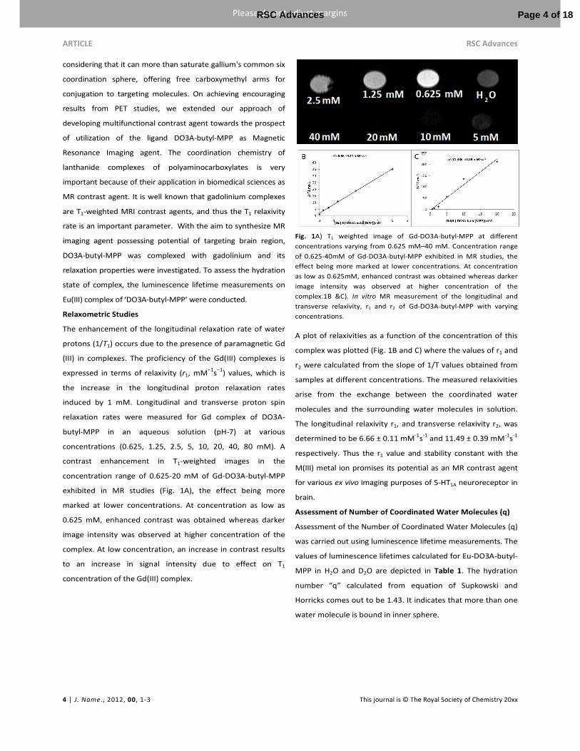
Relaxometric Studies
The enhancement of the longitudinal relaxation rate of water
protons (1/T1) occurs due to the presence of paramagnetic Gd
(III) in complexes. The proficiency of the Gd(III) complexes is
expressed in terms of relaxivity (r1, mM−1
s−1
) values, which is
the increase in the longitudinal proton relaxation rates
induced by 1 mM. Longitudinal and transverse proton spin
relaxation rates were measured for Gd complex of DO3A-
butyl-MPP in an aqueous solution (pH-7) at various
concentrations (0.625, 1.25, 2.5, 5, 10, 20, 40, 80 mM). A
contrast enhancement in T1-weighted images in the
concentration range of 0.625-20 mM of Gd-DO3A-butyl-MPP
exhibited in MR studies (Fig. 1A), the effect being more
marked at lower concentrations. At concentration as low as
0.625 mM, enhanced contrast was obtained whereas darker
image intensity was observed at higher concentration of the
complex. At low concentration, an increase in contrast results
to an increase in signal intensity due to effect on T1
concentration of the Gd(III) complex.
Fig. 1A) T1 weighted image of Gd-DO3A-butyl-MPP at different
concentrations varying from 0.625 mM–40 mM. Concentration range
of 0.625-40mM of Gd-DO3A-butyl-MPP exhibited in MR studies, the
effect being more marked at lower concentrations. At concentration
as low as 0.625mM, enhanced contrast was obtained whereas darker
image intensity was observed at higher concentration of the
complex.1B &C). In vitro MR measurement of the longitudinal and
transverse relaxivity, r1 and r2 of Gd-DO3A-butyl-MPP with varying
concentrations.
A plot of relaxivities as a function of the concentration of this
complex was plotted (Fig. 1B and C) where the values of r1 and
r2 were calculated from the slope of 1/T values obtained from
samples at different concentrations. The measured relaxivities
arise from the exchange between the coordinated water
molecules and the surrounding water molecules in solution.
The longitudinal relaxivity r1, and transverse relaxivity r2, was
determined to be 6.66 ± 0.11 mM-1
s-1
and 11.49 ± 0.39 mM-1
s-1
respectively. Thus the r1 value and stability constant with the
M(III) metal ion promises its potential as an MR contrast agent
for various ex vivo imaging purposes of 5-HT1A neuroreceptor in
brain.
Assessment of Number of Coordinated Water Molecules (q)
Assessment of the Number of Coordinated Water Molecules (q)
was carried out using luminescence lifetime measurements. The
values of luminescence lifetimes calculated for Eu-DO3A-butyl-
MPP in H2O and D2O are depicted in Table 1. The hydration
number “q” calculated from equation of Supkowski and
Horricks comes out to be 1.43. It indicates that more than one
water molecule is bound in inner sphere.
Page 4 of 18RSC Advances
Journal Name ARTICLE
This journal is © The Royal Society of Chemistry 20xx J. Name., 2013, 00, 1-3 | 5
Please do not adjust margins
Please do not adjust margins
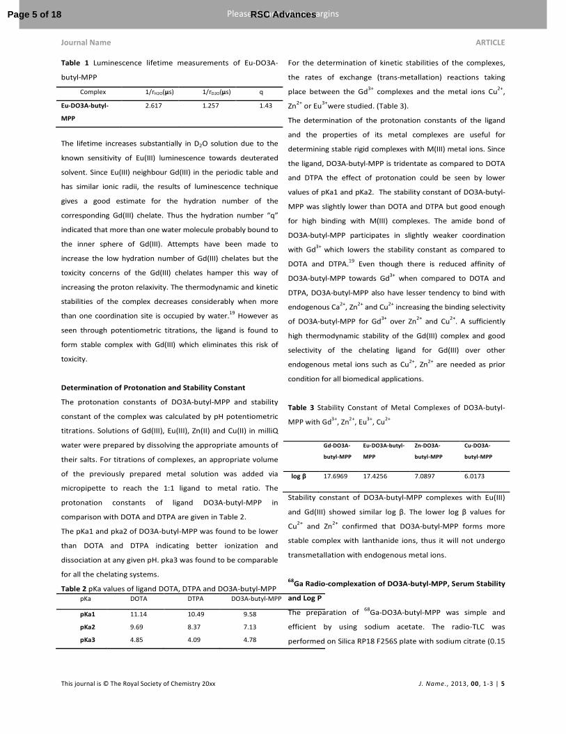
Table 1 Luminescence lifetime measurements of Eu-DO3A-
butyl-MPP
Complex 1/гH2O(µs) 1/гD2O(µs) q
Eu-DO3A-butyl-
MPP
2.617 1.257 1.43
The lifetime increases substantially in D2O solution due to the
known sensitivity of Eu(III) luminescence towards deuterated
solvent. Since Eu(III) neighbour Gd(III) in the periodic table and
has similar ionic radii, the results of luminescence technique
gives a good estimate for the hydration number of the
corresponding Gd(III) chelate. Thus the hydration number “q”
indicated that more than one water molecule probably bound to
the inner sphere of Gd(III). Attempts have been made to
increase the low hydration number of Gd(III) chelates but the
toxicity concerns of the Gd(III) chelates hamper this way of
increasing the proton relaxivity. The thermodynamic and kinetic
stabilities of the complex decreases considerably when more
than one coordination site is occupied by water.19
However as
seen through potentiometric titrations, the ligand is found to
form stable complex with Gd(III) which eliminates this risk of
toxicity.
Determination of Protonation and Stability Constant
The protonation constants of DO3A-butyl-MPP and stability
constant of the complex was calculated by pH potentiometric
titrations. Solutions of Gd(III), Eu(III), Zn(II) and Cu(II) in milliQ
water were prepared by dissolving the appropriate amounts of
their salts. For titrations of complexes, an appropriate volume
of the previously prepared metal solution was added via
micropipette to reach the 1:1 ligand to metal ratio. The
protonation constants of ligand DO3A-butyl-MPP in
comparison with DOTA and DTPA are given in Table 2.
The pKa1 and pka2 of DO3A-butyl-MPP was found to be lower
than DOTA and DTPA indicating better ionization and
dissociation at any given pH. pka3 was found to be comparable
for all the chelating systems.
Table 2 pKa values of ligand DOTA, DTPA and DO3A-butyl-MPP
pKa DOTA DTPA DO3A-butyl-MPP
pKa1 11.14 10.49 9.58
pKa2 9.69 8.37 7.13
pKa3 4.85 4.09 4.78
For the determination of kinetic stabilities of the complexes,
the rates of exchange (trans-metallation) reactions taking
place between the Gd3+
complexes and the metal ions Cu2+
,
Zn2+
or Eu3+
were studied. (Table 3).
The determination of the protonation constants of the ligand
and the properties of its metal complexes are useful for
determining stable rigid complexes with M(III) metal ions. Since
the ligand, DO3A-butyl-MPP is tridentate as compared to DOTA
and DTPA the effect of protonation could be seen by lower
values of pKa1 and pKa2. The stability constant of DO3A-butyl-
MPP was slightly lower than DOTA and DTPA but good enough
for high binding with M(III) complexes. The amide bond of
DO3A-butyl-MPP participates in slightly weaker coordination
with Gd3+
which lowers the stability constant as compared to
DOTA and DTPA.19
Even though there is reduced affinity of
DO3A-butyl-MPP towards Gd3+
when compared to DOTA and
DTPA, DO3A-butyl-MPP also have lesser tendency to bind with
endogenous Ca2+
, Zn2+
and Cu2+
increasing the binding selectivity
of DO3A-butyl-MPP for Gd3+
over Zn2+
and Cu2+
. A sufficiently
high thermodynamic stability of the Gd(III) complex and good
selectivity of the chelating ligand for Gd(III) over other
endogenous metal ions such as Cu2+
, Zn2+
are needed as prior
condition for all biomedical applications.
Table 3 Stability Constant of Metal Complexes of DO3A-butyl-
MPP with Gd3+
, Zn2+
, Eu3+
, Cu2+
Gd-DO3A-
butyl-MPP
Eu-DO3A-butyl-
MPP
Zn-DO3A-
butyl-MPP
Cu-DO3A-
butyl-MPP
log β 17.6969 17.4256 7.0897 6.0173
Stability constant of DO3A-butyl-MPP complexes with Eu(III)
and Gd(III) showed similar log β. The lower log β values for
Cu2+
and Zn2+
confirmed that DO3A-butyl-MPP forms more
stable complex with lanthanide ions, thus it will not undergo
transmetallation with endogenous metal ions.
68Ga Radio-complexation of DO3A-butyl-MPP, Serum Stability
and Log P
The preparation of 68
Ga-DO3A-butyl-MPP was simple and
efficient by using sodium acetate. The radio-TLC was
performed on Silica RP18 F256S plate with sodium citrate (0.15
Page 5 of 18 RSC Advances

ARTICLE RSC Advances
6 | J. Name., 2012, 00, 1-3 This journal is © The Royal Society of Chemistry 20xx
Please do not adjust margins
Please do not adjust margins
M) as the mobile phase. Radio TLC showed peak at 5.73 for
free 68
Ga-Acetate (< 2%) and 3.21 for 68
Ga-DO3A-butyl-MPP.
Radiochemical yield was calculated from decay-corrected 68
Ga
at the end of synthesis (EOS) including SPE purification (Fig.S19
ESI†).The decay corrected radiochemical yield was found to be
86.42 ± 3.9%, n= 40 runs with specific activity of 529.08
GBq/µmoL (Decay corrected and SPE purification for 15 min
EOS). After tc18 cartridge purification of 68
Ga-DO3A-butyl-
MPP, it showed no evidence of presence of free 68
Ga. Radio
HPLC analysis of the purified product 68
Ga-DO3A-butyl-MPP
showed 99% radiochemical purity and the presence of only
one peak (RT = 7.8 min) confirms the formation of only one
species of the complex which quantitatively reflected the
radiochemical purity of the sample (Fig. S20, ESI†). The
optimisation of radiolabeling with respect to pH, temperature,
duration of heating and the amount of precursor used in
labeling is described in detail in supplementary Information
(Fig. S21 ESI†).
The rate of decomplexation of the radiolabeled complex was
studied in human serum under physiological conditions at
different time intervals of 5, 10, 15, 30, 60 120 and 180 min.
No measurable loss of metal ion from the macrocyclic core and
high stability of the radiolabeled DO3A-butyl-MPP were
observed. (See Fig. S22 ESI†). Thus high specific activity,
stability was achieved and log P was found to be 2.59. The log P
of the radiolabeled DO3A-butyl-MPP was found to be lipophilic
in nature, appropriate to cross the blood brain barrier.
Cell viability Studies of DO3A-butyl-MPP
The in vitro cytotoxicity of the ligand, DO3A-butyl-MPP was
studied by incubating Human embryonic kidney (HEK) cells
with varying concentrations of the compound at time intervals
of 24, 48 and 72 h. Using 100 µL of HBSS (Hank Balance Saline
Solution) as control, experiment was conducted. Significant
percentage of viable cells was obtained when HEK cells were
treated with low concentrations (1-100 µM) of the compound.
After 24 and 48 h incubation of 10 mM concentration of the
compound cell survival (percentage relative to untreated
controls) in the range of 70-75% was observed. (Fig. S23,
ESI†). The cytotoxicity studies assessed in normal HEK cells
showed high IC50 indicating its safe and favorable applications
to be used in nuclear medicine imaging techniques.
Radioligand Binding Assay
The specificity of the 1-(2-methoxyphenyl)piperazine
functionalized compound to bind to the cell surface receptors
on primary hippocampal culture cells was examined by
radioligand receptor binding assays using 68
Ga-DO3A-butyl-MPP
as the labeled ligand. Primary hippocampal cells were cultured
as reported in literature.20
It has been demonstrated previously
that moderate to heavy signal were detected for 5-HT1A mRNA
transcripts in hippocampal neurons and glial cells.21
The
specificity of the 1-(2-methoxyphenyl)piperazine functionalized
compound to bind to the cell surface receptors on primary
hippocampal culture cells was examined by radioligand receptor
binding assays using 68
Ga-DO3A-butyl-MPP as the labeled ligand.
Non-specific binding was obtained by using 100-fold excess of
unlabeled serotonin and WAY-100635. Analysis of the binding
curve exhibited saturable binding of the radio-conjugate in
subnanomolar range. Scatchard plot analysis revealed that the
labeled compound exhibited high affinity on hippocampal
neuronal cultures with a Kd value of 0.64 nM and 0.27 nM in
competitive binding assay with serotonin and WAY-1000635
respectively.
The binding affinity of the compound, DO3A-butyl-MPP on the
targeted receptors was determined by harvesting different
parts of brain expressing serotonin receptors and transporters.
5-HT1A receptor binding assay exhibited lower affinity of 68
Ga-
DO3A-butyl-MPP towards 5-HT2A receptors (Kd =136 ± 3.4) than
5-HT1A receptors (Kd=0.39 ± 0.08) in brain homogenates (Table
4). Scatchard plot of the radioligand binding assays of 5HT1A,
5HTT and 5HT2A brain homogenate are given in ESI† (Fig
S24).
A subnanomolar affinity for the 5-HT1A receptor was obtained
for the above derivative in the in vitro binding assay which was
sufficiently high to carry out imaging. The selectivity of the
complex for 5-HT1A receptors was observed to be 1,000 times as
high as for 5-HT2A receptors (Table 4). 5-HTT also showed high
affinity with a value of 56 ± 0.7 nM.
Table 4 Dissociation constants of DO3A-butyl-MPP for 5-HT1A,
5-HT2A, and 5-HTT receptors
*Nonspecific binding was determined by serotonin, ketanserin and
paroxetine.
Kd (nM) 5-HT1A 5-HT2A 5-HTT
68Ga-DO3A-butyl-MPP 0.39±0.09 136 ±3.4 56 ±0.7
Page 6 of 18RSC Advances

Journal Name ARTICLE
This journal is © The Royal Society of Chemistry 20xx J. Name., 2013, 00, 1-3 | 7
Please do not adjust margins
Please do not adjust margins
PET image of rat whole brain
The results found in the micro PET imaging of SD (Sprague
Dawley) rat brain indicated that the radiotracer showed
remarkable accumulation in the rat brain striatum over 30 min
compared with surrounding normal tissue(Fig. 2). The decline
in activity was observed after 30 minutes. This could be well
supported by the fact that terminal elimination half life of
antagonist WAY 100635 is also fast with a value of 33 min.22
The radioactivity distribution was in concordance with the known 5-
HT1A receptor distribution, with high uptake of radioactivity in
hippocampus, caudate putamen and amygdala. (Fig. 2A) The time
activity curves revealed accumulation of the radioligand in whole
brain over the first few minutes which were followed by a clearance
of the compound from hippocampus (Fig. 2).
Fig. 2A. 0-30 minutes summation images of rat brain coronal microPET Scan depicting activity in different section (Slices) of the Brain. B. Time activity
curve(TAC) of different regions for 30 minutes and C. TAC for 60 minutes post 68
Ga-DO3A-butyl-MPP injection in rat (n=5).
The pre-treatment experiment with WAY-100635 (known 5HT1A
antagonist) markedly reduced the level of radioactivity in all
examined regions. Cerebellum was used as a reference region for
the calculation of binding potential due to the low 5HT1A receptor
density. Rat MRI atlas was superimposed on the micro PET for
anatomical confirmation of the brain regions. VOI analysis was done
using PMOD, Data is determined as VOI (CPS/cm3)Brain Region/ VOI
(CPS/cm3)Cerebellum. VOI analysis showed highest uptake in
hippocampus followed by hippocampus and amygdala (Fig. 3). VOI
of 5.606 in hippocampus region and 2.59 in caudate putamen was
observed. VOI of 3.2 and 2.48 were in amygdala and hypothalamus
respectively as compared to the cerebellum with a VOI of 1.1.
Page 7 of 18 RSC Advances

ARTICLE RSC Advances
8 | J. Name., 2012, 00, 1-3 This journal is © The Royal Society of Chemistry 20xx
Please do not adjust margins
Please do not adjust margins
Fig 3. MicroPET images of Rat brain showing Coronal, transverse and sagittal
sections respectively after the intravenous injection of 37 MBq of 68
Ga-
DO3A-butyl-MPP. A. SD Rat depicting high accumulation of radioactivity in
hippocampus, amygdala and Caudate Putamen. B. Coregisteration of rat
MRI atlas on the microPET scan for anatomical confirmation of the brain
regions was done using PMOD and C. Blocking studies with WAY-100635.
Regional uptake in Rat brain
Regional binding indices were calculated by % ID/g of the
different regions of the brain by their respective weights (Fig.
4). In regional biodistribution experiments, the hippocampus
showed the highest uptake of radiolabeled compound,
followed by the caudate putamen which showed a
Fig. 4 Regional distribution of 68
Ga-DO3A-butyl-MPP in SD rat brain.
WBS = whole brain section Hip = hippocampus, Th = thalamus, Ctx =
frontal and parietal cortex, CPu = caudate putamen, Cb =cerebellum at
10, 30 and 45 min post injection.
binding of 1.499% ID/g and cerebellum showed least uptake
with a value of 0.08%ID/g. Whole brain uptake at 30 min
increased to 3.724% ID/g and 73% fall of activity at 60 min. The
values were concordant with the VOI obtained in PET brain
scan. The 5-HT receptor-rich regions showed significantly
higher binding index than the cerebellum.
Plasma Clearance and Biodistribution
The plasma clearance and in vivo biodistribution of 68
Ga-DO3A-
butyl-MPP was studied in SD rats at different time intervals.
Fig. 5 Plasma Clearance of 68
Ga-DO3A-butyl-MPP (Inset: Initial
Clearance up to 20 min)
Plasma clearance indicates the clearance rate from the body.
The biological half life t1/2 (fast) results in the increase in target
to non target ratio. Biodistribution studies were performed to
study the accumulation pattern of the tracer in the whole body.
It was observed that the clearance of the complex was through
the renal and hepatobilliary routes.
The plasma clearance followed a biphasic trend with a rapidly
clearing initial phase followed by slow phase. Plasma clearance
rate (CR) was found to be 37.39 ± 0.31 µg/mL.min-1
(Fig. 5).
In biodistribution studies, major accumulation of the labeled
compound was observed in kidneys 14.62 ± 0.52 % ID/g
followed by liver 6.19 ± 0.61 % ID/g) at 2 h showing that the
complex is excreted by renal and hepatobiliary routes.
Retention of radioactivity in the non-target organs (lungs,
spleen, intestine etc.) was less than 2.0 %ID/g at 4 h post
injection. Accumulation in lungs attributed to the lipophilic
nature of the complex. The retention of activity follows a
declining trend in the all the organs for the later time points
except for the kidneys at 4 h, wherein the clearance was
observed at 8-10 h.
0
0.5
1
1.5
2
2.5
3
3.5
4
4.5
WBS OB CPu Hip Th Ctx Cb
%ID
/g
10 min
30 min
45 min
Page 8 of 18RSC Advances

Journal Name ARTICLE
This journal is © The Royal Society of Chemistry 20xx J. Name., 2013, 00, 1-3 | 9
Please do not adjust margins
Please do not adjust margins
In vivo biodistribution of 68
Ga-DO3A-butyl-MPP in SD rats
showed 3.52 ± 0.39 % ID/g activity in brain at 10 min and 3.91
± 0.54% ID/g activity at 30 min which drastically decreased at 2
h with a value of 0.48 ± 0.03% ID/g activity. The selective high
brain uptake could be seen in regional biodistribution studies
which were found concomitant with the VOIs generated from
the PET studies.Cardiac uptake was seen at early time points.
(Fig.6)
Fig.6 Biodistribution of 68
Ga-DO3A-butyl-MPP in SD Rats following
intravenous injection of 8.14 MBq activity.
MR Imaging
The preliminary in vivo imaging was done on rat brain with and
without injection of the Gd(III)-complex, which shows that the
ligand Gd-DO3A-butyl-MPP at 2 mM was showing enhanced images
as compared to the control brains. T1-weighted signal enhancement
was observed on an in vivo MRI, notably within hippocampus,
which further supports the specificity of the complexed ligand
towards 5-HT1A receptors.
Fig. 7A. In vivo MR imaging in control and treated SD Rats depicting T1
shortening in 30 min images post Gd-DO3A-butyl-MPP (0.176mmol/Kg)
injection. B. Representation of relaxivity with respect to the different
regions in the brain.
The T1 were statistically decreased by Gd-DO3A-butyl-MPP at 2 mM
administration (0.176 mmol/Kg) for hippocampus compared to
baseline scan (Fig. 7A). The enhancement of in vivo MRI in the
control brains was statistically significant with a positive signal at 7
T. It is worthwhile to note that the shortening of T1 relaxation was
observed in treated rats at 30 min post injection at a concentration
of 0.176 mmol/Kg of Gd-DO3A-butyl-MPP, as compared to control
rats. There was increase in T1 relaxation on different regions of
brain when WAY100635 (0.2 mmol/Kg) and Gd-DO3A-butyl-MPP
(0.176 mmol/Kg) were co administered at 30 min pi (Fig. 7B).
It was also observed that the cerebral blood flow was increased
when the Gd(III)-DO3A-buyl-MPP was given alone which is an
indication of antagonistic property of the compound. It can be
hypothesized from the study conducted on rats that in
pathological state when there is an up regulation of 5-HT1A
receptors, this contrast agent could be useful in visualizing the
uptake at high density and high concentration. It could be useful
as a surrogate tracer of 68
Ga-DO3A-buyl-MPP for MR imaging.
Theoretical Evidence
Molecular modeling and docking studies with monomeric
5-HT1A receptor models
Homology modeling and flexible docking studies has been
performed for DO3A-butyl-MPP with human 5-HT1A homology
modeled receptor. The 5-HT1A model was optimized; refined and
Ramachandran plot validates the models where residues are
present in allowed region without any steric clashes (Fig. 8A). The
reliable modeling of TM region is important since ligand binding
pockets lies in TM region of the 5-HT1A monomer (Fig. 8A).
0
2
4
6
8
10
12
14
16
18
20
%ID
/g
10 min
30min
60 min
2h
4h
0
2
4
6
8
10
12
HIP1 HIP2 Th HyTH CTX
r1 (
mM
-1 s
-1)
Brain Regions
Control Gd-DO3A-Butyl-MPP Coinjection WAY100635
Page 9 of 18 RSC Advances

ARTICLE RSC Advances
10 | J. Name., 2012, 00, 1-3 This journal is © The Royal Society of Chemistry 20xx
Please do not adjust margins
Please do not adjust margins
Fig. 8 (A) Model of the human 5-HT1A receptor and Ramachandran Plot
showing the transmembrane helices in different colours. (B) Two
dimensional view of DO3A-butyl-MPP with 5-HT1A receptor where
green colored are hydrophobic, light blue are polar, red are negative
and purple are positive charged residues. Pink lines are hydrogen
bonds, red lines are π-cation interactions. (C) Three dimensional view
of DO3A-butyl-MPP (pink carbon layout) at the binding pocket of 5-
HT1A homology modeled receptor. Yellow dashed lines are showing the
hydrogen bonds.
Three antagonist; p-MPPF, BMMP and WAY-100635 were used
in the docking studies to understand the binding pose of our
ligand with 5-HT1A receptor.23
Our molecular modelling results showed a promising results
for thermodynamic parameters obtained by docking of the
DO3A-butyl-MPP and interactive residue present in the active
site of the 5-HT1A receptor. DO3A-butyl-MPP was aligned in
hydrophobic pocket conserved in all three studied antagonists,
p-MPPF, BMMP and WAY-100635, the Glide XP scoring mode
in flexible docking has predicted the minimized energy
complexes in terms of XP score (Table 5). The antagonist p-
MPPF is involved in π-π interactions with Phe362 and
hydrogen bonding with backbone of Ile189 whereas MefWAY
is involved in π-π interactions Arg181 and hydrogen bonding
with backbone of Ile189. But in case of WAY-100635, one
additional π-π interactions with Phe361 was obtained. (See Fig.
S25 ESI†)
The docking pose of DO3A-butyl-MPP at the shallow
antagonist binding pocket of 5-HT1A was found to have high G
score of -12.132.
Table 5. IFD Glide score and ligand interactions of Antagonists
MefWAY, WAY-100635 and p-MPPF and DO3A-butylMPP
The high G score signifies the contribution due to important
hydrophobic residues surrounding the ligands. In the ligand,
DO3A-butyl-MPP, the carboxylate groups and carbonyl moiety
of DOTA is involved in hydrogen bonding with side chain
residues, His376, Gly382, Ile189 and Lys191. In addition, π-
cation interactions were observed for protonated -NH of
piperazine moiety with Phe 112. This interaction is an example
of non covalent bonding between a monopole (cation) and
a quadrupole (π system). Here, the cation is nitrogen which is
protonated and π- electrons are in residue Phe112.
In addition to the above interactions, the aromatic ring of
piperazine moiety of DO3A-butyl-MPP was involved in π-π
stacking with Phe361, Phe362 and hydrogen bond interactions
with the side chain of Asp116. The resident binding pocket of
both uncomplexed and complexed DO3A-butyl-MPP was
found to be conserved and comparable to our pervious
results24
with known antagonists in which docking was
performed in flexible mode showing appreciable G-score.
The Asp116 interactions with azapirones, buspirone and
sunepitron are well known with piperazine amine25
as in case
of DO3A-butyl-MPP. This binding mode is in general
agreement with putative binding modes suggested in a
published ligand-supported homology model of 5-HT1A
receptor.26
Also the binding pose is quite consistent with the
available experimental studies25
and importance of these
residues is evidenced by site-directed mutagenesis. From
docking pose, the antagonist binding pocket was found to be
Ligand Residual Interactions GScore
Monomeric
Non-polar Residues Polar Residues
IFD
DO3A-
butyl-
MPP
Ile189,Asp116, Phe112,
Phe362, Gly382
Lys191, Thr200,
Asn386
-12.132
p-MPPF
Val117, Cys120,Ile189,
la203,Phe361,Phe362,
Val364,Ala365, Leu368
Lys191, Thr200
-9.11
MefWAY
Ile189,
Phe361,Leu368,Gly382
, Arg181
Thr196,Ser190,
Lys191,Ser199,
Thr379
-9.81
WAY
100635
Ile189,
Tyr195,Phe361,Leu368,
Gly382
Thr196,Thr200,Ser19
0, Lys191,Ser199,
Thr379
-8.81
A
B C
Page 10 of 18RSC Advances

Journal Name ARTICLE
This journal is © The Royal Society of Chemistry 20xx J. Name., 2013, 00, 1-3 | 11
Please do not adjust margins
Please do not adjust margins
relatively shallow and involved in various interactions at the
site.
Although promising, PET compounds, particularly 11
C
compounds, are generally limited to those facilities with a
cyclotron and the high cost hinders commercialization of viable
agents which is driving the need to develop 68
Ga-based agents
that can target 5-HT1A. Progress in receptor pharmacology with
PET radiotracers has led to the design of numerous candidate
for CNS imaging. Fragments of WAY100635, (i.e. arylpiperazine),
have been combined with various bifunctional chelates
consisting of a ligand that binds to technetium-99m and a linker
that joins to the targeting vector. Johannsen et al. prepared a
WAY-based complex using an N2S2 chelate to bind to 99m
Tc
including similar derivatives of arylpiperazine linked to other
technetium(V) chelates (NxSy or NxPy, x = 1-4, y = 4-
x).27
Although their IC50 and Ki values appear promising, these
agents have low or negligible brain uptake. To date, no 68
Ga
labeled compound has been developed which can image the 5-
HT1A receptor in vivo. DO3A-butyl-MPP provides efficient
macrocyclic framework for complexation of M3+
metal ions for
multiple biomedical applications. Its improved affinity and
selectivity with high brain uptake is evident through PET imaging
in rat brain which could plausibly be translated clinically.
Conclusions
We have efficiently synthesized a chelating system containing
1-(2-methoxyphenyl)piperazine unit as a vector for evaluation
of 5-HT1A receptors. Keeping in mind the concept of multiple
imaging modalities, the compound has been labeled with
Gd(III), Eu(III), and 68
Ga so that it can be exploited for MRI and
PET imaging agent. Encouraging results have been obtained
with all three modalities. The compound (Gd-DO3A-butyl-
MPP) has shown longitudinal relaxivity, r1 of 6.66mM-1
s-1
and
transverse relaxivity, r2 of 11.49 mM-1
s-1
on 500MHz at
21°C.Hydration number, ‘q’ was found to be 1.43 from
luminescence lifetime studies. Compound (68
Ga-DO3A-butyl-
MPP) shows high affinity for hippocampal cells found in brain,
proving its relevance as a CNS imaging system, shown also by
the appreciable uptake of radiolabeled compound in brain in
PET images. The desired molecule also shows antidepressant
actions in the behavioral tests conducted on CMS model of
rats, and completely reverses the depression levels. The action
of the DO3A-butyl-MPP can be speculated by acting on 5-HT1A
via SSRI effect, which further supports its potency in treating
depression. So the preliminary results and preclinical
evaluations of the given compound DO3A-butyl-MPP show it
as a favorable molecule and promises good potential as a CNS
imaging system which can be used as a MR contrast, and PET
imaging system. This will also encourage the development of
brain imaging compounds for different applications.
Experimental
General. 1-(2-methoxyphenyl)piperazine, N-(4-
bromobutyl)phthalimide, hydrazine hydrate, tert-butyl
bromoacetate, potassium carbonate, 1,4,7,10-
tetraazacyclododecane (cyclen), trifluoroacetic acid, chloroacetyl
chloride, sodium bicarbonate, serotonin hydrochloride, WAY-
100635 maleate were purchased from Sigma- Aldrich. Acetonitrile
(HPLC grade), N, N-dimethylformamide (HPLC grade),
dichloromethane (HPLC grade), water (HPLC grade), MTT and
trifluoroacetic acid were obtained from E. Merck Ltd., India. Metal
salts (GaCl3, EuCl3 and GdCl3), DMEM F12, EDTA, nystatin,
formaldehyde, D-glucose, agarose and sodium chloride were
purchased from Aldrich (99.9%). Penicillin, streptomycin sulfate,
FBS and trypsin were purchased from GIBCO (invitrogen). The
reactions demanding anhydrous conditions or involving moisture
sensitive reactants were carried out under an atmosphere of dry
nitrogen using oven dried (80°C) glassware. All reaction
temperatures reported indicate the temperature of the bath in
contact with the reaction vessel. Sep-Pak tC18 Plus Short Cartridge
was purchased from Waters. TLC was run on the silica gel coated
aluminum sheets (Silica gel 60 F254, Merck, Germany). Instant thin
layer chromatography used to detect radiocomplexation and
radiochemical purity. Radio TLC was analysed on Peak Simple 3.29.
Gallium-68 complex Radio HPLC was performed on semi automated
GE FXFN TracerLab integrated with radioactivity detection and UV
detection at 254 nm (fixed wavelength detector K-2001, Knauer,
Germany), using isocratic pump SykamS-1021, elution with
methanol and water (25/80, v/v) mobile phase and flow rate of 2
mL/min on C-18 reversed phase Nucleosil column (250 mm X 10
mm, 5 μ). 1H and
13C NMR spectra were recorded on a Bruker
Avance II 400 MHz system (Ultra shield). Chemical shifts ‘δ’ are
reported in ppm relative to TMS. Mass spectra (ESI-MS in positive
and negative ion mode) were performed on in-house Agilent 6310
Page 11 of 18 RSC Advances

ARTICLE RSC Advances
12 | J. Name., 2012, 00, 1-3 This journal is © The Royal Society of Chemistry 20xx
Please do not adjust margins
Please do not adjust margins
system ion trap. High resolution Mass spectroscopy was done using
Accurate-massQuadrupole Time-of-Flight (Q-TOF) LC/MS at Delhi
University. All the computational studies were performed with
Schrödinger, LLC, New York, NY, 2014 maestro 9.7.28
MR studies
was done using an inhouse 7T Bruker Biospec USR 70/30, (AVANCE
III) horizontal bore animal MRI scanner tuned to 20 MHz In
vivo imaging were performed using micro PET imaging as non-
invasive technology. The acquisitions were performed using a GE
FLEX Triumph (TriFoil Imaging) micro-PET/SPECT/CT scanner. This
scanner consists of a micro-PET module (LabPET4) with 2'2'10
mm3
LYSO/LGSO scintillators in an 8-pixel, quad-APD detector
module arrangement. Potentiometric measurements were carried
out with an automatic titration system consisting of metrohm 713
pH meter equipped with Metrohem A.60262.100 electrode, 800
Dosino autoburet.The image processing was done using PMOD 3.2
and Amide 1.0.4 software.
Cell Culture. Monolayer cultures of normal embryonic kidney cells,
HEK, in DMEM (Sigma, USA) supplemented with 10% fetal bovine
serum (GIBCO), 50 U/mL penicillin, 50 μg/mL streptomycin sulfate,
and 2 μg/mL nystatin were maintained at 37 °C in a humidified CO2
incubator (5% CO2, 95% air). Cells were routinely subcultured using
0.0 5% Trypsin (Sigma, USA) in 0.02% EDTA in humidified
atmosphere of 95 % air and 5 % CO2 at 37 °C twice a week.
Ethics statement.The human serum study was approved by the
institutional ethical committee. Blood samples for human serum
study were collected from healthy volunteers who were well
informed about the study. Written consent from the volunteers
above 18 years of age was obtained.
Animal Models. All animal experiments were conducted in
accordance with the guidelines of INMAS animal ethics
committee(CPCSEA Regn No.8/GO/a/99).Blood clearance, imaging,
and biodistribution were carried on SD rats. Rats were housed
under conditions of controlled temperature of 22 ± 2°C and normal
diet ad libitum.
Data Analysis.The competition curve of the receptor binding
experiments was analyzed by nonlinear regression using algorithms
in GraphPad PRISM 5.0 (San Diego, CA). Biodistribution Data
isreported as mean ± Standard Deviation (S.D.). VOI analyses were
carried out on PMOD 3.2.
Synthetic Approach. Synthesis of Compounds 2-(4-(4-(2-
methoxyphenyl)piperazin-1-yl)butyl)isoindoline-1,3-dione (1) and 4-
(4-(2-methoxyphenyl)piperazin-1-yl)butan-1-amine (2) have been
done as reported previously by our group.18
Synthesis of 2-chloro-N-(4-(4-(2-methoxyphenyl)piperazin-1-
yl)butylacetamide (3). 2-Chloroacetylchloride (410 mg, 3.636 mmol
in 10 mL of chloroform) and potassium carbonate (627mg, 4.5mmol
in 10 mL of water) were added simultaneously through dropping
funnels to a stirred solution of 2 (800 mg, 3.03mmol) in 10 mL of
chloroform at 0˚C over a period of 1 h. The reaction mixture was
stirred for an additional 4 h at room temperature. The chloroform
layer was separated and washed with water (50 mL) twice. The
organic layer was dried over anhydrous sodium sulfate and
evaporated to product 3 (820 mg, 80%). The product was used as
such without any further purification.1H NMR (400 MHz, 25 °C,
CDCl3), δ (ppm): 1.5 (s, 4H, CH2), 2.3 (s, 2H, CH2), 2.5 (s, 4H, CH2),
3.1 (s, 4H, CH2), 3.3(t, 2H, J=6Hz, CH2), 3.8(s, 3H, CH3), 4.0(s,2H,
CH2), 6.7-6.9 ( m, 4H , CH); 13
CNMR(100 MHz, 25°C, CDCl3),
δ(ppm):24.2, 27.3, 39.7, 42.7, 50.6, 53.5, 55.3, 58.3, 111.1, 118.2,
120.9, 122.9, 141.2, 152.2, 165.8 ; MS (ESI+) m/z calculated for
C17H26ClN3O6 339.8, found [M + H]+ 340.5.
Synthesis of 1,4,7-tri(tert-butoxymethane)-1,4,7,10-
tetraazacyclododecane (t-Bu-DO3A) (4). Under an atmosphere of
nitrogen, 1,4,7,10-tetraazacyclododecane (2.906 mmol, 500 mg)
was dissolved in dry acetonitrile (10 mL) at 0°C. To the reaction flask
NaHCO3 (8.72 mmol, 732 mg) was added and stirred for 30 min.
tert-Butyl bromoacetate (8.72 mmol, 1.28 mL) was added slowly
from a dropping funnel in 2 h at 0 °C. The reaction was stirred for
additional 1 h at 0 °C and then at room temperature for 40 h. The
progress of the reaction was checked by TLC (9: 1;
dichloromethane: methanol). After the completion of the reaction,
the reaction mixture was filtered and the filtrate was evaporated to
dryness. The crude compound was purified by column
chromatography (silica gel, 10% methanol in dichloromethane, Rf
0.67) to give final compound t-Bu-DO3A (1120 mg, 75%) as white
powder. 1H NMR (400 MHz, 25 °C, CDCl3); 1.4 (27H, s, C(CH3)3), 2.8–
3.1 (16H, m, CH2), 3.2-3.3 (6H, m, CH2) ; 13
CNMR (400 MHz, 25 °C,
CDCl3): 28.0, 47.5 , 49.1, 51.1, 55.6, 58.2, 81.7 , 170.5 , 172.9 ; m/z
calculated for C26H50N4O6 514.7, found 515.8 [M+H]+.
Page 12 of 18RSC Advances

Journal Name ARTICLE
This journal is © The Royal Society of Chemistry 20xx J. Name., 2013, 00, 1-3 | 13
Please do not adjust margins
Please do not adjust margins
Synthesis of tert-butyl 2,2’,2’’-(10-(2-(4-(4-(2-
methoxyphenyl)piperazin-1-yl)butylamino)-2-oxoethyl)-1,4,7,10-
tetraazacyclododecane-1,4,7-triyl)triacetate (5). Under nitrogen
atmosphere, to a stirring solution of 1, 4, 7-
tris(carbobutoxymethyl)-1,4,7,10-tetraaza-cyclododecane (500 mg,
0.97 mmol) in dry acetonitrile (20 mL), K2CO3 (669 mg, 4.85mmol)
was added. After 30min, (3) (396 mg,1.17 mmol) dissolved in
acetonitrile was added drop wise and heated to 70˚C. After 24 h,
the reaction mixture was cooled to room temperature, filtered, and
evaporated under reduced pressure to give crude oily residue. The
compound was purified by column chromatography (silica gel, 5%
methanol in dichloromethane) to give the ligand (5) as a brown
solid (600 mg, 75%). 1H NMR (400 MHz, 25 °C, CDCl3), δ (ppm): 1.4
(s, 27H, C(CH3)3), 1.6-3.3 (m, 40H, CH2) 3.8 (s, 3H, CH3), 6.8-6.9 ( m,
4H, CH), 8.4 (br,1H, NH) ; 13
CNMR(100MHz, 25°C, CDCl3),
δ(ppm):22.9, 26.6, 27.9, 38.5, 49.2, 52.8, 55.7, 56.1, 57.6, 81.8,
111.2, 118.4, 120.9, 133.2, 140.5, 152.2, 171.5, 172.3. HRMS (ESI+)
m/z calculated for C43H75N7O8 817.5677, found [M + H]+ 818.5756,
[M+Na]+
840.5572. (See ESIϮ Fig S9)
Synthesis of 2,2’,2’’-(10-(2-(4-(4-(2-methoxyphenyl)piperazin-
1yl)butylamino)-2-oxoethyl)-1,4,7,10-tetraazacyclododecane-
1,4,7-triyl)triacetic acid (6). The ligand 5 (500 mg, 0.595 mmol) was
dissolved in dichloromethane (4 mL) and 1 mL of trifluoroacetic acid
was added at 0 °C and stirred for 4 h. The reaction mixture was
stirred at room temperature for additional 10 h. The solvent was
evaporated and residue was dissolved in 1 mL of methanol,
followed by addition of 30 mL of diethyl ether drop wise at 0-5 °C
and stirring for 1 h at room temperature. The solvent was decanted
and the precipitated compound dried under reduced pressure. (370
mg,96%). 1H NMR (400 MHz, 25 °C, D2O), δ (ppm): 1.4 (m, 2H, CH2),
1.6(m, 2H, CH2), 2.6 - 3.9 (m, 39H, CH2 and CH3 of -OCH3), 6.8 - 6.9
(m, 2H, CH), 7.0 -7.1(m, 2H, CH); 13
CNMR(100MHz, 25°C, CDCl3),
δ(ppm): 13.9, 20.6,25.3, 38.3, 48.1, 48.7, 50.1, 53.8, 54.1, 55.2,
56.2, 65.9, 111.9, 114.8, 116.2, 117.7, 120.6, 121.2, 126.7, 135.9,
151.8, 162.3, 163.4, 171.5 HRMS (ESI+) m/z calculated for
C31H51N7O8 649.3799, found [M + H]+ 650.3999. (See ESIϮ Fig S11)
Complexation of DO3A-butyl-MPP with EuCl3. To a solution of
DO3A-butyl-MPP (200 mg; 0.3076 mmol) in water (10 mL) was
added EuCl3.6H2O (112.5 mg; 0.3076 mmol) and the pH of the
reaction mixture was adjusted to 5.5. The reaction was stirred at 60
°C for 6 h. The reaction mixture was filtered and the solvent was
evaporated to obtain the Eu-complex as a pale green solid. HR-ESI-
MS calculated for C31H48EuN7O8 800.2855, found [M+H]+
800.2840.
Complexation of DO3A-butyl-MPP with GdCl3.To a solution of
DO3A-butyl-MPP (200 mg; 0.3076 mmol) in water (10 ml) was
added GdCl3.6H2O (114 mg; 0.3076 mmol) and the pH of the
reaction mixture was adjusted to 5.5. The reaction was stirred at 60
°C for 12 h. The reaction mixture was filtered and the solvent was
evaporated to obtain the Gd complex as a pale white solid. HR-ESI-
MS C31H48GdN7O8 calculated for 804.2805, found [M+H]+
805.2896.
Complexation of DO3A-butyl-MPP with GaCl3. 10
mg(0.0153mmol)DO3A-butyl-MPP was dissolved in water and pH
was adjusted to 4.5 with the help of 1M sodium acetate solution.
The resulting solution was perched with nitrogen to maintain inert
atmosphere. Then GaCl3 (3.2mg) was added. The temperature of
reaction mixture was then raised to 80˚C for 8 h. After that the
solution was brought to room temperature and chelex100 was
added to it which was then stirred for 30 min. Then the solution
was filtered and evaporated under reduced pressure to give a white
colored compound. HRMS (ESI+) calculated for C31H48GaN7O8:
715.2820, found [M]+ 713.0644.
Relaxation measurements
MR imaging was done using an inhouse 7T Bruker Biospec USR
70/30, (AVANCE III) horizontal bore animal MRI scanner.For all
experiments, RARE T1+T2 sequence was used to acquire relaxometry
images. The scan was acquired at 5 TE times (11, 33, 55, 77, 99 ms)
and at different TR’s (6000, 5000, 4500, 4000, 3500, 3000, 2500 and
1981 ms).Relaxation data were analyzed with Topspin 2.0 (bruker)
software.
Spectroscopic Measurement
The luminescence lifetime was obtained by measuring the decay at
the maximum of emission spectra and the signals were analyzed as
single exponential decays. The instrument settings were: gate-time
of 10ms, integration time of 1s, flash count - 8 and proper slit
widths were used. The excitation wavelength used for Eu-DO3A-
butyl-MPP was 365 nm and luminescence lifetimes are the average
of five independent experiments. The slit widths for both excitation
and emission monochromators were kept open. The number of
Page 13 of 18 RSC Advances

ARTICLE RSC Advances
14 | J. Name., 2012, 00, 1-3 This journal is © The Royal Society of Chemistry 20xx
Please do not adjust margins
Please do not adjust margins
coordinated water molecules (q) present in the inner sphere of the
complex is calculated by using equation of Supkowski and Horricks
for Eu(III).
q = AEu (1/τH2O – 1/τD2O– aEu)
Solutions of Eu(III) complexed DO3A-butyl-MPP complex were
prepared by mixing its appropriate volume in milliQ water or in D2O
and ligand in Tris buffer 0.1 M (pH = 7.4) or in D2O followed by
addition of NaOD until pH = 7. Measurement of the Ln(III)
luminescence lifetimes in solutions of H2O and D2O gave the
estimation of the number of coordinated water molecules present
in complex by using above equation.
Potentiometric Titrations
Potentiometric titrations were carried out at 25oC and ionic
strength “I” = 1M tert-butylammoniumchloride (TBACl). The
protonation constant of DO3A-butyl-MPP were determined
potentiometrically by titrating 2mM of HCl (2 mL) in the presence of
0.1mM DO3A-butyl-MPP (15 mL) with 0.01 mM (3ml) tert-
butylammoniumhydroxide (TBAOH). Titrations were performed in
the pH range of 1.0 – 10.0 for protonation constants. The
protonation constant of ligand is expressed by equation 1 and
stability constant of the metal complex is given by eq.
�� =�����
���������
……eq (1)
� ��� =� ����
� ��������
Where i=0,1,2,3……eq (2)
Radiolabeling of DO3A-butyl-MPP with 68
Ga
68Ga was eluted from
68Ge/
68Ga generator (Eckert and Ziegler) with
0.1 N HCl (3 mL) to obtain 480 MBq of 68
Ga. Sodium acetate was
added to adjust pH of the elute 4. Then compound (200 μg) was
added from 1 mg/ml stock solution of the compound and the
mixture incubated at 90 °C for 10 min. It was then cooled and
passed through C-18 cartridge (preconditioned with ethanol and
water). The compound was eluted with 1 ml ethanol. Then ITLC was
run in 0.15 M sodium acetate. The radiochemical purity was
assessed by omniscan EZ-TLC scanner and then the radiolabeled
conjugate was purified using a C-18 reversed phase extraction
cartridge which was preconditioned with 20 mL methanol and
subsequently activated with 30% methanol. The cartridge was
successively rinsed with 5 mL distilled water and radiolabeled
conjugate was eluted in 5 mL of 40 % ethanol. The 68
Ga-DO3A-
butyl-MPP was reconstituted in saline, and filtered through a sterile
0.22 µm Millipore (Milford, MA) Millex-GV® disposable syringe filter.
Radiotracer purity and stability were monitored using radio-HPLC.
In each HPLC analysis, the eluent was collected and counted along
with a standard prepared from the injectate.
Determination of logP and stability in blood serum.The partition
coefficient of the complex was determined by measuring the activity
that partitioned between the 1-octanol and aqueous phosphate
buffer (0.025 mol/L, pH 7.4) under strict equilibrium conditions. 2mL
1-octanol and 2mL of 68
Ga-DO3A-butyl-MPP phosphate buffer were
mixed in a centrifuge tube. The mixture was vortexed at room
temperature for 5 min and then centrifuged at 5000 rpm/min for 5
min. The counts in 0.1 mL samples of both organic and inorganic
layers were determined by a well type gamma counter. The
measurement was repeated three times. The partition coefficient (P)
was calculated as a ratio of the counts in the octanol fraction to the
counts in the water fraction (average of n=5).
Human Serum Stability. Fresh Human serum was prepared by
allowing blood collected from healthy volunteers to clot for 1 h at 37
°C in a humidified incubator maintained at 5% carbon dioxide/95%
air. Then, the samples were centrifuged at 400 g, and the serum was
filtered through 0.22 μm syringe filter into sterile tubes. The
radiolabeled DO3A-butyl-MPP was immediately placed in a CO2
chamber incubated at 37 °C and then analyzed to check for any
dissociation of the complex. The mixture was stored in the same
environment conditions, and aliquots of 100 mL (in triplicate) were
taken at appropriate periods of time (0 min, 30 min, 1 h and 3 h).
The aliquots were treated with 200 µL of ethanol, cooled (4 °C), and
centrifuged during 15 min at 4000 rpm, at 4 °C, in order to
precipitate the serum proteins. A 200 µL aliquot of supernatant was
collected for activity counting in a γ well-counter. The sediment was
washed twice with 1 mL of ethanol and its activity was counted. The
activity of the supernatant was compared to that of the sediment in
order to determine the percentage of the chelate associated to the
proteins. The activity of the supernatant at 3 h was evaluated by
TLC in order to check whether the chelate remained intact.
Biological Studies
Page 14 of 18RSC Advances
Journal Name ARTICLE
This journal is © The Royal Society of Chemistry 20xx J. Name., 2013, 00, 1-3 | 15
Please do not adjust margins
Please do not adjust margins
Cell viability assay of DO3A-butyl-MPP. To test the cytotoxic effect
of DO3A-butyl-MPP exposure on cells, MTT assays were conducted.
Exponentially growing cells were plated in a 96 well microtitre plate
at a cell density of 4000 cells/well for 24 h before treatment. Cells
were treated with the varying concentrations of the drug(0.001-10
mM) at different time intervals 24 h, 48 h, 72 h. At the end of
treatment, both the treated cells and negative control were
incubated with MTT at a final concentration of 0.05 mg/ml for 2 h
at 37oC and followed by removal of the medium. Triplicate wells
from each treatment were lysed and the formazan crystals were
dissolved using 150μl of DMSO. Optical density of 150 µl of extracts
at 570 nm was measured (reference filter: 630 nm). Surviving
fraction at (0.001-10 mM) concentration range was plotted against
concentration for DO3A-butyl-MPP.
Measurement of 68
Ga-DO3A-butyl-MPP Uptake in Hippocampal
Primary Cultures. Primary cultured cortical neuronal cells were
obtained from the hippocampus of fetal SD rats at 17–20 days of
gestation. Single cell suspension from the hippocampus of fetal rats
was seeded on the poly-L-Lysine coated culture slide at 3 x 10⁵
cells/ well. Culture was incubated in DMEM (F-12) medium
supplemented with 10% heat inactivated horse serum and 5% fetal
bovine serum for 1–7 days after plating together with glutamine 2.5
mM, glucose 17.5mM, and NaHCO3 14.3 mM. The cultures were
maintained at 37 °C in a humidified 5% CO2 atmosphere. After 3
days non-neuronal cells were removed by addition of cytosine
arabinoside (10 µM). Only mature cultures surviving 15–18 days in
vitro were used for the experiments. After 18 days of culture in
vitro, successful cultured neurons were selected for binding studies
which were carried out as described previously.20
The amount of
radioactivity (CPM) in cell lysates was determined by gamma
scintillation counting. Uptake was then calculated and expressed in
units of pmoles μg protein-1.
min-1
.Competitive binding of 68
Ga-
DO3A-butyl-MPP with unlabeled serotonin and WAY-100635 were
assessed to calculate dissociation constant(kd). Final concentrations
in the well were 10pM–10µM. Hippocampal cultures were washed
with HBSS and were incubated in HBSS at 37 °C for 20 min prior to
the experiment. Binding experiments were performed at 37°C. Cells
were incubated for 30 min with increasing concentrations (10 pM–
10 μM) of 68
Ga -DO3A-butyl-MPP in the absence and presence of
the 100 folds excess unlabeled serotonin/ WAY100635 to estimate
the total binding and non-specific binding respectively. Specific
binding was obtained by subtracting non-specific binding from total
binding. The cells were washed with cold PBS and 0.9% saline four
times at the end of each experiment. Gamma scintillation counting
was used to determine the cell-associated radioactivity.
5-HT1A receptor binding assay. The hippocampus of rat brain was
homogenized in 10 volumes of ice-cold buffer (50 mM Tris-HCl pH-
7.6) using an Ultra Turrax T10 (IKA). The homogenate was
centrifuged for 10 min at 20,000 g. The resulting pellet was
resuspended with the Ultra-Turrax and centrifuged again at 20,000
g for 10 min. The same procedure was repeated again. After that
the pellet was resuspended in 10 volumes of buffer and stored at –
80°C until used in binding studies.
The binding assay was carried out in a final volume of 2.5 mL Tris-
HCl buffer (50 mM, pH 7.4, 0.1% ascorbic acid, 2 mM CaCl2)
containing varied concentrations (1nM-1µM) of the 68
Ga-DO3A-
butyl-MPP complex and membrane homogenate (about 0.5 mg/mL
protein). Nonspecific binding was determined as the amount of
68Ga-DO3A-butyl-MPP bound in the presence of 10 mM serotonin.
The binding assay was conducted in triplicates at 20°C for 120 min.
The incubation was terminated by rapid filtration through GF/B
glass fiber filters. The filters were rapidly washed with 4 mL portions
of ice cold buffer, transferred into 4 mL scintillation cocktail and
analyzed for radioactivity by scintillation counting.
5-HT2A receptor binding assay. The cortex of rat brain was
homogenized and prepared analogously to the procedure described
above and stored at –80°C until used in binding studies. The assay
was performed in a volume of 5.0 mL Tris-HCl buffer (pH 7.6)
containing 100 folds excess of ketanserin tartarate, membrane
homogenate (about 0.9 mg/mL protein) and various concentrations
(1nM-1µM) of 68
Ga-DO3A-butyl-MPP. Triplicates of the samples
were incubated at 20°C for 60 min. Filtration and counting of the
samples were the same as described above.
5-HT transporter binding assay. The caudate nucleus of rat brain
was homogenized and prepared analogously to the procedure
described above and stored at –80°C until used in binding studies.
The final volume of the binding assay was 5.0 Tris-HCl buffer (pH
7.4) containing 120 mM NaCl, 5mM KCl, membrane homogenate
(about 1.2 mg/mL protein) and various concentrations (1nM-1µM)
of the 68
Ga-DO3A-butyl-MPP complex in presence and absence of
Page 15 of 18 RSC Advances
ARTICLE RSC Advances
16 | J. Name., 2012, 00, 1-3 This journal is © The Royal Society of Chemistry 20xx
Please do not adjust margins
Please do not adjust margins
100 fold excess of paroxetine. Filtration and counting of the
samples were the same as described above.
PET Acquisition. Animals were anesthetized by inhalation of 2%
isoflurane in 2 L/min oxygen in the prone position. Animals were
injected with 37 MBq activity of 68
Ga-DO3A-butyl-MPPimmediately
prior to their micro PET scan on the camera bed at the start of a
dynamic acquisition. Frames of 1x1 and 5x5 min were accordingly
sequentially recorded for 60 min. The resulting PET was
reconstructed using 50 iterations of the Maximum Likelihood
Expectation Maximization algorithm
In-vivo MR Imaging. The animals were anasthesized and injected
with the Gd-DO3A-butyl-MPP at 2 mM concentration through tail
vein. MR images were acquired using loop coil on brain. T1
measurements were performed using a fast spin echo sequence
with TRs from 140-6000 msec with TE= 8msec. The slice thickness,
matrix and pixel size were same for all treated and control animals.
Plasma Clearance and Biodistribution. Plasma clearance is a
quantitative study and reveals about the clearance kinetics of the
drug. Radiolabeled conjugate 68
Ga-DO3A-butyl-MPP 200 μL (10
μmol/kg, 14.8 MBq activity) was administered intravenously through
the tail vein of the SD Rats (n=3). Blood samples were withdrawn at
different time intervals (0, 5, 10, 15, 20, 30, 60,120 and 240 min).
Decay corrected radioactivity is expressed as % injected dose
assuming theoretical blood volume to be approximately 7% of the
body weight. Plasma clearance was plotted as µg/ml/min against
time.
For Biodistribution, twelve SD rats (weighing 110-140 g) were divided
into 4 groups (n=3) for time intervals-10 min, 30 min, 1 h and 4 h.
Intravenous injection of 68
Ga-DO3A-butyl-MPP conjugate in a volume
of 100 μL (5 μmol/kg, 8.14 MBq activity) was injected through the tail
vein of each rat. Rats were dissected at 10 min, 30 min, 1h and 4h
post injection. Various organs were removed, made free of adhering
tissue, rinsed with chilled saline, blotted to remove excess liquid,
weighed, and radioactivity measured in each organ in a gamma
counter calibrated for 68
Ga energy. Uptake of the radiotracer in each
tissue was evaluated and expressed as percentage injected dose per
gram of the tissue (%ID/g). Animal protocols have been approved by
Institutional Animal Ethics Committee and in accordance with the
guidelines of INMAS animal ethics committee (CPCSEA Regn
No.8/GO/a/99).
Brain Biodistribution Experiment. Three adult S.D rats weighing
between 300 and 350 g were used in the procedure. Intravenous
injection of 0.5 mL of labeled 68
Ga-DO3A-butyl-MPP solution in 5 %
ethanol with an activity of 6.1 MBq was injected through the tail
vein to each rat. After 10 , 30 and 45 min, the rats were dissected
and the brains were removed and placed in ice, washed with
physiological saline, their weight measured on an analytical balance
and their activity counted. The brains were then dissected, and the
weights of the following regions were calculated: hippocampus,
Caudate Putamen, cortex and cerebellum. The radioactivity in each
section was counted and regional uptake of the ligand was
assessed.
Computational Methodology
The homology model for human 5-HT1A receptor was prepared as
per our previously reported study24
by using Prime Homology
Modeling25
. The 5-HT1A model was built on crystal structure of the
β2-adrenergic receptor as template. The model was refined by using
protein preparation wizard of Schrödinger28
and optimized for
further docking studies. Also, the ligand, ‘DO3A-butyl-MPP’ was
prepared using the Ligprep v2.9 module.23
All the possible
protonation states at pH=7.0 ± 2.0 were generated using ionization
tool. Specified chiralities of these ligands were retained during
ligand preparation. Low energy conformers were obtained in the
force field OPLS2005 without any constraints. These conformers
were retained for docking studies. We have performed docking
studies of all structured 5-HT1A models on the known binding sites
and corresponding grids were generated. Flexible (IFD) docking
studies were performed to predict the binding mode of the ligand
with high accuracy where the Glide docking under extra precision
(XP) mode was employed. Post docking minimizations were
performed by including five poses per ligand.
Acknowledgements
We are thankful to Dr. R. P. Tripathi, Director of Institute of
Nuclear Medicine and Allied Sciences, Defence Research and
Development Organization and Department of Chemistry,
University of Delhi for providing excellent research facilities.
We are also thankful Dr Poonam Rana for MR imaging at NMR
Research Centre, INMAS. This work was supported by INMAS
Project INM-311(3.1).
Page 16 of 18RSC Advances
Journal Name ARTICLE
This journal is © The Royal Society of Chemistry 20xx J. Name., 2013, 00, 1-3 | 17
Please do not adjust margins
Please do not adjust margins
Notes and references
1 J. Passchier, A.V. Waarde, Eur J Nucl Med, 2001, 28,113.
2 (a) N. Aznavour, L. Zimmer, Neuropharmacology. 2007, 52, 695; (b) C. Gross, X. Zhuang, K. Stark, S. Ramboz, R. Oosting, L. Kirby, L. Santarelli, S. Beck, R. Hen, 2002, 416, 396.
3 (a) W.C. Drevets, E. Frank, J.C. Price, D.J. Kupfer, P.J. Greer, C. Mathis, Nucl. Med. Biol.,2000, 27, 499; (b) F. Fiorino, B. Severino, E. Magli, A. Ciano, G. Caliendo, V. Santagada, F.
Frecentese, E. Perissutti, J. Med. Chem., 2014, 57, 4407. 4 (a) L. Rodriguez, M.L. Ayala, D. Benhamu, M.J. Morcillo, Curr.
Med. Chem., 2002, 9, 443; (b) H. Pessoa-Mahana, R. Araya-
Maturana, B.C. Saitz, C.D. Pessoa-Mahana, Minirev. Med.
Chem., 2003, 3, 77; (c) P. Zajdel, G. Subra, A.J. Bojarski, B. Duszynska, M. Pawlowski, J. Martinez, Bioorg.Med. Chem.
Lett., 2006, 16, 3406; (d) J.L. Wang, E.C. Deutsch, S. Oya, H.F. Kung, Nucl. Med. Biol.,2010, 37, 577; (e) J.P. Holland, M.W. Jones, P.D. Bonnitcha, J.S. Lewis, J.R. Dilworth New J. Chem.,
2009, 33, 1845; (f) S. Houle, J.N. DaSilva, A.A. Wilson, Nucl.
Med. Biol., 2000, 27, 463; (g) R. A. Hussainy, J. Verbeek, D.V.D. Born, J. Booij, J.D.M. Herscheid, Euro. J. Med. Chem.,
2011, 46, 5728; (h) R.K. Raghupati, L. Rydelek-Fitzgerald, M. Teitler, R. A. Glennon, J. Med. Chem., 1991, 34, 2633.
5 (a) A.A. Wilson, J.N. Dasilva, S. Houle, Nucl. Med. Biol., 1996,
23, 487; (b) S. Marchais, B. Nowicki, H. Wikstrom, L.T. Brennum, C. Halldin, V.W. Pike, Bioorg. Med. Chem., 2001, 9, 695.
6 (a) D.L. Bars, C. Lemaire, N. Ginovart, A. Plenevaux, J. Aerts, C. Brihaye, W. Hassoun, V. Leviel, P. Mekhsian, D. Weissmann, J.F. Pujol, A. Luxen, D. Comar, Nucl. Med. Biol.,
1998, 25, 343.; (b) L. Lang, E. Jagoda, B. Schmall, M. Sassaman, Y. Ma, W.C. Eckelman, Nucl. Med. Biol., 2000, 27, 457;
7 F. Rosch, R.P. Baum, Dalton Trans., 2011, 40, 6104. 8 L. Lattauda, A. Barge, G. Cravotto, G.B. Giovenzana, L. Tei,
Chem. Soc. Rev., 2011, 40, 3019.
9 K. Tanaka, K. Fukase, Org. Biomol. Chem., 2008, 6, 815. 10 S. Liu, Adv. Drug Deliv. Rev., 2008, 60, 1347–1370. 11 (a) C.J. Anderson, M.J. Welch, Chem. Rev., 1999, 99, 2219.;
(b) M.J.Welch, C.S. Redvanly, N.J. Hoboken, Wiley Chichester
England, 2003, 848. 12 L.E. Jennings, N.J. Long, Chem. Commun., 2009, 24, 3511.
13 M. Suchy, R. Bartha, R.H.E. Hudson, RSC Adv., 2013, 3, 3249; 14 R. A. Glennon,. Drug Dev. Res., 1992, 26, 251. 15 J.L. Mokrosz, M.J. Mokrosz, S.Charackchieva-Minol,
M.H.Paluchwaska, A.J. Bojarski, B. Duszyriska, Arch Pharm Weinheim 1995, 328, 143)
16 A. Orjales, L. Alonso-Cires, L. Labeaga, R. Corcostegui. J. Med.
Chem., 1995, 38, 1273. 17 M.L. Lopez-Rodriguez, M.J. Morcillo, E.Fernandez, M.L.
Rosado, L. Pardo, K.J. Schaper, J. Med. Chem., 2001, 44, 198.
18 N.Singh, P.P. Hazari, S. Prakash, K. Chuttani, H. Khurana, H. Chandra, A.K. Mishra, Med. Chem. Commun., 2012, 3, 814.
19 A.E. Merbach, E. Toth, The Chemistry of Contrast Agents in
Medical Magnetic Resonance Imaging, Wiley, England, 2001 20 A.K. Pandey, P.P. Hazari, R. Patnaik, A.K. Mishra, Brain Res.,
2011, 1383, 289.
21 M. Nishi, and E. C. Azmitia, Brain Res., 1996, 722, 190. 22 K.P. Zuideveld, N. Treijtel, H.J. Maas, J.M. Gubbens-stibbe,
L.A.Peletier, P.H. Van der graaf, M.Danhof, J. Pharmacol.
Exp. Ther., 2001, 300, 330. 23 (a) Y. D. Paila, S. Tiwari, D. Sengupta, A. Chattopadhyay, Mol.
BioSys., 2011,7, 224; (b) F. M. McRobb , B. Capuano, I.T.
Crosby, D. K. Chalmers, and E. Yuriev. J. Chem. Inf. Model. 2010, 50, 626.
24 P.P. Hazari, J. Schulz, D. Vimont, N. Chadha, M. Allard, M.
Szlosek-Pinaud, E Fouquet, A.K. Mishra, Chem. Med. Chem., 2013, 9, 337.
25 O. M. Becker, Y. Marantz, S. Shacham, B. Inbal, A. Heifetz, O.
Kalid, S. Bar-Haim, D. Warshaviak, M. Fichman, S. Noiman, Proc. Natl. Acad. Sci.,2004, 101, 11304.
26 B. Y. Ho, A. Karschin, T. Branchek, N. Davidson, H. A. Lester,
FEBS Lett., 1992, 312, 259. 27 I. Heimbold, A. Drews, M. Kretzschmar, K. Varnas, H. Hall, C.
Halldin, R. Syhre, W. Kraus, H.-J. Pietzsch, S. Seifert, P. Brust,
B. Johannsen, Nuclear Medicine and Biology, 2002, 29, 375 28 Schrödinger, LLC, New York, NY, 2014, Protein prepwizard,
Ligprep version 3.0, Glide version 6.2.
Page 17 of 18 RSC Advances

Synthesis, Preclinical Evaluation and Molecular Modelling of Macrocyclic Appended 1-(2-methoxyphenyl)piperazine for 5-HT1A Neuroreceptor Imaging
Puja Panwar Hazaria, Surbhi Prakasha,b, Virendra Kumar Meenaa,b, Niraj Singha, Krishna Chuttania, Nidhi Chadhaa ,Pooja Singha,b, Shrikant Kukretib and Anil Kumar Mishraa* *
The synthesized 5-HT1A imaging
probe showed appreciable uptake
in brain observed PET Image
HN
ON N
N N
OHO
O
OH
OHO Phenylpiperazine system:
R= 2,3- dichloro: partial agonist = 2-methoxy: potent agonist
Butyl linker: Enhanced Affinity and Selectivity for 5HT1A
Ring system: Complexation with M3+ = 157Gd3+ ,153Eu3+ ,68Ga3+
ᵞ
NN
R
G Score -12.132
Page 18 of 18RSC Advances