Synthesis & characterization of magnesium ferrites & exploring its microwave applications

ORIGINAL ARTICLE
Synthesis of nanostructured pirochromite magnesium chromatewith egg shell membrane template
Tholkappiyan Ramachandran1 • Fathalla Hamed1
Received: 6 June 2016 / Accepted: 3 August 2016 / Published online: 11 August 2016
� The Author(s) 2016. This article is published with open access at Springerlink.com
Abstract In this work, we report on the use of fresh and
boiled egg shell membrane as bio-templates in the co-
precipitation of pirochromite. The structure evolution,
microstructure and optical properties of the ESM templated
MgCr2O4 nanomaterial were studied by X-ray diffraction,
Fourier transform infrared spectroscopy, scanning electron
microscopy, Energy dispersive X-ray spectroscopy, ele-
mental mapping and UV–Vis–NIR spectrophotometry. The
annealing of the untemplated and templated co-precipitated
MgCr2O4 powders at 1000 �C for 4 h produced single
phased nanostructured material with spinel cubic crystal
structure. The FTIR results showed slight variations in the
band positions m1 and m2 which are attributed to the change
in the microstructure due to the introduction of ESM in the
preparation of MgCr2O4. The morphologies and average
crystallite size of the annealed MgCr2O4 nanocrystalline
powders depended on the type of the template. 3D hier-
archical flake-like and mesh like structures were observed.
The annealed MgCr2O4 nanocrystalline powders have
shown excitonic absorptions in the visible range
300–500 nm due to the transitions that took place from the
O-2p level to the Cr-3d level. The optical band gap ener-
gies were found to be 3.68–3.71 eV for the direct band gap
and 3.30–3.37 eV for the indirect band gap. This could
make these MgCr2O4 nanocrystalline powders possible
photocatalysts in the visible range 300–500 nm.
Keywords Pirochromite � Magnesium chromate �X-ray diffraction � Vibrational � Optical
Introduction
Spinel structures with the general chemical formula
AB2O4, where A, and B are two metal cations that occupy
either the tetrahedral or the octahedral sites have made an
impact on material science and technology. Depending on
site occupancy, spinels are classified into three major types:
normal, inverse, and random. Pirochromite MgCr2O4 is
classified as normal spinel with cubic crystal structure
where Mg and Cr ions occupy the tetrahedral and octahe-
dral sites; it belongs to the space group (Fd3m), with 56
atoms per unit cell (O’Neill and Dollase 1994). MgCr2O4
has attracted the attention of many researchers because it
could be utilized in applications such as interconnection
material for solid oxide fuel cells (Schoonman et al. 1991),
high temperature ceramics (Kim et al. 2001), humidity
sensor elements (Drazic and Trontelj 1989), catalysts-
support (Andrade et al. 2006), strengthening agents
(Hashimoto and Yamagushi 1995) and combustion cata-
lysts (Finocchio et al. 1995). In addition, MgCr2O4
refractories are important to the steel, cement, and copper
industries (Deng et al. 2008; Ghosh et al. 2007). Recently,
MgCr2O4 is used as an efficient complete combustant in the
oxidation of propane and propene (Finocchio et al. 1994).
The development of a synthesis route capable of producing
MgCr2O4 nanomaterials with controlled size and mor-
phology is important due to their potential applications as
smart and functional materials. Various methods, such as
solid state reaction (Finocchio et al. 1995), sol-gel method
(SG) (Andrade et al. 2006), co-precipitation (Zhang et al.
1987), solution combustion (Arai et al. 1986), co-
& Fathalla Hamed
1 Department of Physics, College of Science, United Arab
Emirates University, Al Ain-15551, Abu Dhabi, United Arab
Emirates
123
Appl Nanosci (2016) 6:1233–1246
DOI 10.1007/s13204-016-0538-7

precipitation within reverse micro-emulsion (Rida et al.
2010), combustion method (Hosterman et al. 2013) and
conventional double sintering ceramic technique (Khalaf
et al. 2016) have been applied in the preparation of pir-
ochromite based nanoparticles. Nevertheless, some of these
methods are generally complicated, non-reliable, time-
consuming, and expensive for large scale production. To
overcome these limitations, a lot of efforts have been
directed towards exploring facile, flexible, reliable and cost
effective methods for the preparation of pirochromite based
nanomaterials either by adding reducing agents or green
synthesis. Among the possible alternative synthesis meth-
ods, co-precipitation synthesis is particularly attractive due
to its low cost, it seems to produce a mixed oxide and it
allows some degree of control on size, morphology, com-
position, homogeneity and surface area (Hashimoto and
Yamagushi 1995). The reducing agents play a fundamental
role in the precipitation of the mixed oxide. Here, sodium
hydroxide (NaOH) was used as a reducing agent because it
reacts non-violently in addition to being cheap and good
complexant for metal cations.
Recently, template assisted nanomaterials have attracted
an increasing interest because of their specific size and
morphology (Balaz 2014; Oliveira et al. 2013). Compared
with artificial templates, biological templates are inherently
complex and hierarchical due to their fascinating structure
which results in unique properties (Mann 2001). Moreover,
they are generally inexpensive, abundant, and environ-
mentally benign (Mann 2000). Biological template like
eggshell membrane (ESM) has been used to synthesize the
spinel nanomaterials with controlled size and hierarchically
ordered microporous structures. It is widely studied
because of its remarkable properties, which could be uti-
lized in catalysis, adsorption of heavy metals [Au(I) and
Au(III)] (Ishikawa et al. 2002), dyes (malachite green)
(Chen et al. 2012) and organics (eosin B) (Ning and Tao
2011), biosensors (immobilized enzyme glucose oxidase)
(Zheng et al. 2011), sulfonates (Wang et al. 2010) and
fluorides (Lunge et al. 2012). In addition, EMS has been
also used in medical industries as a possible new effective
and safe therapeutic option for the treatment of pain and
inflexibility associated with joint and connective tissue
(JCT) disorders (Ruff et al. 2009). ESM has found appli-
cations in ophthalmology as a cheap and versatile eye
model which could be used during vitreous surgery training
(Benson et al. 2012). Moreover; ESM was found to rep-
resent an interesting biomaterial for electrochemical
application such as a separator in supercapacitors (Li et al.
2012).
We report herein on a novel biotemplate method using
ESM, environmentally friendly and natural biomembrane
for preparing pirochromite MgCr2O4 nanostructured
materials. ESM is constituted of three layers in the egg
shell (ES): (1) outer layer, (2) inner layer and (3) limiting
membrane layer. The inner ESM and limiting membrane
layer can be easily pealed; however, the outer ESM is
strongly bonded into the calcite (CaCO3) which is the major
component in ES. To separate the outer ESM layer from the
ES, a general acidic treatment has been used by various
researchers such as (1) dissolving the ES in acid medium
(e.g., dilute acetic acid, HCl), (2) immersing the ES in acid
medium which does not completely dissolve the ES, but it
will allow the separation of the ESM, (3) dissolved air
flotation separation unit and (4) simply stripping the outer
ESM from the ES. To the best of our knowledge, using ESM
in the preparation of spinel type MgCr2O4 by co-precipita-
tion is not available yet. In this research article, we report a
novel biomorphic technique to obtain self-supported
MgCr2O4 fibrous networks of nanocrystallites with high
surface area using egg shell membrane (ESM) as templates.
Co-precipitated templated MgCr2O4 on boiled or fresh ESM
were prepared. The structural, morphological and optical
properties of the ESM templated MgCr2O4 samples were
characterized by X-ray diffractometry (XRD), Fourier
transfer infrared spectroscopy (FTIR), Scanning electron
microscopy (SEM), Energy dispersive X-ray spectrometry
(EDS), and UV–Vis–NIR spectrophotometry.
Experimental
Materials
Reagent grades of chemicals were used from all chemical
sources; such as magnesium nitrate hexahydrate (MgN2-
O6�6H2O, C98 % purity, Sigma-Aldrich), chromium (III)
nitrate nonahydrate (CrN3O9�9H2O, 99 % purity, Sigma-
Aldrich). Sodium hydroxide pellets purify LR (NaOH,
97 % purity, SDFCL fine chem limited).
Characterizations
The X-ray diffraction patterns were recorded by SHI-
MADZU Lab X-XRD-6100 with CuKa (k = 1.5418 A)
line at scanning rate of 0.02�/min. FTIR measurements
were performed in VARIAN 3100 FTIR, Excalibur series
spectrophotometer. Small amounts of the powders were
mixed with potassium bromide (KBr) and pressed into
1 mm thick discs. FTIR spectra were collected from these
discs over the range 400–2300 cm-1 at room temperature.
SEM studies were performed in JEOL JSM-6010LA
operated at 20 keV accelerating voltage. SEM images were
obtained in the secondary electron imaging (SEI) mode.
Elemental composition and mapping were obtained from
the analyses of EDS. The optical absorption spectra were
measured by Jasco V-670 spectrophotometer over a UV–
1234 Appl Nanosci (2016) 6:1233–1246
123

Vis–NIR region in the diffuse reflectance mode. Small
amounts *1 mg of sample powders were completely dis-
solved into 5 ml of ethanol to form transparent solutions.
UV–Vis–NIR spectra were collected from these solutions.
The light sources used are a deuterium (D2) lamp for the
UV region and a halogen (WI) lamp for the VIS/NIR
region.
Co-precipitation of MgCr2O4 nanocrystalline
powders
Magnesium chromate nanocrystalline powders with the
chemical formula of MgCr2O4 were prepared by co-pre-
cipitation technique. The starting metal nitrates were
weighted in accordance with the desired nominal stoi-
chiometric ratio and dissolved into 20 ml of de-ionized
water. To get complete mixing of the solvents, the solution
was stirred for 30 minutes using magnetic stirrer. Then,
few grams of sodium hydroxide (NaOH) were added
dropwise into the metal nitrates solution. The aqueous
solution was maintained at a pH level = 12 while stirring
for 4 hours at a temperature of 60 �C. Then the mixed
solution was aged for 5 hours at room temperature. The
aged solution was placed in the ultrasonic water bath
(XUB5 series–Grant scientific instruments LTD, UK).
Then, the solution was sonicated for 1.5 hours at a power
of 5 W/cm2. The obtained precipitates were washed with
de-ionized water and acetone several times, dried in
vacuum at 100 �C for 8 hours to remove the water content.
The resulted dried powder was ground using an agate
mortar and pestle to a fine powder. The obtained fine
powder was subjected to heat treatment at 1000 �C for
4 hours in ambient atmosphere. All experiments were
carried out in ambient conditions under atmosphere. The
schematic diagram represents the synthesized magnesium
chromate nanocrystalline powders are shown in Fig. 1.
Separation of fresh egg-shell membrane from egg-
shell
Fresh eggs were purchased from a local super market. They
were gently washed with tap water to remove any albumen
from the egg shell membrane, and then rinsed with de-
ionized water. The eggs were carefully broken at the blunt
end, yolk and white contents were manually removed.
Then, the eggshells were repeatedly washed with de-ion-
ized water. Using tweezers, the inner shell membrane and
the limiting membrane were removed carefully and dipped
in 10 ml hydrochloric acid to dissolve any CaCO3 from the
shell membrane. The obtained EMS was then washed in
de-ionized water for three times and kept in an oven at
60 �C to remove any water content. The resulting dried
membranes were used as the template for the synthesis of
magnesium chromate materials. Figure 1 shows an SEM
image of an egg-shell membrane. It shows the unique
intricate microstructures with porous morphologies.
Fig. 1 Schematic diagram of
co-precipitation of boiled egg-
shell membrane templated and
fresh egg-shell membrane
templated of MgCr2O4
nanocrystalline powders
Appl Nanosci (2016) 6:1233–1246 1235
123

Fabrication of egg-shell membrane templated
MgCr2O4 nanocrystalline powder
In a typical process, the egg-shell membranes were dis-
persed in 25 ml de-ionized water and subsequently mixed
with 40 ml of co-precipitated precursor solution (‘‘Co-
precipitation of MgCr2O4 nanocrystalline powders’’) under
vigorous magnetic stirring for 4 hours. To obtain a
homogeneous solution, the mixed solution was then soni-
cated for 1.5 hours at room temperature. The resulted
product was washed with de-ionized water and acetone six
times. It was then dried in vacuum at 100 �C for 8 hours to
remove any water content. The resultant dried product was
ground by hand with a pestle in an agate mortar to a fine
powder. The obtained fine powder was subjected to heat
treatment at 1000 �C for 4 hours in ambient atmosphere,
then it was allowed to cool down to room temperature. A
schematic representation of the formation of MgCr2O4/
egg-shell membrane composite is illustrated in Fig. 1.
Results and discussion
Fresh ESM studies
The XRD pattern of a fresh ESM sample is shown in
Fig. 2a. The figure clearly shows the amorphous structure
of the fresh ESM with a broad peak around 20�–30�. The
morphology of this ESM was examined by SEM as shown
in Fig. 2b. The fresh ESM is a double layer membrane, the
outer layer of the ESM which on the right side on the SEM
image and the inner layer of ESM which is on the left side
of the SEM image. These membrane layers appeared to be
porous fiber like network, composed of interlaced proteins.
In comparison to the outer layer, the inner layer has spatial
voids and branches in the fibrous network as shown in
Fig. 2d. It appears to be more uniform with finer structure.
The individual fibers have an average diameter of *1 lm;
however, they are randomly oriented.
The elemental analysis of fresh ESM was determined
from EDS. Figure 2c shows the EDS spectra for the freshly
prepared ESM. The analysis of these EDS spectra indicates
that the presence of C, N, O, S and Cl. No other foreign
elements such as Ca, Mg and P were detected in the fresh
ESM. Usually these foreign elements/species are present in
the calcium based egg shell. This confirms the complete
removal of the egg shell membrane from the egg shell.
Figure 3a–f shows the elemental mapping and distribution
of C, N, O, S, and Cl within the fresh ESM sample. Different
colors are subjected to the diverse emission lines where each
color in the EDS mapping refers to the single emission line
of C, N, O, S and Cl elements. It can be seen that the C, N,
O, S, and Cl are generally well distributed and uniform. The
analysis of EDS mapping indicates that these elements are
incorporated within the entire fresh ESM.
Boiled ESM studies
Figure 4a shows the SEM image of the boiled ESM. It
shows that the spatial voids and branches in the fibrous
Fig. 2 a XRD pattern of fresh
ESM, b SEM images of fresh
ESM sample: the inner layer is
on left and the outer layer is on
right, c EDS spectra for fresh
ESM, d magnified image of the
inner layer of ESM
1236 Appl Nanosci (2016) 6:1233–1246
123

network had undergone shrinkage. Figure 4b shows that
the fibrous network in the boiled ESM had become col-
lapsed and coarsened into lumps that vary between 30 and
40 lm in size. Trace amount of non-volatile elements such
as S and Cl were completely removed and only C, N, and O
were detected in these lumps as shown in Fig. 4c.
X-ray diffraction (XRD) studies
The crystal structure of the prepared MgCr2O4 nanocrys-
talline powders was examined by X-ray diffractometer.
Figure 5a shows the powder X-ray diffraction (XRD) pat-
tern of the co-precipitated MgCr2O4 nanocrystalline pow-
der annealed at 800 �C for 4 hours. The figure shows all
the diffraction peaks (111), (220), (311), (222), (400),
(331), (422), (511), (440), (531), (620), (533), (622), (441),
(551), (642), (731), (800), (751), (840), and (911) that
correspond to the normal spinel-cubic structure of
magnesium chromate. These peaks were well matched with
JCPDS card no. 10-0351 for Fd-3m space symmetric
group. No additional secondary diffraction peaks were
detected in the XRD pattern, which indicates the formation
of single phase MgCr2O4 nanocrystalline powders. Fig-
ure 6a shows the XRD pattern of the as-synthesized boiled
egg shell membrane templated MgCr2O4 sample. It shows
a very broad peak near 20�–40�, and the same thing is
observed for fresh egg shell membrane templated MgCr2O4
sample as shown in Fig. 7a with the exception to a very
weak peak near 29.3�. This peak might be due to the
presence of minute traces of CaCO3 left over from the
process of dissolving the egg shell. The co-precipitated
boiled and fresh ESM templated MgCr2O4 has shown an
amorphous nature, to bring out the spinel phase of
MgCr2O4 further calcination is needed. Similar heat treat-
ments had to be carried out in the preparation of MgCr2O4
by sol-gel method (Tripathi and Nagarajan 2016).
Fig. 3 a SEM micrograph and
b–f EDS mapping of C, N, O, S
and Cl for fresh ESM sample
Appl Nanosci (2016) 6:1233–1246 1237
123

Heat treatments
To investigate the effect of annealing on the crystal
structure of the co-precipitated MgCr2O4 nanocrystalline
powders, samples of co-precipitated powders were
annealed at 1000 �C for 4 hours. Figure 5b shows the XRD
patterns (top curve) for the 1000 �C annealed MgCr2O4
sample, all the diffraction peaks match those of the 800 �Cannealed sample. The peaks match very well to the crystal
structure of MgCr2O4 in accordance with JCPDS card no.
00-010-0351. In comparison to the 800 �C annealed
Fig. 4 a SEM image,
b magnified image and c EDS
spectra for boiled ESM sample
Fig. 5 X-ray diffraction (XRD) pattern of co-precipitated un-tem-
plated MgCr2O4 nanocrystalline powder annealed for 4 h a a at
800 �C and b at 1000 �C
Fig. 6 X-ray diffraction (XRD) pattern of co-precipitated boiled
ESM templated MgCr2O4 nanocrystalline powder a as co-precipi-
tated, b annealed at 800 �C for 4 h and c annealed at 1000 �C for 4 h.
Asterisk marks indicate peaks attributed to Cr2O3 phase
1238 Appl Nanosci (2016) 6:1233–1246
123

MgCr2O4 nanocrystalline sample, the diffraction peaks of
the 1000 �C annealed MgCr2O4 sample are more intense
with less broadening as shown in the inset of Fig. 5. The
higher the annealing temperature the greater the degree of
crystallinity. The average crystallite size (t) of the annealed
co-precipitated MgCr2O4 nanocrystalline powders was
calculated from the Scherrer formulism in accordance with
the following equation (Hamed et al. 2016),
t ¼ kk=ðb cos hÞ; ð1Þ
where k is a constant and it is equal to 0.9, k is the CuKa
X-ray line at 1.5405 A, b is the full width at half maximum
after instrumental broadening correction, and h is the
diffraction angle. The calculated values of average crys-
tallite size (t) were found to be 30.8 and 63.2 nm for the
800 and 1000 �C annealed samples, respectively, these
values are listed in Table 1. In comparison, these values are
within range of the reported values of 28.7 nm for particles
prepared by conventional solid phase reaction (Khalaf et al.
2016), 30.2 nm by sol-gel method (He 2010) and 52.4 and
6.4 nm by sol-gel and co-precipitation with reverse micro-
emulsion (Rida et al. 2010).
Figure 6band c is a presentation of the powder X-ray
diffraction profiles for the annealed boiled ESM templated
MgCr2O4 nanocrystalline powders. The diffraction peaks
of the 800 �C annealed sample do not match well with the
crystal structure of MgCr2O4 (JCPDS card no. 00-010-
0351). The additional peaks could be attributed to the
presence of segregated secondary Cr2O3 phase. However,
the diffraction peaks of the 1000 �C annealed sample
match well with those of the normal spinel-cubic crystal
structure of MgCr2O4 (JCPDS 00-010-0351). Therefore,
the possible existence of a secondary (Cr2O3) phase in the
1000 �C annealed sample is excluded. The average crys-
tallite size (t) of the 1000 �C annealed boiled ESM tem-
plated MgCr2O4 nanocrystalline powder was estimated to
46.9 nm, this is 25 % less than the 1000 �C annealed un-
templated MgCr2O4 nanocrystalline powder. The powder
X-ray diffraction pattern of the 1000 �C annealed fresh
ESM templated MgCr2O4 nanocrystalline powder is shown
in Fig. 7b. The diffraction peaks in the pattern can be
indexed with the normal spinel-cubic crystal structure of
MgCr2O4 (JCPDS no. 10-0351). The average crystallite
size (t) in the 1000 �C annealed fresh ESM templated
MgCr2O4 nanocrystalline powder was calculated to be
64.5 nm, this is approximately the same size as the one in
the 1000 �C annealed un-templated MgCr2O4 nanocrys-
talline powder.
The lattice constant (a) of the 1000 �C annealed un-
templated, boiled ESM templated and fresh ESM templated
MgCr2O4 nanocrystalline powders were calculated using
the following relation (Hamed et al. 2016).
kffiffiffiffiffiffiffiffiffiffiffiffiffiffiffiffiffiffiffiffiffiffiffiffi
h2 þ k2 þ l2p
2 sin h¼ a; ð2Þ
where k is the X-ray wave length (1.5405 9 10-10 m),
(hkl) are the Miller indices and h is the diffraction angle
that corresponds an (hkl) plane. The calculated values of
lattice constants of the 1000 �C annealed un-templated,
boiled ESM templated and fresh ESM templated MgCr2O4
nanocrystalline powders are 8.347, 8.323 and 8.324 A,
respectively as listed in Table 1. In comparison, these
values are in good agreement with the reported values of
8.333 A for MgCr2O4 (Stefanescua et al. 2011), 8.39 A for
MgFeCrO4 (Hankare et al. 2009) and 8.381 A for Mg0.5-
Zn0.5FeCrO4 (Haralkar et al. 2012).
Fig. 7 X-ray diffraction (XRD) pattern of co-precipitated fresh ESM
templated MgCr2O4 nanocrystalline powders a as co-precipitated and
b annealed at 1000 �C for 4 h
Table 1 Average crystallite size (t) nm, X-ray density (dX), surface area (m2/g), lattice constant (a) (A) for the 1000 �C annealed co-precipitated
un-templated, boiled ESM templated and fresh ESM templated MgCr2O4 nanocrystalline powders for 4 h
Samples annealed at 1000 �C for 4 h Average crystallite size, t (nm) X-ray density(dX)
g/cm3Surface area (S) (m2/g) Lattice constant (a) (A)
Un-templated MgCr2O4 63.2 4.39 21.59 8.347
Boiled ESM templated MgCr2O4 46.9 4.42 28.84 8.323
Fresh ESM templated MgCr2O4 64.5 4.43 21.0 8.324
Appl Nanosci (2016) 6:1233–1246 1239
123

The X-ray density (dX) of the 1000 �C annealed un-
templated, boiled ESM templated and fresh ESM templated
MgCr2O4 nanocrystalline powders were determined from
the following simple relation (Hamed et al. 2016).
dX ¼ 8M=NAV; ð3Þ
where M is the molecular weight of the sample, V is the unit
cell volume (V = a3 for cubic) and NA is the Avogadro’s
number (NA = 6.023 9 1023/mol). The calculated values of
dX are 4.39, 4.42 and 4.43 g/cm3 for the un-templated, boiled
ESM templated and fresh ESM templated, respectively
(Table 1). These values are almost the same and are in good
agreement with the reported value of 4.43 g/cm3 for MgCr2O4
prepared by co-crystallization method (Haralkar et al. 2012).
The specific surface area (S) of the 1000 �C annealed samples
were calculated with the help of X-ray density (dX) and
average crystallite size (t) in accordance with the following
expression (Hamed et al. 2016)
S ¼ 6=dXðtÞ: ð4Þ
The values of specific surface area of the 1000 �Cannealed un-templated, boiled ESM templated and fresh
ESM templated MgCr2O4 nanocrystalline powders were
determined to be 21.59, 28.84 and 21.0 m2/g, respectively.
The 1000 �C annealed boiled ESM templated MgCr2O4
nanocrystalline powder has a surface area that 33 % higher
than the other two samples, this is due to its smaller
average crystallite size.
Fourier transfer infrared spectroscopy (FTIR)
studies
FTIR studies were conducted to shade some light on the
position of the cations within the crystal structure of the
1000 �C annealed un-templated, boiled ESM templated
and fresh ESM templated MgCr2O4 nanocrystalline pow-
ders. Figure 8 shows the recorded FTIR spectra at room
temperature over the shorter wavelength range of
400–2300 cm-1. The strongest observed absorption bands
are within this range 400–700 cm-1. These bands corre-
spond to the intrinsic stretching vibrational bonds in the
single-phase spinel structure with two sublattices: tetrahe-
dral (A) sites and octahedral (B) sites (White and De
Angelis 1967). The position and intensity of these bands
depend on the nature of cations distribution and their
occupancy in the sub-lattices of the spinel structure. For
pirochromite, the absorption band m1 observed around
*631 cm-1 is attributed to the stretching vibrational mode
between metal ions and oxygen ions in the tetrahedral sites;
whereas the absorption band m2 observed around
*440 cm-1 is attributed to the stretching vibrational mode
between metal ions and oxygen ions in the octahedral site.
The difference between the band positions of m1 and m2 is
expected due to the difference in the Fe3?–O2- bond
lengths in the octahedral and the tetrahedral sites. Similar
observations have been reported for various spinel systems
(Morozova and Popov 2010; Bhosale and Chougule 2006;
Zaki and Sc 2010). The m1 and m2 band positions in our
samples are almost same, this makes us believe that we
have the right cations distribution for the normal spinel-
cubic structure in our MgCr2O4 samples.
Morphology and elemental analysis
Here we present morphological and elemental mapping and
compositional studies conducted on different co-precipi-
tated MgCr2O4 samples. Figure 9a, c, d, e shows SEM
images of the 1000 �C annealed un-templated MgCr2O4
sample. The images show the formation of nano-sized
structures with irregular shapes. One can find that many
nanorod-like shapes (Fig. 9c, d, e) are assembled to form
isolated nanoflakes. These MgCr2O4 nanoflakes have sharp
tip (*10 nm), width (*190 nm) and lengths that range
from 430 to 500 nm. The SEM image also show clusters of
nanoparticles, aggregates and cube like shapes (highlighted
by yellow color in Fig. 9a). The 1000 �C annealed un-
templated MgCr2O4 sample is a mix of different nanos-
tructured shapes. The elemental composition was deter-
mined from the analyses of energy dispersive X-ray (EDS)
spectroscopy. Figure 9b is an energy dispersive spectrum
of the 1000 �C annealed un-templated MgCr2O4 sample.
The spectrum shows the presence of Mg, Cr and O. No
foreign elements or impurity were detected within the
limits of our EDS detector. The elemental analysis of the
EDS spectrum is in good agreement with the expected
nominal chemical composition of MgCr2O4. The results of
Fig. 8 FTIR spectrum of the 1000 �C annealed co-precipitated
MgCr2O4 nanocrystalline powders for 4 h a un-templated, b boiled
ESM templated and c fresh ESM templated
1240 Appl Nanosci (2016) 6:1233–1246
123
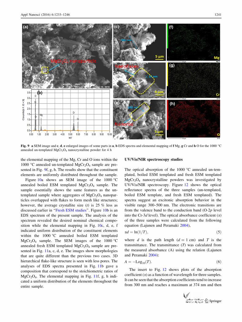
the elemental mapping of the Mg, Cr and O ions within the
1000 �C annealed un-templated MgCr2O4 sample are pre-
sented in Fig. 9f, g, h. The results show that the constituent
elements are uniformly distributed throughout the sample.
Figure 10a shows an SEM image of the 1000 �Cannealed boiled ESM templated MgCr2O4 sample. The
sample essentially shows the same features as the un-
templated sample where aggregates of MgCr2O4 nanopar-
ticles overlapped with flakes to form mesh like structures;
however, the average crystallite size (t) is 25 % less as
discussed earlier in ‘‘Fresh ESM studies’’. Figure 10b is an
EDS spectrum of the present sample. The analysis of the
spectrum revealed the desired nominal chemical compo-
sition while the elemental mapping in Fig. 10c, d, e, f
indicated uniform distribution of the constituent elements
within the 1000 �C annealed boiled ESM templated
MgCr2O4 sample. The SEM images of the 1000 �Cannealed fresh ESM templated MgCr2O4 sample are pre-
sented in Fig. 11a, c, d, e. The images show morphologies
that are quite different than the previous two cases. 3D
hierarchical flake-like structure is seen with less pores. The
analyses of EDS spectra presented in Fig. 11b gave a
composition that correspond to the stoichiometric ratios of
MgCr2O4. The elemental mapping in Fig. 11f, g, h indi-
cated a uniform distribution of the elements throughout the
entire sample.
UV/Vis/NIR spectroscopy studies
The optical absorption of the 1000 �C annealed un-tem-
plated, boiled ESM templated and fresh ESM templated
MgCr2O4 nanocrystalline powders was investigated by
UV/Vis/NIR spectroscopy. Figure 12 shows the optical
reflectance spectra of the three samples (un-templated,
boiled ESM template, and fresh ESM templated). The
spectra suggest an excitonic absorption behavior in the
visible range 300–500 nm. The electronic transitions are
from the valence band to the conduction band (O-2p level
into the Cr-3d level). The optical absorbance coefficient (a)
of the three samples were calculated from the following
equation (Lajunen and Peramaki 2004),
ad ¼ ln 1=Tð Þ; ð5Þ
where d is the path length (d = 1 cm) and T is the
transmittance. The transmittance (T) was calculated from
the measured absorbance (A) using the relation (Lajunen
and Peramaki 2004):
A ¼ �Log10 Tð Þ: ð6Þ
The insert to Fig. 12 shows plots of the absorption
coefficient (a) as a function of wavelength for three samples.
It can be seen that the absorption coefficients tend to increase
from 300 nm and reaches a maximum at 374 nm and then
Fig. 9 a SEM image and c, d, e enlarged images of some parts in a, b EDS spectra and elemental mapping of f Mg, g Cr and h O for the 1000 �Cannealed un-templated MgCr2O4 nanocrystalline powder for 4 h
Appl Nanosci (2016) 6:1233–1246 1241
123

decrease as the wavelength increases. This could be
attributed to inelastic scattering of charge carriers by
phonons, lattice deformations and internal electric fields
within the crystals. Similar behavior was observed for many
semiconducting materials (Willardson and Beer 1967).
The optical band gap energies of the 1000 �C annealed
un-templated, boiled ESM templated and fresh ESM tem-
plated MgCr2O4 nanocrystalline powders were calculated
from the wavelength value corresponding to the intersec-
tion point of the vertical and horizontal part of the spec-
trum, in accordance with following simple relation (Hamed
et al. 2016),
Eg ¼ 1240=k; ð7Þ
where Eg is the band gap energy (eV) and k is the
wavelength in (nm). The calculated values of energy band
gap from the above relation correspond to the absorption
limit. These values are listed in Table 2. In order to get
more precise values of the optical band gaps, the values of
Eg were calculated with the help of the Tauc equation
(Hamed et al. 2016),
ðaEpÞ ¼ CðEp � EgÞm; ð8Þ
where Ep (Ep = hm) is the incident photon energy, C is a
constant that depends on the transition probability and
m depends on the nature of the optical absorption transition
(Hamed et al. 2016). The value of m is 1/2 for direct
allowed electronic transition (direct band gap) and 2 for
indirect allowed electronic transition (indirect band gap)
(Zaki and Sc 2010). For this purpose, (ahm)1/2 was plotted
as a function of photon energy hm (eV) for the indirect gap
and (ahm)2 against photon energy hm (eV) for the direct gap.
Figure 13 is presentation of these plots. The linear intercept
at the hm on x-axis (shown in Fig. 13) gives the value of the
optical bandgap. The estimated optical band gap energies
are listed in Table 2. The band gap energies for the three
samples are very close, they range from 3.68 to 3.71 eV for
the direct band gap and from 3.30 to 3.37 eV for the
indirect band gap. These values are not too far from the
reported value of 3.4 eV for normal spinel ZnCr2O4 (Parhia
and Manivannan 2008). The 1000 �C annealed un-tem-
plated, boiled ESM templated and fresh ESM templated
MgCr2O4 nanocrystalline powders are sensitive to visible
light which could make them suitable photocatalysts
(Borse et al. 2011; Parhia and Manivannan 2008).
From the preceding sections, we can say that the use of
ESM as a template in the co-precipitation of MgCr2O4 has an
effect on the morphologies and the crystallite grain sizes in
the annealed templated MgCr2O4 nanocrystalline powders.
At 1000 �C, the ESM is completely burnt out and none of it is
Fig. 10 a, c, d SEM image, b EDS spectra and elemental mapping of g Mg, f Cr and h O and e elemental distribution for the 1000 �C annealed
boiled ESM templated MgCr2O4 nanocrystalline powder for 4 h
1242 Appl Nanosci (2016) 6:1233–1246
123

left within the annealed MgCr2O4 nanocrystalline powders.
It seems that the ESM plays a role during the co-precipitation
stage and possibly during the annealing stage when the
temperature is raised from room temperature to 1000 �C. A
simple experiment has confirmed the disappearance of ESM
when it was annealed in a matter of 15 min. As far as we
know, there are no reports on the optical band gap energies of
nanocrystalline MgCr2O4 except for the report by Tripathi
et al. They have reported a value of 1.71 eV for MgCr2O4
nanoparticles (Tripathi and Nagarajan 2016), we feel that
this value is too low in comparison of 3.46 eV reported by
Peng et al. for ZnCr2O4 nanoparticles (Peng and Gao 2008)
even though both groups reported similar features in their
optical absorption spectra. Both groups reported two extra
peaks centered on 440 nm and 600 nm in their optical
absorption spectra which we do not observe for our
nanocrystalline MgCr2O4 powders. Here we have reported
optical band gap energies of nanocrystalline MgCr2O4
powders. There were small variations in the values of the
optical band gap energies between the different samples.
There was also a little shading in the color of the three
nanocrystalline MgCr2O4 powders.
Conclusion
Fresh and boiled eggshell membranes (ESM) were used as
a biotemplates during the co-precipitation of pirochromite
MgCr2O4. The un-templated and templated co-precipitated
Fig. 11 a SEM image, b EDS spectra and elemental mapping of c Mg, d Cr and e O and e elemental distribution for the 1000 �C annealed fresh
ESM templated MgCr2O4 nanocrystalline powder for 4 h
Fig. 12 UV absorbance spectra for of the 1000 �C annealed MgCr2-
O4 nanocrystalline powder for 4 h a un-templated, b boiled ESM
templated and c fresh ESM templated
Appl Nanosci (2016) 6:1233–1246 1243
123

MgCr2O4 powders were subjected to heat treatment at
1000 �C for 4 h to produce single phase nanocrystalline
MgCr2O4 powders with spinel cubic crystal structure. The
un-templated and templated nanocrystalline MgCr2O4
powders were characterized by XRD, FTIR, SEM, EDS
and UV/Vis/NIR spectroscopy. The morphologies and the
crystallite grain sizes of the 1000 �C annealed templated
nanocrystalline MgCr2O4 powders were found to depend
on whether the ESM is boiled or fresh. The fresh ESM
templated nanocrystalline MgCr2O4 powders were found to
form 3D hierarchical cascading flake-like structure, the
boiled ESM templated nanocrystalline MgCr2O4 formed
mesh like structures whereas the un-templated nanocrys-
talline MgCr2O4 powders were found to form nano-sized
structures with irregular shapes. The un-templated and
templated nanocrystalline MgCr2O4 powders were found to
be sensitive to visible light absorption over the range
300–500 nm. This could make them possible photocata-
lysts over this range. The optical band gap energies were
found to vary from 3.68 to 3.71 eV for the direct band gap
and from 3.30 to 3.37 eV for the indirect band gap. The
concept of using ESM as a template should be extended to
other metal oxides nanomaterial.
Acknowledgments This research was supported by the UAEU Pro-
gram for Advanced Research (UPAR) under grant G00001647, UAE
University, Al Ain, United Arab Emirates.
Open Access This article is distributed under the terms of the
Creative Commons Attribution 4.0 International License (http://
creativecommons.org/licenses/by/4.0/), which permits unrestricted
use, distribution, and reproduction in any medium, provided you give
appropriate credit to the original author(s) and the source, provide a
link to the Creative Commons license, and indicate if changes were
made.
References
Andrade MJ, Lima MD, Bonadiman R, Bergmann CP (2006a)
Nanocrystalline pirochromite spinel through solution combus-
tion synthesis. Mater Res Bull 41:2070–2079
Andrade MJ, Lima MD, Bonadiman R, Bergmann CP (2006b)
Nanocrystalline pirochromite spinel through solution combus-
tion synthesis. Mater Res Bull 41:2070–2079
Table 2 Optical band gap energies for the 1000 �C annealed un-templated, boiled ESM templated and fresh ESM templated MgCr2O4
nanocrystalline powders for 4 h
Samples annealed at 1000 �C for 4 h Band gap (eV)
Simple method Kubelka–Munk function
Direct Indirect
Un-templated MgCr2O4 3.41 3.68 3.30
Boiled ESM templated MgCr2O4 3.53 3.71 3.37
Fresh ESM templated MgCr2O4 3.43 3.69 3.32
Fig. 13 Tauc plots for direct band gap (left side) and indirect band gap (right side) for the 1000 �C annealed MgCr2O4 nanocrystalline powders
for 4 h a un-templated, b boiled ESM templated and c fresh ESM templated
1244 Appl Nanosci (2016) 6:1233–1246
123

Arai H, Yamada T, Eguchi K, Seiyama T (1986) Catalytic combus-
tion of methane over various perovskite-type oxides. Appl Catal
26:265–276
Balaz M (2014) Eggshell membrane biomaterial as a platform for
applications in materials science. Acta Biomater 10:3827–3843
Benson KF, Ruff KJ, Jensen GS (2012) Effects of natural eggshell
membrane (NEM) on cytokine production in cultures of
peripheral blood mononuclear cells: increased suppression of
tumor necrosis factor-alpha levels after in vitro digestion. J Med
Food 15:360–368
Bhosale AG, Chougule BK (2006) X-ray, infrared and magnetic
studies of Al-substituted Ni ferrites. Mater Chem Phys
97:273–276
Borse PH, Jang JS, Lee JS, Khan FN, Ha MG, Kim JP, Bae JS, Jeong
ED, Kim HG (2011) Enhanced photocatalytic properties due to
electron-rich Ti-ion doping in ZnFe2O4 under visible light
irradiation. J Korean Phys Soc 59:2750–2755
Chen H, Liu J, Cheng X, Peng Y (2012) Adsorption for the removal
of malachite green by using eggshell membrane in environment
water samples. Adv Mater Res 573–754:63–67
Deng YY, Wang HZ, Zhao HZ (2008) Influence of chrome-bearing
sols vacuum impregnation on the properties of magnesia-chrome
refractory. Ceram Int 34:573–580
Drazic G, Trontelj M (1989) Preparation and properties of ceramic
sensor elements based on MgCr2O4. Sens Actuators 18:407–414
Finocchio E, Busca G, Lorenzelli V, Willey RJ (1994) FTIR studies
on the selective oxidation and combustion of light hydrocarbns
at metal oxide surfaces. Propane and propene oxidation on
MgCr2O4. J Chem Soc Faraday Trans 90:3347–3356
Finocchio E, Busca G, Lorenzelli V, Willey RJ (1995a) The
activation of hydrocarbon C–H bonds over transition metal
oxide catalysts: a FTIR study of hydrocarbon catalytic combus-
tion over MgCr2O4. J Catal 151:204–215
Finocchio E, Busca G, Lorenzelli V, Willey RJ (1995b) The
activation of hydrocarbon C–H bonds over transition metal
oxide catalysts: a FTIR study of hydrocarbon catalytic combus-
tion over MgCr2O4. J Catal 151:204–215
Ghosh A, Haldar MK, Das SK (2007) Effect of MgO and ZrO2
additions on the properties of magnesite-chrome composite
refractory. Ceram Int 33:821–825
Hamed F, Tholkappiyan R, Vishista K (2016) The effect of induced
strains on the optical band gaps in lanthanum doped zinc ferrite
nanocrystalline powders. Mod Phys Lett B 30:1650230
Hankare PP, Vader VT, Sankpal UB, Gavali LV, Sasikala R, Mulla IS
(2009) Effect of sintering temperature and thermoelectric power
studies of the system MgFe2-xCrxO4. Solid State Sci
11:2075–2079
Haralkar SJ, Kadam RH, More SS, Shirsath SE, Mane ML, Patil S,
Mane DR (2012) Substitutional effect of Cr3? ions on the
properties of Mg–Zn ferrite nano particles. Phys B
407:4338–4346
Hashimoto S, Yamagushi A (1995) Growth of MgCr2O4 whiskers.
J Cryst Growth 154:329–333
He H (2010) Catalysis and photocatalysis of MgCr2O4 powder at
room temperature. Recent Pat Chem Eng 3:74–77
Hosterman BD, Farley John W, Johnson Allen L (2013) Spectro-
scopic study of the vibrational modes of magnesium nickel
chromite, MgxNi1-xCr2O4. J Phys Chem Solids 74:985–990
Ishikawa S, Suyama K, Arihara K, Itoh M (2002) Uptake and
recovery of gold ions from electroplating wastes using eggshell
membrane. Bioresour Technol 81:201–206
Khalaf KAM, Al-Rawas AD, Widatallah HM, Al-Rashdi KS, Sellai
A, Gismelseed AM, Hashim M, Jameel SK, Al-Ruqeishi MS, Al-
Riyami KO, Shongwe M, Al-Rajhi AH (2016) Influence of Zn2?
ions on the structural and electrical properties of Mg1–xZnx-
FeCrO4 spinels. J Alloys Compd 657:733–747
Kim BN, Hiraga K, Morita K, Sakka Y (2001) A high-strain-rate
superplastic ceramic. Nature 413:288–291
Lajunen LHJ, Peramaki P (2004) Spectrochemical analysis by atomic
absorption and emission. Royal Society of Chemistry,
Cambridge
Li Z, Zhang L, Amirkhiz BS, Tan X, Xu Z, Wang H, Olsen BC, Holt
CMB, Mitlin D (2012) Carbonized chicken eggshell membranes
with 3D architectures as high-performance electrode materials
for supercapacitors. Adv Energy Mater 2:431–437
Lunge S, Thakre D, Kamble S, Labhsetwar N, Rayalu S (2012)
Alumina supported carbon composite material with exception-
ally high defluoridation property from eggshell waste. J Hazard
Mater 237:161–169
Mann S (2000) The chemistry of form. Angew Chem Int Ed Engl
39:3392–3406
Mann S (2001) Biomineralization. Principles and concepts in bioinor-
ganic materials chemistry. Oxford University Press, Oxford
Morozova LV, Popov VP (2010) Synthesis and investigation of
magnesium chromium spinel. Glass Phys Chem 36:86–91
Ning L, Tao L (2011) Adsorption and decoloration of nitroso dye
based on eggshell membrane. Adv Mater Res
183–185:963–966
O’Neill HSC, Dollase WA (1994) Crystal structures and cation
distributions in simple spinels from powder XRD structural
refinements: MgCr2O4, ZnCr2O4, Fe3O4 and the temperature
dependence of the cation distribution in ZnAl2O4. Phys Chem
Miner 20:541–555
Oliveira DA, Benelli P, Amante ER (2013) A literature review on
adding value to solid residues: egg shells. J Clean Prod 46:42–47
Parhia P, Manivannan V (2008) Microwave metathetic approach for
the synthesis and characterization of ZnCr2O4. J Eur Ceram Soc
28:1665–1670
Peng C, Gao L (2008) Optical and photocatalytic properties of spinel
ZnCr2O4 nanoparticles synthesized by a hydrothermal route.
J Am Ceram Soc 91:2388–2390
Rida K, Benabbas A, Bouremmad F, Pena MA, Martinez-Arias A
(2010) Influence of the synthesis method on structural properties
and catalytic activity for oxidation of CO and C3H6 of
pirochromite MgCr2O4. Appl Catal A 375:101–106
Ruff KJ, DeVore DP, Leu MD, Robinson MA (2009) Eggshell
membrane: a possible new natural therapeutic for joint and
connective tissue disorders. Results from two open-label human
clinical studies. Clin Interv Aging 4:235–240
Schoonman J, Dekker JP, Broers JW (1991) Electrochemical vapor
deposition of stabilized zirconia and interconnection materials
for solid oxide fuel cells. Solid State Ion 46:299
Stefanescua M, Barbu M, Vlase T, Barvinschi P, Barbu-Tudoran L,
Stoia M (2011) Novel low temperature synthesis method for
nanocrystalline zinc and magnesium chromites. Thermochim
Acta 526:130–136
Tripathi VK, Nagarajan R (2016) Rapid synthesis of mesoporous,
nano-sized MgCr2O4 and its catalytic properties. J Am Ceram
Soc 99:814–818
Wang WD, Chen B, Huang YM, Cao J (2010) Evaluation of eggshell
membrane-based bio-adsorbent for solid-phase extraction of
linear alkylbenzene sulfonates coupled with high-performance
liquid chromatography. J Chromatogr A 1217:5659–5664
White WB, De Angelis BA (1967) Interpretation of the vibrational
spectra of spinels. Spectrochim Acta A 23:985–995
Willardson RK, Beer AC (1967) Semiconductor and semimetals:
optical properties of III–V compounds. Academic Press, New
York
Appl Nanosci (2016) 6:1233–1246 1245
123

Zaki SM, Sc M (2010) Thesis in physics, Faculty of science. Tanta
University, Egypt
Zhang HM, Teraoka Y, Yamazoe N (1987) Preparation of perovskite-
type oxides with large surface area by citrate process. Chem Lett
16:665–668
Zheng BZ, Xie SP, Qian L, Yuan HY, Xiao D, Choi MMF (2011)
Gold nanoparticles coated eggshell membrane with immobilized
glucose oxidase for fabrication of glucose biosensor. Sens
Actuators B Chem 152:49–55
1246 Appl Nanosci (2016) 6:1233–1246
123