Synthesis, Characterization, Toxicity of...
27
15 Synthesis, Characterization, Toxicity of Nanomaterials for Biomedical Applications A. K. Pradhan, K. Zhang, M. Bahoura, J. Pradhan, P. Ravichandran, R. Gopikrishnan and G. T. Ramesh Norfolk State University, United State of America 1. Introduction Nanomaterials are widely used for biomedical applications as their sizes are comparable with most of the biological entities. Many diagnostic and therapeutic techniques based on nanoscience and nanotechnologies are already in the clinical trial stages, and encouraging results have been reported. The progress in nanoscience and nanotechnology has led to the formation and development of a new field, nanomedicine, which is generally defined as the biomedical applications of nanoscience and nanotechnology. Nanomedicine stands at the boundaries between physical, chemical, biological and medical sciences, and the advances in nanomedicine have made it possible to analyze and treat biological systems at the cell and sub-cell levels, providing revolutionary approaches for the diagnosis, prevention and treatment of some fatal diseases, such as cancer. Nanomagnetism is at the forefront of nanoscience and nanotechnology, and in the field of nanomedicine, magnetic nanomaterials are among the most promising for clinical diagnostic and therapeutic applications. Similarly, luminescent materials are equally important for tagging and imaging applications. The nanomaterials used for biomedical purposes generally include zero-dimensional nanoparticles, one-dimensional nanowires and nanotubes, and two-dimensional thin films. For example, magnetic nanoparticles and nanotubes are widely used for labeling and manipulating biomolecules, targeting drugs and genes, magnetic resonance imaging (MRI), as well as hyperthermia treatment. Magnetic thin films are often used in the development of nanosensors and nanosystems for analyzing biomolecules and diagnosing diseases. As the synthesis and characterization of these nanostructures are completely interdisciplinary, there is a need of coordinated efforts for the successful implementation of these nanomaterials. The synthesis of nanoparticles with required shape, size, and core-shell configuration (surface coating) along with proper characterization are still in the early stage of research. On the other hand, due to the similar size to biological systems, nanoparticles pose potential threats to health and they could consequently have a large impact on industry and society. Hence, apart from successful synthesis and characterization of various nanomaterials, an effort to understand the toxicological impacts of nanomaterials much research has to be done to establish standards and protocols for the safe use of nanomaterials in industry as well as in the public arena, including academia and research laboratories. Nanoparticles have sparked intense interest in anticipation that this unexplored range of material dimensions will yield size-dependent properties. The physical and chemical www.intechopen.com
-
Upload
trannguyet -
Category
Documents
-
view
222 -
download
0
Transcript of Synthesis, Characterization, Toxicity of...

15
Synthesis, Characterization, Toxicity of Nanomaterials for Biomedical Applications
A. K. Pradhan, K. Zhang, M. Bahoura, J. Pradhan, P. Ravichandran, R. Gopikrishnan and G. T. Ramesh
Norfolk State University,
United State of America
1. Introduction
Nanomaterials are widely used for biomedical applications as their sizes are comparable with most of the biological entities. Many diagnostic and therapeutic techniques based on nanoscience and nanotechnologies are already in the clinical trial stages, and encouraging results have been reported. The progress in nanoscience and nanotechnology has led to the formation and development of a new field, nanomedicine, which is generally defined as the biomedical applications of nanoscience and nanotechnology. Nanomedicine stands at the boundaries between physical, chemical, biological and medical sciences, and the advances in nanomedicine have made it possible to analyze and treat biological systems at the cell and sub-cell levels, providing revolutionary approaches for the diagnosis, prevention and treatment of some fatal diseases, such as cancer. Nanomagnetism is at the forefront of nanoscience and nanotechnology, and in the field of nanomedicine, magnetic nanomaterials are among the most promising for clinical diagnostic and therapeutic applications. Similarly, luminescent materials are equally important for tagging and imaging applications. The nanomaterials used for biomedical purposes generally include zero-dimensional nanoparticles, one-dimensional nanowires and nanotubes, and two-dimensional thin films. For example, magnetic nanoparticles and nanotubes are widely used for labeling and manipulating biomolecules, targeting drugs and genes, magnetic resonance imaging (MRI), as well as hyperthermia treatment. Magnetic thin films are often used in the development of nanosensors and nanosystems for analyzing biomolecules and diagnosing diseases. As the synthesis and characterization of these nanostructures are completely interdisciplinary, there is a need of coordinated efforts for the successful implementation of these nanomaterials. The synthesis of nanoparticles with required shape, size, and core-shell configuration (surface coating) along with proper characterization are still in the early stage of research. On the other hand, due to the similar size to biological systems, nanoparticles pose potential threats to health and they could consequently have a large impact on industry and society. Hence, apart from successful synthesis and characterization of various nanomaterials, an effort to understand the toxicological impacts of nanomaterials much research has to be done to establish standards and protocols for the safe use of nanomaterials in industry as well as in the public arena, including academia and research laboratories. Nanoparticles have sparked intense interest in anticipation that this unexplored range of material dimensions will yield size-dependent properties. The physical and chemical
www.intechopen.com
Biomedical Engineering, Trends in Materials Science
350
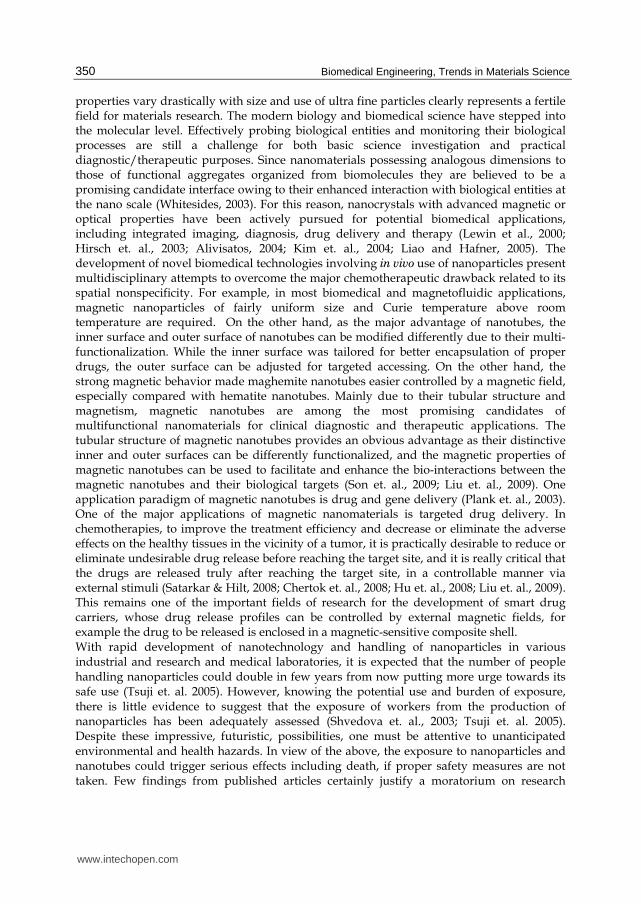
properties vary drastically with size and use of ultra fine particles clearly represents a fertile field for materials research. The modern biology and biomedical science have stepped into the molecular level. Effectively probing biological entities and monitoring their biological processes are still a challenge for both basic science investigation and practical diagnostic/therapeutic purposes. Since nanomaterials possessing analogous dimensions to those of functional aggregates organized from biomolecules they are believed to be a promising candidate interface owing to their enhanced interaction with biological entities at the nano scale (Whitesides, 2003). For this reason, nanocrystals with advanced magnetic or optical properties have been actively pursued for potential biomedical applications, including integrated imaging, diagnosis, drug delivery and therapy (Lewin et al., 2000; Hirsch et. al., 2003; Alivisatos, 2004; Kim et. al., 2004; Liao and Hafner, 2005). The development of novel biomedical technologies involving in vivo use of nanoparticles present multidisciplinary attempts to overcome the major chemotherapeutic drawback related to its spatial nonspecificity. For example, in most biomedical and magnetofluidic applications, magnetic nanoparticles of fairly uniform size and Curie temperature above room temperature are required. On the other hand, as the major advantage of nanotubes, the inner surface and outer surface of nanotubes can be modified differently due to their multi-functionalization. While the inner surface was tailored for better encapsulation of proper drugs, the outer surface can be adjusted for targeted accessing. On the other hand, the strong magnetic behavior made maghemite nanotubes easier controlled by a magnetic field, especially compared with hematite nanotubes. Mainly due to their tubular structure and magnetism, magnetic nanotubes are among the most promising candidates of multifunctional nanomaterials for clinical diagnostic and therapeutic applications. The tubular structure of magnetic nanotubes provides an obvious advantage as their distinctive inner and outer surfaces can be differently functionalized, and the magnetic properties of magnetic nanotubes can be used to facilitate and enhance the bio-interactions between the magnetic nanotubes and their biological targets (Son et. al., 2009; Liu et. al., 2009). One application paradigm of magnetic nanotubes is drug and gene delivery (Plank et. al., 2003). One of the major applications of magnetic nanomaterials is targeted drug delivery. In chemotherapies, to improve the treatment efficiency and decrease or eliminate the adverse effects on the healthy tissues in the vicinity of a tumor, it is practically desirable to reduce or eliminate undesirable drug release before reaching the target site, and it is really critical that the drugs are released truly after reaching the target site, in a controllable manner via external stimuli (Satarkar & Hilt, 2008; Chertok et. al., 2008; Hu et. al., 2008; Liu et. al., 2009). This remains one of the important fields of research for the development of smart drug carriers, whose drug release profiles can be controlled by external magnetic fields, for example the drug to be released is enclosed in a magnetic-sensitive composite shell. With rapid development of nanotechnology and handling of nanoparticles in various industrial and research and medical laboratories, it is expected that the number of people handling nanoparticles could double in few years from now putting more urge towards its safe use (Tsuji et. al. 2005). However, knowing the potential use and burden of exposure, there is little evidence to suggest that the exposure of workers from the production of nanoparticles has been adequately assessed (Shvedova et. al., 2003; Tsuji et. al. 2005). Despite these impressive, futuristic, possibilities, one must be attentive to unanticipated environmental and health hazards. In view of the above, the exposure to nanoparticles and nanotubes could trigger serious effects including death, if proper safety measures are not taken. Few findings from published articles certainly justify a moratorium on research
www.intechopen.com
Synthesis, Characterization, Toxicity of Nanomaterials for Biomedical Applications
351
involving nanoparticles, if not all nanoparticles, until proper safeguards can be put in place, moreover safety tests need to be carried out keeping in view the type of nanomaterials present. Currently, the literature surveys on suggested nanotoxicity are few to draw any conclusion on the exposure dose of nanoparticles required for toxicity.
2. Eu3+
doped Gd2O3 luminescent nanostructures
The nanoscale structures, which include nanoparticles, nanorods, nanowires, nanotubes and
nanobelts (He et. al., 2003; Chang et. al., 2005; Li et. al., 2007; Mao, et. al., 2008; Zhang et al.,
2009), have been considerably investigated due to their unique optical, electronic properties
and prospective application in diverse fields, such as high quality luminescent devices,
catalysts, sensors, biological labeling and other new functional optoelectronic devices. The
precise architectural manipulation of nanomaterials with well-defined morphologies and
accurately tunable sizes remains a research focus and a challenging issue due to the fact that
the properties of the materials closely interrelate with geometrical factors such as shape,
dimensionality, and size. The properties and performances of nanostructures strongly
depend on their dimensions, size, and morphologies (Liu et. al., 2007). Therefore, synthesis,
growth, and control of morphology in the crystallization process of nanostructures are of
critical importance for the development of novel technologies.
Rare earth doped oxides are promising new class of luminescent material due to their
electronic and optical properties that arises from their 4f electrons. Therefore, much
attention has been paid to their luminescent characteristics such as their large stokes shift,
sharp emission visible spectra, long fluorescence lifetime (1-2 ms), and lack of photo-
bleaching compared with dyes (Wang et. al., 2005; Nichkova et. al., 2006). These materials,
especially in the nanostructure, have been widely used in the lighting industry and
biotechnology, including plasma display panel, magnetic resonance imaging enhancement,
and microarray immunoassays for fluorescence labels (Seo et. al., 2002; Nichkova et. al.,
2005; Bridot et. al., 2007; Petoral et. al., 2009). Since the morphology and dimensionality of
nanostructures are of vital factors, which particularly have an effect on the physical,
chemical, optical, and electronic properties of materials, it is expected that rare earth doped
oxides synthesized in the form of nanoscale may take on novel spectroscopic properties of
both dimension controlled and modified ion-phonon confinement effect compared to their
bulk counterparts. Gd2O3, as a rare earth oxide, is a useful paramagnetic material and good
luminescent rare earth doped host. Eu3+ ions can be doped into Gd2O3 easily since they are
all trivalent ions and have the same crystal structure. Furthermore, 5D0 -7F2 of Eu3+
transitions exhibit red characteristic luminescence at a wavelength of 611 nm. Therefore,
lanthanide oxide doped nanostructures can be used as electrical, magnetic or optical
multifunction materials.
Recently, considerable efforts have been made to synthesize low dimensional
nanostructures (Chang et. al., 2005; Li et. al., 2007; Liu et. al., 2008). However, these
processes have to be involved in hydrothermal routine, template, and catalysts. The
nanostructure formed depends somehow on the pressure, template, and catalysts. This
results in experimental complexity, impurities, defects and high cost. In addition, these
methods especially could not meet large scale produce in industry. Therefore, it is necessary
to find new methods to synthesize shape, size, and dimensionality controlled lanthanide
doped oxides. On the other hand, because of the distinct low effective density, high specific
www.intechopen.com
Biomedical Engineering, Trends in Materials Science
352
surface area, and encapsulation ability in hollow nanotubes these nanostructures are
exceptionally promising in various fields such as confined catalysis, biotechnology, photonic
devices, and electrochemical cells (Xu & Asher, 2004; Lou et. al., 2006; Wei et. al., 2008).
Although lanthanide oxides are excellent host lattices for the luminescence of various
optically active lanthanide ions (Mao et. al., 2009), Gd2O3 is a promising host matrix for
down- and up conversion luminescence because of its good chemical durability, thermal
stability, and low phonon energy (Yang et. al., 2007; Jia et. al., 2009).
3. Synthesis of Gd2O3: Eu+3
nanostructures
Gd2O3 doped with Eu3+ nanostructures were synthesized by either sol-gel or co-
precipitation wet chemical solution methods. Nanoparticles were synthesized by a sol-gel
method from their acetate hydrate precursors, which were dissolved in water. This solution
was mixed with citric acid solution in 1:1 volume ratio ultrasonically for about 30 min. The
mixture was heated in a water bath at 80 °C until all water is evaporated, yielding a
yellowish transparent gel. The gel was further heated in an oven at 100 °C which formed a
foamy precursor. This precursor decomposed to give brown-colored flakes of extremely fine
particle size on further heating at 400 °C for 4 h. The flakes were ground and sintered at 800
°C for duration of 2 h. Further heating in O2 ambient removed the carbon content.
The nanoparticles of Eu:Gd2O3 were coated by adopting a base-catalyzed sol-gel process.
100 mg of Eu:Gd2O3 were dispersed in 20 ml of 2-propanol solution and sonicated for 30
min. 75 µl of tetraethoxysilane (TEOS) and 25 µL of 25% NH3H2O solution were injected into
the above mixture and sonicated for 30 min at 60 ºC. By means of centrifugation the
suspended silica capsulated Eu:Gd2O3 were obtained. The coated particles were washed
several times by using acetone and methanol in order to remove any excess unreacted
chemicals. The purified powder was naturally dried. This procedure produces a very
uniform SiO2 coating, as determined using a transmission electron microscope (TEM). By
changing the formulation of the coating solution, we can control the coating thickness.
In the co-precipitation method, 0.5 M aqueous solution was prepared by dissolving
Gd(NO3)3 and Eu(NO3)3 in deionized H2O. The nitrate solutions with cationic molar ratio of
Gd to Eu is 0.95: 0.05 were mixed together and stirred for 30 minutes. The aqueous solution
of 0.2 M NH4HCO3 was prepared and mixed with the nitrate solution drop wise while
stirring to form the precipitate. It is noted that in this experiment extra 10 mol% NH4HCO3
was added in order to ensure all the rare earth ions reacted completely to obtain rare earth
carbonates. The white precipitate slurry obtained was aged for 24 hours at room
temperature with continuous stirring. Then the precipitates were centrifugated and washed
with deionized water for 5 times in order to completely remove NO3-, NH4+ and HCO3-
followed by drying at about 75 oC in the stove. After drying, the white precursor was
ground several times. It is noted that the dried precursor powders were very loosely
agglomerated and can be pulverized very easily. To get Gd2O3 doped with Eu3+
nanostructures, the as-synthesized samples were further calcined at 600, 800, and 1000 oC in
air for 2 hours in the furnace, respectively.
Eu3+ doped Gd2O3 nanotubes were synthesized according to a modified wet chemical method (He et. al., 2003). A mixture of 30 ml of 0.08 M Gd(NO3)3 and Eu(NO3)3 with a nominal molar ratio of Eu/Gd 5 atom %, in a form of clear solution, were added into flasks through ultrasound for 10 min. 30 ml of 25 wt % of ammonia solution was added quickly
www.intechopen.com
Synthesis, Characterization, Toxicity of Nanomaterials for Biomedical Applications
353
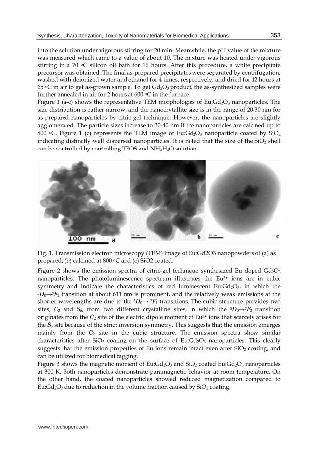
into the solution under vigorous stirring for 20 min. Meanwhile, the pH value of the mixture was measured which came to a value of about 10. The mixture was heated under vigorous stirring in a 70 oC silicon oil bath for 16 hours. After this procedure, a white precipitate precursor was obtained. The final as-prepared precipitates were separated by centrifugation, washed with deionized water and ethanol for 4 times, respectively, and dried for 12 hours at 65 oC in air to get as-grown sample. To get Gd2O3 product, the as-synthesized samples were further annealed in air for 2 hours at 600 oC in the furnace. Figure 1 (a-c) shows the representative TEM morphologies of Eu:Gd2O3 nanoparticles. The
size distribution is rather narrow, and the nanocrytallite size is in the range of 20-30 nm for
as-prepared nanoparticles by citric-gel technique. However, the nanoparticles are slightly
agglomerated. The particle sizes increase to 30-40 nm if the nanoparticles are calcined up to
800 oC. Figure 1 (c) represents the TEM image of Eu:Gd2O3 nanoparticle coated by SiO2
indicating distinctly well dispersed nanoparticles. It is noted that the size of the SiO2 shell
can be controlled by controlling TEOS and NH3H2O solution.
Fig. 1. Transmission electron microscopy (TEM) image of Eu:Gd2O3 nanopowders of (a) as prepared, (b) calcined at 800 oC and (c) SiO2 coated.
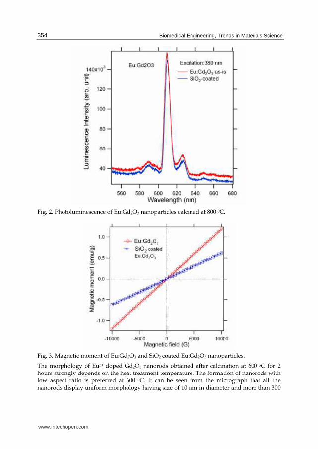
Figure 2 shows the emission spectra of citric-gel technique synthesized Eu doped Gd2O3
nanoparticles. The photoluminescence spectrum illustrates the Eu3+ ions are in cubic
symmetry and indicate the characteristics of red luminescent Eu:Gd2O3, in which the 5D0→7F2 transition at about 611 nm is prominent, and the relatively weak emissions at the
shorter wavelengths are due to the 5D0→ 7F1 transitions. The cubic structure provides two
sites, C2 and S6, from two different crystalline sites, in which the 5D0→7F2 transition
originates from the C2 site of the electric dipole moment of Eu3+ ions that scarcely arises for
the S6 site because of the strict inversion symmetry. This suggests that the emission emerges
mainly from the C2 site in the cubic structure. The emission spectra show similar
characteristics after SiO2 coating on the surface of Eu:Gd2O3 nanoparticles. This clearly
suggests that the emission properties of Eu ions remain intact even after SiO2 coating, and
can be utilized for biomedical tagging.
Figure 3 shows the magnetic moment of Eu:Gd2O3 and SiO2 coated Eu:Gd2O3 nanoparticles
at 300 K. Both nanoparticles demonstrate paramagnetic behavior at room temperature. On
the other hand, the coated nanoparticles showed reduced magnetization compared to
Eu:Gd2O3 due to reduction in the volume fraction caused by SiO2 coating.
www.intechopen.com
Biomedical Engineering, Trends in Materials Science
354
Fig. 2. Photoluminescence of Eu:Gd2O3 nanoparticles calcined at 800 0C.
Fig. 3. Magnetic moment of Eu:Gd2O3 and SiO2 coated Eu:Gd2O3 nanoparticles.
The morphology of Eu3+ doped Gd2O3 nanorods obtained after calcination at 600 oC for 2 hours strongly depends on the heat treatment temperature. The formation of nanorods with low aspect ratio is preferred at 600 oC. It can be seen from the micrograph that all the nanorods display uniform morphology having size of 10 nm in diameter and more than 300
www.intechopen.com

Synthesis, Characterization, Toxicity of Nanomaterials for Biomedical Applications
355
nm in length (Figure 4(a)). In contrast, the nanorods grow bigger in diameter (about 25 nm) and shorter in length (about 100 nm) after the heat treatment at 800 oC as shown in Figure 4(c). However, it is evident that Eu3+ doped Gd2O3 nanorods maintain the anisotropic shape during heat treatment from 600 oC to 800 oC. It can also be observed that the formation of nanorods is related to the fact that the growth direction are preferred along the [211] crystallographic orientation. This is because the spacing between fringes along nanorod axes is about 0.40 nm which is close to the interplanar distance of the cubic (211) plane as shown in Figure 4 (b) and (d). Figure 4(e) presents the TEM images of Eu3+ doped Gd2O3 nanoparticles with size of 60 nm in diameter obtained by heat treatment at 1000 oC. The morphology of Eu3+ doped Gd2O3 nanostructure dependent on the heat treatment temperature is possibly attributed to meta-stable states which are able to recrystallize at 1000 oC. A favorable growth pattern parallel to the (222) plain corresponding to interplanar spacing of 0.3 nm dominates the recrystallization of nanorods and transFigures to form nanoparticles as shown in Figure 4(f).
Fig. 4. Eu3+ doped Gd2O3 nanostructures TEM photographs of low and high magnification after annealing at (a) and (b) 600 oC, (c) and (d) 800 oC, and (e) and (f) 1000 oC, respectively.(b), (d) and (f) represent the HR-TEM images of respective nanostructures.
www.intechopen.com
Biomedical Engineering, Trends in Materials Science
356
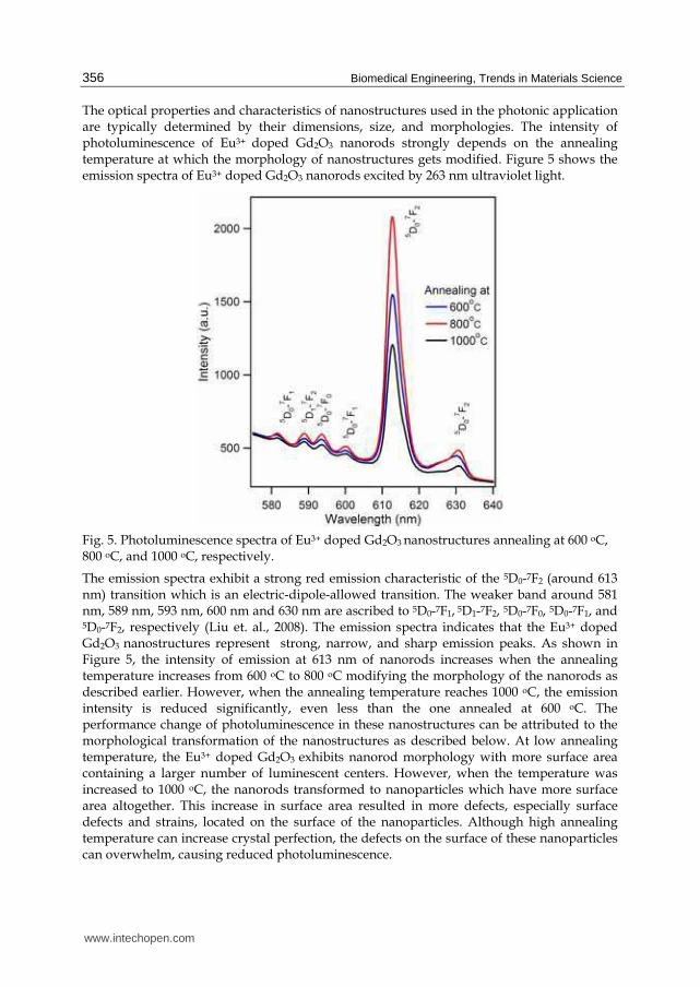
The optical properties and characteristics of nanostructures used in the photonic application are typically determined by their dimensions, size, and morphologies. The intensity of photoluminescence of Eu3+ doped Gd2O3 nanorods strongly depends on the annealing temperature at which the morphology of nanostructures gets modified. Figure 5 shows the emission spectra of Eu3+ doped Gd2O3 nanorods excited by 263 nm ultraviolet light.
Fig. 5. Photoluminescence spectra of Eu3+ doped Gd2O3 nanostructures annealing at 600 oC, 800 oC, and 1000 oC, respectively.
The emission spectra exhibit a strong red emission characteristic of the 5D0-7F2 (around 613 nm) transition which is an electric-dipole-allowed transition. The weaker band around 581 nm, 589 nm, 593 nm, 600 nm and 630 nm are ascribed to 5D0-7F1, 5D1-7F2, 5D0-7F0, 5D0-7F1, and 5D0-7F2, respectively (Liu et. al., 2008). The emission spectra indicates that the Eu3+ doped Gd2O3 nanostructures represent strong, narrow, and sharp emission peaks. As shown in Figure 5, the intensity of emission at 613 nm of nanorods increases when the annealing temperature increases from 600 oC to 800 oC modifying the morphology of the nanorods as described earlier. However, when the annealing temperature reaches 1000 oC, the emission intensity is reduced significantly, even less than the one annealed at 600 oC. The performance change of photoluminescence in these nanostructures can be attributed to the morphological transformation of the nanostructures as described below. At low annealing temperature, the Eu3+ doped Gd2O3 exhibits nanorod morphology with more surface area containing a larger number of luminescent centers. However, when the temperature was increased to 1000 oC, the nanorods transformed to nanoparticles which have more surface area altogether. This increase in surface area resulted in more defects, especially surface defects and strains, located on the surface of the nanoparticles. Although high annealing temperature can increase crystal perfection, the defects on the surface of these nanoparticles can overwhelm, causing reduced photoluminescence.
www.intechopen.com
Synthesis, Characterization, Toxicity of Nanomaterials for Biomedical Applications
357
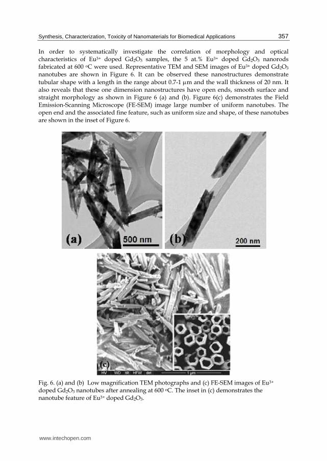
In order to systematically investigate the correlation of morphology and optical characteristics of Eu3+ doped Gd2O3 samples, the 5 at.% Eu3+ doped Gd2O3 nanorods fabricated at 600 oC were used. Representative TEM and SEM images of Eu3+ doped Gd2O3 nanotubes are shown in Figure 6. It can be observed these nanostructures demonstrate
tubular shape with a length in the range about 0.7-1 μm and the wall thickness of 20 nm. It also reveals that these one dimension nanostructures have open ends, smooth surface and straight morphology as shown in Figure 6 (a) and (b). Figure 6(c) demonstrates the Field Emission-Scanning Microscope (FE-SEM) image large number of uniform nanotubes. The open end and the associated fine feature, such as uniform size and shape, of these nanotubes are shown in the inset of Figure 6.
Fig. 6. (a) and (b) Low magnification TEM photographs and (c) FE-SEM images of Eu3+ doped Gd2O3 nanotubes after annealing at 600 oC. The inset in (c) demonstrates the nanotube feature of Eu3+ doped Gd2O3.
www.intechopen.com
Biomedical Engineering, Trends in Materials Science
358
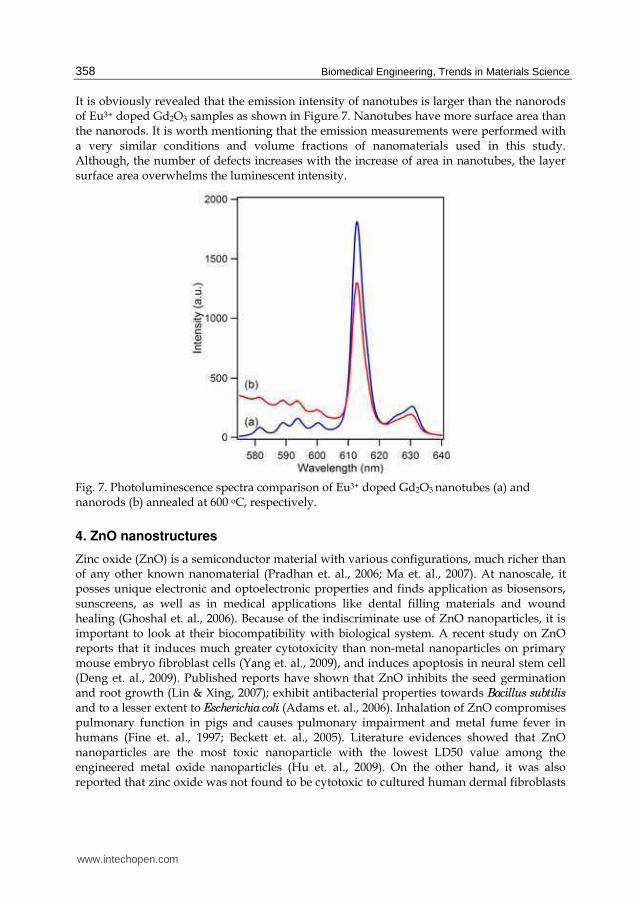
It is obviously revealed that the emission intensity of nanotubes is larger than the nanorods of Eu3+ doped Gd2O3 samples as shown in Figure 7. Nanotubes have more surface area than the nanorods. It is worth mentioning that the emission measurements were performed with a very similar conditions and volume fractions of nanomaterials used in this study. Although, the number of defects increases with the increase of area in nanotubes, the layer surface area overwhelms the luminescent intensity.
Fig. 7. Photoluminescence spectra comparison of Eu3+ doped Gd2O3 nanotubes (a) and nanorods (b) annealed at 600 oC, respectively.
4. ZnO nanostructures
Zinc oxide (ZnO) is a semiconductor material with various configurations, much richer than of any other known nanomaterial (Pradhan et. al., 2006; Ma et. al., 2007). At nanoscale, it posses unique electronic and optoelectronic properties and finds application as biosensors, sunscreens, as well as in medical applications like dental filling materials and wound healing (Ghoshal et. al., 2006). Because of the indiscriminate use of ZnO nanoparticles, it is important to look at their biocompatibility with biological system. A recent study on ZnO reports that it induces much greater cytotoxicity than non-metal nanoparticles on primary mouse embryo fibroblast cells (Yang et. al., 2009), and induces apoptosis in neural stem cell (Deng et. al., 2009). Published reports have shown that ZnO inhibits the seed germination and root growth (Lin & Xing, 2007); exhibit antibacterial properties towards Bacillus subtilis
and to a lesser extent to Escherichia coli (Adams et. al., 2006). Inhalation of ZnO compromises pulmonary function in pigs and causes pulmonary impairment and metal fume fever in humans (Fine et. al., 1997; Beckett et. al., 2005). Literature evidences showed that ZnO nanoparticles are the most toxic nanoparticle with the lowest LD50 value among the engineered metal oxide nanoparticles (Hu et. al., 2009). On the other hand, it was also reported that zinc oxide was not found to be cytotoxic to cultured human dermal fibroblasts
www.intechopen.com

Synthesis, Characterization, Toxicity of Nanomaterials for Biomedical Applications
359
(Zaveri et. al., 2009). In recent years, there has been an escalation in the development of techniques for synthesis of nanorods and subsequent surface functionalization. ZnO nanorods exhibit characteristic electronic, optical, and catalytic properties significantly different from other nano metals. Keeping in view of the unique properties and the extensive use of ZnO in many fields and also contradictory results on ZnO toxicity from both in-vitro and in-vivo studies, we report here to synthesize and characterize the ZnO nanorods on hela cells for its biocompatibility/toxicity.
5. Synthesis: ZnO nanotubes
The typical method employed is as follows. Equal volume of 0.1 M aqueous Zinc acetate anhydrous and Hexamethylenetetramine were mixed in a beaker using ultrasonication for 30 min. After the mixture was mixed well, it was heated at 80 °C in water bath for 75 min, during which white precipitates were deposited at the bottom. Then it was incubated for 30 min in ice cold water to terminate the reaction. The product was washed several times (till the pH of solution becomes neutral) using the centrifuge with deionized water and alcohol, alternatively to remove any by-product and excess of hexamethyleneteteamine. After washing, the solution was centrifuged at 10,000 rpm (12,000×g) for 20 min and the settled ZnO was dried at 80 ◦C for 2 h. Fig. 8 (a, b) shows the SEM micrograph collected on synthesized ZnO nanorods surface morphology. The nanorod was grown perpendicular to the long-axis of the matrix rod and grew along the [001] direction, which is the nature of ZnO growth. The morphology of ZnO nanorod was further confirmed by the TEM image as shown in Fig. 8 (c, d). Though the rod cores were monodisperse, the length of the nanorod was estimated to be around 21 nm in diameter and the length around 50 nm.
Fig. 8. (a and b) Scanning electron micrograph of ZnO nanorods. (c and d) Transmission electron micrograph of ZnO nanorods.
www.intechopen.com
Biomedical Engineering, Trends in Materials Science
360
6. Toxicity studies: Eu:Gd2O3 nanoparticles
For cell culture and treatments, rat lung epithelial cell line (LE, RL 65, ATCC; CRL- 10354)
from ATTC was grown at 37 °C in an atmosphere of 5% CO2 and in complete growth
medium supplemented with 1% penicillin/streptomycin and 10% fetal bovine serum (FBS).
Eu:Gd2O3 were suspended in Dimethyl formamide (DMF) and sonicated for 5 minutes and
henceforth in all control experiments the cells were treated with equivalent volume of DMF.
The cells were incubated with or without nanoparticles in 96 well plates for time intervals as
indicated in the respective Figure legends.
The measurements of intracellular reactive oxygen species (ROS) were performed in the
following way. Oxygen radicals collectively called as reactive oxygen species play a key role
in cytotoxicity. Increased ROS levels in cells by chemical compounds reflect toxicity and cell
death. To study the induction of oxidative stress in LE cells, 1x10⁴ cells/well were seeded in
96 well plate and grown overnight under standard culture conditions. The cells were then
treated with 10 µM of dichlorofluorescein [5-(and-6)-carboxy-2,7`-dichloro-
dihydroxyfluorescein diacetate, H2DCFDA, (C-400, Molecular Probes, Eugene, OR) for 3 h in
Hank’s balanced salt solution (HBSS) in incubator. Following 3 h of incubation, cells were
washed with phosphate buffered saline (PBS) and treated with different concentrations of
Eu:Gd2O3 nanoparticles. Following incubation the intensity of fluorescence is measured at
different time intervals at excitation and emission of wavelength at 485/527 nm,
respectively and expressed as fluorescence units.
LE cells were seeded at 5x10³ cells/well in a 96 well plate and allowed to grow overnight.
After 18 h in serum-free medium, cells were treated with different concentrations of
nanoparticles and grown for 72 h. At the end of the incubation, cells were additionally
treated with 3-[4, 5-dimethylthiazol- 2-yl]-2,5-diphenyltetrazolium bromide] MTT for 3 h.
The cells were then washed with chilled PBS and formazon formed was solubilized in 100
µL of acidic propanol and the absorbance was read at 570 nm.
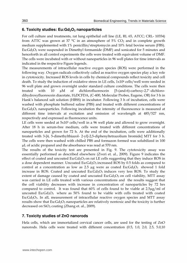
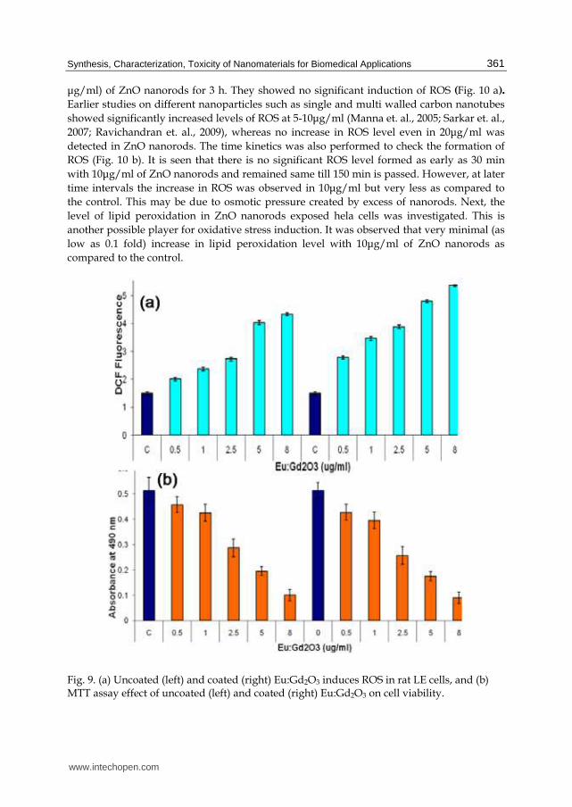
The results of the toxicity test are presented in Fig. 9. The cytotoxicity assay was essentially performed as described elsewhere (Zveri et. al., 2009). Figure 9 indicates the effect of coated and uncoated Eu:Gd2O3 on rat LE cells suggesting that they induce ROS in a dose dependent manner. Uncoated Eu:Gd2O3 increased ROS by 0.5 folds as compared to control at a concentration as low as 2.5 µg were as coated Eu:Gd2O3 showed 1 fold increase in ROS. Coated and uncoated Eu:Gd2O3 induces very less ROS. To study the extent of damage caused by coated and uncoated Eu:Gd2O3 on cell viability, MTT assay was carried in LE cells treated with various concentrations and the results suggest that the cell viability decreases with increase in concentration of nanoparticles by 72 hrs compared to control. It was found that 60% of cells found to be viable at 2.5µg/ml of uncoated Eu:Gd2O3 where as 50% found to be viable with cells treated with coated Eu:Gd2O3. In all, measurement of intracellular reactive oxygen species and MTT assay results show that Eu:Gd2O3 nanoparticles are relatively nontoxic and the toxicity is further decreased on SiO2 coating (Zhang et. al., 2009).
7. Toxicity studies of ZnO nanorods
Hela cells, which are immortalized cervical cancer cells, are used for the testing of ZnO
nanorods. Hela cells were treated with different concentration (0.5, 1.0, 2.0, 2.5, 5.0,10
www.intechopen.com
Synthesis, Characterization, Toxicity of Nanomaterials for Biomedical Applications
361
μg/ml) of ZnO nanorods for 3 h. They showed no significant induction of ROS (Fig. 10 a).
Earlier studies on different nanoparticles such as single and multi walled carbon nanotubes
showed significantly increased levels of ROS at 5-10μg/ml (Manna et. al., 2005; Sarkar et. al.,
2007; Ravichandran et. al., 2009), whereas no increase in ROS level even in 20μg/ml was
detected in ZnO nanorods. The time kinetics was also performed to check the formation of
ROS (Fig. 10 b). It is seen that there is no significant ROS level formed as early as 30 min
with 10μg/ml of ZnO nanorods and remained same till 150 min is passed. However, at later
time intervals the increase in ROS was observed in 10μg/ml but very less as compared to
the control. This may be due to osmotic pressure created by excess of nanorods. Next, the
level of lipid peroxidation in ZnO nanorods exposed hela cells was investigated. This is
another possible player for oxidative stress induction. It was observed that very minimal (as
low as 0.1 fold) increase in lipid peroxidation level with 10μg/ml of ZnO nanorods as
compared to the control.
Fig. 9. (a) Uncoated (left) and coated (right) Eu:Gd2O3 induces ROS in rat LE cells, and (b) MTT assay effect of uncoated (left) and coated (right) Eu:Gd2O3 on cell viability.
www.intechopen.com
Biomedical Engineering, Trends in Materials Science
362
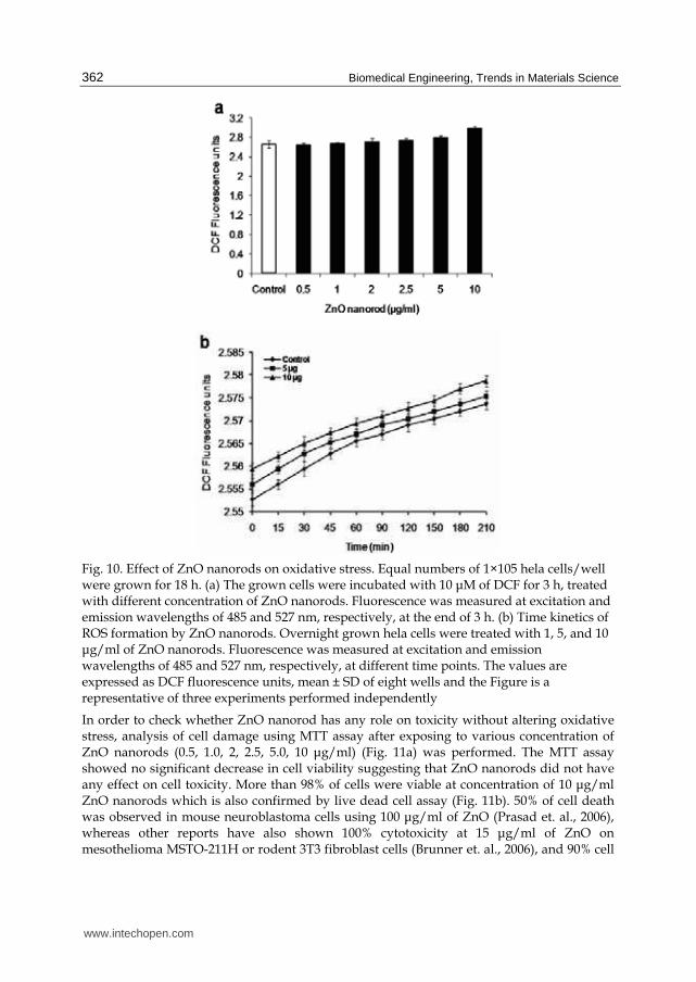
Fig. 10. Effect of ZnO nanorods on oxidative stress. Equal numbers of 1×10⁵ hela cells/well were grown for 18 h. (a) The grown cells were incubated with 10 μM of DCF for 3 h, treated with different concentration of ZnO nanorods. Fluorescence was measured at excitation and emission wavelengths of 485 and 527 nm, respectively, at the end of 3 h. (b) Time kinetics of ROS formation by ZnO nanorods. Overnight grown hela cells were treated with 1, 5, and 10 μg/ml of ZnO nanorods. Fluorescence was measured at excitation and emission wavelengths of 485 and 527 nm, respectively, at different time points. The values are expressed as DCF fluorescence units, mean ± SD of eight wells and the Figure is a representative of three experiments performed independently
In order to check whether ZnO nanorod has any role on toxicity without altering oxidative stress, analysis of cell damage using MTT assay after exposing to various concentration of ZnO nanorods (0.5, 1.0, 2, 2.5, 5.0, 10 μg/ml) (Fig. 11a) was performed. The MTT assay showed no significant decrease in cell viability suggesting that ZnO nanorods did not have any effect on cell toxicity. More than 98% of cells were viable at concentration of 10 μg/ml ZnO nanorods which is also confirmed by live dead cell assay (Fig. 11b). 50% of cell death was observed in mouse neuroblastoma cells using 100 μg/ml of ZnO (Prasad et. al., 2006), whereas other reports have also shown 100% cytotoxicity at 15 μg/ml of ZnO on mesothelioma MSTO-211H or rodent 3T3 fibroblast cells (Brunner et. al., 2006), and 90% cell
www.intechopen.com
Synthesis, Characterization, Toxicity of Nanomaterials for Biomedical Applications
363
death with 20mgL−1 of ZnO nanoparticles on HELF cells (Yuan et. al., 2010). Also, 5 mM of ZnO nanoparticle are shown to be less toxic to human T cells (Reddy et. al., 2007). Previous studies from our laboratory on hela cells and other cells such as lung epithelial, H1299, A549 and HaCaT cells showed the decrease in cell viability at 5 μg/ml when they were exposed to SWCNT and MWCNT (Manna et. al., 2005; Sarkar et. al., 2007; Ravichandran et. al., 2009). Toxicological studies on hela cells and conclude that ZnO nanorods could be the safe nanomaterials (Gopikrishnan el. al., 2010) for biological applications.
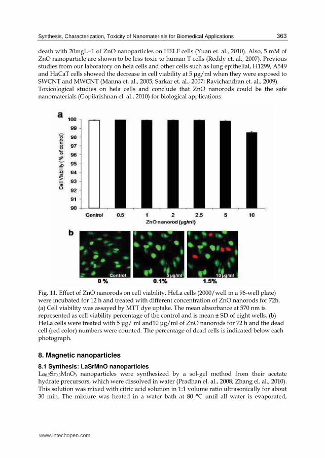
Fig. 11. Effect of ZnO nanorods on cell viability. HeLa cells (2000/well in a 96-well plate) were incubated for 12 h and treated with different concentration of ZnO nanorods for 72h. (a) Cell viability was assayed by MTT dye uptake. The mean absorbance at 570 nm is represented as cell viability percentage of the control and is mean ± SD of eight wells. (b) HeLa cells were treated with 5 μg/ ml and10 μg/ml of ZnO nanorods for 72 h and the dead cell (red color) numbers were counted. The percentage of dead cells is indicated below each photograph.
8. Magnetic nanoparticles
8.1 Synthesis: LaSrMnO nanoparticles La0.7Sr0.3MnO3 nanoparticles were synthesized by a sol-gel method from their acetate hydrate precursors, which were dissolved in water (Pradhan el. al., 2008; Zhang el. al., 2010). This solution was mixed with citric acid solution in 1:1 volume ratio ultrasonically for about 30 min. The mixture was heated in a water bath at 80 °C until all water is evaporated,
www.intechopen.com

Biomedical Engineering, Trends in Materials Science
364
yielding a yellowish transparent gel. The gel was further heated in an oven at 100 °C which formed a foamy precursor. This precursor was decomposed to give black-colored flakes of extremely fine particle size on further heating at 400 °C for 4 h. The flakes were ground and sintered at 800 °C for duration of 2 h. Further heating in O2 ambient removed the carbon content. The ball milling was used with methanol to reduce the size of nanoparticles of LSMO (Fig. 12). The solution containing suspended LSMO nanoparticles was separated using ultra-high centrifuge using methanol for several times.
Fig. 12. FE-EM image of LSMO nanoparticles annealed at 800 oC, showing the individual nanoparticles.
The nanoparticles of ball milled LSMO were coated by adopting a base-catalyzed sol-gel process. 100 mg of LSMO were dispersed in 20 ml of 2-propanol solution and sonicated for 30 min and the nanoparticles were shown in Fig. 13 (a). 75 µl of TEOS and 25 µL of 25% NH3H2O solution were injected into the above mixture and sonicated for 30 min at 60 ºC. The suspended silica capsulated LSMO nanoparticles were obtained by means of centrifugation. The coated nanoparticles were washed several times by using acetone and methanol in order to remove any excess unreacted chemicals. The purified powder was naturally dried. This procedure produces a very uniform SiO2 coating, as determined using a transmission electron microscope. By changing the formulation of the coating solution, the coating thickness can be controlled.
8.2 FeCo nanoparticles FeCo nanoparticles were synthesized by a coprecipitation method under Ar atmosphere from their chloride hydrate precursors. The FeCo nanopowders were dried in Ar gas, and were dispersed in 2- propanol solvent with 10-2 M and sonicated for 1 hour followed by addition of TEOS and 25% ammonia solution of volume ration 3:1. The mixture was sonicated for 1 h to coat the SiO2 onto the surface of FeCo nanoparticles. The solution containing suspended FeCo-SiO2 nanoparticles was decanted and purified using methanol several times in order to remove unreacted Fe and organic materials from the surface. The coated nanopowders were naturally dried in air. Figure 14 (a) shows XRD pattern of the as-synthesized samples, indicating typical amorphous phase. The amorphous phase in FeCo nanoparticles is generated because the coprecipitation reaction takes place below the glass
www.intechopen.com
Synthesis, Characterization, Toxicity of Nanomaterials for Biomedical Applications
365
transition temperature and boron atoms are presented in the nanoparticles. The solution containing suspended FeCo-SiO2 nanoparticles was decanted and purified using methanol several times in order to remove unreacted Fe and organic materials from the surface. The coated nanopowders were naturally dried in air.
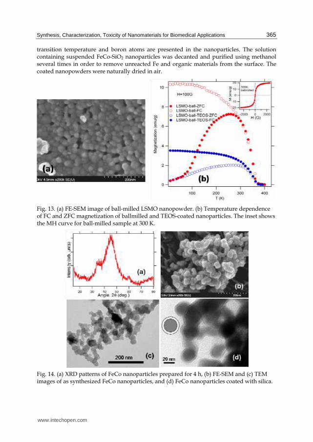
Fig. 13. (a) FE-SEM image of ball-milled LSMO nanopowder. (b) Temperature dependence of FC and ZFC magnetization of ballmilled and TEOS-coated nanoparticles. The inset shows the MH curve for ball-milled sample at 300 K.
Fig. 14. (a) XRD patterns of FeCo nanoparticles prepared for 4 h, (b) FE-SEM and (c) TEM images of as synthesized FeCo nanoparticles, and (d) FeCo nanoparticles coated with silica.
www.intechopen.com
Biomedical Engineering, Trends in Materials Science
366
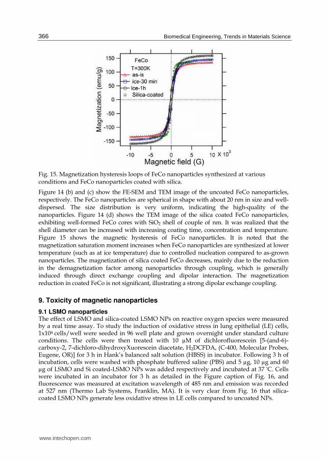
Fig. 15. Magnetization hysteresis loops of FeCo nanoparticles synthesized at various conditions and FeCo nanoparticles coated with silica.
Figure 14 (b) and (c) show the FE-SEM and TEM image of the uncoated FeCo nanoparticles, respectively. The FeCo nanoparticles are spherical in shape with about 20 nm in size and well-dispersed. The size distribution is very uniform, indicating the high-quality of the nanoparticles. Figure 14 (d) shows the TEM image of the silica coated FeCo nanoparticles, exhibiting well-formed FeCo cores with SiO2 shell of couple of nm. It was realized that the shell diameter can be increased with increasing coating time, concentration and temperature. Figure 15 shows the magnetic hysteresis of FeCo nanoparticles. It is noted that the magnetization saturation moment increases when FeCo nanoparticles are synthesized at lower temperature (such as at ice temperature) due to controlled nucleation compared to as-grown nanoparticles. The magnetization of silica coated FeCo decreases, mainly due to the reduction in the demagnetization factor among nanoparticles through coupling, which is generally induced through direct exchange coupling and dipolar interaction. The magnetization reduction in coated FeCo is not significant, illustrating a strong dipolar exchange coupling.
9. Toxicity of magnetic nanoparticles
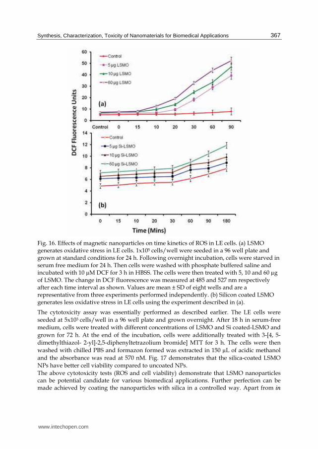
9.1 LSMO nanoparticles The effect of LSMO and silica-coated LSMO NPs on reactive oxygen species were measured by a real time assay. To study the induction of oxidative stress in lung epithelial (LE) cells, 1x10⁴ cells/well were seeded in 96 well plate and grown overnight under standard culture conditions. The cells were then treated with 10 µM of dichlorofluorescein [5-(and-6)-carboxy-2, 7-dichloro-dihydroxyXuorescein diacetate, H2DCFDA, (C-400, Molecular Probes, Eugene, OR)] for 3 h in Hank’s balanced salt solution (HBSS) in incubator. Following 3 h of incubation, cells were washed with phosphate buffered saline (PBS) and 5 µg, 10 µg and 60 µg of LSMO and Si coated-LSMO NPs was added respectively and incubated at 37 ºC. Cells were incubated in an incubator for 3 h as detailed in the Figure caption of Fig. 16, and fluorescence was measured at excitation wavelength of 485 nm and emission was recorded at 527 nm (Thermo Lab Systems, Franklin, MA). It is very clear from Fig. 16 that silica-coated LSMO NPs generate less oxidative stress in LE cells compared to uncoated NPs.
www.intechopen.com
Synthesis, Characterization, Toxicity of Nanomaterials for Biomedical Applications
367
Fig. 16. Effects of magnetic nanoparticles on time kinetics of ROS in LE cells. (a) LSMO generates oxidative stress in LE cells. 1x10⁵ cells/well were seeded in a 96 well plate and grown at standard conditions for 24 h. Following overnight incubation, cells were starved in serum free medium for 24 h. Then cells were washed with phosphate buffered saline and incubated with 10 µM DCF for 3 h in HBSS. The cells were then treated with 5, 10 and 60 µg of LSMO. The change in DCF fluorescence was measured at 485 and 527 nm respectively after each time interval as shown. Values are mean ± SD of eight wells and are a representative from three experiments performed independently. (b) Silicon coated LSMO generates less oxidative stress in LE cells using the experiment described in (a).
The cytotoxicity assay was essentially performed as described earlier. The LE cells were
seeded at 5x10³ cells/well in a 96 well plate and grown overnight. After 18 h in serum-free
medium, cells were treated with different concentrations of LSMO and Si coated-LSMO and
grown for 72 h. At the end of the incubation, cells were additionally treated with 3-[4, 5-
dimethylthiazol- 2-yl]-2,5-diphenyltetrazolium bromide] MTT for 3 h. The cells were then
washed with chilled PBS and formazon formed was extracted in 150 µL of acidic methanol
and the absorbance was read at 570 nM. Fig. 17 demonstrates that the silica-coated LSMO
NPs have better cell viability compared to uncoated NPs.
The above cytotoxicity tests (ROS and cell viability) demonstrate that LSMO nanoparticles can be potential candidate for various biomedical applications. Further perfection can be made achieved by coating the nanoparticles with silica in a controlled way. Apart from in
www.intechopen.com
Biomedical Engineering, Trends in Materials Science
368
vivo biomedical applications, LSMO nanoparticles can also be utilized in protein purification due to their size-dependent magnetic properties, where large size (> 50 nm) NPs show strong ferromagnetic properties at room temperature. The LSMO nanoparticles may be complementary to paramagnetic nanoparticles composed of Ni and NiO (Rodríguez-Llamazares et. al, 2008; Wong et. al., 2008). The in situ modification of the surface during the precipitation (Wong et. al., 2008) used for LSMO nanoparticles becomes very effective in reducing the cytotoxicity.
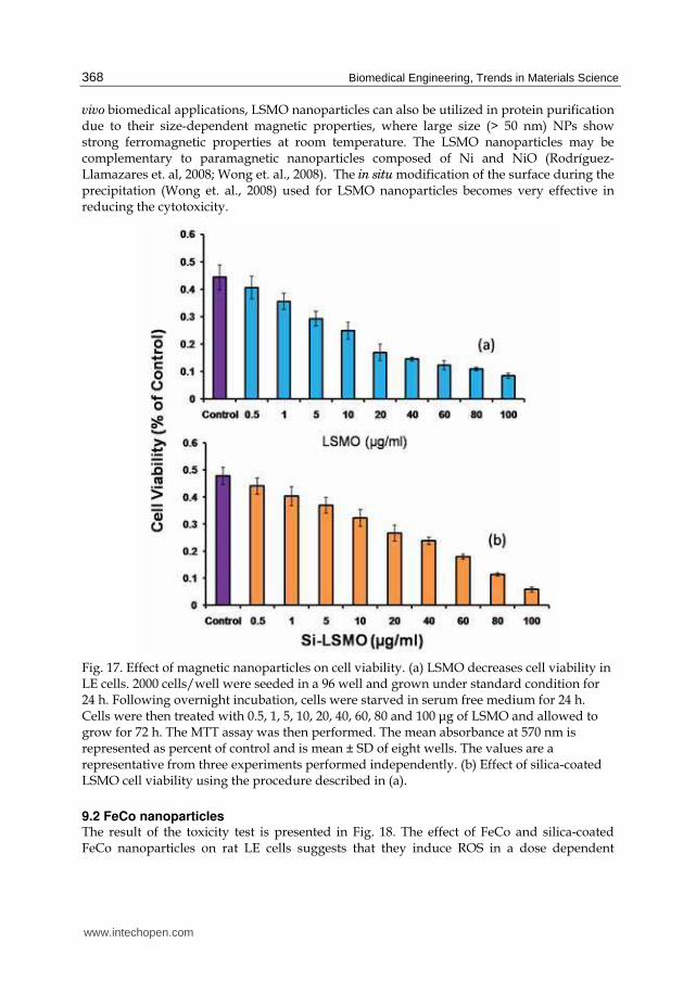
Fig. 17. Effect of magnetic nanoparticles on cell viability. (a) LSMO decreases cell viability in LE cells. 2000 cells/well were seeded in a 96 well and grown under standard condition for 24 h. Following overnight incubation, cells were starved in serum free medium for 24 h. Cells were then treated with 0.5, 1, 5, 10, 20, 40, 60, 80 and 100 μg of LSMO and allowed to grow for 72 h. The MTT assay was then performed. The mean absorbance at 570 nm is represented as percent of control and is mean ± SD of eight wells. The values are a representative from three experiments performed independently. (b) Effect of silica-coated LSMO cell viability using the procedure described in (a).
9.2 FeCo nanoparticles The result of the toxicity test is presented in Fig. 18. The effect of FeCo and silica-coated FeCo nanoparticles on rat LE cells suggests that they induce ROS in a dose dependent
www.intechopen.com

Synthesis, Characterization, Toxicity of Nanomaterials for Biomedical Applications
369
manner. Uncoated FeCo nanoparticles increased ROS by 3.2 folds as compared to control at a concentration as low as 2.5 μg. The coated FeCo nanoparticles showed 3.6 fold increase in ROS (Fig. 18 (b)). To study the extent of damage caused by coated and uncoated FeCo on cell viability, MTT assay was carried in LE cells treated with various concentrations and the results suggest that the cell viability decrease with increase in concentration of FeCo nanoparticles by 72 hrs compared to control. Only 40% of cells found to be viable at 2.5 μg/ml of uncoated FeCo, where as 35% found to be silica-coated FeCo nanoparticles. This suggests that the silica shell thickness should be increased in order to reduce the toxicity of FeCo nanoparticles for any biomedical applications.
Fig. 18. MTT assay effect of (a) uncoated and (b) coated FeCo nanoparticles on cell viability.
10. Conclusion
Nanomaterials are widely used for biomedical applications because their sizes are comparable with most of the biological entities. The development of novel biomedical technologies involving in vivo use of nanoparticles presents multidisciplinary attempts to overcome the major chemotherapeutic drawbacks. Nanomaterials stand at the boundaries between physical, chemical, biological and medical sciences, and the advances in this field impact analyzing and treating biological systems at the cell and sub-cell levels, providing
www.intechopen.com
Biomedical Engineering, Trends in Materials Science
370
revolutionary approaches for the diagnosis, prevention and treatment of some fatal diseases, such as cancer. However, the synthesis, characterization and use of these nanomaterials need thorough studies. The synthesis and characterization of several kinds of nanomaterials, such as luminescent, semiconducting and magnetic, are discussed. The toxicity associated with these nanomaterials is also discussed. Luminescent nanostructures. Eu3+ doped Gd2O3 nanomaterials are very promising luminescent as well as magnetic material. Some typical growth process for varieties of nanostructures, such as nanoparticles, nanorods, nanotubes and encapsulated nanoparticles, are described with some insight into their microstructures and their optical and magnetic properties. The toxicity studies of some of these nanostructures demonstrate that Eu:Gd2O3
nanoparticles are relatively nontoxic and the toxicity is further decreased on silica coating. Semiconductor nanostructures. A typical chemical route was explored to synthesize large scale ZnO nanorods with about 21 nm in diameter and 50 nm in length. Toxicological studies on hela cells show that ZnO nanorods could be the safe nanomaterials for biological applications. Magnetic nanostructures. Manganites and FeCo nanoparticles were synthesized by the chemical technique and the nanostructures were coated with TEOS and other macromolecules. The manganites display essential magnetic properties applicable for hyperthermia applications. On the other hand, FeCo nanoparticles display strong magnetism appropriate for protein purification. The cytotoxicity tests (ROS and cell viability) demonstrate that both manganites and FeCo nanoparticles can be potential candidate for various biomedical applications. Further perfection can be made achieved by coating the nanoparticles with silica in a controlled way. The silica shell thickness should be increased in order to reduce the toxicity of FeCo nanoparticles for any biomedical applications.
11. Acknowledgments
This work is supported by the NSF for Research Infrastructure in Science and Education (RISE) grant No. HRD-0734846 and RISE-HRD-0931373. The authors are thankful to T. Holloway for experimental help.
12. References
Adams, L. K.; Lyon, D. Y. & Alvarez, P. J. (2006). Comparative eco-toxicity of nanoscale TiO2, SiO2, and ZnO water suspensions. Water Research, 40, 19, pp. 3527-3532 (November 2006), ISSN 0043-1354
Alivisatos, P. (2004). The use of nanocrystals in biological detection. Nature Biotechnology, 22,1, (January 2004) pp. 47–52, ISSN 1087-0156
Beckett, W. S.; Chalupa, D. F.; Pauly-Brown, A.; Speers, D. M.; Stewart, D. J.; Frampton, M. W.; Utell, D. J.; Huang, L. S.; Cox, C.; Zareba, W. & Oberdorster, G. (2005). Comparing Inhaled Ultrafine vs Fine Zinc Oxide Particles in Healthy Adults:A Human Inhalation Study. American Journal of Respiratory and Critical Care Medicine, 171, 10, (May 2005) pp. 1129-1135, ISSN 1073-449X
Bridot, J.-L.; Faure, A.-C.; Laurent, S.; Rivière, C.; Billotey, C.; Hiba, B.; Janier, M.; Josserand, V.; Coll, J.-L.; Vander Elst, L.; Muller, R.; Roux, S.; Perriat, P. & Tillement, O. (2007). Hybrid gadolinium oxide nanoparticles: multimodal contrast agent for in vivo
www.intechopen.com
Synthesis, Characterization, Toxicity of Nanomaterials for Biomedical Applications
371
imaging. Journal of the American Chemical Society, 129, 16, (March 2007) pp. 5076-5084, ISSN 0002-7863
Brunner, T. J.; Wick, P.; Manser, P.; Spohn, P.; Grass, P. N.; Limbach, L.; Bruinink, A. & Stark, W. J. (2006). In Vitro Cytotoxicity of Oxide Nanoparticles: Comparison to Asbestos, Silica, and the Effect of Particle Solubility. Environmental Science &
Technology, 40, 14, (March 2006) pp. 4374-4381, ISSN 0013-936X Chang, C.; Kimura, F.; Kimura, T. & Wada, H. (2005). Preparation and characterization of
rod-like Eu:Gd2O3 phosphor through a hydrothermal routine. Materials Letters, 59, 8-9, (April 2005) 1037-1041, ISSN 0167-577X
Chertok, B; Moffat, B. A.; David, A.E.; Yu F. Q.; Bergemann, C.; Ross, B. D. & Yang, V. C. (2008). Iron oxide nanoparticles as a drug delivery vehicle for MRI monitored magnetic targeting of brain tumors. Biomaterials, 29, 4, (February 2008) pp. 487-496, ISSN 0142-9612
Deng, X. Y.; Luan, Q. X.; Chen, W. T.; Wang, Y. L.; Wu, M. H.; Zhang, H. J. & Jiao, Z. (2009). Nanosized zinc oxide particles induce neural stem cell apoptosis. Nanotechnology, 20, 11, (February 2009) 115101, ISSN 0957-4484
Dosev, D.; Nichkova, M.; Liu, M.; Guo, B.; Liu, G.; Hammock, B. B. & Kennedy, L. M. (2005). Application of luminescent Eu:Gd2O3 nanoparticles to the visualization of protein micropatterns. Journal of Biomedical Optics, 10,6, (November 2005) 064006, ISSN 1083-3668
Fine, J. M.; Gordon, T.; Chen, L. C.; Kinney, P.; Falcone, G. & Beckett, W. S. (1997). Metal Fume Fever: Characterization of Clinical and Plasma IL-6 Responses in Controlled Human Exposures to Zinc Oxide Fume at and Below the Threshold Limit Value. Journal of Occupational & Environmental Medicine, 39, 8, (August 1997) pp. 722-726, ISSN 1076-2752
Ghoshal, T.; Kar, S. & Chaudhuri, S. (2006). Synthesis and optical properties of nanometer to micrometer wide hexagonal cones and columns of ZnO. Journal of Crystal Growth, 293, 2, (August 2006) pp. 438-446, ISSN: 0022-0248
Gopikrishnan, R.; Zhang, K.; Ravichandran, P.; S. Baluchamy, Ramesh, V.; Biradar, S.; Ramesh, P.; Pradhan, J.; Hall, J. C.; Pradhan, A. K. & Ramesh, G. T. (2010). Synthesis, Characterization and Biocompatibility Studies of Zinc oxide (ZnO) Nanorods for Biomedical Application. Nano-Micro Letters, 2, 1, (2010) pp. 27-30, ISSN: 2150-5551
He, Y.; Tian, Y. & Zhu, Y. (2003). Large-scale-synthesis of luminescent Y2O3: Eu nanobelts. Chemistry Letters, 32, 9, (August 2003) pp. 862-863, ISSN 0366-7022
Hirsch, L. R.; Stafford, R. J.; Bankson, J. A.; Sershen, S. R.; Rivera, B.; Price, R. E.; Hazle, J. D.; Halas, N. J. & West, J. L. (2003). Nanoshell-mediated near-infrared thermal therapy of tumors under magnetic resonance guidance. Proceedings of the National Academy
of Sciences of the United States of America, 100, 23, (November 2003) pp. 13549–13554, ISSN 1091-6490
Hu, S. H.; Chen, S. Y.; Liu, D. M. & Hsiao, C. S. (2008). Core/single-crystal-shell nanospheres for controlled drug release via a magnetically triggered rupturing mechanism. Advanced Materials, 20, 14, (July, 2008) pp. 2690-2695, ISSN 1521-4095
Hu, X.; Cook, S.; Wang, P. & Hwang, H. (2009). In vitro evaluation of cytotoxicity of engineered metal oxide nanoparticles. Science of The Total Environment, 407, 8, (April 2009) pp. 3070-3072, ISSN 0048-9697
Jia, G.; Liu, K.; Zheng, Y.; Song, Y.; Yang, M. & You, H. P. (2009). Highly uniform Gd(OH)3 and Gd2O3:Eu3+ nanotubes: facile synthesis and luminescence properties. Journal of
Physical Chemistry C, 113,15, (March 2009) pp. 6050-6055, ISSN 1932-7447
www.intechopen.com

Biomedical Engineering, Trends in Materials Science
372
Kim, S.; Lim, Y. T.; Soltesz, E. G.; De Grand, A. M.; Lee, J.; Nakayama, A.; Parker, J. A.; Mihaljevic, T.; Laurence, R. G.; Dor, D. M.; Cohn, L. H.; Bawendi, M. G.; & Frangioni, J. V. (2003). Near-infrared fluorescent type II quantum dots for sentinel lymph node mapping. Nature Biotechnology, 22, 1, (December 2003) pp. 93–97, ISSN 1087-0156
Lewin, M.; Carlesso, N.; Tung, C.-H.; Tang, X.-W.; Cory, D.; Scadden, D. T.; & Weissleder, R. (2000). Tat peptide-derivatized magnetic nanoparticles allow in vivo tracking and recovery of progenitor cells. Nature Biotechnology, 18, 4, (April 2000) pp. 410–414, ISSN 1087-0156
Li, S.; Song, H.; Yu, H.; Lu, S.; Bai, X.; Pan, G.; Lei, Y.; Fan, L. & Wang, T. (2007). Influence of annealing temperature on photoluminescence characteristics of Gd2O3:Eu/AAO nanowires. Journal of Luminescence, 122-123, 1, (January-April 2007) pp. 876-878, ISSN 0022-2313
Liao, H. W. & Hafner, J. H. (2005). Gold nanorod bioconjugates. Chemistry of Materials, 17, 18, (August 2005) pp. 4636–4641, ISSN 0897-4756
Lin, D. H. & Xing, B. S. (2007). Phytotoxicity of nanoparticles: Inhibition of seed germination and root growth. Environmental Pollution, 150, 2, (November 2007) pp. 243-250, ISSN 0269-7491
Liu, G. X.; Hong, G.Y.; Dong, X.T. & Wang, J.X. (2008). Preparation and characterization of Gd2O3:Eu3+ luminescence nanotubes. Journal of Alloys and Compounds, 466, 1-2, (December 2007) pp. 512–516, ISSN 0925-8388
Liu, L.; Ma, E.; Li, R.; Liu, G. & Chen, X. (2007) Effects of phonon confinement on the luminescence dynamics of Eu3+ in Gd2O3 nanotubes. Nanotechnology, 18, 1, (January 2007) pp. 015403-015407, ISSN 0957-4484
Liu, T. Y.; Hu S. H.; Liu, D. M., Chen, S. Y. & Chen, I. W. (2009). Biomedical nanoparticle carriers with combined thermal and magnetic responses. Nano Today, 4, 1, (February 2009) pp. 52-65, ISSN 1748-0132
Lou, X. W.; Wang, Y.; Yuan, C.; Lee, J. Y. & Archer, L. A. (2006). Template-free synthesis of SnO2 hollow nanostructures with high lithium storage capacity. Advanced Materials, 18, 17, (September 2006) pp. 2325-2329, ISSN 1521-4095
Ma, T.; Guo, M.; Zhang, M.; Zhang, Y. & Wang, X. (2007). Density-controlled hydrothermal growth of well-aligned ZnO nanorod arrays. Nanotechnology, 18, 3, (January 2007) 035605, ISSN 0957-4484
Manna, S. K.; Sarkar, S.; Barr, J.; Wise, K.; Barrera, E. V.; Jejelowo, O.; Rice-Ficht, A. C. & Ramesh, G. T. (2005). Single-Walled Carbon Nanotube Induces Oxidative Stress and Activates Nuclear Transcription Factor-κB in Human Keratinocytes. Nano
Letters, 5, 9, (August 2005) pp. 1676-1684, ISSN: 1530-6984 Mao, Y. B.; Tran, T.; Guo, X.; Huang, J. Y.; Shih, C. K.; Wang, K. L. & Chang, J. P. (2009).
Luminescence of nanocrystalline erbium-doped yttria. Advanced Functional
Materials, 19, 5, (March 2009) pp. 748-754, ISSN 1616-3028 Mao, Y.; Huang, J. Y.; Ostroumov, R.; Wang, K. L. & Chang, J. P. (2008). Synthesis and
luminescence properties of Erbium-doped Y2O3 nanotubes. Journal of Physical
Chemistry C, 112, 7, (January 2008) pp. 2278-2285, ISSN 1932-7455 Nichkova, M.; Dosev, D.; Gee, S. J.; Hammock, B. D. & Kennedy, I. M. (2005). Microarray
immunoassay for phenoxybenzoic acid using polymer encapsulated Eu:Gd2O3 nanoparticles as fluorescent labels. Analytical Chemistry, 77, 21, (September 2005) pp. 6864-6873, ISSN 0003-2700
Nichkova, M.; Dosev, D.; Perron, R.; Gee, S. J.; Hammock, B. D. & Kennedy, I. M. (2006). Eu3+-doped Gd2O3 nanoparticles as reporters for optical detection and visualization
www.intechopen.com
Synthesis, Characterization, Toxicity of Nanomaterials for Biomedical Applications
373
of antibodies patterned by microcontact printing. Analytical and Bioanalytical
Chemistry, 384, 3, (February 2006) pp. 631-637, ISSN 1618-2642 Petoral Jr., R. M.; Söderlind, F.; Klasson, A.; Suska, A.; Fortin, M. A.; Abrikossova, N.;
Selegård, L.; Käll, P.; Engström, M. & Uvdal, K. (2009). Synthesis and characterization of Tb3+-doped Gd2O3 nanocrystals: a bifunctional material with combined fluorescent labeling and MRI contrast agent properties. Journal of
Physical, Chemistry C, 113, 17, (April 2009) pp. 6913-6920, ISSN 1932-7447 Plank, C.; Schillinger, U.; Scherer, F.; Bergemann, C.; Rémy, J. S.; Krötz, F.; Anton, M.;
Lausier, J. & Rosenecker, J. (2003). The magnetofection method: using magnetic force to enhance gene delivery. Biological Chemistry, 384, 5, (May 2003) pp. 737-747, ISSN 1431-6730
Pradhan, A.K.; Bah, R.; Konda, R. B.; Mundle, R.; Mustafa, H.; Bamiduro, O.; Rakhimov, R. R.; Wei, Xiaohui; & Sellmyer, D. J. (2008). Synthesis and magnetic characterizations of manganite-based composite nanoparticles for biomedical applications. Journal of
Applied Physics, 103, 7, (2008) 07F704, ISSN 0021-8979 Pradhan, A.K.; Williams, T.M.; Zhang, K.; Hunter, D.; Dadson, J.B.; Lord, K.; Roy, U.N.; Cui,
Y. & Burger, A. (2006). Growth of aligned ZnO Nanorods. Journal of Nanoscience and
Nanotechnology, 6, 7, (July 2006) pp. 1985-1989, ISSN 1550-7033 Prasad, V.; Souza, C. D.; Yadav, D.; Shaikh, A. J.; & Vigneshwaran, N. (2006). Spectroscopic
characterization of zinc oxide nanorods synthesized by solid-state reaction. Spectrochimica Acta Part A, 65, 1, (September 2006) pp. 173-178, ISSN 1386-1425
Ravichandran, P.; Periyakaruppan, A.; Sadanandan, B.; Ramesh, V.; Hall, J. C.; Jejelowo, O. and Ramesh, G. T. (2009). Induction of apoptosis in rat lung epithelial cells by multiwalled carbon nanotubes. Journal of Biochemical and Molecular Toxicology, 23, 5, (October 2009) pp. 333-344, ISSN 1099-0461
Reddy, K. M.; Feris, K.; Bell, J.; Wingett, D. J.; Hanley, C. & Punnoose, A. (2007). Selective toxicity of zinc oxide nanoparticles to prokaryotic and eukaryotic systems. Applied
Physics Letters, 90, 21, (May 2007) 213902, ISSN 0003-6951 Rodríguez-Llamazares, S.; Merchán, J.; Olmedo, I.; Marambio, H. P.; Muñoz, J. P.; Jara, P.;
Sturm, J. C.; Chornik, B.; Peña, O.; Yutronic, N.; & Kogan, M. J. (2008). Ni/Ni Oxides Nanoparticles with Potential Biomedical Applications Obtained by Displacement of a Nickel-Organometallic Complex. Journal of Nanoscience and
Nanotechnology, 8, 8, (August 2008) pp. 3820-3827, ISSN 1550-7033 Sarkar, S.; Sharma, C.; Yog, R.; Periakaruppan, A.; Jejelowo, O.; Thomas, R.; Barrer, E. V.; Rice-
Ficht, A. C.; Wilson, B. L. & Ramesh, G. T. (2007). Analysis of Stress Responsive Genes Induced by Single-Walled Carbon Nanotubes in BJ Foreskin Cells. Journal of Nanoscience and Nanotechnology, 7, 2, (February 2007) pp. 584-592, ISSN 1550-7033
Satarkar, N. S. & Hilt, J. Z. (2008). Magnetic hydrogel nanocomposites for remote controlled pulsatile drug release. Journal of Controlled Release, 130, 3, (September 2008) pp. 246-251, ISSN 0168-3659
Seo, S. Y.; Sohn, K. S.; Park, H. D. & Lee, S. (2002). Optimization of Gd2O3-based red phosphors using combinatorial chemistry method. Journal of the Electrochemical
Society, 149, 1, (December 2002) pp. H12-H18, ISSN 0013-4651 Sharma, C. S.; Sarkar, S.; Periyakaruppan, A.; Barr, J.; Wise, K.; Thomas, R.; Wilson, B. L. &
Ramesh, G. T. (2007). Journal of Nanoscience and Nanotechnology, 7, 7, (July 2007) pp. 2466-2472, ISSN 1550-7033
Shvedova A. A.; Castranova V.; Kisin E. R.; Schwegler-Berry D.; Murray A. R.; Gandelsman V. Z.; Maynard A. & Baron P. (2003). Exposure to carbon nanotube material: assessment
www.intechopen.com
Biomedical Engineering, Trends in Materials Science
374
of nanotube cytotoxicity using human keratinocyte cells. Journal of Toxicology and Environmental Health, Part A, 66, 20, (June 2003) pp. 1909-1926, ISSN 1528-7394
Son, S. J.; Reichel, J.; He, B.; Schuchman, M. & Lee, S. B. (2005). Magnetic nanotubes for magnetic-field-assisted bioseparation, biointeraction, and drug delivery. Journal of
the American Chemical Society, 127, 20, (April 2005) pp. 7316-7317, ISSN 0002-7863 Tsuji, J. S.; Maynard, A. D.; Howard, P. C.; James, J. T.; Lam, C. W.; Warheit, D. B. &
Santamaria, A. B. (2005). Research Strategies for Safety Evaluation of Nanomaterials, Part IV: Risk Assessment of Nanoparticles. Toxicological Sciences, 89, 1, (September 2005) pp. 42-50, ISSN 1096-0929
Wang, H.; Uehara, M.; Nakamura, H.; Miyazaki, M. & Maeda, H. (2005). Synthesis of well-dispersed Y2O3:Eu nanocrystlas and self-assembled nanodisks using a simple non-hydrolytic route. Advanced Materials, 17, 20, (October 2005) pp. 2506-2509, ISSN 1521-4095
Wei, W.; Ma, G. H.; Hu, G.; Yu, D.; Mcleish, T.; Su, Z. G. & Shen, Z. Y. (2008). Preparation of hierarchical hollow CaCO3 particles and the application as anticancer drug carrier. Journal of the American Chemical Society, 130, 47, (November 2008) pp. 15808-15810, ISSN 0002-7863
Whitesides, G. M. (2003). The ‘right’ size in nanobiotechnology. Nature Biotechnology, 21, 10, (September 2003) pp. 1161-1165, ISSN 1087-0156
Wong, J. E.; Gaharwar, A. K.; Müller-Schulte, D.; Bahadur, D. & Richtering, W. (2008). Magnetic Nanoparticle-Polyelectrolyte Interaction: A Layered Approach for Biomedical Applications. Journal of Nanoscience and Nanotechnology, 8, 8, (August 2008) pp. 4033-4040, ISSN 1533-4880
Xu, X. L. & Asher, S. A. (2004). Synthesis and utilization of monodisperse hollow polymeric particles in photonic crystals. Journal of the American Chemical Society, 126, 25, (June 2004) pp. l7940-7945, ISSN 0002-7863
Yang, H.; Liu, C.; Hui, Yang, D.; Zhang, H. & Xi, Z. (2009). Comparative study of cytotoxicity, oxidative stress and genotoxicity induced by four typical nanomaterials: the role of particle size, shape and composition. Journal of Applied
Toxicology, 29, 1, (August 2009) pp. 69-78, ISSN 1099-1263 Yang, J.; Li, C. X.; Cheng, Z. Y.; Zhang, X. M.; Quan, Z. W.; Zhang, C. M. & Lin, J. (2007).
Size-tailored synthesis and luminescent properties of one-dimensional Gd2O3: Eu nanorods
and microrods. Journal of Physical Chemistry C, 111, 49, (November 2007) pp. 18148-18154, ISSN 1932-7447
Yuan, J. H.; Chen, Y.; Zha, H. X.; Song, L. J.; Li, C. Y.; Li, J. Q. & Xia, X. H. (2010). Determination, characterization and cytotoxicity on HELF cells of ZnO nanoparticles. Colloids and Surfaces B: Biointerfaces, 76, 1, (March 2010) pp. 145-150, ISSN 0927-7765
Zaveri, T.; Dolgova, N.; Chu, B. H.; Lee, J.; Lele, T.; Ren, F. & Keselowsky, B. G. (2009). Proceedings of IFMBE, pp. 119-120, ISSN 1680-0737, Miami, Florida, USA, May 2009, Springer, Berlin, Heidelberg
Zhang, K.; Holloway, T.; Pradhan, J.; Bahoura, M.; Bah, R.; Rakhimov, R. R.; Pradhan, A. K.; Prabakaran, R. & Ramesh, G. T. (2010). Synthesis and Magnetic Characterizations of La1-xSrxMnO3 Nanoparticles for Biomedical Applications. Journal of Nanoscience and
Nanotechnology, 10, 9, (September 2010) pp. 5520-5526, ISSN 1550-7033 Zhang, K.; Holloway, T.; Bahoura, M.; Pradhan, A. K.; Prabakaran, R.; Pradhan, J.; Smith, S.;
Hall, J. C.; Ramesh, G. T.; Sahu, D. R.; Huang, J.-L. (2009) Europium doped Gd2O3 and FeCo nanoparticles for biomedical application. Proceedings of SPIE, 7291, (March 2009) 729104-10, ISSN 0277-786X
www.intechopen.com
Biomedical Engineering, Trends in Materials ScienceEdited by Mr Anthony Laskovski
ISBN 978-953-307-513-6Hard cover, 564 pagesPublisher InTechPublished online 08, January, 2011Published in print edition January, 2011
InTech EuropeUniversity Campus STeP Ri Slavka Krautzeka 83/A 51000 Rijeka, Croatia Phone: +385 (51) 770 447 Fax: +385 (51) 686 166www.intechopen.com
InTech ChinaUnit 405, Office Block, Hotel Equatorial Shanghai No.65, Yan An Road (West), Shanghai, 200040, China
Phone: +86-21-62489820 Fax: +86-21-62489821
Rapid technological developments in the last century have brought the field of biomedical engineering into atotally new realm. Breakthroughs in materials science, imaging, electronics and, more recently, the informationage have improved our understanding of the human body. As a result, the field of biomedical engineering isthriving, with innovations that aim to improve the quality and reduce the cost of medical care. This book is thesecond in a series of three that will present recent trends in biomedical engineering, with a particular focus onmaterials science in biomedical engineering, including developments in alloys, nanomaterials and polymertechnologies.
How to referenceIn order to correctly reference this scholarly work, feel free to copy and paste the following:
A. K. Pradhan, K. Zhang, M. Bahoura, J. Pradhan, P. Ravichandran, R. Gopikrishnan and G. T. Ramesh(2011). Synthesis, Characterization, Toxicity of Nanomaterials for Biomedical Applications, BiomedicalEngineering, Trends in Materials Science, Mr Anthony Laskovski (Ed.), ISBN: 978-953-307-513-6, InTech,Available from: http://www.intechopen.com/books/biomedical-engineering-trends-in-materials-science/synthesis-characterization-toxicity-of-nanomaterials-for-biomedical-applications