Synthesis of copper and zinc nanostructures by discharges ...

NANO REVIEW Open Access
Synthesis and Functions of Ag2SNanostructuresChunyan Cui, Xiaoru Li*, Jixian Liu, Yongchao Hou, Yuqing Zhao and Guocheng Zhong
Abstract
The paper presents a review about synthesis and applications of Ag2S nanostructures. As the modernphotoelectric and biological materials, Ag2S nanomaterials are potentially useful for both structure andfunction purposes. Ag2S is a direction narrow band gap semiconductor with special properties. Ag2Snanostructures have been widely researched in chemistry and biochemistry fields because of their unusualoptical, electrical, and mechanical properties. It can also be used in many fields, such as photovoltaic cellsand infrared detector. In the past few years, Ag2S nanostructures have been synthesized by various methods.The article mainly discusses the four types of preparation methods. Moreover, this article shows a detailedreview on the new properties, fabrication, and applications of Ag2S nanocrystals.
Keywords: Ag2S nanostructure, Synthesis, Properties, Application
ReviewIntroductionNanoparticles are different from molecular and blockmaterials and have many special physical and chemicalproperties. In the past decades, the synthetic methodsand tuning morphologies of single-component nanocrys-talline have made great progress. The metal sulfidenanomaterials have attracted widespread attention dueto their suitable band gap, easy manufacturing, low cost,and high performance [1–4].As an important metal sulfide, Ag2S is a direction
narrow band gap semiconductor (~1.5 eV), and high-absorption coefficient is approximately 104 m−1 [5, 6].It also has good chemical stability and optical proper-ties [7, 8], so it is widely used in various fields, suchas semiconductor, photovoltaic cells, infrared detector,and superionic conductor [9–12]. Recently, it also hasbeen used in the photoelectric switch and oxygensensor at room temperature [13]. So far, Ag2S nano-particles were successfully prepared by microemulsionmethod, sol–gel method, particles embedded technologytemplate method, etc. [14–17]. But, it often requires strictpreparation conditions, time-consuming, energy con-sumption, and large size distribution in the traditional
production methods. Therefore, simple and economicroutes of uniform size distribution of Ag2S nanomaterialsstill have challenges.In this review, we summarized the preparation and
properties of Ag2S materials as follows.
Synthetic MethodsAg2S nanostructures have been synthesized by variousmethods, such as sol–gel method, particles embeddedtechnology template method, etc. Every method has bothadvantages and limitations. So the preparation methodshave still challenges. The new performance of the Ag2Snanostructures needs to be further exploded. So, the art-icle summarized four types of preparation methods toexpect to provide a little help for the workers who areengaging in this field.
Formation of Different Forms of Ag2S NanoparticlesRecently, the great efforts have been focused on themorphology control of the semiconductor nanocrystalsdue to their morphology-dependent properties [18].Thus, the preparation of different forms of Ag2S nano-particles (such as urchin-[19], snow-[20], dendrite-like[21, 22] nanocrystals, and so on) has been caused manyscientific research in order to expend their current appli-cations. Various methods in the preparation of Ag2Snanostructures and their mechanism have been
* Correspondence: [email protected] of Chemistry Science and Chemical Engineering, Qingdao University,No. 308 Ningxia Road, Qingdao 266071, China
© 2015 Cui et al. Open Access This article is distributed under the terms of the Creative Commons Attribution 4.0 InternationalLicense (http://creativecommons.org/licenses/by/4.0/), which permits unrestricted use, distribution, and reproduction in anymedium, provided you give appropriate credit to the original author(s) and the source, provide a link to the Creative Commonslicense, and indicate if changes were made.
Cui et al. Nanoscale Research Letters (2015) 10:431 DOI 10.1186/s11671-015-1125-7

explored. For example, Zhao et al. [23] prepared rod-likeAg2S nanocrystallines by using Na2S2O3 as a chalcogensource via gamma ray irradiation at room temperature.In this experiment, polyvinylpyrrolidone (PVP) as aguide reagent of crystal growth plays an important rolein the formation of rod-like Ag2S nanocrystallines. It iswell known that high-energy gamma ray irradiation canmake H2O producing strong reductive eaq
− that can initi-ate many redox reactions to generate ions, which cannothappen in a common atmosphere. And, the homoge-neously dispersed S2O3
2− ions reacted with the generatedreductive particles to form S2− [24]. And then, Ag+ couldreact with S2− to form Ag2S nanoparticles. Ag2S nano-rods were formed in the solution of the PVP [25].Figure 1 shows that the diameters of the nanorods rangfrom 200 to 500 nm, and the length is up to several ten
micrometers. The band gap of Ag2S nanorods calculatedfrom the UV–vis spectrum is 2.34 eV, which shows anobvious blue shift in UV–vis absorption compared withthe bulk Ag2S (Fig. 2).Chen et al. [26] reported that the leaf-like Ag2S nano-
sheets were prepared successfully by a facile hydrothermalmethod in alcohol–water homogenous medium. In the ex-periment, CS2 used as sulfur source was dissolved in alco-hol and AgNO3 and water at the beginning, respectively.Then the two solutions were mixed together. The reactionis as follows:
2NH3 þ CS2→NH4NHCSSH ð1Þ
2Ag NH3ð Þþ þNH4NHCSSH→Ag2S↓þNH4SCN þ 2NH4þ
ð2Þ
The picture of TEM (Fig. 3) shows all of the samplesare leaf-like.However, Xu et al. [27] reported that the Ag2S was
prepared with an alcohol solution method using CS2as sulfur source also. But, all the products were ir-regular nanoparticles. The reaction medium waschanged from water and alcohol–water to alcohol.So, the morphology changed from big irregularnanosheets and microspheres, leaf-like nanosheetsnanoparticles to big microspheres, respectively. It isfound that alcohol–water homogenous condition andsulfur source of NH3-CS2 played the key roles inconstructing this unique morphology. That is be-cause the formation of nucleation and growth is ex-pected to be strongly dependent on the properties ofthe solvent during processes such as coarsening andaggregation [28, 29]. The different morphologies of
Fig. 1 a TEM image, b SAED pattern of Ag2S [23]
Fig. 2 UV–vis absorption spectrum of as-prepared Ag2Snanorods [23]
Cui et al. Nanoscale Research Letters (2015) 10:431 Page 2 of 21

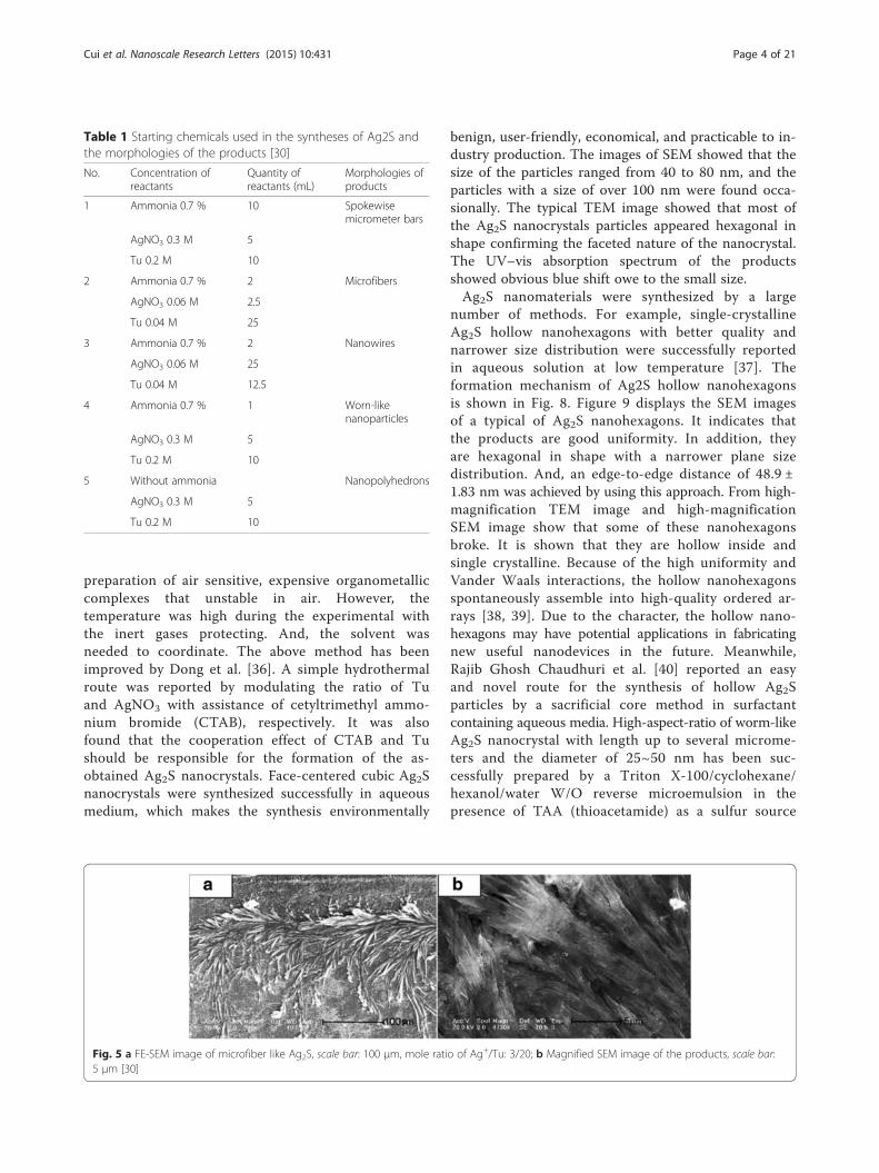
Ag2S nano- and micromaterials, including spokewisemicrometer bars, nanowires, and nanopolyhedronshave been gained by a facile one-step method atroom temperature [30]. In the route, organic tem-plate materials were not added into the reactioncontainer. It only changed the ratios of Ag+, S2−,and ammonia, which may produce the different sizeand morphologies of the products. In this route, thespokewise microbars of Ag2S were successfully pre-pared. Firstly, 10 mL of 0.2 M Tu ((NH2)2CS) wasadded in 10 mL of 0.7 % ammonia. Then, 5 mL of0.3 M Ag2NO3 was quickly added in the solutionwith stirring for 20 min. Last, the product was puri-fied and redispersed in the water several times bycentrifugation and sonication (Fig. 4). The methods
with other morphologies of Ag2S were similar to theabove process, except for the concentration of am-monia, AgNO3, or Tu. By controlling the concentra-tion of ammonia, AgNO3, or Tu, the morphology ofAg2S could be easily tuned (Table 1 and Figs. 5, 6,and 7).Recently, polyhedral nanocrystals including nano-
cubes have been attracted intensive attention and avariety of face-centered cubicin organic nanocrystals[31–33] have been successfully fabricated. The Ag2Snanocrystals were also prepared by Lim et al. [34]and Wang et al. [35] by decomposing exothermicallyorganometallic precursor silver thiobenzoate (Ag(SCOPh))and Ag[S2P(OR)2] (R = CnH2n + 1), respectively. How-ever, the method often suffers from elaborate
Fig 3 The typical leaf-like morphology TEM image of Ag2S nanosheets with different magnification, angle and part prepared in alcohol-watermedium by hydrothermal treatment at 160 °C for 10h, the insert in (d) shows the SAED spots [26]
Fig. 4 a FE-SEM image of spokewise microbars of Ag2S, scale bar: 20 μm, mole ratio of Ag+/Tu: 3/4; b Magnified SEM image of the products scalebar: 5 μm [30]
Cui et al. Nanoscale Research Letters (2015) 10:431 Page 3 of 21
preparation of air sensitive, expensive organometalliccomplexes that unstable in air. However, thetemperature was high during the experimental withthe inert gases protecting. And, the solvent wasneeded to coordinate. The above method has beenimproved by Dong et al. [36]. A simple hydrothermalroute was reported by modulating the ratio of Tuand AgNO3 with assistance of cetyltrimethyl ammo-nium bromide (CTAB), respectively. It was alsofound that the cooperation effect of CTAB and Tushould be responsible for the formation of the as-obtained Ag2S nanocrystals. Face-centered cubic Ag2Snanocrystals were synthesized successfully in aqueousmedium, which makes the synthesis environmentally
benign, user-friendly, economical, and practicable to in-dustry production. The images of SEM showed that thesize of the particles ranged from 40 to 80 nm, and theparticles with a size of over 100 nm were found occa-sionally. The typical TEM image showed that most ofthe Ag2S nanocrystals particles appeared hexagonal inshape confirming the faceted nature of the nanocrystal.The UV–vis absorption spectrum of the productsshowed obvious blue shift owe to the small size.Ag2S nanomaterials were synthesized by a large
number of methods. For example, single-crystallineAg2S hollow nanohexagons with better quality andnarrower size distribution were successfully reportedin aqueous solution at low temperature [37]. Theformation mechanism of Ag2S hollow nanohexagonsis shown in Fig. 8. Figure 9 displays the SEM imagesof a typical of Ag2S nanohexagons. It indicates thatthe products are good uniformity. In addition, theyare hexagonal in shape with a narrower plane sizedistribution. And, an edge-to-edge distance of 48.9 ±1.83 nm was achieved by using this approach. From high-magnification TEM image and high-magnificationSEM image show that some of these nanohexagonsbroke. It is shown that they are hollow inside andsingle crystalline. Because of the high uniformity andVander Waals interactions, the hollow nanohexagonsspontaneously assemble into high-quality ordered ar-rays [38, 39]. Due to the character, the hollow nano-hexagons may have potential applications in fabricatingnew useful nanodevices in the future. Meanwhile,Rajib Ghosh Chaudhuri et al. [40] reported an easyand novel route for the synthesis of hollow Ag2Sparticles by a sacrificial core method in surfactantcontaining aqueous media. High-aspect-ratio of worm-likeAg2S nanocrystal with length up to several microme-ters and the diameter of 25~50 nm has been suc-cessfully prepared by a Triton X-100/cyclohexane/hexanol/water W/O reverse microemulsion in thepresence of TAA (thioacetamide) as a sulfur source
Table 1 Starting chemicals used in the syntheses of Ag2S andthe morphologies of the products [30]
No. Concentration ofreactants
Quantity ofreactants (mL)
Morphologies ofproducts
1 Ammonia 0.7 % 10 Spokewisemicrometer bars
AgNO3 0.3 M 5
Tu 0.2 M 10
2 Ammonia 0.7 % 2 Microfibers
AgNO3 0.06 M 2.5
Tu 0.04 M 25
3 Ammonia 0.7 % 2 Nanowires
AgNO3 0.06 M 25
Tu 0.04 M 12.5
4 Ammonia 0.7 % 1 Worn-likenanoparticles
AgNO3 0.3 M 5
Tu 0.2 M 10
5 Without ammonia Nanopolyhedrons
AgNO3 0.3 M 5
Tu 0.2 M 10
Fig. 5 a FE-SEM image of microfiber like Ag2S, scale bar: 100 μm, mole ratio of Ag+/Tu: 3/20; b Magnified SEM image of the products, scale bar:5 μm [30]
Cui et al. Nanoscale Research Letters (2015) 10:431 Page 4 of 21

and EDTA (ethylenediaminetetraacetic acid) as achelating ligand [41]. The as-synthesized Ag2S nano-crystals exhibit strong absorption in UV region, andthe absorption edge at about 290 nm (Fig. 10) corre-sponding to the band gap of 4.3 eV. Compared tothe absorption band of bulk Ag2S (1240 nm, Eg =1.0 eV) [42], the observed absorption edge is a sig-nificant blue-shift. The result is due to the position-dependent quantum-size effect and shape effect.Figures 11, 12, and 13 show the TEM images ofAg2S nanocrystal synthesized with the typical experi-mental procedure, in which change one condition.The results indicate that the morphology and size ofAg2S nanocrystal can be readily controlled by modu-lating the mole ratio of Ag+ to EDTA, the molar ra-tio of water to surfactant (ω0), and the aging time.The diameter distribution of Ag2S nanocrystal be-comes wider with the increasing ω0. It can beexplanted that at low ω0 water inside the reverse mi-celles is considered to be “bound”. Therefore, insuffi-ciently available to dissolve the surfactant headgroup and counterion, the microemulsion becomesmore fluid with increasing ω0, which accelerated the
growth of nanocrystalline Ag2S. The effect of EDTAconcentration on the formation of worm-like Ag2Snanocrystals is important. It can coordinate Ag+ toform relative stable Ag-EDTA complex, which lowersthe effective concentration of Ag+; TAA could re-lease S2− very slowly in the solution. The two as-pects make the Ag+and S2−react slowly, which couldresult in the separation of nucleation and growthstep and is favorable for the directional growth ofthe crystal nuclei [43]. Ag2S nanorice was syn-thesized by reaction between [Ag(NH3)2]
+ andNa2S·9H2O in the presence of PVP through hydro-thermal method [44]. And, the feature of the Ag2Snanostructure depends mainly on the type of sliversource, influence of the pyrrolidone rings of PVP, re-action time, and temperature. TEM technique wasemployed to inspect the morphological variation ofthe Ag2S nanoparticles obtained by using differentsliver sources (Fig. 14 a and b) at 160 °C for 10 h. Itwas found that the well-dispersed Ag2S nanoparticlespresented an approximately uniformed rice-shapedmorphology. But when AgNO3 was changed intoAg[(NH3)2]
+ as the sliver source without a control
Fig. 6 a FE-SEM image of the aggregated microwires of Ag2S, scale bar: 50 μm, mole ratio of Ag+/Tu: 3/1; b SEM image of Ag2S nanowires, scalebar: 5 μm [30]
Fig. 7 FE-SEM images of Ag2S, mole ratio of Ag+/Tu: 3/4. a worm-like nanoparticles, the initial concentration of ammonia: 7 %; b Nanopolyhedronswithout ammonia [30]
Cui et al. Nanoscale Research Letters (2015) 10:431 Page 5 of 21

over the Ag+ release rate provided by ammonia com-plexation during the reaction, the rice-shaped mor-phological feature of Ag2S nanoparticles would notbeen seen, and anamorphous appearance would bepresented instead. So, Ag+ concentration had a keyimpact over the formation of the Ag2S nanorice.Similar condition also happened while reaction timeinfluences on the experimental results (Fig. 14 c andd). It can be explained by the famous Ostwald ripen-ing mechanism. The FI-IR spectra of pure PVP, Ag2Snanorice-associated PVP are shown in Fig. 15. It iseasy to find that the pyrrolidone ring plays an
important role in the formation of the Ag2S na-norice. Normal and flattened rhombic dodecahedralsubmicrometer Ag2S particles were prepared byadjusting the ratio of Tu to AgNO3 and volume ofconcentrated HCl aqueous solution [45].
Preparation by Using Bionic TechnologyIn recent years, the synthesis of quantum dots (QDs)with biological macromolecules route has attached greatattention [46]. In previous works, CdTe QDs with goodbiocompatibility by using RNase A as the template wassynthesized successfully [48]. But, it is not popular
Fig. 8 Schematic representation of the formation mechanism of hollow nanohexagons: (a) the soluble CTA+- Ag(S2O3) and CTA+-[Ag(S2O3)2]3-
ion pairs, (b) hexagon-like micellar composites with Ag(S2O3)- and [Ag(S2O3)2]
3-, (c) Ag2S nuclei, (d) hollow nanohexagon of Ag2S [37]
Fig. 9 a Low-magnification SEM image of hollow nanohexagons, b Plane size distribution of hollow nanohexagons, c High-magnification TEMimage of hollow nanohexagons, and d High-magnification SEM image of hollow nanohexagons. The inset is the corresponding electronicdiffraction pattern from one nanohexagon. Scale bars, a 100 nm, b 5 nm, and c 100 nm [37]
Cui et al. Nanoscale Research Letters (2015) 10:431 Page 6 of 21

enough because of the toxic nature of Cd and Te. Ag2SQDs is treated as an ideal optical probe because it haslower toxicity compared with previous prepared near-infrared (NIR) QDs which was synthesized in organicphase. But, the process may cause extra environmentpollution [49, 50]. The synthesis of QDs with fluores-cence emission from UV to NIR regions has made greatprogresses as optical probes for in vitro and in vivo mo-lecular imaging [47]. So, the highly monodisperse andwater soluble RNase-Acopped-Ag2S QDs clusters weresynthesized via biomimetic route in aqueous phase [51].The QDs have low cytotoxicity and good biocompatibil-ity. Meanwhile, the produce process is environmental
friendly. From the images (Fig. 16), it indicates thatRNase A-Ag2S QDs clusters with irregular morphologieswere dispersed, and the Ag2S nanocrystals have clear lat-tice fringes. Furthermore, X-ray diffraction (XRD) imageshows that the prepared Ag2S QDs assumed the crystal-line structure of monoclinic α-Ag2S. Tetrazolium-basedcolorimetric assay (M77 test) shows that RNase A dosenot only serves as a stabilizer agent in the formation ofAg2S QDs to avoid aggregation but also is a biomoleculeto modify the surface of Ag2S QDs to decrease toxicity.So, the products have great potential application in mo-lecular imaging in living cells and tissues. Biomoleculesassisted the formation of inorganic nanostructures, fa-cilitate the electrostatic stabilization, and thus improvingoptical properties.Siva C et al. [52] reported that Ag2S nanostructure
was obtained using Ag0 nuclei as a core. Biomol-ecule (L-cysteine) can act as a sulfur source andstabilizing agent which prevents the agglomeration[53]. Meanwhile, they also have obtained the novelAg3AuS2 nanocrystals by adopting the gold ions inthe L-cysteine-assisted Ag2S formation. The FT-IRimages (Fig. 17) show that one L-cysteine moleculeis interconnected to another via hydrogen bonding.From Fig. 18, it is clear that the Ag2S nanocrystalsare almost uniform in their size and are intercon-nected among themselves, and their average particlesize is 5.2 nm. Meanwhile, the Ag3AuS2 nanocrystalsare also connected with themselves. The Ag3AuS2nanocrystals are non-uniform in their sizes, and theaverage size is 9.1 nm. Self-organized nanocrystal ar-chitectures with subnanometric spatial resolutionalso obtained by mimicking the biological crystalgrowth [54].
Fig. 10 UV–vis absorption spectra of Ag2S nanocrystal synthesized in W/O microemulsion ([TAA] = 0.3 mol/L, [Ag+] = 0.1 mol/L, [Ag+]/[EDTA] = 1)aged for 3d with ω0 = 10 [41]
Fig. 11 TEM images of Ag2S nanocrystal synthesized in W/O microemulsion ([TAA] = 0.3 mol/L, [Ag+] = 0.1 mol/L, [Ag+]/[EDTA] = 1) a,b aged for3d, c aged for 24d with ω0 = 10 [41]
Cui et al. Nanoscale Research Letters (2015) 10:431 Page 7 of 21

Preparation by a Single Molecular Precursor ofDecompositionAmong the many methods for synthesizing metal chalco-genide materials, the single molecular precursor route hassome appealing features [55, 56]. On the one hand, it of-fers the distinct advantages of mildness, simplicity, safety,and particular compatibility with the metalorganic chem-ical vapor deposition [57]. On the other hand, the molecu-lar precursor may be related to the unusual crystal growthselectivity or metastable phase formation of the resultantproducts, which are sometimes unattainable via con-ventional synthesis techniques [55, 56]. For example,Ag2S nanocrystals were achieved via a modified hot-injection process from a single-source precursor molecule
Ag(SCOph). When the precursor molecule is in-jected into a preheated reaction system at 160 °C,spherical Ag2S nanocrystals are directly obtained[58]. Wang et al. [59] obtained the Ag2S crystallitesby heating the Ag-DDTA in air at 200 °C for 3 hand used an air-stable single-source molecular pre-cursor (Ag-DDTA) as the react source. The methodis both economic and non-toxic. Monodispersed andsize-controlled Ag2S nanoparticles were synthesizedsuccessfully via a green and simple surfactant-freesolventless thermolysis of silver xanthates as single-source precursors [60]. In the experiment, the diam-eter of Ag2S nanoparticles is from 8.9 ± 1.2 nm to48.3 ± 3.6 nm (Fig. 19). And, the “Size control” was
Fig. 12 TEM images of Ag2S nanocrystal synthesized in W/O microemulsion ([TAA] = 0.3 mol/L, [Ag+] = 0.1 mol/L, [Ag+]/[EDTA] = 1) aged for 3dwith a ω0 = 6, b ω0 = 15 [41]
Fig. 13 TEM images of Ag2S nanocrystal synthesized in W/O microemulsion ([TAA] = 0.3 mol/L, [Ag+] = 0.1 mol/L) with various concentrations ofEDTA a [Ag+]/[EDTA] = 2, b [Ag+]/[EDTA] = 4 aged for 3d with ω0 = 10 [41]
Cui et al. Nanoscale Research Letters (2015) 10:431 Page 8 of 21

achieved by simply changing the alkyl chain lengthin the precursor. The grain size of Ag2S nanoparti-cles decreases with the increase of the alkyl chainlength of the precursors. At the same time, with thetemperature increasing, the xanthate ligand will beabsorbed onto the surface of Ag2S nanoparticles tocontrol particle growth. Figure 20 shows the possiblegrowth mechanism.
Other Methods of PreparationIn addition, there are many other methods. For example,Ag2S nanocrystals were prepared via a facile solution
growth method, in which Ag2S and sulfur powder areused as precursors. Oleylamine is used and act as bothreducing agent and stabilizer during the syntheticprocess [61]. The obtained Ag2S nanocrystals can beused as substrates for surface-enhanced Raman scatter-ing (SERS) detection in this method. SERS spectra ofrhodamine 6G can be detected, the synthesis strategy issimple, and the obtained samples have great potentialfor high sensitive optical detection. This character mayattract much interest in fundamental physics as well asdevice application points of view. Maryam Shakouri-Arani et al. [62] have produced the Ag2S nanoparticlesby a solvothermal process; a new sulfuring agent fromclass of thio Schiff-base benzenethiol was used in thepresence of various solvents. In this paper, we also foundthat the shape and size of the Ag2S can be controlled bymeans of setting certain reaction parameters such as thereaction temperature, presence of surfactant, and type ofsolvent (Fig. 21).
Performance StudyApplication in BiotechnologyIn recent years, Ag2S nanometer materials in the appli-cation of biotechnology have gradually aroused people’sconcern and attention. Many efforts have been devotedto identifying NIR-II emitting agents for in vivo imagingapplications. QDs such as PbSe, [63] PbS, [64] andCdHgTe [65], with NIR emission, were successfully
Fig. 14 TEM image of Ag2S nanoparticles synthesized under different experimental conditions: a typical experiment, b using AgNO3 as the silversource, c 160 °C, 4 h, and d 160 °C, 13 h[44]
Fig. 15 a FT-IR spectra of PVP-AgPVP-Ag2S, and b pure PVP [44]
Cui et al. Nanoscale Research Letters (2015) 10:431 Page 9 of 21

obtained. But, the highly toxic nature of Pb, Cd, andHg is of concern for in vivo applications [66]. And,well-designed carbon nanotubes also have beenregarded as biological imaging agent in the NIR-II re-gion. However, the disadvantage is the lower fluores-cence quantum yield of carbon nanotubes [67, 68].So, Zhang et al. [69] made a study for Ag2S QDs,which combined with other biomacromolecules to be-come the imaging agent. Because Ag2S QDs shouldbe more biocompatible owning to the absence of anytoxic metals such as Cd, Pb, and Hg. And, Ag2S alsoexhibits an ultra-low solubility product constant (Ksp= 6.3 × 10−50) which ensures the minimum amount ofAg+ released into the biological surroundings. Ag2S
QDs have high-emission efficiency in the unique NIR-II imaging window. So, there are a lot of characterssuch as deep tissue penetration, high sensitivity, andelevated spatial and temporal resolution; the water-soluble Ag2S QDs terminated with carboxylic acidgroup were synthesized by one-step method reported[70]. The Ag2S QDs exhibited bright photolumines-cence and excellent photo stabilities. Therefore, thephotoluminescence emission could be turned fromvisible region to near-infrared (NIR) region (from 510to 1221 nm). So, it has the opportunity to studynanodiagnostics and imaging. In vivo imaging experi-ment, the Ag2S QDs were injected into the nudemice subcutaneous tissue or abdominal cavity. As
Fig. 16 a TEM image of fresh prepared RNase A-Ag2S QDs. b The high-resolution TEM (HR-TEM) image of an individual RNase A-Ag2S QD.c Powder X-ray diffraction (XRD) pattern of RNase A-CdS QDs. d EDS spectrum of RNase A-CdS QDs [51]
Cui et al. Nanoscale Research Letters (2015) 10:431 Page 10 of 21

shown in Fig. 22a–c, bright spot of Ag2S QDs fluor-escence was observed in the mice with subcutaneousand celiac injection compared with the ordinary mice.From the PL spectra (Fig. 22d), it can be seen thatthe fluorescence emitted from the injection regiondiffer with the auto fluorescence from the other re-gion of the mice body. It indicated that the fluores-cence of the as-prepared Ag2S QDs can penetrate thebody of nude mice. And, the fluorescence emittedfrom the celiac region was clear and bright. It sug-gested that the Ag2S QDs fluorescence was less af-fected by the body auto fluorescence. At the sametime, the Ag2S QDs do not contain toxic elements tobody. Thus, it has great potential in vivo imaging.And then, Ag2S nanocrystals were applied into DNA
hybridization analysis [71]. A DNA probe labeled withAg2S nanoparticles, which detection limit can beattained up to picomoles per liter. It indicated thatthe product have high sensibility and selectivity. Further-more, this surfactant-capped Ag2S product is likely tobe of potential application value in electrochemicaldetection and biosensors.
Application in the Catalytic and DecompositionNowadays, a shortage of clean water can lead to seriousproblems and diseases. So, water purification problemsbecome more and more important. But, many textiledyes are difficult to degrade with the common methodsdue to their synthetic origin and the presence of a com-plex aromatic structure [72, 73] TiO2 is often used as
Fig. 17 a FT-IR spectrum of L-cysteine, Ag2S, and Ag3AuS2 nanocrystals. b and c are the magnified graphs to show the-SH stretching, and carboxylicgroup interaction [52]
Cui et al. Nanoscale Research Letters (2015) 10:431 Page 11 of 21

catalytic to remove dyes and phenolics for theirhigher photocatalytic activity, good photo stability,non-toxicity, and low price. However, the large bandgap of TiO2 (3.2 eV) limits its photocatalytic applica-tions in the UV range and reduces its catalytic effi-ciency. Because of the unique structure of Ag2S, it isexpected to be the new type catalyst. And because ofthe unique structure of Ag2S, it can expect to be thenew type catalyst. For example, Ag2S nanoparticleswere prepared by using a hydrothermal method andNi was doped via a photo-assisted deposition method[74]. The XRD images of the parent Ag2S and Ni/Ag2S nanoparticles are contrasted in Fig. 23. Theyfound that the structural characteristics of Ag2S andNi/Ag2S are mainly composed of Ag2S. It indicatedthat the Ag2S structure remained conserved after theapplication of the photo-assisted deposition method-ology. From the UV–vis diffuse reflectance spectra of
the Ag2S and Ni/Ag2S nanoparticles, they calculatedthat the energy gap decreased with the increasing Niions. Ni used as a trapping site captures photo-generated electrons from the conduction band andseparates the photo-generated electron–hole pairs.This change would force Ag2S to be activated moreeasily in the visible region, so it can enhance the lightabsorption ability of the catalysts. And, the catalystcould be reused without any loss in activity for thefirst 5 cycles. Compared with pure semiconductors,Ag2S loaded mesoporous materials in general possessgreater photocatalytic activity.The advantages using zeolite or mesoporous support
for semiconductor photocatalysis include formation ofultrafine semiconductor particles during sol–gel de-position, increased adsorption in the pores, surfaceacidity which enhances electron-abstraction, and de-creased UV-light scattering as the main component of
Fig. 18 HRTEM images of Ag2S (a, b), Ag3AuS2 (c), and nanocrystals (d). Insets are the histogram of particle size distribution [52]
Cui et al. Nanoscale Research Letters (2015) 10:431 Page 12 of 21
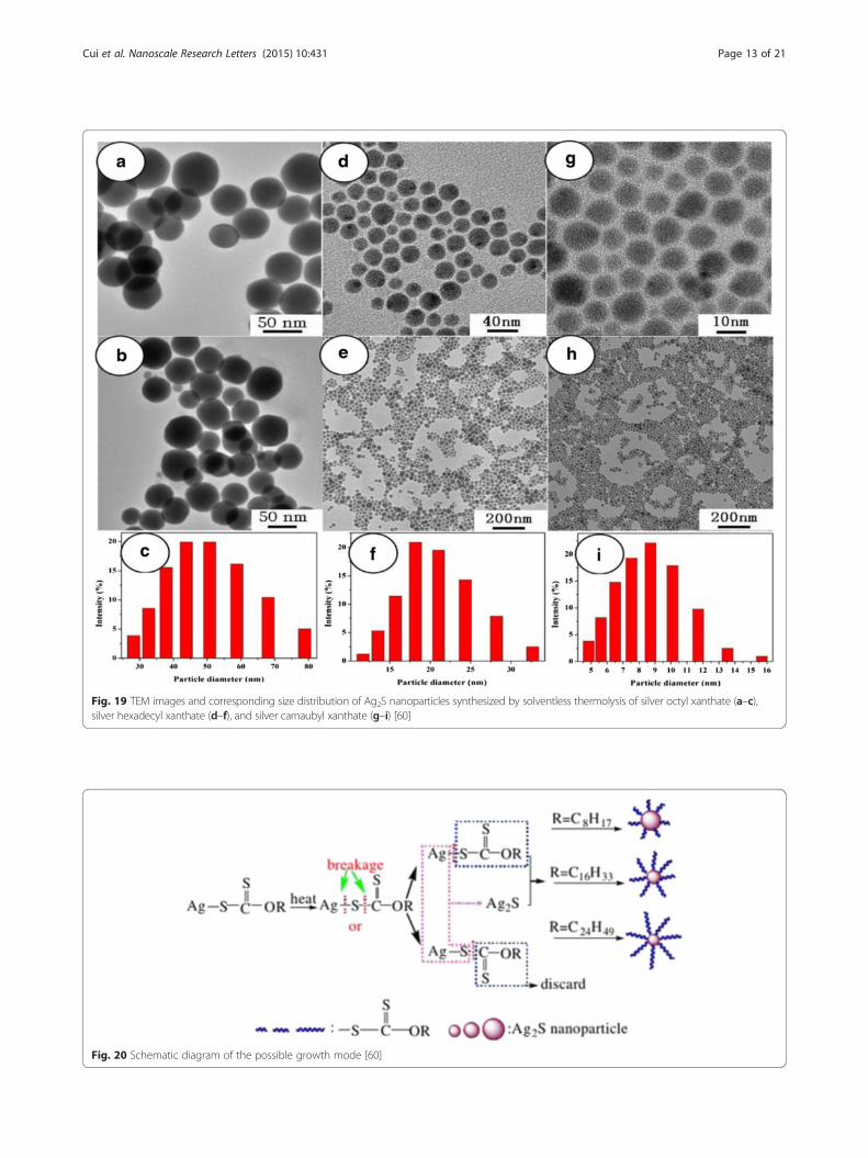
Fig. 19 TEM images and corresponding size distribution of Ag2S nanoparticles synthesized by solventless thermolysis of silver octyl xanthate (a–c),silver hexadecyl xanthate (d–f), and silver carnaubyl xanthate (g–i) [60]
Fig. 20 Schematic diagram of the possible growth mode [60]
Cui et al. Nanoscale Research Letters (2015) 10:431 Page 13 of 21

zeolite is silica [72, 73]. A. Pourahmad prepared theAg2S/MCM-41 photocatalysts by ion exchange methodand is used for the photocatalytic degradation ofmethylene blue [75]. Figure 24 is the time-dependentelectronic absorption spectra of dye during photo ir-radiation. After 20 min of irradiation under UV lightin a Ag2S/MCM-41 suspension, 94 % of dye wasdecomposed and decolorized. And, no new bandsappear in the UV–vis region due to the reaction in-termediates formed during the degradation process.Under UV irradiation, Ag2S, MCM-41, and Ag2S/MCM-41 materials on photodegradation of methy-lene blue are shown in Fig. 25. It is observed thatAg2S supported system has a higher rate of degrad-ation than Ag2S or MCM-41 alone. And, there aremany factors that can influence the efficiency ofnanocomposite catalyst, such as the amount of Ag2Sloading, PH, and initial concentration of dye. Severalmethods have been reported concerning the photo-sensitization of TiO2 by MxSy or MxOy nanoparticlesfor heterogeneous photocatalysis [76] including CdS[77] or WO3 [78]. In fact, nanocrystalline Ag2S is agood candidate for the photosensitization of TiO2
catalysts, for Ag2S has a direct band gap of 0.9–1.05 eV, and its conduction band (−0.3 eV) is lessanodic than the corresponding TiO2 band (−0.1 eV),and the valence band (+0.7 eV) is more cathodicthan the TiO2 valence band (+3.1 eV). So, distinctTiO2/Ag2S nanocomposites were prepared by a
single-source decomposition method [79]. After, thesensitized TiO2 materials were evaluated as photoca-talysts on the degradation of aqueous phenol solutions,and the photocatalytic activity of nanocompositeswas enhanced with the existence of Ag2S over theTiO2 surface. And, the efficiency of this photocata-lysts is considerably improved comparing with pureTiO2. The best phenol photocatalyst was obtainedwhen atomic ratio of Ti/Ag is 2.40. NanostructuredAg2S/CdS was synthesized by two-step precipitationmethod [80]. And, the composite materials havecertain photocatalytic performance. When the con-centration of Ag2S was 5 % by weight, Ag2S/Cdsshowed the highest photocatalytic activity for hydro-gen evolution, with the solar-hydrogen energy con-version efficiency approximately 0.7 %. So, afterdoped Ag2S, the photocatalytic activity of CdS haveenhanced obviously.
Application in Optoelectronic DevicesCompared with bulk counterparts, the sheet-likephotocatalysts are much better for continuous flowsystem because of ease separation and recovery fromthe reaction system [86]. The sheet-like photocata-lysts can also help to harvest light more efficiently[87]. So, a novel graphene sheet/Ag2S composite wassynthesized through a facile solvothermal method,and its electrochemical performance was carried ona modified glassy carbon electrode in a three-electrode
Fig. 21 Schematic diagram illustrating the formation of Ag2S samples at various conditions [62]
Cui et al. Nanoscale Research Letters (2015) 10:431 Page 14 of 21

electrochemical cell [81]. In Fig. 26, it can be clearlyseen that the pure graphene oxide sheets naturallyaggregate and stack to multilayers with numerousedges, and the surface of graphene oxide was verysmooth compared with graphene sheets doped withAg2S NPs. So, the morphology of G-Ag2S compositeshas a substantial difference from that of the Go
sheet. Figure 27 shows that the composite modifiedGCE shows redox peaks, but graphene modified ex-hibits a perfect rectangle curve, and the redox peaksof the composites, often a characteristic of pseudoca-pacitance mainly result from the redox transition ofAg2S between a semiconducting state and a conduct-ing state. And, it is calculated that the redox peaks
Fig. 22 In vivo NIR fluorescence imaging (pseudocolored image) of nude mice. Control experiment (a), with subcutaneous injection (b) and withceliac injection (c) of Ag2S quantum dots emitting at 910 nm; Unmixed image of Ag2S quantum dots fluorescence signal (C inset); The correspondingemission spectra of the auto fluorescence and QD fluorescence of mice with celiac injection (d). (In images a–d, the blue corresponded to the miceauto fluorescence and the red corresponded to QD fluorescence.) (For interpretation of the reference to color in this figure legend, the reader isreferred to the web version of this article.) [70]
Cui et al. Nanoscale Research Letters (2015) 10:431 Page 15 of 21

with the specific capacitance is 1063 Fg−1, but thespecific capacitance of graphene modified is 316 Fg−1. So,it is believed that the nanocomposites would be apromising candidate as supercapacitor materials forpractical applications in future electronic devices. At
the same time, graphene-like Co3S4 nanosheet/Ag2Snanocomposite was prepared using a simple method.The nanocomposite photocatalyst displays excellentstability and photocatalytic activity compared withpure Co3S4 nanosheet or Ag2S nanoparticles [85].Ag2S is an important material for optoelectronic, be-cause it has an energy gap of Eg~1.1 eV, which issimilar to the ideal band gap of 1.13 eV for a photo-voltaic device [82] indicating that Ag2S could be anoptimal solar absorber performance which was mea-sured to the battery. The Ag2S QDs were synthe-sized by the successive ionic layer adsorption andreaction deposition method [83]. And, the assembledAg2S-QD solar cell in λ = 530 nm has the biggest ex-ternal quantum efficiency (EQE) which was 59 %,and when the spectral range in 400–1000 nm, theaverage of EQE was 42 %. The effective scope ofphotovoltaic is full of visible light and near-infraredspectral regions. Therefore, the results indicate thatAg2S QDs can be used as a highly efficient andbroad band sensitizer for solar cells. R. Karimzadehet al. [84] found that about 3 nm Ag2S semicon-ductor nanocrystals in concentrations of dimethylsulfoxide solution has different non-linear refractiveproperties; it can be used as a low power optical-limiting device. In addition, Ag2S also has importantapplication in other areas, for example nanometer-scale non-volatile memory devices. [88].
Other ApplicationsAg2S nanomaterials also have many other propertiesin various fields, such as electronic, magnetic, andso on. Ag2S belongs to I-VI semiconductor materialswith monoclinic crystal structure. Thin films ofAg2S have applications in photoconducting cells[89], IR detectors [90], and solar selective coating.Thus, Ag2S is a promising material for the conver-sion of solar energy into electricity as its band gap isbetween 1 and 2 eV. Usually, the material design forthese technological applications is based on thin filmpreparation techniques, in which the film thicknessranges from micrometer to submicrometer. It is wellknown that the surface contribution to the electrictransport process could be evaluated when thin filmswith different thicknesses, i.e., with various surface-to-volume ratios, are investigated [91]. So, D. Karashanovaet al. [92] evaluated the surface contribution to theelectronic or ionic transport in the epitaxial silversulfide films using electron-conducting and electron-blocking contacts, respectively. At the same time,Ag/Ag2S also can become the electrode materials,but disadvantages of the solid-state Ag/Ag2S elec-trode such as non-ideal response, signal drifting, anda long response time at low sulfide levels have
Fig. 23 XRD pattern of Ag2S and Ni/Ag2S nanoparticles [74]
Fig. 24 Spectra change that occur during the photocatalytic ofaqueous solution of methylene blue: PH = 7. [20 wt% Ag2S/MCM=41] = 0.6 g/L. C0 = 0.32 ppm [75]
Cui et al. Nanoscale Research Letters (2015) 10:431 Page 16 of 21

limited its application [93]. Thus, increasing the pre-cision of the Ag/Ag2S electrode by surface micromo-tion can help the research work under extremecircumstances, such as hydrothermal vents [94, 95].So, Ding et al. [96] have enhanced the sensitivity ofthe Ag/Ag2S electrode by using direct current carrierpower to electroplate silver nanoparticles on a silverwire. Three types of the Ag/Ag2S electrode each haddifferent physical structures under SEM (Fig. 28),which indicated that the different surfaces of theseelectrodes demonstrated that the preparation proce-dures affected the physical structures of the elec-trodes. Among all these electrodes, the directcurrent carrier electroplating electrode has the high-est detection limit while the typical electrode has thelowest limit. From Table 2, we can see that the re-sponse time of the electrode prepared by direct
Fig. 25 Effect of UV light and different photocatalysts on photocatalystic degradation of methylene blue. C0 = 0.32 ppm. [20 wt% Ag2S/MCM-41] =0.6 g/L, PH = 7 [75]
Fig. 26 SEM images of a the pristine graphene oxide and b Gs-Ag2Scomposites [81]
Fig. 27 CV curves of graphene and Gs-Ag2S composites at 100 mV−1
in 1 M H2SO4 in potential range from −0.6 to 0.2 V [81]
Cui et al. Nanoscale Research Letters (2015) 10:431 Page 17 of 21

current carrier electroplating for detecting a concen-tration of 10−7 mol L−1 S2− was less than 60 s. And,the detection limits of the Ag/Ag2S electrodes pre-pared by direct current electroplating and directcurrent carrier electroplating were improved to 1 ×10⁻⁵ and 1 × 10⁻⁷ mol L−1, respectively. The RMSE(root mean square deviation) of the linear regressionfor the electrode using the direct current carrierelectroplating method verified the accuracy and pre-cision of this type of electrode (Fig. 29). In additionto the above mention, Ag2S also has many other
Fig. 28 SEM observation of the surface of a typical electrode, b direct current electroplating electrode, and c direct current carrier electroplatingelectrode [96]
Table 2 Correlation of EMF (mv) with-log[S2−] for the types ofelectrode [96]
slope R2 n RMSE p
Typical 30.81 0.998 4 1.763 <0.001
DC electroplating 29.49 0.998 5 2.231 <0.0001
DC carrier electroplating 33.22 0.983 7 10.24 <0.0001
Fig. 29 Response curves of the three types of electrode. The linearcorrelation curve is generated using the direct current carrierelectroplating method [96]
Cui et al. Nanoscale Research Letters (2015) 10:431 Page 18 of 21

properties in applications, but it still needs people toexplore it gradually.
ConclusionsAg2S, playing important functions in a number of op-tical, electrochemical, and biochemical process, has beenregarded as a promising sensor and biological imagingagent in living creature. The preparation process andproduct of Ag2S have many disadvantages in traditionalpreparation methods. For example, it usually needs hightemperature, complicated processes, easy gather, andparticle size bed control. Recently, considerable effortshave been made to optimize the productive process ofAg2S and enhance properties and values of products.This work reviewed the progress in the development ofAg2S nanomaterials in the field of synthesis and applica-tion. Different forms of Ag2S nanostructures have beensynthesized such as rod-shaped, leaf-shaped, and cubic.Ag2S nanostructure obtained by bionic technology andprecursor of decomposition were prepared successfully.Meanwhile, it has been applied to many fields success-fully, such as optical, electrical, and biology, and it is ex-pected to use in other fields. In fact, there are stilllimitations for their practical use in photoelectric andmedical fields because it often requires complex prepar-ation process, and the yield is very low. In most cases,Ag2S nanoparticles are very prone to gather, which willgreatly reduce its optical properties. Therefore, it is oftennecessary to composite with other materials to achieve agood effect. Although, there are so many challenges, theadvances in nanoscience and nanotechnology of Ag2Sstill promise a better future for kinds of industries.
Competing InterestsThe authors declare that they have no competing interests.
Authors’ ContributionsCC researched the existing literatures and wrote the manuscript. XLdeveloped the concept and designed the manuscript. JL developed theconcept. All the authors read and approved the final manuscript.
AcknowledgementsThis work was supported by the Specialized Research Fund for the DoctoralProgram of the Higher of Education (No. 20123706120003), ShandongProvince Natural Science Fund of China (No. ZR2014EMQ002).
Received: 26 June 2015 Accepted: 16 October 2015
References1. Jun HK, Gareem MA, Arof AK, Sust R (2013) Quantum dot-sensitized solar
cells-perspective and recent development: a review of Cd chalcogenidequantum dots as sensitizers. Energy Rev 22:148–167
2. Selinsky RS, Ding Q, Faber MS, Wright JC, Jin S (2013) Quantum dotnanoscale heterostructures for solar energy conversion. Chem Soc Rev42:2963–2985
3. PanL LT, Liu X, Lu T, Zhu G, Sun Z, Sun CQ (2012) Metal-free photocatalyticdegradation of 4-chlorophenol in water by mesoporous carbon nitridesemiconductors. Catal Sci Technol 2:754–758
4. Zhao Z, Liu Z, Miyauchi M (2010) Tailored remote photochromic colorationof in situ synthesized CdS quantum dot loaded WO3 films. Adv Funct Mater20:4162–4167
5. Ezenwa IA, Okereke NA, Egwunyenga NJ (2012) Optical properties ofchemical bath deposited Ag2S thin films. Int J Sci Technol 2:101–106
6. Hwang I, Yong K (2013) Environmentally benign and efficient Ag2S-ZnOnanowires as photoanodes for solar cells: comparison with CdS-ZnOnanowires. Chem Phys Chem 14:364–368
7. Jiang F, Tian Q, Tang M, Chen Z, Yang J, Hu J (2011) One-pot synthesis oflarge–scaled Janus Ag-Ag2S nanoparticles and their photocatalyticproperties. Cryst Eng Comm 13:7189–7193
8. Dlala H, Amlouk M, Belgacem S, Girard P, Barjon D (1998) Structural andoptical properties of Ag2S thin films prepared by spray pyrolysis. Eurphys j-applphys 2:13–16
9. Brelle MC, Zhang JZ (1998) Femtosecond study of photo-induced electrondynamics in AgI and core/shell structural AgI/Ag2S colloidal nanoparticles.Chem J Phys 108:3119
10. Bruhwiler D, Leigener C, Glaus S, Calzaferri G (2002) Luminescent silversulfide clusters. J Phys Chem B 106:3770
11. Hull S, Keen DA, Sioia DS, Madden PA, Wilson M (2002) The high-temperature superionic behaviour of Ag2S. J Phys Conclens Matter 14:19
12. Kitova S, Eneva J, Panov A, Haefke H (1994) Infrared photography based onvapor-deposited silver sulfide thin films. Imaging J Sci Fechnol 38:484
13. Wang DS, Hao CH, Zhong W, Peng Q, Wang TH, Liao ZM, Yu DP, Li YD(2008) Ultralong single-crystalline Ag2S nanowires: promising candidates forphotoswitches and room-temperature oxygen sensors. Adv Mater 20:2628
14. Liu JC, Raveendran P, Shervani Z, Ikushima Y (2004) Synthesis of Ag2Squantum dots in water-in-CO2 microemulsions. Chem Commun 22:2582–3
15. Xiao JP, Xie Y, Tang R, Luo W (2002) Template-based synthesis of nanoscaleAg2E (E = S,Se) dendrites. J Mater Chem 12:1148–51
16. Chen M, Xie Y, Chen HY, Qiao ZP, Qian YT (2001) Preparation andcharacterization of metal sulfides in ethylenediamine under ambientcondition through a γ-irradiation route. J Colloid Interf Sci 237:47–53
17. Lim WP, Zhang ZH, Low HY, Chin WS (2004) Preparation of Ag2Snanocrystals of predictable shape and size. Angew Chem Int Ed 43:5685–9
18. Buda C, Chen XB, Narayanan R, Sayed EI (2005) Chemistry and properties ofnanocrystals of different shapes. Chem Rev 105:1025
19. Zhang WQ, Xu LQ, Tang KB, Li FQ, Qian WT (2005) Solvothermal synthesisof NiS 3D nanostructures. Eur J Inorg Chem 4:653–656
20. Liu QY, Guo F, Komarneni S (2004) Biomolecule-assisted synthesis of highlyordered snowflakelike structures of bismuth sulfide nanorods. J Am ChemSoc 126:54–55
21. Kuang D, Xu A, Fang Y, Liu H, Frommen C, Fenske D (2003) Surfactant-assisted growth of novel PbS dendritic nanostructures via facilehydrothermal process. Adv Mater 15:1747–1750
22. Chen XG, Wang X, Wang ZG, Yang XG, Qian YT (2005) Hierarchical growthand shape evolution of HgS dendrites. Cryst Growth Des 5:347–350
23. Zhao Y, Zhang DW, Shi WF (2007) A gamma-ray irradiation reduction routeto prepare rod-like Ag2S nanocrystallines at room temperature. Mater Lett61:3232–3234
24. Yin YD, XuXG GXW, Lu Y, Zhang ZC (1999) Synthesis and characterization ofZnS colloidal particles via γ-radiation. Radiat Phys Chem 55:353–356
25. Chen AH, Wang HQ, Li XY (2005) One-step process to fabricate Ag-polypyrrole coaxial nanocables. Chem Commun 14:1863–1864
26. Chen MH, Gao L (2006) Synthesis of leaf-like Ag2S nanosheets by hydrothermalmethod in water alcohol homogenous medium. Mater Lett 60:1059–1062
27. Xu CG, Zhang ZC, Ye Q (2004) A novel facile method to metal sulfide(metal = Cd, Ag, Hg) nanocrystallite. Mater Lett 58:1671–1676
28. Cheng B, Russell JM, Sheng W, Zhang L, Samulski ET (2004) Large-Scale,solution-phase growth of single-crystalline SnO2 nanorods. J Am Chem Soc126:5972–5973
29. Wong EM, Bonevich JE, Searson PC (1998) Growth kinetics ofnanocrystalline ZnO particles from colloidal suspensions. J Phys Chem B102:7770–7775
30. Zhai HJ, Wang HS (2008) Ag2S morphology controllable via simpletemplate-free solution route. Mater Res Bull 43:2354–2360
31. Gou LF, Murphy CJ (2003) Solution-phase synthesis of Cu2O nanocubes.Nano Lett 3:231–234
32. Lee SM, Jun Y, Cho SN, Cheon J (2002) Single-crystalline star-shapednanocrystals and their evolution: programming the geometry of nano-building blocks. J Am Chem Soc 124:11244–11245
Cui et al. Nanoscale Research Letters (2015) 10:431 Page 19 of 21

33. Seo WS, Shim JH, Oh SJ, Lee EK, Hur NH, Park JT (2005) Phase- and size-controlled synthesis of hexagonal and cubic CoO nanocrystals. J Am ChemSoc 127:6188–6189
34. Lim WP, Zhang ZH, Low HY, Chin WS (2004) Preparation of Ag2S nanocrystalsof predictable shape and size. Angew Chem Int Ed 42:5803–5807
35. Wang XB, Liu WM, Hao JC, Fu XG, Xu BS (2005) A simple large-scalesynthesis of well-defined silver sulfide semiconductor nanoparticles withadjustable size. Chem Lett 43:1664–1665
36. Dong LH, Chu Y, Liu Y (2008) Synthesis of faceted and cubic Ag2Snanocrystals in aqueous solution. J Colloid Interf Science 317:485–492
37. Sun YZ, Zhou BB (2010) Single-crystalline Ag2S hollow nanohexagons andtheir assembly into ordered arrays. Mater Lett 64:1347–1349
38. Zhuang ZB, Peng Q, Zhang B, Li YD (2008) Controllable synthesis of Cu2Snanocrystals and their assembly into superlattice. J Am Chem Soc130:10428–3
39. Zhuang Z, Peng Q, Wang X, Li Y (2007) Tetrahedral colloidal crystals of Ag2Snanocrystals. Angew Chem Int Ed 46:8174–8177
40. Chaudhuri RG, Paria S (2012) A novel method for the templated synthesis ofAg2S hollow nanospheres in aqueous surfactant media. J Colloid InterfScience 369:117–122
41. Liu MY, Xu ZL, Li BN, Lin CM (2011) Synthesis of worm-like Ag2Snanocrystals in W/O reverse microemulsion. Mater Lett 65:555–558
42. Brelle MC, Zhang JZ, Nguyen L, Mehra RK (1999) Synthesis and ultrafaststudy of cysteine- and glutathione-capped Ag2S semiconductor colloidalnanoparticles. J Phys Chem A 103:10194–201
43. Ortega EV, Berk D (2006) Precipitation of silver powders in the presence ofethylenediamine tetraacetic acid. Ind Eng Chem Res 45:1863–1868
44. Lv LY, Wang H (2014) Ag2S nanorice: hydrothermal synthesis andcharacterization study. Mater Lett 121:105–108
45. Yu C, Ming ML, Liu Z, Yu Y (2012) Synthesis of normal and flattenedrhombic dodecahedral Ag2S particles. Cryst Eng Comm 14:3772
46. McGrath KM (2001) Probing material formation in the presence of organicand biological molecules. Adv Mater 13(12–13):989–992
47. Cai W, Shin DW, Chen K, Gheysens O, Cao Q, Wang SX (2006) Peptide-labeled near-infrared quantum dots for imaging tumor vasculature in livingsubjects. Nano Lett 6:669–676
48. Kong Y, Chen J, Gao F, Li W, Xu X, Pandoli O (2010) A multifunctionalribonuclease-A-conjugated CdTe quantum dot cluster nanosystem forsynchronous cancer imaging and therapy. Small 6:2367–2373
49. Yarema M, Pichler S, Sytnyk M, Seyrkammer R, Lechner RT, Fritz-Popovski G(2011) Infrared emitting and photoconducting colloidal silver chalcogenidenanocrystal quantum dots from a silylamide-promoted synthesis. ACS Nano5:3758–3765
50. Du Y, Xu B, Fu T, Cai M, Li F, Zhang Y (2010) Near-infrared photoluminescentAg2S quantum dots from a single source precursor. J Am Chem Soc132:1470–1471
51. Chen J, Zhang T, Feng LL (2013) Synthesis of ribonuclease-A conjugateAg2S quantum dots clusters via biomimetic route. Mater Lett 96:224–227
52. Siva C, Chandrasekaran Nivedhini I (2014) L-cysteine assisted formation ofmesh like Ag2S and Ag3AuS2 nanocrystals through hydrogen bonds.MaterLett 134:56–59
53. Koneswaran M, Narayanaswamy R (2009) L-cysteine-capped ZnS quantumdots based fluorescence sensor for Cu2+ ion. Sens Actuator B Chem139:104–9
54. de la Rica R, Velders AH (2011) Biomimetic crystallization of Ag2Snanoclusters in nanopore assemblies. JAm Chem Soc 133:2875–2877
55. Brennan JG, Siegrist T, Carroll PJ, Stuczynski SM, Brus LE, Steigerwald ML.The preparation of large semiconductor clusters via the pyrolysis of amolecular precursor. J. Am. Chem. Soc. 1989:111;4141-4143.
56. Fan D, Afzaal M, O'Brien P (2007) Using coordination chemistry to develop newroute to semiconductor and other materials. Coord Chem Rev 251:1878–1888
57. Esteves ACC, Trindade T (2002) Synthesis studies on II/VI semiconductorquantum dots. Curr Opin Solid State Mater Sci 6:347–353
58. Tang Q, Yoon SK, Yang HJ (2006) Selective degradation of chemical bonds:from single source molecular precursors to metallic Ag and semiconductingAg2S nanocrystals via instant thermal activation. Langmuir 22:2802–2805
59. Wang TX, Xiao H, Zhang YC (2008) Simple solid state synthesis of Ag2Scrystallites using a single source molecular precursor. Mater Lett 62:3736–3738
60. Zhang CL, Zhang SM, Yu LG, Zhang ZJ (2012) Size-Controlled synthesis ofmonodisperse Ag2S nanoparticles by a solventless thermolytic method.Mater Lett 85:77–80
61. Hou XM, Zhang XL, Yang W, Liu Y (2012) Synthesis of SERS active Ag2Snanocrystals using oleylamine as solvent reducing agent and stabilizer.Mater Res Bull 47:2579–2583
62. Shakouri-Arani M, Salavati-Niasari M (2014) Structural and spectroscopiccharacterization of prepared Ag2S nanoparticles with a novel sulfuringagent. Mol Biomol Spectrosc 133:463–471
63. Wehrenberg BL, Wang C, Guyot-Sionnest P (2002) Interband and intrabandoptical studies of PbSe colloidal quantum dots. J Phys Chem B 106:10634–10640
64. Bakueva L, Gorelikov I, Musikhin S, Zhao XS, Sargent EH, Kumacheva E(2004) PbS quantum dots with stable efficient luminescence in the near-IRspectral range. Adv Mater 16:926–929
65. Harrison MT, Kershaw SV, Burt MG, Eychmuller A, Weller H, Rogac AL (2000)Wet chemical synthesis and spectroscopic study of CdHgTe nanocrystalswith strong near-infrared luminescence. Mater Sci Eng B 69:355–360
66. Zrazhevskiy P, Senawb M, Gao X (2010) Designing multifunctional quantumdots for bioimaging, detection, and drug delivery. Chem Soc Rev 39:4326–4354
67. O'Connell MJ, Bachilo SM, Huffman CB, Moore VC, Strano MS, Haroz EH,Rialon KL, Boul PJ, Noon WH, Kittrell C (2002) Band gap fluorescence fromindividual single-walled carbon nanotubes. Science 297:593–596
68. Crochet J, Clemens M, Hertel T (2007) Quantum yield heterogeneities ofaqueous single-wall carbon nanotube suspensions. J Am Chem Soc129:8058–8059
69. Zhang Y, Hong GS, Zhang YJ (2012) Ag2S quantum dot: a bright andbiocompatible fluorescent nanoprobe in the second near-infrared window.ACS Nano 6(5):3659–3702
70. Jiang P, Zhu CN, Zhang ZL (2012) Water-soluble Ag2S quantum dots fornear-infrared fluorescence imaging in vivo. Biomaterials 33:5130–5135
71. Zhou XD, Shi HQ, Huang DM, Jia SM (2008) Room temperature synthesisand electrochemical application of imidazoline surfactant-modified Ag2Snanocrystals. Mater Lett 62:2407–2410
72. Sohrabnezhad S, Pourahmad A (2010) Comparison absorption of newmethylene blue dye in zeolite and nanocrystal zeolite. Desalination 256:84–89
73. Pourahmad A, Pourahmad A, Sohrabnezhad S, Rakhshaee R (2011) Ternarymetal sulphide nanocrystals in MCM-41 nanoparticles matrix: preparationand properties. Micro Nano Lett 6:918–921
74. Aazam ES (2014) Photocatalytic oxidation of methylene blue dye undervisible light by Ni doped Ag2S nanoparticles. J Ind Eng Chem 20:4033–4038
75. Pourahmad A (2012) Ag2S nanoparticle encapsulated in mesoporousmaterial nanoparticles and its application for photocatalytic degradation ofdye in aqueous solution. Superlattices and Microst 52:276–287
76. Robert D (2007) Photosensitization of TiO2 by MxOy and MxSy nanoparticlesfor heterogeneous photocatalysis applications. Catal Today 122:20–26
77. Kim JC, Choi J, Lee YB, Hong JH, Lee JI, Yang JW, Lee WI, Hur NH. Enhancedphotocatalytic activity in composites of TiO2 nanotubes and CdSnanoparticles. Chem. Commun. 2006, 5024–5026. Epub 2006 Oct 27.
78. Puddu V, Mokaya R, Puma GL. Novel one step hydrothermal synthesis ofTiO2/WO3 nanocomposites with enhanced photocatalytic activity.Chem.Commun. 2007, 4749–4751. Epub 2007 Sep 7.
79. Neves MC, Nogueira JMF, Trindade T, Mendonca MH (2009)Photosensitization of TiO2 by Ag2S and its catalytic activity on phenolphotodegradation. J Photochem Photobiol A Chem 204:168–173
80. Shen SH, Guo LJ, Chen XB, Ren F (2010) Effect of Ag2S on solar-drivenphotocatalytic hydrogen evolution of nanostructured CdS. Int J HydrogenEnergy 35:7110–7115
81. Mo ZL, Liu PE, Gou RB, Deng ZP (2012) Graphene Sheets/Ag2Snanocomposites: synthesis and their application in supercapacitor materials.Mater Lett 68:416–418
82. Marti A, Araujo GL (1996) Limiting efficiencies for photovoltaic energyconversion in multigap systems. Sol Enrgy Mater Sol Cells 43:203
83. Auttasit T, Wu KL, Tung HY (2010) Ag2S quantum dot-sensitized solar cells.Electrochem Commun 12:1158–1160
84. Karbmzadeh R, Aleali H, Mansour N (2011) Thermal nonlinear refractionproperties of Ag2S semiconductor nanocrystals with its application as a lowpower optical limiter. Opt Commun 284:2370–2375
85. Xu MY, Niu HL, Huang JJ, Song JM, Mao CG, Zhang SY, Zhu CF, Chen CL(2015) Facile synthesis of graphene-like Co3S4 nanosheet/Ag2S nanocompositewith enhanced performance in visible light photocatalysis. Appl Surf Sci351:374–381
86. Li ZH, Shen J, Wang JQ, Wang DJ, Huang YJ, Zou J (2012) Single crystaltitanate–zirconate nanoleaf: synthesis, growth mechanism and enhancedphotocatalytic hydrogen evolution properties. Cryst Eng Comm 14:1874–1880
Cui et al. Nanoscale Research Letters (2015) 10:431 Page 20 of 21

87. Sun YF, Sun ZH, Gao S, Cheng H, Liu QH, Lei FC, Wei SQ, Xie Y (2014) All-surface-atomic-metal chalcogenide sheets for high-efficiency visible lightphotoelectrochemical water splitting. Adv Energy Mater 4:1300611
88. Gubicza A, Csontos M, Halbritter A, Mihaly G (2015) Non-exponentialresistive switching in Ag2S memristors: a key to nanometer-scale non-volatile memory devices. Nanoscale 7:3493
89. Nasrallah TB, Dlala H, Amlouk M, Belgacem B (2005) Some physicalinvestigations on Ag2S thin films prepared by sequential thermalevaporation. Synth Met 151:225–230
90. Karashanova D, Nihtianova D, Starbov K (2004) Crystalline structure andphase composition of epitaxially grown Ag2S thin films. Solid State lonics171:269–275
91. Hamilton JF (1988) The silver halide photographic process. Adv Phys37:359–441
92. Karashanova D, Starbov N (2006) Surface assisted electric transport in Ag2Sthin films. Appl Surf Sci 252:3011–3022
93. Kuhl M, Steuckart C, Eickert G, Jeroschewski P (1998) A H2S microsensor forprofiling biofilms and sediments: application in an acidic lake sediment.Aquat Microb Ecol 15:201–209
94. Ding K, Seyfried WE, Tivey MK, Bradley AM (2001) In situ measurement ofdissolved H2 and H2S in high temperature hydrothermal vent fluids at theMain Endeavour Field, Juan de Fuca Ridge. Earth Planet Sci Lelt 186:417–425
95. Zhang RH, Zhang XT, Hu SM (2013) Novel sensor based on Ag/Ag2Selectrode for in situ measurement of dissolved H2S in high temperatureand pressure fluids. Sens Actuators B Chem 177:163–171
96. Ding Q, Pan YW, Huang YF. The optimization of Ag/Ag2S electrode usingcarrier electroplating of nano silver particles and its preliminary applicationto offshore Kueishan Tao, Taiwan. Continental Shelf Research. 2015, 25
Submit your manuscript to a journal and benefi t from:
7 Convenient online submission
7 Rigorous peer review
7 Immediate publication on acceptance
7 Open access: articles freely available online
7 High visibility within the fi eld
7 Retaining the copyright to your article
Submit your next manuscript at 7 springeropen.com
Cui et al. Nanoscale Research Letters (2015) 10:431 Page 21 of 21