Synthesis and characterization of new modified metabolites ...
98
UNIVERSITA’ DEGLI STUDI DI NAPOLI FEDERICO II Ph.D. Thesis in Chemical Sciences XXIX Course Synthesis and characterization of new modified metabolites, molecules with strong pharmacological activities Raffaele Gravante Tutor Supervisor Dr. Giovanni Di Fabio Dr. Annalisa Guaragna Coordinator Prof. Luigi Paduano
Transcript of Synthesis and characterization of new modified metabolites ...

UNIVERSITA’ DEGLI STUDI DI NAPOLI
FEDERICO II
Ph.D. Thesis in Chemical Sciences
XXIX Course
Synthesis and characterization of new modified metabolites, molecules with strong pharmacological activities
Raffaele Gravante
Tutor Supervisor
Dr. Giovanni Di Fabio Dr. Annalisa Guaragna
Coordinator
Prof. Luigi Paduano

Summary
Polyphenols are the most widely distributed class of plant secondary metabolites and several
thousand different compounds have been identified. They play many different roles in plant biology
and human life, as UV protective agents, defensive compounds against herbivores and pathogens,
they contribute to plant colors and to the taste of food and drink. Moreover it is widely known that
natural metabolites have played a crucial role in the identification of many of the drugs that are on
the market today. Nowadays natural products used pure in pharmacological preparations represent
only 5% of the total, but their semi-synthesis derivatives represent more than 50% of the drugs in
use.1
My PhD project is focused on the chemistry of natural substances, and I developed some strategies
to synthetize new silibinin conjugates. Silibinin is the major biologically active component of the
seeds extract of the milk thistle (Silybum marianum) also known as silymarin.2 Structurally,
silibinin is a diastereoisomeric mixture of two flavonolignans, silybin A and silybin B in a ratio of
approximately 1:1 (Figure 1).3
Figure 1
Silibinin is a metabolite with multiple biological activities operating at various cell levels.4
Unfortunately, its therapeutic efficiency is rather limited by its low bioavailability due to its very
low water solubility.
Aiming to improve the solubility, during the first year of my PhD project, we have developed an
efficient synthetic procedure to obtain new 9”-phosphodiester silibinin conjugates with different
mono- and di-saccharide labels through the anomeric hydroxyl group (Figure 2).5
O
OOH
OHOCH3
silybin A(2R, 3R, 7''R, 8''R)
silybin B(2R, 3R, 7''S, 8''S)
O
OH
O
OOH
OOH
OCH3
HO
OH
2
345
8
6
8''
7''
5'
2'
6'
1" 5"
2"
9''
silibinin
O
OOH
OHOCH3
+
17
1'
4'
3'
6"
4"
3"
A C E
B D

Figure 2
In our approach 9’’-phosphoramidite has been used as silibinin substrate and 1-OH full protected
mono- and disaccharide derivatives as sugar starting materials. We initially converted full
acetylated mono and di-saccharides into 1-OH derivatives and these compounds were coupled with
9’’-phosphoramidite silibinin. The oxidation and the deprotection treatments led to the desired
phosphodiester derivatives in good yields. The crude materials were then subjected to the
purification by reverse phase analysis (RP-18 HPLC), using a variety of columns and elution
conditions, but unfortunately it was too difficult to purify the mixture of diastereoisomers. In the
end new silibinin analogues were obtained as a mixture of diastereoisomers, observed by 31P NMR
analysis. The NMR analysis has proved to be very complex, in fact 1H and 31P NMR spectra of all
compounds showed a dramatic complexity, due to the presence of a lot of diastereoisomers. This
drawback has not allowed a complete and detailed NMR characterization of the new derivatives.
The structures were confirmed by 31P NMR and ESI-MS mass spectra signals. In preliminary study,
new derivatives were subjected to DPPH free radical scavenging and Xanthine Oxidase inhibition
assays to evaluate their antioxidant activities. Independently of the sugar moiety present, all
compounds exhibited a radical scavenging activities slightly higher than that of the silibinin, and
Xanthine Oxidase inhibition at least as that of the silibinin. On the other hand the new derivatives
showed a water solubility well above that of silibinin, in fact it was possible to prepare solutions of
about 70 mg/mL in water.
These two data encouraged our studies during my second year of PhD to improve this synthetic
strategy and to realize libraries of optically pure glyco-conjugated silibinins.
The 9''-phosphoramidite has been used as silibinin substrate and fully protected 6-OH mono- and
di-saccharide derivatives as sugar starting materials (Figure 3).6

Figure 3
We initially converted opportunely protected mono- and di-saccharides (Glucose, Mannose,
Galactose, N- Acetylglucosamine, Trehalose and Lactose) into 6-OH derivatives and then they were
coupled with 9"-phosphoramidite silibinin following typical phosphoramidite chemistry procedure.
The crude materials were purified by reverse phase chromatography (RP-18 HPLC) and
characterized by NMR and MALDI- TOF/TOF-MS analyses. All compounds were obtained in
good yields and as a mixture of two silibinin diastereoisomers (A and B). Finally new
phosphodiester derivatives were converted into the corresponding sodium salt by cation exchange
resin carrying crystalline samples. New derivatives showed water solubility well above that of
silibinin, with the possibility to prepare solutions of about 70 mg/mL in water. The stability of new
glyco-conjugates was investigated in human serum by HPLC analyses and the 50% disappearance
of the peak corresponding to the intact glyco-conjugate was observed after ca. 40-68 hours. All
derivatives were subjected to DPPH free radical scavenging assay and they exhibited radical
scavenging activities slightly higher than that of the silibinin. In order to verify the potential
biological properties of these derivatives, a biological assay was used to evaluate the cytotoxicity of
the new glyco-conjugates, compared with that of the silibinin, on human liver cancer cell line (Hep
G2). No significant changes in the viability of treated cells were observed when they were subjected
to the action of glyco-conjugates, also considering the longest incubation (72 hours) at the
maximum dose taken (30 µM). Also, in order to investigate the role of the different OH groups in
the antioxidant activity of new glyco-conjugates in comparison with silibinin, their acidity and
redox capacity have been evaluated. A redox-deep characterization was carried out in collaboration
with Prof. Mauro Iuliano and Dr Gaetano De Tommaso of our Department, using potentiometric
and voltammetric techniques. The determination of the acidity constants was conducted by

potentiometric titrations measuring the concentration of hydrogen ions with a glass electrode. By
processing data, it was possible to define three equilibrium constants. Analysis of the data shows
that the conjugation in position 9" does not affect the redox behaviour of the silibinin scaffold.
As a part of our continuing research effort towards the synthesis of new natural product analogues
exploiting the phosphoramidite chemistry, in the last year of my PhD project the attention was
focused on the development of synthetic methods to obtain oligoflavonoids based on silibinin, and
to investigate their anti-radical activity. Exploiting the selective protection to hydroxyl groups of the
silibinin we have developed an efficient strategy for the synthesis of new 3-9", 3-3 and 9"-9" dimers
of silibinin in good yields (Figure 4).7
Figure 4
In order to obtain suitable building blocks for the dimers synthesis, we have developed a selective
protective reaction for the different hydroxyl groups with isobutyric anhydride. The building blocks
obtained were coupled in the three different provisions with 3- and 9”-silibinin phosphoramidite
using the well known phosphoramidite chemistry. After oxidation, deprotection and RP-18 HPLC
purification the products were converted into the corresponding sodium salts by cation exchange on
a DOWEX (Na+ form) resin, leading to the desired phosphodiester dimers derivatives in good
yields. The structures of new analogues were confirmed by 31P NMR and MALDI-TOF/TOF-MS
analyses. The stability of new silibinin dimers was investigated in human serum by HPLC analysis
and the 50% disappearance of the peak corresponding to the intact molecule (t1/2) was observed
after ca. 80 hours. All derivatives were subjected to DPPH free radical scavenging assay and they
exhibited activities quite higher than silibinin.
Moreover, the solubility in water of silibinin and its dimers as well as their ability to react with

reactive oxygen species (ROS) were determined by estimating their second order rate constant with
singlet oxygen (1O2) and hydroxyl radical (HO∙) in solution. This data were obtained in the
laboratories of Prof. Marcello Brigante during my experience work in his research team at the
University Blaise Pascal, Institute of Chemistry of Clermont-Ferrand (France). Solubility
experiments indicate that dimers were completely dissolved (0.1 mg in 10 mL) in water and
solubility can be estimated to be ≥ 19.5 µM corresponding to > 20 mg/L. Reactivity between silibinin and dimers with 1O2 and HO∙ was determined by use of Rose Bengal
(RB) and hydrogen peroxide as respective ROS sources. For this purpose, 545 nm centered
excitation of RB and laser flash photolysis (LFP) experiments were coupled with a kinetic
competition approach. Dimers reactivity toward singlet oxygen results to be close to the value
determined for silibinin, or about 35% lower. Second order rate constant fare in the same order of
magnitude reported in literature for molecules with similar structure. Morales and co-workers8
reported a reactivity ranging from 2.4 to 13.4 × 10⁷ M-1s-1 for flavonoid derivative such as quercetin
and morin. Estimation of second order rate constant reactivity with HO∙ indicates that some dimers
showed a second order rate constant ≥ 1.5 × 10¹⁰ M–1s–1. Wang and co-workers9 investigated the
reactivity of hydroxyl radical with phenolic compounds in order to estimate their anti-oxidative
ability using aqueous pulse radiolysis. The value of 1.5 × 10¹⁰ M–1s–1 was found for quercetin that is
close to those estimates for green tea polyphenols. Interestingly, Husain at al.10 reported that
reactivity of flavonoids toward photo-generated hydroxyl radical increases with the number of
hydroxyl groups in the aromatic ring.
References 1 Newman, D. J.; Cragg, G. M. J. Nat. Prod. 2012, 75, 311. 2 Gažák, R.; Walterová, D.; Křen, V. Curr. Med. Chem. 2007, 14, 315. 3 Napolitano, J. G.; Lankin, D. C.; Graf, T. N.; Friesen, J. B.; Chen, S-N.; McAlpine, J. B.; Oberlies, N. H.;
Pauli, G. F. J. Org. Chem. 2013, 78, 2827. 4 Zhan, T.; Digel, M.; Küch, E.-M. J. Cell. Biochem. 2011, 112, 849 and references therein. 5 Zarrelli, A.; Romanucci, V.; Tuccillo, C.; Federico, A.; Loguercio, C.; Gravante, R.; Di Fabio, G. Bioorg.
Med. Chem. Lett. 2014, 24, 5147. 6 Romanucci, V.; Gravante, R.; Di Marino, C.; Iuliano, M.; De Tommaso, G.; Caruso, T.; Zarrelli, A.; Di
Fabio, G. submitted for publication. 7 Gravante, R .; Romanucci, V.; Cimafonte, M.; Di Marino, C.; Mailhot, G.; Brigante, M.; Zarrelli, A.; Di
Fabio, G. submitted for publication. 8 Morales, J.; Günther, G.; Zanocco, A. L.; Lemp, E. PLoS One 2012, 7, e40548. 9 Wang, W. F.; Luo, J. S.; Yao, D.; Lian, Z. R.; Zhang, J. S.; Lin, N. Y. Radiat. Phys. Chem. 1993, 42,
985. 10 Rafat Husain, S.; Cillard, J.; Cillard, P. Phytochemistry 1987, 26, 2489.

1
Chapter 1: Introduction 3 1.1 Natural products and secondary metabolism 3
1.2 Plant polyphenols: molecules with a high degree of molecular diversity 4
1.2.1 A bit of history 5
1.2.2 What Are plant polyphenols really? 6
1.3 Poliphenols: focus on the antioxidant activity 11
1.4 Polyphenol-protein interactions: pharmacological properties of natural polyphenols 15
1.5 Silibinin: a flavonolignan extracted from the milk thistle 20
1.5.1 Biological activities of silibinin 22
1.6 Synthesis of silibinin modifieds: state of art 25
References 32
Chapter 2: Results and discussion 36 2.1 Aim of the research work 36
2.1.1 Synthesis, characterization, evaluation of anti-oxidant and biological properties and
investigation of electrochemical behavior of new silibinin glyco-conjugates 36
2.1.2 Synthesis, spectroscopic properties, solubility and anti-oxidative ability of silibinin
oligomers 37
2.2 New silibinin glyco-conjugates: synthesis and evaluation of anti-oxidant properties 39
2.3 Synthesis, biological and electrochemical investigation of new silibinin glyco-phosphodiester
conjugates 43
2.4 Dimeric phosphate-linked silibinins: synthesis, spectroscopic properties, solubility and radical
scavenger ability 58
References 69
Chapter 3: Experimental Session 71 3.1 New silibinin glyco-conjugates: synthesis and evaluation of anti-oxidant properties 71
3.1.1 General procedure for the synthesis of conjugates 6–9 71
3.1.2 DPPH radical scavenging activity assay 72
3.1.3 Xanthine Oxidase inhibition assay 72
3.2 Synthesis, biological and electrochemical investigation of new silibinin glyco-phosphodiester
conjugates 73
3.2.1 Synthesis of silibinin phosphoramidite building block 3 73

2
3.2.2 General procedure for the synthesis of monosaccharide building blocks 8–12 74
3.2.3 Synthesis of lactose building blocks 13 74
3.2.4 General procedure for the synthesis of silibinin phosphodiester glyco-conjugates 14–19 75
3.2.5 Analysis to serum stability of silibinin glyco-conjugates 76
3.2.6 DPPH radical scavenging activity assay 76
3.2.7 Citotoxicity on on human liver cancer cell Hep G2 76
3.2.7 Determination of the acidity constants by potentiometric titrations, UV-Vis
spectrophotometric measures and Circular Dichroism (CD-UV) of silibinin solutions. Redox
behaviour carried out by cyclic voltammetry 77
3.3 Dimeric phosphate-linked silibinins: synthesis, spectroscopic properties, solubility and radical
scavenger ability 80
3.3.1 Chemicals 80
3.3.2 General methods 80
3.3.3 Synthesis of silibinin building block 2 80
3.3.4 Synthesis of silibinin building block 3 81
3.3.5 Synthesis of silibinin building block 4 82
3.3.6 Synthesis of silibinin building block 5 82
3.3.7 Synthesis of dimeric phosphate-linked silibinins 6–8 83
3.3.8 HPLC purification of 6–8 84
3.3.9 DPPH radical scavenging activity assay 84
3.3.10 Serum stability 85
3.3.11 Singlet oxygen reactivity 85
3.3.12 Hydroxyl radical generation and reactivity estimation 86
References 89

3
Chapter 1: Introduction
1.1 Natural products and secondary metabolism
For million years, humankind is completely dependent on plants as source of carbohydrates, proteins
and fats for food and shelter. In addition, plants are a valuable source of a wide range of secondary
metabolites, which are used as pharmaceuticals, agrochemicals, flavours, fragrances, colours,
biopesticides and food additives.
The secondary metabolites, as the name suggests, are so-called because they are produced by the plant
secondary metabolism. Primary metabolism refers to the processes producing the carboxylic acids of
the Krebs cycle, α-amino acids, carbohydrates, fats, proteins and nucleic acids, all essential for the
survival and well-being of the organism.1 All organisms possess the same metabolic pathways by
which these compounds are synthesized and utilized. Secondary metabolites, on the other hand, are
non-essential to life but contribute to the species fitness for survival.1 Secondary metabolites are also
produced using other metabolic pathways than primary metabolites. These pathways are more
characteristic for the particular family or genus and are related to the mechanism of evolution of
species. In fact, the specific constituents in a certain species have been used to help with systematic
determination, groups of secondary metabolites being used as markers for botanical classification.1
The division between primary and secondary metabolism is not clear, but the two types are linked
together because primary metabolism provides the small molecules that are the starting materials of the
secondary metabolic pathways (Figure 1.1).

4
Figure 1.1 Primary metabolites and their links to secondary metabolism.
One of the most important families of secondary metabolites is that of polyphenols. Polyphenols are the
most widely distributed class of plant secondary metabolites and several thousand different compounds
have been identified. They play many different roles in plant biology and human life, including UV
protection, defense against herbivores and pathogens, and they contribute to plant colors, taste of food
and drink, and are used as pharmaceuticals.
1.2 Plant polyphenols: molecules with a high degree of molecular diversity
“Eating five servings of fruits and vegetables per day”
This is what is highly recommended and heavily advertised nowadays to the general public to stay fit
and healthy. “Drinking green tea on a regular basis”, “eating chocolate from time to time”, as well as
“savoring a couple of glasses of red wine per day” have been claimed to increase life expectancy even
further. Why? The answer is in fact still under scientific scrutiny, but a particular class of compounds,

5
naturally occurring in fruits and vegetables, is considered to be crucial for the expression of such
human health benefits: the polyphenols.
1.2.1 A bit of history
Before being called polyphenols, these plant-derived natural products were globally referred to as
“vegetable tannins” as a consequence of the use of various plant extracts containing them in the
conversion of animal skins into leather. The first mentions of vegetable tanning in the classical
literature are accredited to the founder of the science of botany, Theophrastus of Eressus (371–286 BC),
in his Historia Plantarum encyclopedia and the origins of this leather-making process get lost in the
depths of the most ancient records of the history of human civilizations.2
Over the centuries, “vegetable tannins” have never ceased to garner general (and commercial) interest,
as well as scientific curiosity,3 and from the beginning of the last century an increasingly numerous of
chemists got involved in this affair, mainly concerned with finding an accurate method for analyzing
tanning extracts used in the leather industry. This was indeed a valuable and quite honorable objective,
but far from trivial given the means of chemical analysis available at the time. Fortunately, over the
years, botanists, plant physiologists, phytochemists and biochemists, as well as a few obstinate organic
chemists, kept on studying polyphenols and underlying their significance not only as major and
ubiquitous plant secondary metabolites, but also as compounds that express properties with numerous
implications and potential exploitations in various domains of general public and commercial interests.
So polyphenols gradually became a topic of intensive investigation in various plant-related scientific
domains, including applied research areas such as agriculture, ecology, food science and nutrition, as
well as medicine.4,5 The development of more and more advanced analytical techniques that paralleled
this gradual expansion of interest in polyphenol research during the second half of the past century
clearly had a major positive impact on both the development of the field and its appreciation by the
scientific community at large.
Nowadays, plant polyphenols enjoy an ever-increasing recognition not only by the scientific
community but also, and most remarkably, by the general public because of their presence and
abundance in fruits, seeds, vegetables and derived foodstuffs and beverages, whose regular
consumption has been claimed to be beneficial for human health. It is their capacity to scavenge
oxidatively generated free radicals, such as those derived from lipids and nucleic acids, that has often
been highlighted as the fundamental chemical event that underlies their utility in reducing the risk of

6
certain age-related degenerations and diseases. Although this so-called antioxidation property is not
listed among the qualifying factors that make a plant phenolic a “true” polyphenol, it has become the
trademark of “polyphenols” in recent exploitations by the agro-food, cosmetic, and parapharmaceutic
industries. However, antioxidation is not a property limited to polyphenols, as numerous simple plant
phenols are strong antioxidants, with many of them being in fact used as the active principles present in
some industrial formulations. The use of the term “plant phenols” by industry would definitely be more
appropriate, but the term “polyphenols” is preferred for commercial communications. As in the case of
earlier confusions surrounding the use of the term “tannins” in the scientific literature, the term
“polyphenols” has been and is still often misused by scientists from industry as well as academia.
1.2.2 What are plant polyphenols really?
According to a common classification, the polyphenols are divided into several sub-classes, based on
the number of phenolic rings present in their structure, the structural elements that bind these rings
between them and to the substituents bound on the rings. On this basis, they can be identified two
major groups: flavonoids and non-flavonoids polyphenol.6
x The flavonoids (Figure 1.2), share a structure formed by two aromatic rings, indicated as A and
B, linked together by three carbon atoms that form an oxygenated heterocycle, the ring C. They
can be further divided into several subclasses, depending on the type of involved heterocycle :
flavones, flavonols, flavanones, flavanonols, flavanols or flavan-3-ols or catechins,
anthocyanins and anthocyanidins, isoflavones, neoflavonoids, chalcones.
Figure 1.2 Basic structure of flavonoids.
x Among the non-flavonoid polyphenols are identified: simple phenol, phenolic acids, benzoic
aldehydes, hydrolysable tannins, acetophenones and phenylacetic acid, hydroxycinnamic acids,
coumarin, benzophenones, xanthones, stilbene, lignans, secoiridoids.

7
The above assortment of structure types is admittedly far from providing a clear picture of the family of
plant polyphenols. Of course, the presence of more than one hydroxy group on a benzene ring or other
arene ring does not make them polyphenolic. Catechol, resorcinol, pyrogallol, and phloroglucinol (all
di- and trihydroxylated benzene derivatives) are still defined as “phenols” according to the IUPAC
official nomenclature rules of chemical compounds7 but many such plant-derived monophenolics
(Figure 1.3) are often quoted as “polyphenols”, not only in cosmetic, parapharmaceutic, or nutraceutic
commercial advertisements, but also in the scientific literature, which has succumbed to today’s
fashionable use of the term.
Figure 1.3 Examples of simple plant-derived “monophenolics”.
The meaning of the chemical term “phenol” includes both the arene ring and its hydroxy substituent(s).
Hence, even if we agree to include polyphenolic compounds with no tanning action in a definition, the
term “polyphenol” should be restricted in a strict chemical sense to structures bearing at least two
phenolic moieties, irrespective of the number of hydroxy groups they each bear. However, as
judiciously pointed out earlier by Jeffrey B. Harborne,8 such a purely chemically based definition of
(poly)phenols needs additional restrictions, since many natural products of various biosynthetic origins
contain more than one phenolic unit. The existence of such alkaloids still gives us another problem
when attempting to define plant polyphenols in an as simple and yet comprehensive manner as possible,
since the tyrosine amino acid from which they are derived is itself a (primary) metabolite of the
phenylpropanoid pathway. It isn’t totally surprising by the difficult qualification of these molecules, but

8
the interest that the (poly)phenols plant arouse in various scientific fields requires greater clarity. This
raises the need to arrive at a revised definition of "real" plant polyphenols, purely chemical mold,
referring to the biosynthetic process by which they are formed. This definition may be as follows: the
term “polyphenol” should be used to define plant secondary metabolites derived exclusively from the
shikimate-derived phenylpropanoid and/or the polyketide pathway(s), featuring more than one
phenolic ring and being devoid of any nitrogen-based functional group in their most basic structural
expression.
This definition leaves out all monophenolic structures, which include di- and trihydroxyphenyl variants,
but contains three main categories of compounds, that can be identified as "true" polyphenols:
1. the proanthocyanidins (condensed tannins) such as procyanidins, prodelphinidins, and
profisetinidins (Figure 1.4), which are derived from the oligomerization of flavan-3-ol units
such as (epi)catechin, epigallocatechin, and fisetinidol (Figure 1.5).9
Figure 1.4 Representative examples of condensed tannins.

9
Figure 1.5 Representative examples of flava/flavonoids.
2. the gallo- and ellagitannins (hydrolyzable tannins), which are derived from the metabolism of
the shikimate-derived gallic acid (3,4,5-trihydroxybenzoic acid) that leads through esterification
and phenolic oxidative coupling reactions to numerous (near 1000) monomeric and oligomeric
polyphenolic galloyl ester derivatives of sugar-type polyols, mainly D-glucose (Figure 1.6),10,11

10
Figure 1.6. Representative examples of hydrolyzable tannins.
3. the phlorotannins that are found in red-brown algae (Figure 1.7) and essentially derived from
the oligomerizing dehydrogenative coupling of phloroglucinol (1,3,5-trihydroxybenzene).12
Figure 1.7 Representative examples of phlorotannins.

11
1.3 Poliphenols: focus on the antioxidant activity
There are numerous reasons to investigate plant polyphenols. From their most basic structural
expressions to their elaboration into further chemically transformed and complex oligo/polymeric
assemblies, plant polyphenols exhibit a remarkably diverse range of bio-physicochemical properties
that makes them rather unique and intriguing natural products. The first question that comes to mind is
why did plants choose to rely so heavily on the production of metabolites with multiple phenolic
moieties. The answer to this question is still a subject of debate and speculation, and possibly differs
for the different types of polyphenols.13 Generally speaking, plant polyphenols, as defined above, have
been implicated in diverse functional roles, including plant resistance against microbial pathogens and
animal herbivores such as insects (antibiotic and antifeeding actions), protection against solar radiation
(screens against DNA-damaging UV-B light), which probably was a determining factor in early
terrestrial plant evolution, as well as reproduction, nutrition, and growth, notably through interactions
with other organisms above and below ground (insects, symbiotic fungi, and bacteria).13 Over the
course of long-term evolution, as well as compulsory quick seasonal adjustments, plants have learnt to
cope with changing environmental conditions and pressures by relying on the formidable chemical
arsenal available to them through their remarkably dynamic secondary metabolisms, endless sources of
structural diversity, and variation.13 Of course, among the main groups of secondary metabolites, others
such as alkaloids and terpenoids have also demonstrated their value in protecting plants during their
evolution, while contributing by chemical means to maintain a fair ecological balance between plants
and other living organisms, many of which feeding on them, including humans. However, plant
phenolics arguably deserve a special mention if we consider the wide-ranging benefits that they offer to
plants and hence to other living organisms are essentially all a result of their inherent physicochemical
properties bundled within the phenol functional group (Scheme 1.1).

12
Scheme 1.1 Basic physicochemical properties and reactivities of the phenol functional group. E = Electrophile, Nu = Nucleophile.
A tremendous increase in the number of scientific publications on “polyphenols” has appeared over the
course of the last 20 years. Such reports include numerous epidemiological studies that have confirmed
the potential value of these natural products for the prevention of agerelated diseases. These studies
show that polyphenols act as scavengers of free radicals and reactive oxygen species, which are
overproduced under oxidative stress conditions and unable to be subdued by the regular action of
endogenous cellular antioxidants such as glutathione (GSH), glutathione peroxidase, or superoxide
dismutase, or by dietary antioxidant vitamins (for example, vitamins E and C, carotenoids).
It did not take long for the cosmetic industry to exploit polyphenols extracted from various plant parts,
including diverse fruits, herbs, nuts, grape seeds, and tree barks, in their development of new lines of
products that aimed to better protect the skin from damages caused by solar radiation and aging. The
food industry did not stand still and initiated the development of functional foods or “nutraceutics”
based on the use of selected natural polyphenolic molecules as additives.14
The most talked about characteristic of polyphenols, and plant phenolics in general, is without doubt

13
their acclaimed capability to scavenge reactive oxygen species (ROS), which include radical and non-
radical oxygen species such as O2-x, HOx, NOx, H2O2, 1O2, HOCl, as well as oxidatively generated free
radicals ROx and ROOx such as those derived from biomolecules such as low-density lipoproteins
(LDLs),15 proteins, and oligonucleic acids (DNA and RNA).16 All these species can have deleterious
effects on human health.17,18,19
This so-called antioxidation ability is frequently cited to be the key property underlying the prevention
and/or reduction of oxidative stress-related chronic diseases and age-related disorders such as
cardiovascular diseases (for example, atherosclerosis), carcinogenesis, neurodegeneration (for example,
Alzheimer’s disease), as well as skin deterioration, by dietary plant (poly)phenolics and other plant
polyphenol-containing commodities.
In view of the overwhelming emphasis that has been rightly or wrongly placed on plant polyphenols as
“super” antioxidants, it’s important to describe the fundamental aspects of the chemistry behind it.
Plant (poly)phenolic compounds can act as antioxidants by chelating metal ions such as
iron(II)/copper(I) and iron(III)/copper(II) ions that are involved in the conversion of O2-x and H2O2 into
highly aggressive HOx through Haber-Weiss/Fenton-type reactions.20,21 They can also block the action
of some enzymes responsible for the generation of O2-x, such as xanthine oxidase and protein kinase
C.22 However, it is through the direct quenching of radical ROS and/or free radicals in general that
(poly)phenols appear to best exhibit their protective role. A synergistic antioxidant action through the
regeneration of other potent antioxidants such as D-tocopherol (D-TOH; D-TOx + ArOH Æ D-TOH +
ArOx) is another conceivable option that has also been examined.22,23
Two main antioxidation mechanisms have been proposed.24 The first is based on the aforementioned
capacity of the phenol functional group to donate a hydrogen atom to a free radical Rx, such as peroxy
radicals LOOx generated during lipid (LH) autoxidation (peroxidation; LH Æ Lx, then Lx + 3O2 Æ
LOOx). In this case, the (poly)phenols act as chain-breaking antioxidants. Through this so-called
hydrogen-atom transfer (HAT) mechanism, the phenolic antioxidant (ArOH) itself becomes a free
radical (ArOx; Scheme 1.2).

14
Scheme 1.2 Hydrogen-atom transfer and single-electron transfer are the main mechanisms through which plant (poly)phenols express their radical-scavenging-based antioxidant action. The dissociation energy (BDE) and the ionization potential (IP) of the phenol are the two basic physicochemical parameters that can be used to determine the potential efficacy of each process, respectively.
The efficiency of the antioxidant action essentially relies on the rapidity of the H-atom transfer to LOOx
(ArOH + LOOx Æ ArOx + LOOH) and on the stability of the resulting phenoxy radical ArOx, which
should neither react back with LOOH nor react with the substrate LH, hence terminating the
propagating radical chain reaction (LOOx + LH Æ LOOH + Lx). The ease of formation and stability of
ArOx is strongly dependent upon the structural features of the ArOH parent compound. The most
important determining factors are the presence, number, and relative position of additional phenolic
hydroxy groups, their implication in the formation of intramolecular hydrogen bonds,25 and the
conformationally dependent possibility of allowing electronic delocalization throughout the largest part
of the molecule. All of these factors affect the dissociation energy of the phenolic O-H bond: the
weaker the O-H bond, the easier the H-atom transfer will be.
The second mechanism is the single-electron transfer (SET) from ArOH to a free radical Rx with
formation of a stable radical cation ArOHx+ (Scheme 1.2). The ionization potential (IP) of ArOH is thus
another important physico-chemical parameter for assessing the antioxidant efficacy of plant
(poly)phenols: the lower the ionization potential, the easier the one- electron transfer is.
Numerous techniques have been developed to evaluate the antioxidant capacity of plant (poly)phenols,
essentially all based on monitoring directly or indirectly the decay of radical species and on
determining the rate constants for radical scavenging.15,22,26,27 For example, Jovanovic et al., then Bors
and Michel, relied on pulse radiolysis to evaluate the reactivity of various polyphenols with HOx, O2-x,
and N3x.28,29 Bors and Michel suggested that flavanols such as (epi)catechin, epigallocatechin,
epicatechin gallate, epigallocatechin gallate (EGCG), and oligomers thereof (proanthocyanidins) are
better radical scavengers than many monomeric flavones and even flavonols. The reason for this is
their (multiple) expression of catecholic and pyrogallolic moieties as privileged radical-scavenging
sites. The increasing rates of reactions with the highly reactive HOx species (t1/2 ≈10-9 s) nicely

15
correlated with the number of phenolic units bearing adjacent hydroxy groups.28 Extensive structure–
activity relationship studies have been carried out on large numbers of plant polyphenols by relying on
numerous antioxidation activity assays, notably based on the ability of an antioxidant to scavenge the
2,2’-azino-bis(3-ethylbenzothiazoline-6-sulfonic acid) radical cation (ABTS+x) in comparison to that of
the water-soluble vitamin E analogue Trolox (6-hydroxy-2,5,7,8-tetramethyl-chroman-2-carboxylic
acid) or to scavenge the 1,1-diphenyl-2-picrylhydrazyl radical (DPPHx). Determining the ability of an
antioxidant to inhibit copper(II) -or 2,2’-azobis(2-amidino-propane) dihydrochloride (AAPH)-induced
LDL peroxidation is another option. All of the assays unveiled more or less the same trends.15,21,26
1.4 Polyphenol-protein interactions: pharmacological properties of natural polyphenols
For a long time, the biological activities of plant polyphenols in plant, as well as in humans, have
arguably been attributed to their capacity to exert antioxidant actions (as discussed above) and/or to
their propensity to form precipitating complexes with proteins in a rather nonspecific manner.31,32
Today, there is compelling evidence that strongly suggests that the mechanisms by which plant
polyphenols exert their protective actions against cardiovascular and neurodegenerative diseases, as
well as cancer and diabetes, are not simply due to their redox properties, but rather to their ability to
directly bind to target proteins (or peptides). Such a mode of action would induce the inhibition of key
enzymes, the modulation of cell receptors or transcription factors, as well as the perturbation of protein
(or peptide) aggregates, which can regulate cell functions related to, for example, growth and
proliferation, inflammation, apoptosis, angiogenesis, metastasis, and immune responses, in various
ways by affecting signal transduction pathways.33,34,35,36 Numerous reports describe the significant
inhibition of various enzymes by various polyphenols.33,34,37,38, Among the most therapeutically
relevant enzymes are inflammatory ones such as COXs and LOXs, CYPs, signal transduction kinases
(generally inhibited more strongly by simple flavonoids, ellagitannins, and ellagic acid than by
gallotannins and condensed tannins39), xanthine oxidase, NADH-oxidase, thioredoxin reductase,40
adenosine deaminase, matrix metalloproteinases,41 telomerase, DNA polymerases,42 topoisomerases
and methyl transferases, ATPase/ATP synthase,43 ornithine decarboxylase, as well as urokinase, an
enzyme required by human tumors to form metastases and notably inhibited by EGCG.44
The current appreciation of the capacity of several plant polyphenols to modulate cellular signaling
cascades by binding to specific target proteins has certainly refreshed opinions on polyphenol-protein

16
interactions, and should provide a new impetus for (re)considering polyphenolic compounds in
pharmacological drug developments. It is important to emphasize, again, that the structural diversity of
plant polyphenols is huge, and that the manner with which they can interact (specifically or not) with
proteins strongly (and mutually) depends on both their physicochemical characteristics and those of
their protein partners.
In the previous years, the ability of polyphenols to strongly associate with proteins with a high proline
content was clearly established45 and the molecular interactions of polyphenols with proline-rich
proteins (PRPs) in saliva were examined in detail, notably in relation to the phenomenon of astringency.
NMR spectroscopic analyses of complexes formed between different polyphenols and model peptides
mimicking extended polyproline helices of PRPs were performed, and some details of the association
between the 1,2,3,4,6-penta-O-galloyl-E-D-glucopyranose (E-PGG) and the mouse salivary proline-rich
peptides were then revealed.46 A preference for an interaction between the pyrolidine ring of prolyl
groups and the aromatic ring of galloyl units (s-p attraction) was thus discerned, in tandem with the
deployment of hydrogen bonds between the carbonyl group of the peptide residue preceding the proline
unit and one of the meta-hydroxy groups of the E-PGG moieties (Figure 1.8).47,48
Figure 1.8 Proposed interaction between the 1,2,3,4,6-penta-O-galloyl-E-D-glucopyranose (β-PGG) and a prolyne residue with formation of a hydrogen bond with its preceding amide bond; G = galloyl (3,4,5-trihydroxybenzoyl).
47,48
This selectivity for proline residues was, however, challenged in the case of the complex formed
between Gly-Pro-Gly-Gly and the procyanidin B3 catechin-(4DÆ8)-catechin, for which no preferential
interaction with the proline residue was observed.49
Numerous other studies, using either peptides or full-length proteins and various polyphenolic

17
molecules, have been carried out over the years to provide further insight into the physicochemical
basics that govern polyphenol–protein complexation (and precipitation). The aim of these studies was
not just understanding the astringent effect of dietary polyphenols, but also how their binding to
proteins could affect (not necessarily negatively) their biological activities, including their antioxidant
action, and their bioavailability.37,50
In the case of PRPs, hydrophobic stacking of phenolic rings against proline rings would constitute the
primary associative driving force, followed by the formation of hydrogen bonds between phenolic
hydroxy groups and carbonyl groups linked to proline amino groups, hence stitching up the resulting
complex (Figure 1.8).32 However, some researchers have suggested that the principal driving forces
towards association are instead governed by hydrogen bonding between the carbonyl groups of proline
residues and the phenolic hydroxy groups.45,51,52 It is from these observations that Haslam proposed that
the less hydrophilic the polyphenol, the better is its ability to complex with proteins,53 at least with
extended random-coil type proteins such as salivary PRPs, collagen (or gelatin), casein, as well as
peptides such as bradykinin, or loosely structured globular proteins such as BSA. Kawamoto et al.
proposed a follow-up two-stage process for the precipitation of BSA by galloylated glucose derivatives
(Scheme 1.3).54
Scheme 1.3 Two-stage process for the precipitating complexation of BSA by gallotannin-like “hydrophobic” galloylglucopyranoses. For complexation, more than 3 galloyl units are required per galloylglucose, for precipitation, a total of more than 30 galloyl units per BSA is required.
The first stage in this process is a complexation between the protein and those polyphenols bearing a
minimum number of three available-for-binding galloyl groups, until a “hydrophobic” coat is formed
around the protein. Precipitation would commence during the second stage, once the total number of
galloyl units bound to BSA reaches 30 units. It certainly does not entirely apply to all types of
polyphenols and proteins, and probably also depends on the experimental conditions used that may or
may not be relevant to the conditions encountered in natural systems. One example is the relative

18
concentrations of the protein and the polyphenol involved.55,56 In this regard, most of the literature data
converge to elect water-soluble higher proanthocyanidin oligomers -especially those harboring
regularly (4DÆ8) linked sequence- as being the best precipitators of PRPs. The multiple phenolic
moieties (catechol and/or pyrogallol Brings) made available for binding by virtue of their helical
threadlike shape57 would be particularly well suited to interact in a cooperative manner with multiple
sites on conformationally extended proteins,58,59,60,61 notably at pH values near their isoelectric points.45
In contrast, tightly coiled globular proteins have much lower affinities for proanthocyanidins. Recent
studies have clearly established that not only resveratrol but also the tea 3-O-galloylated flavanol
EGCG exerts antifibrillogenic properties, which are of value for the fight against human protein
misfolding disorders involved in neurodegenerative pathologies. For example, Wanker and co-workers
showed that EGCG binds directly to natively unfolded amyloid-E (AE) and D-synuclein (DS)
polypeptides, and hence prevents their aggregation into toxic E-sheet-rich fibrillar AE and DS
oligomers that are implicated in the development of Alzeihmer’s and Parkin- son’s diseases,
respectively.62 These authors proposed interesting mechanisms of the inhibitory action of EGCG
against E-sheet formation (and aggregation) of DS. By preferentially binding to a highly flexible region
of this peptide, EGCG would promote the rapid self-assembly of EGCG-bearing monomers into highly
stable unstructured DS oligomers, thus redirecting E-sheet-forming and aggregation-prone molecules
toward a different and nontoxic assembly pathway (Figure 1.9 A). Furthermore, EGCG-stabilized
monomers and lower oligomers would not be incorporated into preformed amyloidogenic E-sheet
intermediates, hence interfering with the seeded aggregation pathway to amyloidogenesis, and might
thus even be able to antagonize the fibrillogenic process even after fibrils had started to accumulate
(Figure 1.9 B).62

19
Figure 1.9 Models to explain the effects of EGCG on aS fibrillogenesis: A) Amyloidogenesis of monomeric polypeptides, which exist in equilib- rium between unfolded and partially folded conformations, proceeds via oligomeric states to amyloid fibrils. EGCG preferentially binds to unfolded polypeptide chains and prevents amyloidogenesis by induc- ing the formation of unstructured, seeding-incompetent, and nontoxic oligomers. B) EGCG prevents monomer and lower oligomer addition to amyloid b-sheet intermediates, thus interfering with the seeded aggregation pathway to amyloidogenesis.
All of these investigations on the interactions between polyphenols and amyloidogenic or prionogenic
polypeptides show great promise for the design of polyphenol-inspired fibrillogenesis inhibitors as
therapeutic agents for the treatment of neurodegenerative diseases.63
However, all of the structure types mentioned, including monomeric flava/flavonoids and
hydroxystilbenes such as resveratrol and even its glucoside piceid, are “true” polyphenols according to
the proposed definition. All of the lignan/neolignan dimers displaying two free phenolic moieties and
lignin polymers also fit this definition. Among other plant-derived phenolic compounds that have been
the subject of intensive investigations on account of their remarkable biological activities, the
ellagitannin metabolite ellagic acid, which is naturally present in many red fruits and berries, the
phenylpropanoid-derived pigment curcumin, isolated from Curcuma spp. such as turmeric (Curcuma
longa), and the flavonolignan silibinin,64 isolated from Silybum marianum seeds, are also “true”
polyphenols (Figure 1.10).

20
Figure 1.10 Plant polyphenolic: to be or not to be!
1.5 Silibinin: a flavonolignan extracted from the milk thistle
Silibinin is a flavonolignan extracted from milk thistle (Silibum marianum L.). As the flavonolignan
name suggests, this class is derived from the oxidative condensation, through a radical reaction,
between a flavonoid and a lignan (phenyl-propanoid). In the specific case, the flavonolignan extracted
from milk thistle are derived from the non-stereoselective reaction between the taxifolin (flavonoid)
and coniferyl alcohol (lignan).
One-electron oxidation of taxifolin generates a free radical, a highly unstable species with a strong
reactivity, which combines with the free radical generated by the coniferyl alcohol. It has, therefore, the
formation of a specific product, which can be structured in different ways, generating a mixture of
metabolites known as Silymarin (Scheme 1.4). The latter consists of several flavonolignan present, in
their turn, in the form of diastereomeric mixtures.

21
Scheme 1.4 Reaction between taxifolin and coniferyl alcohol to form the silibinin precursor.
The milk thistle is a biennial spontaneous plant of the Asteraceae family (Figure 1.11), very abundant
in the entire Mediterranean basin, in southern Russia, North Africa and the eastern part of the United
States of America. Its fruits are harvested in late summer to be subjected to beating and drying; what is
obtained from the extract is a complex of flavonolignan named by Wagner "Silymarin", in which the
main component (about 50-70% of the entire complex) appears to be the silibinin (Figure 1.11),
mixture of two diastereoisomers, silybin A and silybin B, present in a ratio of 45:55 respectively.65
Figure 1.11 Silybum marianum (Carduus marianus L., Asteraceae; milk thistle); structure of silibinin and its diastereoisomers: silybin A and silybin B.
O
OOH
OHOCH3
silybin A(2R, 3R, 7''R, 8''R)
silybin B(2R, 3R, 7''S, 8''S)
O
OH
O
OOH
OOH
OCH3
HO
OH
2
345
8
6
8''
7''
5'
2'
6'
1" 5"
2"
9''
silibinin
O
OOH
OHOCH3
+
17
1'
4'
3'
6"
4"
3"
A C E
B D

22
Over the years different modes of numbering for silibinin have been proposed.66 In this thesis we used
a systematic numbering as shown in Figure 1.11.
1.5.1 Biological activities of silibinin
Over the last two decades silymarin, and so it’s most abundant component silibinin, returned to the
attention of the scientific community for reasons not directly related to its antioxidant capacity, but for
it’s numerous biological activities (Figure 1.12).
Figure 1.12 Biological properties recognized to the silymarin.
For example, a great number of studies conducted on silibinin to assess the anti-cancer activity, have
shown that it is capable of:
• stop the cell cycle in G1 with a cyclin-dependent mechanism;67
• reduce tumor growth following the downregulation of EGFR;68

23
• reduce the expression of HER2 in breast cancer;
• induce apoptosis in p53 wild-type cells;69
• inhibit the activation of ERK1/2 and Akt, overexpressed in aberrant phenotypes of some solid tumors;
• inhibit angiogenesis by preventing the expression of VEGF, iNOS, COX-2 and NOS3;69
• modulate the activity of P-glycoprotein by binding competitively to the intracellular domain that binds ATP and inhibits the extrusion of cytotoxic chemotherapy drugs;70
• inhibit the expression of telomerase.71
In view of these activities, silibinin can fill the role of adjuvant antineoplastic terapy.
Also, some researchers have investigated the anti-inflammatory activity of the silibinin, due to its
ability to inhibit the nuclear translocation of the nuclear factor kappa-light-chain-enhancer of acvated B
cells (NF-kB), preventing the cascade of transduction that is under the control of this important
transcription factor (Figure 1.13).72
Figure 1.13 Mechanism of NF-kB activation.

24
NF-kB is sequestered in the cytosol by its inhibitor IκB-α which is degraded when phosphorylated by
IκK, and the transcription factor can move into nu-cleo increasing the transcription of mRNA encoding
cytokines (IL-1β, IL- 6, IL-11, TNF-α), chemokines (IL-8, CCL5), proinflammatory enzymes (iNOS,
COX-2, LA-2, 5-LOX), adhesion molecules (CD-54, CD106, CD62E), receptors (CD25, β chain of the
TCR) and proteins involved in apoptosis (bcl-2, IAPs, cCD95).
The neuroprotective action of silibinin, instead, is explicated through antioxidant activity and
inhibition of NF-kB: it is known that oxidative stress is one of the major causes of diseases such as
Alzheimer or multiple sclerosis. The silibinin was tested as in vitro neuroprotectant, proving to increase
the cell survival in cultures dispossesed of NGF (nerve growth factor).73
Some researcher proved that silibinin has, also, antiviral properties. Hepatitis C is an infectious
disease caused by the hepatitis C virus (HCV, Hepatitis C Virus). In most cases the infection is
asymptomatic, but in severe cases it may progress to cirrhosis, fibrosis and hepatic cancer. It is known
that fibrosis is accompanied by loss of tissue function and is manifested as a result of damage to
stromal or parenchymal cells, which are then replaced by connective tissue. In this context it can placed
the most important and recognized biological activity of silibinin and, more general of silymarin,
against chronic liver diseases, cirrhosis and hepatocellular carcinoma, because of antioxidant, anti-
inflammatory and antifibrotic power. Indeed, the anti-oxidant and anti-inflammatory effect of silymarin
is oriented towards the reduction of virus-related liver damages through inflammatory cascade
softening and immune system modulation. It also has a direct antiviral effect associated with its
intravenous administration in hepatitis C virus infection. With respect to alcohol abuse, silymarin is
able to increase cellular vitality and to reduce both lipid peroxidation and cellular necrosis.
Furthermore, silymarin/silybin use has important biological effects in non-alcoholic fatty liver disease.
These substances antagonize the progression of non-alcoholic fatty liver disease, by intervening in
various therapeutic targets: oxidative stress, insulin resistance, liver fat accumulation and mitochondrial
dysfunction. Silymarin is also used in liver cirrhosis and hepatocellular carcinoma that represent
common end stages of different hepatopathies by modulating different molecular patterns.74 In vivo
studies have demonstrated that silibinin is capable of reducing the deposition of collagen, lipid
peroxidation and the proliferation of hepatic cells in rats with induced fibrosis,75 thanks to the
inhibition of NF-kB factor. The latter controls the synthesis of factors involved in inflammatory states

25
such as IL-1, which, in turn, induces the synthesis of MCP-1, protein responsible for the chemo-
attraction of monocytes 1 and tissue remodeling of fibrotic phenotype.
It also has been shown that silibinin acts on the glucose metabolism as it is capable of modulating the
up-take of glucose in adipocytes, blocking GLUT 4 (between the conveyor-insulin-dependent
glucose).76 In addition, studies in rat hepatocytes have shown that it, in a concentration range between
25 and 100 mmol/L, can reduce gluconeogenesis and glycogenolysis, block the hydrolysis of glucose-
6-phosphate, inhibit pyruvate kinase and glucose-6-phosphatase.
From the many biological data available, it is possible to observe that the silibinin may be used in the
field of pharmacology for treatment of numerous diseases. Pharmacokinetic studies have shown that
the silibinin, taken orally (in the form of silymarin), is not toxic, even at high doses. However, in vivo
studies have shown that its bioavailability is very poor not only because it is susceptible to metabolism
in liver enzymes of phase II, but also because its low solubility in water limits the absorption (which
takes place at the stomach level).
Both the free form of the silibinin and its conjugates show a rapid distribution tissue that occurs thanks
to the presence of endogenous lipoproteins, which act as carriers for transport to the extra-hepatic
compartments. The bile concentration of silibinin is two orders of magnitude higher than plasma: in
fact, its elimination is mainly through the biliary excretion (although a discrete portion enters the
enterohepatic recirculation), while a small percentage (about 5% of the total dose) is excreted in the
urine.77
1.6 Synthesis of silibinin modified: state of art
The growing number of experimental data for pharmacological studies to understand silibinin action
mechanisms have directed research towards the synthesis of new analogues, molecules with greater
solubility and bioavailability. In this context, however, the synthetic efforts to date are still limited and
the number of new analogues synthesized and subjected to biological assays is very small. The first and,
currently, only drug that present silibinin as active ingredient came into therapy is Legalon® SIL,
produced by Rottapharm Madaus, and used in the treatment of hepatic acute intoxication by
mycotoxins.78 Its active ingredient is the sodium salt of silibinin bis-succinate (Figure 1.14) that has a
greater solubility in water; in fact, the preparation can be administered intravenously but presents

26
considerable limitations, because it is an extremely expensive drug that requires patient hospitalization.
Over the years the poiesis of analogues has not stopped and between all the available examples (ethers,
esters, glycoside, product of oxidation and isomerization),79 worthy of note emerges the silibinin
phosphate (Figure 1.14), synthesized for the first time in 1997 that appears to be a pro-drug activated
by endogenous phosphatase, and exhibits greater solubility in water.80
Figure 1.14 Structures of silibinin bis-succinate and silibinin phosphate.
Moreover, another drug based on silibinin is commercially available. It comes to a lipophilic
preparations not obtained by chemically modifying the molecule, but creating a non-covalent complexe
with phosphatidyl choline (Silipide, IdB 1016, Indena, IT).81,82 This complex possess not only better
bioavailability than silibinin but also exhibited higher antioxidant activities.83,84 Silipide, also, exhibits
promising anticancer activities as was demonstrated by its significant inhibition of the growth of human
ovarian cancer xenografts.85
Some researchers, over the years, have focused their work on the development of new analogues based
on silibinin. For example, Křen and his collaborators have developed two selective acylation methods
for silibinin esterification with long-chain fatty acids were, yielding a series of silibinin 7-O- and 9’’-O-
acyl-derivatives of varying acyl chain lengths (Figure 1.15). These compounds were tested for their
antioxidant (inhibition of lipid peroxidation and DPPH-scavenging) and anti-influenza virus activities.
The acyl chain length is an important prerequisite for both biological activities, as they improved with
increasing length of the acyl moiety.

27
Figure 1.15 Silibinin 7-O- and 9’’-O-acyl-derivatives.
At the same way, in the research group of Křen have been prepared silibinin dimers and glycosides.
Silibinin glycosides (9’’-O-β-glucoside, β-galactoside, β-lactoside and β-maltoside, Figure 1.16) have
been synthesized by different methods (Helferich glycosylation, Lewis acid catalysis) and their
cytoprotective effects of were studied in isolated rat hepatocytes intoxicated by CCl4 (10 mM).86
Reduction in the lactate dehydrogenase (LDH) leakage was higer for the new synthesized compounds
than the silibinin. Similar results were obtained in hepatocytes intoxicated with tert-butyl
hydroperoxide and allyl alcohol.87
Figure 1.16. Silibinin glycosides.

28
The same researchers have proposed, also, the chemical synthesis of silibinin dimers (Figure 1.17), in
which the monomer units are linked to each other by an ether spacer. Various biological in vitro tests
have been used to investigate the behavior of the products, and, as expected, DPPH-scavenging and
inhibition of microsomal lipid peroxidation was slightly improved by dimerization.
Figure 1.17 Silibinin dimers.
Our research team, in recent years, is engaged in the synthesis of new silibinin analogues with chemical
changes for the improvement of the water solubility of this metabolite, without affecting the properties
of radical scavenger.88 For this purpose has been developed a synthetic strategy for obtaining analogues
modified in position 9" of the silibinin and the 2,3-dehydro-silibinin, with functional groups able to
confer to the molecule a greater solubility. From a structural analysis of silibinin, we can see that five
hydroxyl groups of different nature are present; in fact, the groups present in positions 5, 7 and 4' are
phenolic type, while in position 3 and in position 9" there are a primary and a secondary hydroxyl
group, respectively. The choice of insert conjugates on position 9" it has also been dictated by the fact
that changes in this position did not seem to alter the antioxidant capacity of silibinin.89
The different hydroxyl groups of the silibinin were protected with orthogonal functional groups. This

29
strategy has enabled the obtaining of a key intermediate useful for insertion of various chemical
changes in position 9". For this purpose, new analogs were obtained by introducing in position 9" a
sulphate function, a phosphodiester function and an amino function (Figure 1.18). Similarly new
analogues were obtained for the 2,3-dehydro-silibinin.
Figure 1.18 Silibinin analogs obtained by introducing in position 9" a sulphate function, a phosphodiester function and an
amino function.
The antioxidant capacity of the analogues obtained was evaluated in vitro with the DCFH-DA (2', 7'-
dichlorofluorescein diacetate), using rat fibroblasts, allowing to measure the levels of ROS converted
into more stable species. The results showed that all analogues have reduced levels of ROS present in
basal conditions and were able to prevent the formation of H2O2 more efficiently than the silibinin. The
analysis of the data led to the conclusion that the modifications made to increase the hydrophilicity of
silibinin, and do not change, as expected, the antioxidant capacity but, instead, in most cases the
antioxidant activity was greater than the silibinin. Were also performed cytotoxicity assays using a
viability test conducted with MMT in which the rat fibroblasts were pre-incubated with increasing
concentrations of analogues (0-800 μM) and their viability was assessed spectrophotometrically after
48 hours. A reduction in cell viability was observed in all compounds with IC50 range of between 124
and 178 μM (the silibinin showed IC50 of 162 μM) and no toxic effects were observed for 30 μM
values (used to evaluate the activity antioxidant).
Moreover, as a part of the continuing research effort towards the synthesis of new natural product
analogues of my research group90,91 an efficient synthetic procedure to obtain 9’’-phosphodiester
O
OH
O
OR
OOH
OCH3
HO
OH
O
OH
O
OR
OOH
OCH3
HO
OH O SO
OONa
O POAr
OONa
N3
NH2
R =

30
silibinin conjugates with different labels, aiming to improve the bioavailability, the delivery as well as
the biological activity, was carried out. Particularly, some molecules known for their ability as
molecular carriers (steroids, bile acids),92,93 radical scavengers (nucleosides),94,95 and capable to
improve water solubility (as polyether)96,97 have been selected (Figure 1.19). The introduction of a
phosphate group may bring pharmaceutical and pharmacokinetic benefits90 and conjugation is usually
considered as an efficient route in drug discovery to improve the biological properties of a large
number of drugs and can improve the bioavailability and delivery as well as the biological activity.
Also in this case the different hydroxyl groups of the silibinin were protected with orthogonal
functional groups, leading to obtain the 9’’-phosphoramidite building block 3 (Figure 19) and, starting
from it, a series of conjugates were used to obtain new analogues by a solution-phase parallel array
protocol, exploiting standard and reliable phosphoramidite chemistry.90
Figure 1.19 Synthetic procedure to obtain 9’’ phosphodiester silybin conjugates with different labels.
As described up to now demonstrates that the application of synthetic strategies to improve the
pharmacological and pharmacodynamic characteristics of the molecule leads to obtain biologically
active molecules. Therefore, the future challenge lies in the application of new synthetic strategies to
increase the number of new analogues. This is, in particular, the objective presented in this PhD thesis,

31
which also aims to identify new molecules that exhibit improved anti-radical and biological activity,
and therefore possible candidates for the identification of new drugs.

32
References [1] K.B.G. Torsell, Natural Product Chemistry. A mechanistic, biosynthetic and ecological approach. Apotekarsocieteten Swedish Pharmaceutical Press, 1997.
[2] C. Van DrielMurray in Ancient Egyptian Materials and Technology (Eds. P.T. Nicholson, I. Shaw), Cambridge University Press, Cambridge, 2000, pp. 299.
[3] T. White in The Chemistry of Vegetable Tannins A Symposium, Society of Leather Trades Chemists, Croydon, 1956, pp. 7–29.
[4] E. Haslam, Practical Polyphenolics From Structure to Molecular Recognition and Physiological Action, Cambridge University Press, Cambridge, 1998.
[5] J. Bruneton in Pharmacognosie Phytochimie Plantes Medicinales, 3rd ed. (Ed.: J. Bruneton), Editions Tec & Doc, Paris, 1999, pp. 369–404.
[6] L.A. de la Rosa, E. AlvarezParrilla, G.A. GonzàlezAguilar, Fruit and vegetable phytochemicals: chemistry, nutritional value, and stability. 1th Edition, J. Wiley & Sons, Inc., 2010.
[7] IUPAC. Compendium of Chemical Terminolgy, 2nd ed. (Compiled by A. D. McNaught, A. Wilkinson), Blackwell Science Oxford, 1997, p. 299.
[8] J.B. Harborne in Methods in Plant Biochemistry Vol. 1 (Ed.: J. B. Harborne), Academic Press, London, 1989, pp. 1–28.
[9] D. Ferreira, D. Slade, J.P.J. Marais in Flavanoids Chemistry, Biochemistry and Applications (Eds. M. Andersen, K. R. Markham), CRC/Taylor & Francis, Boca Raton, FL, 2006, pp. 553.
[10] E. Haslam, Y. Cai, Nat. Prod. Rep. 1994, 11, 41.
[11] T. Okuda, T. Yoshida, T. Hatano, H. Ito in Chemistry and Biology of Ellagitannins An Underestimated Class of Bioactive Plant Polyphenols (Ed.: S. Quideau), World Scientific, Singapore, 2009, pp.1.
[12] B. Sailler, K.W. Glombitza, Phytochemistry 1999, 50, 869.
[13] V. Lattanzio, A. Cardinali, C. Ruta, I.M. Fortunato, V.M.T. Lattanzio, V. Linsalata, N. Cicco, Environ. Exp. Bot. 2009, 65, 54.
[14] M.G. Sajilata, P.R. Bajaj, R.S. Singhal, Compr. Rev. Food Sci. Food Saf. 2008, 7, 229.
[15] A. Neudörffer, J.P. Desvergne, D. BonnefontRousselot, A. Legrand, M.B. Fleury, M. Largeron, J. Agric. Food Chem. 2006, 54, 1898.
[16] A.S.H. Li, B. Bandy, S.S. Tsang, A.J. Davison, Free Radical Res. 2000, 33, 551.
[17] J.B. Harborne, C.A. Williams, Phytochemistry 2000, 55, 48.
[18] D.S. Leake in Phytochemistry of Fruits and Vegetables (Eds.: F. A. TomasBarberan, R. J. Robins), Clarendon Press, Oxford, 1997, pp. 287.
[19] L.R. Ferguson, Mutat. Res. 2001, 475, 89.
[20] M. Andjelkovic, J. Van Camp, B. De Meulenaer, G. Depae melaere, C. Socaciu, M. Verloo, R. Verhe, Food Chem. 2006, 98, 23, and references therein.
[21] N. Sugihara, M. Ohnishi, M. Imamura, K. Furuno, J. Health Sci. 2001, 47, 99.

33
[22] P.G. Pietta, J. Nat. Prod. 2000, 63, 1035, and references therein.
[23] J.G. Fang, B. Zhou, J. Agric. Food Chem. 2008, 56, 11458.
[24] J.S. Wright, E.R. Johnson, G.A. DiLabio, J. Am. Chem. Soc. 2001, 123, 1173.
[25] M.I. de Heer, H.G. Korth, P. Mulder, J. Org. Chem. 1999, 64, 6969.
[26] K. Furuno, T. Akasako, N. Sugihara, Chem. Pharm. Bull. 2002, 25, 19.
[27] D. Huang, B. Ou, R.L. Prior, J. Agric. Food Chem. 2005, 53, 1841.
[28] W. Bors, C. Michel, Free Radical Biol. Med. 1999, 27, 1413.
[29] S.V. Jovanovic, Y. Hara, S. Steeken, M.G. Simic, J. Am. Chem. Soc. 1997, 119, 5337.
[30] T. Yokozawa, C.P. Chen, E. Dong, T. Tanaka, G.-I. Nonaka, I. Nishioka, Biochem. Pharmacol. 1998, 56, 213.
[31] E. Haslam, T.H. Lilley, Y. Cai, R. Martin, D. Magnolato, Planta Med. 1989, 55, 1.
[32] J.E. Beart, T.H. Lilley, E. Haslam, Phytochemistry 1985, 24, 33.
[33] M. Zhu, D. Phillipson, P.M. Greengrass, N.E. Bowery, Y. Cai, Phytochemistry 1997, 44, 441.
[34] C.S. Yang, X. Wang, G. Lu, S.C. Picinich, Nat. Rev. Cancer 2009, 9, 429.
[35] S. Sang, Z. Hou, J. D. Lambert, C.S. Yang, Antioxid. Redox Signaling 2005, 7, 1704.
[36] J. P. E. Spencer, Chem. Soc. Rev. 2009, 38, 1152.
[37] O. Dangles, C. Dufour in Recent Advances on Polyphenol Research, Vol. 1 (Eds.: F. Daayf, V. Lattanzio), WileyBlack well, Oxford, 2008, pp. 67.
[38] A. Scalbert, C. Manach, C. Morand, C. Rémésy, Crit. Rev. Food Sci. Nutr. 2005, 45, 287.
[39] G.M. Polya, B.H. Wang, L.Y. Foo, Phytochemistry 1995, 38, 307.
[40] J. Fang, J. Lu, A. Holmgren, J. Biol. Chem. 2005, 280, 25284.
[41] J.S.Shim, J.H.Kim, H.Y.Cho,Y.N. , S.H.Kim, H.J. Park, B.S. Shim, S.H. Choi, H.J. Kwon, Chem. Biol. 2003, 10, 695.
[42] D.J. Maloney, J.Z. Deng, S.R. Starck, Z. Gao, S.M. Hecht, J. Am. Chem. Soc. 2005, 127, 4140.
[43] J. Zheng, V. D. Ramirez, Br. J. Pharmacol. 2000, 130, 1115.
[44] J. Jankun, S.H. Selman, R. Swiercz, E. SkrzypczakJankun, Nature 1997, 387, 561.
[45] A.E. Hagerman, L.G. Butler, J. Biol. Chem. 1981, 256, 4494.
[46] A.J. Charlton, N.J. Baxter, T.H. Lilley, E. Haslam, C.J. McDonald, M.P. Williamson, FEBS Lett. 1996, 382, 289.
[47] N.J. Baxter, T.H. Lilley, E. Haslam, M.P. Williamson, Biochemistry 1997, 36, 5566.
[48] N.J. Murray, M.P. Williamson, T.H. Lilley, E. Haslam, Eur. J. Biochem. 1994, 219, 923.
[49] T. Hatano, R.W. Hemingway, Chem. Commun. 1996, 2537.

34
[50] C. Dufour, M. Loonis, O. Dangles, Free Radical Biol. Med. 2007, 43, 241.
[51] C. Simon, K. Barathieu, M. Laguerre, J.M. Schmitter, E. Fouquet, I. Pianet, E.J. Dufourc, Biochemistry 2003, 42, 10385.
[52] A.E. Hagerman, M.E. Rice, N.T. Ritchard, J. Agric. Food Chem. 1998, 46, 2590.
[53] E. Haslam, J. Nat. Prod. 1996, 59, 205 , and references therein.
[54] H. Kawamoto, F. Nakatsubo, K. Murakami, Phytochemistry 1996, 41, 1427.
[55] C. Pascal, C. Poncet-Legrand, A. Imberty, C. Gautier, P. Sarni Manchado, V. Cheynier, A. Vernhet, J. Agric. Food Chem. 2007, 55, 4895.
[56] J.P. McManus, K.G. Davis, J.E. Beart, S.H. Gaffney, T.H. Lilley, E. Haslam, J. Chem. Soc. Perkin Trans. 2 1985, 1429.
[57] E. Haslam, Phytochemistry 1977, 16, 1625.
[58] W.V. Zucker, Am. Nat. 1983, 121, 335.
[59] C. Poncet-Legrand, C. Gautier, V. Cheynier, A. Imberty, J. Agric. Food Chem. 2007, 55, 9235.
[60] A.E. Hagerman, M.E. Rice, N.T. Ritchard, J. Agric. Food Chem. 1998, 46, 2590.
[61] T. Hofmann, A. Glabasnia, B. Schwarz, K.N. Wisman, K.A. Gangwer, A.E. Hagerman, J. Agric. Food Chem. 2006, 54, 9503.
[62] D.E. Ehrnhoefer, J. Bieschke, A. Boeddrich, M. Herbst, L. Masino, R. Lurz, S. Engemann, A. Pastore, E.E. Wanker, Nat. Struct. Mol. Biol. 2008, 15, 558.
[63] Y. Porat, A. Abramowitz, E. Gazit, Chem. Biol. Drug Des. 2006, 67, 27.
[64] R. Gazák, D. Walterová, V. Křen, Curr. Med. Chem. 2007, 14, 315.
[65] F. Capasso, Farmacognosia: botanica, chimica e farmacologia delle piante medicinali; Spinger, II edizione 2011
[66] D. Biedermann, E. Vavříková, L. Cvak, V. Křen, Nat. Prod. Rep. 2014, 31, 1138.
[67] T.W. Flaig, D.L. Gustafson, L.J. Su, J. A. Zirrolli, F. Crighton, G. S. Harrison, A. Scott Pierson, R. Agarwal, L.M. Glodé, New Drugs 2007, 25, 139.
[68] G. Deep, S.C. Gangar, S. Rajamanickam, K. Raina, M. Gu, C. Agarwal, N.H. Oberlies, R. Agarwal, VEGFVEGFR Signaling 2012, 7, e34630.
[69] M. Provinciali, F. Papalini, F. Orlando, S. Pierpaoli, A. Donnini, P. Morazzoni, Riva, A. Smorlesi, Cancer Res. 2007, 5, 67.
[70] P. Dubàk, M. Hajdùch, R. Gaẑàk, A. Svobodovà, J. Psotovà, D. Walterovà, P. Sedmeraand, V. Křen, Bioorg. Med. Chem. 2006,14, 3793.
[71] W.P. Thelen, H. Wuttke, M. Jarry, M. Grzmil, R.H. Ringert, J. Urol. 2004, 171, 1934.
[72] C. Loguercio, D. Festi, World J. Gastroent. 2011, 17, 2288.
[73] D. Walterovà, V. Křen, Biomed. Papers 2005, 149, 29.

35
[74] A. Federico, M. Dallio, C. Loguercio, Molecules 2017, 22, 191.
[75] A. Di Sario, E. Bendia, S. Taffetani, A. Omenetti, C. Candelaresi, M. Marzioni, S. De Minicis, A. Benedetti, Dig. Liv. Dis. 2005, 37, 869.
[76] T. Zhan, M.E. Digel, M. Küch, W. Stremmel, J. Füllekrug, J. Cell. Biochem. 2011, 112, 849.
[77] J.L.W. Wua, C. Linb, T.H. Tsai, J. Ethnoph. 2009, 121, 185.
[78] U. Mengs, R.T. Pohl, T. Mitchell, Curr. Pharm. Biotechnol. 2012, 13, 1964.
[79] D. Biedermann, E. Vavříková, L. Cvak , V. Křen, Nat. Prod. Rep. 2014, 31, 1138.
[80] G. Pifferi, R. Pace, M. Cont, Il Farmaco 1994, 49, 75.
[81] M. Conti, S. Malandrino, M.J. Magistretti, Jpn. J. Pharmacol. 1992, 60, 315.
[82] G. Buzzelli, S. Moscarella, A. Gusti, A. Duchini, C. Marena, M. Lampertico, Int. J. Clin. Pharmacol. Ther. Toxicol. 1993, 31, 456460.
[83] P. Morazzoni, M.J. Magistretti, C. Giachetti, G. Zanolo, Eur. J. Drug Metab. Pharmokinet. 1992, 17, 39.
[84] N. Barzaghi, F. Crema, G. Gatti, G. Pifferi, E. Perucca, Eur. J. Drug Metab. Pharmokinet. 1990, 15, 333.
[85] D. Gallo, S. Giacomelli, C. Ferlini, G. Raspaglio, P. Apollonio, S. Prisley, A. Riva, P. Morazzoni, E. Bombardelli, G. Scambia, Eur. J. Canc. 2003, 39, 2403.
[86] V. Křen, J. Kubisch, P. Sedmera, P. Halada, V. Prikrylová, A. Jegorov, L. Cvak, R. Gebhardt, J. Ulrichovád, V. Simánekd, J. Chem. Soc., Perkin Trans. 1997, 1, 2467.
[87] V. Křen, R. Gebhardt, Cell. Biol. Toxicol. 1997, 13 (Suppl. 1), 58.
[88] A. Zarrelli, A. Sgambato, V. Petito, L. De Napoli, L. Previtera, G. Di Fabio, Bioorg&Med. Chem. Lett. 2011, 21, 4389.
[89] R. Gažák, P. Sedmera, M. Vrbacký, J. Vostálová, Z. Drahota, P. Marhol, D. Walterová, V. Křen, Free Radic. Biol. Med. 2009, 46, 745.
[90] A. Zarrelli, A. Sgambato, V. Petito, L. De Napoli, L. Previtera, G. Di Fabio, Bioorg. Med. Chem. Lett. 2011, 21, 4389.
[91] L. De Napoli, G. Di Fabio, J. D’Onofrio, D. Montesarchio, Chem. Commun. 2005, 2586; and references cited therein.
[92] P.W. Swaan, K.M. Hillgren, F.C. Szoka, S. Oie, Bioconjugate Chem. 1997, 8, 520.
[93] V. Janout, C. Di Giorgio, S.L. Regen, J. Am. Chem. Soc. 2000, 122, 2671.
[94] A.V. Kachur, Y. Manevich, J.E. Biaglow, Free Rad. Res. 1997, 26, 399.
[95] Y. Richter, B. Fischer, J. Bio. Inorg. Chem. 2006, 11, 1063.
[96] S.S. Banerjee, N. Aher, R. Patil, J. Khandare, J. Drug Del. 2012, 10, 39.
[97] M.E. Giorgi, R. Agusti, R.M. de Lederkremer, Beil. J. Org. Chem 2014, 10, 1433.

36
Chapter 2: Results and discussion
2.1 Aim of the research work
It is known that natural metabolites have played a crucial role in the identification of many of the drugs
that are on the market today. The natural products used as such as pharmacological preparations
represent only 5% of the total, but their semi-synthesis derivatives represent more than 50% of the
drugs in use.1 In the context of the chemistry of natural substances is focused this PhD thesis, in which
the attention is mainly aimed on the developement of strategies for the realization of new modified
metabolites based on silibinin. In particular, in the research group where I worked during my PhD
period, we focused at first on the synthesis, characterization and evaluation of biological activity of
new glycosylated silibinin derivatives, through a phosphate bridge. At a later stage, the attention was
aimed at the development of synthetic methods to obtaining oligoflavonoids based on silibinin, and
investigates their anti-radical activity.
2.1.1 Synthesis, characterization, evaluation of antioxidant and biological properties and investigation of electrochemical behavior of new silibinin glyco-conjugates
Silibinin is a diastereoisomeric mixture of two flavonolignans, namely silybin A and silybin B in a
approximately 1:1 ratio. Silibinin is one of the flavonoid antioxidants with multiple biological activities,
mostly related to its radical-scavenging activity. Recently, this metabolite has received attention due to
its anticancer and chemopreventive actions,2 as well as hypocholesterolemic, cardioprotective, and
neuroprotective activities.3 Unfortunately, the pharmacological use in vivo of silibinin is dramatically
limited for its very low water solubility.4,5 To remedy this problem, various procedures are known in
literature that improve the solubility and bioavailability, and therefore the pharmacokinetic and
pharmacological properties of the molecule. An example could be the coniugation with phosphorous or
PEG groups. The presence of phosphate group increase the water solubility and brings the possibility of
prodrug approach making possible the improvement of its pharmaceutical, pharmacokinetic and/or
pharmacodynamic properties. In recent years, many efforts have been made to overcome these
limitations leading to the production of two water soluble derivatives of silibinin, the FlavobionTM and
LegalonTM, endowed with high hepatoprotective activities are used in chronic liver diseases caused by
oxidative stress (such as alcoholic and non-alcoholic fatty liver diseases or drug- and chemically-
induced hepatic toxicity).6

37
Another family of molecules suitable to improve the solubility, and therefore the bioavailability, is sure
that of carbohydrates. In this contest, it has been reported, for the quercetin, natural product endowed
with potent antioxidant activitiy, that the therapeutic absorption of its modified quercetin glycoside, is
more efficient than the quercetin aglycon, ascribing this benefit to the hydrophilic character of
saccharide moieties that increase the water solubility of the aglycon, and enhance its bioavailability.7,8
Moreover, carbohydrate-based conjugates allow targeting of a certain class of cell membrane receptors
that recognize a specific carbohydrate motif and internalize their ligands by endocytosis. The presence
of hydroxyl groups brings the possibility of prodrug approach making possible the improvement of its
pharmaceutical, pharmacokinetic and/or pharmacodynamic properties.9
While remaining within of chemistry of silibinin, we dealt with the low water solubility of silibinin that
dramatically limits its pharmacological use.5 In this frame, we developed a synthetic strategy to obtain
new silibinin derivatives linking suitable chemical conjugations, in particular glico-conjugation, able to
improve its applications in biomedicine and biochemisty.10,11
2.1.2 Synthesis, spectroscopic properties, solubility and anti-oxidative ability of silibinin dimers
As described before, it is shown from the literature that plant polyphenol exhibit an important and
remarkable antioxidant activity. This activity is mainly performed through two mechanisms: the first
regards the capacity of the phenol functional group to donate a hydrogen atom to a free radical, acting
as chain-breaking antioxidants (HAT mechanism) and the second mechanism is the single-electron
transfer (SET) from the polyphenol to a free radical with formation of a stable radical cation. The most
important determining factors in this activity are the presence, number, and relative position of
additional phenolic hydroxy groups, their implication in the formation of intramolecular hydrogen
bonds,12 and the conformationally dependent possibility of allowing electronic delocalization
throughout the largest part of the molecule.
In this context, divalent or multivalent molecules often exhibit enhanced biological and antioxidant
activity relative to the simple monovalent units.13 It is not only the multivalency effect that modifies
their biological activity (multi-ligand binding) but also their pharmacokinetic parameters, as shown
recently in artemisinin dimers used as antimalarial drugs.14 The dimerization of flavonoids, aiming at
the preparation of more effective P-glycoprotein inhibitors, has been accomplished by the synthesis of
apigenin homodimers linked with polyethylene glycol spacers.15 This study also demonstrated that the
length of the spacer is a critical parameter for the binding of dimers to the target protein(s). Křen and

38
his collaborators have prepared dimers of silibinin, wherein the monomers are linked to each other by
short aliphatic chain, using an enzyme-catalyzed and also chemical approach and they show a DPPH-
scavenging and inhibition of microsomal lipid peroxidation slightly improved by dimerization.
Due to this information, as a part of our continuing research effort towards the synthesis of new natural
product analogues by a prodrug approach, we have developed an efficient synthesis of dimers linked-
phosphate silibinin.

39
2.2 New Silibinin glyco-conjugates: synthesis and evaluation of anti-oxidant properties
As previously described, therapeutic efficiency of silibinin is rather limited by its low bioavailability
that limited its affectivity.6 In this contest, it has been reported, for the quercetin, natural product
endowed with potent antioxidant activitiy, that the therapeutic absorption of its modified quercetin
glycoside, is more efficient than the quercetin aglycon, ascribing this benefit to the hydrophilic
character of saccharide moieties that increase the water solubility of the aglycon, and enhance its
bioavailability.7,8 Moreover, the presence of hydroxyl groups brings the possibility of prodrug approach
making possible the improvement of its pharmaceutical, pharmacokinetic and/or pharmacodynamic
properties.9
Starting from these considerations, we synthesized and biologically characterized, new 9''-silibinin
conjugates connecting the mono- and di-saccharide residues to hydroxyl anomeric position through
phosphodiester bond (Scheme 2.1). In our approach 9''-phosphoramidite 1 has been used as silibinin
substrate and 1-OH full protected mono- and di-saccharide derivatives (2−5, Scheme 2.1) chosen as
sugar starting materials. We initially converted full acetylated mono and di-saccharides into 1-OH
derivatives by a reaction with benzylamine in THF at room temperature.16,17
Thus compounds 2−5 (Scheme 2.1) were coupled with derivative 1 using 0.45 M tetrazole in
CH3CN/CH2Cl2 (1:1, v:v). After the treatment with 5.5 M tert-butyl hydroperoxide solution in decane
and subsequently with Et3N/pyridine (1:1, v:v) at 50 °C and conc. aq. Ammonia/MeOH (1:1, v:v) at
room temperature, the phosphodiester derivatives 6−9 were obtained in good yields.

40
Scheme 2.1. General structures of new silibinin glycoconjugates (top); synthesis of new 9"-glyco-silibinin analogues (bottom).
The crude materials were then subjected to the purification step, by reverse phase analysis (RP-18
HPLC), using a variety of columns and elution conditions, but unfortunately the mixture of
diastereoisomers was too much complicated to be purified. In the end new silibinin analogues (6−9)
were eluted onto Sep-Pak C18 cartridge and were obtained as a mixture of diastereoisomers, as
observed 31P NMR analysis. All compounds were then converted into the corresponding sodium salts
by cation exchange on a DOWEX (Na+ form) resin to have crystalline samples. The NMR analysis has
proved very complex, in fact 1H and 31P NMR spectra of all compound showed a dramatic complexity,
due to the presence of a complex mixture of diastereoisomers. This drawback has not allowed a
complete and detailed NMR characterization of the new derivatives. In this preliminary study we show
the values of the 31P NMR and ESI-MS mass spectra signals. In order to evaluate their antioxidant
activity, all silibinin derivatives were subjected to 2,2-diphenyl-1-picrylhydrazyl (DPPH) free radical
scavenging assay18 and the Xanthine Oxidase (XO) inhibition models assay (Table 2.1).

41
Table 2.1. Free Radical Scavenging capacity (DPPH) and Xanthine Oxidase Inhibition (X/XO)
Samples IC50 (PM)a Cell viability (%)b
DPPH scavenging
after induction of oxidative stress
Silibinin-9"-phosphoryl-D-glucopyranoside (6)
301.7 ± 15.1 89.2 ± 3.1 42.7 ± 3.2
Silibinin-9”-phosphoryl-D-mannopyranoside (7)
108.0 ± 3.2 89.5 ± 2.3 42.3 ± 4.2
Silibinin-9"-phosphoryl-D-galactopyranoside (8)
233.0 ± 14.0 84.1 ± 2.4 42.8 ± 3.6
Silibinin-9"-phosphoryl-D-lactopyranoside (9)
154.7 ± 6.2 91.8 ± 2.1 48.4 ± 3.8
Silibinin 392.2 ± 7.8 45.3 ± 2.5 42.4 ± 2.9
2,3-Dehydrosilybin 27.0 ± 0.8 - -
Quercetin 0.31 ± 0.01 - - a. IC50 values (defined as concentrations that inhibites activity by 50%) were calculated
using data obtained from at least three independent experiments. b. (Mean ± SD from three separate experiments run in duplicate, at 10, 25, 50, 100, and
200 µM).
The DPPH test is a non-enzymatic method currently used to provide basic information on the
scavenging potential of stable free radicals in vitro. On the other hand XO is considered to be the most
important biological source of free radicals. Many references reveal cerebral microvascular injury
resulting from XO production of superoxide free radicals.19 XO inhibition is thereby implicated as
useful approach in treating cerebrovascular pathological changes or Central Nervous System (CNS)
diseases.20,21 From the DPPH assay shown in Table 2.1, we observed that introducing any sugar
moieties in position 9" did not led to a significant reduction of quenching properties. For comparison
purposes, the antioxidant activities of 2,3-dihydrosilybin and quercetin were evaluated as controls. In
particular the new silibinin derivatives (6−9) showed DPPH radical scavenging activities similar to that
of the silibinin, confirming that the introduction in 9'' of the sugar moiety did not influence the radical
scavenging activity of this metabolite.22,23 In basal conditions, the pre-incubation of MKN28 cells with
6−9 and silibinin lead to two different results (Table 2.1). In fact, 6−9 analogues don’t affect cell
viability, while silibinin induced a cell death of about 50%, also at the lower dose used. The evaluation
of cell viability in MKN28 cultured cells after incubation with 6−9 and silibinin, and the subsequent
induction of oxidative stress, have shown that these molecules protect from cell death under oxidative
stress, at least as the silibinin.

42
In conclusion, new silibinin glyco-conjugates were synthesized and their antioxidant properties were
evaluated. Exploiting our solution phase strategy, a variety of structurally diverse silibinin glyco-
conjugates were successfully realized in a short time and in very good yields. New derivatives were
subjected to DPPH free radical scavenging assay and the Xanthine Oxidase inhibition models assay in
preliminary study to evaluate their antioxidant activities. Irrespective of the sugar moiety examined, all
compounds exhibited a radical scavenging activities slightly higher than to silibinin and XO inhibition
at least as silibinin. In the other hand the new derivatives showed water solubility well above that of
silibinin, in fact it was possible to prepare solutions of about 70 mg/mL of analogues in water. These
two data encouraged our studies aimed to improve this synthetic strategy to realize libraries of optically
pure glyco-conjugated silibinin.

43
2.3 Synthesis, biological and electrochemical investigation of new silibinin glyco-phosphodiester conjugates
In our work, of which the details are explained in the previous paragraph, we have combined two
important aspects of the design and synthesis of soluble prodrugs: the first is the possibility to create
phosphate ester useful for phenolic and other metabolites containing poorly water soluble drugs to
enhance their aqueous solubility. Moreover, phosphate prodrugs display excellent chemical stability
and rapid bioconversion in vivo to the parent drug by phosphatases present in the intestinal brush
border or in the liver. On the other hand, the second essential aspect for the prodrug strategy was that
of using a carbohydrate-based conjugates that may be useful as carrier and a target of a certain class of
cell membrane receptors, which recognize a specific carbohydrate motif and internalize in the cell their
ligands by endocytosis. In this frame we reported the synthesis of a new class of phosphodiester
silibinin glyco-conjugates with a good water solubility as well as an attractive antioxidant properties.24
Unfortunately, NMR analysis has proved very complex, in fact 1H and 31P NMR spectra of all
compound showed a dramatic complexity, due to the presence of a complex mixture of
diastereoisomers. This drawback has not allowed a complete and detailed NMR characterization of the
new derivatives. This aspect, combined with the fact that the new conjugates are much more soluble in
water and exhibit interesting biological properties compared to the starting molecule, have mainly
encouraged us on the improving this synthetic strategy in order to realize libraries of optically pure
glyco-conjugated silibinin derivatives. Infact, as a part of our continuing research effort towards the
synthesis of silibinin conjugates, our project aimed at developing an efficient method for the synthesis
of a new silibinin glyco-conjugates in which the structural elements necessary for the antioxidant
activity are maintained and the saccharide units are linked by a phosphodiester bridge, which is
hydrolytically labile in the biological fluid. Results of a recent study on the antiradical activity of
silibinin have elucidated the functional groups responsible for this activity and suggest that the C-9"
position could be a site for useful modifications aimed to improve the bioavailability.
Key point of our strategy was the synthesis of 3,5,7,4"-O-tetra-isobutyryl-silibinin-9"-phosphoramidite,
useful building block to obtain a wide range of silibinin 9"-conjugates with a many type of labels,
following a reliable and well known phosphoramidite chemistry. Exploiting a combinatorial solution
phase strategy, structurally diverse silibinin glyco-conjugates (gluco, manno, galacto, lacto, trehalo,
and N-acetylgluco) were successfully obtained in very good yields (60-72% as sodium salts) and in
short time (Scheme 2.2).

44
Scheme 2.2 Synthesis and general structures of new silibinin glycoconjugates.
New 9''-phosphoramidite (3, Scheme 2.3) have been used as silibinin substrate and primary OH full
protected mono- and di-saccharide derivatives (8−13, Scheme 2.3) chosen as sugar starting materials.
Using an a selectively and orthogonal approach we started from silibinin and from selected mono- and
di-saccharides, to synthesized both 3 and 8−13 building blocks in a good yields. We initially converted
silibinin 1 into 9-ODMT ether by a reaction with DMT-chloride (DMTCl) in pyridine and after an
exhaustive aceylation treatment with an excess of isobutyryl anhydride, the successive treatment using
5% formic acid in CH2Cl2 allowed the removal of the DMT protecting group to led 2 in a 61% yield.
The choice of isobutyryl group was necessary for obtaining a more stable and easily manipulated
intermediate. Indeed in the detrytilation step it was observed a partial lability of the acetyl group, used
in previous works, which did not ensure a good efficiency in the purification procedures.
The intermediate 2 was reacted with 2-cyanoethyl-N,N-diisopropylamino-chlorophosphoramidite and
DIEA in anhydrous DCM. After purification the identity of compounds 3, obtained in good yields
(65%), was confirmed by NMR (1H, 13C and 31P) and HRMS-ESI data.

45
Scheme 2.3 a. DMTCl, pyridine, 50 °C; b. isobutyl anhydride, pyridine; c. 5% formic acid in CH2Cl2; d. DMTCl,
pyridine; e. acetyl anhydride, pyridine; f. 5% formic acid in CH2Cl2; g. I2/Et3SiH, DMC; h. dry CH3OH, Ag2CO3, 3Å MS; i. 28% NH4OH acq./CH3OH (1:1, v/v), r.t.
For the synthesis of saccharide units (8−13, Scheme 2.3) were followed different routes according to
the sugar units chosen. Starting from D-1-OMe-monosaccharides (4−7) we initially converted into 6-
ODMT ether and then fully acylated by treatment with an excess of acetyl anhydride in pyridine. The
treatment using 5% formic acid in CH2Cl2 allowed the removal of the DMT protecting group to led
8−11 in a good yields (50−70%). The same procedure was used starting from the commercially
available D,D-trehalose to synthesized the eptaacetyl-trehalose (12, 60%). Regarding the lactose, we
started from the precursor octaacetil lactose, converting it into the corresponding glycosyl iodide
intermediate by reaction with iodine (I2) and triethylsilane (Et3SiH) in CH2Cl2 at reflux, for 40 minutes.
The intermediate obtained was subjected to glycosidation in situ for reaction with MeOH and silver
carbonate (Ag2CO3) at room temperature for 6 hours, keeping the reaction mixture in low light

46
conditions. Later, also in this case were protected in a orthogonal way the primary and secondary
hydroxyls with DMT-Cl and acetic anhydride, respectively.
Building blocks 8−13 (Scheme 2.4) were coupled with silibinin phosphoramidite 3 using 0.45 M 4,5-
dicyanoimidazole (DCI). The treatment with 5.5 M tert-butyl hydroperoxide solution in decane (1 h,
r.t.) and the subsequent treatment with conc. aq. ammonia and MeOH (1:1, v:v) at 50 °C, allowing full
deprotection from isobutyryl and cyanoethyl groups.
Scheme 2.4 Coupling reaction between Silibinin phosphoramidite 3 and sugar buinding blocks 8−13, giving conjugates
14−19.
The crude materials were then subjected to reverse phase analysis and purification (RP-C18 HPLC)
leading to the desired phosphodiester derivatives 14−19 (Scheme 2.4). All compounds were then
converted into the corresponding sodium salts by cation exchange on a DOWEX (Na+ form) resin to
have crystalline samples. New compounds obtained in good yields (50−60%) were thus fully
characterized by NMR (1H, 13C, 31P) and MALDI-MS. The spectra of all analogues look like that of a
single compound and the structures of new silibinin conjugates, were determined by NMR
spectroscopy and the complete assignment was proved by COSY, HMBC and HSQC NMR
experiments.

47
Table 2.2 13C NMR of 14−19 in CD3OD at 400 MHz (flavonolignan portion).
sample carbon
14 15 16 17 18 19
2 85.46 85.00
85.03 84.97
85.05 84.97
85.04 84.97
83.33 83.26
83.36 83.28
3 74.29 73.96 73.98 73.96 72.25 71.37
4 198.54 198.51 198.67 198.62 198.70 198.68 198.43
4a 102.02 103.02 102.98 102.13 102.09 102.14 101.94
5 165.64 164.70 164.68 165.59 165.59 165.58 165.62
6 97.80 97.84 97.71 97.74 97.73 97.90
7 169.51 169.46 169.54 169.10 169.25 169.14 169.88
169.82 8 96.77 96.79 96.67 96.72 96.71 96.90 8a 164.70 164.68 164.70 164.70 164.70 164.69
1' 131.68 131.80 131.81 131.75 131.87 131.83
131.83 131.79
2' 117.98 117.85
117.96 117.86
117.95* 117.87*
117.98 117.85 117.97 118.00
3' 145.34 145.35 145.33 145.33 145.37
145.32 145.37 145.33 117.90
4' 145.65 145.61 145.62 145.61
145.67 145.62
145.59 145.57
145.68 145.61
5' 118.25 118.17
118.27 118.19
118.23* 118.17*
118.25 118.17
118.37 118.29 145.33
6' 122.55 122.37
122.57 122.46
122.59 122.49
122.59 122.43
122.71 122.68
168.30 118.23
1" 129.55 129.54 129.50 129.57 129.49 129.57 129.55
2" 112.64 112.60
112.59 112.56
112.58 112.49 112.59 112.60 112.64
112.60
3" 149.47 149.47 149.49 149.48 149.45 149.48 149.50
149.44
4" 148.53 148.54 148.51
148.55 148.54
148.53 148.47 148.54 148.53
148.49
5" 116.65 116.60
116.66 116.62 116.63 116.69
116.64 116.70 116.68 116.68
6" 122.08 122.05
122.09 122.02
122.00 121.96 122.06 122.13
122.08 122.07
7" 77.89 77.82
77.82 77.78 77.76 77.83 77.79
77.75 77.92 77.81
8" 78.72 d (4.3)§ 78.63 d (3.7)§
78.74 m 78.67 m
78.80 m 78.72 m
78.74 d (8.0)§ 78.69 d (7.4)§
78.83 d (7.5)§ 78.79 d (7.1)§
78.76 m 78.64 m
9" 66.11 m 66.36 d (5.3) 65.81 m 65.81 m 68.79 m 65.75 66.08 m
65.80 m
C-4"-OCH3 56.87 56.85
56.89 56.87 56.86 56.88
57.85 57.65 56.90
* Overlapped signals; § In parenthesis shows JCP in Hz.

48
Table 2.3 13C NMR of 14−19 in CD3OD at 400 MHz (saccharide portion). sample
carbon 14 15 16 17 18 19
1 101.40 102.01 101.73 100.01 105.62 95.47 95.46
2 73.95 73.80 72.30 72.30 55.59 75,90
75.87 73.53 73.51
3 75.13 75.07 72.68 71.47 72.12
73.08 76.47 76.44
74.66 74.57
4 71.54 68.47 70.68 70.59
72.14 72.13 82.01 72.19
71.75
5 72.79 d (7.4)§ 72.71 d (6.8)§
73.69 d (7.2)§ 73.60 d (7.1)§
71.15 d (6.6)§ 71.08 d (6.0)§
72.86 d (7.9)§ 72.79 d (7.7)§ 72.69 m 74.07 m
73.98 m
6 65.83 d (5.1)§ 65.81 d (3.8)§
65.85 m 65.78 m
65.63 d (3.4)§ 65.63 d (3.7)§
66.17 d (5.2)§ 66.10 d (5.1)§ 65.87 m 66.08 m
65.80 m
1' - - - - 105.31 95.39 95.33
2' - - - - 74.89 74.84
73.53 73.51
3' - - - - 75.80 75.70 74.74
4' - - - - 70.03 69.99
72.19 71.75
5' - - - - 76.67 74.07 73.98
6' - - - - 62.29 62.95
1-OCH3 55.86 55.84 55.59 56.07
56.03 55.85 55.83 56.92 -
NHCOCH3 - - - 173.99 - - NHCOCH3 - - - 22.91 - -
§ In parenthesis shows JCP in Hz.
Table 2.4 1H NMR of 14−19 in CD3OD at 400 MHz (saccharide portion).
proton
sample
1 2 3 4 5 6 1 2 3 4 5 6 OCH3
14 4.55
d (4.5)
3.36 m
3.62 m
3.39 m
3.59 m
4.01 m - - - - - - 3.34 s
15 4.60 *
3.78 m
3.67 m
3.71 m
3.56 m
4.11 m
4.14 m
- - - - - - 3.30 s
16 4.67
d (3.7)
3.76 m
3.94 m
4.06 m
3.89 m
4.00 m - - - - - - 3.33 s
17 4.55 3.92 m
3.65 m
3.45 m
3.60 m
4.03 m - - - - - - 3.37 s
18 4.36
d (1.3)
3.55 m
3.78 m
3.53 m
3.58 m
4.10 * 4.23 3.27
m 3.52 m
3.86 m
3.43 m
3.91 m
3.86 m
3.53 s
19 5.06 m
3.48 m
3.82 m
3.36 m
3.84 m
4.11 m
4.02 m
5.09 3.50 m
3.77 m
3.32 m
3.82 m
3.72 m
3.82 m
-
*Overlapped signals

49
Table 2.5. 1H NMR of 14 19 in CD3OD 400 MHz (flavonolignan portion)
proton
sample
2 3 6 8 2' 5’ 6' 2" 5" 6" 7" 8" 9" 4"-OCH3
1-OCH3
14 4.99
d (11.2)
4.57 d
(11.2) 4.54
d (11.2)
5.93 d
(1.9)
5.91 d
(1.9)
7.12 d
(1.8) 7.11
d (1.9)
7.02 d
(8.2) 7.01
d (8.2)
7.06 m
7.09 d
(1.8)
6.87 d
(8.1) 6.86
d (8.1)
6.98 dd (1.8;8.1)
5.01 d
(7.5) 5.00
d (7.5)
4.35 m
4.33 m
4.11 m
3.85 m
3.91 s 3.34 s
15 4.98
d (11.5)
4.56 d
(11.4)
5.94* d
(2.0)
5.91* d
(2.0)
7.12 d
(1.8) 7.11
d (1.7)
7.03 d
(7.5)
7.05 m
7.09 m
6.85 d
(8.2)
6.97 dd
(1.7;8.2)
5.03 d
(7.8) 5.01
d (7.8)
4.31 m
4.12 m
3.85 m
3.91 s 3.30 s
16 5.03
d (11.5)
4.57 d
(11.5)
5.95 d
(1.8)
5.93 d
(1.8)
7.12 s
7.03 d
(8.3) 7.02
d (8.3)
7.06 m
7.08 m
6.86 d
(8.1)
6.97 dd (1.8;8.1)
5.02 d
(7.5)
4.27 m
4.10 m
3.85 m
3.91 s 3.33 s
17 4.99
d (11.8)
4.55 m
4.53 m
5.95 d
(2.0)
5.93 d
(2.0)
7.12 d
(1.8) 7.11
d (1.7)
7.02 d
(8.3) 7.00
d (8.3)
7.04 m
7.09 m
6.87 d
(8.1) 6.86
d (8.3)
6.97 dd (2.0;8.3)
5.01 d
(7.7) 5.00
d (7.7)
4.32 m
4.10 m
3.85 m
3.91 s 3.90 s 3.37 s
18 4.99 m
4.57* 4.55 m
5.95 d
(2.1)
5.93 d
(2.1)
7.12 s
7.05 d
(8.5)
7.07 m
7.08 m
6.88 d
(8.2) 6.87
d (8.2)
6.98 m
5.02 m
5.01 m
4.27 m
4.12 m
4.00 m
3.83 m
3.91 s 3.53 s
19 5.01
d (11.7)
4.56 d
(11.5)
5.93 d
(2.2)
5.92 d
(2.2)
7.12 d
(1.9) 7.11
d (1.9)
7.03 d
(8.2) 7.02
d (8.2)
7.06 m
7.08 d
(1.8)
6.88 d
(8.0) 6.87
d (8.0)
6.97 dd (1.8;8.2)
5.02 d
(8.0) 5.00
d (8.0)
4.33 m
4.28 m
4.13 m
4.11 m
3.91 s
*Overlapped signals

50
New derivatives were subjected to 2,2-diphenyl-1-picrylhydrazyl free radical scavenging assay (Table
2.6) and their activities results to be better than that of silibinin 1, which confirmed that the
introduction in 9'' of the sugar moiety had little effect on radical scavenging activity. As expected the
new glycosylated were very soluble in water and they exhibited a DPPH radical scavenging activities
slightly higher than to silibinin.
Also, since the presence in blood and extracellular fluid of a great number of hydrolytic enzymes,
among which phosphatase that can break a phosphodiester bond before the active complex can reach its
molecular target, the stability of the new synthesized conjugates was evaluated in human serum and
monitored over time by of HPLC analysis. New silibinins glyco-conjugates (14−19) were incubated at
37 °C with human serum from human male AB plasma in Tris•HCl buffer (pH = 8.0 containing 10 mM
MgCl2). Small aliquots of the mixture were taken at defined time intervals, kept at 85 °C for 5 min,
equilibrated at room temperature, and analyzed by HPLC on an RP column. As shown in Table 2.6 the
conjugates (14−19) are degraded with a time half-lives of 40 to 68 hours.

51
Table 2.6 Summary table of MALDI analysis, HPLC retention times, IC50 and serum half lives for compound 14−19.
MALDI tR (min) DPPH IC50(mM) t1/2 (hours)
14 calcd for C32H35O18P = 738.16 [M-H]- = 737.68; [MNa-H]- = 759.80; [MK-H]- = 775.20
14.04 1.23 ± 0.075 40.2
15 calcd for C32H35O18P = 738.16 [M-H]- = 737.22; [MNa-H]- = 759.53 [MK-H]- = 775.28
14.07 1.00 ± 0.068 47.6
16 calcd for C32H35O18P = 738.16 [M-H]- = 737.49; [MNa-H]- = 759.19 [MK-H]- = 775.68
14.03 0.89 ± 0.078 42.7
17 calcd for C34H38NO18P = 779.18 [M-H]- = 778.58; [MNa-H]- = 800.37 [MK-H]- = 816.74
14.01 0.98 ± 0.069 45.6
18 calcd for C38H45O23P = 900.21 [M-H]- = 900.35; [MNa-H]- = 921.43 [MK-H]- = 937.94
13.45 0.93 ± 0.084 59.0
19 calcd for C37H43O23P = 886.19 [M-H]- = 886.65; [MNa-H]- = 907.34 [MK-H]- = 923.55
13.32 0.94 ± 0.071 68.1
Silibinin (1) 1.4 ± 0.06
Quercetin 0.18 ± 0.01
In order to verify the potential biological properties of these derivatives, a biological assay was utilized
to evaluate the cytotoxicity of new glyco-conjugates, compared with the silibinin, on human liver
cancer cell line (Hep G2). In this field, no significant changes in the viability of treated cells were
observed when they were subjected to the action of glyco-conjugates, also considering the longest
incubation (72 hours) and the maximum dose taken (30 µM).
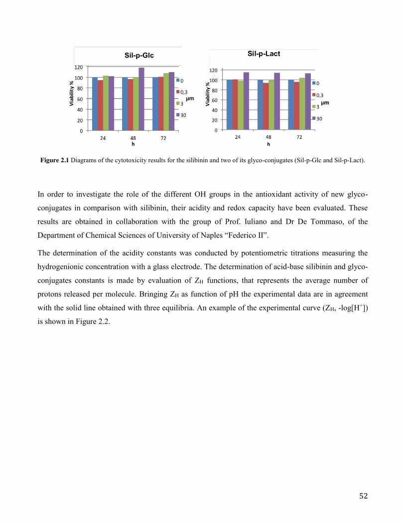
52
Figure 2.1 Diagrams of the cytotoxicity results for the silibinin and two of its glyco-conjugates (Sil-p-Glc and Sil-p-Lact).
In order to investigate the role of the different OH groups in the antioxidant activity of new glyco-
conjugates in comparison with silibinin, their acidity and redox capacity have been evaluated. These
results are obtained in collaboration with the group of Prof. Iuliano and Dr De Tommaso, of the
Department of Chemical Sciences of University of Naples “Federico II”.
The determination of the acidity constants was conducted by potentiometric titrations measuring the
hydrogenionic concentration with a glass electrode. The determination of acid-base silibinin and glyco-
conjugates constants is made by evaluation of ZH functions, that represents the average number of
protons released per molecule. Bringing ZH as function of pH the experimental data are in agreement
with the solid line obtained with three equilibria. An example of the experimental curve (ZH, -log[H+])
is shown in Figure 2.2.

53
Figure 2.2 ZH (pH) function, for Sil-p-Glc, Sil-p-Gal and Sil-p-Man systems. The solid line is calculated using the constants
of Table 2.7.
By processing data, it was possible to define three equilibrium constants, both for silibinin and its
conjugates. The obtained values for silibinin derivatives are similar to silibinin acidity constants. New
glycosylated show a redox behavior similar to the silibinin and the acidic constants of the phenolic
groups, very often implicated in the complexation with metals such as (Cu, Zn, Fe) remain unchanged.
Table 2.7. Protolisys constant of Silibinin and some conjugates in 0.1 M NaClO4.
Compound Silibinina Sil-p-Glc Sil-p-Man Sil-p-Gal Sil-p-Lact pKa1 7.00 ± 0.1 7.20 r 0.06 7.32 r 0.08 7.28 r 0.08 7.27 r 0.07
pKa2 10.25 ± 0.1 9.45 r 0.09 9.56 r 0.09 9.39 r 0.09 9.40 r 0.09
pKa3 11.8 ± 0.1 10.9 r 0.1 11.0 r 0.1 11.0 r 0.1 11.1 r 0.1 a. Protolisys constant measured in 0.1 M NaClO4/50% CH3OH.
By analogy with the acid-base behavior of known molecules, the values of the acid constants can be
assigned according to Figure 2.3.
0
1
2
3
2 4 6 8 10 12
Z H=(
[H+ ]
-CH
/CL
pH
Silibinin
O
OH
O
O
HO
OH O
OH
OCH3
OH
Ka1=7.0±0.1
Ka1=10.25±0.1Ka1=11.8±0.1

54
Figure 2.3 Molecular structure of silibinin where the OH groups relating to protolytic constants are shown.
Acidity constants obtained by spectrophotometric measurements (by Hyperquad program) were in
good agreements with constants determined by potentiometric method.
Figure 2.4 UV-Vis spectra of a Sil-p-Man solution of 5.0 x 10⁻⁵ M to varying pH (from 2 to 11.1).
CD-UV measurements in solution, at different pH, show variations in the CD spectrum that follow the
same changes registered in UV-Vis spectra.
0
0,05
0,1
0,15
0,2
0,25
0,3
0,35
0,4
200 250 300 350 400
Abs
nm
pH 2
pH 4.4
pH 7.2
pH 8.6
pH 9.4
pH 10.6
pH 11.1

55
Figure 2.5 CD-Vis spectra of the glyco-conjugate Sil-p-Man, concentration of 5.0 x 10⁻⁵ M to vary pH from 2 to 11.1.
The electrochemical oxidation of flavonoids has been investigated using cyclic voltammetry (CV). All
the above studies were based on in situ analyses using glassy carbon electrodes. Silibinin (1) and glyco-
conjugates (14−19) are subjected to a single oxidation. This peak around 0.7 V (v.s. a saturated silver
chloride reference electrode) appears in all the compounds under study and its anodic currents are
probably associated with oxidation involving the OH group on C-4" (as Gyorgy el al. reported for
silibinin)31. Cyclic voltammograms for silibinin gluco-conjugate Sil-p-Man, recorded at different pH,
are reported in Figure 2.6.
Peak oxidation potential (EP) and half-peak oxidation potential (EP/2) are deducing from
voltammograms. The number of electrons, ne, involved in the process are obtained by equation:
EP - EP/2 = 56.5 mV/ne,
The value of EP - EP/2 in the range pH 3.0−7.0, is (37 r 8) mV, which corresponds to ne | 1.6
(probably two electrons involved).
-5
-4
-3
-2
-1
0
1
2
3
4
5
220 270 320 370
∆A
nm
pH 2
pH 4.4
pH 7.2
pH 8.6
pH 9.4
pH 10.6
pH 11.1

56
Figure 2.6. Cyclic voltammogramms for Sil-p-Man recorded at different pH (from 3 to 9) (scan rate 50mV/s)
The reducing properties of compounds are very often characterized by formal redox potentials E°’,
determinated at pH = 7.0 (v.s. saturated silver chloride reference electrode). This parameter can be
obtained by half-wave potential (E1/2), which is related to EP/2 from equation:
E1/2 = EP/2 - 28 mV/ne
The results are presented in Table 2.8.
Table 2.8 Formal redox potentials at pH = 7.0 (v.s. saturated silver chloride reference electrode), for some silibinin
glyco-conjugates.
Compound Silibinin Sil-p-Glc Sil-p-Man Sil-p-Gal Sil-p-Lact
E°’, Volt 0.65 ± 0.1 0.67 r 0.02 0.64 r 0.02 0.69 r 0.02 0.68 r 0.02
The electrochemical behavior is dependent on pH (Figure 2.7). The slope of Ep/2 vs pH is (33 r 8)
mV, corresponding at two protons involved in the oxidation.
0,00E+00
1,00E-05
2,00E-05
3,00E-05
4,00E-05
5,00E-05
6,00E-05
0 0,2 0,4 0,6 0,8 1 1,2
A
V vs Ag/AgCl
pH 3
pH 4
pH 6
pH 7
pH 9

57
Fig. 2.7 Ep/2 values (mV, v.s. saturated silver chloride reference electrode) for Sil-p-Man determined over the pH range
3.0−9.0.
New glycosylated show a redox behavior similar to the silibinin and the acidic constants of the
phenolic groups, very often implicated in the complexation with metals such as (Cu, Zn, Fe).
In conclusion, new silibinin glyco-phosphodiester conjugates were synthesized and their biological
and electrochemical investigation were evaluated. Their synthesis was successfully realized in a short
time and in very good yields. The serum stability of the new conjugates was evaluated, as well as their
redox behavior was studied with different approaches. Infact, new derivatives were subjected to DPPH
free radical scavenging assay in preliminary study to evaluate their antioxidant activities. Irrespective
of the sugar moiety examined, all compounds exhibited a radical scavenging activities slightly higher
than to silibinin. In the other hand, the new derivatives showed a water solubility well above that of
silibinin, in fact it was possible to prepare solutions of about 70 mg/mL of analogues in water.
Moreover, their acidity and redox capacity have been evaluated by potentiometric and voltammetric
studies. The glyco-conjugation by phosphodiester bond has been to improve the water solubility of
silibinin core without making any changes to the behavior redox. In collaboration with Prof.
Pannecouque of Rega Institute of Leuven (Belgium) and prof. Agarwal of Department of
Pharmaceutical Sciences, University of Colorado, future studies are in progress to evaluate the antiviral
and anticancer activity, respectively.
0,3
0,5
0,7
0,9
2 4 6 8 10
E P/2
pH

58
2.4 Dimeric phosphate-linked silibinins: synthesis, spectroscopic properties, solubility and radical scavenger ability
Oxidative stress is a state present in all aerobic organisms. It occurs when the production of reactive
oxygen species (ROS) exceeds the limited capacity of the cellular antioxidant system. ROS are small
molecules, commonly produced in radical reactions, with the capacity to quickly interact with cellular
structures. They are represented by various chemical entities: oxygen radicals (such as hydroxyl radical
HO• and singlet oxygen 1O2) or nonradical substances (such as hydrogen peroxide H2O2). ROS are
usually very reactive; they do not have long lifetimes and cannot be transported for long distances in an
organism. They therefore damage the cell structures that are closest to the site of their formation. They
readily attack nucleic acids, proteins, and lipids. The molecule that is attacked is either damaged,
destroyed, or has its function altered. Oxidative stress plays a key role in the origin and development of
many diseases connected with inflammation, such as neurodegeneration, cardiovascular diseases, and
cancer.
Free radicals are molecules containing unpaired electrons, which makes them very reactive. This
reactivity leads to donating or accepting electrons from surrounding molecules to achieve a more stable
state. The reaction of such a radical with a nonradical causes a radical chain reaction and creates new
radicals, which, in turn, react with other molecules. This process is called propagation. The reaction is
terminated when two radicals react with each other to form a nonradical. Hydroxyl radical is the most
potent oxidant and one of the most reactive natural free radicals known. It has a very short half-life and
thus reacts with molecules at the site of its formation. The reaction rate with other molecules is high,
approximately 109 to 1010 M1 × s−1.
As can be expected from the enormous reactivity of HO•, the exposure of DNA (or RNA) to it yields a
large number of products. To prevent or overcome the damage to biomolecules and pre- vent the
development of the diseases previously mentioned, the cells take advantage of various ways and
enzymatic systems. Sometimes, the capacity of the endogenous enzymes and antioxidants is
overwhelmed. For these cases human cells may use exogenous antioxidants supplied in the diet, such
as vitamins, carotenoids, and flavonoids, such as polyphenols. Polyphenols are widely distributed in
nature and are, in many cases, the active compounds of the medicinal plants from which they can be
isolated. Despite the few and common biosynthetic origins, polyphenols encompass many subclasses of
structurally diverse entities and from the different pharmacological properties. Many studies suggests

59
that the mechanisms by which plant polyphenols exert their protective actions against cardiovascular
and neurodegenerative diseases, as well as cancer and diabetes, are not simply due to their redox
properties, but rather to their ability to directly bind to bio-targets (peptides, proteins, nucleic acids).
It is the task of scientists to develop suitable methods for determining the antioxidant activity and to
search for new antioxidants in natural sources. There are various ways of measuring the scavenging
activity of HO• and 1O2. The choice of method substantially influences and determines the results.
Obviously, we cannot expect the same results for all of the different methods used to measure ROS
scavenging in antioxidant activity assays of flavonoids. Basically, there are 2 main steps in the
procedure of ROS-scavenging methods: (1) production of ROS: this includes, in the case of HO•, the
employment of various physical processes, such as pulse radiolysis, γ -irradiation, or UV photolysis of
H2O2, and also different chemical reactions, among which the most widely used is the Fenton reaction;
(2) reaction of ROS with a suitable probe and the consequent detection of a reaction product or
corresponding change in some physical parameter. Because HO• is a very reactive substance, it is not
possible to measure its presence and concentration directly; but the first reaction of HO• with another
molecule, a probe or trap, followed by the detection of a reaction product is crucial.32
Recently we reported the synthesis of new silibinin conjugated by phosphodiester bond with a mono-
and di-saccharides in the position 9". The presence of carbohydrate moiety and phosphate group
increase the water solubility of the aglycon and brings the possibility of prodrug approach making
possible the improvement of the its pharmaceutical, pharmacokinetic and/or pharmacodynamic
properties. New derivatives showed a better solubility in water while maintaining a good antioxidant
activity compared with that of the silibinin.
As a part of our continuing research effort towards the synthesis of new natural product analogues
exploiting the phosphoramidite chemistry, we present here the efficient synthesis of new dimeric
linked-phosphate silibinins and the related study of redox behavior. In order to achieve a more
complete picture of spectroscopic properties, solubility and radical scavenger ability, we have studied
the silibinin dimers in collaboration with Marcello Brigante, Maitre de Conférences at Université
Clermont Auvergne, Institut de Chimie de Clermont-Ferrand (ICCF), Equipe Photochimie (France).
Notably, during the period that I spent in the Brigante’s group, I performed analysis to characterize
spectroscopically the dimers in comparison with silibinin and assess their reactivity against some
oxygen free radicals.

60
Exploiting the selective protection to hydroxyl groups of the silibinin we have developed an efficient
strategy for the synthesis of new 3-9", 3-3 and 9"-9" dimers of silibinin (Scheme 2.5) in good yields.
Scheme 2.5 Synthetic strategy for the new 3-9", 3-3 and 9"-9" dimers of silibinin.
Solubility in water of silibinin and dimers as well as their ability to react with reactive oxygen species
(ROS) were determined. Reactivity with ROS was quantified by estimating their second order rate
constant with singlet oxygen and hydroxyl radical in solution. For this purpose, 545 nm centered
excitation of Rose Bengal (RB) and laser flash photolysis (LFP) experiments were coupled with a
kinetic competition approach. Laser Flash Photolysis is used for a kinetic study of short lived species,
and was developed by George Porter and Ronald Norrish (Nobel Prize 1967). This technique provides
direct transient measurement of reactions involving species such as radicals or excited states. In LFP
the sample is firstly excited by a strong pulse of monochromatic light from a laser of a nanosecond
pulse width. This first pulse starts a chemical reaction or leads to an increased population for energy
levels other than the ground state within a sample of atoms or molecules. Differences in absorption
compared to the ground state are measured with time resolved spectroscopy.

61
Silibinin is a flavonolignan with five hydroxy groups (three phenolic, one secondary and one primary)
with a different reactivity. To obtain the suitable building blocks 2 and 3 (Scheme 2.6) we have
developed a regioselective protection of the different hydroxyl functions with isobutyryl chloride e in
THF dry at low temperature. The desired compounds 2 and 3 were obtained with a 15 and 28% of
yields, respectively.
Scheme 2.6 Silibinin building blocks and their corresponding phosphoramidites. (a) isobutyryl chloride, TEA, THF dry (b) 2-cyanoethyl-N,N-diisopropylamino-chlorophosphoramidite, DIEA, DCM dry, r.t.
In order to prepare the corresponding phosphoramidites, we converted 2 and 3 (Scheme 2.6) into the
phosphoramidites 4 and 5 by a classic reaction with 2-cyanoethyl-N,N-diisopropylamino-
chlorophosphoramidite. After work-up the silica gel chromatography afforded desired compounds 4
and 5 in a 65% yields. The identity of compounds 2−5 was confirmed by NMR (1H, 13C and 31P) and
ESI-MS data. Compounds 2 and 3 were coupled, in the three different provisions (Scheme 2.7), with
phosphoramidites 4 and 5 using 0.45 M 4,5-dicyanoimidazole (DCI) in ACN. After oxidation treatment
with 5.5 M tert-butyl hydroperoxide solution in decane, the crude material was purified by gel
chromatography. Several complex fractions were recovered by gel chromatography, but only those
with the signal 31P NMR active, were treated with conc. aq. ammonia and MeOH (1:1, v:v) at 50 °C,
allowing full deprotection from isobutyryl and cyanoethyl groups. After RP-18 HPLC purification the
products were converted into the corresponding sodium salts by cation exchange on a DOWEX (Na+
form) resin, leading to the desired phosphodiester derivatives 6−8 in good yields (50, 45 and 60 %
respectively, Scheme 2.7).

62
Scheme 2.7 Phosphodiester silibinin dimers derivatives.
Given the very complex mixture (there are at least three diastereoisomers for each dimer) the NMR
analysis was not possible. The products obtained by cation exchange were suspended in DMSO-d6, but
the goodness of NMR spectra did not allow a detailed assignment of different spin systems. The
structures of 6−8 were confirmed by 31P NMR and MALDI-MS analyses.
In order to evaluate their antioxidant activity, all silibinin dimers were subjected to 2,2-diphenyl-1-
picrylhydrazyl free radical scavenging assay.18 The DPPH test is the non-enzymatic method currently
used to provide basic information on the scavenging potential of stable free radicals in vitro. From the
DPPH assay shown in Table 2.8, we observed that conjugating two moieties of silibinin did not led to a
significant reduction of quenching properties. For comparison purposes, the antioxidant activities of
quercetin were evaluated as controls. In particular the new silibinin dimers 6−8 showed DPPH radical
scavenging activities higher to that of the silibinin, confirming that the introduction in 9'' of the moiety
did not influence the radical scavenging activity of this metabolite.22, 23
Blood and extracellular fluid contain a large number of hydrolytic enzymes, such as cholinesterase,
aldolase, lipase, dehydropeptidase, alkaline and phosphatase. Several functional groups, such as ester,
amide, carbamate, lactam, lactone, sulphonamide and, of course, phosphate, are susceptible to plasma

63
degradation. If the new compound has affinity for one of these enzymes and it has a hydrolyzable
group in the right position, it can be decomposed in the plasma.
Hydrolysis in plasma can be a major cause of compound clearance, and pharmacologically
efficacious concentrations may not be achievable in vivo pre-clinical study. Since the presence of a
junction of phosphodiester type, which could be susceptible to the action of endogenous phosphatase
before the active complex can reach its molecular target, the stability of the new synthesized conjugates
was evaluated in human serum, and monitored over time by of HPLC analysis. New dimeric
phosphate-linked silibinins (6−8) were incubated at 37 °C with human serum from human male AB
plasma in Tris•HCl buffer (pH = 8.0 containing 10 mM MgCl2). Small aliquots of the mixture were
taken at defined time intervals, kept at 85 °C for 5 min, equilibrated at rt, and analyzed by HPLC on an
RP column. As shown in Table 2.8 the dimers 6−8 are degraded with a time half-lives of 81 to 87
hours.
Table 2.8 Summary table of MALDI analysis, HPLC retention times, IC50 and serum half lives for compound 6−8.
MALDI tR (min) DPPH IC50(mM) t1/2 (hours)
6 calcd for C50H43O22P = 1026.20 found [M-H]- = 1025.79; [MNa-H]- = 1047.25
17.32 0.99 ± 0.04 86.7
7 calcd for C50H43O22P = 1026.20 found [M-H]- = 1025.11; [MNa-H]- = 1047.03
17.24 0.63 ± 0.05 85.6
8 calcd for C50H43O22P = 1026.20 found [M-H]- = 1025.66; [MNa-H]- = 1047.05
17.17 0.99 ± 0.07 81.2
Silibinin (1) 1.4 ± 0.06
Quercetin 0.18 ± 0.01
As reported the water solubility of silibinin change according to the experimental conditions such as
pH and temperature.25,26 In this work we determined the solubility of the new dimers (6−8) and then
compared to the water solubility of the silibinin (1).
Solubility experiments were performed by dissolving selected compound in milli-Q water leaving that
10 min under magnetic stirring in the dark. Molar absorption coefficients (ε) for each compound in
water were determined from calibration curves (absorbance vs concentration) with successive dilutions
of a stock solution using the Beer-Lamberts law. For silibinin, stock solution was prepared in CH3CN
(1.18 mg in 1 mL) and absorption spectra were recorded by diluting silibin solution in H2O (2% of

64
CH3CN used to increase the solubility and, as consequence, quantification limit) UV-Vis spectra were
presented in Figure 2.7 revealing the presence of absorption band centered around 288 corresponding
to the π−π* transition and around 334 nm that should be also due to the transition n−π* on the oxygen
of ketones groups.
Figure 2.7. UV-vis spectra of Silibinin in H2O:CH3CN 98:2 solution and dimers in water at pH 6.5.
Silibinin results only slightly dissolved when 0.5 mg were suspended in 10 mL of water and, after
filtration, the absorption spectrum reveals a maximum at 288 nm with an absorption of ~ 0.018. The
saturated concentration of silibinin in water was estimated to be 0.9 µM corresponding to ~ 0.44 mg L1.
The solution was also keep in the dark and stirred during 12 hours without any significant solubility
increase.
In order to investigate the possible increase of solubility as function of pH, a phosphate buffer
solution at pH 8.0 was prepared (buffer concentration of 1 mM) and 0.35 mg of silibinin is suspended
in 15 mL. After 10 min of magnetic stirring followed by 0.45µm filtration, the UV-vis spectrum reveals
the presence of two absorption maxima: the first at 288 attributed to the silibinin with an absorption of
~ 0.02 (in agreement with solubility determination) and a second centered around 330 nm indicating
the formation of degradation products of silibinin in alkaline solutions.
Solubility experiments that are summarized in Table 2.9 indicate that dimers were completely dissolved
(0.1 mg in 10 mL) in water and solubility can be estimated to be ≥ 19.5 µM corresponding to > 20 mg
L1.

65
Table 2.9. Spectroscopic characteristics and solubility of silibinin and dimers in solution. *solution is saturated and suspension is filtered before analysis.
Compound 288nmH (M1 cm1) Suspended in 10 mL
Theoretical concentration
Abs288 nm after filtration
Solubility in water
1 22158 (CH3CN)
19469 (H2O:CH3CN 9.8:0.2) 0.44 mg 91.2 µM* 0.018 ≤ 0.9 µM
6 9100 (H2O) 0.10 mg 19.5 µM 0.18 ≥ 19.5 µM
7 14435 (H2O) 0.10 mg 19.5 µM 0.30 ≥ 19.5 µM
8 18536 (H2O) 0.10 mg 19.5 µM 0.35 ≥ 19.5 µM
Reactivity between silibinin and dimers with 1O2 and HO● was determined by use of Rose Bengal
and hydrogen peroxide as respective ROS sources. For singlet oxygen, the steady-state concentration in
solution ( ) in water is determined following tryptophan disappearance at different initial
concentrations. The kinetic approach is summarized in reaction R5-R7
(R5)
(R6)
(R7)
With the formation rate of upon irradiation of RB, the pseudo-first decay of singlet
oxygen in water (2.4 × 10⁵ s1) and the second order rate constant between and TRP (3.2 ×
107 M1s1).27 Application of the steady-state approximation to [ ] leads to the following equation:
(EQ1)
12[ ]ssO
21
2
, 1 12 ( )O h f
ORB O R MsQ ����o
12
1 3 12 2 ( )
OO O k s�o
12
1 1 12 ,
( )O TRP
O TRP products k M s� �� o
12
fO
R 12O 1
2Ok
12 ,O TRP
k 12O
12O
12
1 12 2 ,
11 (1 )[ ]
Ofd
TRP O O TRP
k
R k TRPR �

66
data vs. different TRP concentrations can be fitted with a linear equation lading to the
quantification of singlet oxygen steady state concentration ( ) that is equal to the ration /
.
Dimers reactivity toward singlet oxygen results to be closed to the value determined for silibinin (see
dimer 6) or about 35% lower in the case of dimer 8 (Table 2.10). Second order rate constant fare in the
same order of magnitude reported in literature for molecules with similar structure. Morales and co-
workers28 reported a reactivity ranging from 2.4 to 13.4 × 10⁷ M-1 s-1 for flavonoid derivative such as
quercetin and morin.
Table 2.10 Singlet oxygen steady state concentrations [1O2]SS and second order rate constant ( 12 ,
IIO X
k ) between singlet
oxygen and silibinin or dimers in THF and H2O solutions.
Compound THF H2O
[1O2]SS (M) (M1s1) [1O2]SS (M) (M1s1)
1 3.79 × 10¹³ 6.1 ± 0.4 ×10⁷ - -
6 - - 4.07 × 10¹³ 6.8 ± 1.2 × 10⁷
7 - - 4.07 × 10¹³ 5.4 ± 0.3 × 10⁷
8 - - 4.07 × 10¹³ 4.0 ± 0.2 × 10⁷
Experimental data like those reported in Figure 2.8 for dimers 7 and 8 (Abs/Abs0 followed at 475 nm
vs dimer concentration) are fitted with a linear equation (E1) leading to the estimation of second order
rate constant between hydroxyl radical and selected molecule.
1dTRPR
y ax b �
12[ ]SSO 1
2
fO
R
12O
k
12 ,
IIO X
k 12 ,
IIO X
k

67
Figure 2.8. Abs/Abs0 measured at 475 nm as function of dimers 6 and 7. The solid line represents the linear fit of the experimental data and dashed lines denote the 95% confidence interval of this fit.
Estimation of second order rate constant indicates that the most reactive species are dimer 7 and 8
with a second order rate constant ≥ 1.5 × 10¹⁰ M1 s1 (see Table 2.11 summarizing experimental results).
Wang and co-workers29 investigated the reactivity of hydroxyl radical with phenolic compounds in
order to estimate their antioxidative ability using aqueous pulse radiolysis. The value of 1.5 × 10¹⁰ M1
s1 was found for quercetin that is closed to those estimates for green tea polyphenols. Interestingly,
Husain at al.30 reported that reactivity of flavonoids toward photogenerated hydroxyl radical increases
with the number of hydroxyl groups in the aromatic ring.
Table 2.11: Second order rate constant ( ,IIHO X
k x ) between hydroxyl radical and silibinin or dimers in H2O/CH3CN (9/1)
solutions.
Compound (M1s1)
1 8.26 ± 1.64 × 10⁹
6 6.73 ± 0.32 × 10⁹
7 1.52 ± 0.08 × 10¹⁰
8 ≥ 2.0 × 10¹⁰
,IIHO X
k x

68
In conclusion, new dimeric linked-phosphodiester silibinins were synthesized and their biological and
radical scavenger behaviour were evaluated. Exploiting our solution phase strategy, a variety of
structurally diverse silibinin dimers were successfully realized in a short time and in very good yields.
New derivatives were subjected to DPPH free radical scavenging assay in preliminary study to evaluate
their antioxidant activities. Thanks to the conjugation through a phosphodiester junction of two units of
molecule, all compounds exhibited radical scavenging activities quite higher than to silibinin. In the
other hand, the new derivatives showed water solubility well above that of silibinin. Since the presence
of a junction of phosphodiester type, which could be susceptible to the action of endogenous
phosphatase before the active complex can reach its molecular target, the stability of the new
synthesized conjugates was evaluated in human serum, and as shown in Table 2.8 the dimers (6−8) are
degraded with a time half-lives of 81 to 87 hours and this makes them ideal candidates for further
clinical trials. Moreover, for all the new analogues, their reactivity against some oxygen free radicals
(1O2 and HO●) was investigated. Dimers reactivity toward singlet oxygen results to be closed to the
value determined for silibinin. Second order rate constant fare in the same order of magnitude reported
in literature for molecules with similar structure. These data encouraged our future studies aimed to
improve the synthetic strategy to realize libraries of optically pure linked-phoshate silibinin dimers.

69
References
[1] D.J. Newman, G.M. Cragg, J. Nat. Prod. 2012, 75, 311.
[2] D. Gallo, S. Giacomelli, C. Ferlini, G. Raspaglio, P. Apollonio, S. Prislei, A. Riva, P. Morazzoni, E. Bombardelli, G. Scambia, Europ. J. Canc. 2003, 39, 2403.
[3] H. Zhao, G.E. Brandt, L. Galam, R.L. Matts, B.S.J. Blagg, Bioorg. & Med. Chem. Lett. 2011, 21, 2659.
[4] E. Theodosiou, K. Purchartová, H. Stamatis, F. Kolisis, V. Křen, Phytochemistry Rev. 2013, 13, 1.
[5] D.J. Kroll, H.S. Shaw, N.H. Oberlies, Integ. Canc. Therap. 2007, 6, 110.
[6] R. Gazak, D. Walterova, V. Křen, Curr. Med. Chem. 2007, 14, 315.
[7] P. Calias, T. Galanopoulos, M. Maxwell, A. Khayat, D. Graves, H.N. Antoniades, M. D’Alarcao, Carb. Res. 1996, 292, 83.
[8] K.V. Hirpara, P. Aggarwal, A.J. Mukherjee, N. Joshi, A.C. Burman, Anti-canc. Ag. Med. Chem. 2009, 9, 138.
[9] M. Morishita, K. Park, Biodrug Delivery Systems: Fundamentals, Applications and Clinical Development 2009, p 488. CRC Press.
[10] A. Zarrelli, V. Romanucci, M. Della Greca, L. De Napoli, L. Previtera, G. Di Fabio, Synlett 2013, 24, 45.
[11] A. Zarrelli, V. Romanucci, L. De Napoli, L. Previtera, G. Di Fabio Helv. Chim. Acta 2015, 98, 399.
[12] a) M. I. de Heer, H.-G. Korth, P. Mulder, J. Org. Chem. 1999, 64, 6969.
[13] G. Berube, Curr. Med. Chem. 2006, 13, 131.
[14] B.T. Mott, A. Tripathi, M.A. Siegler, C.D. Moore, D.J. Sullivan, G.H. Posner, J. Med. Chem. 2013, 56, 2630.
[15] K.F. Chan, Y.Z. Zhao, B.A. Burkett, I.L.K. Wong, L.M.C. Chow, T.H. Chan, J. Med. Chem. 2006, 49, 6742.
[16] T.B. Cai, D. Lu, X. Tang, Y. Zhang, M. Landerholm, P. G. Wang, J. Org. Chem. 2005, 70, 3518.
[17] D. Majumdar, G.A. Elsayed, T. Buskas, G.-J. Boons, J. Org. Chem 2005, 70, 1691.
[18] L. Yang, J. Gong, F. Wang, Y. Zhang, Y. Wang, X. Hao, X. Wu, H. Bai, J. Stöckigt, Y. Zhao, J. Enz. Inhib. Med. Chem. 2006, 21, 399.
[19] J.W. Beetsch, T.S. Park, L.L. Dugan, A.R. Shah, J.M. Gidday, Brain Res. 1998, 786, 89.
[20] A.Y. Abramov, A. Scorziello, M.R. Duchen, J. Neur. 2007, 27, 1129.
[21] C.X. Qin, X. Chen, R.A. Hughes, S.J. Williams, O. L. Woodman, J. Med. Chem. 2008, 51, 1874.
[22] R. Gažák, P. Sedmera, M. Vrbacký, J. Vostálová, Z. Drahota, P. Marhol, D. Walterová, V. Křen, Free Rad. Biol. Med. 2009, 46, 745.

70
[23] P. Trouillas, P. Marsal, A. Svobodová, J. Vostálová, R. Gazák, J. Hrbác, P. Sedmera, V. Kren, R. Lazzaroni, J.-L. Duroux, D. Walterová, J. Phys. Chem. A 2008, 112, 1054.
[24] A. Zarrelli, V. Romanucci, C. Tuccillo, A. Federico, C. Loguercio, R. Gravante, G. Di Fabio, Bioorg. Med. Chem. Lett. 2014, 24, 5147.
[25] D. Biedermann, E. Vavříková, L. Cvak, V. Křen, Nat. Prod. Rep. 2014, 31, 1138.
[26] T.-C. Bai, J.-J. Zhu, H.-J. Zhang, C.-G. Huang, Fluid Phase Equilib. 2007, 254, 204.
[27] F. Wilkinson, W.P. Helman, A.B. Ross, J. Phys Chem. Ref. Data 1995, 41, 663.
[28] J. Morales, G. Günther, A.L. Zanocco, E. , P., Lem, PLoS One 2012, 7, e40548.
[29] W.F. Wang, J. Luo, S.D. Yao, Z.R. Lian, J.S. Zhang, N. Y. Lin, Radiat. Phys. Chem. 1993, 42, 985.
[30] S. Rafat Husain, J. Cillard, P. Cillard, Phytochemistry. 1987, 26, 2489.
[31] I. Gyorgy, S. Antus, A. Blazovics, G. Foldiak, Radiat. Phys. Chem. 1992, 39, 81.
[32] J., Treml, K., Smejkal, Compr. Rev. Food. Sci. Food. Saf. 2016, 15, 720-739.

71
Chapter 3: Experimental Session
3.1 New Silibinin glyco-conjugates: Synthesis and evaluation of antioxidant properties.
3.1.1 General procedure for the synthesis of conjugates (6−9):
Phosphoramidite 1 (150 mg, 0.18 mmol) and the select saccharide compound 2−5 (0.19 mmol,
previously dried and kept under reduced pressure, were reacted with a 0.45 M tetrazole solution in
anhydrous CH3CN (1.0 mL, 0.45 mmol). The reaction was left under stirring at room temperature and
monitored by TLC in the eluent system n-hexane:AcOEt, (1:2, v:v). After 1 hour, a 5.5 M tert-butyl
hydroperoxide (tert-BuOOH) solution in decane (70 μL, 0.40 mmol) was added to the mixture and left
under stirring at room temperature. After 30 minutes the reaction mixture was diluted with CHCl3,
transferred into a separatory funnel, washed three times with water and dried under reduced pressure.
The next treatment with TEA/pyridine (1:1, v/v) to 1 hour at 50 °C and then with aq. ammonia
(28%)/CH3OH (1:1, v:v) for 1 hour at room temperature, allowed full deprotection from acetyl and 2-
cyanoethyl groups. The dried mixtures were purified by a Sep-Pak C18 Cartridge and then converted
into the corresponding sodium salts by cation exchange on a DOWEX (Na+ form) resin to have a
homogeneous samples in good yield (60-72%).
Compound 6 (brown powder, 88.1 mg, 68%): 31P NMR (161.98 MHz, D2O, room temperature, mixture
of diastereoisomers) δ 2.9, 1.4, 0.8. HRMS (MALDI-TOF, negative ions) m/z calcd for C31H32O18P =
723.1332, found 723.1332 [M H]-.
Compound 7 (brown powder, 94.0 mg, 72%): 31P NMR (161.98 MHz, D2O, room temperature, mixture
of diastereoisomers) δ 1.3, 0.9, 0.7. HRMS (MALDI-TOF, negative ions) m/z calcd for C31H32O18P =
723.1332, found 723.1333 [M H]-.
Compound 8 (brown powder, 82.3 mg, 63%): 31P NMR (161.98 MHz, D2O, room temperature, mixture
of diastereoisomers) δ 2.4, 1.6, 1.1, 0.8. HRMS (MALDI-TOF, negative ions) m/z calcd for
C31H32O18P = 723.1332, found 723.1333 [M H]-.
Compound 9 (brown powder, 104.8 mg, 60%): 31P NMR (161.98 MHz, D2O, room temperature,
mixture of diastereoisomers) δ 1.1, 0.8. HRMS (MALDI-TOF, negative ions) m/z calcd for C37H42O23P
= 885.1860, found 885.1859 [M H]-.

72
3.1.2 DPPH radical scavenging activity assay
The antioxidant activities of the compounds was assessed by examining their abilities to scavenge the
1,1-diphenyl-2-picrylhydrazyl free radical. DPPH solution (20 mg/mL) was prepared in methanol. The
compounds was dissolved in methanol to prepare the stock solution (1 mM). Freshly prepared DPPH
solution was taken in test tubes and analogue solutions (1 mM−1 μM) were added to every test tube so
that the final volume was 2.25 mL and after 10 min, the absorbance was read at 515 nm. Quercetin and
2,3-dehydrosilybin were used as a reference standards and dissolved in methanol to make the stock
solution with the same concentration (1 mM).
3.1.3 Xanthine Oxidase Inhibition Assay.
Oxidative stress was induced by incubating MKN28 cells with XO (10-100 mU/mL) in the presence of
its substrate xanthine (X) (1 mM) for periods of up to 3 hours. We examined the effect of silibinin and
of our analogues 6−9 on X/XO induced cell damage. In particular, cells were incubated with serum free
medium (control) for 1 - 48 hours; with serum free medium for 148 hours and then with X (1 mM) XO
(50 mM) for 2 hours (X/XO control); with 6−9 and silibinin (10−200 µM) for 148 hours and then, after
washing, with X (1 mM) XO (50 mM) for 2 hours. Subsequently, we determined cell viability by [3-
(4,5-dimethylthiazol-2-yl)-2,5-diphenyl tetrazoliumbromide] MTT assay in MKN28 cultured cells.

73
3.2 Synthesis, biological and electrochemical investigation of new silibinin glyco-phosphodiester conjugates
3.2.1 Synthesis of silibinin phosphoramidite building block 3
Silibinin (1.6 g, 3.3 mmol) previously dried by repeated coevaporate with anhydrous THF, was
dissolved in 8 mL of anhydrous pyridine and DMTCl (1.48 g, 4.37 mmol) was added. The mixture was
kept at 50 °C for 3 hours. After monitoring by TLC, the reaction was stopped by addition of CH3OH
and the mixture dried under reduced pressure. The crude material was purified by column
chromatography with a gradient of n-hexane in CHCl3, from 10% to 0%, and then increasing the
polarity eluting with CHCl3 and increasing percentages of CH3OH from 0% to 5%. In all of the eluants,
it was used 0.5% of Et3N. The derivative 9"-ODMT was obtained in good yield. (2.5 g, 3.18 mmol,
96%).
9"-ODMT silibinin (2.5 g, 3.18 mmol) was dissolved in 8 mL of pyridine and isobutyric anhydride was
added (6.5 mL, 60 mmol). The reaction mixture was kept under stirring for 2 hours at room
temperature. After monitoring by TLC, the reaction was stopped by addition of CH3OH and then kept
at 0 °C for 1 hour. The mixture was extracted with a saturated aqueous solution of NaHCO3 and CHCl3
and the organic layer was dried over anhydrous Na2SO4 and concentrated under reduced pressure to
lead 3,5,7,4"-tetra-O-isobutyryl-9"-O-DMT silibinin (2.70 g, 2.53 mmol, 79%).
3,5,7,4"-tetra-O-isobutyryl-9"-ODMT silibinin (2.70 g, 2.53 mmol) was selectively deprotected from
DMT group with a solution of HCOOH 5% in CH2Cl2 (150 mL). The reaction mixture was kept under
stirring for 1 hour at room temperature. After monitoring by TLC, the mixture was washed with a
saturated aqueous solution of NaHCO3 and the organic phase was dried over anhydrous Na2SO4 and
evaporated under reduced pressure. The crude material was purified by column chromatography eluting
with a gradient of ethyl acetate in hexane from 50% to 70% to obtain the compound 2 in good yield
(1.5 g, 2.05 mmol, 81%).
To 3,5,7,4"-tetra-O-isobutyryl silibinin (240.0 mg, 0.33 mmol) dissolved in anhydrous CH2Cl2 (5 mL),
DIEA (390 μL, 2.22 mmol), and 2-cyanoethyl-N,N-diisopropylamino-chlorophosphoramidite (107 μL,
0.48 mmol) were mixed. After 30 min the solution was diluted with EtOAc, and the organic phase was
washed twice with brine and then concentrated. Silica gel chromatography of the residue (eluent n-
hexane:EtOAc, 3:7, v/v, in the presence of 1% of Et3N), afforded desired compound 3 (205.0 mg, 0.24
mmol) in a 65% yield.

74
3.2.2 General procedure for the synthesis of monosaccharide building blocks 8−12
Different 1-O-methyl-mono- and di-saccharides (Glc, Man, Gal, GlNAc, Thre, 2.07 mmol), previously
coevaporated with anhydrous pyridine, were dissolved in anhydrous pyridine (8 mL) and reacted with
DMTCl (1.86 mmol). The reaction mixture was stirred at room temperature for 2 hours (checked by
TLC analysis) then quenched with MeOH and evaporated to dryness. The residue was extracted with
CHCl3, the organic phase was dried over anhydrous Na2SO4 and concentrated under reduced pressure.
The crude material was purified by column chromatography (98:2 chloroform/methanol with 0.5% of
pyridine), leading a good yield of the 6-O-DMT glycosides (55-85%). Then, the 6-O-DMT-1-O-
methyl-glycoside (1.45 mmol) was solubilized in anhydrous pyridine (2 mL), and to the resulting
mixture was added acetic anhydride (1.3 mL, 14 mmol). The mixture was stirring at room temperature
for 12 hours (checked by TLC analysis). The reaction was quenched with MeOH at 0 °C and washed
with CHCl3, the organic phase was dried over anhydrous Na2SO4 and concentrated under reduced
pressure. Subsequently, we proceeded to remove the DMT group of full protected 1-O-methyl-
glycoside (1.45 mmol) with a solution of HCOOH 5% in CH2Cl2 and triisopropylsilane (1.45 mmol)
under stirring at 0 °C.
On completion of reaction, the resulting mixture was diluted with CH2Cl2 and washed with a saturated
aqueous solution of NaHCO3. The combined organic layers were dried over anhydrous Na2SO4 and
concentrated under reduced pressure. The crude material was purified by column chromatography
(chloroform/methanol, 98:2, v:v), leading the compounds 8−12 in good yields (55−65%).
3.2.3 Synthesis of lactose building blocks 13
Lactose octaacetate (1 g, 1.47 mmol) and I2 (523 mg, 2.06 mmol) were dissolved in anhydrous
dichloromethane (10 mL). After completed dissolution, the reaction mixture was cooled to 0 °C and the
Et3SiH (329 μL, 2.06 mmol) was added. After 5 minutes the reaction was refluxed until TLC analysis
displayed complete consumption of the peracetylated sugar (the glycosyl iodides are partially unstable
on TLC). The mixture was cooled to RT and then diluted with CH2Cl2. The organic phase was washed
with a 5% solution of NaHCO3 containing Na2S2O3 (to remove residual amounts of iodine). The
aqueous phase was re-extracted with CH2Cl2, and the combined organic layers were dried over
anhydrous Na2SO4 and concentrated under reduced pressure. The glycosyl iodide intermediate was
obtained in good yield (1.01 g, 1.3 mmol, 90%).

75
A mixture of appropriate glycosyl iodide (1.01 g, 1.3 mmol) and Ag2CO3 (69 mg, 0.65 mmol) was
stirred at room temperature in anhydrous MeOH (20 mL) in presence of 3-Å molecular sieves for one
day. On completion of the reaction (checked by TLC analysis), the mixture was filtered with CHCl3,
concentrated under reduced pressure and directly deprotected with a solution of MeOH/NH4OHaq (1:1,
v:v) for 30 minutes. The mixture was purified by column chromatography (chloroform/methanol, 9:1)
to give the pure product β-1-O-methyl-lactoside (454 mg, 1.27 mmol, 98%).
The methyl lactoside (454 mg, 1.27 mmol) was subsequently dissolved in anhydrous pyridine (8 mL)
andreacted with DMTCl (386 mg, 1.14 mmol). The reaction mixture was stirred at room temperature
for 2 hours, then quenched with CH3OH and evaporated to dryness. The residue was extracted with
CHCl3, the organic phase was dried over anhydrous Na2SO4 and concentrated under reduced pressure.
The 6-O-DMT-β-1-O-methyl-lactoside was purified by column chromatography (9:1
chloroform/methanol with 0.5% of pyridine) (343 mg, 0.52 mmol, 41%).
Subsequently, 6-O-DMT-β-1-O-methyl-lactoside (343 mg, 0.52 mmol) was fully acetylated with acetic
anhydride (4 mL, 42.3 mmol) in anhydrous pyridine (8 mL) for 12 hours. The reaction mixture was
quenched with MeOH and then washed with CHCl3, the organic phase was dried over anhydrous
Na2SO4 and concentrated under reduced pressure. Finally, the fully acetylated 6-O-DMT-β-1-O-
methyl-lactoside was regioselectively deprotected from DMT group at the position 6 of the galactosyl
residue with a solution of 5% HCOOH in CH2Cl2 and triisopropylsilane (1 mmol) under stirring at 0 °C
for 1 hour. The reaction mixture was diluted with CH2Cl2 and washed with 5% of NaHCO3 aqueous
solution and finally with H2O. The combined organic layers were dried over anhydrous Na2SO4 and
concentrated under reduced pressure. The product 13 was obtained in pure form and in good yield (196
mg, 0.32 mmol, 61%) by column chromatography (chloroform/methanol, 9:1).
3.2.4 General procedure for the synthesis of silibinin phosphodiester glyco-conjugates (14−19)
The coupling reaction between the phosphoramidite and the mono and disaccharides appropriately
protected was carried out by suspending the derivatives 7−12 (0.6 mmol) in a solution of DCI in
acetonitrile (0.45 M, Activator 42®) and by adding 3-Å molecular sieves. After few minutes, the
mixture was added to the silibinin phosphoramidite (0.54 mmol) and was kept under stirring for 1 hour.
On completion of reaction monitoring by TLC analysis, tert-butyl hydroperoxide solution in decane
(5.5 M) was added and after 30 minutes, the solvent was removed under vacuum and the crude material

76
was purified by column chromatography (chloroform/methanol, 99:1, 95:5). The products obtained
were subsequently subjected to treatment with ammonia aqueous solution and methanol (1:1, v/v) at
25 ° C for 3 hours for the full deprotection of protecting groups.
After the deprotection treatment, the crude material was purified by reverse phase HPLC using a
Phenomenex Luna® C18 column eluted with ammonium acetate 0.1 M with a linear gradient 20-100%
ACN in 50 minutes (flow = 7 mL/min). Then, the material was desalted on Chromafix® C18 column
(deionized H2O/CH3OH, 8:2, 1:1, CH3OH) and converted into the corresponding sodium salt by cation
exchange on DOWEX resin (Na+) leading the compounds 14−19 in good yields (45−60 %).
3.2.5 Analysis to serum stability of silibinin glyco-conjugates
Reactions (500 PL reaction volume) were performed in 50 mM Tris•HCl pH 8.0 containing MgCl2 (10
mM), silibinin conjugates (50 PL vol of a 10 PM solution), and human serum from human male AB
plasma (400 PL). All reactions were incubed at 37 °C. From this solutions were taken samples of
constant volume at different times, and the HPLC profile was analyzed in comparison with that of the
intact compound.
3.2.6 DPPH radical scavenging activity assay
The antioxidant activities of the compounds was assessed by examining their abilities to scavenge the
1,1-diphenyl-2-picrylhydrazyl free radical. DPPH solution (20 mg/mL) was prepared in methanol. The
compounds was dissolved in methanol to prepare the stock solution 2 mM). Freshly prepared DPPH
solution was taken in test tubes and analogue solutions (2 mM−5 μM) were added to every test tube so
that the final volume was 2.25 mL and after 10 min, the absorbance was read at 515 nm. Quercetin and
Trolox were used as a reference standards and dissolved in methanol to make the stock solution with
the same concentration (2 mM).
3.2.7 Citotoxicity on on human liver cancer cell (Hep G2)
Silibinin and its new glyco-conjugates were solubilised in DMSO at a final concentration of 13.5 mM
and then diluted in water to obtain the operational concentrations to be used for the tests on cells.
Cytotoxic activity of different amounts of silibinin or its derivatives (0.3, 3 and 30 μM) was measured

77
at different incubation times (24, 48 and 72 hours) by a colorimetric assay. As a control, cell viability
was assessed in the absence of the effectors.
3.2.7 Determination of the acidity constants by potentiometric titrations, UV-Vis spectrophotometric measures and Circular Dichroism (CD-UV) of silibinin solutions. Redox behaviour carried out by cyclic voltammetry
In according to the chemical structure of the molecule, silibinin shows many OH groups, that are
ionizable. The acid-base behavior can be represented by general equilibria:
H4-nL1-n + H2O H3-nL1-n + H3O+ KAn (3.1)
The study is conduced by potentiometric methods at 25 qC in 0.1 M NaClO4 as ionic medium. The
measurements are carried out as potentiometric titrations where the hydrogenionic concentration is
determined by the e.m.f. of cell (A):
(�) RE/Test Solution/GE (+) (A)
in which GE symbolizes glass electrode and RE is the reference half�cell:
RE = 0.1 M NaClO4/0.01 M AgClO4, 0.09 M NaClO4/AgCl/Ag
The test solution has the general composition:
CL M L, CA M HClO4, CB M NaOH, (0.1 � CA) M NaClO4
CHnL is chosen to be from 2.0 x 10�3 M to 8.0 x �10�3 M for silibinin glyco-conjugates (for silibinin the
range is 2.0 ≤ CL≤ 6.0�10�3 M, with 50% oh CH3OH), whereas (CA � CB) = CH is varied into �1.0 x
10�3 M � 9.0�10�3 M. The e.m.f. of cell (A) at 25 °C may be written as:
E = E0 � 0.05916 x log([H+]yH) + EJ (3.3)
where E0 is a constant in each titration, yH is the activity coefficient of the H+ ion and EJ represents the
liquid junction potential at the test solution 0.1 M NaClO4 boundary.1
It depends on the hydrogen ions concentration in the equation:

78
EJ = 0.35 x h (3.4)
Activity coefficients tend to 1 and EJ tend to 0 as the composition of test solution approaches 0.1 M
NaClO4.
Each titration consists of two parts. In first, E is measured in the absence of silibinin, or its conjugates,
and (CA � CB) = [H+] is varied between 2 x 10�3 M and 5 x 10�5 M. From these data E0 values constant
to within r 0.05 V is calculated, assuming EJ = �0.51 x [H+] + 0.24 x Kw[H+]�1. In the second part, in
the presence of ligand, acid-base behavior is followed by alcalification and/or acidification.
Placing:
EG0 = EG
0’ + 0.05916 x log yH
The equation (3.3) takes the form:
EG = EG0 + 0.05916 x log h + EJ (3.5).
From experimental data (CL, CHnL, [H+]) the average number of protons (ZH) released per L molecule is
evaluated by:
ZH = ([H+] � CH � Kw/[H+])/CHnL (3.6)
where a value Kw = 10�13.78 was utilized.
Bringing ZH as function of pH the experimental data are in agreement with the solid line obtained with
three equilibria.
A speciation model was obtained by processing the experimental data using Hyperquad program.2
The criterion used is to determine the values of the constants that make KAn minimize the sum U of
squared weighted residuals:
U EG,i exp � EG,i calc)2 (3.7)
where, for the i-th measurement, EG,i exp and EG,icalc represent, respectively, the experimental potential
and that calculated for a set of constant value KAn..
The minimum of the function U is found by the method of Newton-Raphson. Once the minimum of U,
Umin, the agreement between the experimental and calculated data is given by the variance σ2 defined
as:

79
σ2 = Umin/(n � m) (3.8)
where n represents the number of experimental data and m is the number of KAn constants.
The constants standard deviations are obtained by the diagonal elements of the variance-covariance
matrix, calculated according to the equation:
Mv = σ2 H�1 (3.9)
where H is the matrix of the normal equations and it is a square matrix of order m.
The acid-base properties of the silibinin and its glyco derivatives are studied also by UV-VIS
spectrophotometric procedures, that confirm the results obtained by potentiometric measurements. The
experimental data collected are processed by the numerical minimization program Hyspec, Hyperquad
package, which minimizes the standard deviation between the measured absorbance and those
calculated by the proposed model constants. The results obtained are in agreement with those obtained
from the potentiometric. Also, circular dichroism (CD-UV) of silibinin solutions were made.
Measurements in solution, at different pH, show variations in the CD spectrum that follow the same
changes registered in the UV-VIS spectra. For the voltammetric measurement, a polarograph Varian
767 METROM it was utilized, with a measurement electod in glassy carbon, auxiliary electrode in Pt
and a reference electrode of Ag/AgCl(s).

80
3.3 Dimeric phosphate-linked silibinins: synthesis, spectroscopic properties, solubility and radical scavenger ability
3.3.1 Chemicals
Silibinin is purchased from Sigma-Aldrich. HPLC grade MeCN, MeOH are purchased from Carlo
Erba Reagents and Sigma-Aldrich. Hydrogen peroxide (30 %), from Fluka. Unless otherwise indicated,
other chemicals are obtained from Sigma Aldrich and purity is > 98 %.
Fresh aqueous solutions of H2O2, Rose Bengal and tryptophan are prepared before each experiment.
The concentration of the stock solution of H2O2 in milli-Q water (Laser flash photolysis experiments) is
determined using a molar absorption coefficient of 38.1 ± 1.4 M�1 cm�1 at 240 nm.9
3.3.2 General methods
Reactions for dimers’ synthesis are monitored by TLC (precoated silica gel plate F254, Merck) and
Column chromatography: Merck Kieselgel 60 (70-230 mesh). The analysis is performed with a
Shimadzu LC-8A PLC system equipped with a Shimadzu SCL-10A VP System control and Shimadzu
SPD-10A VP UV-VIS Detector. HPLC purifications are carried out on Phenomenex RP18 column (5-
μm particle size, 10.0 mm × 250 mm i.d.) using a linear gradient of ACN in 0.1 M TEAA in H2O, pH
7.0 from 5% to 100% over 30 minutes at a flow rate of 1.5 mL/min with detection at 260 nm.
For the ESI MS analyses, a Waters Micromass ZQ Instrument equipped with an Electrospray source,
is used. MALDI TOF mass spectrometric analyses are performed on a PerSeptive Biosystems Voyager-
De Pro MALDI mass spectrometer.
Absorption spectra are measured with a Varian Cary 100 Scan UV-vis spectrophotometer using 1 cm
pathlength quartz cell.
3.3.3 Synthesis of silibinin building block 2
Silibinin 1 (1.5 g, 3.1 mmol) previously dried by repeated coevaporate with anhydrous THF and then
was dissolved in 8 mL of anhydrous THF and TEA (2.1 mL, 15.4 mmol). Isobutyryl chloride (0.980
mL, 9.3 mmol) was added and the mixture was kept at 0 °C for 30 minutes. After monitoring by TLC,
the reaction was stopped by addition of MeOH and the mixture was extracted with CH2Cl2. The
organic phase was dried over anhydrous Na2SO4 and concentrated under reduced pressure, the crude
material was purified by column chromatography (CHCl3/acetone, 96:4, v/v). The derivative 3,5,7,4"-
tetra-O-isobutyryl silibinin was obtained as pale amorphous solid (0.365 g, 0.47 mmol, 15%).

81
2 Rf = 0.6 (benzene/AcOEt, 80:20, v/v). 1H NMR (CDCl3, 400 MHz, room temperature, mixture
of two diastereoisomers): δ = 7.15−6.96 (complex signals, 6H, H-2', H-5', H-6', H-2'', H-5'', H-6''), 6.77
(complex signal, 1H, H-8), 6.58 (s, 1H, H-6), 5.71 and 5.69 (d, 1H, J = 12.2/12.3 Hz, H-3), 5.38 (m,
1H, H-2), 5.08 and 5.06 (d, 1H, J = 8.3/8.7 Hz, H-7"), 4.04 (m, 1H, H-8''), 3.88−3.83 (overlapped
signals, 4H, OCH3, H-9''a), 3.58 (dd, 1H, J = 3.2 Hz, J = 12.0 Hz, H-9''b), 2.98-2.74 (complex signals,
3H, CH of isobutyryl groups), 2.62-2.52 (m, 1H, CH of isobutyryl group), 1.41−1.25 (m, 18H, CH3 of
isobutyryl groups), 1.15−1.10 (m, 6H, CH3 of isobutyryl group) ppm. 13C NMR (CDCl3, 100 MHz,
room temperature, mixture of diastereoisomers): δ = 185.0, 175.2, 175.1, 175.0, 174.1, 162.5, 162.4,
156.5, 151.7, 151.6, 144.0, 144.1, 143.7, 140.6, 134.5, 128.7, 123.1, 123.0, 121.0, 120.8, 119.8, 119.7,
117.1, 117.0, 116.6, 116.4, 111.2, 111.1, 108.7, 81.1, 81.0, 78.3, 78.4, 76.0, 75.9, 72.9, 72.8, 61.5, 56.1,
56.0, 34.3, 34.0, 33.7, 19.0, 18.8, 18.7, 18.6 ppm. ESI-MS (positive ions): m/z calculated for C41H46O14
= 762.29; found: 763.84 [M-H]+.
3.3.4 Synthesis of silibinin building block 3
Silibinin 1 (1.0 g, 2.7 mmol) previously dried by repeated coevaporate with anhydrous THF, was
dissolved in 8 mL of anhydrous THF and TEA (2.1 mL, 15.4 mmol) and isobutyryl chloride (1.1 mL,
10.4 mmol) were added. The mixture was kept at 0°C for 30 minutes. After monitoring by TLC, the
reaction was stopped by addition of MeOH and the mixture was extracted with CH2Cl2, the organic
phase was dried over anhydrous Na2SO4 and concentrated under reduced pressure. The crude material
was purified by column chromatography (eluent CHCl3/acetone, 96:4, v/v). The derivative 5,7,4",9"-
tetra-O-isobutyryl silibinin was obtained as pale amorphous solid (0.447 g, 0.58 mmol, 28%).
3 Rf = 0.9 (benzene/AcOEt, 80:20, v/v). 1H NMR (CDCl3, 400 MHz, room temperature, mixture
of two diastereoisomers): δ = 7.21−6.98 (complex signals, 6H, H-2', H-5', H-6', H-2'', H-5'', H-6''), 6.76
(complex signal, 1H, H-8), 6.59 (s, 1H, H-6), 5.07 (d, 1H, J = 12.8 Hz, H-2), 5.00 (d, 1H, J = 8.3 Hz,
H-7"), 4.56 and 4.55 (d, 1H, J = 12.3/12.4 Hz, H-3), 4.44 (dd, 1H, J = 2.3 Hz, J = 12.9 Hz, H-9"a), 4.26
(m, 1H, H-8"), 3.98 (dd, 1H, J = 2.3 Hz, J = 12.8 Hz, H-9"b), 3.86 (s, 3H, OCH3), 2.98-2.76 (complex
signals, 3H, CH of isobutyryl groups), 2.61 (ept, 1H, J = 6.8 Hz, CH of isobutyryl group), 1.36
(complex signals, 18H, CH3 of isobutyryl groups), 1.21 (m, 6H, CH3 of isobutyryl group) ppm. 13C
NMR (CDCl3, 100 MHz, room temperature, mixture of diastereoisomers): δ = 191.2, 176.4, 175.0,
174.9, 174.1, 163.1, 157.0, 151.7, 151.5, 144.0, 143.5, 140.8, 134.3, 129.3, 123.2, 121.4, 121.2, 119.8,

82
117.5, 117.4, 116.6, 116.5, 111.1, 110.9, 109.7, 108.9, 83.2, 76.2, 75.9, 75.8, 73.1, 73.0, 62.4, 56.0,
34.3, 34.1, 34.0, 33.9, 19.0, 18.9, 18.7, 18.6 ppm.
ESI-MS (positive ions): m/z calculated for C41H46O14 = 762.29; found: 763.66 [M H]+.
3.3.5 Synthesis of silibinin building block 4
To 3,5,7,4"-tetra-O-isobutyryl silibinin 2 (240.0 mg, 0.33 mmol) dissolved in anhydrous CH2Cl2 (5
mL), DIEA (390 μL, 2.22 mmol), and 2-cyanoethyl-N,N-diisopropylamino-chlorophosphoramidite
(107 μL, 0.48 mmol) were mixed. After 30 min the solution was diluted with EtOAc, and the organic
phase was washed twice with brine and then concentrated. Silica gel chromatography of the residue
(eluent n-hexane/EtOAc, 30:70, v/v, in the presence of 1% of Et3N), afforded desired compound 4
(205.0 mg, 0.24 mmol) in a 65% yield.
4 Rf = 0.8 (n-hexan/AcOEt, 60:40, v/v). 1H NMR (CDCl3, 500 MHz, room temperature, mixture
of four diastereoisomers): δ = 7.11−6.94 (overlapped signals, 6H, H-2', H-3', H-6', H-2'', H-5'', H-6''),
6.76 (comkplex signals, 1H, H-8), 6.56 (s, 1H, H-6), 5.69 (x2), 5.68 and 5.67 (d, 1H, J = 12.2 Hz, H-3),
5.38, 5.37 (x2) and 5.36 (d, 1H, J = 12.2 Hz, H-2); 5.03 (x2) and 5.01 (x2) (d, 1H, J = 7.2/7.5 Hz, H-
7''), 4.00- 3.51 (complex signals, 12H, OCH3, H-8'', H-9''a, H-9''b, OCH2CH2CN, N[CH(CH3)2]2
OCH2CH2CN), 3.01−2.68 (m, 3H, CH of isobutyryl groups), 2.56 (m, 1H, CH of isobutyryl group)
1.36−0.95 (complex signals, 36H, CH3 of isobutyryl groups, N[CH(CH3)2]2) ppm. 13C NMR (CDCl3,
100 MHz, room temperature, mixture of diastereoisomers): δ = 185.0, 175.2, 175.1, 175.0, 174.1,
162.5, 162.4, 156.5, 151.7, 151.5, 144.3, 143.5, 143.4, 140.5, 134.7, 128.3, 128.2, 123.0, 122.9, 121.0,
120.9, 120.8, 119.9, 119.8, 119.7, 117.7, 117.5, 117.1, 116.4, 116.3, 111.5, 111.4, 111.3, 111.1, 111.0,
110.8, 108.7, 81.2, 81.1, 76.2, 75.9, 75.8, 72.9, 72.8, 62.7, 62.5, 62.2, 62.0, 58.8, 58.6, 58.5, 58.3, 56.0,
55.9, 53.4, 43.3, 43.2, 34.2, 34.0, 33.9, 33.6, 24.6, 24.5, 19.0, 18.8, 18.7, 18.6 ppm. 31P NMR (CDCl3,
161.98 MHz): δ = 150.2, 149.8, 149.7 ppm. ESI-MS (positive ions): m/z calculated for C50H63N2O15P =
962.40; found: 963.92 [MH]+.
3.3.6 Synthesis of silibinin building block 5
To 5,7,4",9"-tetra-O-isobutyryl silibinin 3 (240.0 mg, 0.33 mmol) dissolved in anhydrous CH2Cl2 (5
mL), DIEA (390 μL, 2.22 mmol), and 2-cyanoethyl-N,N-diisopropylamino-chlorophosphoramidite
(107 μL, 0.48 mmol) were mixed. After 1 hour the solution was diluted with EtOAc, and the organic
phase was washed twice with brine and then concentrated. Silica gel chromatography of the residue

83
(eluent n-hexane/EtOAc , 3:7, v/v, in the presence of 1% of Et3N), afforded desired compound 5 (205.0
mg, 0.24 mmol) in a 65% yield.
5 Rf = 0.8 (n-hexan/AcOEt, 60:40, v/v). 1H NMR (CDCl3, 500 MHz, room temperature, mixture
of four diastereoisomers): δ = 7.30−6.97 (overlapped signals, 6H, H-2', H-3', H-6', H-2'', H-5'', H-6''),
6.73 (comkplex signals, 1H, H-8), 6.54 (complex signal, 1H, H-6), 5.02 (x2) and 5.01 (x2) (d, 1H, J =
7.8/8.0 Hz, H-7"), 4.94 (d, 1H, J = 9.3 Hz, H-2), 4.43 (complex signal, 1H, H-3), 4.34 (complex signal,
1H, H-9"a), 4.20 (complex signal, 1H, H-8"), 4.10−3.94 (complex signals, 1H, H-9"a), 3.86 (complex
signal, 3H, OCH3), 3.54−3.34 (complex signals, 9H, H-9''b, OCH2CH2CN, N[CH(CH3)2]2
OCH2CH2CN) 2.96−2.74 (complex signals, 3H, CH of isobutyryl groups), 2.65 (complex signal, 1H,
CH of isobutyryl group), 1.40-1.10 (complex signals, 36H, CH3 of isobutyryl groups, N[CH(CH3)2]2)
ppm. 13C NMR (CDCl3, 100 MHz, room temperature, mixture of diastereoisomers): δ = 189.0, 188.3,
176.4, 176.3, 174.9, 174.2, 174.1, 162.3, 156.3, 156.1, 149.9, 143.9, 143.8, 143.4, 143.3, 140.8, 135.9,
134.2, 129.5, 129.4, 129.3, 123.2, 121.7, 121.6, 119.7, 119.6, 117.8, 117.5, 117, 4, 117.3, 116.8, 116.6,
111.3, 111.1, 111.0, 108.6, 83.1, 82.9, 76.2, 75.8, 75.6, 75.2, 62.4, 62.3, 59.7, 59.5, 58.5, 58.3, 56.0,
43.5, 43.4, 34.2, 34.1, 33.9, 24.8, 24.7, 24.6, 24.2, 24.1, 19.0, 18.9, 18.8, 18.7, 18.6 ppm. 31P NMR
(CDCl3, 161.98 MHz): δ = 153.2, 153.0, 152.3, 152.2 ppm. ESI-MS (positive ions): m/z calculated for
C50H63N2O15P = 962.40; found: 963.43 [MH]+.
3.3.7 Synthesis of dimeric phosphate-linked silibinins 6−8
The coupling reaction between the phosphoramidites 4 or 5 and the suitable silibinin building blocks 2
or 3 was carried out by suspending 240 mg (0.32 mmol) the derivatives 2 or 3 in a solution of DCI in
acetonitrile (0.45M, Activator 42®) and by adding 3-Å molecular sieves. After few minutes, the
mixture was added to 270 mg (0.28 mmol) of silibinin phosphoramidites 4 or 5 and was kept under
stirring for 1 hour. On completion of reaction monitoring by TLC analysis, 150 PL of tert-butyl
hydroperoxide solution in decane (5.5 M) was added and after 30 minutes, the solvent was removed
under vacuum and the crude material was purified by column chromatography (eluent CHCl3/CH3OH,
99:1 to 95:5, v/v). The material obtained were subsequently subjected to treatment with ammonia
aqueous solution (28%) and methanol (1:1, v/v) at 50 °C for 3 hours for the full deprotection. After the
deprotection treatment, the crude material was analyzed by RP-HPLC using a Phenomenex Luna C18
(5 μm particle size, 10.0 mm × 250 mm i.d.) eluted with 0.1 M ammonium acetate with a linear
gradient 20-100% ACN in 30 minutes (flow = 3.0 mL/min) and then purified by RP-HPLC using a

84
Gemini C18-110A preparative column (10 μm particle size, 250 mm × 21.2 mm i.d.) eluted with 0.1 M
ammonium acetate with a linear gradient 20−100% ACN in 50 minutes (flow = 7 mL/min). The
recovered material was desalted on Chromafix® C18 column eluted with H2O/CH3OH 8:2 (v/v) and
converted into the corresponding sodium salt by cation exchange on DOWEX resin (Na+) leading the
compounds 6 - 8 in good yields (50, 45 and 60 % respectively).
3.3.8 HPLC purification of 6−8
The sample solutions were prepared by dissolving and sonicating the accurately weighed compound (6
- 8) in H2O/CH3OH (1:1, v/v). The obtained solution (ca 10 mg/mL) was then applied to Nylon filters
(pore size = 0.45 µm) and then purified.
The HPLC analysis was performed with a Shimadzu LC-9A system equipped with a Shimadzu SPD-
6A Detector using a RP18 column Phenomenex LUNA (5-μm particle size, 10.0 mm × 250 mm i.d.)
and eluted with ammonium acetate 0.1 M with a linear gradient 20−100% ACN in 30 minutes (flow =
1.5 mL/min). The chromatograms were monitored at 288 nm.
The preparative HPLC method was performed with a Shimadzu LC-8A PLC system equipped with a
Shimadzu SCL-10A VP System control and Shimadzu SPD−10A VP UV-VIS Detector. A
Phenomenex Gemini C18-110A preparative column (10-μm particle size, 250 mm × 21.2 mm i.d.) was
used and eluted with ammonium acetate 0.1 M with a linear gradient 20−100% ACN in 30 minutes
(flow = 7 mL/min). The chromatograms were monitored at 288 and 254 nm. The HPLC system was
controlled by LC Real Time Analysis software (Shimadzu Corporation).
3.3.9 DPPH radical scavenging activity assay.
The antioxidant activities of the compounds was assessed by examining their abilities to scavenge the
1,1-diphenyl-2-picrylhydrazyl free radical. DPPH solution (20 mg/mL) was prepared in methanol. The
compounds was dissolved in methanol to prepare the stock solution (2 mM). Freshly prepared DPPH
solution was taken in test tubes and analogue solutions (2 mM−5 μM) were added to every test tube so
that the final volume was 2.25 mL and after 10 min, the absorbance was read at 515 nm. Quercetin and
Trolox were used as a reference standards and dissolved in methanol to make the stock solution with
the same concentration (2 mM).

85
3.3.10 Serum stability
Tests were performed in 500 PL of 50 mM Tris•HCl (pH 8.0 containing 10 mM MgCl2), 50 PL of
dimeric phosphate-linked silibinin (10 PM solution), and human serum from human male AB plasma
(400 PL). All reactions were incubed at 37 °C. From this solutions were taken samples of constant
volume at different times, and the HPLC profile was analyzed in comparison with that of the intact
compound.
3.3.11 Singlet oxygen reactivity
In order to estimate the reactivity of silibinin and dimers toward singlet oxygen, solutions are
irradiated in a quartz cell of 1cm pathlength using 545 nm centered irradiation (1000 W Xenon lamp
equipped with a monochromator) in the presence of 100 µM of RB as photosensitizer for singlet
oxygen generation.3 An aliquot of solution was withdrawn at fixed interval time and analyzed by liquid
chromatography
Figure 3.1 shows absorption spectra of RB and overlap with emission spectrum reaching the solution
recorded using a fiber optics coupled with a CCD spectrophotometer (Ocean Optics USD 2000+UV-
VIS). A reference lamp (DH-2000-CAL, Ocean Optics) is used for calibration.
Figure 3.1 Emission spectrum reaching the solution (1000 W Xenon lamp equipped with a monochromator) and
absorptions spectrum of 10µM RB solution.

86
Silibinin is solubilized in THF, while dimers in H2O/CH3CN (9:1, v/v). THF and CH3CN are used as
solvent to increase the silibinin and dimer solubility and due to their similar reactivity with singlet
oxygen compared to the water (i.e. only limited singlet oxygen scavenging is accounted for by the
solvent).4 Silibinin and dimers degradations are determined as following: an aliquot (200 µL) was taken
at fixed interval times during irradiation, stored in the dark and monitored by ultra-high performance
liquid chromatography, using a Waters Acquity UPLC instrument equipped with a diode array detector
and a Waters 2695 separation module.
The gradient is a linear increasing from 1% CH3CN and 99% water acidified with 0.1% of formic
acid to 50% acetonitrile within 5 min. Then the CH3CN is increased linearly to 80% for 2 min and
maintained constant for 2 min. The column is a Water Acquity UPLC C18 (100 mm × 2.1 mm × 1.7
µm) and flow rate of 0.3 mL min‒N. Retention times of silibinin, 3-3, 3-9" and 9"-9" dimers are 5.4,
6.3, 7.0 and 7.3 min respectively and detection wavelength set at 288 nm.
The time evolution of silibinin and dimers in the presence of singlet oxygen could be fitted with a
pseudo-first order equation 12
0 exp Ok t
tC C�
where C0 is the initial concentration of compound, Ct the
concentration at time t and 12O
k the pseudo-first order degradation rate constant of target compound (X).
The degradation rate ( dXR ) is determined as 1
20
dX O
R k C .
The transformation rate of silibinin or dimers in the presence of singlet oxygen is given by the
following relation: 12
12,
[ ] [ ]d IIX SSO X
R k O X where [ ]X is the concentration of silibinin or dimers in
solution. The second order rate constant between target molecule and singlet oxygen ( 12 ,
IIO X
k (M1 s1))
can be determined as: 12 1,
2[ ] [ ]
dII XO X
SS
RkO X
Singlet oxygen steady-state concentration [1O2]ss is quantified with tryptophan as probe using
experimental conditions adopted for silibinin and dimmers considering a second order rate constant
between TRo and [1O2] of 3.2 × 10⁷ M1 s1.10
3.3.12 Hydroxyl radical generation and reactivity estimation
Due to the high reactivity of THF with hydroxyl radical (~ 4.0 × 10⁹ M-1 s-1),5 stock solutions of
H2O2, SCN � , silibinin and dimers are prepared in Milli-Q water or CH3CN (for silibinin) and an
appropriate volume is mixed just before each experiment, to obtain the desired concentration of all the
species. Experiments in pure water are performed for dimmers while for silibinin, CH3CN/water (9:1,

87
v/v) solution is used. All experiments are performed at ambient temperature (295 ± 2 K) and in aerated
solution.
Hydroxyl radical are generated from 266 nm excitation of H2O2 solution, corresponding to the fourth
harmonic of a Quanta Ray GCR 130-01 Nd:YAG laser system instrument, used in a right-angle
geometry with respect to the monitoring light beam. The single pulse is of 9 ns in duration, and energy
of 40 mJ. Individual cuvette samples (~3 mL volume) are used for a maximum of two consecutive laser
shots to avoid degradation products formation and interferences. The transient absorbance at the pre-
selected wavelength is followed by a detection system consisting of a pulsed xenon lamp (150 W),
monochromator and a photomultiplier (1P28). A spectrometer control unit is used for synchronising the
pulsed light source and programmable shutters with the laser output. The signal from the
photomultiplier is digitised by a programmable digital oscilloscope (HP54522A) and analysed using a
32 bits RISC-processor kinetic spectrometer workstation.
The UV-vis spectrum of HO● presents a maximum centered at 240 nm and direct quantification is
difficult due to the low molar absorption coefficient (ε240nm) of ~ 600 M-1 cm-1.6 For this reason a
kinetic approach based on the thiocyanate (SCN-) competition reactivity is generally adopted to
estimate the second order rate constant of target compounds with HO ● in solution based on the
following equations:
266
2 2 2LFP nmH O HOx����o (R1)
HO SCN HO SCNx � � x� o � (R2)
2SCN SCN SCNx � x�� o (R3)
HO X productsx � o (R4)
The first step (R1) is the hydrogen peroxide photolysis using a laser pulse at 266 nm followed by the
competition reactivity of generated HO● with SCN � (R2) and silibinin (or dimers) (R4). In the presence
of high concentration of SCN � , thiocyanate radical ( SCN x ) reacts with another equivalent of
thiocyanate to form di-thiocyanate radical anion ( 2SCN x� ) (R3), which has a strong absorption molar
coefficient at 475 nm (ε475nm = 7600 M-1 cm-1).7

88
The absorption of 2SCN x� in the presence of different concentration of HO● quenchers (X) allows the
determination of the second order rate constant between hydroxyl radical and silibinin or dimers from
the following equation:
,0
,
[ ]1
[ ]
IIHO X
IIHO SCN
k XAbsAbs k SCN
x
x �� � (E1)
in which Abs0 and Abs are the absorption of 2SCN x� transient followed at 475 nm without and with
quencher respectively; ,
IIHO SCN
k x � is the second order rate constant between HO● and thiocyanate that is
1.2 × 10¹⁰ M-1 s-1,8 [ ]SCN � is the initial concentration of thiocyanate (0.1 or 0.5 mM in our study);
[ ]X is the concentration of target compound and ,
IIHO X
k x the second order rate constant with hydroxyl
radical.
0AbsAbs
values vs concentration of quencher ([ ]SY ) can be fitted using a linear equation y ax b � in
which 0AbsyAbs
, 1,b [ ]x X and ,
,[ ]
IIHO X
IIHO SCN
ka
k SCNx
x �� .
,IIHO X
k x value can be determined to be equal to
,[ ]II
HO SCNa k SCNx �
�u .

89
References
[1] E. van Wenum, R. Jurczakowski, G. Litwinienko, J. Org. Chem. 2013, 78, 9103.
[2] P. Gans, A. Sabatini, A. Vacca, Talanta 1996, 43, 1739.
[3] M.C. DeRosa, R.J. Crutchley, Coord. Chem. Rev. 2002, 233, 351.
[4] F. Wilkinson, W. P. Helman, A.B. Ross, J. Phys Chem. Ref. Data 1995, 41, 663.
[5] G.V. Buxton, C. L. Greenstock, W.P. Helman, A.B. Ross, J. Phys. Chem. Ref. Data 1988, 3, 513.
[6] H. Herrmann, D. Hoffmann, T. Schaefer, P. Bräuer, A. Tilgner, Chem. Phys. Chem. 2010, 11, 3796.
[7] L. Dogliotti, E. Hayon, J. Phys. Chem. 1968, 72, 1800.
[8] G.V. Buxton, C.L. Greenstock, W. P. Helman, A.B. Ross, J. Phys. Chem. Ref. Data 1988, 34, 513.
[9] M.S. Morgan, P.F. Van Trieste, S. M. Garlick, M.J. Mahon, A.L. Smith, Anal. Chim. Acta 1998, 215, 325.
[10] S.G. Bertolotti, N.A. Garcia, J. Photochem. Photobiol. B 1991, 10, 57.

PhD Course Activity Summary
Candidate: Dr. Raffaele Gravante Supervisor: Prof. Giovanni Di Fabio
1) Attended Courses:
x Tecniche di estrazione solido-liquido, 8 hours, June 2014 (Prof. Naviglio);
x Spettrometria di Massa, 10 hours, July 2014 (Prof. Pucci);
x Sintesi, struttura ed applicazioni di oligonucleotide naturali e modificati, 8 hours, February
2015 (Prof. Montesarchio);
x Glicoscienza, 8 hours, July 2015 (Prof. Parrilli and Dr. Bedini);
x Produzione ricombinante di protein naturali e mutanti, 8 hours, July 2015 (Prof. Duilio);
x Neutron scattering, 8 hours, October 2015 (Prof. Zorn);
x Chimica Fisica degli Acidi Nucleici, 8 hours, July 2016 (Prof. Petraccone).
2) Attended Seminars:
Title Speaker Date Place
Impact factor, citations,
journal reputation, editorial
policy: how to choose the
right journal.
Dott. Thierry
Tron;
Dott.ssa Jalila
Siman;
Prof. Danilo
Porro.
26/06/2014
Università degli
Studi di Napoli
“Federico II”
Evidenze genetiche sulle
prime espansioni umane
nell’Africa.
Prof. Guido
Barbujani 23/10/2014
CEINGE
BIOTECNOLOGIE
AVANZATE
Pharmaceutical Companies:
external manufacturing and
quality assurance.
Prof. Domenico
De Masi 25/05/2015
Università degli
Studi di Napoli
“Federico II”
Alessandro Ballio e la
chimica a Napoli negli anni
‘60
Dr. Andrea
Carpentieri 18/06/2015
Università degli
Studi di Napoli
“Federico II”
Le transglutaminasi..dalle
poliammine alle bioplastiche
Prof. Raffaele
Porta 30/06/2015
Università degli
Studi di Napoli

“Federico II”
Synthetic and biofunctional
studies of microbial and
animal glycans.
Dr. Fukase 29/09/2015
Università degli
Studi di Napoli
“Federico II”
Scientific calculator. Prof. Vacatello 11/11/2015
Introduction to
bioinformatics Dr. Remo Sanges 18/12/2015
Università degli
Studi di Napoli
“Federico II”
Biopesticides which target
voltage-gated ion channels:
efficacy and biosafety
Prof. Angharad
M.R.Gatehouse 14/01/2016
Università degli
Studi di Napoli
“Federico II”
1,1,2,2-tetraethoxybutyne-
Exciting to make and work
with
Prof. Leiv K.
Sydnes 14/01/2016
Università degli
Studi di Napoli
“Federico II”
Functionalized and artificial
enzymes:newbio-derived
catalysts
Prof. Therrytron 14/01/2016
Università degli
Studi di Napoli
“Federico II”
Secondary metabolites from
higher land and sea plants in
Chemical ecology and
Chemosystematics
Prof. Christian
Zidorn 27/01/2016
Università degli
Studi di Napoli
“Federico II”
Modified nucleotides and
oligonucleotides for
biomedical applications.
Prof. Daniela
Montesarchio 18/01/2017
Università degli
Studi di Napoli
“Federico II”
3) Attended Integration Exams (for candidates not graduated in Chemical Science):
Title Professor Date
Chimica Fisica I Castronuovo 11/02/2015
Chimica Inorganica Vitagliano 29/02/2016

4) Visiting periods in Institutions different from University of Naples “Federico II”:
Host Institution Country Start Date End Date
University Blaise
Pascal, Institute
of Chemistry of
Clermont-
Ferrand.
France 02/10/2016 02/01/2017
5) Publications (include submitted and in preparation):
x New Silibinin glyco-conjugates: Synthesis and evaluation of antioxidant properties Armando Zarrelli, Valeria Romanucci, Concetta Tuccillo, Alessandro Federico, Carmela Loguercio, Raffaele Gravante and Giovanni Di Fabio; Bioorg Med Chem Lett. 2014; 24(22): 5147-9.
x Isolation, structural determination, and biological activities of gymnemic acid, a molecule that does not exist Armando Zarrelli , Valeria Romanucci, Raffaele Gravante, Cinzia Di Marino and Giovanni Di Fabio; Nat Prod Commun. 2014; 9(10): 1429-32.
x Scavenging Effect of Various Solvent Extracts of Gymnema sylvestre R. Br. and in vivo Antioxidant Activity of Isolated Triterpenes Armando Zarrelli, Valeria Romanucci, Giovanni Di Fabio, Daniele D'Alonzo, Cinzia Di Marino, Raffaele Gravante, Achille Cittadini and Alessandro Sgambato (forthcoming).
x Synthesis, biological and electrochemical investigation of new Silibinin glyco-phosphodiester conjugates Valeria Romanucci, Raffaele Gravante, Cinzia Di Marino, Mauro Iuliano, Gaetano de Tommaso, Tonino Caruso, Armando Zarrelli and Giovanni Di Fabio (forthcoming).
x Dimeric phosphate-linked Silibinins: Synthesis, spectroscopic properties, solubility and radical scavenger ability Raffaele Gravante, Valeria Romanucci, Martina Cimafonte, Cinzia Di Marino, Gilles Mailhot, Marcello Brigante, Armando Zarrelli and Giovanni Di Fabio (forthcoming).
6) Attended congresses/workshops/summer schools/contribution:
x New Silibinin glyco conjugates: synthesis, characterization and preliminary evaluation of antioxidant properties Raffaele Gravante1, Valeria Romanucci1,Valeria Sgambati2, Elio Pizzo2, Daniele D’Alonzo1, Annalisa Guaragna1, Armando Zarrelli1 and Giovanni Di Fabio1 1Department of Chemical Sciences; 2Department of Biology, University of Naples Federico II, Via Cintia 4, I-80126, Napoli, Italy Poster session at “XXXVI CONVEGNO DELLA DIVISIONE DI CHIMICA ORGANICA - CDCO 2015”.
x GEM-DCA: synthesis of a novel codrug Raffaele Gravante1, Daniele D’Alonzo1, Valeria Romanucci1, Armando Zarrelli1, Giovanni Di Fabio1, Giovanni Palumbo1 and Annalisa Guaragna1 1Department of Chemical Sciences, Università di Napoli "Federico II", Via Cintia 21, I-80126, Napoli, Italy. Poster session at “XXXVI CONVEGNO DELLA DIVISIONE DI CHIMICA ORGANICA - CDCO 2015”.