Synopsis of Causation Fractures of the Lower Limb ... · Tibial plateau fracture: an...
31
Ministry of Defence Synopsis of Causation Fractures of the Lower Limb (includes foot) Author: Mr Benedict Clift, Ninewells Hospital and Medical School, Dundee Validator: Mr Sheo Tibrewal, Queen Elizabeth Hospital, London September 2008
-
Upload
trinhtuong -
Category
Documents
-
view
214 -
download
0
Transcript of Synopsis of Causation Fractures of the Lower Limb ... · Tibial plateau fracture: an...
Ministry of Defence
Synopsis of Causation
Fractures of the Lower Limb (includes foot)
Author: Mr Benedict Clift, Ninewells Hospital and Medical School, Dundee Validator: Mr Sheo Tibrewal, Queen Elizabeth Hospital, London
September 2008
Disclaimer This synopsis has been completed by medical practitioners. It is based on a literature search at the standard of a textbook of medicine and generalist review articles. It is not intended to be a meta-analysis of the literature on the condition specified. Every effort has been taken to ensure that the information contained in the synopsis is accurate and consistent with current knowledge and practice and to do this the synopsis has been subject to an external validation process by consultants in a relevant specialty nominated by the Royal Society of Medicine. The Ministry of Defence accepts full responsibility for the contents of this synopsis, and for any claims for loss, damage or injury arising from the use of this synopsis by the Ministry of Defence.
2
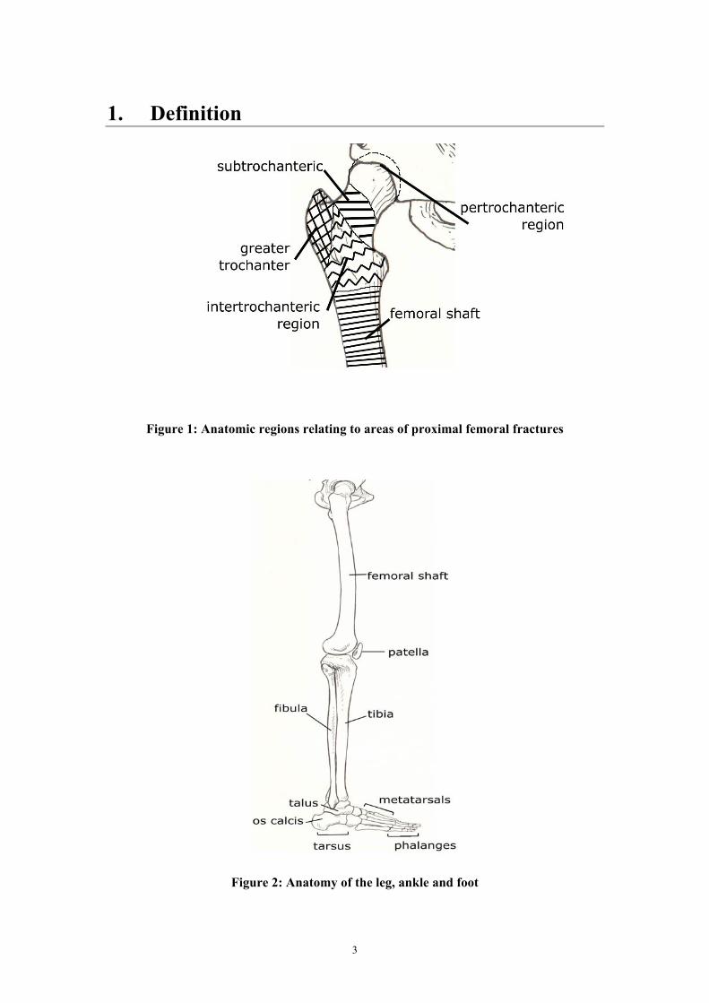
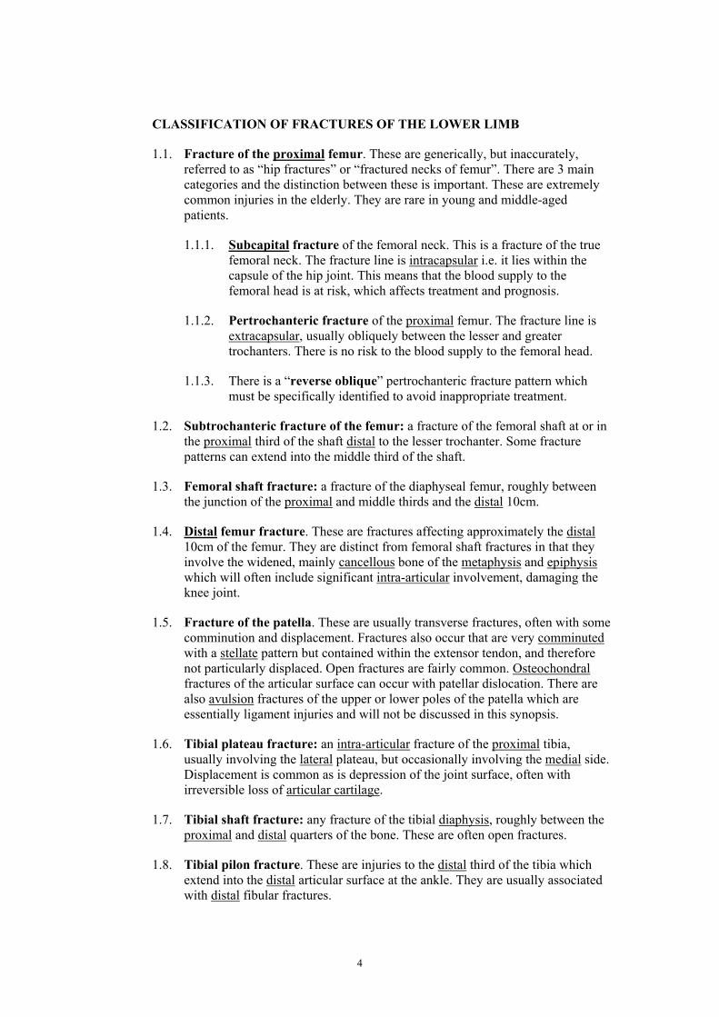
1. Definition
Figure 1: Anatomic regions relating to areas of proximal femoral fractures
Figure 2: Anatomy of the leg, ankle and foot
3
CLASSIFICATION OF FRACTURES OF THE LOWER LIMB
1.1. Fracture of the proximal femur. These are generically, but inaccurately, referred to as “hip fractures” or “fractured necks of femur”. There are 3 main categories and the distinction between these is important. These are extremely common injuries in the elderly. They are rare in young and middle-aged patients.
1.1.1. Subcapital fracture of the femoral neck. This is a fracture of the true femoral neck. The fracture line is intracapsular i.e. it lies within the capsule of the hip joint. This means that the blood supply to the femoral head is at risk, which affects treatment and prognosis.
1.1.2. Pertrochanteric fracture of the proximal femur. The fracture line is extracapsular, usually obliquely between the lesser and greater trochanters. There is no risk to the blood supply to the femoral head.
1.1.3. There is a “reverse oblique” pertrochanteric fracture pattern which must be specifically identified to avoid inappropriate treatment.
1.2. Subtrochanteric fracture of the femur: a fracture of the femoral shaft at or in the proximal third of the shaft distal to the lesser trochanter. Some fracture patterns can extend into the middle third of the shaft.
1.3. Femoral shaft fracture: a fracture of the diaphyseal femur, roughly between the junction of the proximal and middle thirds and the distal 10cm.
1.4. Distal femur fracture. These are fractures affecting approximately the distal 10cm of the femur. They are distinct from femoral shaft fractures in that they involve the widened, mainly cancellous bone of the metaphysis and epiphysis which will often include significant intra-articular involvement, damaging the knee joint.
1.5. Fracture of the patella. These are usually transverse fractures, often with some comminution and displacement. Fractures also occur that are very comminuted with a stellate pattern but contained within the extensor tendon, and therefore not particularly displaced. Open fractures are fairly common. Osteochondral fractures of the articular surface can occur with patellar dislocation. There are also avulsion fractures of the upper or lower poles of the patella which are essentially ligament injuries and will not be discussed in this synopsis.
1.6. Tibial plateau fracture: an intra-articular fracture of the proximal tibia, usually involving the lateral plateau, but occasionally involving the medial side. Displacement is common as is depression of the joint surface, often with irreversible loss of articular cartilage.
1.7. Tibial shaft fracture: any fracture of the tibial diaphysis, roughly between the proximal and distal quarters of the bone. These are often open fractures.
1.8. Tibial pilon fracture. These are injuries to the distal third of the tibia which extend into the distal articular surface at the ankle. They are usually associated with distal fibular fractures.
4
1.9. Ankle fracture: a fracture of one or both bones (the tibia and fibula) at the ankle, which may extend into the articular surface. There are numerous different patterns, best summarised by the Lauge-Hansen classification. This is detailed, but useful, as it clarifies the mechanism of injury, guides treatment, and, to some extent, indicates the possible prognosis. Some ankle fractures feature significant damage to the distal tibiofibular joint (the syndesmosis between these two bones), which it is essential to recognise when planning treatment. Basic fracture types under the Lauge-Hansen classification are:
Supination-external rotation fractures •
•
•
•
Pronation-external rotation fractures
Pronation-abduction fractures
Supination-adduction fractures
Within each group there are different categories which indicate the anatomical extent of the injury. Exact classification requires careful scrutiny of the x-rays and, for an inexperienced assessor, reference to the textbooks.
1.10. Talus fracture: a fracture of the talus, the weight bearing bone sandwiched between the tibia and the os calcis. The commonest fracture is at the talar neck, roughly dividing the bone into its posterior half (the body) and the anterior half (the talar head). The bone is unusual for its complex shape and the fact that most of its surface is covered in articular cartilage. This is because it forms part of 3 important joints – the subtalar, the talonavicular and the ankle joints. These 3 joints are often disrupted to varying degrees with displaced talar neck fractures. This extensive cartilage covering also means that the blood supply to the bone is mainly through small areas where ligaments attach. The precarious blood supply is important in prognosis. The bone may also be fractured through the body itself and through its medial process. These rare injuries will not be specifically discussed.
1.11. Os calcis fracture: a fracture of the os calcis (also known as the calcaneum or calcaneus), which is the bone of the heel. It has articulations with the talus above it at the subtalar joint, and the cuboid anteriorly at the calcaneocuboid joint. These joints are often involved in the fracture. The bone has a complex shape with a low peak superiorly (Bohler’s angle). This peak is often flattened by the fracture.
1.12. Midfoot fracture-dislocation. The complex arrangement of small bones and joints in the midfoot is disrupted by blunt trauma, usually with a forced dorsiflexion and/or a rotational element. These are essentially dislocations of the midfoot, at the level of tarsometatarsal joints, often with associated fractures. The fracture element is usually a marker of a soft tissue disruption, such as a flake of bone pulled off a metatarsal base by the attached ligament, although major fractures of any of the bones can be present. They are commonly grouped together as Lisfranc injuries, named after a Napoleonic surgeon. The 5 metatarsals can be dislocated from the tarsal bones in one big group, or usually in smaller groups, often with displacement of these groups in different directions.
1.13. Metatarsal fracture. A fracture of any of the 5 metatarsal bones of the forefoot. Those of the fifth metatarsal constitute a specific subgroup.
5

1.14. Stress fracture. A stress fracture is fatigue failure of bone due to repeated mechanical loading, typically the cyclical loading of walking, for durations longer than an individual is accustomed to. They are undisplaced and may not be visible on x-ray.
6
2. Clinical Features
2.1. Fractures of the proximal femur. All displaced fractures tend to cause shortening and external rotation of the leg. Weight bearing is not possible. Undisplaced or impacted subcapital fractures can occasionally be fairly stable and allow limited weight bearing, which is uncommon but can lead to delays in diagnosis. Surgery is almost always required for all types of fracture, but the exact operation depends on the fracture site and pattern.
2.2. Subtrochanteric fracture of the femur. Like all femoral shaft fractures, there is localised swelling, deformity and tenderness. The limb is shortened. They are rarely open fractures.
2.3. Femoral shaft fractures. There is localised swelling, deformity and tenderness. The limb is shortened. They are often open fractures with major damage to the quadriceps muscle in particular. There is a high incidence of associated soft tissue injuries of the knee, such as ligamentous injury and/or meniscal tears.
2.4. Distal femur fractures. There is localised swelling, tenderness and deformity. Open fractures are fairly common. There will be a knee haemarthrosis if the joint is involved. There is a small risk of local neurovascular damage.
2.5. Fractures of the patella. There is localised swelling and tenderness at the front of the knee. With displaced fractures that have disrupted the extensor mechanism of the knee, the patient is unable to actively straighten the knee. The gap between the fracture ends may be palpable.
2.6. Tibial plateau fractures. These fractures are fairly common in the elderly as osteoporotic bone is easily crushed and compression is the main injurious force. In young patients they are usually high energy injuries, often with a significant soft tissue element. These are occasionally open fractures. They have a small but significant risk of developing acute compartment syndrome affecting the extensor muscles in the anterior compartment of the leg. Neurovascular injury is a recognised complication, particularly with displaced high energy fractures. The meniscus, a shock absorbing cartilage which sits on the tibial plateau, may be damaged and this probably plays a part in the outcome.
2.7. Tibial shaft fractures. The tibia has a subcutaneous border over its whole length. The swelling, deformity and crepitus are usually obvious. Soft tissue injuries are common, including open fractures. There is a small incidence of neurovascular injury but a much higher incidence of compartment syndrome which, if untreated, typically leads to loss of the anterior muscles of the leg.
2.8. Tibial pilon fractures. Comminution and fragment displacement are typically present in younger patients. The tibia here has very little soft tissue cover. Open fractures are very common and most injuries, even if not open, have an extensive soft tissue component that can make any form of open surgery risky in terms of wound healing. Necessary surgery is often delayed by up to 2 weeks for this reason. Plastic surgery input is often required, although this is a difficult area in which to create a good soft tissue reconstruction. Traction and external fixation may be part of the management.
7
2.9. Ankle fractures. All ankle fractures demonstrate swelling, tenderness and bruising. This may represent the actual fracture site but also may indicate the presence of an important ligament injury, particularly on the medial side of the ankle, which contributes to instability. Many ankle fractures present with the joint actually dislocated, which is clinically obvious with gross deformity and severe pain. The reduction of this dislocation is an emergency to minimise the risk to the skin and the neurovascular structures. Major swelling, which can lead to a delay in surgery, is not unusual and “fracture blisters” may develop rapidly, particularly in pronation-abduction and supination-adduction fractures. Open fractures are rare but usually affect the medial side, and plastic surgery input is often needed for soft tissue cover in this situation.
2.10. Talus fractures. There is always a lot of swelling, which may render early surgery risky, and considerable disruption to the hindfoot joints with permanent articular cartilage damage. The foot is often deformed at presentation. Open fractures are fairly common (about 20%) occasionally with extrusion of bone into the sock. Associated injuries, particularly spine, pelvis and os calcis should be looked for. Radiological imaging usually requires a CT scan for the best information.
2.11. Os calcis fractures
2.11.1. The bone gives the heel its shape and height. Most of the fractures are compression injuries which flatten and widen the bone and the shape of the heel changes accordingly; this can give problems with footwear at a later stage. The soft tissues are often severely damaged with occasional open fractures and a high incidence of severe swelling, bruising and fracture blisters. This dramatic soft tissue damage can delay surgery (if deemed appropriate) by 2 to 3 weeks. The damage to the 2 joints creates stiffness, loss of motion and pain, culminating in post-traumatic osteoarthritis in some cases.
2.11.2. Some cases have so much muscle trauma in the tight compartments of the foot that the muscle dies. The late effect of untreated compartment syndrome of this kind is toe clawing, weakness and abnormal gait.
2.12. Midfoot fracture-dislocations. It is essential to realise that these injuries are associated with severe internal soft tissue damage. There is major swelling, bruising and occasionally skin necrosis. Fracture blisters are common. Compartment syndrome of the foot is fairly common, and easily missed. There may be nerve injury which is difficult to assess accurately at the time of presentation. The foot may be misshapen as well as swollen. Plain x-rays tend to underestimate the extent of the damage and CT imaging is preferred.
2.13. Metatarsal fractures. There is localised swelling and tenderness with difficultly in weight bearing. Bruising may appear on the sole of the foot.
2.14. Stress fractures. There is localised pain and tenderness. The pain is of insidious onset. It is aggravated by walking and running. There is rarely significant swelling. There is no associated soft tissue injury. There needs to be a high index of suspicion based on the history, classically shin or foot pain after a long march or “square bashing” in new recruits. It can also be present in athletes who have effectively gone beyond the mechanical endurance limits of their skeleton, despite being very fit. If not evident on x-ray, further imaging, particularly isotope bone scanning and MRI, is essential.
8
3. Aetiology 3.1. Fractures of the proximal femur. Most proximal femoral fractures of all types
are due to a twisting injury at the hip, associated with a fall. This is a very common scenario with the elderly where simple falls can lead to such a fracture, particularly if the patient has osteoporosis. In younger patients with normal bone density, these fractures are usually high energy injuries, such as with falls from a height or high speed motor vehicle accidents. This synopsis will focus on the issues in relation to younger, active patients.
3.2. Subtrochanteric fracture of the femur. In the young and middle-aged adult, these are high energy injuries usually due to falls from a height and motor vehicle accidents. There is a group of injuries with a long spiral pattern that are due to substantial rotational force applied to the limb.
3.3. Femoral shaft fractures. These are high energy injuries in most patients. Comminution is common. Falls from a height and motor vehicle accidents are the usual causes. In combat situations, ballistic injuries to the femur with major soft tissue damage are common.
3.4. Distal femur fractures. These are high energy injuries. Motor vehicle accidents resulting in direct impact are probably the commonest cause, such as the front of the knee striking the dashboard.
3.5. Fractures of the patella. These fractures can be due to direct trauma to the front of the knee, such as striking a dashboard during a motor vehicle accident, which causes either a transverse or comminuted fracture. An alternative mechanism involves sudden knee flexion with the quadriceps muscle contracting, such as in a sports accident, which causes a transverse fracture.
3.6. Tibial plateau fractures. The distal femur is forced onto the tibial plateau by a combination of axial loading and bending. The lateral plateau is the most commonly injured, which means that at the moment of injury a valgus (knock kneed) bending force was applied as well as compression. The opposite force (varus) is present in the rarer medial injuries. Occasionally both medial and lateral plateaus are fractured, with primarily axial loading without valgus or varus. Any compressive force applied to the leg can therefore do this, ranging from a simple fall in the elderly to a fall from a height or motor vehicle accident in younger patients with stronger bone. Extreme bending on its own, such as having one’s leg trapped by a toppling weight can also create this fracture. The knee ligament on the opposite side of the joint, usually the medial collateral ligament, can be damaged in this situation.
3.7. Tibial shaft fractures. Direct trauma in contact sport, such as a football tackle, is a very common cause. Rotational injuries tend to cause lower energy spiral fractures, for example, football studs catching on turf. High energy comminuted fractures are typically from motor vehicle accidents and falls from a height. Tibial shaft fractures are a common component of multiple injuries.
3.8. Tibial pilon fractures. These are usually high energy injuries. There is a large compressive force applied to a very small area and the distal tibia shatters on the underlying bone, the talus. They are often due to a fall from a height but are also seen in motor vehicle accidents, where the feet have been caught in the pedals and associated with difficulties in extrication. A small group of distal
9
tibial intra-articular fractures are spiral lower energy rotational injuries which happen to involve the joint. These are a much more benign group to treat. In older published series the good results are mostly in this subgroup.
3.9. Ankle fractures. Most injuries involve a fall or stumble with a twisting component, causing the spiral fibular fracture. This is often combined with a specific incident, such as a football tackle. The two rare groups of pronation-abduction and supination-adduction fractures typically have less of a twisting element and more direct valgus or varus force. The soft tissue injury is often worse in these groups.
3.10. Talus fractures. These are high energy injuries thought to be due to sudden forced dorsiflexion of the ankle joint, as could happen in falling from a height, such as a bad parachute landing, or a head-on collision with the foot forced against the pedals.
3.11. Os calcis fractures
3.11.1. These are nearly all compression injuries, classically a fall from a height, landing on the heel. The calcaneal bone is compressed, fractures and crunches together. The subtalar joint in particular is often shattered, with considerable comminution and loss of articular cartilage. Some injuries do not damage the subtalar joint to this extent; instead the compressive force impacts on an anterior area of the bone, often including the posterior part of the subtalar joint, and elevates a tongue of bone posteriorly as part of one large fragment.
3.11.2. Compression forces of this kind classically also damage the spine, pelvis and the opposite foot. These areas must be carefully assessed.
3.11.3. A small number of fractures, usually in osteoporotic bone, are simply avulsions of bone at the Achilles tendon insertion. These will not be discussed further.
3.12. Midfoot fracture-dislocations. Although direct crushing injuries occur in this area, most injuries are due to an indirect force with a significant rotational element. The common component of almost all of these injuries is dislocation of the base of the second metatarsal with rupture of the associated ligament in the plantar aspect of the foot. The other ligaments involved are those connecting the metatarsals with each other and the cuneiform bones.
3.13. Metatarsal fractures
3.13.1. Direct trauma, such as dropping a heavy object that is being carried onto the foot, is a common cause. Indirect trauma with twisting injuries to the foot can cause fractures that are typically oblique or spiral on x-ray. A careful assessment is essential to be sure that there is not a Lisfranc type of midfoot fracture-dislocation.
3.13.2. Fractures of the base of the fifth metatarsal are associated with ankle inversion injuries and are due to the suddenly stretched attached soft tissue structures pulling off the base of the bone.
10
3.14. Stress fractures
3.14.1. The main predisposing factor is repeated mechanical loading; normal bone repair and remodelling mechanisms cannot respond adequately in a situation where the application of forces is ongoing, as may occur during military or intensive athletic training. This is often related to poor overall physical condition prior to such intensive training. Stress fractures are also more likely in women, suggesting a possible hormonal influence, but also reflecting the fact that women have smaller bones than men.
3.14.2. Certain anatomical features may also predispose to stress fractures and should be looked for – high longitudinal arch of the foot, leg length inequality, and forefoot varus.1
3.14.3. The commonest sites are probably the posteromedial tibial cortex and the foot metatarsals.
11
4. Prognosis
4.1. Fractures of the proximal femur
4.1.1. Subcapital fractures. A truly undisplaced fracture can be treated with internal fixation with screws or a screw and short plate, thus retaining the femoral head. Displaced fractures in younger patients (less than 60 years would be a reasonable cut off) would still be treated with reduction and fixation, but there is a relatively high risk of complications. It is generally accepted that it is best to try to preserve the femoral head in such patients. In the elderly, i.e. most patients, some sort of joint replacement is preferred as a reliable definitive solution which simply replaces the femoral head.
There is a huge published literature on subcapital femoral fractures in the elderly which does not necessarily correlate with the situation in younger active adults. This is because the technical problems of internal fixation are less in younger adults without osteoporosis but, conversely, it takes more force to fracture strong bone and so the risks of nonunion and avascular necrosis (AVN) are higher in the younger patient.
In a study of 29 patients with a mean age of 46 years, who underwent reduction and internal fixation, the incidence of AVN was 21%, although it seemed that the earlier surgery took place the less likely this complication became.2 It should be emphasised that the presence of AVN on an x-ray does not mean that the patient will inevitably develop pain and/or need further surgery. In a German study of 51 patients with a mean age of 37 years, followed for a mean of 10.1 years, there was an overall incidence of nonunion (which usually needs further surgery) of about 7%. AVN occurred in about 10%, and importantly at this relatively late follow up, the incidence of arthritis was 33%. Anatomical reduction significantly improved these outcome measures.3 The prognosis of younger patients who undergo hip replacement for this fracture, which should be a rare event, is similar to that of younger hip replacements in general, i.e. later revisional surgery is inevitable if the patient lives long enough.
4.1.2. Pertrochanteric fractures are almost universally treated with internal fixation, either with a sliding hip screw and plate (such as the dynamic hip screw, or DHS) or using a short intramedullary nail. These fractures almost always heal rapidly but may have significant limb shortening of up to 3cm in some cases. In a study of 66 pertrochanteric fractures in patients younger than 40 years, 47 of which were due to high energy outdoor trauma, many patients had associated significant injuries. All healed on average 70 days after surgery. Nonunion is very rare in these fractures. Ten percent of patients had complications related to the proximal femoral fracture but the final functional outcome and disability were most influenced by the associated injuries.4
4.1.3. The reverse oblique fracture pattern requires internal fixation with a low angle onlay device such as a blade plate, or with an intramedullary nail. If an ordinary DHS is used, this fracture will often displace and shorten leading to fixation failure. In one series there was a 32% failure
12
rate of fracture healing or fixation.5 In younger patients, these fractures should be treated as subtrochanteric injuries, ideally with an intramedullary device. The outcomes are those of subtrochanteric injuries.
4.2. Subtrochanteric fracture of the femur
4.2.1. Like all high energy injuries, the prognosis is in part dependent on the extent of associated injuries and the local soft tissue trauma. They require surgical treatment most commonly with a reconstruction nail, a form of intramedullary fixation, with proximal fixation extending into the femoral head and neck or, less commonly, with an onlay device in the form of a blade plate or similar. They usually heal predictably, albeit there are the usual risks of shortening and rotational malalignment.
4.2.2. This is an injury where inappropriate implant selection, usually due to an inadequate assessment of the fracture anatomy, can lead to early failure of fixation, which is a very difficult situation to salvage effectively. Likewise, failure to preserve the medial soft tissues and failure to use bone graft medially, if indicated, can cause early catastrophic failure. The damaged medial proximal femur undergoes the highest stresses in the whole of the skeleton when weight bearing.
4.2.3. If one follows up appropriately treated patients, the injury heals fairly predictably. For a group of 90 patients treated with an intramedullary device at a mean of 2 years follow up, all fractures united, although 3% of patients required unplanned secondary surgery for failure of the fixation. There was a higher rate of minor planned surgery, such as the removal of a screw – possibly unnecessarily – to encourage union.6 Some intramedullary implants may have higher complication rates,7 which may be relevant in the detailed assessment of a specific case. For an onlay device, in 31 cases treated with a 95° dynamic condylar screw (DCS) followed up for a mean of 3 years, there was 100% union and 6.4% malunion, when “biological” reduction and fixation techniques were employed.8 However, it is devices like the DCS that are often used poorly leading to early failure and further surgery.
4.2.4. Over and above these specific fixation issues, subtrochanteric fractures carry the same long-term risks in relation to functional compromise as to similar femoral shaft fractures.9
4.2.5. There is a trend towards minimally invasive percutaneous plate fixation (MIPPO) with these injuries, which may shorten the time to bony union in some cases.
4.3. Femoral shaft fractures
4.3.1. Treatment is routinely by intramedullary nail fixation. This is highly effective and early mobilisation is the rule. The final outcome is influenced by the extent of the soft tissue injury, by associated injuries elsewhere, and by specific bone-related factors such as shortening and rotational malalignment. Bony healing is predictable, but many patients remain symptomatic in some way at one year, and beyond. This will
13
often manifest itself by some restriction, although often fairly modest, in physical activities such as sport.
4.3.2. The early results of antegrade intramedullary nailing (the usual operation) are, in most respects, predictably good. In a series of 551 fractures, the union rate was 98.9%. Significant malalignment and infection were very rare.10 Retrograde nailing is similar, although healing tends to be a bit slower, and there may be a higher incidence of knee pain.11
4.3.3. Plating, as opposed to nailing, has been greatly facilitated in recent years by the introduction of anatomically designed plates with locking screws, such as the less invasive stabilisation system (LISS) plate. These are inserted with relatively little iatrogenic soft tissue trauma.
4.3.4. However, in the longer term there may be some disability. In a prospective study, 37% of patients had pain related to cold and damp weather, which was present continually or on activity, and 39% had some sort of limitation in walking or standing, to the extent that 9% changed or modified their employment.12 This is probably due to the extent of muscle damage, particularly the quadriceps.9
4.4. Distal femur fractures
4.4.1. They are ordinarily treated surgically with either a short intramedullary supracondylar nail, inserted through the knee, or a lateral onlay device, such as a plate with screws. There is a modest risk of nonunion of about 5% requiring further surgery. With less than perfect fixation, there is a significant risk of varus malunion, which creates marked deformity requiring surgical correction. There is a significant incidence of knee stiffness and loss of knee flexion. Intra-articular fractures carry a risk of post-traumatic osteoarthritis. The exact location of the main fracture lines and the exactness of the reduction are the main influences on this.
4.4.2. The exact incidence of each of these complications is not known in the sense that contemporary fixation techniques are very different from the management used in the older published series. Certainly malunion and nonunion should be fairly rare events with appropriate contemporary practice. In functional terms, assuming that the bones heal satisfactorily, the joint and muscle involvement may lead to restrictions. In a series of these fractures treated with internal fixation, about 70% of these injuries had “good and excellent” results.13 Using specific distal femoral intramedullary nails in a series of 24 patients, the average knee range of movement was -5° to 102° of flexion, therefore moderately restricted, with a 12% malunion incidence.14 The very latest percutaneous fixation techniques are likely to lead to better outcomes, although this has yet to be proven. In a study of 116 fractures stabilised with LISS with minimal soft tissue dissection, 90% of fractures healed at an average of 13 months follow up, but 20% of patients required further surgery. It was clear from this study that outcomes were poorer in more complex injuries.15
14
4.5. Fractures of the patella
4.5.1. Completely undisplaced fractures can be treated in a cast and, after rehabilitation, knee function is essentially normal. Displaced fractures require open reduction and internal fixation. With comminuted fractures, residual defects in the articular surface are common and are a likely cause of longer term symptoms. If the whole patella is unreconstructable, complete excision (patellectomy) is very occasionally necessary. Excision of the comminuted lower half, with tendon reattachment, is carried out for some fractures.
4.5.2. Although surgery is usually fairly standardised, 22% of surgically treated fractures undergo some loss of fixation which may affect the long-term outcome.16 For open fractures followed up for an average of 3 years, “good or excellent” knee scores were found in 77% of patients.17 In closed fractures, one assumes that the late results are at least on a par with these. The results of partial patellectomy (of the lower pole) are marginally worse, with 72% “good or excellent” at a median 5.2 year follow up.18
4.5.3. Functional deficits relate to loss of movement (losing 20-30 degrees of flexion is not unusual), symptomatic crepitus, extensor lag and difficulty in kneeling. It is quite common (in as many as 65% of cases) for a second operation to be needed to remove symptomatic metalwork.
4.5.4. The incidence of late post-traumatic osteoarthritis of the patellofemoral articulation is not known precisely. However, even if present on x-ray, it would only rarely be a reason for surgery, although it may contribute to functional disability typically on kneeling, squatting and descending slopes and stairs.
4.6. Tibial plateau fractures
4.6.1. This is a fracture which damages the smooth articular surface of a weight-bearing joint. Prognosis is therefore heavily influenced by how accurately this surface is reconstructed (which is often not possible) and how much weight this part of the joint is taking. The end product of a badly damaged joint is typically post-traumatic osteoarthritis.
4.6.2. The lateral tibial plateau takes much less of the body weight than the medial side, and this is one reason why some poorly reduced fractures can still have very good outcomes.
4.6.3. There is a reasonable track record in treating selected fractures conservatively but the vast majority of patients of working age undergo surgery. This is classically open reduction and internal fixation with a plate combined with a bone graft to support the joint surface. There is also the valuable option of external fixation, usually combined with limited internal fixation.
4.6.4. It is difficult to generalise about outcomes, given the wide spectrum of injuries, but the literature does give some guidance. A group of 29 younger patients followed for an average of 8 years had 83% “good or excellent” results. X-ray changes of arthritis were present in 35% of patients but this did not necessarily mean that they were symptomatic.
15
65% were still working, including heavy labour.19 Older patients do less well. 72% of cases treated operatively in patients aged 50 years or more had “unsatisfactory” outcomes.20 This figure has been challenged in more recent work,21 although the basic principle remains true that older patients fare less well. If patients do develop painful post-traumatic arthritis to the point of requiring total knee replacement, then the results of that procedure are not as good as for “ordinary” arthritis (i.e. that with no previous fracture or surgery) with a 33% failure rate at an average of 6 years.22
4.7. Tibial shaft fractures
4.7.1. The outcome is very dependent on the soft tissue injury. If the soft tissues are unproblematic, most tibial shaft fractures in adults are treated with an intramedullary nail, usually a straightforward procedure. A small number of fractures, mostly undisplaced stable injuries, are treated with a cast. Fractures with severe soft tissue injury and bone loss often require relatively complex external fixation. The prognoses for these different fracture types are likely to be different.
4.7.2. Compartment syndrome does not lead to poor outcomes if promptly treated, but if it is missed there is a huge functional problem due to muscle loss. New cases of osteomyelitis are most commonly, in the UK, due to open tibial fractures. These 2 groups i.e. neglected compartment syndrome and chronic post-traumatic osteomyelitis have very poor outcomes with significant long-term disability and pain.
4.7.3. The main question, however, is how does the operatively treated isolated tibial shaft fracture fare? There is some evidence in the literature on this. A study of 83 patients treated with intramedullary nailing followed up for at least 3 years showed that bony healing was straightforward in 77% of patients but that nearly a quarter needed 2 or more operations. 35% had knee pain at rest, 71% had difficulty kneeling, and 16% still had some fracture site pain. The conclusion was that about 70% of patients had “excellent” results.23 Looking at the more severe injuries, those with major soft tissue damage, the results are worse. At an average of 7 years follow up, physical and psychosocial functioning deteriorated, so that the functional results of saving and reconstructing a badly damaged leg were no better than those for patients who had amputation and prosthetic fitting.24 A prospective one year outcome study of 64 patients, average age 46 years, treated by routine methods showed that about 50% of patients still had functional limitations due to the fracture and also had reduced quality of life parameters with 42% of patients experiencing problems with employment and 65% struggling with leisure activities. This disability was not correlated to specific complications, which were rare.25
4.8. Tibial pilon fractures. The prognosis for these depends on 2 main areas, the soft tissue injury and the articular surface reconstruction.
4.8.1. The soft tissue cover at this level is very poor as the bone is separated from the skin by a few tendons and little else. Skin grafts here are difficult and the plastic surgeon often has to resort to complex soft tissue flaps with unpredictable outcomes. The soft tissue damage and
16
swelling are such that after open surgery there is often difficulty in getting the skin closed. Skin necrosis and wound breakdown are not unusual in this situation and infection can enter easily. This is an area where post-traumatic osteomyelitis is a real risk, and there is a small, but by no means negligible incidence of transtibial amputation as a result of all this. Smokers have a higher risk of soft tissue complications.
4.8.2. In relation to the articular surface, in normal stance half of the body weight passes through a few square centimetres of joint surface. The articular cartilage needs to be in pristine condition to avoid arthritic changes, but in these injuries it is frequently damaged beyond accurate reconstruction.
4.8.3. If one is to operate, the best choice of surgery depends on the specifics of the bone comminution, the soft tissue injury, and the patient characteristics, e.g. a history of diabetes. Open reduction and plating, usually with bone grafting, is the classic technique but it has a relatively high complication rate. Various kinds of external fixation with limited internal fixation with screws are providing an alternative means of stabilisation with less risk to the soft tissues.
4.8.4. When these fractures were routinely opened and plated, the results were alarming.26 As there is a lot of primary damage to, and loss of, articular cartilage in the comminuted injuries, some evidence of osteoarthritis is almost routinely present on x-rays in the longer term. This radiological evidence of arthritis does not, however, inevitably correlate with a clinical problem. The best long-term results of patients treated with hybrid fixation using external fixation and screws are still not very good. At a minimum of 5 years after surgery, 5 of 40 ankles (12.5%) had undergone a fusion operation for osteoarthritis. Limited physical recreational activity was the norm (87% unable to run), and 50% of patients had changed their job as a result of the injury.27 In a large study of 80 patients followed up for a mean of 3.2 years, general heath as measured by SF-36 was poorer than age and sex-matched controls. Significant stiffness, swelling, and pain were each reported in about a third of patients. 43% of patients employed at the time of injury were unemployed post-injury, mostly due to the fracture.28
4.9. Ankle fractures. The trouble with giving prognoses for “ankle fractures” is, as should be evident from previous sections, that they are a mix of often very different injuries which have been inappropriately grouped together. For example, supination-adduction fractures have more in common with tibial pilon injuries than they do with the commonest ankle fracture, the supination-external rotation group.
4.9.1. Most displaced injuries are treated with surgery, although absolute rules about this are not possible. The main factors in prognosis are:
The extent of the soft tissue injury. This may be in relation to specific anatomical damage, e.g. to the local cutaneous nerves, or to ill-localised soft tissue trauma leading to long-term swelling, presumably due to damage to the lymphatic drainage of the area
•
17
The presence of talar shift and/or tilt. The talus should sit anatomically beneath the tibial plafond and between the malleoli. As little as 1mm of lateral talar shift can greatly reduce the articular surface contact, leading to increased stresses on the cartilage. Residual shift and tilt are due to failure to anatomically reduce the fracture and possibly due to primary articular cartilage damage. The final result is usually a degree of ongoing pain, reduced movement and, in some cases, post-traumatic osteoarthritis
•
Primary articular cartilage damage. This cannot be objectively measured, but it is probably fairly common. It is associated with pain, swelling, and eventual post-traumatic osteoarthritis in some cases
•
The presence of a significant syndesmosis injury requiring specific operative intervention
•
The presence of complications of treatment, such as wound infection, which may lead to loss of fixation or osteomyelitis.
•
4.9.2. Every effort should be made to classify the ankle fracture type accurately. The factors described above should be considered for each case.
4.9.3. Long-term results of ankle fractures in the literature are limited. It is stated that up to 25% have “less than satisfactory outcomes” and 20-25% of patients have been shown to be experiencing functional difficulties 2 years post-treatment.29 Most patients are discharged well before they have reached their final functional and symptomatic result.30 The longest-term follow-up for operatively treated injuries (10-14 years) indicates a slight worsening of outcome over time, with only 52% having “good” or “excellent” results.31
4.10. Talus fractures
4.10.1. There are a number of areas to consider. Most of these injuries are operated on with screw fixation and closed or open reduction, depending on the degree of displacement and associated joint movement. The Hawkins classification (grades I to IV) reflects this.
4.10.2. Malunion, usually with varus angulation of the distal talus, may be difficult to recognise but it causes pain and difficulty in walking and may lead to arthritis. Its exact incidence is not known but it should be looked for in a symptomatic patient.
4.10.3. Post-traumatic arthritis is secondary to avascular necrosis, primary cartilage damage, and malunion. In a series of 60 patients with displaced injuries reviewed at 7 years, 43% had ankle arthritis and 62% subtalar arthritis visible on x-ray although there was still an 82% incidence of “good or very good” results.32 Joint stiffness, particularly the subtalar and talonavicular joints, is common and will affect gait and the functional outcome.
4.10.4. The biggest prognostic issue is avascular necrosis (AVN) of the talar body due to damage to its blood supply at the time of injury. The
18
incidence ranges from about 10-90% roughly correlating with grades I-III of the Hawkins classification, even with good surgery.33 Therefore in some injuries it is virtually inevitable. It can be hard to diagnose without an MRI scan, which is not feasible if steel implants have been used. AVN itself causes pain but its late secondary effects of arthritis of the ankle and/or subtalar joints are the main problem, along with loss of height of the bone, which gives rise to problems of ankle joint loss of motion and with footwear fitting. The late arthrodesis incidence, which is the main form of salvage surgery, is between 15 and 30%.34 Another series of 50 patients showed clearly that fractures without an associated dislocation (Hawkins types I and II) had 95% “good and excellent” results, whereas an associated dislocation of the talar body (type III) managed only 70%. With an additional dislocation of the talar head (type IV) the results were terrible – only 10% “good and excellent” results.35
4.11. Os calcis fractures. Prognosis for these depends on several factors, particularly the damage to the 2 joints, the height and width of the heel, soft tissue injury, and whether or not a compartment syndrome has damaged the muscles of the foot.
4.11.1. There is ongoing controversy about the best method of treatment. The surgical option of open reduction and internal fixation with a plate and screws has been in vogue. However, the procedure is potentially difficult and very dependent on the expertise of the surgeon. There is a 10-15% incidence of wound problems, with the real possibility of osteomyelitis. Many surgeons feel that conservative management with early movement and limited weight bearing is as beneficial and has fewer early complications than surgery. This controversy was examined in a well-constructed prospective randomised controlled trial, looking at operative and conservative treatment. Three hundred and nine patients were assessed at a minimum of 2 years. The general conclusion was that there was no difference between the 2 groups. However, if one removed those patients seeking compensation, it became evident that the results of surgery, judged by a general health survey (the SF-36) and by a disease-specific score, were slightly better. This was particularly obvious in women and young active adults.36
4.11.2. The poorest prognosis, however they were treated, has been reported in males, those working in “medium or heavy labour”, those with bilateral fractures, and those seeking compensation. It was stated that, “male patients always were less able to return to work at the same level as before the injury.”37
4.11.3. A smaller study, comparing different treatments, showed no difference in treatment types, as judged by a foot and ankle scoring system and the SF-36. The big difference was a 31.5% wound infection rate in the operative group.38 Wound infection in this situation has a relatively high chance of leading to deep bone infection and, occasionally, amputation.
4.11.4. There is no doubt that litigation and compensation claims can appear to adversely affect clinical outcomes. Fifty-four patients with os calcis fractures were followed for an average of 40 months. 30% were
19
seeking compensation. This correlated strongly with poor outcome scores, difficulties with footwear fitting, and time off work.39
4.11.5. If one looks at outcomes in general, without specifically comparing the different treatment options, nearly all patients lose movement at the subtalar joint, which makes walking on uneven ground or on a camber more awkward. Gait analysis studies confirm this.40 About two-thirds of patients manage only 50% of their preinjury subtalar motion. In terms of overall function, there is strong evidence on SF-36 assessment that displaced fractures, however they were treated, are at least on a par with myocardial infarction and organ transplantation in terms of being “serious life-changing events”.41 That is to say that they have a similar impact on an individual’s quality of life, recreational activities, employment and activities of daily living as would, in this example, a myocardial infarction. In all studies, no more than 20% of patients are pain free in the longer term. If the patient has had either a compartment syndrome or a deep infection, then there is a significant risk of a very poor outcome.
4.11.6. The incidence of late subtalar arthritis is hard to quantify. In a study of 22 nonoperatively treated patients with a mean follow up of 6 years, 18 (80%) had radiographic evidence of “severe or moderate” subtalar arthritis.40 Proponents of surgery postulate that this incidence is reduced by operation. However, it is recognised that the standard operation for this (subtalar fusion) is a rare event, probably less than 5% of all cases.
4.12. Midfoot fracture-dislocations
4.12.1. The best prognosis is dependent on the surgeon recognising the full extent of the injury, treating any compartment syndrome promptly and being “aggressive” in reducing and stabilising all the involved joints. The important structures that need to heal properly are the interosseous ligaments, and ligament healing is usually slower and less predictable than that of bone. Most cases require open reduction of at least part of the injury and rigid internal fixation with screws, followed by a period of limited weight bearing. There is almost always a significant loss of midfoot motion in the longer term with associated stiffness, even if the implants are removed. Functionally all weight bearing activities tend to be limited, commonly with problems in getting comfortable footwear.
4.12.2. The prognosis for these injuries is rarely very good. In a study of 256 injuries with a mean follow up of 36 months, using a variety of outcome measures, only 4 patients (16%) had an “excellent” outcome.42 In a detailed follow up at a mean of 41 months, 11 patients with excellent radiological reductions were assessed. Seventy per cent had evidence of arthritis in the foot, 80% had loss of motion and, although ordinary walking seemed virtually normal, subjective patient outcomes were not good.43 The biggest modern study, of 48 cases followed for a mean of 52 months and treated with the best techniques, showed that anatomic reduction was correlated with the best results. Nevertheless, there was 25% incidence of arthritis, a 12.5% incidence of further surgery, and significant functional and recreational restrictions were the norm.44
20
4.13. Metatarsal fractures
4.13.1. Prognosis very much depends on the site of the fracture and which metatarsal is affected. Fractures of the first to fourth metatarsal bases and shafts usually heal quickly with a short period of cast immobilisation, if needed for pain relief. If there is good alignment they do not give rise to any long-term disability.
4.13.2. Complex and displaced injuries may need surgery and, if there is a severe soft tissue injury, it is this that may determine longer term disability and pain.
4.13.3. Stress fractures are discussed in section 4.14.
4.13.4. A fracture of the base of the fifth metatarsal also heals uneventfully – the associated ankle ligament injury is the bigger problem. It is essential, however, that a distinction is made between the common very proximal fracture and the slightly more distal Jones fracture at the metaphysis. The latter has a definite tendency to nonunion and often requires surgery. If it is recognised and acted upon, then the prognosis is very good, but it can be missed and cause pain and functional impairment.
4.13.5. If fractures heal with significant plantar or dorsal angulation or displacement, there may be pain on weight bearing due to the subsequent high pressure concentrations on the sole of the foot, and possibly from pressure of the shoe on the dorsum of the foot. This can be an indication for surgery to obtain good alignment.45 Uncorrected pressure abnormalities causing pain require orthoses (insoles) with an unpredictable level of success.
4.14. Stress fractures
4.14.1. The single most important issue is to make the diagnosis, which can be elusive. If that is done then, in nearly all cases, management is easy i.e. rest or immobilisation. Although these injuries are common they are not typically associated with serious complications or long-term disability and their significance should not be overestimated. A certain incidence of stress fractures is inevitable in military training and does not automatically imply negligence or “fault”.
4.14.2. Most stress fractures heal with rest for up to 6 weeks, followed by graded training with low impact activities, with full intensive activity only when the individual has reached the necessary level of fitness. Therefore in the majority of cases the prognosis is excellent.
4.14.3. The highest risk injuries for ongoing problems include those in the femoral neck, the anterior cortex of the tibia, the talus, and the tarsal bones of the foot. These require either cast immobilisation or surgery (the femoral neck) but should still heal.46
4.15. Overall outcomes of trauma
4.15.1. It is appropriate to consider the evidence for the general effects of significant musculoskeletal trauma. Beyond the issues related to a
21
specific anatomical site, there is a cumulative effect, both physical and mental, that may have a huge impact on employment, family life, self image, recreation etc.
4.15.2. This is difficult to quantify, and to an extent depends on the personality, motivation and support available to the injured party.
4.15.3. In a study of 302 patients with lower extremity fractures followed for one year, the degree of physical impairment accounted for only a small amount of the variance in disability. The other highly relevant factors were age, socioeconomic status, pre-injury health, and social support.47
4.15.4. Looking at employment in relation to 312 patients with lower extremity trauma, 25% of patients were not back at work after a year despite relatively high rates of recovery from the fractures. The chances of returning to work were higher in younger patients, those with “white collar” jobs, high incomes, and evidence of higher education. Conversely, claims for, or receipt of, disability compensation significantly reduced the chances of a return to work.48
4.15.5. It should be realised that apparently successful fracture reconstruction does not necessarily equate with a good overall outcome. This is strikingly shown by evidence that the preservation of severely injured limbs, when compared to amputation for similar injuries, did not give any improvements in measures such as the Sickness Impact Profile or, indeed, any return to work.49
4.15.6. The factors that most reliably correlate with poor overall outcomes, unrelated to the specific injury and its management are:
Rehospitalisation •
•
•
•
•
•
Low educational level
Poverty
Poor social-support network
Smoking
Involvement in disability and compensation claims
4.15.7. These factors should all be considered as relevant to the assessment of overall outcome and apparent disability in patients with significant musculoskeletal injury.
22
5. Summary
5.1. Fractures of the proximal femur. These fractures are very common in the elderly, with standardised treatment protocols and predictable results. It is a different situation in active younger adults, for whom the incidence of specific complications may be higher. Moreover in this group, modest degrees of stiffness, weakness, pain etc. can create real issues of disability in terms of sport, limitations in employment and physical endurance in weight bearing activities in general.
5.2. Subtrochanteric fracture of the femur. These are serious, high energy injuries that require surgery. The outcome is usually good in terms of bone healing but function is dependent on the extent of any soft tissue injury and other associated injuries, which are not unusual. They are more prone to technical error than femoral shaft fractures, and such errors tend to cause early complications.50 However, in most respects the long-term outcomes are like those of femoral shaft fractures.
5.3. Femoral shaft fractures. These are easily stabilised and heal readily. Most patients regain normal function. There is a significant percentage of patients with longer term pain and reduced function, which appears to relate to the soft tissue injury, particularly that of the muscles.
5.4. Distal femur fractures. These injuries require careful surgery and early mobilisation. In younger patients, there is frequently significant soft tissue damage with associated loss of knee movement and stiffness, despite aggressive rehabilitation techniques. Intra-articular fractures have a risk of arthritis in the future. Most high-level physical activities, such as running, competitive sport or climbing will be significantly affected by these fractures. About a third of injuries are associated with relatively poor function outcomes. This can often be predicted by assessing the overall severity of the injury.
5.5. Fractures of the patella. These usually require surgery. Complications and secondary procedures are fairly common, but the longer term outcomes are regarded as good in about 75% of cases.
5.6. Tibial plateau fractures. These injuries are usually best treated with surgery. The most difficult part of that is getting an anatomical reduction, and this plays a large part in determining outcome. Younger, active patients do better than older patients. The salvage operation, total knee replacement, has relatively poor outcomes in this situation.
5.7. Tibial shaft fractures. These can range from simple undisplaced cracks to mangled limbs. Not surprisingly the final outcomes are variable. A very important factor is the extent of any soft tissue injury. Long-term symptoms, even if not disabling, are common.
5.8. Tibial pilon fractures. These are very serious injuries. Most require surgery. Even in the best hands there is a relatively high complication rate. Post-traumatic osteoarthritis is common, and function is limited to the point of leading frequently to a change in employment and recreational activities. This is one of the relatively few injuries where amputation is a real risk if complications develop.
23
5.9. Ankle fractures. A reasonable way of looking at outcomes in the most general sense is that at about 15 years after injury more than half of the patients will be fine, with the residual number (at least 25%) having less good outcomes for the reasons outlined in Section 4.9.1. Specific fracture types are important as they will have potentially very different final outcomes.
5.10. Talus fractures. These are unique injuries because of the shape of the bone, its articulations and its blood supply. Surgery is usually required. Very few patients become symptom free and a degree of long-term functional impairment is common. At least 15% of patients will require further surgery. Accurate classification with the Hawkins method greatly aids prognosis.
5.11. Os calcis fractures. This is not a rare injury, and the displaced intra-articular calcaneal fracture almost always leads to permanent symptoms of varying degrees of severity. There are major implications for employment and quality of life. Litigation is also common and has an adverse effect on the outcome. Surgery may be a better option for some patients, but it is not a clear-cut decision in many cases. The complications of the surgery itself can be very serious.
5.12. Midfoot fracture-dislocations. These are very disabling injuries, which are easy to underestimate and therefore undertreat. Even with the best techniques, some sort of functional restriction and pain are common. There is a significant risk of late post-traumatic arthritis and subsequent surgery.
5.13. Metatarsal fractures. Nearly all metatarsal fractures, which are common, heal uneventfully and without any pain and disability. It is essential to identify the Jones fracture of the fifth metatarsal and any displacement which could give rise to abnormal pressure on the sole of the foot.
5.14. Stress fractures. The overall prognosis for these injuries is excellent, as long as they are identified – which can be difficult – and, where necessary, treated actively. By their nature they are associated with a transient period of pain and disability but long-term problems are extremely unlikely.
24

6. Related synopses Osteoarthritis Osteoarthritis of the Hip Osteoarthritis of the Knee Osteoporosis
25

7. Glossary
articular cartilage Connective tissue covering the articular (joint)
surfaces of bones within a moveable joint.
avulsion The forcible tearing away of a bony fragment by stronger ligament and muscle attachments.
cancellous Bone that has a lattice-like or spongy structure, generally found at the end of long bones.
comminuted fracture A fracture where the bone is broken into several pieces.
compartment syndrome A build-up of pressure in the muscles due to swelling within a closed anatomic space, which causes the constriction of blood vessels, nerves and tendons.
crepitus Grating sound, often associated with fracture sites or arthritis.
diaphysis The shaft of a long bone.
distal Farther from the body, cf. proximal.
epiphysis The end of a long bone containing the growth plate.
extracapsular Situated outside the capsule of a joint.
fracture blisters Vesicles containing watery fluid that form on the swollen skin overlying a fracture site.
haemarthrosis Blood in a joint.
iatrogenic Caused inadvertently by medical or surgical intervention.
intra-articular Within the joint.
intracapsular Within the capsule of a joint.
lateral Direction: the part furthest from the middle.
malleoli Bony projections at either side of the ankle joint, the inner being part of the tibia and the outer part of the fibula.
medial Direction: the part closest to the middle.
metaphysis Section of bone between the diaphysis and epiphysis.
neurovascular Relating to nerves and blood vessels.
26

osteochondral Pertaining to both bone and cartilage.
osteomyelitis Inflammation of bone caused by infection.
plafond The articular surface at the distal end of the tibia.
proximal Closer to the body, cf. distal.
stellate Star-shaped.
subcapital A point on the femur where the neck of the bone joins the head.
syndesmosis Joint articulation formed by ligaments.
27
8. References
1. Korpelainen R, Orava S, Karpakka J, Siira P, Hulkko A. Risk factors for recurrent stress fractures in athletes. Am J Sports Med 2001 May-Jun;29(3):304-10.
2. Jain R, Koo M, Kreder HJ, Schemitsch EH, Davey JR, Mahomed NN. Comparison of early and delayed fixation of subcapital hip fractures in patients of sixty years of age or less. J Bone Joint Surg Am 2002 Sep;84-A(9):1605-12.
3. Fuchtmeier B, Hente R, Maghsudi M, Nerlich M. Reduction of femoral neck fractures in adults. Valgus or anatomical position? Unfallchirurg 2001 Nov;104(11):1055-60.
4. Hwang LC, Lo WH, Chen WM, Lin CF, Huang CK, Chen CM. Intertrochanteric fractures in adults younger than 40 years of age. Arch Orthop Trauma Surg 2001;121(3):123-6.
5. Haidukewych GJ, Israel TA, Berry DJ. Reverse obliquity fractures of the intertrochanteric region of the femur. J Bone Joint Surg 2001 May;83A(5):643-50.
6. Borens O, Wettstein M, Kombot C, Chevalley F, Mouhsine E, Garofalo R. Long gamma nail in the treatment of subtrochanteric fractures. Arch Orthop Trauma Surg 2004 Sep;124(7):443-7.
7. Broos PL, Reynders P. The use of the unreamed AO femoral intramedullary nail with spiral blade in nonpathologic fractures of the femur: experiences with eighty consecutive cases. J Orthop Trauma 2002 Mar;16(3):150-4.
8. Vaidya SV, Dholakia DB, Chatterjee A. The use of a dynamic condylar screw and biological reduction techniques for subtrochanteric femur fracture. Injury 2003 Feb;34(2):123-8.
9. Kapp W, Lindsey RW, Noble PC, Rudersdorf T, Henry P. Long-term residual musculoskeletal deficits after femoral shaft fractures treated with intramedullary nailing. J Trauma 2000 Sep;49(3):446-9.
10. Wolinsky PR, McCarty E, Shyr Y, Johnson K. Reamed intramedullary nailing of the femur: 551 cases. J Trauma 1999 Mar;46(3):392-9.
11. Papadokostakis G, Papakostidis C, Dimitriou R, Giannoudis PV. The role and efficacy of retrograde nailing for the treatment of diaphyseal and distal femoral fractures: a systematic review of the literature. Injury 2005 Jul;36(7):813-22.
12. Benirschke SK, Melder I, Henley MB, Routt ML, Smith DG, Chapman JR, Swiontkowski MF. Closed interlocking nailing of femoral shaft fractures: assessment of technical complications and functional outcomes by comparison of a prospective database with a retrospective review. J Orthop Trauma 1993;7(2):118-22.
13. Sanders R, Regazzoni P, Ruiedi TP. Treatment of supracondylar-intracondylar fractures of the femur using the dynamic condylar screw. J Orthop Trauma 1989;3(3):214-22.
28
14. Watanabe Y, Takai S, Yamashita F, Kusakabe T, Kim W, Hirasawa Y. Second-generation intramedullary supracondylar nail for distal femoral fractures. Int Orthop 2002;26(2):85-8.
15. Schütz M, Müller M, Krettek C, Höntzsch D, Regazzoni P, Ganz R, Haas N. Minimally invasive fracture stabilization of distal femoral fractures with the LISS: a prospective multicenter study. Results of a clinical study with special emphasis on difficult cases. Injury 2001 Dec;32 Suppl 3:SC48-54.
16. Smith ST, Cramer KE, Karges DE, Watson JT, Moed BR. Early complications in the operative treatment of patella fractures. J Orthop Trauma 1997 Apr;11(3):183-7.
17. Catalano JB, Iannacone WM, Marczyk S, Dalsey RM, Deutsch LS, Bonn CT, Delong WG. Open fractures of the patella: long-term functional outcome. J Trauma 1995 Mar;39(3):439-44.
18. Kesemenli CC, Subasi M, Kirkgoz T, Arslan H, Necmioglu S. The middle period outcome of partial patellectomy for the treatment of comminuted patellar fractures. Ulus Travma Derg 2001 Apr;7(2):117-21.
19. Weigel DP, Marsh JL. High energy fractures of the tibial plateau. Knee function after longer term follow up. J Bone Joint Surg Am 2002 Sep;84-A(9):1541-51.
20. Schwartsman R, Brinker MR, Beaver R, Cox DD. Patient self-assessment of tibial plateau fractures in 40 older adults. Am J Orthop 1998 Jul;27(7):512-9.
21. Su EP, Westrich GH, Rana AJ, Kapoor K, Helfet DL. Operative treatment of tibial plateau fractures in patients older than 55 years. Clin Orthop Relat Res 2004 Apr;421:240-8.
22. Saleh KJ, Sherman P, Katkin P, Windsor R, Haas S, Laskin R, Sculco T. Total knee arthroplasty after open reduction and internal fixation of fractures of the tibial plateau: a minimum five-year follow up study. J Bone Joint Surg Am 2001 Aug;83-A(8):1144-8.
23. Dogra AS, Ruiz AL, Marsh DR. Late outcome of isolated tibial fractures treated by intramedullary nailing: the correlation between disease-specific and generic outcome measures. J Orthop Trauma 2002 Apr;16(4):245-9.
24. Mackenzie EJ, Bosse MJ, Pollak AN, Webb LX, Swiontkowski MF, Kellam JF et al. Long-term persistence of disability following severe lower-limb trauma. Results of a seven-year follow up. J Bone Joint Surg Am 2005 Aug;87-A:1801-9.
25. Skoog A, Söderqvist, Törnkvist H, Ponzer S. One-year outcome after tibial shaft fractures: results of a prospective fracture registry. J Orthop Trauma 2001 Mar-Apr;15(3):210-5.
26. Teeny SM, Wiss DA. Open reduction and internal fixation of tibial plafond fractures. Variables contributing to poor results and complications. Clin Orthop Relat Res 1993 Jul;(292):108-17.
29
27. Marsh JL, Weigel DP, Dirschl DR. Tibial plafond fractures. How do these ankles function over time? J Bone Joint Surg Am 2003 Feb;85-A(2):287-95.
28. Pollak AN, McCarthy ML, Bess RS, Agel J, Swiontkowski MF. Outcomes after treatment of high energy tibial plafond fractures. J Bone Joint Surg Am 2003 Oct;85-A(10):1893-1900.
29. Lash N, Horne G, Fielden J, Devane P. Ankle fractures: functional and lifestyle outcomes at 2 years. ANZ J Surg 2002 Oct;72(10):724-30.
30. Obremskey WT, Dirschl DR, Crowther JD, Craig WL 3rd, Driver RE, LeCroy CM. Change over time of SF-36 functional outcomes for operatively treated unstable ankle fractures. J Orthop Trauma 2002 Jan;16(1):30-3.
31. Day GA, Swanson CE, Hulcombe BG. Operative treatment of ankle fractures: a minimum ten-year follow up. Foot Ankle Int 2001 Feb;22(2):102-6.
32. Szyszkowitz R, Reschauer R, Seggl W. Eighty-five talus fractures treated by ORIF with five to eight years of follow-up study of 69 patients. Clin Orthop Relat Res 1985 Oct;(199):97-107.
33. Berlet GC, Lee TH, Massa EG. Talar neck fractures. Orthop Clin North Am 2001 Jan;32(1):53-64.
34. Archdeacon M, Wilber R. Fractures of the talar neck. Orthop Clin North Am 2002 Jan;33(1):247-62.
35. Pajenda G, Vecsei V, Reddy B, Heinz T. Treatment of talar neck fractures: clinical results of 50 patients. J Foot Ankle Surg 2000 Nov-Dec;39(6):365-75.
36. Buckley R, Tough S, McCormack R, Pate G, Leighton R, Petrie D, Galpin R. Operative compared with nonoperative treatment of displaced intra-articular calcaneal fractures: a prospective, randomized, controlled multicenter trial. J Bone Joint Surg Am 2002 Oct;84-A(10):1733-44.
37. Tufescu TV, Buckley R. Age, gender, work capability and worker’s compensation in patients with displaced intra-articular calcaneal fractures. J Orthop Trauma 2001 May;15(4):275-9.
38. Kennedy JG, Jan WM, McGuinness AJ, Barry K, Curtin J, Cashman WF, Mullan GB. An outcomes assessment of intra-articular calcaneal fracture, using patients and physician’s assessment profiles. Injury 2003 Dec;34(12):932-6.
39. Thornes BS, Collins AL, Timlin M, Corrigan J. Outcome of calcaneal fractures treated operatively and nonoperatively. The effect of litigation on outcomes. Ir J Med Sci 2001 Jul-Sep;171(3):155-7.
40. Kitaoka HB, Schaap EJ, Chao EY, An KN. Displaced intra-articular fractures of the calcaneus treated nonoperatively. Clinical results and analysis of motion and ground-reaction and temporal forces. J Bone Joint Surg Am 1994 Oct;76-A(10):1531-40.
41. Van Tetering EA, Buckley RE. Functional outcome (SF-36) of patients with displaced calcaneal fractures compared to SF-36 normative data. Foot Ankle Int 2004 Oct;25(10):733-8.
30
31
42. O’Connor PA, Yeap S, Noël J, Khayyat G, Kennedy JG, Arivindan S, McGuinness AJ. Lisfranc injuries: patient- and physician-based functional outcomes. Int Orthop 2003;27(2):98-102.
43. Teng AL. Pinzur MS, Lomasney L, Mahoney L, Havey R. Functional outcome following anatomic restoration of tarsal-metatarsal fracture dislocation. Foot Ankle Int 2002 Oct;23(10):922-6.
44. Kuo RS, Tejwani NC, Digiovanni CW, Holt SK, Benirschke SK, Hansen ST Jr, Sangeorzan BJ. Outcome after open reduction and internal fixation of Lisfranc joint injuries. J Bone Joint Surg Am 2000 Nov;82-A(11):1609-18.
45. Rammelt S, Heineck J, Zwipp H. Metatarsal fractures. Injury 2004 Sep;35 Suppl 2:SB77-86.
46. Boden BP, Osbahr DC. High-risk stress fractures: evaluation and treatment. J Am Acad Orthop Surg 2000 Nov-Dec;8(6):344-53.
47. Mock C, MacKenzie E, Jurkovich G, Burgess A, Cushing B, deLateur B et al. Determinants of disability after lower extremity fracture. J Trauma 2000 Dec;49(6):1002-11.
48. MacKenzie EJ, Morris JA Jr, Jurkovich GJ, Yasui Y, Cushing BM, Burgess AR et al. Return to work following injury: the role of economic, social, and job-related factors. Am J Public Health 1998 Nov;88(11):1630-7.
49. Bosse MJ, MacKenzie EJ, Kellam JF, Burgess AR, Webb LX, Swiontkowski MF et al. An analysis of outcomes of reconstruction or amputation after leg-threatening injuries. N Engl J Med 2002 Dec;347(24):1924-31.
50. Nungu KS, Olerud C, Rehnberg L. Treatment of subtrochanteric fractures with the AO dynamic condylar screw. Injury 1993 Feb;24(2):90-2.