
Synergistic functions of E2F7 and E2F8 are critical to ... · OPEN ORIGINAL ARTICLE Synergistic...
12
University of Groningen Synergistic functions of E2F7 and E2F8 are critical to suppress stress-induced skin cancer Thurlings, I.; Lopez-Martinez, Maria J.; Westendorp, B.; Zijp, M.; Kuiper, R.; Tooten, PCJ; Kent, Lindsey N.; Leone, Gustavo; Vos, H.J.; Burgering, B Published in: ONCOGENE DOI: 10.1038/onc.2016.251 IMPORTANT NOTE: You are advised to consult the publisher's version (publisher's PDF) if you wish to cite from it. Please check the document version below. Document Version Publisher's PDF, also known as Version of record Publication date: 2017 Link to publication in University of Groningen/UMCG research database Citation for published version (APA): Thurlings, I., Lopez-Martinez, M. J., Westendorp, B., Zijp, M., Kuiper, R., Tooten, PCJ., ... de Bruin, A. (2017). Synergistic functions of E2F7 and E2F8 are critical to suppress stress-induced skin cancer. ONCOGENE, 36(6), 829-839. https://doi.org/10.1038/onc.2016.251 Copyright Other than for strictly personal use, it is not permitted to download or to forward/distribute the text or part of it without the consent of the author(s) and/or copyright holder(s), unless the work is under an open content license (like Creative Commons). Take-down policy If you believe that this document breaches copyright please contact us providing details, and we will remove access to the work immediately and investigate your claim. Downloaded from the University of Groningen/UMCG research database (Pure): http://www.rug.nl/research/portal. For technical reasons the number of authors shown on this cover page is limited to 10 maximum. Download date: 24-05-2020
Transcript of Synergistic functions of E2F7 and E2F8 are critical to ... · OPEN ORIGINAL ARTICLE Synergistic...
University of Groningen
Synergistic functions of E2F7 and E2F8 are critical to suppress stress-induced skin cancerThurlings, I.; Lopez-Martinez, Maria J.; Westendorp, B.; Zijp, M.; Kuiper, R.; Tooten, PCJ;Kent, Lindsey N.; Leone, Gustavo; Vos, H.J.; Burgering, BPublished in:ONCOGENE
DOI:10.1038/onc.2016.251
IMPORTANT NOTE: You are advised to consult the publisher's version (publisher's PDF) if you wish to cite fromit. Please check the document version below.
Document VersionPublisher's PDF, also known as Version of record
Publication date:2017
Link to publication in University of Groningen/UMCG research database
Citation for published version (APA):Thurlings, I., Lopez-Martinez, M. J., Westendorp, B., Zijp, M., Kuiper, R., Tooten, PCJ., ... de Bruin, A.(2017). Synergistic functions of E2F7 and E2F8 are critical to suppress stress-induced skin cancer.ONCOGENE, 36(6), 829-839. https://doi.org/10.1038/onc.2016.251
CopyrightOther than for strictly personal use, it is not permitted to download or to forward/distribute the text or part of it without the consent of theauthor(s) and/or copyright holder(s), unless the work is under an open content license (like Creative Commons).
Take-down policyIf you believe that this document breaches copyright please contact us providing details, and we will remove access to the work immediatelyand investigate your claim.
Downloaded from the University of Groningen/UMCG research database (Pure): http://www.rug.nl/research/portal. For technical reasons thenumber of authors shown on this cover page is limited to 10 maximum.
Download date: 24-05-2020
OPEN
ORIGINAL ARTICLE
Synergistic functions of E2F7 and E2F8 are critical tosuppress stress-induced skin cancerI Thurlings1,5, LM Martínez-López1,5, B Westendorp1,5, M Zijp1, R Kuiper1,6, P Tooten1, LN Kent2, G Leone2, HJ Vos3, B Burgering3
and A de Bruin1,4
E2F transcription factors are important regulators of the cell cycle, and unrestrained activation of E2F-dependent transcription isconsidered to be an important driver of tumor formation and progression. Although highly expressed in normal skin and skincancer, the role of the atypical E2Fs, E2F7 and E2F8, in keratinocyte homeostasis, regeneration and tumorigenesis is unknown.Surprisingly, keratinocyte-specific deletion of E2F7 and E2F8 in mice did not interfere with skin development and wound healing.However, the rate for successful isolation and establishment of E2f7/8-deficient primary keratinocyte cultures was much higher thanfor wild-type keratinocytes. Moreover, E2f7/8-deficient primary keratinocytes proliferate more efficiently under stress conditions,such as low/high confluence or DNA damage. Application of in vivo stress using the DMBA/TPA skin carcinogenesis protocolrevealed that combined inactivation of E2f7/8 enhanced tumorigenesis and accelerated malignant progression. Loss of atypicalE2Fs resulted in increased expression of E2F target genes, including E2f1. Additional loss of E2f1 did not rescue, but worsened skintumorigenesis. We show that loss of E2F7/8 triggers apoptosis via induction of E2F1 in response to stress, indicating that the tumor-promoting effect of E2F7/8 inactivation can be partially compensated via E2F1-dependent apoptosis. Importantly, E2F7/8 represseda large set of E2F target genes that are highly expressed in human patients with skin cancer. Together, our studies demonstratethat atypical E2Fs act as tumor suppressors, most likely via transcriptional repression of cell cycle genes in response to stress.
Oncogene (2017) 36, 829–839; doi:10.1038/onc.2016.251; published online 25 July 2016
INTRODUCTIONDisruption of pathways controlling E2F activity is considered acritical event of tumorigenesis.1 Transcriptional activation ofcell cycle genes by E2Fs is inhibited through direct interactionwith the retinoblastoma protein (RB).2 Importantly, most humancancers show alterations in the upstream regulators of RB, such ascyclins, or carry RB mutations resulting in disruption of RB/E2Finteraction and consequently the upregulation of E2F-dependenttranscription.3
Recent studies demonstrated that expression of E2F targetgenes is induced by transcriptional activator E2Fs, and counter-balanced by transcriptional repressors E2F7 and E2F8 duringS- and G2-phase.1,4,5 In addition to their role to control thisdownswing of oscillating cell cycle genes, there is evidence thatE2F7/8 are also involved in the transcriptional repression of cellcycle genes during DNA damage.6,7 E2F7 and E2F8 expression isinduced in response to DNA damage, which is partially dependenton P53.6–8 The exact function of E2F7/8 during DNA damage isunclear, but transcriptional inhibition of cell cycle progression toenable optimal DNA repair is likely, as high E2F7/8 levels inhibitproliferation in vitro.4,9–14
There is evidence that E2F7/8 have a particularly important rolein the maintenance of the squamous epithelium of the skin.In vitro experiments suggested that E2F7 downregulation drivessquamous differentiation.15 Furthermore, E2F7 levels in squamous
carcinoma cell lines affected proliferation and sensitivity to thechemotherapeutic drug doxorubicin.16 In vivo studies on thefunction of E2F7 and E2F8 in skin are currently lacking. We showhere that E2F7/8 are dispensable for normal skin function andwound healing, but deletion leads to increased numbers of skintumors and enhanced malignant progression in a two-stage skincarcinogenesis model. E2F7/8-deficient keratinocytes displayedincreased expression of E2F1 and enhanced apoptosis duringtumor initiation. Loss of E2F1 reduced apoptosis and acceleratedtumorigenesis in E2F7/8-deficient tumors. These findings suggestthat E2F1-induced apoptosis might partially compensate forthe loss of the tumor suppressor functions of E2F7/8.
RESULTSAtypical E2F function is not required for skin development,maintenance and regenerationOur previous studies demonstrated that synergistic functionsof atypical E2Fs are essential for placental development andembryonic survival in mice.17,18 To investigate the role in adulttissues, we conditionally ablated E2F7 and E2F8 in the skin.9,14 Wecrossed E2f7LoxP/LoxP;E2f8LoxP/LoxP mice (from here on referred to ascontrol) with transgenic K14-Cre mice. We confirmed keratinocyte-specific Cre activity by interbreeding the K14-Cre;E2f7Δ/Δ;E2f8Δ/Δ
mice (from here on referred to as E2f7Δ/ΔE2f8Δ/Δ mice) with
1Department of Pathobiology, Faculty of Veterinary Medicine, Utrecht University, Utrecht, The Netherlands; 2Department of Molecular Virology, Immunology, Medical Genetics,Comprehensive Cancer Center, The Ohio State University, Columbus, OH, USA; 3Molecular Cancer Research, University Medical Center Utrecht, Utrecht, The Netherlands and4Department of Pediatrics, Division of Molecular Genetics, University Medical Center Groningen, Groningen, The Netherlands. Correspondence: Professor A de Bruin, Departmentof Pathobiology, Faculty of Veterinary Medicine, Utrecht University, Yalelaan 1, Utrecht 3584CL, The Netherlands.E-mail: [email protected] authors contributed equally to this work.6Current address: Karolinska Institute, Department of Laboratory Medicine, Karolinska University Hospital Huddinge, 17177 Stockholm, Sweden.Received 13 November 2015; revised 21 April 2016; accepted 1 June 2016; published online 25 July 2016
Oncogene (2017) 36, 829–839
www.nature.com/onc
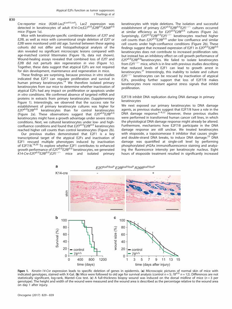
Cre-reporter mice (R26R-LacZLoxP/LoxP). LacZ expression wasdetected in keratinocytes of adult K14-Cre;E27Δ/Δ;E2f8Δ/Δ;R26RΔ/Δ
mice (Figure 1a).Mice with keratinocyte-specific combined deletion of E2f7 and
E2f8, as well as mice with conventional single deletion of E2f7 orE2f8 were monitored until old age. Survival between these ageingcohorts did not differ and histopathological analysis of theskin revealed no significant microscopic lesions compared withage-matched control littermates (Figure 1b, data not shown).Wound-healing assays revealed that combined loss of E2f7 andE2f8 did not perturb skin regeneration in vivo (Figure 1c).Together, these data suggest that atypical E2Fs are not requiredfor skin development, maintenance and regeneration in mice.These findings are surprising, because previous in vitro studies
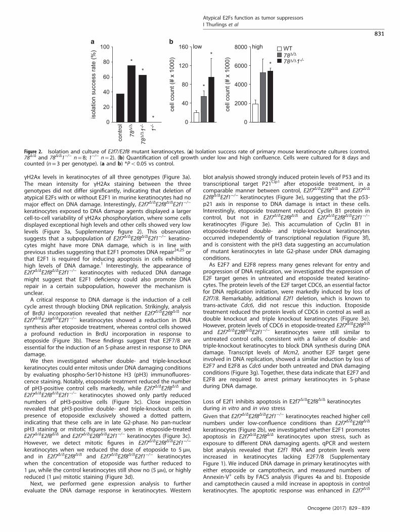
indicated that E2F7 can regulate proliferation and survival inhuman primary keratinocytes.19 We therefore isolated primarykeratinocytes from our mice to determine whether inactivation ofatypical E2Fs had any impact on proliferation or apoptosis underin vitro conditions. We confirmed absence of targeted mRNA andproteins in extracts from primary keratinocytes (SupplementaryFigure 1). Interestingly, we observed that the success rate forestablishment of primary keratinocyte cultures was higher forE2f7Δ/ΔE2f8Δ/Δ keratinocytes than for control keratinocytes(Figure 2a). These observations suggest that E2f7Δ/ΔE2f8Δ/Δ
keratinocytes might have a growth advantage under severe stressconditions. Next, we cultured keratinocytes under low- and high-confluence conditions and found that E2f7Δ/ΔE2f8Δ/Δ keratinocytesreached higher cell counts than control keratinocytes (Figure 2b).Our previous studies demonstrated that E2F1 is a key
transcriptional target of the atypical E2Fs and inactivation ofE2F1 rescued multiple phenotypes induced by inactivationof E2F7/8.18,20 To explore whether E2F1 contributes to enhancedgrowth performance of E2f7Δ/ΔE2f8Δ/Δ keratinocytes, we generatedK14-Cre-E2f7Δ/ΔE2f8Δ/Δ;E2f1−/− mice and isolated primary
keratinocytes with triple deletions. The isolation and successfulestablishment of primary E2f7Δ/ΔE2f8Δ/ΔE2f1−/− cultures occurredat similar efficiency as for E2f7Δ/ΔE2f8Δ/Δ cultures (Figure 2a).Surprisingly, E2f7Δ/ΔE2f8Δ/ΔE2f1−/− keratinocytes reached highercell counts than E2f7Δ/ΔE2f8Δ/Δ under low confluence and similarcell counts under high-confluence conditions (Figure 2b). Thesefindings suggest that increased expression of E2F1 in E2f7Δ/ΔE2f8Δ/Δ
keratinocytes does not contribute to increased proliferation rate,but instead has an inhibitory effect on cell growth performance ofE2f7Δ/ΔE2f8Δ/Δkeratinocytes. We failed to isolate keratinocytesfrom E2f1−/− mice, which is in line with previous studies describingthat reduced levels of E2F1 can lead to growth arrest inkeratinocytes.21 Interestingly, this inability to isolate and cultureE2f1−/− keratinocytes can be rescued by inactivation of atypicalE2Fs, providing further support that loss of E2F7/8 makeskeratinocytes more resistant against stress signals that inhibitproliferation.
E2F7/8 inhibit DNA replication during DNA damage in primarykeratinocytesWe next exposed our primary keratinocytes to DNA damageagents, as previous studies suggest that E2F7/8 have a role in theDNA damage response.6–8,22 However, these previous studieswere performed in transformed human cancer cell lines, in whichthe physiological DNA damage response might already be altered.Furthermore, mechanisms how E2F7/8 participate in the DNAdamage response are still unclear. We treated keratinocyteswith etoposide, a topoisomerase II inhibitor that causes single-and double-strand DNA breaks, to induce DNA damage.23 DNAdamage was quantified at single-cell level by performingphosphorylated γH2Ax immunofluorescence staining and analyz-ing the fluorescence intensity per keratinocyte nucleus. Eighthours of etoposide treatment resulted in significantly increased
E2f7loxP/loxP E2f8loxP/loxP R26RloxP/loxP
- +K14-cre
skin
time (days)12008004000
Sur
viva
l (%
)
100
80
60
40
20
0 78∆/∆control
0
20
40
60
80
100
1 3 5 7 9 11 13 15
wou
nd s
ize
(%)
time (days after injury)
78∆/∆control
Figure 1. Keratin-14-Cre expression leads to specific deletion of genes in epidermis. (a) Microscopic pictures of normal skin of mice withindicated genotypes, stained with X-Gal. (b) Mice were followed to old age for survival analysis (control n= 5; 78Δ/Δ n= 12). Differences are notstatistically significant, log-rank, Mantel–Cox test. (c) A full-thickness biopsy wound was induced on the dorsal midline of mice (n= 3 pergenotype). The height and width of the wound were measured and the wound area is described as the percentage relative to the wound areaon day 1 after injury.
Atypical E2Fs function as tumor suppressorsI Thurlings et al
830
Oncogene (2017) 829 – 839
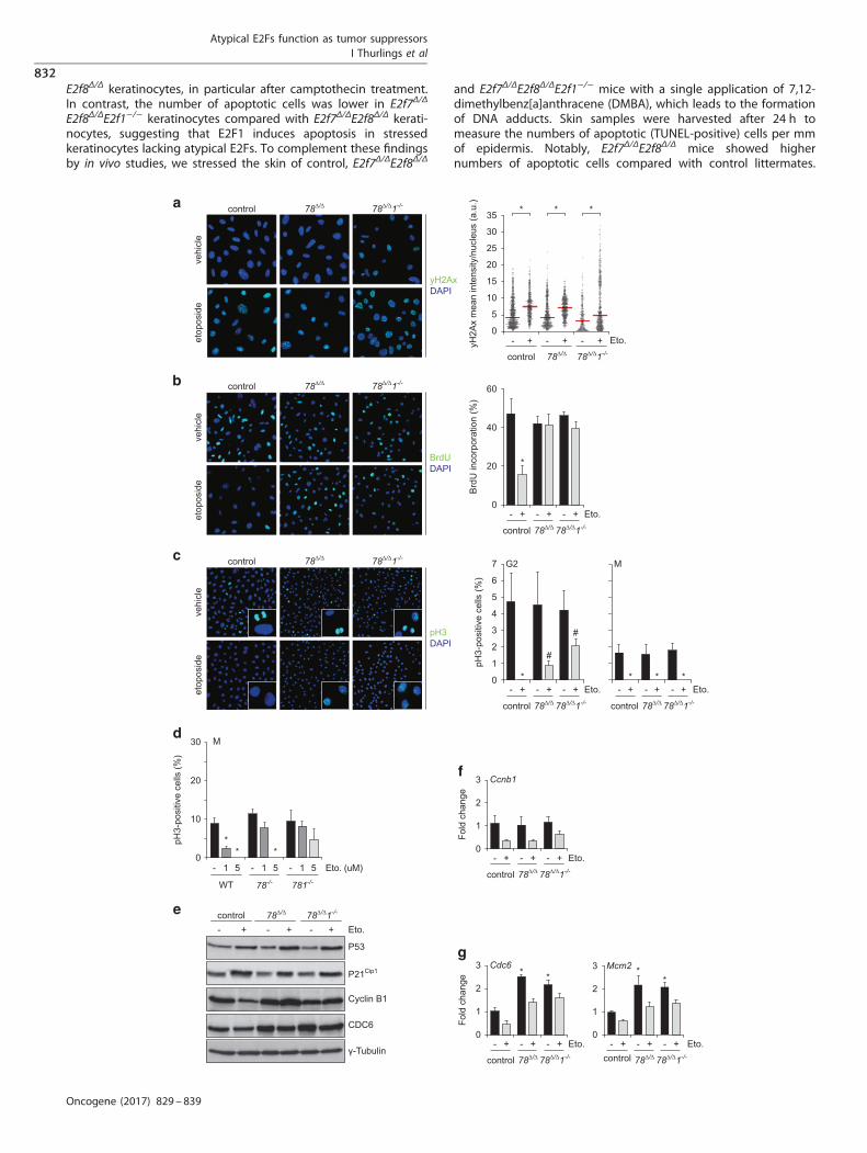
γH2Ax levels in keratinocytes of all three genotypes (Figure 3a).The mean intensity for γH2Ax staining between the threegenotypes did not differ significantly, indicating that deletion ofatypical E2Fs with or without E2F1 in murine keratinocytes had nomajor effect on DNA damage. Interestingly, E2f7Δ/ΔE2f8Δ/ΔE2f1−/−
keratinocytes exposed to DNA damage agents displayed a largercell-to-cell variability of γH2Ax phosphorylation, where some cellsdisplayed exceptional high levels and other cells showed very lowlevels (Figure 3a, Supplementary figure 2). This observationsuggests that a subpopulation of E2f7Δ/ΔE2f8Δ/ΔE2f1−/− keratino-cytes might have more DNA damage, which is in line withprevious studies suggesting that E2F1 promotes DNA repair24,25 orthat E2F1 is required for inducing apoptosis in cells exhibitinghigh levels of DNA damage.1 Interestingly, the appearance ofE2f7Δ/ΔE2f8Δ/ΔE2f1−/− keratinocytes with reduced DNA damagemight suggest that E2F1 deficiency could also promote DNArepair in a certain subpopulation, however the mechanism isunclear.A critical response to DNA damage is the induction of a cell
cycle arrest through blocking DNA replication. Strikingly, analysisof BrdU incorporation revealed that neither E2f7Δ/ΔE2f8Δ/Δ norE2f7Δ/ΔE2f8Δ/ΔE2f1−/− keratinocytes showed a reduction in DNAsynthesis after etoposide treatment, whereas control cells showeda profound reduction in BrdU incorporation in response toetoposide (Figure 3b). These findings suggest that E2F7/8 areessential for the induction of an S-phase arrest in response to DNAdamage.We then investigated whether double- and triple-knockout
keratinocytes could enter mitosis under DNA damaging conditionsby evaluating phospho-Ser10-histone H3 (pH3) immunofluores-cence staining. Notably, etoposide treatment reduced the numberof pH3-positive control cells markedly, while E2f7Δ/ΔE2f8Δ/Δ andE2f7Δ/ΔE2f8Δ/ΔE2f1−/− keratinocytes showed only partly reducednumbers of pH3-positive cells (Figure 3c). Close inspectionrevealed that pH3-positive double- and triple-knockout cells inpresence of etoposide exclusively showed a dotted pattern,indicating that these cells are in late G2-phase. No pan-nuclearpH3 staining or mitotic figures were seen in etoposide-treatedE2f7Δ/ΔE2f8Δ/Δ and E2f7Δ/ΔE2f8Δ/ΔE2f1−/− keratinocytes (Figure 3c).However, we detect mitotic figures in E2f7Δ/ΔE2f8Δ/ΔE2f1−/−
keratinocytes when we reduced the dose of etoposide to 5 μM,and in E2f7Δ/ΔE2f8Δ/Δ and E2f7Δ/ΔE2f8Δ/ΔE2f1−/− keratinocyteswhen the concentration of etoposide was further reduced to1 μM, while the control keratinocytes still show no (5 μM), or highlyreduced (1 μM) mitotic staining (Figure 3d).Next, we performed gene expression analysis to further
evaluate the DNA damage response in keratinocytes. Western
blot analysis showed strongly induced protein levels of P53 and itstranscriptional target P21Cip1 after etoposide treatment, in acomparable manner between control, E2f7Δ/ΔE2f8Δ/Δ and E2f7Δ/Δ
E2f8Δ/ΔE2f1−/− keratinocytes (Figure 3e), suggesting that the p53-p21 axis in response to DNA damage is intact in these cells.Interestingly, etoposide treatment reduced Cyclin B1 protein incontrol, but not in E2f7Δ/ΔE2f8Δ/Δ and E2f7Δ/ΔE2f8Δ/ΔE2f1−/−
keratinocytes (Figure 3e). This accumulation of Cyclin B1 inetoposide-treated double- and triple-knockout keratinocytesoccurred independently of transcriptional regulation (Figure 3f),and is consistent with the pH3 data suggesting an accumulationof mutant keratinocytes in late G2-phase under DNA damagingconditions.As E2F7 and E2F8 repress many genes relevant for entry and
progression of DNA replication, we investigated the expression ofE2F target genes in untreated and etoposide treated keratino-cytes. The protein levels of the E2F target CDC6, an essential factorfor DNA replication initiation, were markedly induced by loss ofE2f7/8. Remarkably, additional E2f1 deletion, which is known totrans-activate Cdc6, did not rescue this induction. Etoposidetreatment reduced the protein levels of CDC6 in control as well asdouble knockout and triple knockout keratinocytes (Figure 3e).However, protein levels of CDC6 in etoposide-treated E2f7Δ/ΔE2f8Δ/Δ
and E2f7Δ/ΔE2f8Δ/ΔE2f1−/− keratinocytes were still similar tountreated control cells, consistent with a failure of double- andtriple-knockout keratinocytes to block DNA synthesis during DNAdamage. Transcript levels of Mcm2, another E2F target geneinvolved in DNA replication, showed a similar induction by loss ofE2F7 and E2F8 as Cdc6 under both untreated and DNA damagingconditions (Figure 3g). Together, these data indicate that E2F7 andE2F8 are required to arrest primary keratinocytes in S-phaseduring DNA damage.
Loss of E2f1 inhibits apoptosis in E2f7Δ/ΔE2f8Δ/Δ keratinocytesduring in vitro and in vivo stressGiven that E2f7Δ/ΔE2f8Δ/ΔE2f1−/− keratinocytes reached higher cellnumbers under low-confluence conditions than E2f7Δ/ΔE2f8Δ/Δ
keratinocytes (Figure 2b), we investigated whether E2F1 promotesapoptosis in E2f7Δ/ΔE2f8Δ/Δ keratinocytes upon stress, such asexposure to different DNA damaging agents. qPCR and westernblot analysis revealed that E2f1 RNA and protein levels wereincreased in keratinocytes lacking E2F7/8 (SupplementaryFigure 1). We induced DNA damage in primary keratinocytes witheither etoposide or camptothecin, and measured numbers ofAnnexin-V+ cells by FACS analysis (Figures 4a and b). Etoposideand camptothecin caused a mild increase in apoptosis in controlkeratinocytes. The apoptotic response was enhanced in E2f7Δ/Δ
0
20
40
60
80
100
cont
rol
1-/-
78∆/
∆
78∆/
∆ 1-/-
isol
atio
n su
cces
s ra
te (%
)
0
40
80
120
160 low
cell
coun
t (#
x 10
00)
0
2000
4000
6000
8000 high
cell
coun
t (#
x 10
00)
WT78∆/∆
78∆/∆1-/-**
*
*
**
Figure 2. Isolation and culture of E2f7/E2f8 mutant keratinocytes. (a) Isolation success rate of primary mouse keratinocyte cultures (control,78Δ/Δ and 78Δ/Δ1−/− n= 8; 1−/− n= 2). (b) Quantification of cell growth under low and high confluence. Cells were cultured for 8 days andcounted (n= 3 per genotype). (a and b) *Po0.05 vs control.
Atypical E2Fs function as tumor suppressorsI Thurlings et al
831
Oncogene (2017) 829 – 839
E2f8Δ/Δ keratinocytes, in particular after camptothecin treatment.In contrast, the number of apoptotic cells was lower in E2f7Δ/Δ
E2f8Δ/ΔE2f1−/− keratinocytes compared with E2f7Δ/ΔE2f8Δ/Δ kerati-nocytes, suggesting that E2F1 induces apoptosis in stressedkeratinocytes lacking atypical E2Fs. To complement these findingsby in vivo studies, we stressed the skin of control, E2f7Δ/ΔE2f8Δ/Δ
and E2f7Δ/ΔE2f8Δ/ΔE2f1−/− mice with a single application of 7,12-dimethylbenz[a]anthracene (DMBA), which leads to the formationof DNA adducts. Skin samples were harvested after 24 h tomeasure the numbers of apoptotic (TUNEL-positive) cells per mmof epidermis. Notably, E2f7Δ/ΔE2f8Δ/Δ mice showed highernumbers of apoptotic cells compared with control littermates.
control 78∆/∆ 78∆/∆1-/-
- + - + - + Eto.0
5
10
15
20
25
30
35 * * *
yH2A
x m
ean
inte
nsity
/nuc
leus
(a.u
.)
vehi
cle
etop
osid
e
yH2AxDAPI
control 78∆/∆ 78∆/∆1-/-
control 78∆/∆ 78∆/∆1-/-
vehi
cle
etop
osid
e
pH3DAPI
control 78∆/∆
78∆/∆
78∆/∆1-/-
control 78∆/∆ 78∆/∆1-/-
control 78∆/∆ 78∆/∆1-/- control 78∆/∆ 78∆/∆1-/-
78∆/∆1-/-
control 78∆/∆ 78∆/∆1-/-
- + - + - + Eto.0
1
2
3
4
5
6
7
pH3-
posi
tive
cells
(%)
G2
#
#
*- + - + - + Eto.
M
* **
control
78∆/∆ 78∆/∆1-/-control
vehi
cle
etop
osid
e
BrdUDAPI
Brd
U in
corp
orat
ion
(%)
0
20
40
60
*
control 78∆/∆ 78∆/∆1-/-
- + - + - + Eto.
γ-Tubulin
P53
P21Cip1
CDC6
Cyclin B1
- +- +- + Eto.
1
2
3 Ccnb1
- + - + - + Eto.0
Fold
cha
nge
0
1
2
3 Cdc6
- + - + - +
* *
Eto.
Fold
cha
nge *
0
1
2
3 Mcm2
- + - + - +
*
Eto.
0
10
20
30
- 1 5
WT 78-/- 781-/-
- 1 5 - 1 5 Eto. (uM)
M
** *
pH3-
posi
tive
cells
(%)
Atypical E2Fs function as tumor suppressorsI Thurlings et al
832
Oncogene (2017) 829 – 839
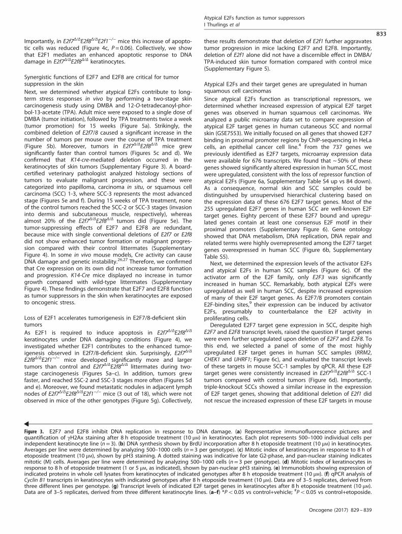
Importantly, in E2f7Δ/ΔE2f8Δ/ΔE2f1−/− mice this increase of apopto-tic cells was reduced (Figure 4c, P= 0.06). Collectively, we showthat E2F1 mediates an enhanced apoptotic response to DNAdamage in E2f7Δ/ΔE2f8Δ/Δ keratinocytes.
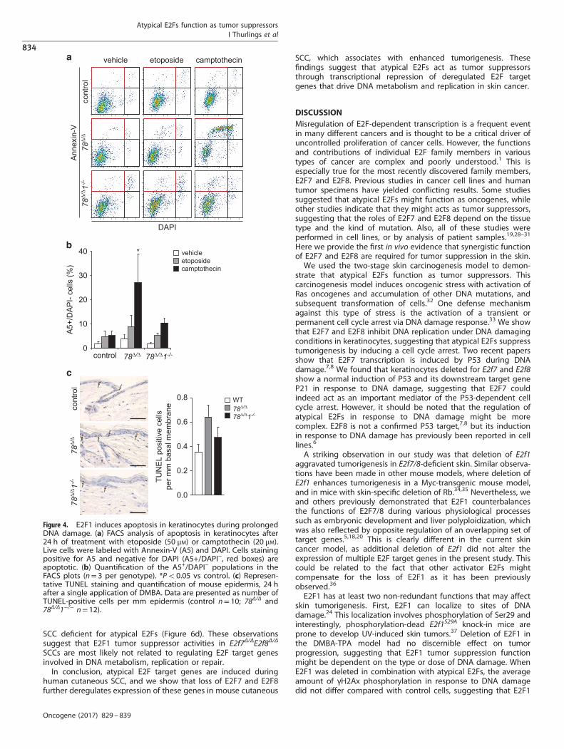
Synergistic functions of E2F7 and E2F8 are critical for tumorsuppression in the skinNext, we determined whether atypical E2Fs contribute to long-term stress responses in vivo by performing a two-stage skincarcinogenesis study using DMBA and 12-O-tetradecanoyl-phor-bol-13-acetate (TPA). Adult mice were exposed to a single dose ofDMBA (tumor initiation), followed by TPA treatments twice a week(tumor promotion) for 15 weeks (Figure 5a). Strikingly, thecombined deletion of E2f7/8 caused a significant increase in thenumber of tumors per mouse over the course of TPA treatment(Figure 5b). Moreover, tumors in E2f7Δ/ΔE2f8Δ/Δ mice grewsignificantly faster than control tumors (Figures 5c and d). Weconfirmed that K14-cre-mediated deletion occurred in thekeratinocytes of skin tumors (Supplementary Figure 3). A board-certified veterinary pathologist analyzed histology sections oftumors to evaluate malignant progression, and these werecategorized into papilloma, carcinoma in situ, or squamous cellcarcinoma (SCC) 1-3, where SCC-3 represents the most advancedstage (Figures 5e and f). During 15 weeks of TPA treatment, noneof the control tumors reached the SCC-2 or SCC-3 stages (invasioninto dermis and subcutaneous muscle, respectively), whereasalmost 20% of the E2f7Δ/ΔE2f8Δ/Δ tumors did (Figure 5e). Thetumor-suppressing effects of E2F7 and E2F8 are redundant,because mice with single conventional deletions of E2f7 or E2f8did not show enhanced tumor formation or malignant progres-sion compared with their control littermates (SupplementaryFigure 4). In some in vivo mouse models, Cre activity can causeDNA damage and genetic instability.26,27 Therefore, we confirmedthat Cre expression on its own did not increase tumor formationand progression. K14-Cre mice displayed no increase in tumorgrowth compared with wild-type littermates (SupplementaryFigure 4). These findings demonstrate that E2F7 and E2F8 functionas tumor suppressors in the skin when keratinocytes are exposedto oncogenic stress.
Loss of E2F1 accelerates tumorigenesis in E2F7/8-deficient skintumorsAs E2F1 is required to induce apoptosis in E2f7Δ/ΔE2f8Δ/Δ
keratinocytes under DNA damaging conditions (Figure 4), weinvestigated whether E2F1 contributes to the enhanced tumor-igenesis observed in E2f7/8-deficient skin. Surprisingly, E2f7Δ/Δ
E2f8Δ/ΔE2f1−/− mice developed significantly more and largertumors than control and E2f7Δ/ΔE2f8Δ/Δ littermates during two-stage carcinogenesis (Figures 5a–c). In addition, tumors grewfaster, and reached SSC-2 and SSC-3 stages more often (Figures 5dand e). Moreover, we found metastatic nodules in adjacent lymphnodes of E2f7Δ/ΔE2f8Δ/ΔE2f1−/− mice (3 out of 18), which were notobserved in mice of the other genotypes (Figure 5g). Collectively,
these results demonstrate that deletion of E2f1 further aggravatestumor progression in mice lacking E2F7 and E2F8. Importantly,deletion of E2f1 alone did not have a discernible effect in DMBA/TPA-induced skin tumor formation compared with control mice(Supplementary Figure 5).
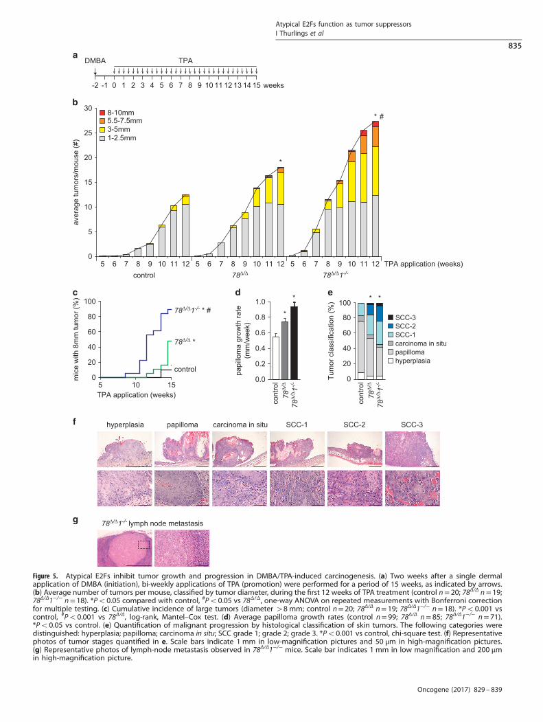
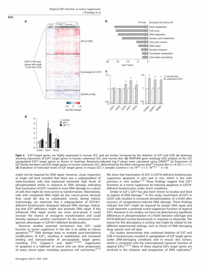
Atypical E2Fs and their target genes are upregulated in humansquamous cell carcinomasSince atypical E2Fs function as transcriptional repressors, wedetermined whether increased expression of atypical E2F targetgenes was observed in human squamous cell carcinomas. Weanalyzed a public microarray data set to compare expression ofatypical E2F target genes in human cutaneous SCC and normalskin (GSE7553). We initially focused on all genes that showed E2F7binding in proximal promoter regions by ChIP-sequencing in HeLacells, an epithelial cancer cell line.4 From the 737 genes wepreviously identified as E2F7 targets, microarray expression datawere available for 676 transcripts. We found that ~ 50% of thesegenes showed significantly altered expression in human SCC, mostwere upregulated, consistent with the loss of repressor function ofatypical E2Fs (Figure 6a, Supplementary Table S4 up vs 84 down).As a consequence, normal skin and SCC samples could bedistinguished by unsupervised hierarchical clustering based onthe expression data of these 676 E2F7 target genes. Most of the255 upregulated E2F7 genes in human SCC are well-known E2Ftarget genes. Eighty percent of these E2F7 bound and upregu-lated genes contain at least one consensus E2F motif in theirproximal promoters (Supplementary Figure 6). Gene ontologyshowed that DNA metabolism, DNA replication, DNA repair andrelated terms were highly overrepresented among the E2F7 targetgenes overexpressed in human SCC (Figure 6b, SupplementaryTable S5).Next, we determined the expression levels of the activator E2Fs
and atypical E2Fs in human SCC samples (Figure 6c). Of theactivator arm of the E2F family, only E2F3 was significantlyincreased in human SCC. Remarkably, both atypical E2Fs wereupregulated as well in human SCC, despite increased expressionof many of their E2F target genes. As E2F7/8 promoters containE2F-binding sites,9 their expression can be induced by activatorE2Fs, presumably to counterbalance the E2F activity inproliferating cells.Deregulated E2F7 target gene expression in SCC, despite high
E2F7 and E2F8 transcript levels, raised the question if target geneswere even further upregulated upon deletion of E2F7 and E2F8. Tothis end, we selected a panel of some of the most highlyupregulated E2F target genes in human SCC samples (RRM2,CHEK1 and UHRF1; Figure 6c), and evaluated the transcript levelsof these targets in mouse SCC-1 samples by qPCR. All these E2Ftarget genes were consistently increased in E2f7Δ/ΔE2f8Δ/Δ SCC-1tumors compared with control tumors (Figure 6d). Importantly,triple-knockout SCCs showed a similar increase in the expressionof E2F target genes, showing that additional deletion of E2f1 didnot rescue the increased expression of these E2F targets in mouse
Figure 3. E2F7 and E2F8 inhibit DNA replication in response to DNA damage. (a) Representative immunofluorescence pictures andquantification of γH2Ax staining after 8 h etoposide treatment (10 μM) in keratinocytes. Each plot represents 500–1000 individual cells perindependent keratinocyte line (n= 3). (b) DNA synthesis shown by BrdU incorporation after 8 h etoposide treatment (10 μM) in keratinocytes.Averages per line were determined by analyzing 500–1000 cells (n= 3 per genotype). (c) Mitotic index of keratinocytes in response to 8 h ofetoposide treatment (10 μM), shown by pH3 staining. A dotted staining was indicative for late G2-phase, and pan-nuclear staining indicatesmitotic (M) cells. Averages per line were determined by analyzing 500–1000 cells (n= 3 per genotype). (d) Mitotic index of keratinocytes inresponse to 8 h of etoposide treatment (1 or 5 μM, as indicated), shown by pan-nuclear pH3 staining. (e) Immunoblots showing expression ofindicated proteins in whole cell lysates from keratinocytes of indicated genotypes after 8 h etoposide treatment (10 μM). (f) qPCR analysis ofCyclin B1 transcripts in keratinocytes with indicated genotypes after 8 h etoposide treatment (10 μM). Data are of 3–5 replicates, derived fromthree different lines per genotype. (g) Transcript levels of indicated E2F target genes in keratinocytes after 8 h etoposide treatment (10 μM).Data are of 3–5 replicates, derived from three different keratinocyte lines. (a–f) *Po0.05 vs control+vehicle; #Po0.05 vs control+etoposide.
Atypical E2Fs function as tumor suppressorsI Thurlings et al
833
Oncogene (2017) 829 – 839
SCC deficient for atypical E2Fs (Figure 6d). These observationssuggest that E2F1 tumor suppressor activities in E2f7Δ/ΔE2f8Δ/Δ
SCCs are most likely not related to regulating E2F target genesinvolved in DNA metabolism, replication or repair.In conclusion, atypical E2F target genes are induced during
human cutaneous SCC, and we show that loss of E2F7 and E2F8further deregulates expression of these genes in mouse cutaneous
SCC, which associates with enhanced tumorigenesis. Thesefindings suggest that atypical E2Fs act as tumor suppressorsthrough transcriptional repression of deregulated E2F targetgenes that drive DNA metabolism and replication in skin cancer.
DISCUSSIONMisregulation of E2F-dependent transcription is a frequent eventin many different cancers and is thought to be a critical driver ofuncontrolled proliferation of cancer cells. However, the functionsand contributions of individual E2F family members in varioustypes of cancer are complex and poorly understood.1 This isespecially true for the most recently discovered family members,E2F7 and E2F8. Previous studies in cancer cell lines and humantumor specimens have yielded conflicting results. Some studiessuggested that atypical E2Fs might function as oncogenes, whileother studies indicate that they might acts as tumor suppressors,suggesting that the roles of E2F7 and E2F8 depend on the tissuetype and the kind of mutation. Also, all of these studies wereperformed in cell lines, or by analysis of patient samples.19,28–31
Here we provide the first in vivo evidence that synergistic functionof E2F7 and E2F8 are required for tumor suppression in the skin.We used the two-stage skin carcinogenesis model to demon-
strate that atypical E2Fs function as tumor suppressors. Thiscarcinogenesis model induces oncogenic stress with activation ofRas oncogenes and accumulation of other DNA mutations, andsubsequent transformation of cells.32 One defense mechanismagainst this type of stress is the activation of a transient orpermanent cell cycle arrest via DNA damage response.33 We showthat E2F7 and E2F8 inhibit DNA replication under DNA damagingconditions in keratinocytes, suggesting that atypical E2Fs suppresstumorigenesis by inducing a cell cycle arrest. Two recent papersshow that E2F7 transcription is induced by P53 during DNAdamage.7,8 We found that keratinocytes deleted for E2f7 and E2f8show a normal induction of P53 and its downstream target geneP21 in response to DNA damage, suggesting that E2F7 couldindeed act as an important mediator of the P53-dependent cellcycle arrest. However, it should be noted that the regulation ofatypical E2Fs in response to DNA damage might be morecomplex. E2F8 is not a confirmed P53 target,7,8 but its inductionin response to DNA damage has previously been reported in celllines.6
A striking observation in our study was that deletion of E2f1aggravated tumorigenesis in E2f7/8-deficient skin. Similar observa-tions have been made in other mouse models, where deletion ofE2f1 enhances tumorigenesis in a Myc-transgenic mouse model,and in mice with skin-specific deletion of Rb.34,35 Nevertheless, weand others previously demonstrated that E2F1 counterbalancesthe functions of E2F7/8 during various physiological processessuch as embryonic development and liver polyploidization, whichwas also reflected by opposite regulation of an overlapping set oftarget genes.5,18,20 This is clearly different in the current skincancer model, as additional deletion of E2f1 did not alter theexpression of multiple E2F target genes in the present study. Thiscould be related to the fact that other activator E2Fs mightcompensate for the loss of E2F1 as it has been previouslyobserved.36
E2F1 has at least two non-redundant functions that may affectskin tumorigenesis. First, E2F1 can localize to sites of DNAdamage.24 This localization involves phosphorylation of Ser29 andinterestingly, phosphorylation-dead E2f1S29A knock-in mice areprone to develop UV-induced skin tumors.37 Deletion of E2F1 inthe DMBA-TPA model had no discernible effect on tumorprogression, suggesting that E2F1 tumor suppression functionmight be dependent on the type or dose of DNA damage. WhenE2F1 was deleted in combination with atypical E2Fs, the averageamount of γH2Ax phosphorylation in response to DNA damagedid not differ compared with control cells, suggesting that E2F1
vehicle etoposide camptothecin
cont
rol
78∆/
∆78
∆/∆ 1
-/-
0
10
20
30
40
control 78∆/∆ 78∆/∆1-/-
A5+
/DA
PI-
cells
(%)
vehicleetoposidecamptothecin
DAPI
Ann
exin
-V
*
0.2
0.0
0.4
0.6
0.8 WT78∆/∆
78∆/∆1-/-
TUN
EL
posi
tive
cells
per m
m b
asal
mem
bran
econt
rol
78∆/
∆ 1-/-
78∆/
∆
Figure 4. E2F1 induces apoptosis in keratinocytes during prolongedDNA damage. (a) FACS analysis of apoptosis in keratinocytes after24 h of treatment with etoposide (50 μM) or camptothecin (20 μM).Live cells were labeled with Annexin-V (A5) and DAPI. Cells stainingpositive for A5 and negative for DAPI (A5+/DAPI−, red boxes) areapoptotic. (b) Quantification of the A5+/DAPI− populations in theFACS plots (n= 3 per genotype). *Po0.05 vs control. (c) Represen-tative TUNEL staining and quantification of mouse epidermis, 24 hafter a single application of DMBA. Data are presented as number ofTUNEL-positive cells per mm epidermis (control n= 10; 78Δ/Δ and78Δ/Δ1−/− n= 12).
Atypical E2Fs function as tumor suppressorsI Thurlings et al
834
Oncogene (2017) 829 – 839
0
5
10
15
20
25
30
5 6 7 8 9 10 11 12control 78∆/∆ 78∆/∆1-/-
aver
age
tum
ors/
mou
se (#
)
TPA application (weeks)
3-5mm1-2.5mm
5.5-7.5mm8-10mm
5 6 7 8 9 10 11 12 5 6 7 8 9 10 11 12
40
60
20
0Tum
or c
lass
ifica
tion
(%)
80
100
SCC-1carcinoma in situ
hyperplasiapapilloma
SCC-2SCC-3
* *
TPA application (weeks)5 10 15
0
20
40
60
80
100
control
78∆/∆ *
78∆/∆1-/- * #
mic
e w
ith 8
mm
tum
or (%
)
0.0
0.2
0.4
0.6
0.8
1.0
papi
llom
a gr
owth
rate
(mm
/wee
k)
cont
rol
cont
rol
78∆/
∆
78∆/
∆
78∆/
∆ 1-/-
78∆/
∆ 1-/-
*
*
TPADMBA
1 2 3 4 5 6 7 8 9 10 11 12 13 14 150-1-2
carcinoma in situpapillomahyperplasia SCC-3SCC-1 SCC-2
* #
*
78∆/∆1-/- lymph node metastasis
weeks
Figure 5. Atypical E2Fs inhibit tumor growth and progression in DMBA/TPA-induced carcinogenesis. (a) Two weeks after a single dermalapplication of DMBA (initiation), bi-weekly applications of TPA (promotion) were performed for a period of 15 weeks, as indicated by arrows.(b) Average number of tumors per mouse, classified by tumor diameter, during the first 12 weeks of TPA treatment (control n= 20; 78Δ/Δ n= 19;78Δ/Δ1−/− n= 18). *Po0.05 compared with control, #Po0.05 vs 78Δ/Δ, one-way ANOVA on repeated measurements with Bonferroni correctionfor multiple testing. (c) Cumulative incidence of large tumors (diameter 48 mm; control n= 20; 78Δ/Δ n= 19; 78Δ/Δ1−/− n= 18). *Po0.001 vscontrol, #Po0.001 vs 78Δ/Δ, log-rank, Mantel–Cox test. (d) Average papilloma growth rates (control n= 99; 78Δ/Δ n= 85; 78Δ/Δ1−/− n= 71).*Po0.05 vs control. (e) Quantification of malignant progression by histological classification of skin tumors. The following categories weredistinguished: hyperplasia; papilloma; carcinoma in situ; SCC grade 1; grade 2; grade 3. *Po0.001 vs control, chi-square test. (f) Representativephotos of tumor stages quantified in e. Scale bars indicate 1 mm in low-magnification pictures and 50 μm in high-magnification pictures.(g) Representative photos of lymph-node metastasis observed in 78Δ/Δ1−/− mice. Scale bar indicates 1 mm in low magnification and 200 μmin high-magnification picture.
Atypical E2Fs function as tumor suppressorsI Thurlings et al
835
Oncogene (2017) 829 – 839
might not be required for DNA repair. However, closer inspectionat single cell level revealed that there was a subpopulation oftriple-knockout cells that expressed extremely high levels ofphosphorylated γH2Ax in response to DNA damage, indicatingthat inactivation of E2F1 resulted in more DNA damage in a subsetof cells that might be more prone to transformation. Alternatively,cells with unrepaired DNA might be less cancer-prone, becausethey could die due to catastrophic events during mitosis.Interestingly, we observed that a subpopulation of E2f7/8/1-deficient keratinocytes displayed reduced DNA damage, indicat-ing that E2f1 deficiency might also promote DNA repair. If thisenhanced DNA repair events are more error-prone it couldincrease the chance of oncogenic transformation and couldthereby represent another mechanism for the enhanced tumor-igenesis phenotype in E2f7/8/1-deficient keratinocytes.However, another potential mechanism how E2F1 could
function as tumor suppressor in the skin is its ability to induceapoptosis.38,39 DNA damage leads to multiple post-translationalmodifications of E2F1, resulting in stabilization and increasedbinding and transactivation of pro-apoptotic target genes,including P73, Caspase-3 and Apaf1.25,39,40 Suppressionof apoptosis is a hallmark of cancer and can drive progressionof many cancer types, including squamous cell carcinomas.41,42
We show that inactivation of E2f1 in E2f7/8 deficient keratinocytessuppresses apoptosis in vitro and in vivo, which is line withprevious in vitro studies.6,19 These findings suggest that E2F1functions as a tumor suppressor by inducing apoptosis in E2f7/8-deficient keratinocytes under stress conditions.Similar to E2F1, E2F7 has also been shown to localize and bind
to regions of DNA damage.22 In this study, inactivation of E2F7 inU2OS cells resulted in increased phosphorylation of γ-H2AX afterrecovery of camptothecin-induced DNA damage. These findingsindicate that E2F7 might be required for proper DNA repair andcould represent a potential tumor-suppressor function of atypicalE2Fs. However in our studies, we have not detected any significantdifferences in phosphorylation of γ-H2AX between wild-type andE2F/8-deficient murine keratinocyte in response to etoposide. Thereason for this discrepancy is unclear, but might be related to thedifferent experimental settings, such as choice of DNA damagingdrug, species and cell type.Our studies demonstrate that combined deletion of E2f7 and
E2f8 in keratinocytes resulted in upregulation of E2F target genesunder DNA-damaging conditions and in mouse skin tumors,which is consistent with the transcriptional repressor function ofatypical E2Fs,4,10,19 Many of these atypical E2Fs target genes areinvolved in the initiation and progression of DNA replication.4
skin cutaneous SCC
UP (n=255)
Unchanged (n=337)
DOWN (n=84)
E2F7 ChIP-seq:genes with peak< 5 kb from TSS
-20 -15 -10 -5 0
Chromatin remodelingPyrimidine metabolismNuclear transportDNA repairCell cycle controlNucleic acid metabolismDNA replicationCell cycleDNA metabolism
P=0.05
log P value
Enriched GO terms UP
-1.0 1.0log fold change
00
0.5
1
1.5 normal skincutaneous SCC
** ** **
UHRF1RRM2
CHEK10
0.5
1
1.5
E2F1
E2F2
E2F3
E2F7
E2F8
**** *
Chek1 Uhrf1 Rrm2
control78∆/∆
78∆/∆1-/-
0
1
2
3
4
5
6*
* *
Figure 6. E2F7-target genes are highly expressed in human SCC and are further increased by the deletion of E2f7 and E2f8. (a) Heatmapshowing expression of E2F7 target genes in human cutaneous SCC and normal skin. (b) PANTHER gene ontology (GO) analysis of the 255upregulated E2F7 target genes as shown in heatmap. Benjamini-adjusted log P-values were calculated using DAVID.49 (c) Expression ofE2F family members and E2F target genes in human cutaneous SCC, determined by the Riker microarray data48 (normal skin n= 4; SCC n= 11).(d) Evaluation of transcript levels of E2F target genes in mouse SCC-1 samples (control n= 6; 78Δ/Δ n= 7; 78Δ/Δ1−/− n= 8).
Atypical E2Fs function as tumor suppressorsI Thurlings et al
836
Oncogene (2017) 829 – 839

Importantly, these atypical E2F7/8 target genes were alsoupregulated in human squamous cell carcinomas, indicating thatderegulation of E2F7/8 target genes might contribute to skintumorigenesis in mice and humans. Evaluation of the COSMIC andTCGA databases revealed that genetic alterations of E2F7 andE2F8 are very infrequent, and the chance that both atypical E2Fsare mutated in the same tumor is extremely small. Therefore,forcing tumor cells to express high levels of E2F7 and/or E2F8could be a promising strategy to inhibit E2F activity in tumor cells.We show that deletion of atypical E2Fs in skin tumors leadsto further deregulation of E2F target genes accompanied withacceleration of tumorigenesis indicating that atypical E2Fs cancounterbalance deregulated E2F activity to suppress tumorigen-esis. For that reason, further enhancement of atypical E2Fs activityto downregulate E2F activity in tumor cells could represent asuccessful strategy to mitigate tumorigenesis, for examplethrough blocking degradation of atypical E2Fs. Further investiga-tions are required to examine how and when E2Fs are degraded,but studies from our group and others provide evidence thatatypical E2Fs are degraded by the APC/CCdh1 complex.43,44 Thesefindings might open new avenues for therapeutic approaches toinhibit abundant E2F activity during tumorigenesis. Future studiesare required to investigate how the activity of activator E2Fs andatypical E2Fs can be optimally manipulated to inhibit tumorgrowth for different cancer types.
MATERIALS AND METHODSAnimalsThe use of laboratory animals was approved by the Animals EthicsCommittee at Utrecht University and performed according to theinstitutional and national guidelines. Conventional and conditional E2f7and E2f8-knockout mice were generated as described.18 K14-Cre and R26R-LacZLoxP/LoxP mice were purchased from Jackson Laboratory (Bar Harbor,ME, USA). Conventional E2f1 knockout mice were provided by MGreenberg (Duke University Medical Center, USA). All mice were bred inFVB genetic background for at least five generations. Genotypic analysis ofmice was performed on DNA from ear-clips by PCR. Primer sequences areshown in Supplementary Table S1. Animals were allocated to experimentalgroups based on their genotype and the analysis of the mice wasperformed in randomized and blinded manner.
β-galactosidase stainingStaining on skin and tumor tissues was performed as describedpreviously.20
Wound healingDorsal skin of 6-week-old mice was shaved and 6 mm diameter circularfull-thickness biopsy wounds were induced at the dorsal midline, removingboth epidermis and dermis. The height and width of the wounds wasmeasured at indicated time points after injury to assess the healing rates ofthe wounds. The wound area is described as the percentage relative to thewound area on day 1 after injury.
Primary keratinocyte isolationPrimary mouse keratinocytes were isolated from E17- to E19-day-oldembryos following previously described protocol45 and cultured in definedkeratinocyte CnT-medium (CELLnTEC, Bern, Switzerland, Advanced CellSystems AG, CnT-07). The success rate was defined as the percentage ofsuccessful isolation and subsequent culturing primary murine keratinocytecell lines in relation to the total number of isolation attempts per genotypegroup. When primary keratinocytes were established and cultured formore than three passages from the skin of a mouse embryo, this wascounted as a successful isolation.
Cell countKeratinocytes were plated under low (7000 cells/cm2) or high (177.000cells/cm2) confluency conditions. Cells growing under low confluency were
counted 8 days after plating. Cells growing under high confluency werepassaged once at 4 days, and counted after 8 days.
Immunofluorescence microscopyKeratinocytes were cultured on glass coverslips, treated with etoposide(10 μM) for 8 h and fixed in 4% PFA for 10 min at room temperature. For 5-bromo-2′-deoxyuridine (BrdU) detection, cells were treated with BrdU(Sigma, St Louis, MO, USA, B5002) 6 h before fixation. Cells were incubatedwith 2 M HCl for 20 min, followed by addition of 0.1 M sodium borate(Na2B4O7), pH 8.5 for 2 min. Cells were washed with PBS and permeabilizedwith 0.2% triton/PBS for 5 min. Immunofluorescence staining was doneby anti-BrdU-FITC for 2 h. Coverslips were mounted on slides usingFluoroshield mounting medium containing DAPI (Sigma, F6057).Cells were fixed with 4% PFA/0.1% triton/PBS for 10 min at room
temperature for immunofluorescence staining for γ-H2Ax and pH3.Cells were blocked with 4% BSA/TBS for 30 min, and incubated for 2 hwith primary antibodies dissolved in 2% BSA/TBS. Cells were washed andincubated with a secondary antibody conjugated with Alexa-Fluor 488 for1 h. DAPI was used as nuclear counterstaining. Images were taken using aLeica TCS SPE-II confocal microscope and analyzed using Fiji software.46
Used antibodies are in Supplementary Table S2.
Western blot analysisProtein isolation and western blot analysis were performed as previouslydescribed.4 Used antibodies are in Supplementary Table S2. Uncroppedversions of the western blots are shown in Supplementary Figure 7.
qPCRRNA isolation, cDNA synthesis and qPCR were done as described.20
Gene expression was calculated using a ΔΔCt method adapted formultiple reference genes correction,47 correcting for b-actin and Gapdh(mouse tumors) or 18 S ribosomal RNA and Gapdh (keratinocytes). Primersequences are included in Supplementary Table S3.
Flow cytometryKeratinocytes were treated 24 h with etoposide (50 μM) or camptothecin(20 μM; Sigma C9911), and trypsinized. Annexin V-staining wasperformed according to the manufacturer’s protocol (Invitrogen, Carlsbad,CA, USA, A13202). DAPI (1 ng/ml) was added to the samples to distinguishbetween apoptotic and necrotic cells. After filtration of the cellsuspensions on 40 μM cell strainers (Falcon, Amsterdam, The Netherlands),analysis was performed with a BD FACS Canto II and quantifications weredone using FlowJo software (TreeStar Inc., Ashland, OR, USA).
ImmunohistochemistryApoptosis in microscopic mouse skin sections was detected using ApoTagPlus peroxidase In Situ Apoptosis Kit (TUNEL) (Chemicon, Southampton,UK, S701) according to manufacturer’s protocol. All slides were counter-stained with hematoxylin. TUNEL-positive cells per mm epidermis wasdetermined manually.
Two-stage carcinogenesis protocolThe two-stage carcinogenesis protocol was performed as describedpreviously.32 For short-term experiments, 25 nmol DMBA/200 μl acetonewas topically applied to skin of 7–9-week-old mice, which were killed24 h after application. Treated and untreated skin sections were fixed in4% PFA for histological analysis.
Comparative gene expression analysisThe Gene Expression Omnibus was used to find previously publishedhuman cutaneous SCC data sets, and found one Affymetrix HGU113plus2data set (GSE755348). The raw data (.CEL) files from the squamous cellcarcinoma (n=11) and normal skin samples (n=4) were analyzed usingFlexarray 1.6.1 software (University of Quebec, Montreal, Canada). RawAffymetrix data were normalized using RMA, and tested for significantdifferences between normal skin and SCC using Empirical Bayes estimation(Wilson & Wright, New Haven, CT, USA). Using a previously published ChIP-sequencing analysis,4 we filtered only those genes from the microarraydata that have significant E2F7 binding within their promoter regions(o5 kb from transcription start site). Unsupervised hierarchical clustering
Atypical E2Fs function as tumor suppressorsI Thurlings et al
837
Oncogene (2017) 829 – 839
based on expression of all E2F7 target genes was performed using theEuclidian distance method. E2F binding motif analysis was performed asdescribed.4 Heatmaps reflecting normalized expression data weregenerated using Gene-E (https://www.broadinstitute.org/cancer/software/GENE-E/). Gene ontology analysis of E2F7 target genes accordingto expression changes in human SCC was performed using PANTHERBiological Pathways tool in DAVID.49
StatisticsData are presented as mean± s.e.m. unless indicated otherwise. Differ-ences between groups were compared using one-way analysis of variancefollowed by Tukey’s post hoc correction. When multiple groups were notnormally distributed, Kruskal–Wallis tests with Dunn’s post hoc correctionwere performed. Analysis was performed using SigmaPlot (Systat Software,San Jose, CA, USA).
CONFLICT OF INTERESTThe authors declare no conflict of interest.
ACKNOWLEDGEMENTSThis work was financially supported by a Dutch Cancer Society grant (KWF:UU2013-5777) to BW and AdB, and the Netherlands Organization of ScientificResearch (NWO-ALW:843.11.002). We like to thank M Greenberg (Duke UniversityMedical Center, USA) for kindly providing the E2f1 conventional knockout mice,and R Caldelari (University of Berne, Switzerland) for technical assistance andadvice with the keratinocyte isolations. We are also thankful for the technicalsupport by Elsbeth van Liere and Ronald Molenbeek (Utrecht University, TheNetherlands).
REFERENCES1 Chen H-Z, Tsai S-Y, Leone G. Emerging roles of E2Fs in cancer: an exit from cell
cycle control. Nat Rev Cancer 2009; 9: 785–797.2 Chellappan SP, Hiebert S, Mudryj M, Horowitz JM, Nevins JR. The E2F transcription
factor is a cellular target for the RB protein. Cell 1991; 65: 1053–1061.3 Johnson DG, Degregori J. Putting the oncogenic and tumor suppressive activities
of E2F into Context. Curr Mol Med 2006; 6: 731–738.4 Westendorp B, Mokry M, Groot Koerkamp MJA, Holstege FCP, Cuppen E, de Bruin
A. E2F7 represses a network of oscillating cell cycle genes to control S-phaseprogression. Nucleic Acids Res 2012; 40: 3511–3523.
5 Chen H-Z, Ouseph MM, Li J, Pécot T, Chokshi V, Kent L et al. Canonical andatypical E2Fs regulate the mammalian endocycle. Nat Cell Biol 2012; 14:1192–1202.
6 Zalmas LP, Zhao X, Graham AL, Fisher R, Reilly C, Coutts AS et al. DNA-damageresponse control of E2F7 and E2F8. EMBO Rep 2008; 9: 252–259.
7 Carvajal LA, Hamard P-J, Tonnessen C, Manfredi JJ. E2F7, a novel target, isup-regulated by p53 and mediates DNA damage-dependent transcriptionalrepression. Genes Dev 2012; 26: 1533–1545.
8 Aksoy O, Chicas A, Zeng T, Zhao Z, McCurrach M, Wang X et al. The atypical E2Ffamily member E2F7 couples the p53 and RB pathways during cellular senes-cence. Genes Dev 2012; 26: 1546–1557.
9 de Bruin A, Maiti B, Jakoi L, Timmers C, Buerki R, Leone G. Identification andcharacterization of E2F7, a novel mammalian E2F family member capable ofblocking cellular proliferation. J Biol Chem 2003; 278: 42041–42049.
10 Di Stefano L, Jensen MR, Helin K. E2F7, a novel E2F featuring DP-independentrepression of a subset of E2F-regulated genes. EMBO J 2003; 22:6289–6298.
11 Logan N, Delavaine L, Graham A, Reilly C, Wilson J, Brummelkamp TR et al. E2F-7: adistinctive E2F family member with an unusual organization of DNA-bindingdomains.Oncogene 2004; 23: 5138–5150.
12 Logan N, Graham A, Zhao X, Fisher R, Maiti B, Leone G et al. E2F-8: an E2F familymember with a similar organization of DNA-binding domains to E2F-7. Oncogene2005; 24: 5000–5004.
13 Christensen J, Cloos P, Toftegaard U, Klinkenberg D, Bracken AP, Trinh E et al.Characterization of E2F8, a novel E2F-like cell-cycle regulated repressor ofE2F-activated transcription. Nucleic Acids Res 2005; 33: 5458–5470.
14 Maiti B, Li J, de Bruin A, Gordon F, Timmers C, Opavsky R et al. Cloningand characterization of mouse E2F8, a novel mammalian E2F familymember capable of blocking cellular proliferation. J Biol Chem 2005; 280:18211–18220.
15 Hazar-Rethinam M, Cameron SR, Dahler AL, Endo-Munoz LB, Smith L, Rickwood Det al. Loss of E2F7 expression is an early event in squamous differentiation andcauses derepression of the key differentiation activator Sp1. J Invest Dermatol2011; 131: 1077–1084.
16 Hazar-Rethinam M, de Long LM, Gannon OM, Topkas E, Boros S, Vargas AC et al. Anovel E2F/sphingosine kinase 1 axis regulates anthracycline response in squa-mous cell carcinoma. Clin Cancer Res 2015; 21: 417–427.
17 Ouseph MM, Li J, Chen H-Z, Pécot T, Wenzel P, Thompson JC et al. Atypical E2Frepressors and activators coordinate placental development. Dev Cell 2012; 22:849–862.
18 Li J, Ran C, Li E, Gordon F, Comstock G, Siddiqui H et al. Synergistic function ofE2F7 and E2F8 is essential for cell survival and embryonic development. Dev Cell2008; 14: 62–75.
19 Endo-Munoz L, Dahler A, Teakle N, Rickwood D, Hazar-Rethinam M, Abdul-Jabbar Iet al. E2F7 can regulate proliferation, differentiation, and apoptotic responses inhuman keratinocytes: implications for cutaneous squamous cell carcinoma for-mation. Cancer Res 2009; 69: 1800–1808.
20 Pandit SK, Westendorp B, Nantasanti S, van Liere E, Tooten PCJ, Cornelissen PWAet al. E2F8 is essential for polyploidization in mammalian cells. Nat Cell Biol 2012;14: 1181–1191.
21 D'Souza SJA, Vespa A, Murkherjee S, Maher A, Pajak A, Dagnino L. E2F-1 isessential for normal epidermal wound repair. J Biol Chem 2002; 277:10626–10632.
22 Zalmas L-P, Coutts AS, Helleday T, La Thangue NB. E2F-7 couples DNA damage-dependent transcription with the DNA repair process. Cell Cycle 2013; 12:3037–3051.
23 Hande KR. Etoposide: four decades of development of a topoisomerase II inhi-bitor. Eur J Cancer 1998; 34: 1514–1521.
24 Biswas AK, Johnson DG. Transcriptional and nontranscriptional functions of E2F1in response to DNA damage. Cancer Res 2012; 72: 13–17.
25 Carnevale J, Palander O, Seifried LA, Dick FA. DNA damage signals through dif-ferentially modified E2F1 molecules to induce apoptosis. Mol Cell Biol 2012; 32:900–912.
26 Schmidt-Supprian M, Rajewsky K. Vagaries of conditional gene targeting. NatImmunol 2007; 8: 665–668.
27 Silver DP, Livingston DM. Self-excising retroviral vectors encoding the Crerecombinase overcome Cre-mediated cellular toxicity. Molecular Cell 2001; 8:233–243.
28 Salvatori B, Iosue I, Mangiavacchi A, Loddo G, Padula F, Chiaretti S et al.The microRNA-26a target E2F7 sustains cell proliferation and inhibitsmonocytic differentiation of acute myeloid leukemia cells. Cell Death Dis 2012; 3:e413.
29 Reimer D, Sadr S, Wiedemair A, Stadlmann S, Concin N, Hofstetter G et al. Clinicalrelevance of E2F family members in ovarian cancer--an evaluation in a training setof 77 patients. Clin Cancer Res 2007; 13: 144–151.
30 Deng Q, Wang Q, Zong W-Y, Zheng D-L, Wen Y-X, Wang K-S et al. E2F8 con-tributes to human hepatocellular carcinoma via regulating cell proliferation.Cancer Res 2010; 70: 782–791.
31 Park S-A, Platt J, Lee JW, López-Giráldez F, Herbst RS, Koo JS. E2F8 asa Novel Therapeutic Target for Lung Cancer. J Natl Cancer Inst 2015; 107:djv151–1, 1–16.
32 Abel EL, Angel JM, Kiguchi K, DiGiovanni J. Multi-stage chemicalcarcinogenesis in mouse skin: fundamentals and applications. Nat Protoc 2009; 4:1350–1362.
33 Di Micco R, Fumagalli M, Cicalese A, Piccinin S, Gasparini P, Luise C et al.Oncogene-induced senescence is a DNA damage response triggered by DNAhyper-replication. Nature 2006; 444: 638–642.
34 Rounbehler RJ, Rogers PM, Conti CJ, Johnson DG. Inactivation of E2f1 enhancestumorigenesis in a Myc transgenic model. Cancer Res 2002; 62: 3276–3281.
35 Costa C, Santos M, Martínez-Fernández M, Dueñas M, Lorz C, García-Escudero Ret al. E2F1 loss induces spontaneous tumour development in Rb-deficient epi-dermis. Oncogene 2013; 32: 2937–2951.
36 Tsai S-Y, Opavsky R, Sharma N, Wu L, Naidu S, Nolan E et al. Mouse developmentwith a single E2F activator. Nature 2008; 454: 1137–1141.
37 Biswas AK, Mitchell DL, Johnson DG. E2F1 responds to ultraviolet radiation bydirectly stimulating DNA repair and suppressing carcinogenesis. Cancer Res 2014;74: 3369–3377.
38 DeGregori J, Leone G, Miron A, Jakoi L, Nevins JR. Distinct roles for E2Fproteins in cell growth control and apoptosis. Proc Natl Acad Sci USA 1997; 94:7245–7250.
39 Polager S, Ginsberg D. p53 and E2f: partners in life and death. Nat Rev Cancer2009; 9: 738–748.
40 Pediconi N, Ianari A, Costanzo A, Belloni L, Gallo R, Cimino L et al. Differentialregulation of E2F1 apoptotic target genes in response to DNA damage. Nat CellBiol 2003; 5: 552–558.
Atypical E2Fs function as tumor suppressorsI Thurlings et al
838
Oncogene (2017) 829 – 839

41 Hanahan D, Weinberg RA. Hallmarks of cancer: the next generation. Cell 2011;144: 646–674.
42 Kim C, Pasparakis M. Epidermal p65/NF-κB signalling is essential for skin carci-nogenesis. EMBO Mol Med 2014; 6: 970–983.
43 Cohen M, Vecsler M, Liberzon A, Noach M, Zlotorynski E, Tzur A. Unbiased tran-scriptome signature of in vivo cell proliferation reveals pro- and antiproliferativegene networks. Cell Cycle 2013; 12: 2992–3000.
44 Boekhout M, Yuan R, Wondergem AP, Segeren HA, van Liere EA, Awol N et al.Feedback regulation between atypical E2Fs and APC/CCdh1 coordinates cell cycleprogression. EMBO Rep 2016; 17: 414–427.
45 Caldelari R, Müller EJ. Short- and long-term cultivation of embryonic and neonatalmurine keratinocytes. Methods Mol Biol 2010; 633: 125–138.
46 Schindelin J, Arganda-Carreras I, Frise E, Kaynig V, Longair M, Pietzsch T et al. Fiji: anopen-source platform for biological-image analysis. Nat Methods 2012; 9: 676–682.
47 Vandesompele J, De Preter K, Pattyn F, Poppe B, Van Roy N, De Paepe A et al.Accurate normalization of real-time quantitative RT-PCR data by geometricaveraging of multiple internal control genes. Genome Biol 2002; 3:RESEARCH0034, 1–12.
48 Riker AI, Enkemann SA, Fodstad O, Liu S, Ren S, Morris C et al. The gene expressionprofiles of primary and metastatic melanoma yields a transition point of tumorprogression and metastasis. BMC Med Genomics 2008; 1: 13.
49 Huang DW, Sherman BT, Lempicki RA. Systematic and integrative analysis oflarge gene lists using DAVID bioinformatics resources. Nat Protoc 2009; 4:44–57.
This work is licensed under a Creative Commons Attribution-NonCommercial-NoDerivs 4.0 International License. The images or
other third party material in this article are included in the article’s Creative Commonslicense, unless indicatedotherwise in the credit line; if thematerial is not included underthe Creative Commons license, users will need to obtain permission from the licenseholder to reproduce the material. To view a copy of this license, visit http://creativecommons.org/licenses/by-nc-nd/4.0/
© The Author(s) 2017
Supplementary Information accompanies this paper on the Oncogene website (http://www.nature.com/onc)
Atypical E2Fs function as tumor suppressorsI Thurlings et al
839
Oncogene (2017) 829 – 839