Syncope - American College of Cardiology/media/Non-Clinical/Files-PDFs-Excel-MS-Word-etc...Syncope:...
63
Syncope: Evaluation of the Weak and Dizzy William M. Miles, MD, FACC, FHRS Professor of Medicine Silverstein Chair for Cardiovascular Education University of Florida College of Medicine
Transcript of Syncope - American College of Cardiology/media/Non-Clinical/Files-PDFs-Excel-MS-Word-etc...Syncope:...
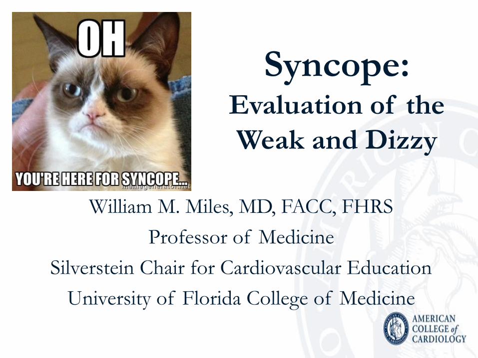
Syncope: Evaluation of the
Weak and Dizzy
William M. Miles, MD, FACC, FHRS
Professor of Medicine
Silverstein Chair for Cardiovascular Education
University of Florida College of Medicine

Disclosures • Medtronic, Inc. (Clinical Events Committee, consultant)
• Biosense-Webster, Boston Scientific, Medtronic, St. Jude
(UF EP Fellowship Support)
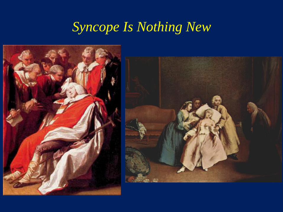
Syncope Is Nothing New

Heaton KW.
BMJ 2006;
333:1335
William Shakespeare (~1564-1616) Works in Which a Character Faints from Strong Emotion
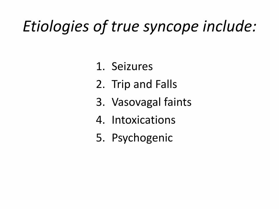
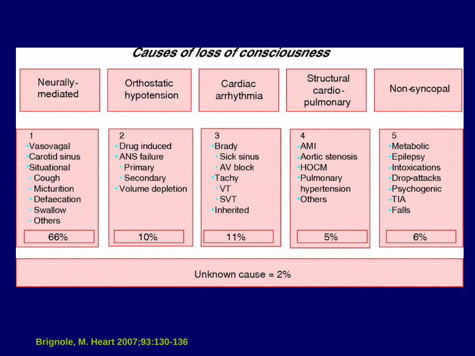
Etiologies of true syncope include:
1. Seizures
2. Trip and Falls
3. Vasovagal faints
4. Intoxications
5. Psychogenic

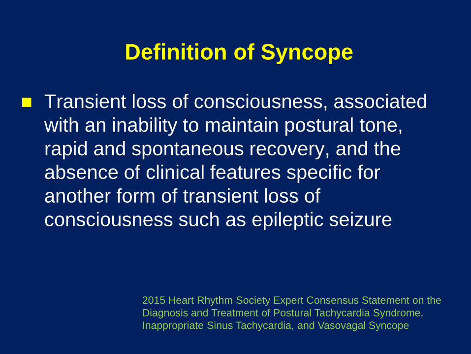
Definition of Syncope
Transient loss of consciousness, associated
with an inability to maintain postural tone,
rapid and spontaneous recovery, and the
absence of clinical features specific for
another form of transient loss of
consciousness such as epileptic seizure
2015 Heart Rhythm Society Expert Consensus Statement on the
Diagnosis and Treatment of Postural Tachycardia Syndrome,
Inappropriate Sinus Tachycardia, and Vasovagal Syncope
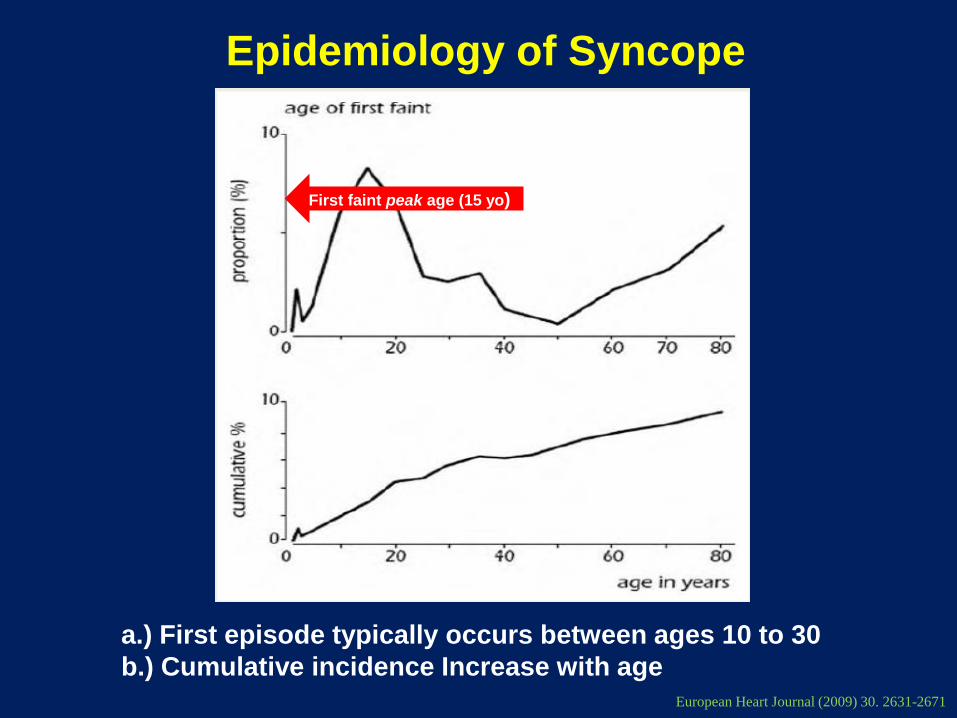
Epidemiology of Syncope
a.) First episode typically occurs between ages 10 to 30
b.) Cumulative incidence Increase with age European Heart Journal (2009) 30. 2631-2671
First faint peak age (15 yo)
Brignole, M. Heart 2007;93:130-136

Syncope Evaluation
European Heart Journal (2009) 30. 2631-2671

Syncope patients with the poorest prognosis are those with:
1. Vasovagal syncope
2. Orthostatic syncope
3. Carotid sinus hypersensitivity
4. Cardiac cause of syncope
5. Syncope of undetermined cause

Survival of Patients With Syncope
Soteriades, N Engl J Med 2002;347:878-85
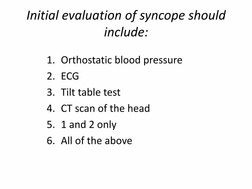
Initial evaluation of syncope should include:
1. Orthostatic blood pressure
2. ECG
3. Tilt table test
4. CT scan of the head
5. 1 and 2 only
6. All of the above
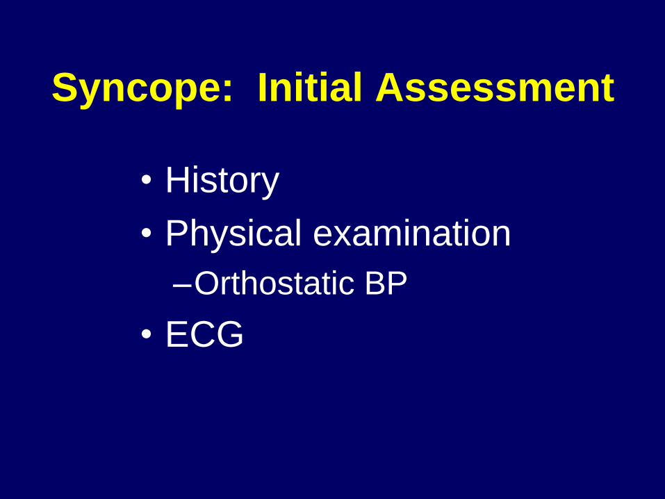
Syncope: Initial Assessment
• History
• Physical examination
–Orthostatic BP
• ECG

Brignole et al, ECS Task Force on Syncope. European Heart Journal 2004;25:2054
Seizure vs. Syncope

Clinical Features Suggestive of Specific
Causes of Loss of Consciousness
• Neurally mediated syncope
– Absence of cardiac disease
– Long history of syncope
– After sudden unexpected unpleasant sight, sound, smell or pain
– Prolonged standing or crowded, hot places
– Nausea, vomiting associated with syncope
– During a meal or in the absorptive state after a meal
– With head rotation, pressure on carotid sinus (as in tumors, shaving, tight collars)
– After exertion
Brignole et al, ESC Task Force on Syncope. European Heart Journal 2004;25:2054
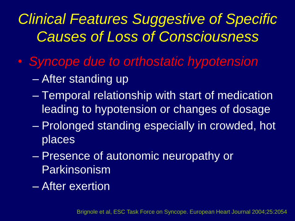
Clinical Features Suggestive of Specific
Causes of Loss of Consciousness
• Syncope due to orthostatic hypotension
– After standing up
– Temporal relationship with start of medication
leading to hypotension or changes of dosage
– Prolonged standing especially in crowded, hot
places
– Presence of autonomic neuropathy or
Parkinsonism
– After exertion
Brignole et al, ESC Task Force on Syncope. European Heart Journal 2004;25:2054

Clinical Features Suggestive of Specific
Causes of Loss of Consciousness
• Cardiac syncope
– Presence of definite structural heart disease
– During exertion, or supine
– Preceded by palpitation
– Family history of sudden death
• Cerebrovascular syncope
– With arm exercise
– Differences in blood pressure or pulse in the
two arms
Brignole et al, ESC Task Force on Syncope. European Heart Journal 2004;25:2054
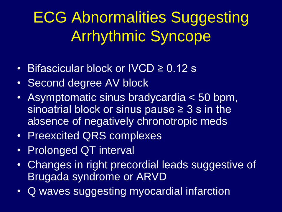
ECG Abnormalities Suggesting
Arrhythmic Syncope
• Bifascicular block or IVCD ≥ 0.12 s
• Second degree AV block
• Asymptomatic sinus bradycardia < 50 bpm, sinoatrial block or sinus pause ≥ 3 s in the absence of negatively chronotropic meds
• Preexcited QRS complexes
• Prolonged QT interval
• Changes in right precordial leads suggestive of Brugada syndrome or ARVD
• Q waves suggesting myocardial infarction
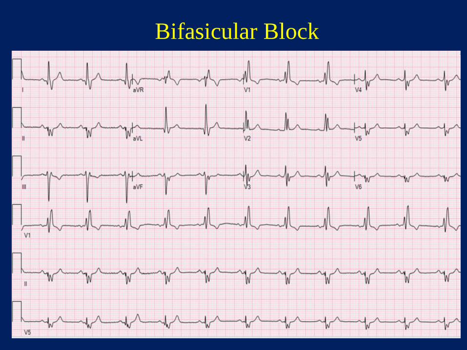
Bifasicular Block
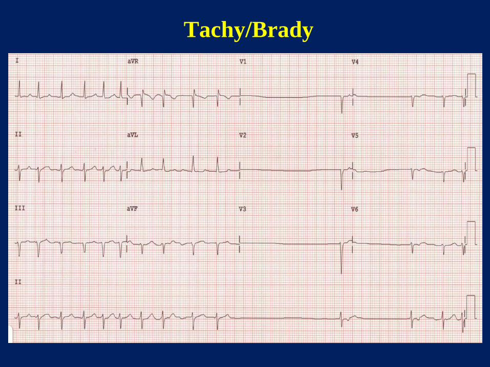
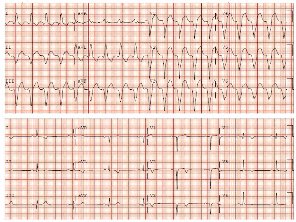
Tachy/Brady

Type II AV Block
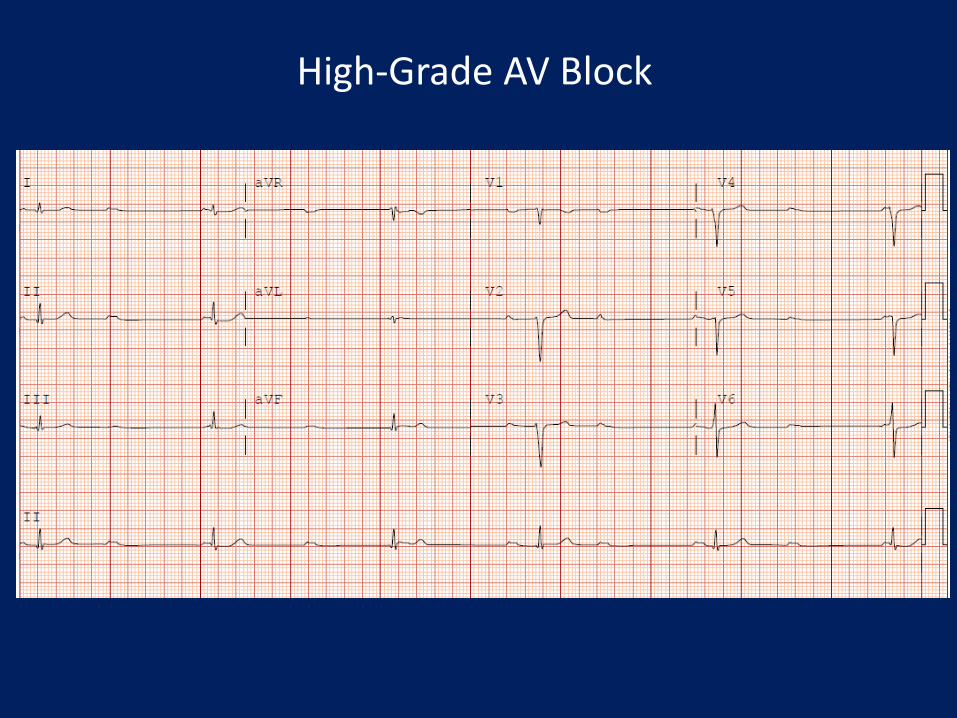
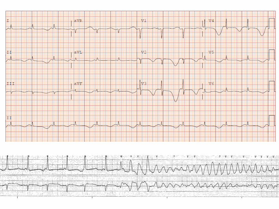
High-Grade AV Block

Wilde et al. Circulation. 2002;106:2514-2519
Brugada Pattern
Coved ST
Elevation

54 y.o. male with palpitations and syncope; family history of sudden death
Arrhythmogenic Right Ventricular Dysplasia
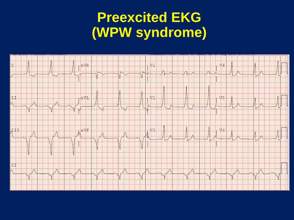
Preexcited EKG (WPW syndrome)
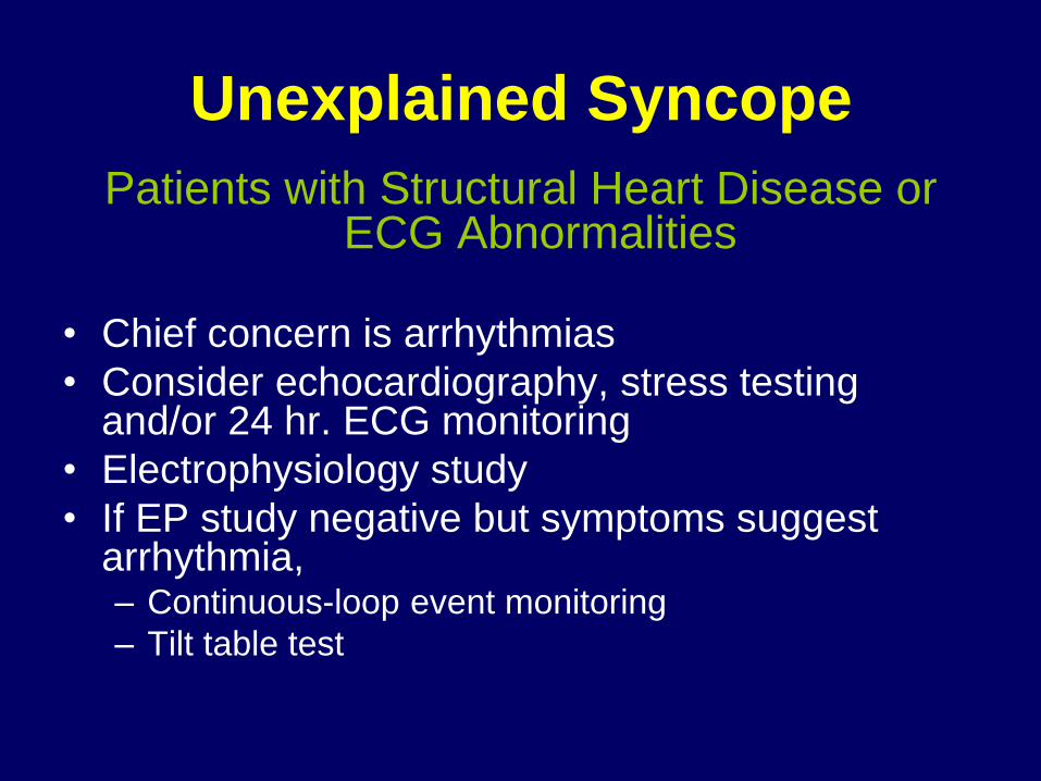
Unexplained Syncope
Patients with Structural Heart Disease or ECG Abnormalities
• Chief concern is arrhythmias
• Consider echocardiography, stress testing and/or 24 hr. ECG monitoring
• Electrophysiology study
• If EP study negative but symptoms suggest arrhythmia, – Continuous-loop event monitoring
– Tilt table test
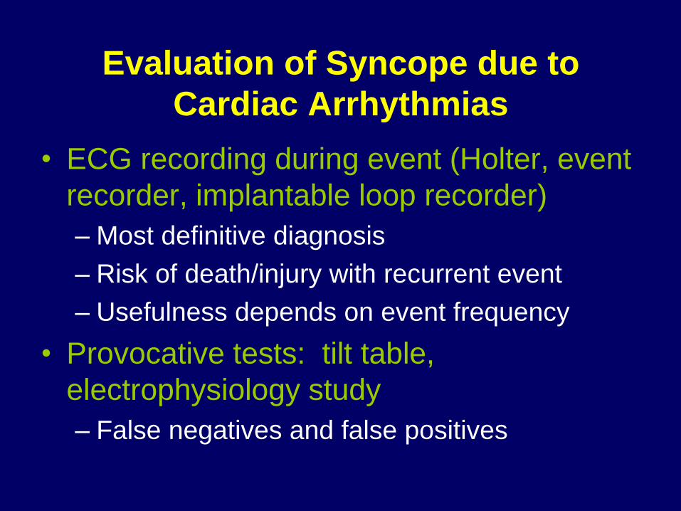
Evaluation of Syncope due to
Cardiac Arrhythmias
• ECG recording during event (Holter, event
recorder, implantable loop recorder)
– Most definitive diagnosis
– Risk of death/injury with recurrent event
– Usefulness depends on event frequency
• Provocative tests: tilt table,
electrophysiology study
– False negatives and false positives
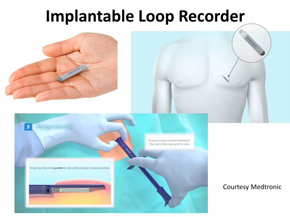
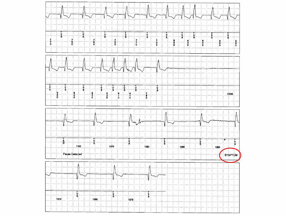
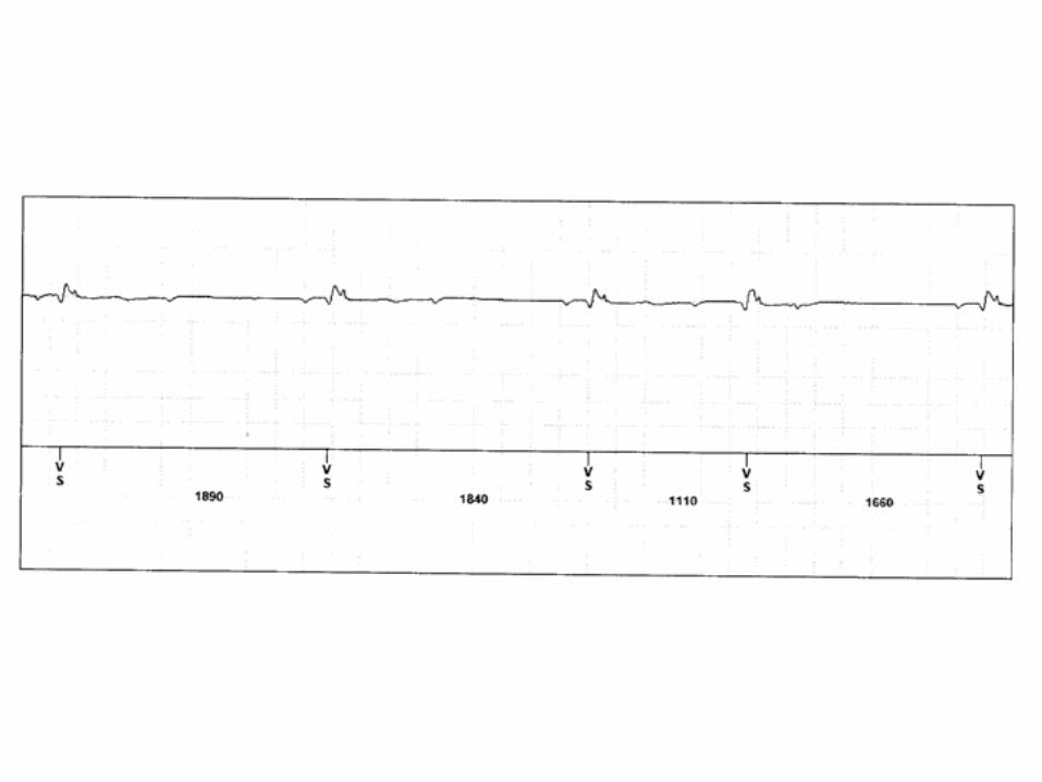
Implantable Loop Recorder
Courtesy Medtronic
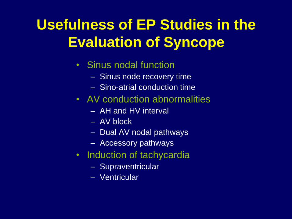
Usefulness of EP Studies in the
Evaluation of Syncope
• Sinus nodal function – Sinus node recovery time
– Sino-atrial conduction time
• AV conduction abnormalities – AH and HV interval
– AV block
– Dual AV nodal pathways
– Accessory pathways
• Induction of tachycardia – Supraventricular
– Ventricular
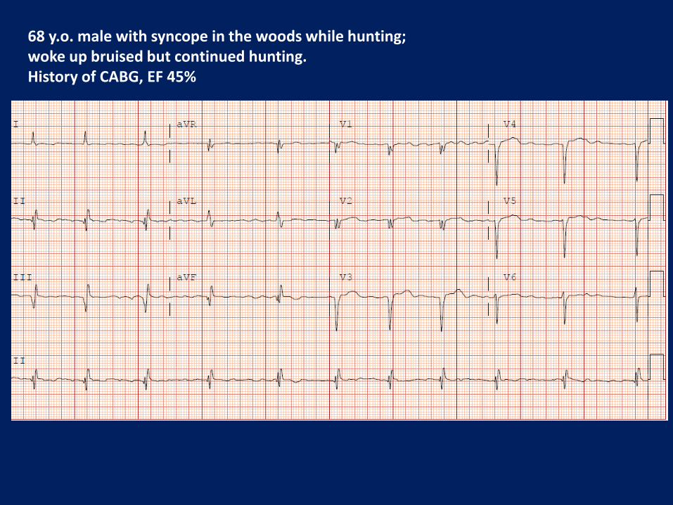
68 y.o. male with syncope in the woods while hunting; woke up bruised but continued hunting. History of CABG, EF 45%
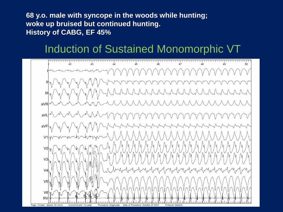
Induction of Sustained Monomorphic VT
68 y.o. male with syncope in the woods while hunting;
woke up bruised but continued hunting.
History of CABG, EF 45%
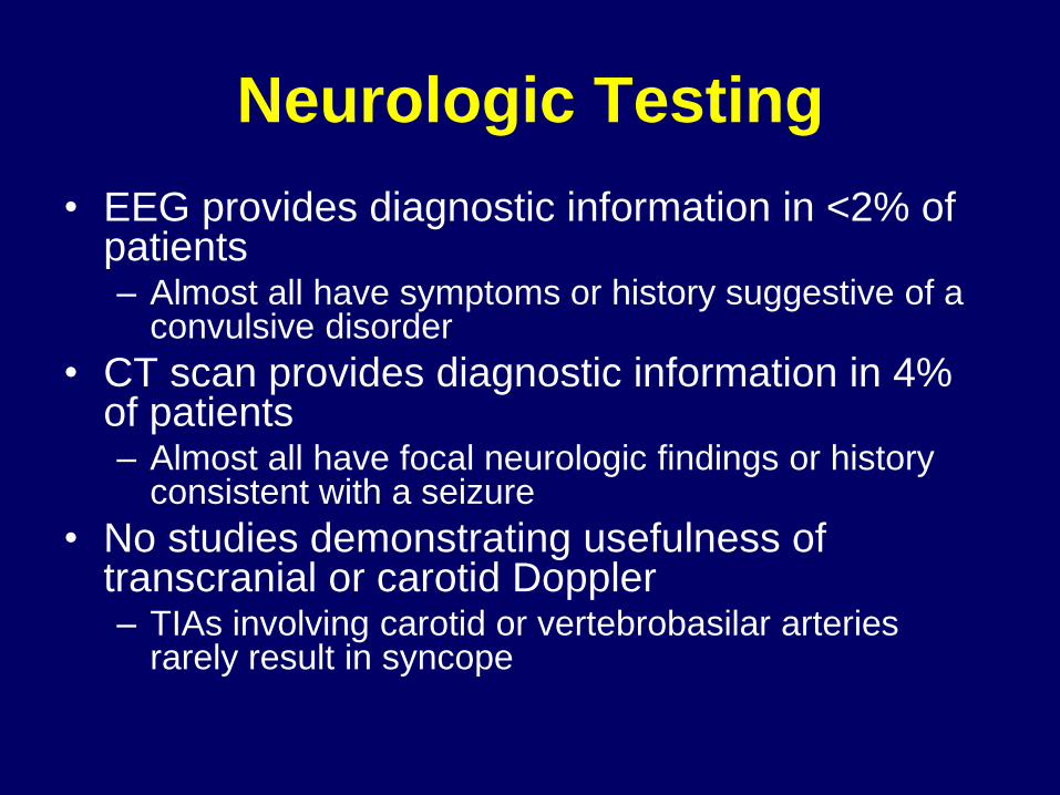
Neurologic Testing
• EEG provides diagnostic information in <2% of patients – Almost all have symptoms or history suggestive of a
convulsive disorder
• CT scan provides diagnostic information in 4% of patients – Almost all have focal neurologic findings or history
consistent with a seizure
• No studies demonstrating usefulness of transcranial or carotid Doppler – TIAs involving carotid or vertebrobasilar arteries
rarely result in syncope

EEG Makes the Diagnosis of
Arrhythmic Syncope

Treatment of Syncope
European Heart Journal (2009) 30. 2631-2671

Which one is not like the other?
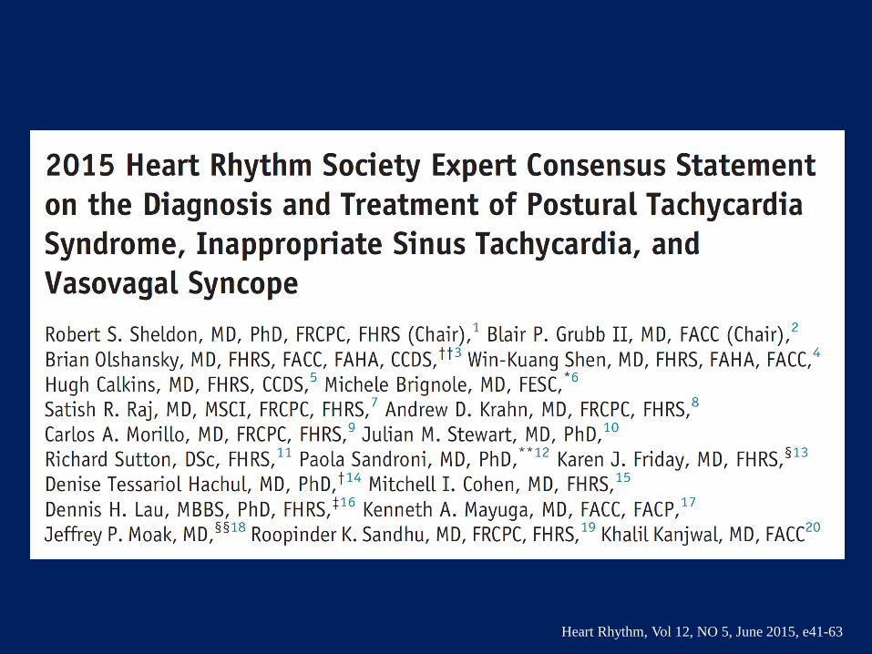
Heart Rhythm, Vol 12, NO 5, June 2015, e41-63
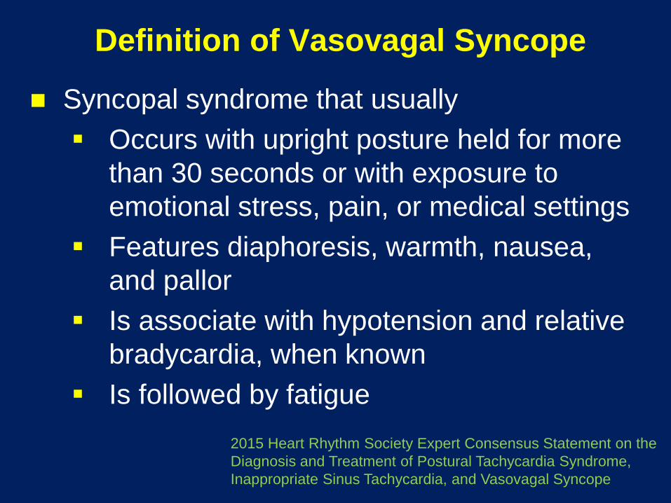
Definition of Vasovagal Syncope
Syncopal syndrome that usually
Occurs with upright posture held for more
than 30 seconds or with exposure to
emotional stress, pain, or medical settings
Features diaphoresis, warmth, nausea,
and pallor
Is associate with hypotension and relative
bradycardia, when known
Is followed by fatigue
2015 Heart Rhythm Society Expert Consensus Statement on the
Diagnosis and Treatment of Postural Tachycardia Syndrome,
Inappropriate Sinus Tachycardia, and Vasovagal Syncope

Neurally Mediated Syncope
• Exaggeration of normal physiology
• Results from autonomic nervous system
reflexes (sympathetic and
parasympathetic)
• Cardioinhibitory and vasodepressor
responses

Pathophysiology of Vasovagal Syncope
Calkins, Am J Cardiol 1999;84 (21 Oct)
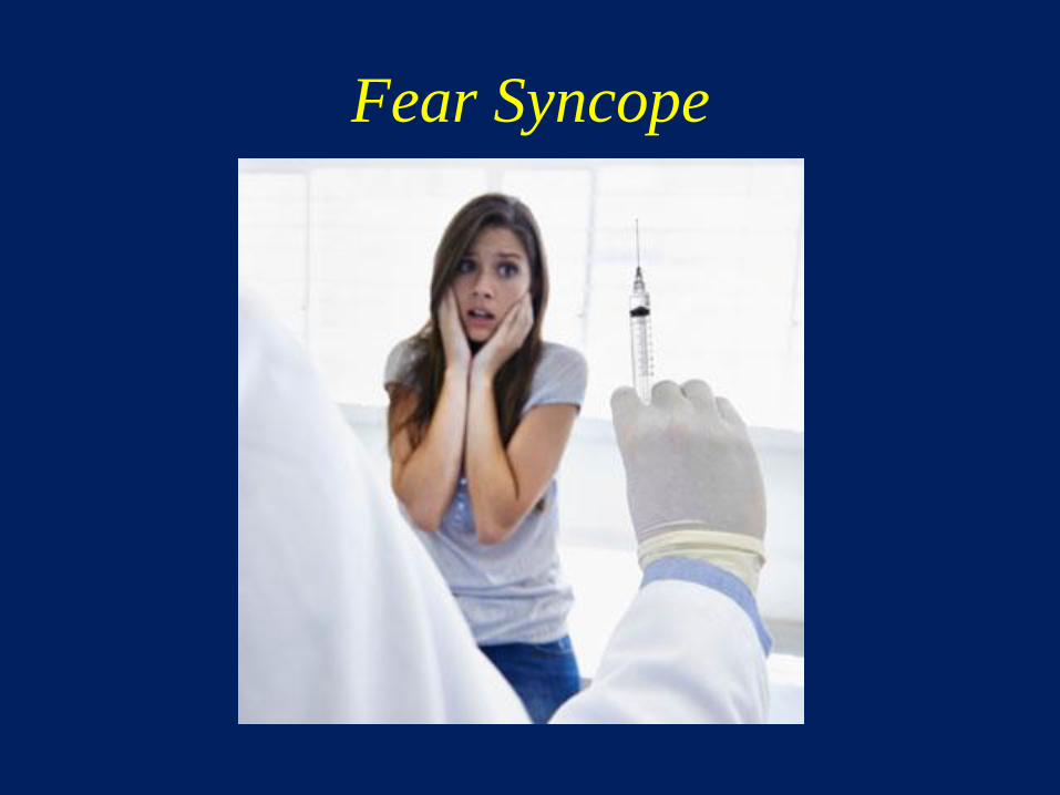
Fear Syncope
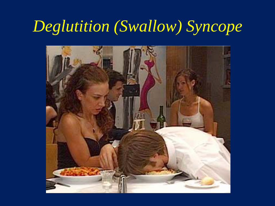
Deglutition (Swallow) Syncope

Micturation Syncope?

Micturation vs. Defecation Syncope?

Grubb, Am J Cardiol 1999;84 (21 Oct)

Tilt Testing for Neurally Mediated
Syncope
Limitations:
• False negatives and false positives (Bayes
theorem applies)
• Reproducibility
• No “gold standard”
• Tilt protocols not standardized
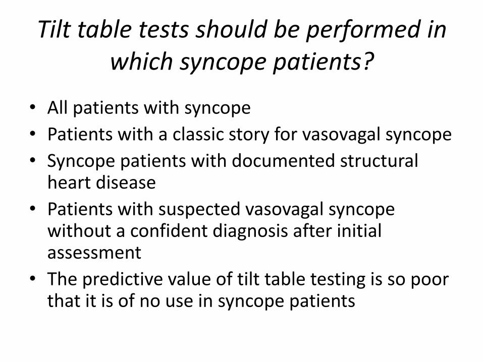
Tilt table tests should be performed in which syncope patients?
• All patients with syncope
• Patients with a classic story for vasovagal syncope
• Syncope patients with documented structural heart disease
• Patients with suspected vasovagal syncope without a confident diagnosis after initial assessment
• The predictive value of tilt table testing is so poor that it is of no use in syncope patients

Investigation of Vasovagal Syncope
Heart Rhythm, Vol 12, NO 5, June 2015, e41-63
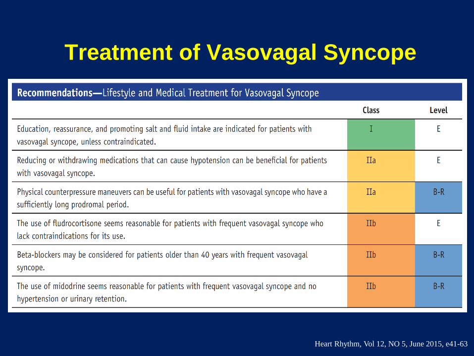
Treatment of Vasovagal Syncope
Heart Rhythm, Vol 12, NO 5, June 2015, e41-63
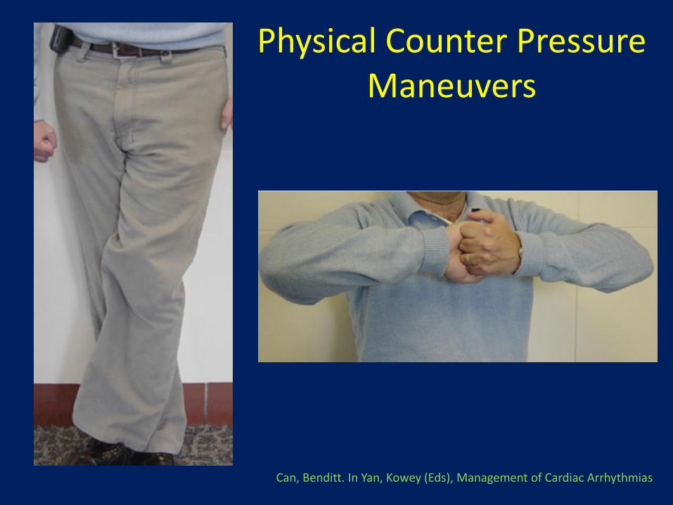
Physical Counter Pressure Maneuvers
Can, Benditt. In Yan, Kowey (Eds), Management of Cardiac Arrhythmias

Vasovagal Syncope Treatment Strategy
• For patients with only occasional syncope: – Reassure
– Stress fluid and salt intake
– Teach counterpressure maneuvers
– Do not treat patients who have not fainted in the past year
• For patients with recurrent episodes: – Begin conservatively as above
– Reduce or withdraw drugs that might cause hypotension
– Consider fludrocortisone, midodrine, or beta blockers (if older than age 40) prior to pacing, recognizing that there is no high-level evidence for their use
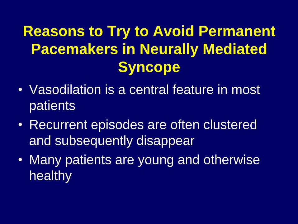
Reasons to Try to Avoid Permanent
Pacemakers in Neurally Mediated
Syncope
• Vasodilation is a central feature in most
patients
• Recurrent episodes are often clustered
and subsequently disappear
• Many patients are young and otherwise
healthy
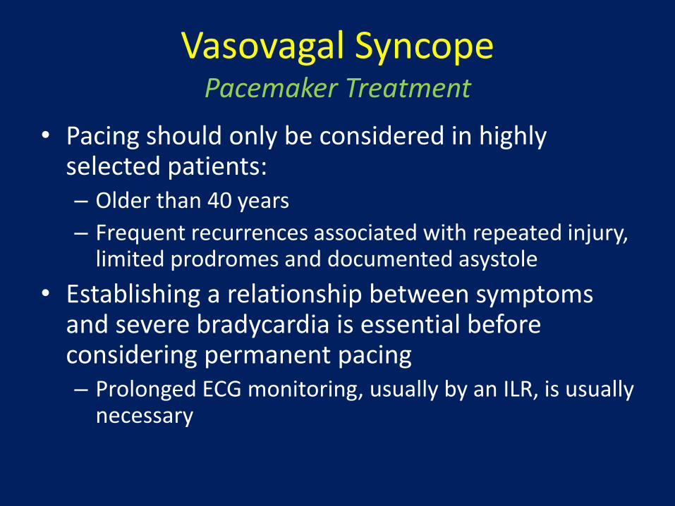
Vasovagal Syncope Pacemaker Treatment
• Pacing should only be considered in highly selected patients: – Older than 40 years
– Frequent recurrences associated with repeated injury, limited prodromes and documented asystole
• Establishing a relationship between symptoms and severe bradycardia is essential before considering permanent pacing – Prolonged ECG monitoring, usually by an ILR, is usually
necessary
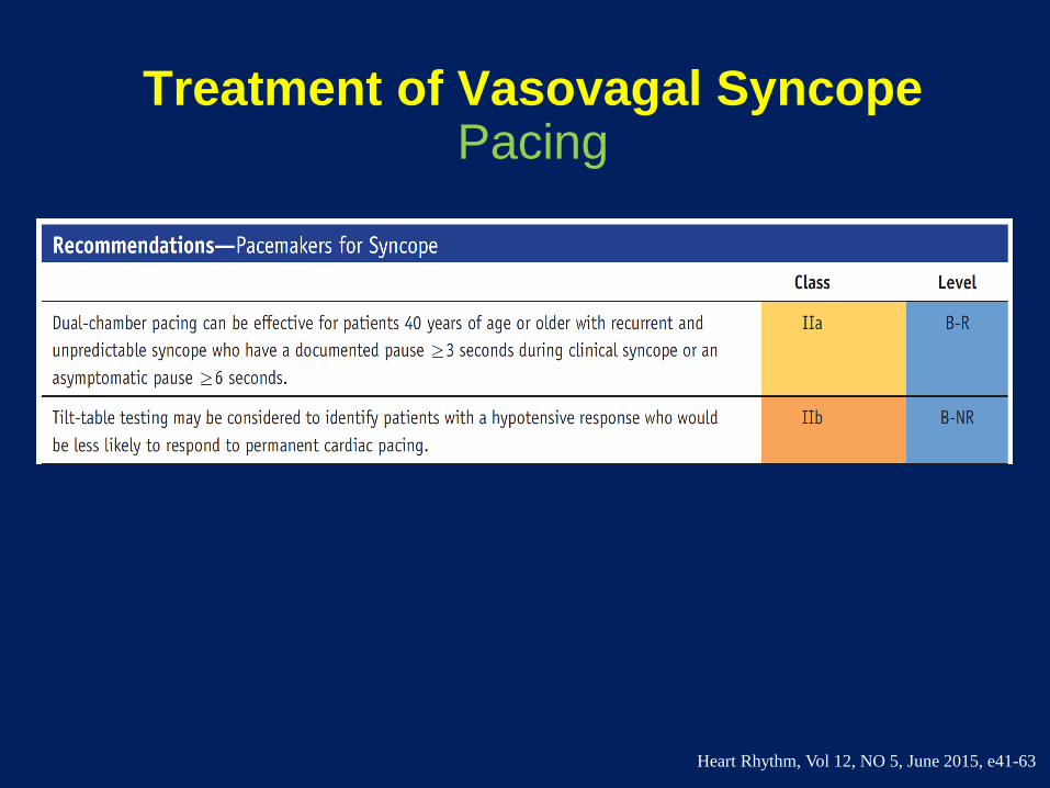
Treatment of Vasovagal Syncope Pacing
Heart Rhythm, Vol 12, NO 5, June 2015, e41-63

Diagnosis and Treatment of Syncope
Conclusions
• The initial evaluation for syncope consists of history and physical examination, including orthostatic blood pressure and ECG
• The initial evaluation may lead to
– Certain diagnosis
– Suspected diagnosis that needs to be confirmed by appropriate diagnostic tests
– No diagnosis
Brignole, M. Heart 2007;93:130-136

Diagnosis and Treatment of Syncope
Conclusions
• The strategy of evaluation varies according to: – The severity and frequency of the episodes
– The presence or absence of heart disease
• In general, the absence of heart disease excludes a cardiac cause of syncope – Conversely, the presence of heart disease
has relatively low specificity, as about half of patients with heart disease have a non-cardiac cause of syncope
Brignole, M. Heart 2007;93:130-136
Diagnosis and Treatment of Syncope
Conclusions
• Determining the mechanism of syncope is a prerequisite for:
– Advising patients with regard to prognosis
– Developing an effective mechanism-specific treatment
• Most patients with syncope require only reassurance and education regarding the nature of the disease and the avoidance of triggering events
Brignole, M. Heart 2007;93:130-136

Thank You!


