INTRODUCTION TO DENTAL HYGIENE INSTRUMENTS Dr. Shahzadi Tayyaba Hashmi [email protected].

Three dimensional transition solid elements for mesh gradation
*Syeda Ammara Shabbir1), Shahzadi Shamaila2), Rehana Sharif3), SairaRiaz4) and Shahzad Naseem5)
1), 2),3)Department of Physics, University of Engineering and Technology, Lahore,
Pakistan4),5)
Centre for Solid State Physics, University of the Punjab, Lahore
ABSTRACT
The Multiwalled carbon nanotubes (MWCNTs) thin film based electrode was fabricatedby electrophoretic deposition and modified with Nickel (Ni) Nanoparticles to fabricateNi/CNTs nanocomposite sensor for nonenzymatic glucose detection. The expensiveglassy carbon (GC) electrode is replaced by Fluorine doped tin oxide (FTO) glasscontaining CNTs film to confine the Ni nanoparticles growth by electrodepositionthrough Cyclic Voltammetry (CV). The UV-VIS and XRD analysis revealed thesuccessful deposition of Ni nanoparticles on the CNTs modified electrode. TheScanning electron microscope (SEM) confirmed the surface morphology ofelectrodeposited Ni on CNT film as uniformly dispersed particles. The electrocatalyticactivity of electrode to the glucose oxidation was investigated in alkaline medium byCyclic Voltammetry (CV) and amperometric measurements. The fabricated sensorexhibited a fast response time of less than 4s and the sensitivity 605.0 μA mM −1cm −2
with linear concentration range (0.005–3.0 mM ) having detection limit 5.0 μM. Due to simple preparation of sensor, Ni-CNTs nanocomposite electrodes are a suitablecandidate for reliable determination of glucose with good stability.
Key words: Electrophoretic deposition (EPD), Fluorine doped tin oxide (FTO) substrate,Carbon Nanotubes, Copper Nanoparticles, Non enzymatic electrocatalysis, Glucosesensor.
1. Introduction
Diabetes mellitus being an extremely extensive disease, transporting metabolicdisorders and affecting about 220 million people around the world. The mortality andmorbidity caused by hyperglycemia and insulin deficiency is imitated by higher or lowerconcentration of blood glucose as compared to the normal range (4.5 – 6.5 mM) [1].Therefore to establish a simple and rapid glucose sensing system is of significant
1)Corresponding Author

importance having high stability, sensitivity and selectivity to distinguish different levelsof glucose. [2-4].The electrochemical glucose sensors, widely used till today, areenzymatic with high sensitivity and good reliability. However, the catalytic response ofthe enzyme glucose oxidase (GOx) is sensitive to chemical reagents, pH, temperatureand humidity, causing the instability and enzyme damage. Therefore, highly selectivenonenzymatic glucose sensors, based on direct glucose oxidation on the surface ofelectrode without using enzyme are desired. The non-enzymatic biosensors [5, 6] haveattractive benefits of simple and inexpensive fabrication with good reproducibility andhigh stability. Most of these non-enzymatic glucose sensors are amperometric.The Electrode material with highly active surface area plays an important role in theglucose electroxidation. Therefore, nanomaterials such as transition metallicnanoparticles and Carbon nanotubes (CNTs) have been utilized extensively inbiosensors. CNTs are of great importance because of its high chemical stability [7-10]and remarkable physical and electrical properties. Transition metallic nanoparticles(NPs) increase the electrochemical activities due to high surface area, enhancedcatalysis and good biocompatibility. [10-12 ]. Hence it is a significant approach to usethem for nonenzymatic glucose sensors , such as Pt NPs [13,14], Cu NPs [15,16],Au NPs [17,18], Ni NPs [19], CNTs modified electrodes [20,21] and Cu NPs with CNTs[22,23] . The fast amperometric glucose detection and high sensitivity have beenreported owing to increase in electroactive surface area. This provides the moreelectron transfer during oxidation reactions of glucose. The electrode materials forglucose analysis based on Nickel NPs are of primary interest for its better catalyticactivity, detection limit, range of response and mainly stability [24]. It has been revealedthat the catalyst properties can be improved by varying the surface profile, grain sizeand texture. Therefore it is a critical requirement to produce nanosized catalysts withimproved activities [25]. Continuous efforts have been made to fabricate effectivelydispersed Ni/CNTs composites with increased catalytic activity; however, during recentyears few reports developed the nonenzymatic glucose sensor with the highlydispersed Ni/CNTs nanocomposites electrodes.In this work a new, inexpensive and facile route have been employed for the Ni/CNTscomposite fabrication for the nonenzymatic sensing of glucose and it delivered fairlyhigh sensitivity, good selectivity and reproducibility and fast current response .In fact,the fabrication of pure Ni Nanoparticles is difficult, as they provide poor stability duringelectroanalysis and easily oxidized in air [12]. In this project, Ni nanoclusters wereelectrodeposited by Cyclic voltammetry on CNTs modified electrode fabricated byelectrophoretic deposition. COOH functionalized CNTs were treated with SDS (Sodiumdodecyl sulphate) to get the most stable suspension for CNTs electrode fabricationthrough Electrophoretic deposition (EPD). The rigid control of deposition rate and filmthickness during EPD provides an excellent film uniformity and strong adhesion onelectrode surface which is not possible in drop casting and other wet chemicaltechniques for thin films fabrication [26–28]. This fabricated Ni/CNTs compositeprovides more uniform copper deposition on CNTs electrode with better sensingparameters as compared to previously published works with other techniques.

2. EXPERIMENTAL DETAILS
2.1 Reagents and materialsMWCNTs (diameter~10–20 nm, length~10 –30 μm, Purity >95%) were purchased fromJinzhou Hancheng Import & Export Co., Ltd. in China and used as received. Thechemicals, Sodium dodecyl sulphate (SDS), Boric acid (H3BO3), Nickel sulphate(NiSO4.6H2O), Nickel Chloride (NiCl2.6H2O), Sodium hydroxide (NaOH), PotassiumFerricyanide (K3 [Fe(CN)6] ) and reagents Nitric acid (HNO3) and Sulphuric acid(H2SO4)were purchased from Merck India. Deionized (DI) water was utilized throughoutthe experiment for aqueous solutions.
2.2 Preparation of Ni/CNTs/FTO nanocomposite electrode.The CNTs were carboxylic acid (COOH) functionalized through sonication at 60◦C for 6hours in HNO3/ H2SO4 (1:3, 50 ml) mixture. The acid mixture was then re-suspended inDI water following centrifugation at 8,500 rpm for 30 min. The repetitive centrifugationwas performed to achieve the neutral pH solution. The residue was then dried at 25°Cfor 24 h to yield the COOH- functionalized MWNTs (f-CNTs). To get the effective stablesuspension for EPD, f-CNTs were dissolved in aqueous micellar solutions of theanionic surfactant Sodium dodecyl sulphate (SDS). EPD was conducted using a DCpower supply by applying optimized DC voltage (35 V/cm) for 3 min deposition time atroom temperature. The working electrode was FTO substrate and the graphite used ascounter electrode. Both electrodes in the EPD cell were fixed parallel to each other. DCvoltage was applied at a constant distance between electrodes. The CNTs coated FTOsubstrate was cleaned with DI water to remove any contaminants and dried at roomtemperature. The obtained electrode was arranged for electrodeposition of copper. TheNickel (Ni) electrodeposition was carried out in Boric acid solution of Nickel sulphate(Ni2So4.6H2O) and Nickel Chloride (NiCl2.6H2O) by cyclic voltammetry. The scan ratewas 100 mV/s and performed in the potential window of 0.5 to 1.1 V. The cyclic numberwas kept 30 to achieve the high reproducibility and sensitivity of the electrodes. Thefabricated Ni/CNTs composite electrode was stored at room temperature.
2.3 Characterization Techniques:The CNTs and Ni/CNTs composite electrode was characterized by 2001 Bruker AXSdiffractometer using CuKα radiation. The absorption spectrum of both electrodes was recorded using UV-VIS spectrophotometer. The surface morphology of the modifiedelectrodes was characterized by scanning electron microscopy (SEM; JSM-6480LV).To study the electrochemical properties of the modified electrode cyclic voltammetrywas conducted using the Princeton 263A electrochemical work station having threeelectrode configurations with graphite as a counter electrode, a saturated calomelreference electrode, and CNT film electrode as working electrode.
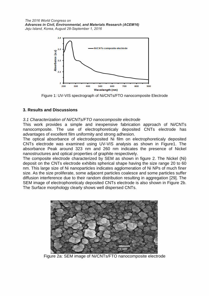
Figure 1: UV-VIS spectrograph of Ni/CNTs/FTO nanocomposite Electrode
3. Results and Discussions
3.1 Characterization of Ni/CNTs/FTO nanocomposite electrodeThis work provides a simple and inexpensive fabrication approach of Ni/CNTsnanocomposite. The use of electrophoreticaly deposited CNTs electrode hasadvantages of excellent film uniformity and strong adhesion.The optical absorbance of electrodeposited NiCNTs electrode was examined using UVabsorbance Peak around 323 nm and 260 nm indicates the presence of Nickelnanostructures and optical properties of graphite respectively.The composite electrode characterized by SEM as shown in figure 2. The Nickel (Ni)deposit on the CNTs electrode exhibits spherical shape having the size range 20 to 60nm. This large size of Ni nanoparticles indicates agglomeration of Ni NPs of much finersize. As the size proliferate, some adjacent particles coalesce and some particles sufferdiffusion interference due to their random distribution resulting in aggregation [29]. TheSEM image of electrophoreticaly deposited CNTs electrode is also shown in FiguThe Surface morphology clearly shows well dispersed CNTs.
Figure 2a: SEM image of Ni/CNTs/FTO nanocomposite electrode
VIS spectrograph of Ni/CNTs/FTO nanocomposite Electrode
3. Results and Discussions
3.1 Characterization of Ni/CNTs/FTO nanocomposite electrodeThis work provides a simple and inexpensive fabrication approach of Ni/CNTsnanocomposite. The use of electrophoreticaly deposited CNTs electrode hasadvantages of excellent film uniformity and strong adhesion.The optical absorbance of electrodeposited Ni film on electrophoreticaly depositedCNTs electrode was examined using UV-VIS analysis as shown in Figure1. Theabsorbance Peak around 323 nm and 260 nm indicates the presence of Nickelnanostructures and optical properties of graphite respectively.
composite electrode characterized by SEM as shown in figure 2. The Nickel (Ni)deposit on the CNTs electrode exhibits spherical shape having the size range 20 to 60nm. This large size of Ni nanoparticles indicates agglomeration of Ni NPs of much finer
e. As the size proliferate, some adjacent particles coalesce and some particles sufferdiffusion interference due to their random distribution resulting in aggregation [29]. TheSEM image of electrophoreticaly deposited CNTs electrode is also shown in FiguThe Surface morphology clearly shows well dispersed CNTs.
Figure 2a: SEM image of Ni/CNTs/FTO nanocomposite electrode
VIS spectrograph of Ni/CNTs/FTO nanocomposite Electrode
This work provides a simple and inexpensive fabrication approach of Ni/CNTsnanocomposite. The use of electrophoreticaly deposited CNTs electrode has
film on electrophoreticaly depositedVIS analysis as shown in Figure1. The
absorbance Peak around 323 nm and 260 nm indicates the presence of Nickel
composite electrode characterized by SEM as shown in figure 2. The Nickel (Ni)deposit on the CNTs electrode exhibits spherical shape having the size range 20 to 60nm. This large size of Ni nanoparticles indicates agglomeration of Ni NPs of much finer
e. As the size proliferate, some adjacent particles coalesce and some particles sufferdiffusion interference due to their random distribution resulting in aggregation [29]. TheSEM image of electrophoreticaly deposited CNTs electrode is also shown in Figure 2b.
Figure 2a: SEM image of Ni/CNTs/FTO nanocomposite electrode

Figure 2b: SEM image of CNTs/FTO modified electrode
The Ni/CNTs nanocomposite crystal structure was confirmed by using XRDdetermination (Figure 3). The X ray diffraction pattern indicates three peaks at 49.43,55.23, and 76.42, corresponding to the planes {111}, {200}, and {220}, respectively,corresponds to the FCC Ni lattice. The peak at 33.05 is assigned to the [002] plane ofthe CNTs [30]. These results clearly show that the Ni nanoparticles have beensuccessfully electrodeposited on the CNTs modified electrodes. Based on theexperimental results and Scherrer formula, the average crystallite size of Ni wascalculated to be 8 nm.
Figure 3: X-ray diffraction Pattern of Ni/CNTs/FTO nanocomposite Electrode

Therefore, the Ni nanoparticles of size 20-60 nm, perceived from the SEM images wereassembled by the average Ni Nanocrystalline size of 8 nm. The growth of Ninanoparticles on CNTs modified substrates provides Ni/CNTs nanocomposites havingporous structure. Thus providing a perfect interface for utilizing in nonenzymaticelectrochemical biosensor.
3.2 Electrochemical Characterization of Ni/CNTs/FTO electrode.Figure 4 shows the comparison of Cyclic voltammograms of CNTs modified FTOsubstrate with Ni/CNTs modified FTO in 0.5 mM K3Fe(CN)6 and 0.1 M KCl at 50 mV/s.The reversible one electron redox behavior of Ferricyanide was observed. Thevoltammetric behavior of Ferricyanide redox couple on the CNTs electrode found dueto the high aspect ratio and surface with electrocatalytic activity. For the Ni/CNTsnanocomposite electrode, the cyclic voltammetric response was comparable to that ofthe CNTs electrode. The voltammograms showed that Ni was efficiently immobilized onCNTs coated FTO surface providing the essential conduction paths to promote thetransfer of electron at the interface of electrode and analyte like an electrode atnanoscale. With the Ni nanoparticles deposition on CNTs modified electrode, Peakcurrent Ip has been increased and Peak separation (ΔEp) decreased as compared tothe CNTs modified electrode. This indicates that the Ni nanoclusters have increasedthe electrocatalytic active surface area.The active surface area was calculated from the Randles–Sevcik equation (Ip =2.69×10⁵ n3/2ACν1/2D1/2), where Ip is the Redox peak current (A), n is the electrontransfer number (= 1), A is the surface area of the electrode (cm2), D is the diffusioncoefficient (0.76 × 10⁻⁵ cm2/s), C is the concentration of electroactive species(K3Fe(CN)6,M/L), and ν is the scan rate (V/s). The calculated active surface areas ofCNTs modified FTO and Ni/CNTs/FTO was 0.75 and 1.60 cm2. These results clearlyshowed the increased active surface area of Ni/CNTs nanocomposite electrode.3.3 Non enzymatic Electrocatalysis of glucose at the Ni/CNTs/FTO electrode.
Figure 4: Cyclic Voltammetry Curves of CNTs and Ni/CNTs/FTO electrode at scan rateof 50 mV/s

The electrocatalytic response of the CNTs/FTO and Ni/CNTs/FTO electrode towardsglucose oxidation in alkaline media was examined by Cyclic voltammetry in 0.1 MNaOH solution at scan rate of 20 mV/s in the presence and absence of 0.5 mM glucoseas shown in Figure 5. The potential range was selected from 0.0 to 0.8 V versussaturated calomel electrode (SCE). The requirement of alkaline medium to improve theeffect of Ni for glucose oxidation is acknowledged. Figure shows that in the glucoseabsence, no Redox peaks appears from electrophoreticaly deposited CNTs electrodeand Ni/CNTs composites electrode. A predominant increase in the reaction peakcurrent was detected upon addition of glucose for Ni/CNTs composite as a workingelectrode. The well-defined pair of redox peak currents at 0.68 and 0.25 are observedfrom the voltammograms (d) corresponding to the glucose oxidation peaks for Ni (II)/Ni(III) [37-40]. Redox peaks from the transition of Ni (II) and Ni (III) have been consideredas a mediator for electron transfer. These results indicate that Ni nanoparticles exhibitexcellent electrocatalytic response towards glucose oxidation and CNTs electrodeprovides the large surface area and a high conductivity to increase the electron transferrate. The detection potential was found 0.59 V.Cyclic voltammetry plot of Ni/CNTs nanocomposite electrode in the presence ofglucose (0.1 M NaOH) solution at altered scan rates 20 mV/s, 60 mV/s, 80 mV/s, 100mV/s, and 150 mV/s is shown in Figure 6. The increasing scan rates resulted in shiftingthe redox peak currents to higher values with more peak to peak separation. Figureshowed the linear variation of cathodic and anodic peak currents with increased squareroot of scan rates demonstrating that the electrochemical reactions are surfacecontrolled by the adsorption of glucose molecule. The correlation coefficients R2 0.985and 0.989 for anodic and cathodic peaks, respectively have been observed.
Figure 5: Cyclic Voltammetry of CNT electrode (Curves a and b) and Ni/CNT electrode(curves c and d) before (curves a and c) and after (Curves b and d) adding 0.5mM
glucose in 0.1mM NaOH solution at scan rate of 20 mV/s
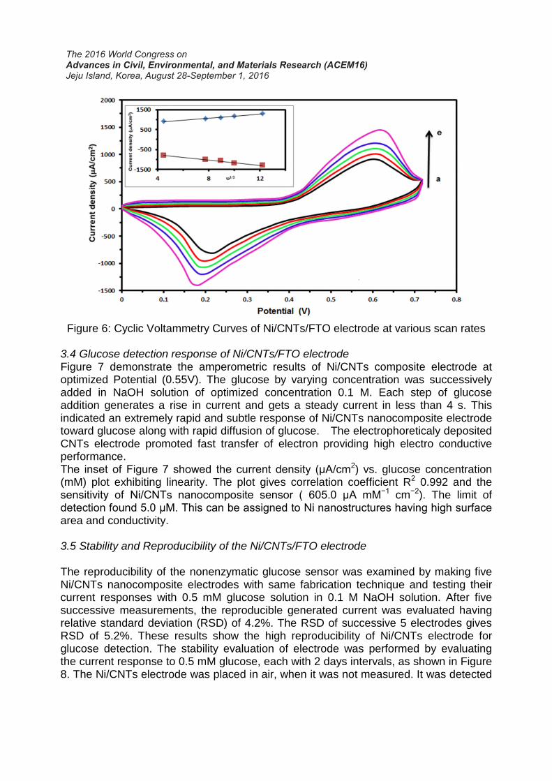
Figure 6: Cyclic Voltammetry Curves of Ni/CNTs/FTO electrode at various scan rates
3.4 Glucose detection response of Ni/CNTs/FTO electrodeFigure 7 demonstrate the amperometric results of Ni/CNTs composite electrode atoptimized Potential (0.55V). The glucose by varying concentration was successivelyadded in NaOH solution of optimized concentration 0.1 M. Each step of glucoseaddition generates a rise in current and gets a steady current in less than 4 s. Thisindicated an extremely rapid and subtle response of Ni/CNTs nanocomposite electrodetoward glucose along with rapid diffusion of glucose. The electrophoreticaly depositedCNTs electrode promoted fast transfer of electron providing high electro conductiveperformance.The inset of Figure 7 showed the current density (μA/cm2) vs. glucose concentration(mM) plot exhibiting linearity. The plot gives correlation coefficient R2 0.992 and thesensitivity of Ni/CNTs nanocomposite sensor ( 605.0 μA mM−1 cm−2). The limit ofdetection found 5.0 μM. This can be assigned to Ni nanostructures having high surface area and conductivity.
3.5 Stability and Reproducibility of the Ni/CNTs/FTO electrode
The reproducibility of the nonenzymatic glucose sensor was examined by making fiveNi/CNTs nanocomposite electrodes with same fabrication technique and testing theircurrent responses with 0.5 mM glucose solution in 0.1 M NaOH solution. After fivesuccessive measurements, the reproducible generated current was evaluated havingrelative standard deviation (RSD) of 4.2%. The RSD of successive 5 electrodes givesRSD of 5.2%. These results show the high reproducibility of Ni/CNTs electrode forglucose detection. The stability evaluation of electrode was performed by evaluatingthe current response to 0.5 mM glucose, each with 2 days intervals, as shown in Figure8. The Ni/CNTs electrode was placed in air, when it was not measured. It was detected
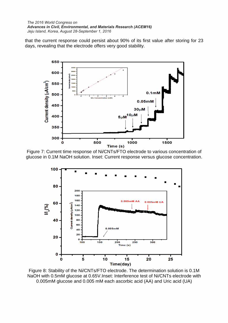
that the current response could persist about 90% of its first value after storing for 23days, revealing that the electrode offers very good stability.
Figure 7: Current time response of Ni/CNTs/FTO electrode to various concentration ofglucose in 0.1M NaOH solution. Inset: Current response versus glucose concentration.
Figure 8: Stability of the Ni/CNTs/FTO electrode. The determination solution is 0.1MNaOH with 0.5mM glucose at 0.65V.Inset: Interference test of Ni/CNTs electrode with
0.005mM glucose and 0.005 mM each ascorbic acid (AA) and Uric acid (UA)
that the current response could persist about 90% of its first value after storing for 23
Figure 7: Current time response of Ni/CNTs/FTO electrode to various concentration ofglucose in 0.1M NaOH solution. Inset: Current response versus glucose concentration.
Figure 8: Stability of the Ni/CNTs/FTO electrode. The determination solution is 0.1MNaOH with 0.5mM glucose at 0.65V.Inset: Interference test of Ni/CNTs electrode with
0.005mM glucose and 0.005 mM each ascorbic acid (AA) and Uric acid (UA)

Some easily oxidized synergetic compounds such as ascorbic acid (AA) and uric acid(UA) gives interfering current signals which are the major encounters in thenonenzymatic detection of glucose. Although these intrusive species of AA (0.002 mM)and UA (0.002 mM) are much lower in concentration than glucose (3– 8 mM) in anordinary biological sample, they generate electrochemical current values analogous toglucose. This might be due to more transfer of electrons [13]. The Inset of Figure 8demonstrated the measurements upon addition of 0.005 mM interfering species withcontinuous addition of glucose in 0.1 M NaOH solution. A significant glucose responsewas detected as compared to glucose thereby neglecting the effect of interferingspecies. This demonstrates that the fabricated Ni/CNTs nanocomposite electrode isprecisely specific towards glucose regardless of the intrusive species.This work provides high sensitivity, fast response, low detection limit and good stabilityas compared to previously reported works [31-36]. In Comparison to other Ni/CNTsnanocomposite electrodes, the demonstrated advantages of the Ni deposition on theuniform and homogeneous CNT films showed that these CNT films played highlyimportant role in glucose electrocatalytic oxidation performance. The improvedperformance can also be attributed to synergistic electrocatalytic behavior of the CNTfilms towards glucose oxidation [31]. This work offers a simple and a promisingapproach to functionalize the CNT thin films for possible application.
Conclusion
The Ni nanoparticles have been electrodeposited by Cyclic Voltammetry on the CNTsfilm prepared on FTO substrate by electrophoretic deposition, constructing a non-enzymatic amperometric glucose biosensor. The prepared Ni/CNTs/FTO electrodeexhibits fast response of less than 4 s, high stability, good reproducibility and excellentsensitivity 605.0 μA mM−1 cm −2 with wide linear range (0.005–3.5 mM) and lowdetection limit (5 μM). This value of sensitivity is higher in comparison to previously reported Ni electrodeposited nonenzymatic glucose sensors, demonstrating theincrease in electrocatalytic active surface area by homogenous deposition of Ninanoparticles on uniformly coated CNTs substrate. The glassy carbon electrode canbe replaced with inexpensive FTO substrate for effective glucose determination withlow detection limit, low over potential and high sensitivity. This project offers apromising approach to incorporate the CNTs thin films, prepared by Electrophoreticdeposition, for potential application. Furthermore nanocomposites Ni/Cu/CNT are inprocess of fabrication by same technique for more sensitive nonenzymatic glucosedetection.
Acknowledgement
This work was supported by University of Engineering and Technology, Lahore,Pakistan. The authors are highly grateful to Department of Physics, GCU, Lahore, andCentre of Advanced Studies in Physics (CASP), GCU, Lahore for providing assistancein XRD and SEM analysis respectively.

References
Ricard Prehn , Montserrat Cortina-Puig and Francesc Xavier Munoz, Journal of TheElectrochemical Society. 159 (2012) F134-F139.
Feihong Menga,Wei Shia, Yanan Suna, Xuan Zhub, Guisen Wua, Changqing Ruana,Xin Liua, Dongtao Gea, Biosensors and Bioelectronics.42 (2013) 141-147.
Bansi D. Malhotraa, Asha Chaubeyb, S.P. Singha, Analytica Chimica Acta, 578 (2006)59-74.
Joseph Wang,Chemical Reviews.108 (2008) 814-825.Sejin Park, Sun Young Lee,Hankil Boo, Hyun-Mi Kim,Ki-Bum Kim, et al, Chemistry in
Materials.19(2007) 3373-3375.Yipeng Sun, Harvey Buck, and Thomas E. Mallouk, Analytical Chemistry. 73 (2001)
1599-1604.Hongtao Zhao, Huangxian Ju, Analytical Biochemistry. 350 (2006) 138–144 .Arben Merkoc¸ Martin Pumera, Xavier Lopis, Briza Perez, Manel del Valle, Salvador
Alegret, Trends in Analytical Chemistry. 24 (2005) 9.Vasilis G. Gavalasa, Stacy A. Lawa, J. Christopher Balla, Rodney Andrewsb, Leonidas
G. Bachasa, Analytical Biochemistry.329 (2004)247-252.Yuehe Lin , Xiaoli Cui1, Xiangrong Ye, Electrochemistry communications.7 (2005) 267-
274.M.L. Mena, P. YanezSedeno, J.M. Pingarron, Analytical Biochemistry.336 (2005)20-27.Christine M. Welch, Richard G. Compton, Analytical and Bioanalytical Chemistry.384
(2006) 601-619.Sejin Park, Taek Dong Chung and Hee Chan Kim, Analytical Chemistry. 75 (2003)
3046-3049.Hankil Boo, Sejin Park, Bonkyung Ku,Yunmee Kim, Jin Hyung Park, Hee Chan Kim
and Taek Dong Chung, American Chemical Society.126 (2004) 4524-4525.Innocenzo G Casella , Maria Gatta, Maria R Guascito, Tommaso R.I Cataldi, Analytica
Chimica Acta. 357 (1997) 63-71.Qin Xu, Yu Zhao, Jin Zhong Xu, Jun-Jie Zhu, Sensors and Actuators B. 8694 (2005)1-8.Masato Tominaga, Toshihiro Shimazoe, Makoto Nagashima, Isao Taniguchi,
Electrochemistry communications.7 (2005) 189-193.Masato Tominaga, Toshihiro Shimazoe, Makoto Nagashima, Hideaki Kusuda,
Atsushi Kubo, Yutaka Kuwahara and Isao Taniguchi, ElectroanalyticalChemistry.590 (2006) 37-46.
Tianyan You ,Osamu Niwa , Zilin Chen , Katsuyoshi Hayashi , Masato Tomita andShigeru Hirono , Analytical chemistry. 75(2003) 5191-5196.
JianShan Yea, Ying Wenb, Wei De Zhangc, Leong Ming Ganc, Guo Qin Xub, FwuShanSheua,, Electrochemistry communications. 6 (2004) 66-70.
Randhir P. Deo, Joseph Wang, Electrochemistry communications. 6 (2004) 284-287.Keith B Malea, Sabahudin Hrapovica, Yali Liua, Dashan Wangb, John H.T Luonga,
Analytica Chimica Acta.516 (2004) 35-41.Joseph Wang, Gang Chen, Mei Wang and Madhu Prakash Chatrathia, Analyst.129
(2004)512-515.Shamsipur, Mojtaba, Mostafa Najafi, and Mohammad-Reza Milani Hosseini,
Bioelectrochemistry 77.2 (2010): 120-124.

Yanhui Xu, Xiangqin Lin, Electrochimica Acta. 52 (2007) 5140-5149.Laxmidhar Besra, Meilin Liu, Progress in Materials Science. 52 (2007)1–61.I ZHITOMIRSKY, L GALOR,Materials in Medicine. 8 (1997)213-219.I. Zhitomirsky, J. European Ceramic Society. 18 (1998)849–856.Linyou Cao , Peng Diao , Tao Zhu and Zhongfan Liu , J. Physical Chemistry B.108
(2004) 3535-3239.M. Terrones, W.K. Hsu, A. Schilder, H. Terrones et al. , Applied Physics A. 66 (1998)
307-317.Sun, Aili, Jianbin Zheng, and Qinglin Sheng. Electrochimica Acta 65 (2012): 64-69.Bittencourt, C., et al. Surface science 601.13 (2007): 2800-2804.Rather, Sami-ullah, and Kee Suk Nahm. Materials Research Bulletin 49 (2014): 525-
530.Hsieh, Chien-Te, Yun-Wen Chou, and Wei-Yu Chen. Journal of Solid State
Electrochemistry 12.6 (2008): 663-669.Zhu, Z. G., et al. Sensors and Actuators B: Chemical 178 (2013): 586-592.Lin, Kuan-Yu, Wen-Ta Tsai, and Jeng-Kuei Chang. International journal of hydrogen
energy 35.14 (2010): 7555-7562.Lu, Li-Min, et al. Biosensors and Bioelectronics 25.1 (2009): 218-223.Salimi, Abdollah, and Mahmoud Roushani. Electrochemistry Communications 7.9
(2005): 879-887.Ai, Hanhua, et al. Biosensors and Bioelectronics 24.4 (2008): 1048-1052.Wang, Yulong, et al. Sensors and Actuators B: Chemical 151.1 (2010): 65-70.