Switch from translation to RNA replication in a positive...
13
Switch from translation to RNA replication in a positive-stranded RNA virus Andrea V. Gamarnik and Raul Andino 1 Department of Microbiology and Immunology, University of California, San Francisco, California 94143-0414 USA In positive-stranded viruses, the genomic RNA serves as a template for both translation and RNA replication. Using poliovirus as a model, we examined the interaction between these two processes. We show that the RNA polymerase is unable to replicate RNA templates undergoing translation. We discovered that an RNA structure at the 5* end of the viral genome, next to the internal ribosomal entry site, carries signals that control both viral translation and RNA synthesis. The interaction of this RNA structure with the cellular factor PCBP up-regulates viral translation, while the binding of the viral protein 3CD represses translation and promotes negative-strand RNA synthesis. We propose that the interaction of 3CD with this RNA structure controls whether the genomic RNA is used for translation or RNA replication. [Key Words: PCBP; positive-stranded virus; RNA replication; translational control; viral polymerase] Received March 26, 1998; revised version accepted June 2, 1998. The genomes of positive-stranded RNA viruses are im- portant in at least three major processes: They act as mRNAs to direct the synthesis of viral proteins; they serve as templates for genome replication; and they are packaged along with structural proteins during viral as- sembly. The balance between these processes must be properly maintained to allow efficient viral proliferation. Thus, once the viral RNA-dependent RNA polymerase and other essential proteins are synthesized, the geno- mic RNA must be used as a template for negative-strand RNA replication (Pogue et al. 1994). In principle, this creates a conflict between the translation and replication machinery: While the ribosomes are moving along the viral RNA in the 58 to 38 direction, the polymerase ini- tiates replication at the 38 end of the same RNA and moves in the opposite direction as it synthesizes the complementary negative strand. It is not known whether the arrangement of ribosomes and polymerases on the RNA template allows both processes to occur simulta- neously or whether translation and RNA replication in- terfere with each other. To examine the interplay between translation and RNA replication, we have used poliovirus as a model, because both in vitro and in vivo systems are available to dissect the viral cycle (Molla et al. 1991; Barton and Flanegan 1993; Gamarnik and Andino 1996). Several ex- periments have suggested that the highly conserved 58- untranslated region (58UTR) of the poliovirus genome plays an important role in the regulation of both trans- lation and RNA replication (Rohll et al. 1994). Two func- tional domains have been defined within this region: a short 58-terminal element involved in RNA replication (Andino et al. 1990a, 1993; Harris et al. 1994; Roehl et al. 1997) and a longer element, termed the internal ribo- somal entry site (IRES), involved in viral translation (Jang et al. 1988; Pelletier et al. 1988; Trono et al. 1988). It was originally thought that these two elements were independent; however, recent evidence suggests a func- tional overlap between them (Simoes and Sarnow 1991; Borman et al. 1994; Shiroki et al. 1995). The regulatory function of the 58 UTR is probably me- diated through its interactions with cellular and viral proteins (Ehrenfeld and Semler 1995; Jackson and Ka- minski 1995; Belsham and Sonenberg 1996). The short 58-terminal element of the 58 UTR folds into a cloverleaf- like structure and forms a ribonucleoprotein complex with the cellular poly(C)-binding proteins 1 and 2 (re- ferred to as PCBP in this report, but also known as hnRNP E or aCP), and the viral protein 3CD, which is the uncleaved precursor of the viral protease (3C pro ) and polymerase (3D pol ) (Gamarnik and Andino 1997; Parsley et al. 1997). Mutations that disrupt the formation of this ribonucleoprotein complex impair viral RNA replication (Andino et al. 1990a; Rohll et al. 1994). On the other hand, the IRES element enables ribosomes to internally enter the RNA without scanning from the 58 end (Jang et 1 Corresponding author. E-MAIL [email protected]; FAX (415) 476-0939. GENES & DEVELOPMENT 12:2293–2304 © 1998 by Cold Spring Harbor Laboratory Press ISSN 0890-9369/98 $5.00; www.genesdev.org 2293 Cold Spring Harbor Laboratory Press on June 5, 2018 - Published by genesdev.cshlp.org Downloaded from
Transcript of Switch from translation to RNA replication in a positive...

Switch from translation to RNAreplication in a positive-strandedRNA virus
Andrea V. Gamarnik and Raul Andino1
Department of Microbiology and Immunology, University of California, San Francisco, California 94143-0414 USA
In positive-stranded viruses, the genomic RNA serves as a template for both translation and RNA replication.Using poliovirus as a model, we examined the interaction between these two processes. We show that theRNA polymerase is unable to replicate RNA templates undergoing translation. We discovered that an RNAstructure at the 5* end of the viral genome, next to the internal ribosomal entry site, carries signals thatcontrol both viral translation and RNA synthesis. The interaction of this RNA structure with the cellularfactor PCBP up-regulates viral translation, while the binding of the viral protein 3CD represses translation andpromotes negative-strand RNA synthesis. We propose that the interaction of 3CD with this RNA structurecontrols whether the genomic RNA is used for translation or RNA replication.
[Key Words: PCBP; positive-stranded virus; RNA replication; translational control; viral polymerase]
Received March 26, 1998; revised version accepted June 2, 1998.
The genomes of positive-stranded RNA viruses are im-portant in at least three major processes: They act asmRNAs to direct the synthesis of viral proteins; theyserve as templates for genome replication; and they arepackaged along with structural proteins during viral as-sembly. The balance between these processes must beproperly maintained to allow efficient viral proliferation.Thus, once the viral RNA-dependent RNA polymeraseand other essential proteins are synthesized, the geno-mic RNA must be used as a template for negative-strandRNA replication (Pogue et al. 1994). In principle, thiscreates a conflict between the translation and replicationmachinery: While the ribosomes are moving along theviral RNA in the 58 to 38 direction, the polymerase ini-tiates replication at the 38 end of the same RNA andmoves in the opposite direction as it synthesizes thecomplementary negative strand. It is not known whetherthe arrangement of ribosomes and polymerases on theRNA template allows both processes to occur simulta-neously or whether translation and RNA replication in-terfere with each other.
To examine the interplay between translation andRNA replication, we have used poliovirus as a model,because both in vitro and in vivo systems are available todissect the viral cycle (Molla et al. 1991; Barton andFlanegan 1993; Gamarnik and Andino 1996). Several ex-
periments have suggested that the highly conserved 58-untranslated region (58UTR) of the poliovirus genomeplays an important role in the regulation of both trans-lation and RNA replication (Rohll et al. 1994). Two func-tional domains have been defined within this region: ashort 58-terminal element involved in RNA replication(Andino et al. 1990a, 1993; Harris et al. 1994; Roehl et al.1997) and a longer element, termed the internal ribo-somal entry site (IRES), involved in viral translation(Jang et al. 1988; Pelletier et al. 1988; Trono et al. 1988).It was originally thought that these two elements wereindependent; however, recent evidence suggests a func-tional overlap between them (Simoes and Sarnow 1991;Borman et al. 1994; Shiroki et al. 1995).
The regulatory function of the 58 UTR is probably me-diated through its interactions with cellular and viralproteins (Ehrenfeld and Semler 1995; Jackson and Ka-minski 1995; Belsham and Sonenberg 1996). The short58-terminal element of the 58 UTR folds into a cloverleaf-like structure and forms a ribonucleoprotein complexwith the cellular poly(C)-binding proteins 1 and 2 (re-ferred to as PCBP in this report, but also known ashnRNP E or aCP), and the viral protein 3CD, which isthe uncleaved precursor of the viral protease (3Cpro) andpolymerase (3Dpol) (Gamarnik and Andino 1997; Parsleyet al. 1997). Mutations that disrupt the formation of thisribonucleoprotein complex impair viral RNA replication(Andino et al. 1990a; Rohll et al. 1994). On the otherhand, the IRES element enables ribosomes to internallyenter the RNA without scanning from the 58 end (Jang et
1Corresponding author.E-MAIL [email protected]; FAX (415) 476-0939.
GENES & DEVELOPMENT 12:2293–2304 © 1998 by Cold Spring Harbor Laboratory Press ISSN 0890-9369/98 $5.00; www.genesdev.org 2293
Cold Spring Harbor Laboratory Press on June 5, 2018 - Published by genesdev.cshlp.orgDownloaded from
al. 1988; Pelletier et al. 1988; Trono et al. 1988; Chen andSarnow 1995). The mechanism by which the translationapparatus recognizes IRES sequences is unknown, but ithas been proposed that many canonical initiation factorsas well as other specific cellular proteins participate inthe process (Meyer et al. 1995; Pestova et al. 1996). Cur-rently, three noncanonical factors that bind to the IREShave been identified: polypyrimidine tract binding pro-tein (PTB; Hellen et al. 1993); La autoantigen (Meero-vitch et al. 1993); and PCBP (Blyn et al. 1996). Interest-ingly, PCBP appears to participate in both viral transla-tion and RNA replication (Blyn et al. 1996, 1997;Gamarnik 1997; Parsley et al. 1997).
We have analyzed the interplay between poliovirustranslation and RNA replication and the contribution ofspecific ribonucleoprotein complexes to the regulationof both processes. We show that the viral polymerase isunable to use the genomic RNA as a template for RNAsynthesis while it is being used by translating ribosomes.We found that the cloverleaf RNA at the 58 end of theviral genome is a bifunctional element involved in theregulation of both viral translation and RNA replication.The binding of the cellular protein PCBP to the clover-leaf enhances viral translation, while the binding of theviral protein 3CD represses translation and facilitatesnegative-strand synthesis. Thus, we propose that over-lapping translation and replication signals within thecloverleaf function as a strategy to coordinate the use ofthe genomic RNA for translation or RNA replication.
Results
Actively translating ribosomes inhibit the elongationactivity of the poliovirus RNA polymerase 3Dpol
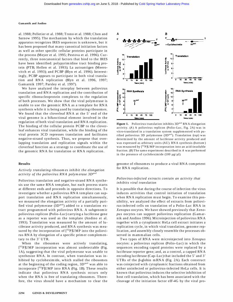
Poliovirus translation and negative-strand RNA synthe-sis use the same RNA template, but each process startsat different ends and proceeds in opposite directions. Toinvestigate whether a poliovirus RNA template can sup-port translation and RNA replication simultaneously,we measured the elongation activity of a partially puri-fied viral polymerase (3Dpol) added to a translation ex-tract programmed with poliovirus RNA. A subgenomicpoliovirus replicon (Polio–Luc) carrying a luciferase geneas a reporter was used as the template (Andino et al.1993). Translation was measured by the amount of lu-ciferase activity produced, and RNA synthesis was mea-sured by the incorporation of [32P]UMP into the poliovi-rus RNA by elongation of a specific primer complemen-tary to the 38 UTR.
When the ribosomes were actively translating,[32P]UMP incorporation was almost undetectable (Fig.1A), suggesting that the viral polymerase was unable tosynthesize RNA. In contrast, when translation was in-hibited by cycloheximide, which stalled the ribosomesat the beginning of the coding region, 3Dpol was able toincorporate [32P]UMP into RNA (Fig. 1B). These resultsindicate that poliovirus RNA synthesis occurs onlywhen the RNA is free of translating ribosomes. There-fore, the virus should have a mechanism to clear the
genome of ribosomes to produce a viral RNA competentfor RNA replication.
Poliovirus-infected extracts contain an activity thatinhibits viral translation
It is possible that during the course of infection the virusinduces activities that control initiation of translationwhen RNA replication must begin. To examine this pos-sibility, we analyzed the effect of extracts from poliovi-rus-infected cells on translation of a Polio–Luc RNA inXenopus oocytes. We have showed previously that Xeno-pus oocytes can support poliovirus replication (Gamar-nik and Andino 1996). Microinjection of poliovirus RNAtogether with a cytoplasmic HeLa cell extract initiates areplication cycle, in which viral translation, genome rep-lication, and assembly closely resemble the processes ob-served in mammalian cells.
Two types of RNA were microinjected into Xenopusoocytes: a poliovirus replicon (Polio–Luc) in which thesequences encoding capsid proteins were replaced by aluciferase reporter gene; and, as a control, a capped RNAencoding luciferase (Cap–Luc) that included the 58 and 38UTRs of the b-globin mRNA (Fig. 2A). Each constructwas coinjected with cytoplasmic fractions obtained fromeither uninfected or poliovirus-infected HeLa cells. It isknown that poliovirus induces the selective inhibition ofhost cell translation, which has been associated with thecleavage of the initiation factor eIF-4G by the viral pro-
Figure 1. Poliovirus translation inhibits 3Dpol RNA elongationactivity. (A) A poliovirus replicon (Polio–Luc, Fig. 2A) was invitro-translated in a translation system supplemented with pu-rified poliovirus 3D polymerase (3Dpol). Translation (top) wasdetermined by the amount of luciferase activity produced andwas expressed as arbitrary units (AU). RNA synthesis (bottom)was measured by [32P]UMP incorporation into an acid-insolublefraction. (B) The same experiment described in A was performedin the presence of cycloheximide (100 µg/µl).
Gamarnik and Andino
2294 GENES & DEVELOPMENT
Cold Spring Harbor Laboratory Press on June 5, 2018 - Published by genesdev.cshlp.orgDownloaded from

tease 2Apro (for review, see Mathews 1996). As expected,crude S10 extracts from infected HeLa cells strongly in-hibited cap-dependent translation, but stimulated Polio–Luc RNA translation by 30% (Fig. 2B). Interestingly, afurther purified fraction obtained from poliovirus-in-fected cells (HeLa S100) contained an activity thatstrongly inhibited viral translation (Fig. 2B, right). Thisnegative effect was specific for poliovirus cap-indepen-dent translation, because Cap–Luc RNA was efficientlytranslated after coinjection with either infected or unin-fected S100 HeLa cell extracts (Fig. 2B, left).
Because the inhibitory effect on translation was ob-served only with injection of infected extracts, the in-hibitory activity must involve either a viral factor or avirally modified cellular factor. To characterize this ac-tivity further, we fractionated the infected S100 extractby chromatography on a HiTrap SP column. Part of theinhibitory activity was recovered in the flowthrough,while a larger part was retained in the column and elutedat 250 mM KCl (Fig. 2C). As observed with the total ex-tract, microinjection of the fractions had no effect ontranslation of Cap–Luc RNA (Fig. 2C). Then, we ana-lyzed whether the fractions showing inhibitory activity
contained any viral protein. Western blot analysis re-vealed that the inhibitory activity copurified with theviral proteins 3Dpol, 3CD, and partially with their pre-cursor P3 (Fig. 2D), but not with other viral proteins (datanot shown). Interestingly, 3Dpol is the RNA-dependentRNA polymerase that is synthesized as a fusion proteinwith 3Cpro, a protease that also binds to specific se-quences of the viral 58 UTR (Andino et al. 1990b, 1993).
The viral protein 3CD represses poliovirus translation
To determine whether any of the 3D-containing proteinswere responsible for the inhibitory effect on translation,we took advantage of the RNA-binding properties of3Cpro. Affinity chromatography with an immobilizedRNA was used to deplete 3CD from the most activecolumn fractions. The treated fraction retained most ofthe 3Dpol protein but <10% of the original amounts of3CD and P3 (Fig. 3A, left). Significantly, the depletedfraction lacked the ability to repress viral translationwhen microinjected into Xenopus oocytes (Fig. 3A,right), suggesting that the 3Cpro domain is required forthis inhibitory activity.
Figure 2. Poliovirus-infected cell extracts contain an activitythat specifically inhibits poliovirus translation. (A) Schematicrepresentation of the chimeric poliovirus luciferase RNA (Po-lio–Luc) and capped luciferase RNA (Cap–Luc). In Polio–Luc,the coding region of the poliovirus capsid proteins was replacedby the luciferase reporter gene, and a cleavage site for 2Apro hasbeen introduced between luciferase and 2Apro (represented bythe arrow). The Cap–Luc RNA consists of the luciferase geneflanked by the 58 and 38 uncoding regions of the b-globinmRNA. (B) Microinjection of infected S100 HeLa cell extractinto Xenopus oocytes specifically inhibits poliovirus cap-inde-pendent translation. Polio–Luc or Cap–Luc RNA was injectedinto oocytes together with uninfected (open bars) or poliovirus-infected S10 or S100 HeLa cell fractions (solid bars) as indicatedin each case. Luciferase activity was determined in oocytes after3 hr of incubation at 22°C and expressed in arbitrary units (AU).(C) Elution profile of the translation inhibitory activity afterion-exchange chromatography. Infected S100 HeLa cell extractwas loaded onto a HiTrap SP column (Pharmacia) and elutedwith a KCl gradient, as indicated at right. The translation in-hibitory activity was determined by coinjection of 20 nl of eachfraction (1–14) together with 5 nl of HeLa S10 (to provide thecellular factor essential for poliovirus translation in oocytes,PTF) and 20 ng of Polio–Luc RNA (j) or Cap–Luc RNA (h) intooocytes. Luciferase activity was determined in oocyte extractsafter 3 hr of incubation at 22°C and expressed in AU. (D) Viralproteins 3Dpol, 3CD, and P3 copurified with the viral transla-tion inhibitory activity. Western blot analysis of fractions 1–14eluted from the HiTrap SP column is shown. Two microliters ofeach fraction was resolved in a 10% SDS–polyacrylamide gel,transferred to nitrocelluose membrane, and probed with specificanti-3CD antibodies. The electrophoretic mobility of P3, 3CD,and 3D is indicated at left.
Poliovirus translational control
GENES & DEVELOPMENT 2295
Cold Spring Harbor Laboratory Press on June 5, 2018 - Published by genesdev.cshlp.orgDownloaded from

We next determined whether 3CD alone is sufficientto inhibit poliovirus translation. Four different proteinswere expressed in Xenopus oocytes by preinjection ofsynthetic mRNAs encoding the corresponding polypep-tide: 3Cpro, 3Dpol, a mutated 3CD with an alteration atthe cleavage site between 3Cpro and 3Dpol (Gln 182 to
Asn; Andino et al. 1993) that completely eliminates theautoproteolytic processing of the precursor 3CD; and anunrelated mRNA control encoding green fluorescentprotein (GFP). Because expression of each proteinreached the highest level between 10 and 15 hr afterinjection, as monitored by Western blot analysis (datanot shown), we injected Polio–Luc and Cap–Luc RNA 15hr after injection of the mRNAs. While translation ofCap–Luc RNA proceeded normally in oocytes expressing3CD (Fig. 3B, right), the translation of Polio–Luc RNAwas inhibited by 60% (Fig. 3B, left). In contrast, none ofthe other preinjections (3C, 3D, or GFP) had a significanteffect on luciferase expressed by either Cap–Luc or Po-lio–Luc. Taken together, these results strongly suggestthat the protease–polymerase fusion 3CD specifically in-hibits viral cap-independent translation.
The cloverleaf RNA controls poliovirus translation
Because 3CD is a known RNA-binding protein thatbinds to the cloverleaf domain of the poliovirus 58 UTR,we reasoned that 3CD might exert its inhibitory effectby interacting with this or other regulatory RNA ele-ments. We have demonstrated previously that 3CD in-teracts specifically with the isolated cloverleaf to form aternary ribonucleoprotein complex with a ribosome-as-sociated cellular factor, PCBP (Andino et al. 1993;Gamarnik and Andino 1997; Parsley et al. 1997); but itwas unknown whether 3CD interacts with other regionsof the viral RNA. To examine other possible sites of 3CDinteractions with the viral UTRs, we performed mobil-ity-shift experiments using several defined domains ofthe poliovirus RNA as probes. The results obtained in-dicated that 3CD binds only to the cloverleaf RNA (A.Gamarnik and R. Andino, in prep.).
To charaterize further the regulatory role of 3CD andthe cloverleaf, we examined whether the cloverleaf RNAdirectly participates in poliovirus translation. We haveshown previously that disrupting the interaction of 3CDwith the cloverleaf RNA affects positive-strand RNAsynthesis without impairing viral translation (Andino etal. 1993). In that previous study, we observed a smallenhancement of translation for mutants in which 3CDwas unable to interact with the cloverleaf RNA. Thosedifferences were originally interpreted as insignificant.However, because the results presented here stronglyimplicate 3CD in translational control, and because ithas been shown previously that PCBP is a positive regu-lator of poliovirus translation (Gamarnik and Andino1997; Parsley et al. 1997), we re-examined the impor-tance of these RNA–protein interactions in the transla-tion process. To this end, we designed Polio–Luc con-structs containing cloverleaf mutations that specificallydisrupted the binding of either 3CD or PCBP.
The polymerase precursor 3CD binds to stem–loop Dof the cloverleaf RNA, whereas PCBP specifically inter-acts with stem–loop B (Fig. 4A) (Gamarnik and Andino1997; Parsley et al. 1997). Three types of mutant RNAswere constructed: one with the entire cloverleaf deleted(DCL); a second type in which the interaction of the RNA
Figure 3. The polymerase–protease precursor, 3CD, repressesviral translation. (A) Depletion of 3CD from infected cell ex-tracts correlates with loss of translation inhibition. The viralprotein 3CD was depleted from a partially purified infectedHeLa fraction by affinity chromatography by use of an immo-bilized cloverleaf RNA (see Materials and Methods). (Left) Twomicroliters of 3CD-depleted extract (lane 2) and 2 µl of a non-depleted control (lane 1) were subjected to Western blot analysisas described for Fig. 2C. (Right) Luciferase activity was deter-mined in oocyte extracts 3 hr after coinjection of Polio–LucRNA with buffer, nondepleted control, or 3CD-depleted frac-tions as indicated in the bottom. (B) Overexpression of mutated3CD (Gln-182 → Asn) in Xenopus oocytes inhibits poliovirustranslation. (Top) Schematic diagram of the microinjection pro-tocol. Oocytes were injected with 4 ng of a capped RNA encod-ing for 3CD, 3C, 3D, or an unrelated RNA encoding GFP, andincubated at 17°C for 15 hr. Then, oocytes were microinjected asecond time with 40 ng of Polio–Luc or Cap–Luc RNA. Lucif-erase expression in oocytes was measured by enzymatic activity6 hr after the second microinjection. Translation of Polio–Lucand Cap–Luc RNA was determined in oocytes that were prein-jected with the mRNAs or with buffer control (−) as indicated atthe bottom. Luciferase activity was expressed in arbitrary units(AU).
Gamarnik and Andino
2296 GENES & DEVELOPMENT
Cold Spring Harbor Laboratory Press on June 5, 2018 - Published by genesdev.cshlp.orgDownloaded from

with PCBP was either abolished by a 4-nucleotide dele-tion at the top of stem–loop B (LB.14) or reduced by asubstitution in stem B (SB.212); and a third type in whichthe interaction of 3CD with the RNA was partially dis-rupted by a 4-nucleotide insertion at the top of stem–loop D (LD.73).
Translation efficiencies of wild-type and mutant Po-lio–Luc RNAs were evaluated by measurement of lucif-erase activity produced as a function of time after trans-fection into HeLa cells or microinjection into Xenopusoocytes. These experiments were carried out under con-ditions in which luciferase activity was produced only bythe input RNA. For the transfections into HeLa cells,luciferase activity was measured prior to RNA replica-tion (Andino et al. 1993), while in Xenopus oocytes, theamount of newly synthesized RNA was negligible incomparison to the injected RNA. The Polio–Luc RNAconstruct with the deleted cloverleaf (DCL) or with themutation that abolished PCBP binding (LB.14) translatedat 10% of the efficiency of wild type (Fig. 4B). The Polio–Luc mutant with reduced binding to PCBP (SB. 212)translated at 40% of the efficiency of wild type. In con-trast, the mutant with a deficiency in 3CD-cloverleafinteraction (LD.73) showed a substantial increase in viraltranslation (Fig. 4B). These results suggest that the bind-ing site for PCBP within the cloverleaf structure is nec-essary for efficient viral translation. The involvement of
the cloverleaf in translation was first postulated by Si-moes and Sarnow (1991). In agreement with our results,these authors reported a poliovirus mutant with a 6-nucleotide insertion at the top of stem–loop B, whichresulted in a significant decrease in viral translation (Si-moes and Sarnow 1991). Furthermore, the increase oftranslation that we observed with LD.73 suggests thatthe inhibitory effect of 3CD may involve its binding tothe cloverleaf structure.
The role of 3CD and PCBP in viral translation wasevaluated further by competition experiments. We mi-croinjected an excess of wild-type or mutated cloverleafcompetitor together with the Polio–Luc reporter con-struct into Xenopus oocytes. We hypothesized that thefree cloverleaf RNAs would interact with 3CD and/orPCBP, sequestering the proteins from their normal func-tion in translation. Indeed, when wild-type or stem–loopD mutant RNA decoys were coinjected with Polio–Luc,we observed an 80% inhibition of luciferase production(Fig. 4C). Because both decoys have intact PCBP-bindingsites, this result suggests further that PCBP is requiredfor efficient translation. In contrast, a cloverleaf com-petitor carrying the stem–loop B mutation (unable tobind PCBP but fully capable of binding 3CD) did notdecrease but rather stimulated viral translation, presum-ably by titrating out 3CD expressed by the Polio–LucRNA. Taken together, these results indicate that the
Figure 4. The cloverleaf structure formed atthe 58 end of the viral genome controls viraltranslation. (A) Schematic representation ofthe ribonucleoprotein complex formedaround the cloverleaf RNA. The predictedcloverleaf structure is composed of stem–loopB (nucleotides 10–34), stem–loop C (nucleo-tides 35–45), and stem–loop D (nucleotides51–78). Viral factor 3CD and cellular proteinPCBP are shown interacting with their spe-cific target sequences. The locations of themutations introduced into the cloverleafstructure of the Polio–Luc RNAs are indi-cated by arrows: LB.14 (nucleotides 23–26,CCCA, were deleted in loop B); SB.212(nucleotides 14–16, GGG, and nucleotides28–30, CCC, were replaced with AAA andUUU, respectively, which maintain the stemB structure); and LD.73 (nucleotides GUACwere inserted in position 70 of loop D). (B)Luciferase activity produced by Polio–Lucconstructs carrying wild-type or mutated clo-verleaf structures. In vitro-transcribed Polio–Luc RNAs were either transfected into HeLacells (top) or microinjected into Xenopusoocytes (bottom). The RNAs are indicatedas WT (wild-type), DCL (cloverleaf-deleted),LB.14 (loop B muted), SB.212 (stem B mu-tated), and LD.73 (loop D mutant). Luciferase
activity was measured in HeLa cell extracts 2 hr after electroporation and in oocyte extracts 10 hr after injection, and expressed inarbitrary units (AU). (C) Microinjection of decoy cloverleaf RNAs into Xenopus oocytes interferes with poliovirus translation. Wild-type Polio-Luc RNA was coinjected with buffer (−), 30 ng of wild-type cloverleaf (WT), or 30 ng of mutant cloverleaf decoys (LB,nucleotides C23 to A26 deleted, or LD, nucleotides GUAC inserted in position 70). Luciferase activity was determined in oocyteextracts 10 hr after injection.
Poliovirus translational control
GENES & DEVELOPMENT 2297
Cold Spring Harbor Laboratory Press on June 5, 2018 - Published by genesdev.cshlp.orgDownloaded from
cloverleaf is a bifunctional element: In addition to itspreviously described function in RNA replication, itplays a central role in the regulation of poliovirus trans-lation.
Synthesis of poliovirus negative-strand RNAin Xenopus oocytes
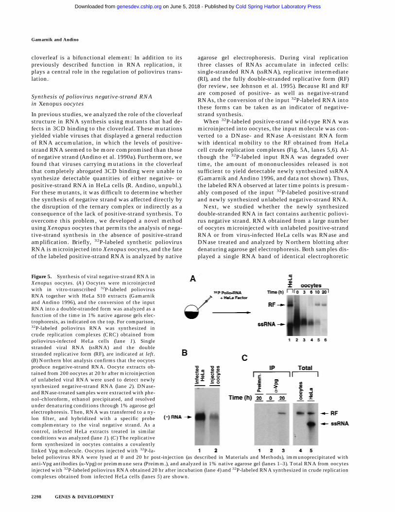
In previous studies, we analyzed the role of the cloverleafstructure in RNA synthesis using mutants that had de-fects in 3CD binding to the cloverleaf. These mutationsyielded viable viruses that displayed a general reductionof RNA accumulation, in which the levels of positive-strand RNA seemed to be more compromised than thoseof negative strand (Andino et al. 1990a). Furthermore, wefound that viruses carrying mutations in the cloverleafthat completely abrogated 3CD binding were unable tosynthesize detectable quantities of either negative- orpositive-strand RNA in HeLa cells (R. Andino, unpubl.).For these mutants, it was difficult to determine whetherthe synthesis of negative strand was affected directly bythe disruption of the ternary complex or indirectly as aconsequence of the lack of positive-strand synthesis. Toovercome this problem, we developed a novel methodusing Xenopus oocytes that permits the analysis of nega-tive-strand synthesis in the absence of positive-strandamplification. Briefly, 32P-labeled synthetic poliovirusRNA is microinjected into Xenopus oocytes, and the fateof the labeled positive-strand RNA is analyzed by native
agarose gel electrophoresis. During viral replicationthree classes of RNAs accumulate in infected cells:single-stranded RNA (ssRNA), replicative intermediate(RI), and the fully double-stranded replicative form (RF)(for review, see Johnson et al. 1995). Because RI and RFare composed of positive- as well as negative-strandRNAs, the conversion of the input 32P-labeled RNA intothese forms can be taken as an indicator of negative-strand synthesis.
When 32P-labeled positive-strand wild-type RNA wasmicroinjected into oocytes, the input molecule was con-verted to a DNase- and RNase A-resistant RNA formwith identical mobility to the RF obtained from HeLacell crude replication complexes (Fig. 5A, lanes 5,6). Al-though the 32P-labeled input RNA was degraded overtime, the amount of mononucleosides released is notsufficient to yield detectable newly synthesized ssRNA(Gamarnik and Andino 1996, and data not shown). Thus,the labeled RNA observed at later time points is presum-ably composed of the input 32P-labeled positive-strandand newly synthesized unlabeled negative-strand RNA.
Next, we studied whether the newly synthesizeddouble-stranded RNA in fact contains authentic poliovi-rus negative strand. RNA obtained from a large numberof oocytes microinjected with unlabeled positive-strandRNA or from virus-infected HeLa cells was RNase andDNase treated and analyzed by Northern blotting afterdenaturing agarose gel electrophoresis. Both samples dis-played a single RNA band of identical electrophoretic
Figure 5. Synthesis of viral negative-strand RNA inXenopus oocytes. (A) Oocytes were microinjectedwith in vitro-transcribed 32P-labeled poliovirusRNA together with HeLa S10 extracts (Gamarnikand Andino 1996), and the conversion of the inputRNA into a double-stranded form was analyzed as afunction of the time in 1% native agarose gels elec-trophoresis, as indicated on the top. For comparison,32P-labeled poliovirus RNA was synthesized incrude replication complexes (CRC) obtained frompoliovirus-infected HeLa cells (lane 1). Singlestranded viral RNA (ssRNA) and the doublestranded replicative form (RF), are indicated at left.(B) Northern blot analysis confirms that the oocytesproduce negative-strand RNA. Oocyte extracts ob-tained from 200 oocytes at 20 hr after microinjectionof unlabeled viral RNA were used to detect newlysynthesized negative-strand RNA (lane 2). DNase-and RNase-treated samples were extracted with phe-nol–chloroform, ethanol precipitated, and resolvedunder denaturing conditions through 1% agarose gelelectrophoresis. Then, RNA was transferred to a ny-lon filter, and hybridized with a specific probecomplementary to the viral negative strand. As acontrol, infected HeLa extracts treated in similarconditions was analyzed (lane 1). (C) The replicativeform synthesized in oocytes contains a covalentlylinked Vpg molecule. Oocytes injected with 32P-la-beled poliovirus RNA were lysed at 0 and 20 hr post-injection (as described in Materials and Methods), immunoprecipitated withanti-Vpg antibodies (a-Vpg) or preimmune sera (Preimm.), and analyzed in 1% native agarose gel (lanes 1–3). Total RNA from oocytesinjected with 32P-labeled poliovirus RNA obtained 20 hr after incubation (lane 4) and 32P-labeled RNA synthesized in crude replicationcomplexes obtained from infected HeLa cells (lanes 5) are shown.
Gamarnik and Andino
2298 GENES & DEVELOPMENT
Cold Spring Harbor Laboratory Press on June 5, 2018 - Published by genesdev.cshlp.orgDownloaded from

mobility that hybridized specifically with a polioviruspositive-strand RNA probe (Fig. 5B).
To characterize further the RF RNA produced in oo-cytes, we examined whether this molecule contains Vpg,a genome-linked viral peptide. During RNA replication,Vpg is added to the 58 end of the growing RNA chains ata very early stage, possibly as a primer of RNA synthesis.This protein is found in both positive- and negative-strand RNAs, suggesting that a similar mechanism isresponsible for initiation of synthesis of both strands(Flanegan et al. 1977; Pettersson et al. 1978). Therefore,in infected cells, the RF RNA contains Vpg linked to the58 end of both strands. However, the RF synthesized afterone round of replication in oocytes should only carry Vpgattached to the 58 end of the negative strand, because thepositive strand is the product of T7 RNA polymerasetranscription. As expected, phenol- and SDS-treateddouble-stranded RNA produced in oocytes was immuno-precipitated by antibodies directed against Vpg but notby pre-immune sera (Fig. 5C, lanes 1,3). In addition, anti-Vpg antibodies did not precipitate 32P-labeled RNA atearly time points before double-stranded RNA was ob-served (Fig. 5C, lane 2), showing that the input ssRNAcannot be precipitated by anti-Vpg antibodies. These re-sults indicate that the double-stranded RNA formed inoocytes contains negative-strand RNA covalently linked
to Vpg, which closely resembles that produced in HeLainfected cells.
Binding of 3CD to the cloverleaf RNA is requiredfor negative-strand RNA synthesis
Using the method described in the previous section, wedetermined whether negative-strand RNA synthesis isaffected by mutations that completely disrupt 3CD in-teraction with the cloverleaf. Two mutants were used(schematically represented in Fig. 6A), one in which thecloverleaf structure was modified by altering 2 bp at thetop of stem–loop D (Polio-315), and the other with analteration in the putative RNA-binding domain of 3CD,which was unable to bind to the cloverleaf RNA butdisplayed normal proteolysis (Polio-181). These mutantsyielded no virus after transfection into HeLa cells, indi-cating that the mutation severely compromised viralreplication.
We first studied the ability of these mutants to directtranslation in the oocyte system. The amount of lucifer-ase activity produced by the mutants during the first 2 hrpostinjection was similar to that of wild type (Fig. 6B). Incontrast, at 4 and 8 hr post-injection, both mutatedRNAs were translated at higher levels than the wild-typeRNA, once again indicating that the inability of 3CD to
Figure 6. The interaction between 3CD and the cloverleaf RNA is required for negative strand RNA synthesis. (A) Schematicrepresentation of wild-type poliovirus genome (Polio-WT), a mutant in the cloverleaf structure in which two Us in positions 60 and68 were replaced by Cs to disrupt stem–loop D (Polio-315), and a mutant in 3CD-coding sequence in which the Asp-85 was replacedby Glu, which abrogates RNA binding (Polio-181). (B) Disrupting 3CD–cloverleaf interaction increases viral translation. Oocytes weremicroinjected with Polio–Luc WT (black bars), Polio–Luc 315 (white bars), or Polio-Luc 181 (gray bars) RNAs and incubated at 22°C.Cytoplasmic extracts were obtained, and luciferase activity was measured at the indicated times (0.5, 2, 4, and 8 hr). (C) Disruptionof 3CD–cloverleaf interaction abolishes negative-strand RNA synthesis. Oocytes were microinjected with 32P-labeled Polio–WT (lanes1–5), mutant Polio-315 (lanes 6–10), or mutant Polio-181 (lanes 11–15) and incubated at 30°C. Total RNA was extracted at 0 , 2, 4, 8,and 12 hr, and analyzed on 1% agarose gels. For comparison, 32P-labeled poliovirus RNA was synthesized in crude replicationcomplexes (CRC) obtained from poliovirus-infected HeLa cells (lane 16). Double-stranded (ds) replicative form and single-stranded (ss)poliovirus RNAs are indicated.
Poliovirus translational control
GENES & DEVELOPMENT 2299
Cold Spring Harbor Laboratory Press on June 5, 2018 - Published by genesdev.cshlp.orgDownloaded from

interact with the cloverleaf structure results in an in-crease in protein synthesis.
We next examined negative-strand RNA synthesis inboth wild type and mutants. When 32P-labeled wild-typeRNA was microinjected into oocytes, the input mol-ecules were readily converted to an RF form (Fig. 6C,lanes 4,5). However, the RNAs corresponding to the mu-tants that were defective in 3CD binding to the clover-leaf were unable to synthesize RF (lanes 9,10,14,15), in-dicating that the interaction of 3CD with the cloverleafRNA not only down-regulates viral translation but isessential for negative-strand RNA synthesis.
Discussion
The replication of positive-stranded RNA virusespresents an unresolved conundrum: How is the negative-strand RNA synthesized in the face of a wave of trans-lating ribosomes moving in the opposite direction? Wehave studied this problem in the context of poliovirusreplication and have found that actively translating ribo-somes prevent RNA synthesis. Because each molecule ofgenomic RNA must be used for translation prior to RNAreplication (Kuge et al. 1986; Collis et al. 1992; Novakand Kirkegaard 1994), the virus should have a mecha-nism to down-regulate translation to begin RNA synthe-sis. We found that an RNA element at the 58 end of theviral genome, the cloverleaf RNA, contains overlappingsignals for translation and RNA replication. The bindingof the cellular protein PCBP to this RNA greatly en-hances viral translation, while the binding of the viralpolymerase precursor, 3CD, represses viral translationand promotes the synthesis of negative-strand RNA. Wepropose that these RNA–protein interactions determinethe switch from translation to RNA replication.
Role of the cloverleaf RNA in translationand RNA replication
The results described here suggest that the cloverleafcoordinates the use of the viral RNA as a template fortranslation or RNA replication. We propose that afterviral entry, the genomic RNA interacts with translationinitiation factors to begin protein synthesis. Once a criti-cal concentration of viral proteins is reached, 3CD bindsto the cloverleaf RNA, shuts down viral translation andpromotes negative-strand synthesis.
We examined the effects of mutations in the 3CDbinding site in the cloverleaf or in the RNA-binding do-main in 3CD on translation and negative-strand RNAsynthesis. The results showed that the mutated viralRNAs, while translating more efficiently than wild type,do not accumulate negative-strand RNA (Fig. 6). Thisphenotype could result from the inability of these mu-tants to shut down translation, as predicted by ourmodel. However, on the basis of the dramatic effect onnegative-strand synthesis, it seems more likely that thecloverleaf participates in both repression of translationand initiation of RNA synthesis.
The cloverleaf structure was originally described as an
element required for positive-strand RNA synthesis.This conclusion was drawn from the study of mutantswith defects either in 3CD or in the cloverleaf that de-bilitated complex formation, which reduced the ratio ofpositive- to negative-strand RNA accumulated in in-fected cells (Andino et al. 1990a). In that previous study,we did not analyze nonviable mutants (unable to form3CD–cloverleaf complexes) because of the lack of a sen-sitive method to study RNA synthesis. Here, using anovel assay that allows us to measure negative-strandsynthesis independently of positive-strand amplifica-tion, we found that mutations that completely abrogatebinding of 3CD to the cloverleaf impaired negative-strand synthesis.
It is possible that the defect on positive-strand synthe-sis detected previously was a consequence of a primaryeffect on negative-strand synthesis. On the other hand, itcould be that, although, the interaction of the cloverleafwith 3CD is required for both initiation of positive- andnegative-strand synthesis, each process has a differentdegree of dependence on complex formation. Thus, a par-tial defect in ribonucleoprotein complex formation couldhave a more profound effect on positive-strand than onnegative-strand synthesis, resulting in the decreased ra-tio of positive to negative strands observed.
Repression of viral translation by the binding of 3CDto the cloverleaf RNA
Two sets of experiments presented in this report dem-onstrate that the binding of 3CD to stem–loop D of thecloverleaf RNA represses viral translation. First, weshow that providing an excess of 3CD in trans specifi-cally inhibits poliovirus translation without interferingwith cap-dependent translation (Figs. 2 and 3). Second,we examined the effect of 3CD in cis produced by Polio–Luc RNA replicons. Mutations either in 3CD or the3CD-binding site of the cloverleaf, resulted in an in-crease of the level of viral translation compare to those ofwild type (Figs. 4 and 6).
In addition, we demonstrate that the cloverleaf RNA isinvolved in the positive regulation of viral translation.We show that the interaction of PCBP with stem–loop Bof the cloverleaf enhances translation 10-fold (Fig. 4).This result is in agreement with recent studies thatshowed that PCBP is required for efficient poliovirustranslation in HeLa cells and oocytes (Blyn et al. 1996,1997; Gamarnik and Andino 1997; Parsley et al. 1997).Furthermore, there is increasing evidence that PCBP par-ticipates in translational control of cellular mRNAs(Holcik and Liebhaber 1997). PCBP is a component of anRNP complex that forms at the 38 UTR of the humana-globin mRNA and determines its stability (Kiledjian etal. 1995). Also, PCBP appears to be responsible for trans-lational silencing of the 15-lipoxigenase mRNA (Os-tareck et al. 1997).
The molecular mechanism by which PCBP partici-pates in cap-independent translation remains unknown.Perhaps the binding of PCBP to the viral 58 UTR medi-ates interactions between the viral RNA and the trans-
Gamarnik and Andino
2300 GENES & DEVELOPMENT
Cold Spring Harbor Laboratory Press on June 5, 2018 - Published by genesdev.cshlp.orgDownloaded from
lation machinery during the internal entry of ribosomes.How could 3CD repress viral translation? It is possiblethat the binding of 3CD to the cloverleaf alters the in-teraction of PCBP with this RNA element, which couldinterfere with the ability of PCBP to promote transla-tion. Thus, the elucidation of the role of PCBP and 3CDin the regulation of viral translation may also clarify cel-lular pathways of translational control.
In addition, other viral and cellular factors may par-ticipate in the regulation of viral translation. For in-stance, it has been showed that the viral protein 3ABinteracts in solution with 3CD and that the complex3AB/3CD tightly binds to the cloverleaf (Harris et al.1994). The relevance of this interaction in the regulationof viral translation and RNA replication warrants study.
Cell compartmentalization and translational control
Given that viral translation must stop to allow RNAsynthesis to proceed, it is intriguing that viral proteinsynthesis in infected cells continues for several hoursafter RNA replication has already started (Levintow1974). Poliovirus RNA is synthesized on membrane-as-sociated structures. It has been speculated that mem-brane compartmentalization may sequester the replica-tion machinery from the rest of the cytoplasm, therebyproviding an adequate environment for RNA synthesis(Caliguiri and Tamm 1969; Bienz et al. 1987; Irurzun etal. 1992). According to our model, the local 3CD concen-tration in a given compartment could determinewhether translation or replication will be favored. Canthis compartmentalization maintain two separate poolsof genomic RNAs, one used only for translation and theother for RNA replication? Previous experiments sug-gested that this is not the case. Viral RNA replicationdepends on translation of the genome in cis, that is, aparticular viral genome must be used first as a templatefor translation to become competent for RNA synthesis(Novak and Kirkegaard 1994). Therefore, each moleculeof viral RNA must be used as a template for both pro-cesses and regulation of the use of the RNA templatewould be need throughout the entire replicative cycle.
Control of translation and RNA replicationin positive-stranded RNA viruses
In the proposed model, the repression of viral translationmust ensure that all viral proteins required for RNA rep-lication have been produced in sufficient quantities. Po-liovirus proteins are expressed as part of a large polypro-tein. Thus, each individual polypeptide accumulates inequimolar concentrations and, in principle, could act asa translation shut-off factor. Our results show that 3CD,the precursor of the poliovirus RNA polymerase, inhibitsviral translation. Interestingly, the RNA phage Qb usesthe interaction of its RNA polymerase with Shine–Dal-garno sequences to control translation of the core protein(Kolakofsky and Weissmann 1971; Weber et al. 1972;Meyer et al. 1981), suggesting that animal RNA viruses
and bacterial phages might use similar mechanisms todown-regulate translation.
Could this strategy be used by other eukaryotic RNAviruses? The entire poliovirus IRES can be replaced withcorresponding sequences of different members of the Pi-cornaviridae family such as coxsackievirus B3, rhinovi-rus 14, mengovirus, encephalomyocarditis virus, with-out major consequences for viral replication (Alexanderet al. 1994; Rohll et al. 1994). Moreover, the same se-quences can be replaced by the IRESs of other positive-stranded RNA viruses such as hepatitis C virus, a mem-ber of the Flaviviridae family (Lu and Wimmer 1996).These observations suggest that the mechanisms andfactors that control the switch from translation to RNAreplication in these viruses have been conserved. Fur-thermore, it is reasonable to speculate that even for vi-ruses with capped genomic positive-stranded RNAs,translation and negative-strand synthesis are antagonis-tic. For these viruses however, a different mechanism fortranslational control is probably used. In conclusion, theresults presented here provide insight into a generalstrategy by which positive-stranded RNA viruses mightuse common RNA structures for translation and initia-tion of RNA replication to coordinate these two pro-cesses.
Materials and methods
HeLa cell transfections, infections, and cytoplasmicextract preparation
To test the translation efficiencies of wild-type and mutant Po-lio–Luc RNAs, 100-mm dishes containing ∼3 × 10⁶ HeLa cellswere trypsinized and transfected with 10 µg of in vitro tran-scribed RNA per plate by standard electroporation procedures.After 2 hr of incubation at 37°C, cells were washed with PBS,scraped from the plates, and lysed in 200 µl of lysis buffer (Pro-mega). Luciferase activity was measured in 10 µl of extract witha luciferase system as recommended by the manufacturer (Pro-mega) and quantified by use of an Optocomp I luminometer.
For poliovirus infection, ∼4 × 10⁸ HeLa cells grown in suspen-sion were mock infected or infected with wild-type poliovirus ata multiplicity of infection of 30 pfu per cell. After absorption atroom temperature for 30 min, 1 liter of fresh medium wasadded, and the cultures were incubated at 37°C for 6 hr. Then,the cells were collected by centrifugation and washed threetimes with cold PBS. The pellet was resuspended in 2 vols ofhypotonic buffer (20 mM HEPES at pH 7.4, 10 mM KCl, 1.5 mM
Mg(CH3CO2)2, 2 mM dithiothreitol), incubated on ice for 20min, and homogenized by 20 strokes with a glass Dounce ho-mogenizer. A postnuclear supernatant was obtained by centrifu-gation at 5000g for 10 min at 4°C. This supernatant was sub-mitted to a second centrifugation (15,000g for 20 min) to obtainS10 cytoplasmic extract. Further centrifugation yielded a post-ribosomal supernatant (S100) and a ribosomal pellet (P100) asdescribed previously (Brown and Ehrenfeld 1979). The fractionswere supplemented with 5% glycerol and stored at −70°C.
Partially purified S100 fractions were depleted of 3CD by useof a biotinylated cloverleaf RNA. The cloverleaf RNA was tran-scribed in vitro in the presence of limiting concentrations ofbiotin–16-UTP to incorporate 2–3 biotinylated nucleotides permolecule of RNA. Thirty micrograms of this RNA was incu-bated with 40 µl of streptavidin beads and washed five times
Poliovirus translational control
GENES & DEVELOPMENT 2301
Cold Spring Harbor Laboratory Press on June 5, 2018 - Published by genesdev.cshlp.orgDownloaded from

with PBS. Finally, 200 µl of the partially purified fraction con-taining 3CD was incubated with the beads for 1 hr on ice, and,after centrifugation, the supernatant was injected directly intoXenopus oocytes or analyzed by Western blotting with anti-3CD antibodies. The control sample was treated under the sameconditions, except that biotin was not added during transcrip-tion of the cloverleaf RNA.
Microinjections in Xenopus oocytes
Oocytes were surgically isolated and enzymatically defollicu-lated as described previously (Gamarnik and Andino 1996).Manually sorted stage VI oocytes were injected with 20 nl of invitro-transcribed Polio–Luc or Cap–Luc RNA (1 µg/µl) and 20 nlof the HeLa cell fraction, to provide the cellular factor essentialfor poliovirus translation in oocytes (PTF; Gamarnik and An-dino 1996). Expression of 3C, 3D, 3CD, or GFP proteins in Xeno-pus oocytes was carried out by injection of a capped RNA en-coding for the respective protein. The capped RNAs were ob-tained by in vitro-transcription with T7 polymerase. Injectedoocytes were incubated for 15 hr at 17°C and injected a secondtime with 40 ng of Polio–Luc RNA or Cap–Luc RNA togetherwith 20 nl of uninfected S10 HeLa cell extract to provide PTF.The effect of decoy cloverleaf RNAs (wild-type, loop B mutant,and loop D mutant) on viral translation was determined by coin-jection of 10 nl of decoy RNA (3 µg/µl) or buffer control with 20nl of Polio–Luc RNA together with 20 nl of S10 HeLa cell pro-teins. For measurement of luciferase expression, 10 oocyteswere lysed in lysis buffer (20 µl per oocyte; Promega) and cen-trifuged for 5 min at 10,000 g. The supernatant (5 µl) was as-sayed by use of a luciferase system as described above.
To analyze negative-strand RNA synthesis, in vitro-tran-scribed 32P-labeled poliovirus RNA (30 ng) was microinjectedinto Xenopus oocytes together with 100 ng of S10 HeLa cellproteins. Oocytes were incubated at 30°C in a media containing50 µg/ml of actinomycin D (Buller and White 1990). Thirtyoocytes were lysed at various times in 400 µl of TENSK buffer(50 mM Tris-HCl at pH 7.5, 5 mM EDTA, 100 mM NaCl, 1%SDS, 200 µg/ml proteinase K), incubated at 37°C for 1 hr, ex-tracted with phenol–chloroform, and precipitated with ethanol.Samples were resuspended in 50 µl of TE, treated with DNasesand analyzed by electrophoresis through 1% native agarose gelsand autoradiographed. rRNA, visualized by ethidium bromide,was used as an internal control for RNA extraction. Crude rep-lication complexes were prepared as described previously (Tak-eda et al. 1986).
To analyze the presence of Vpg-linked RNA, 60 oocytes wereinjected with in vitro-transcribed 32P-labeled poliovirus RNAand processed as described above with the exception that pro-teinase K was omitted in the TENSK buffer and the incubationat 37°C was not performed. After phenol-chloroform extractionand ethanol precipitation (to remove noncovalently bound Vpg),the samples were diluted to 0.5 ml with NE buffer (50 mM
Tris-HCl at pH 7.4, 100 mM NaCl, 0.02% NP-40), plus 10 µl ofpreimmune or anti-Vpg antibodies, and the mixture was incu-bated for 1 hr on ice. Then, 50 µl of protein A–agarose (Boeh-ringer) equilibrated in NE buffer was added, and the mixturewas incubated for 1 hr rocking at 4°C. After incubation, thesamples were centrifuged at maximum speed for 10 sec, and thebeads were washed four times with 1 ml of NE buffer. After thefinal wash, the beads were resuspended in NE buffer containing1% SDS and removed by centrifugation. Ten micrograms ofglycogen was added, and the samples were phenol extracted,ethanol precipitated, analyzed through 1% native agarose gelsand autoradiographed.
For Northern blot analysis, 200 oocytes were injected with
unlabeled poliovirus RNA (30 ng) together with 100 ng of S10HeLa cell proteins. Oocytes were incubated at 30°C for 20 hr,lysed in TENSK buffer and incubated for 1 hr at 37°C. Then, thesamples were extracted with phenol-chloroform, precipitatedwith ethanol, treated with DNases, separated on a denaturingagarose gel, transferred to a nylon filter, and hybridized with aspecific probe complementary to the poliovirus negative-strandRNA. As a control an infected HeLa cell extract was treatedunder the same conditions as the oocyte extracts.
Translation/replication
Reticulocyte translation lysates were obtained from Promega.Thirty-five microliters of lysate was supplemented with 4 µg ofS10 HeLa cell extract, a mixture of the 20 amino acids at 50 µM
final concentration, and 4 µl of buffer 3D (50 mM HEPES at pH8.0, 4 mM DTT, 3 mM Mg(CH3CO2)2, 5 µM ZnCl2, 0.1% NP-40).One microliter of Polio–Luc RNA was used as a template. Aprimer complementary to the 38 UTR (CAATCCAATTC-GACT) was annealed to the template by 5 min of incubation at60°C. The translation reaction was initiated by incubating themixture at 30°C with or without cycloheximide. After 15 minof incubation to allow for translation to begin, one-half of thetranslation reaction was combined with ATP, GTP, and CTP(0.25 mM), [32P]UTP (0.3 µCi, 25 µM final concentration), and 3µl of a partially purified poliovirus polymerase. Both reactions(translation and RNA replication) were allowed to proceed at30°C for 90 min; samples were removed every 15 min, nucleo-tide incorporation into RNA was determined by TCA precipi-tation, and translation was monitored by luciferase activity pro-duced over time. Poliovirus polymerase was obtained from po-liovirus-infected HeLa cells as described (Hey et al. 1986) andpartially purified by means of a HiTrapQ chromatography (Phar-macia).
Acknowledgments
We are grateful to Judith Frydman, Alan Frankel, ElizabethBlackburn, and members of Andino’s laboratory particularly toNina Boddeker, Shane Crotty, and Debbie Silvera for their use-ful comments on the manuscript; and Amy Corder for graphics.This work was supported by funds provided by the Departmentof Microbiology and Immunology, University of California, SanFrancisco and U.S. Public Health Service grant AI40085 to R.A.
The publication costs of this article were defrayed in part bypayment of page charges. This article must therefore be herebymarked ‘‘advertisement’’ in accordance with 18 USC section1734 solely to indicate this fact.
References
Alexander, L., H.H. Lu, and E. Wimmer. 1994. Polioviruses con-taining picornavirus type 1 and/or type 2 internal ribosomalentry site elements: Genetic hybrids and the expression of aforeign gene. Proc. Natl. Acad. Sci. 91: 1406–1410.
Andino, R., G.E. Rieckhof, and D. Baltimore. 1990a. A func-tional ribonucleoprotein complex forms around the 58 end ofpoliovirus RNA. Cell 63: 369–380.
Andino, R., G.E. Rieckhof, D. Trono, and D. Baltimore. 1990b.Substitutions in the protease (3Cpro) gene of poliovirus cansuppress a mutation in the 58 noncoding region. J. Virol.64: 607–612.
Andino, R., G.E. Rieckhof, P.L. Achacoso, and D. Baltimore.1993. Poliovirus RNA synthesis utilizes an RNP complex
Gamarnik and Andino
2302 GENES & DEVELOPMENT
Cold Spring Harbor Laboratory Press on June 5, 2018 - Published by genesdev.cshlp.orgDownloaded from

formed around the 58-end of viral RNA. EMBO J. 12: 3587–3598.
Barton, D.J. and J.B. Flanegan. 1993. Coupled translation andreplication of poliovirus RNA in vitro: Synthesis of func-tional 3D polymerase and infectious virus. J. Virol. 67: 822–831.
Belsham, G.J. and N. Sonenberg. 1996. RNA-protein interac-tions in regulation of picornavirus RNA translation. Micro-biol. Rev. 60: 499–511.
Bienz, K., D. Egger, and L. Pasamontes. 1987. Association ofpolioviral proteins of the P2 genomic region with the viralreplication complex and virus-induced membrane synthesisas visualized by electron microscopic immunocytochemis-try and autoradiography. Virology 160: 220–226.
Blyn, L.B., K.M. Swiderek, O. Richards, D.C. Stahl, B.L. Semler,and E. Ehrenfeld. 1996. Poly(rC) binding protein 2 binds tostem-loop IV of the poliovirus RNA 58 noncoding region:Identification by automated liquid chromatography-tandemmass spectrometry. Proc. Natl. Acad. Sci. 93: 11115–11120.
Blyn, L.B., J.S. Towner, B.L. Semler, and E. Ehrenfeld. 1997.Requirement of poly(rC) binding protein 2 for translation ofpoliovirus RNA. J. Virol. 71: 6243–6246.
Borman, A.M., F.G. Deliat, and K.M. Kean. 1994. Sequenceswithin the poliovirus internal ribosome entry segment con-trol viral RNA synthesis. EMBO J. 13: 3149–3157.
Brown, B.A. and E. Ehrenfeld. 1979. Translation of poliovirusRNA in vitro: Changes in cleavage pattern and initiationsites by ribosomal salt wash. Virology 97: 396–405.
Buller, A.L. and M.M. White. 1990. Functional acetylcholinereceptors expressed in Xenopus oocytes after injection ofTorpedo beta, gamma, and delta subunit RNAs are a conse-quence of endogenous oocyte gene expression. Mol. Pharma-col. 37: 423–428.
Caliguiri, L.A. and I. Tamm. 1969. Membranous structures as-sociated with translation and transcription of poliovirusRNA. Science 166: 885–886.
Chen, C.Y. and P. Sarnow. 1995. Initiation of protein synthesisby the eukaryotic translational apparatus on circular RNAs.Science 268: 415–417.
Collis, P.S., B.J. O’Donnell, D.J. Barton, J.A. Rogers, and J.B.Flanegan. 1992. Replication of poliovirus RNA and subge-nomic RNA transcripts in transfected cells. J. Virol.66: 6480–6488.
Ehrenfeld, E. and B.L. Semler. 1995. Anatomy of the poliovirusinternal ribosome entry site. Curr. Top. Microbiol. Immu-nol. 203: 65–83.
Flanegan, J.B., R.F. Petterson, V. Ambros, N.J. Hewlett, and D.Baltimore. 1977. Covalent linkage of a protein to a definednucleotide sequence at the 58-terminus of virion and repli-cative intermediate RNAs of poliovirus. Proc. Natl. Acad.Sci. 74: 961–965.
Gamarnik, A.V. and R. Andino. 1996. Replication of poliovirusin Xenopus oocytes requires two human factors. EMBO J.15: 5988–5998.
———. 1997. Two functional complexes formed by KH domaincontaining proteins with the 58 noncoding region of poliovi-rus RNA. RNA 3: 882–892.
Harris, K.S., W. Xiang, L. Alexander, W.S. Lane, A.V. Paul, andE. Wimmer. 1994. Interaction of poliovirus polypeptide3CDpro with the 58 and 38 termini of the poliovirus genome.Identification of viral and cellular cofactors needed for effi-cient binding. J. Biol. Chem. 269: 27004–27014.
Hellen, C.U., G.W. Witherell, M. Schmid, S.H. Shin, T.V.Pestova, A. Gil, and E. Wimmer. 1993. A cytoplasmic 57-kDa protein that is required for translation of picornavirusRNA by internal ribosomal entry is identical to the nuclear
pyrimidine tract-binding protein. Proc. Natl. Acad. Sci.90: 7642–7646.
Hey, T.D., O.C. Richards, and E. Ehrenfeld. 1986. Synthesis ofplus- and minus-strand RNA from poliovirion RNA tem-plate in vitro. J. Virol. 58: 790–796.
Holcik, M. and S.A. Liebhaber. 1997. Four highly stable eukary-otic mRNAs assemble 38 untranslated region RNA- proteincomplexes sharing cis and trans components. Proc. Natl.Acad. Sci. 94: 2410–2414.
Irurzun, A., L. Perez, and L. Carrasco. 1992. Involvement ofmembrane traffic in the replication of poliovirus genomes:Effects of brefeldin A. Virology 191: 166–175.
Jackson, R.J. and A. Kaminski. 1995. Internal initiation of trans-lation in eukaryotes: The picornavirus paradigm and beyond.RNA 1: 985–1000.
Jang, S.K., H.G. Krausslich, M.J. Nicklin, G.M. Duke, A.C. Pal-menberg, and E. Wimmer. 1988. A segment of the 58 non-translated region of encephalomyocarditis virus RNA directsinternal entry of ribosomes during in vitro translation. J.Virol. 62: 2636–2643.
Johnson K. and P. Sarnow. 1995. Viral RNA synthesis. In Hu-man enterovirus infections (ed. H. Rotbart), pp. 95–112.ASM, Washington, DC.
Kiledjian, M., X. Wang, and S.A. Liebhaber. 1995. Identificationof two KH domain proteins in the alpha-globin mRNP sta-bility complex. EMBO J. 14: 4357–4364.
Kolakofsky, D. and C. Weissmann. 1971. Q replicase as repres-sor of Q RNA-directed protein synthesis. Biochim. Biophys.Acta. 246: 596–599.
Kuge, S., I. Saito, and A. Nomoto. 1986. Primary structure ofpoliovirus defective-interfering particle genomes and pos-sible generation mechanisms of the particles. J. Mol. Biol.192: 473–487.
Levintow, L. 1974. The reproduction of picornaviruses. In Com-prehensive virology (ed. H. Fraenkel-Conrat and R. Wagner),pp. 109–164. Plenum Press, New York, NY.
Lu, H.H. and E. Wimmer. 1996. Poliovirus chimeras replicatingunder the translational control of genetic elements of hepa-titis C virus reveal unusual properties of the internal ribo-somal entry site of hepatitis C virus. Proc. Natl. Acad. Sci.93: 1412–1417.
Mathews, M.B. 1996. Interaction between viruses and the cel-lular machinery for protein synthesis. In Translational con-trol (ed. J.W. Hershey, M.B. Mathews, and N. Sonenberg), pp.505–548. Cold Spring Harbor Laboratory Press, Cold SpringHarbor, NY.
Meerovitch, K., Y.V. Svitkin, H.S. Lee, F. Lejbkowicz, D.J.Kenan, E.K. Chan, V.I. Agol, J.D. Keene, and N. Sonenberg.1993. La autoantigen enhances and corrects aberrant trans-lation of poliovirus RNA in reticulocyte lysate. J. Virol.67: 3798–3807.
Meyer, F., H. Weber, and C. Weissmann. 1981. Interactions of Qbeta replicase with Q beta RNA. J. Mol. Biol. 153: 631–660.
Meyer, K., A. Petersen, M. Niepmann, and E. Beck. 1995. Inter-action of eukaryotic initiation factor eIF-4B with a picorna-virus internal translation initiation site. J. Virol. 69: 2819–2824.
Molla, A., A.V. Paul, and E. Wimmer. 1991. Cell-free, de novosynthesis of poliovirus. Science 254: 1647–1651.
Novak, J.E. and K. Kirkegaard. 1994. Coupling between genometranslation and replication in an RNA virus. Genes & Dev.8: 1726–1737.
Ostareck, D.H., A. Ostareck-Lederer, M. Wilm, B.J. Thiele, M.Mann, and M.W. Hentze. 1997. mRNA silencing in ery-throid differentiation: hnRNP K and hnRNP E1 regulate 15-lipoxygenase translation from the 38 end. Cell 89: 597–606.
Poliovirus translational control
GENES & DEVELOPMENT 2303
Cold Spring Harbor Laboratory Press on June 5, 2018 - Published by genesdev.cshlp.orgDownloaded from

Parsley, T.B., J.S. Towner, L.B. Blyn, E. Ehrenfeld, and B.L. Sem-ler. 1997. Poly (rC) binding protein 2 forms a ternary com-plex with the 58- terminal sequences of poliovirus RNA andthe viral 3CD proteinase. RNA 3: 1124–1134.
Pelletier, J., G. Kaplan, V.R. Racaniello, and N. Sonenberg. 1988.Cap-independent translation of poliovirus mRNA is con-ferred by sequence elements within the 58 noncoding region.Mol. Cell. Biol. 8: 1103–1112.
Pestova, T.V., C.U. Hellen, and I.N. Shatsky. 1996. Canonicaleukaryotic initiation factors determine initiation of transla-tion by internal ribosomal entry. Mol. Cell. Biol. 16: 6859–6869.
Pettersson, R.F., V. Ambros, and D. Baltimore. 1978. Identifica-tion of a protein linked to nascent poliovirus RNA and to thepolyuridylic acid of negative-strand RNA. J. Virol. 27: 357–365.
Pogue, G.P., C.C. Huntley, and T.C. Hall. 1994. Common rep-lication strategies emerging from the study of diverse groupsof positive-strand RNA viruses. Arch. Virol. Suppl. 9: 181–194.
Roehl, H.H., T.B. Parsley, T.V. Ho, and B.L. Semler. 1997. Pro-cessing of a cellular polypeptide by 3CD proteinase is re-quired for poliovirus ribonucleoprotein complex formation.J. Virol. 71: 578–585.
Rohll, J.B., N. Percy, R. Ley, D.J. Evans, J.W. Almond, and W.S.Barclay. 1994. The 58-untranslated regions of picornavirusRNAs contain independent functional domains essential forRNA replication and translation. J. Virol. 68: 4384–4391.
Shiroki, K., T. Ishii, T. Aoki, M. Kobashi, S. Ohka, and A. No-moto. 1995. A new cis-acting element for RNA replicationwithin the 58 noncoding region of poliovirus type 1 RNA. J.Virol. 69: 6825–6832.
Simoes, E.A. and P. Sarnow. 1991. An RNA hairpin at the ex-treme 58 end of the poliovirus RNA genome modulates viraltranslation in human cells. J. Virol. 65: 913–921.
Takeda, N., R.J. Kuhn, C.F. Yang, T. Takegami, and E. Wimmer.1986. Initiation of poliovirus plus-strand RNA synthesis in amembrane complex of infected HeLa cells. J. Virol. 60: 43–53.
Trono, D., J. Pelletier, N. Sonenberg, and D. Baltimore. 1988.Translation in mammalian cells of a gene linked to the po-liovirus 58 noncoding region. Science 241: 445–448.
Weber, H., M.A. Billeter, S. Kahane, C. Weissmann, J. Hindley,and A. Porter. 1972. Molecular basis for repressor activity ofQ replicase. Nat. New. Biol. 237: 166–170.
Gamarnik and Andino
2304 GENES & DEVELOPMENT
Cold Spring Harbor Laboratory Press on June 5, 2018 - Published by genesdev.cshlp.orgDownloaded from

10.1101/gad.12.15.2293Access the most recent version at doi: 12:1998, Genes Dev.
Andrea V. Gamarnik and Raul Andino virus
Switch from translation to RNA replication in a positive-stranded RNA
References
http://genesdev.cshlp.org/content/12/15/2293.full.html#ref-list-1
This article cites 48 articles, 32 of which can be accessed free at:
License
ServiceEmail Alerting
click here.right corner of the article or
Receive free email alerts when new articles cite this article - sign up in the box at the top
Cold Spring Harbor Laboratory Press
Cold Spring Harbor Laboratory Press on June 5, 2018 - Published by genesdev.cshlp.orgDownloaded from