
SWIMMING MECHANICS AND ENERGETICS OF …glauder/reprints_unzipped/Lauder.di... · Hydrodynamics of...
35
6 SWIMMING MECHANICS AND ENERGETICS OF ELASMOBRANCH FISHES GEORGE V. LAUDER VALENTINA DI SANTO 1. Introduction 2. Elasmobranch Locomotor Diversity 3. Elasmobranch Kinematics and Body Mechanics 4. Hydrodynamics of Elasmobranch Locomotion 5. The Remarkable Skin of Elasmobranchs and Its Locomotor Function 6. Energetics of Elasmobranch Locomotion 7. Climate Change: Effects on Elasmobranch Locomotor Function 8. Conclusions The remarkable locomotor capabilities of elasmobranch fishes are evident in the long migrations undertaken by many species, in their maneuverability, and in specialized structures such as the skin and shape of the pectoral and caudal fins that confer unique locomotor abilities. Elasmobranch locomotor diversity ranges from species that are primarily benthic to fast open-ocean swimmers, and kinematics and hydrodynamics are equally diverse. Many elongate-bodied shark species exhibit classical undulatory patterns of deformation, while skates and rays use their expanded wing-like locomotor structures in oscillatory and undulatory modes. Experimental hydrodynamic analysis of pectoral and caudal fin function in leopard sharks shows that pectoral fins, when held in the typical cruising position, do not generate lift forces, but are active in generating torques during unsteady swimming. The heterocercal (asymmetrical) tail shape generates torques that would rotate the body around the center of mass except for counteracting torques generated by the ventral body surface and head. The skin of sharks, with its hard surface denticles embedded in a flexible skin, alters flow dynamics over the surface and recent experimental data suggest that shark skin both reduces drag and enhances thrust on oscillating propulsive surfaces such as the tail. Analyses of elasmobranch 219 Physiology of Elasmobranch Fishes: Structure and Interaction with Environment: Volume 34A Copyright r 2016 Elsevier Inc. All rights reserved FISH PHYSIOLOGY DOI: http://dx.doi.org/10.1016/B978-0-12-801289-5.00006-7
Transcript of SWIMMING MECHANICS AND ENERGETICS OF …glauder/reprints_unzipped/Lauder.di... · Hydrodynamics of...
6
SWIMMING MECHANICS AND ENERGETICS
OF ELASMOBRANCH FISHES
GEORGE V. LAUDER
VALENTINA DI SANTO
1. Introduction
2. Elasmobranch Locomotor Diversity
3. Elasmobranch Kinematics and Body Mechanics
4. Hydrodynamics of Elasmobranch Locomotion
5. The Remarkable Skin of Elasmobranchs and Its Locomotor Function
6. Energetics of Elasmobranch Locomotion
7. Climate Change: Effects on Elasmobranch Locomotor Function
8. Conclusions
The remarkable locomotor capabilities of elasmobranch fishes are
evident in the long migrations undertaken by many species, in their
maneuverability, and in specialized structures such as the skin and shape of
the pectoral and caudal fins that confer unique locomotor abilities.
Elasmobranch locomotor diversity ranges from species that are primarily
benthic to fast open-ocean swimmers, and kinematics and hydrodynamics
are equally diverse. Many elongate-bodied shark species exhibit classical
undulatory patterns of deformation, while skates and rays use their
expanded wing-like locomotor structures in oscillatory and undulatory
modes. Experimental hydrodynamic analysis of pectoral and caudal fin
function in leopard sharks shows that pectoral fins, when held in the typical
cruising position, do not generate lift forces, but are active in generating
torques during unsteady swimming. The heterocercal (asymmetrical) tail
shape generates torques that would rotate the body around the center of
mass except for counteracting torques generated by the ventral body surface
and head. The skin of sharks, with its hard surface denticles embedded in a
flexible skin, alters flow dynamics over the surface and recent experimental
data suggest that shark skin both reduces drag and enhances thrust on
oscillating propulsive surfaces such as the tail. Analyses of elasmobranch
219Physiology of Elasmobranch Fishes:
Structure and Interaction with Environment: Volume 34A Copyright r 2016 Elsevier Inc. All rights reservedFISH PHYSIOLOGY DOI: http://dx.doi.org/10.1016/B978-0-12-801289-5.00006-7
locomotor energetics are limited in comparison to data from teleost fishes,
and data from batoids are particularly scarce. We present an overview
of comparative data on elasmobranch energetics, with comparisons to
selected teleost fishes: generally, teleost fishes exhibit low costs of transport
compared to elasmobranchs. Many elasmobranch species are particularly
susceptible to changing oceanic conditions in response to climate change as
a result of benthic habitat, reproductive mode, or reproductive site fidelity.
Experimental studies on skates demonstrate that even small changes
in water temperature can negatively impact locomotor performance, and
locally adapted populations can differ in how they respond to abiotic
stressors.
1. INTRODUCTION
Elasmobranch fishes exhibit remarkable locomotor diversity, sometimes
forming large aggregations of individuals (Clark, 1963) for feeding and
reproduction, exhibiting long distance oceanic migrations, and inhabiting
ecological zones that vary from benthic to coral reefs to the open ocean.
Associated with this behavioral and ecological diversity is an array of
morphological and physiological adaptations that range from specialized
skin structure, to the arrangement of muscle fibers in the segmental body
musculature, to the structure and shape of the body wall and fins. Recent
studies of elasmobranch locomotion have involved analysis of patterns of
body and fin bending, how body muscles function to generate thrust,
hydrodynamic effects of tail and fin movement, and analysis of the effects of
the shark skin surface on swimming function. Furthermore, energetic
analyses of elasmobranch swimming, while still few in number, are
beginning to allow broader comparisons to teleost fishes for which a large
database on the energetics of locomotion exists.
Although the topic of elasmobranch locomotion has been addressed in
a number of overviews recently (Lauder, 2006, 2015; Maia et al., 2012;
Shadwick and Goldbogen, 2012; Shadwick and Lauder, 2006; Wilga and
Lauder, 2004a) our goal in this chapter is to focus on selected recent results,
place these new data in the context of previous work, and provide some
original data on locomotor hydrodynamics and batoid energetics. We
conclude with a discussion of the possible effects of climate change on ocean
chemistry and how this will affect locomotor function and energetics in key
elasmobranch species, an area of considerable topical importance but one
that has not received much previous attention.
GEORGE V. LAUDER AND VALENTINA DI SANTO220
2. ELASMOBRANCH LOCOMOTOR DIVERSITY
Elasmobranchs display a remarkable diversity of locomotor designs and use
a variety of anatomical systems to execute some amazing feats of movement.
Chimeras use pectoral fin flapping motions in addition to undulatory body
undulations in a manner very similar to pectoral fin swimming in teleost fishes
(Daniel, 1988). Rays swim using complex motions of their pectoral fins, which
can be undulated in a wave-like motion as in stingrays (Blevins and Lauder,
2012; Rosenberger and Westneat, 1999), oscillated primarily in the vertical
plane as illustrated by swimming manta rays (Moored et al., 2011a,b), or a
combination of both (Rosenberger, 2001). A series of recent papers has shown
how some species of skates can use their modified pelvic fins as “limbs” to push
against the substrate during benthic locomotion, a behavior termed “punting”
(Koester and Spirito, 2003; Macesic and Kajiura, 2010; Macesic et al., 2013;
Macesic and Summers, 2012).
Most shark species possess an elongated body with one or more dorsal,
pectoral, and anal fins that can be actively moved even as the body
undergoes undulatory motion during swimming. The body shapes of sharks
are themselves quite diverse and the classic paper by Thomson and Simanek
(1977) provided the first general overview of the diversity of body shapes,
and the Thomson–Simanck framework has been used by a number of
subsequent authors (e.g., Shadwick and Goldbogen, 2012; Wilga and
Lauder, 2004a). Dorsoventrally flattened angel sharks use expanded
pectoral fins in addition to body undulation to power locomotion, while
the diversity of shark body shapes includes species with classic heterocercal
(asymmetrical) tail fins and fast pelagic swimmers such as lamnid sharks
with a wing-like tail fin that is externally almost symmetrical in shape, with
the upper and lower lobes of nearly equal area. Body shape is relevant to
locomotion not only for discussions of streamlining and drag reduction in
pelagic species, but also because sharks use their body and head shapes to
generate lift and to regulate body torques as we discuss below. Parameters
such as head shape (which can range from conical to flattened or blunt), and
flattening of the ventral body surface to create lift when the body is inclined,
are key to understanding the process of undulatory locomotion in sharks,
which differs from that of the more completely studied teleost fishes due to
differences in body density and tail shape (Maia et al., 2012).
Morphological diversity in elasmobranch body and fin shapes is
associated with a great diversity of ecological locomotor behaviors which
include migration across vast oceanic distances as well as diel migrations
probably associated with feeding (see Chapter 8) (Graham et al., 2012;
Weng and Block, 2004; Weng et al., 2007). And yet the study of
6. LOCOMOTION OF ELASMOBRANCHS 221
elasmobranch body form in relation to ecology and migratory patterns has
just begun and is a rich area for future research (e.g., Irschick and
Hammerschlag, 2014a,b).
In addition, we wish to draw attention to the relative lack of information
on the routine swimming speeds of elasmobranchs (see Chapter 8) (Lowe,
1996; Parsons and Carlson, 1998). There is a natural focus both in the
literature and in the popular press on the high speeds and on the maximal
swimming performance of fishes in general and sharks in particular. But the
most ecologically relevant locomotor factor that may dictate locomotor
efficiency during long migrations to reproductive sites, diel migrations, or the
day-to-day search for food is the average, routine swimming speed that can be
aerobically maintained at relatively low energetic cost. Such activity might
take place at swimming speeds near the minimum cost of transport, but this
has yet to be documented. Routine swimming speeds in many elasmobranch
species are reported to be around 1 body length per second (e.g., Parsons,
1990; Parsons and Carlson, 1998), and routine slow swimming in Greenland
sharks (Somniosus microcephalus) involves tail beat frequencies below 1 Hz
(Shadwick, personal communication). This may be in part due to a scaling
effect of the larger body size in most elasmobranchs compared to teleost
fishes, where routine swimming speeds tend to be greater than a body length
per second but absolute body lengths are smaller. There is a notable lack of
data on routine swimming speeds in chimaeras, skates, rays, and indeed in
most shark species also. Elasmobranch species can be challenging to study
ecologically under field conditions, and are not amenable to direct visual
tracking to obtain routine swimming speed estimates. Tagging studies with
high temporal resolution capable of recording tail beat frequencies and water
flow speed past the body probably represent the main means of obtaining this
information, and we hope that future work will provide a much clearer
picture of the frequency distribution of swimming speeds with high temporal
fidelity in a variety of elasmobranch species.
3. ELASMOBRANCH KINEMATICS AND BODY MECHANICS
The general descriptive terminology that is associated with elasmobranch
locomotion mirrors that used to describe swimming in bony fishes (Maia et al.,
2012; Shadwick, 2005).Most sharks swimusing bodyundulations in a generally
“anguilliform” or eel-like mode with waves of body bending passing down the
body (Fig. 6.1), although measurement of specific parameters such as body
wavelength and amplitude place many shark species in the “subcarangiform”
classification with the body length containing less than a half wavelength
GEORGE V. LAUDER AND VALENTINA DI SANTO222
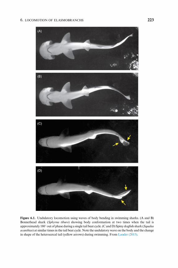
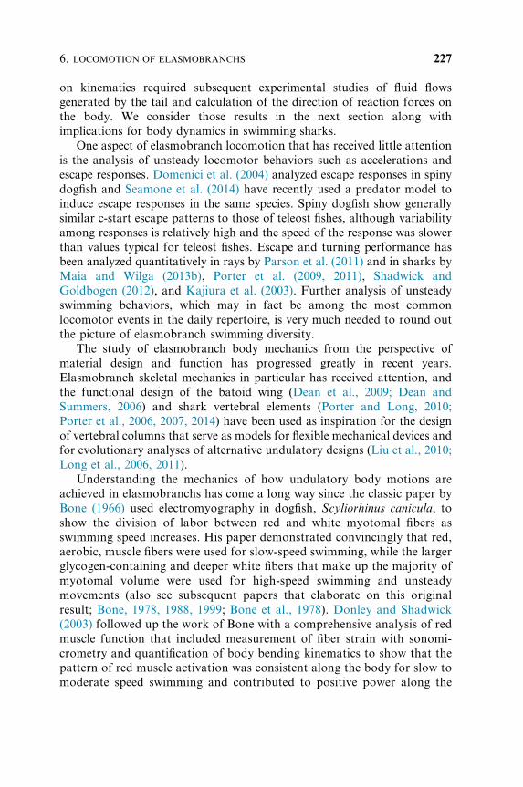
Figure 6.1. Undulatory locomotion using waves of body bending in swimming sharks. (A and B)
Bonnethead shark (Sphyrna tiburo) showing body conformation at two times when the tail is
approximately 1801out of phaseduring a single tail beat cycle. (CandD)Spinydogfish shark (Squalus
acanthias) at similar times in the tail beat cycle.Note the undulatorywave on the body and the change
in shape of the heterocercal tail (yellow arrows) during swimming. From Lauder (2015).
6. LOCOMOTION OF ELASMOBRANCHS 223
(Webb andKeyes, 1982).While this classificatory terminologydates back to the
classic papers of Breder (1926) and Lindsey (1978) and has been expanded in
recent years to differentiate between BCF (body and caudal fin) and MPF
(median and paired fin) locomotion, we believe that much of the diversity of
locomotor function in fishes is obscured by this descriptive terminology.
Specifically, even during undulatory locomotion using waves of body
bending, the median and paired fins of sharks play an important role in
balancing pitch, roll, and yaw torques, and the dorsal fins in particular are also
capable of generating thrust by acceleratingwater posteriorly (Maia andWilga,
2013a,b;Wilga andLauder, 2000).Medianandpairedfins in elasmobranchs are
under active control by intrinsic musculature and their function during
undulatory locomotion is integral tounderstandinghowdestabilizing forces are
managed by swimming elasmobranchs. However, compared to the fins of
teleost fishes, which possess highly flexible fin rays with a bilaminar structure
that allows active bending (Lauder, 2006), elasmobranchfinswith their rod-like
fin rays appear to be less flexible and capable of lower curvatures and range of
motion. An additional function ofmedian fins during swimming in sharks is the
interaction between flows generated by the dorsal fins and the caudal fin
(Maia andWilga, 2013a;Webb and Keyes, 1982), a phenomenon that has also
been studied extensively in teleost fishes (Drucker and Lauder, 2001; Standen
andLauder, 2007).Depending on the relative timingof active dorsal and caudal
fin movements, fluid vortices shed from the dorsal fins can interact with caudal
fin flows to substantially alter free-stream fluid motion incident to the tail.
The roles of fins can change considerably during unsteady locomotor
movements compared to their function during steady swimming. Studies of the
function of shark pectoral fins, for example, have shown that fin conformations
maintained during steady horizontal swimming are adjusted during vertical
maneuvering in order to generate torques that pitch the body up or down
(Fish and Shannahan, 2000; Wilga and Lauder, 2000, 2001), which facilitates
vertical movement in the water column. Control of pitching bodymotions may
be especially important in elasmobranchs, which tend to be negatively buoyant
and lack the gas-filled swimbladder common to most teleost fishes.
Batoid fishes are known for their use of pectoral fins during locomotion
(Blevins and Lauder, 2012; Fontanella et al., 2013; Klausewitz, 1964; Parson
et al., 2011; Rosenberger, 2001), and skates and rays also show changes in
fin and body position during transitions from steady horizontal swimming
to vertical maneuvering. Study of locomotion in skates illustrates the
changing role that fins can take when maneuvers are initiated and during
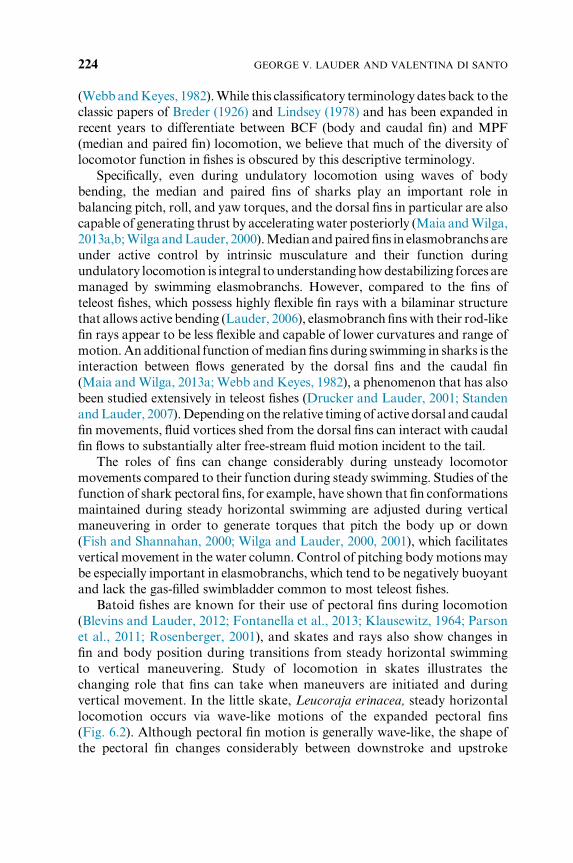
vertical movement. In the little skate, Leucoraja erinacea, steady horizontal
locomotion occurs via wave-like motions of the expanded pectoral fins
(Fig. 6.2). Although pectoral fin motion is generally wave-like, the shape of
the pectoral fin changes considerably between downstroke and upstroke
GEORGE V. LAUDER AND VALENTINA DI SANTO224
(compare Fig. 6.2, panels A and C). Lateral views show that during the
downstroke, a relatively sharp transition point can occur in the middle of
the fin margin as the wave is propagated posteriorly (Fig. 6.2A). During the
upstroke, the fin margin takes on a more rounded shape (Fig. 6.2C); the
effect of these downstroke–upstroke shape changes on patterns of fluid flow
and force production are as yet unknown.
Fin kinematics in the little skate show some interesting changes while
executingverticalmaneuvers.Ascending in thewater column isactivelypowered
by higher amplitude fin wave-like motions than those used during steady
forward swimming (Fig. 6.2E). However, descending occurs by reorienting the
body at a negative angle of attack and is largely passivewith only low amplitude
movements of the pectoral fins (Fig. 6.2F). Very few kinematic studies of
locomotion in freely swimming skates or rays are available, so it is not currently
possible to say how general these observations are.
An additional noteworthy aspect of pectoral fin locomotion in batoids was
described by Blevins and Lauder (2012) in their study of three-dimensional
Figure 6.2. Swimming kinematics in the little skate, Leucoraja erinacea. (A and B) Frames from
high-speed video recordings of body and pectoral conformations during steady horizontal
locomotion at a swimming speed of 1.2 BL/s. (C and D) Body and pectoral fin conformation at
a time 0.24 s after the images shown in (A) and (B). Note the change in pectoral wave shape.
(E and F) Body and pectoral conformation during vertical (ascending) locomotion (E), and
during downward (ventral) descent (F). Ascending is active and often accompanied by high
amplitude and rapid pectoral fin motions, while descending can be passive following body
reorientation at a negative angle of attack to oncoming flow.
6. LOCOMOTION OF ELASMOBRANCHS 225
pectoral fin kinematics in the freshwater stingray Potamotrygon orbignyi.
They observed that the outer margin of pectoral fin, during the downstroke,
can be rather substantially cupped downward, and hence curved into the
flow. If pectoral fin kinematics were dominated by the fluid loading
occurring as the fin is moved down against the fluid, then the fin margin
would be expected to be curved upward as is seen on the pectoral fin in the
oscillatory motion of manta rays. But the opposite conformation often
occurred in the freshwater stingray, and suggests that active control of the
fin margin allows detailed shape changes during swimming, which perhaps
function to control how water flows over the fin edge and to increase thrust
by directing more water posteriorly.
Batoids are also noted for benthic locomotion where fin movements
occur near the substrate and are subject to “ground effect” hydrodynamic
influences. Ground effects are well known for flying animals and man-made
aircraft where both rigid and flapping wings alter flows near surfaces (see
review in Rayner, 1991), but the dynamics of aquatic animals interacting
with the substrate are very different given the flexible undulating propulsive
modes observed in fishes. The ground effect has proven challenging to study
in live elasmobranchs, but several recent papers have addressed some of the
kinematic and hydrodynamic factors involved with rays swimming in
ground effect using simple physical models. Studies using flexible
membranes as models of ray wings, either with dual actuators driving a
rubber membrane (Blevins and Lauder, 2013) or a single leading edge
actuator controlling a flexible panel (Quinn et al., 2014a,b), have shown
that swimming near the bottom can greatly alter flow patterns over
the undulating membrane. Remarkably, even though kinematics of the
membranes can remain relatively unchanged, substantial improvements in
swimming efficiency can be achieved.
In sharks the function of the asymmetrical (heterocercal) shape of the tail
received renewed attention beginning with the paper by Thomson (1976)
who first suggested that the asymmetrical shape acts to direct locomotor
force through the center of body mass and thus avoids inducing rotational
torques tending to pitch the head down. This contrasts with the classical
view that the heterocercal tail generates lift forces (Affleck, 1950). The
heterocercal tail moves in a complex manner (Fig. 6.1C and D: yellow
arrows) and requires a full three-dimensional study to determine the
orientation of different regions of the tail during swimming. Experimental
analysis of three-dimensional motion of the heterocercal tail in leopard
sharks (Ferry and Lauder, 1996) suggested that the classical model was
correct, and that orientation of the tail surface during side-to-side movement
indicates that lift forces are produced, which pitch the head ventrally around
the center of mass (COM). However, confirmation of this suggestion based
GEORGE V. LAUDER AND VALENTINA DI SANTO226
on kinematics required subsequent experimental studies of fluid flows
generated by the tail and calculation of the direction of reaction forces on
the body. We consider those results in the next section along with
implications for body dynamics in swimming sharks.
One aspect of elasmobranch locomotion that has received little attention
is the analysis of unsteady locomotor behaviors such as accelerations and
escape responses. Domenici et al. (2004) analyzed escape responses in spiny
dogfish and Seamone et al. (2014) have recently used a predator model to
induce escape responses in the same species. Spiny dogfish show generally
similar c-start escape patterns to those of teleost fishes, although variability
among responses is relatively high and the speed of the response was slower
than values typical for teleost fishes. Escape and turning performance has
been analyzed quantitatively in rays by Parson et al. (2011) and in sharks by
Maia and Wilga (2013b), Porter et al. (2009, 2011), Shadwick and
Goldbogen (2012), and Kajiura et al. (2003). Further analysis of unsteady
swimming behaviors, which may in fact be among the most common
locomotor events in the daily repertoire, is very much needed to round out
the picture of elasmobranch swimming diversity.
The study of elasmobranch body mechanics from the perspective of
material design and function has progressed greatly in recent years.
Elasmobranch skeletal mechanics in particular has received attention, and
the functional design of the batoid wing (Dean et al., 2009; Dean and
Summers, 2006) and shark vertebral elements (Porter and Long, 2010;
Porter et al., 2006, 2007, 2014) have been used as inspiration for the design
of vertebral columns that serve as models for flexible mechanical devices and
for evolutionary analyses of alternative undulatory designs (Liu et al., 2010;
Long et al., 2006, 2011).
Understanding the mechanics of how undulatory body motions are
achieved in elasmobranchs has come a long way since the classic paper by
Bone (1966) used electromyography in dogfish, Scyliorhinus canicula, to
show the division of labor between red and white myotomal fibers as
swimming speed increases. His paper demonstrated convincingly that red,
aerobic, muscle fibers were used for slow-speed swimming, while the larger
glycogen-containing and deeper white fibers that make up the majority of
myotomal volume were used for high-speed swimming and unsteady
movements (also see subsequent papers that elaborate on this original
result; Bone, 1978, 1988, 1999; Bone et al., 1978). Donley and Shadwick
(2003) followed up the work of Bone with a comprehensive analysis of red
muscle function that included measurement of fiber strain with sonomi-
crometry and quantification of body bending kinematics to show that the
pattern of red muscle activation was consistent along the body for slow to
moderate speed swimming and contributed to positive power along the
6. LOCOMOTION OF ELASMOBRANCHS 227
length, unlike previous results obtained for many teleost fishes. Recent
overviews of shark muscle function can be found in Shadwick and
Gemballa (2006), Shadwick and Goldbogen (2012) and Syme (2006) (see
also Chapter 5).
Analysis of the mechanics of shark musculature changes considerably
when fast-swimming pelagic species such as lamnid sharks are considered
because the red muscle is internalized and located medially (Bernal et al.,
2003a,b; Graham et al., 1994; Sepulveda et al., 2005), and these red fibers
possess an elevated temperature with respect to ambient water (Bernal et al.,
2005, 2009; see also Chapter 8). The remarkable evolutionary similarity
between pelagic lamnid sharks and tuna (Bernal et al., 2001; Donley et al.,
2004; Shadwick, 2005) in the location of the red musculature and in the
attachment of body muscle fibers to collagenous myosepts is one of the most
outstanding known examples of convergent biomechanical evolution.
4. HYDRODYNAMICS OF ELASMOBRANCH LOCOMOTION
Experimental analyses of water flow over the bodies and fins of swimming
elasmobranchs have enabled a number of hypotheses about body and fin
function to be addressed. Quantitative flow visualization borrows approaches
from engineering to visualize and analyze patterns of water movement over
the surface and in the wake of the body and fins (Drucker and Lauder, 1999).
In swimming leopard sharks, Wilga and Lauder (2002, 2004b) showed that
the heterocercal tail generated a momentum jet that is directed posteriorly
and ventrally, and thus produces a reaction force aimed above the center of
mass (COM) (Fig. 6.3). This confirmed the canonical model of heterocercal
tail function and indicates that the classic heterocercal tail shape generates
lift forces and body torques. Flammang et al. (2011) applied a recently
developed volumetric flow imaging approach to heterocercal tail function
and showed a more complex vortex wake signature than previously suspected
(Fig. 6.3D), while confirming earlier work on the direction of forces and the
momentum jet produced by heterocercal tails (Fig. 6.3E). Borazjani and
Daghooghi (2013) have shown an important additional feature of fish tails
that applies to both heterocercal and homocercal tail hydrodynamics: the tail
appears to generate an attached leading edge vortex that enhances thrust in
a manner similar to proposed previously for insect and bird wings.
The significance of differences in shape between the upper and lower lobes
in the heterocercal tail for leading edge vortex structure is as yet unknown,
but the computational fluid dynamic approach promises new insights into the
function of elasmobranch caudal fins.
GEORGE V. LAUDER AND VALENTINA DI SANTO228
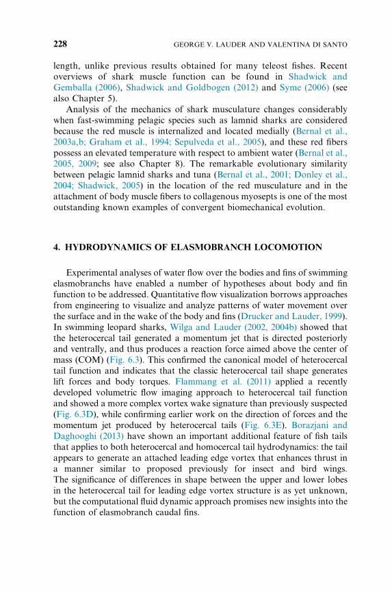
Figure 6.3. Dynamics of locomotion in sharks studied with three-dimensional (3D) kinematics
and particle image velocimetry. (A) 3D analysis of pectoral fin conformation in swimming
leopard sharks (Triakis semifasciata, 21–26 cm total length) shows that the pectoral fins are held
in a position that generates minimal vorticity during steady swimming. (B) Summary of forces
acting on the body of a steadily swimming shark (see text for discussion). (C) Plot of the angle of
the fluid dynamic jet formed by the tail vortex ring versus body angle. Jets are negative (below
the horizontal) no matter what the body angle in leopard sharks, which indicates that the tail
generates torques around the center of mass. These torques are counteracted by lift forces on the
body. (D) Vortex ring conformation generated by one tail beat in a swimming spiny dogfish
(Squalus acanthias). (E) Vertical slice through the vortex wake of a swimming leopard shark
showing two centers of vorticity and the central fluid jet inclined below the horizontal. Modified
from Flammang et al. (2011) and Wilga and Lauder (2000, 2002).
6. LOCOMOTION OF ELASMOBRANCHS 229
Data on flow visualization over the body and pectoral fins (Wilga
and Lauder, 2000) combined with analysis of flows generated by the tail
provide an overall picture of the balance of forces on the body of freely
swimming sharks (Fig. 6.3). During steady horizontal locomotion, pectoral
fins are held in a conformation that results in near zero net lift forces
(Fig. 6.3A). However, this changes dramatically during maneuvering when
pectoral fins change their angle of attack to initiate pitch moments about
the COM.
During horizontal steady swimming, the body is inclined to the
horizontal and generates lift forces and also counter-rotating torques
around the COM (Fig. 6.3B). Both net lift and torques must balance and
this is achieved dynamically during each tail beat as the head, body, and tail
lift forces match gravitational forces, and oppositely-signed torques balance
to prevent net rotation (Fig. 6.3B). In leopard sharks, the angle of the fluid
dynamic tail jet is independent of the angle of the body during swimming,
while in bamboo sharks, Chiloscyllium punctatum, Wilga and Lauder (2002)
observed changes in jet angle, which became more horizontal as body angle
increased during slow speed swimming. Sharks adjust their body angle as
swimming speed changes, and during slow horizontal swimming at 0.5 L/s
the body may be inclined at an angle of approximately 101 or more, while at
a speed of 2.0 L/s the body is nearly horizontal. However, a cautionary note
is in order here. Studies that generate accurate kinematic data on the body
and fins of elasmobranchs necessarily involve using smaller animals that are
suitable for laboratory flumes and camera arrangements, and it is still
unclear if these conclusions apply to larger freely-swimming sharks in
unrestricted open-ocean conditions.
The heterocercal tail of sharks has also inspired the construction of
simple physical models that have been used to understand some of the basic
kinematic and hydrodynamic properties of propulsive surfaces with angled
trailing edges as compared to homocercal (externally symmetrical) shapes
(Lauder et al., 2011, 2012). Interestingly, analysis of the self-propelled
speeds of simple flexible plastic panels with different trailing edge shapes
showed that the heterocercal shape had increased swimming speeds
(approximately 7% faster on average) compared to panels with the same
area but a vertical trailing edge. However, this occurs with a slightly
increased cost of transport (Lauder et al., 2011), and the vortex wake shed
by the heterocercal panel differed in several ways from that of freely
swimming leopard sharks. This difference could be due to obvious
differences in structure between the simple plastic panel models and the
tail of live sharks, and also to active stiffening via muscle fibers intrinsic to
the tail and changes in stiffness through pressure changes in the tail as
sharks swim (Flammang, 2010).
GEORGE V. LAUDER AND VALENTINA DI SANTO230
Experimental hydrodynamic data on batoid locomotion are not
currently available for live animals swimming steadily, and most hydro-
dynamic data relevant to skate and ray swimming come from panel models
or robotic systems (Moored et al., 2011a,b). Here we present experimental
hydrodynamic measurements of flow around the body and in the wake of
the little skate, L. erinacea, during steady locomotion at a moderate
swimming speed of 2.0 L/s (Fig. 6.4). Particle image velocimetry of flows
around the body and wake of the undulating pectoral fins shows clear
momentum jets that alternate from posterodorsal to posteroventral
(compare Fig. 6.4, panels B and E). These momentum jets are not
symmetrical and jet velocities resulting from the upstroke appear to be of
higher speed and carry greater momentum than downstroke flows. In
addition to the associated vortex wake, flow slowed by interaction with the
body and wing is evident as ribbon-like strips of vorticity over the upper
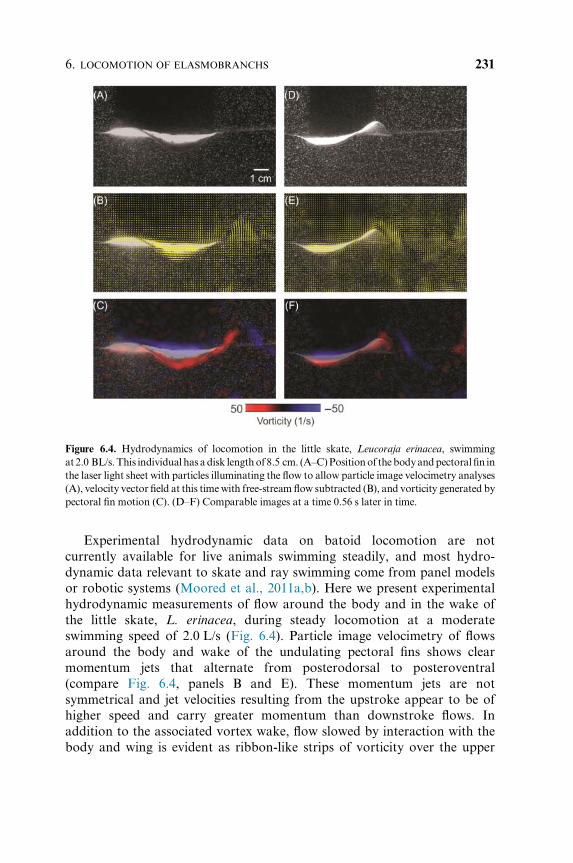
Figure 6.4. Hydrodynamics of locomotion in the little skate, Leucoraja erinacea, swimming
at2.0 BL/s.This individual has adisk lengthof 8.5 cm. (A–C)Positionof thebodyandpectoral fin in
the laser light sheet with particles illuminating the flow to allow particle image velocimetry analyses
(A), velocity vector field at this timewith free-stream flow subtracted (B), and vorticity generated by
pectoral fin motion (C). (D–F) Comparable images at a time 0.56 s later in time.
6. LOCOMOTION OF ELASMOBRANCHS 231
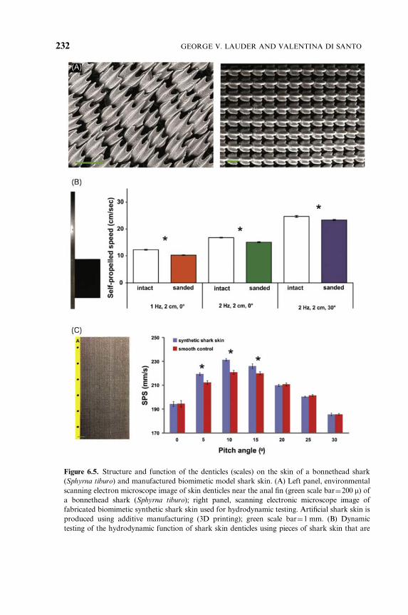
Figure 6.5. Structure and function of the denticles (scales) on the skin of a bonnethead shark
(Sphyrna tiburo) and manufactured biomimetic model shark skin. (A) Left panel, environmental
scanning electron microscope image of skin denticles near the anal fin (green scale bar¼200 m) ofa bonnethead shark (Sphyrna tiburo); right panel, scanning electronic microscope image of
fabricated biomimetic synthetic shark skin used for hydrodynamic testing. Artificial shark skin is
produced using additive manufacturing (3D printing); green scale bar¼1 mm. (B) Dynamic
testing of the hydrodynamic function of shark skin denticles using pieces of shark skin that are
GEORGE V. LAUDER AND VALENTINA DI SANTO232
body and lateral pectoral edge (Fig. 6.4C). These preliminary data suggest
that further studies including a diversity of batoid species would be useful
for understanding how different patterns of pectoral fin motion and body
positions alter flows produced by the pectoral fins, and the relative balance
of upstroke and downstroke momentum production.
5. THE REMARKABLE SKIN OF ELASMOBRANCHS AND ITS
LOCOMOTOR FUNCTION
As the fin and body surfaces of elasmobranchs undulate and are moved
through the water, the skin encounters a time-dependent flow pattern.
Friction between skin and the water is likely to be an important (although
still of unknown magnitude) source of drag, and there is now a large
literature on how the skin of elasmobranchs might have special drag-
reducing properties. Much of this literature is based on engineered models
(Bechert et al., 1986; Bechert and Hage, 2007; Dean and Bhushan, 2010) and
shows that surface texture, of an appropriate size and spacing, can result in
drag reduction as fluid is moved steadily past a textured surface.
Shark skin has inspired much of this research on the function of surface
texture due to its remarkably complex structure (Kemp, 1999; Liem et al.,
2001; Motta et al., 2012; Reif, 1982, 1985). Small (100 um to 1 mm) bony
dermal denticles (Fig. 6.5A) cover the skin. Denticles are embedded into the
dermis (Kemp, 1999; Motta, 1977) with a small expanded bases, and have
stalk-like structures that extend through the skin to support ridged flanges
exposed to water flow at the surface (Fig. 6.5A).
Although studies of shark skin models under static conditions where the
textured surface does not move have provided a solid baseline of data on
attached to a flat support (shown on the left) which in turn is attached to a mechanical flapping
foil device that allows controlled side-to-side and rotational motions of the shark skin membrane.
Graph shows the self-propelled swimming speed of the shark skin membrane with intact denticles
and after the denticles have been sanded off (to produce a relatively smooth surface) under three
different motion programs. Note that in each case the swimming speed of the shark skin with
denticles intact is significantly greater (*) than after the denticles have been removed by sanding.
(C) Assembly of the tested flexible biomimetic shark skin foil (on left) and hydrodynamic analysis.
A flat support attaches to the yellow area with holes on the left side of the foil, and this support is
moved by a mechanical flapping device. Graph of the results from measuring the self-propelled
swimming speed of the biomimetic shark skin foil (blue bars) compared to the smooth control
(red bars) at different pitch angles. Heave motion was 71.5 cm at 1 Hz for all trials. At pitch
angles of 51, 101, and 151 the biomimetic shark skin foils swim significantly faster (*) than the
smooth controls. At the other four pitch angles, the swimming speeds are similar. Modified from
Oeffner and Lauder (2012), Wen et al. (2014), and Lauder (2015).
6. LOCOMOTION OF ELASMOBRANCHS 233
drag reduction, the lack of dynamic testing is of special concern. Shark skin,
during free-swimming, is exposed to time-dependent flows with changing
angles of attack and also flow separation over leading edges of the fins and
tail, and also possibly along the body (Anderson et al., 2001). This suggests
that dynamic testing is needed where skin and biomimetic versions of shark
skin can be moved under a controlled motion program that mimics that of
freely-swimming sharks, and forces, fluid flow patterns, and swimming
performance measured. Wen et al. (2014) manufactured a biomimetic shark
skin, and tested its function in comparison to a smooth control (Fig. 6.5A,
right panel). Manufactured shark skin mimics have the advantage of
allowing good experimental control, as smooth surfaces with the same mass
as the artificial skin can also be studied and swimming performance
compared.
Oeffner and Lauder (2012) used pieces of real shark skin to make flexible
membranes attached to a supporting rod (Fig. 6.5B). By attaching this rod
to a computer controlled mechanical flapping apparatus (Lauder et al.,
2007, 2011) that allows dynamic testing, they were able to move the pieces of
shark skin in an undulatory motion program with realistic angles of attack
and to achieve curvatures of the shark skin that match that of freely-
swimming sharks. They found that the textured denticle surface increased
swimming speed by an average of 12.3% compared to a smoothly sanded
control in which the denticles have been removed, moving with the same
motion program of heave and pitch (Fig. 6.5B). An additional key result was
that the increased swimming speeds did not occur in shark skin membranes
that were attached to rigid surfaces, indicating that flexibility and bending of
the shark skin membrane is critical to the increased performance with the
roughened denticle surface.
Analysis of the flow field around swimming shark skin membranes and
the sanded controls revealed a possible new role for the denticle-covered
shark skin surface. Oeffner and Lauder (2012) found changes in the intensity
and location of the leading edge vortex attached to the swimming shark skin
membrane suggesting that the roughened surface might enhance thrust by
promoting leading edge suction compared to a smooth control. They
hypothesize that this effect may have been at least partially responsible for
the observed increased swimming speeds, and may apply to regions of the
shark body where flow separation occurs, such as the tail.
One limitation of studying real shark skin is that it is difficult to modify
the denticle pattern and make experimental alterations to determine which
specific features of shark denticles most affect swimming mechanics. To
address this issue, Wen et al. (2014) manufactured a biomimetic shark skin
and smooth controls and compared their performance under dynamic
swimming conditions. Fig. 6.5C shows how the self-propelled swimming
GEORGE V. LAUDER AND VALENTINA DI SANTO234
speed of the biomimetic shark skin and smooth controls compares when
moved at a constant heave of 71.5 cm at 1 Hz under a variety of different
pitch angles. Manufactured shark skin swam significantly faster at pitch
angles of 51, 101, and 151, but at the same speed as the smooth control at low
(01) and high (20–301) pitch angles. The effect of the shark skin surface on
locomotor performance thus depends critically on the motion program and
how the surface interacts dynamically with oncoming flow as it moves
through water.
These results suggest that the function of the roughened denticle-covered
skin of sharks is complex and motion-dependent, and it is clear that much
more remains to be discovered about the diversity of denticle patterns over
the body and among species, the effect of possible movement of individual
denticles during swimming (Lang et al., 2008, 2014), and the functional
significance of the ornamentation on the denticle surface.
6. ENERGETICS OF ELASMOBRANCH LOCOMOTION
Studies that estimate the energetic costs incurred by elasmobranchs during
swimming are scarce. In a few cases, costs of locomotion have been measured
using a Brett-type swim tunnel (Brett, 1971), although recent technological
advances have allowed researchers to approximate energetic costs in free-
swimming sharks using speed and tail-beat sensors that have been calibrated
to oxygen consumption rates (e.g., Graham et al., 1990; Scharold andGruber,
1991; Sepulveda et al., 2004; Bernal et al., 2012; see also Chapter 8). Most
studies on the metabolism of elasmobranchs have focused on a few benthic
and inactive species, resting on the bottomof a respirometer (restingmetabolic
rate; Brett andBlackburn, 1978; Ferry-GrahamandGibb, 2001;Di Santo and
Bennett, 2011b).However, some studies alsomeasured restingmetabolic rates
of more active sharks, such as the mako shark, which exhibit oxygen
consumption rates at rest similar to comparably sized yellowfin tuna Thunnus
albacares (240 vs. 253 mg O2�kg�1 h�1, respectively; Graham et al., 1990;
Dewar andGraham, 1994). A few other researchers have used swim tunnels to
measure swimming metabolic rates in sharks. The metabolic rates of
elasmobranchs during steady swimming have been reported for mako
(Graham et al., 1990; Sepulveda et al., 2007), leopard (Scharold et al.,
1988a), lemon (Scharold and Gruber, 1991), blacknose sharks (Carlson et al.,
1999), and for one batoid, the little skate (Di Santo and Kenaley, in
preparation), and these comparative data are summarized in Fig. 6.6 and
Table 6.1. The little skate exhibits lower energetic costs during steady
swimming when compared to more active ectothermic and lamnid sharks.
Oxygen consumption decreases somewhat in the little skate as a function of
6. LOCOMOTION OF ELASMOBRANCHS 235
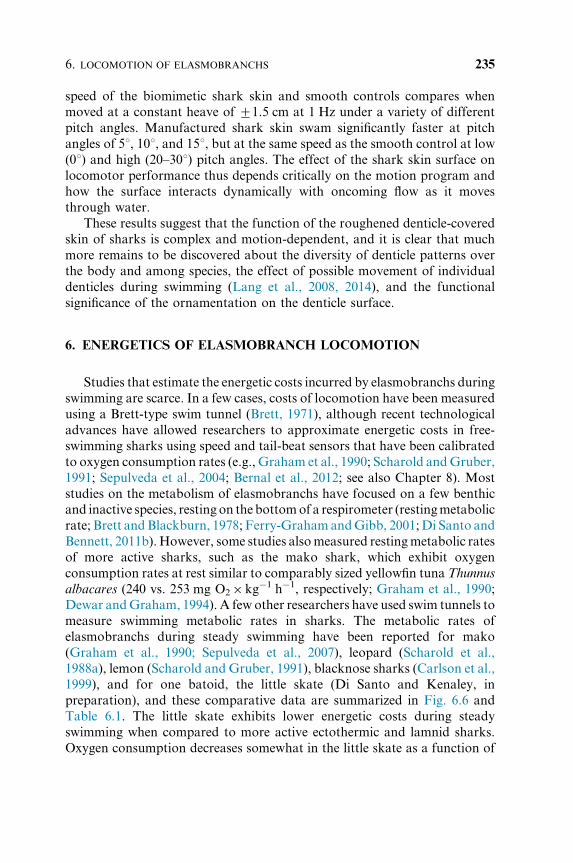
speed within the range tested (0.75–1.25 BL/s). It is possible that the slightly
highermetabolic rates of skates swimming at the lowest speed (Fig. 6.6)maybe
due to the fact that they were swimming below the velocity necessary to
maintain hydrostatic equilibrium (Bernal et al., 2012), and thus incurred
additional costs to maintain body stability. Even though there are only a few
studies on elasmobranch energetics during activity, it is apparent that there is a
positive association between fast-swimming fishes and swimming metabolic
rates (Table 6.1). For instance, even after adjusting for temperature and body
size, scalloped hammerhead sharks Sphyrna lewini consume nearly double the
amount of oxygen that leopard sharks use when both are swimming about
1 body length per second (Scharold et al., 1988; Lowe, 1996, 2002).
Basic measures of oxygen consumption such as the resting metabolic rate
and active metabolic rate are useful for a gross determination of the costs of
activity in a species. However, calculating the aerobic scope (i.e., the
difference between the maximum and the resting metabolic rates) can be
used to estimate the capacity for activity in fishes (Fry, 1947; Farrell et al.,
2008, 2009; Roche et al., 2013). Not surprisingly, lamnid sharks exhibit high
Figure 6.6. Oxygen consumption rates (VO2 7 SE) of five species of elasmobranchs at different
swimming speeds (in Body Lengths per second, BL/s): open triangle: Isurus oxyrinchus (Graham
et al., 1990); closed triangle: Negaprion brevirostris (Scharold and Gruber, 1991); open circle:
Triakis semifasciata (Scharold et al., 1988); closed circle: Carcharhinus acronotus (Carlson et al.,
1999); closed square: Leucoraja erinacea (Di Santo and Kenaley, unpublished data).
GEORGE V. LAUDER AND VALENTINA DI SANTO236
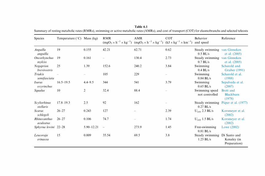
Table 6.1
Summary of resting metabolic rates (RMRs), swimming or active metabolic rates (AMRs), and cost of transport (COT) for elasmobranchs and selected teleosts
Species Temperature (1C) Mass (kg) RMR
(mgO2�h�1�kg�1)
AMR
(mgO2�h�1�kg�1)
COT
(kJ�kg�1�km�1)
Behavior
and speed
Reference
Anguilla
anguilla
19 0.155 42.21 62.71 0.62 Steady swimming
0.5 BL/s
van Ginneken
et al. (2005)
Oncorhynchus
mykiss
19 0.161 – 130.4 2.73 Steady swimming
0.7 BL/s
van Ginneken
et al. (2005)
Negaprion
brevirostris
25 1.39 152.6 240.2 3.84 Swimming
0.4 BL/s
Scharold and
Gruber (1991)
Triakis
semifasciata
– – 105 229 – Swimming
0.84 BL/s
Scharold et al.
(1988)
Isurus
oxyrinchus
16.5–19.5 4.4–9.5 344 541 3.79 Swimming
0.65 BL/s
Sepulveda et al.
(2007)
Squalus 10 2 32.4 88.4 – Swimming speed
not controlled
Brett and
Blackburn
(1978)
Scyliorhinus
stellaris
17.8–19.3 2.5 92 162 – Steady swimming
0.27 BL/s
Piiper et al. (1977)
Scarus
schlegeli
26–27 0.243 127 – 2.39 Ucrit 2.3 BL/s Korsmeyer et al.
(2002)
Rhinecanthus
aculeatus
26–27 0.106 74.7 – 1.74 Ucrit 1.5 BL/s Korsmeyer et al.
(2002)
Sphyrna lewini 22–28 5.90–12.21 – 273.9 1.45 Free-swimming
0.81 BL/s
Lowe (2002)
Leucoraja
erinacea
15 0.009 35.54 69.5 3.8 Steady swimming
1.25 BL/s
Di Santo and
Kenaley (in
Preparation)
active metabolic rates (Fig. 6.6) which correlate with long-distance
migrations and the ability to catch fast-moving prey (Bonfil et al., 2005;
Sepulveda et al., 2007; Jorgensen et al., 2010). Lamnid sharks also exhibit
relatively high resting metabolic rates to accommodate morphological and
physiological adaptations for high swimming performance. In fact, it has
been estimated that white sharks use about 46% of their total energy intake
just to sustain basal metabolic demands (Ezcurra et al., 2012).
The deployment of small activity loggers has enabled measurement of the
costs of locomotion in free swimming fishes (Scharold et al., 1988; Scharold
and Gruber, 1991; Farrell et al., 2008; Gleiss et al., 2010). Although precise
speed sensors, accelerometers, and heart-rate monitors are now available
and can be calibrated with standard swimming tests in the laboratory, there
are many abiotic and biotic factors that can alter metabolic and heart rates
in fishes beside swimming speed. It is unlikely that an elasmobranch would
swim constantly at the same depth and in the same area, and it is therefore
likely to encounter a variety of thermal environments, salinities, pH, and
dissolved oxygen levels. All these abiotic factors are known to affect
physiological processes in fishes (Fry, 1971; Carlson and Parsons, 2001;
Meloni et al., 2002; Farrell et al., 2008; Di Santo and Bennett, 2011a; Di
Santo, 2015). In addition, the presence or absence of predators, prey, and
conspecifics can alter cost of activity (Wurtsbaugh and Li, 1985; Trudel
et al., 2001; Allan et al., 2013; Binning et al., 2013). These factors make
interpreting the data obtained from loggers challenging, but for pelagic
elasmobranchs there is often little alternative.
The energetics of steady free swimming in elasmobranchs can be quite
difficult to measure, especially in benthic species that are ordinarily mostly
sedentary, and an alternative measure of locomotor capacity can be obtained
by measuring excess post-exhaustion oxygen consumption in fishes that have
been manually chased (Cutts et al., 2002; Svendsen et al., 2011; Roche et al.,
2013). In this protocol, researchers gently and repeatedly tap the fish thus
eliciting “burst-and-glide” swimming until the fish is unresponsive to
handling, that is, fatigue is achieved, and the fish is immediately transferred
to a respirometer to measure the “oxygen debt” accumulated as a result of
intense short exercise (Svendsen et al., 2011; Clark et al., 2013; Di Santo, in
review). For many benthic species, this method represents, to date, the only
effective way to measure maximum metabolic rates, and it also may result in
higher oxygen consumption rates than those measured during swimming,
allowing a better estimation of maximal aerobic capacity (Clark et al., 2013;
Roche et al., 2013). In the section ‘Climate Change: Effects on Elasmobranch
Locomotor Function’ we describe results from thismethod applied to the little
skate in an effort to understand the effects of changing oceanic conditions on
locomotor energetics.
GEORGE V. LAUDER AND VALENTINA DI SANTO238
Finally, we emphasize that there is a near total lack of studies on the cost
of locomotion in batoids, and the data shown in Fig. 6.6 for the little skate
represent the only measurements that we are aware of. Without broader
taxonomic representation of elasmobranch energetic data, we will not be
able to make general conclusions as to energetic underpinnings associated
with different modes of locomotion, and this is an especially fruitful area for
future research.
7. CLIMATE CHANGE: EFFECTS ON ELASMOBRANCH
LOCOMOTOR FUNCTION
Since the industrial revolution, global atmospheric carbon dioxide
(CO2) concentrations have increased from 280 to about 400 ppm (in 2015),
thus reaching levels that have not been experienced in the past 65 million
years (IPCC, 2013). The two major consequences of the accelerating
increase in atmospheric CO2 concentration are a rise in ocean temperature
and acidification (i.e., lower pH). Temperature alone is considered to be
the “abiotic master factor” as it is known to affect nearly every metabolic
process in fishes (Fry, 1971; Brett, 1971). In fact, fishes are known to
select different temperatures in order to enhance physiological processes
(Sims et al., 2006; Wallman and Bennett, 2006; Di Santo and Bennett,
2011a). As ocean temperature increases as a consequence of climate change,
we might expect fish to adjust to the new thermal environment through
physiological acclimatization (Somero, 2010; Donelson et al., 2012),
adaptation (Angilletta et al., 2004; Baumann and Conover, 2011; Di Santo,
2015), or behavioral thermoregulation (Perry et al., 2005; Greenstein and
Pandolfi, 2008).
Similarly to other metabolic processes, swimming performance in fishes is
known to be affected by abiotic climate-related factors such as temperature and
pH (Chin and Kyne, 2007; Ishimatsu et al., 2008; Pang et al., 2011; Chen et al.,
2011). Swimming performance increases with temperature up to a thermal
optimum, beyond which performance declines rapidly. As organisms often live
close to their thermal optimum, even a slight increase in ambient temperature
may drastically reduce the energy available for highly demanding aerobic
activities, such as locomotion (Rummer et al., 2014; Di Santo, in review; Brill
and Lai, 2015). Locomotor efficiency is key to survival and reproduction in
fishes, as swimming isused tomigrate daily and seasonally to forage, spawn, and
avoid predators. In addition, in light of environmental change, efficient
locomotion may also ensure that elasmobranch fishes are able to find
suitable refugia. Relocation to more favorable areas assumes that populations
6. LOCOMOTION OF ELASMOBRANCHS 239
of elasmobranchs will be able to find and move to the new locations, and this
assumption should be treated with caution (Chin and Kyne, 2007). The ability
(or lack of thereof) to undertake large-scale migrations raises serious concerns
especially for lessmobile, or philopatric species that tend to remain close to their
home areas. When comparing the costs of transport and metabolic rates of a
range of elasmobranch and teleost fishes, it becomes apparent that metabolic
rates per se are not a good indicator to predict the ability of a fish to migrate
(Table 6.1). In fact, even though the European eel, Anguilla Anguilla, has
comparable swimming metabolic rates to the little skate (62.7 vs.
69.5 mgO2�kg�1� h�1), it uses over five times less energy than the little skate
to cover the same distance (Table 6.1). These results correspond to observations
of activity of these two species in nature. TheEuropean eel has a very low cost of
transport as a result of the combination of undulatory locomotor mode, a long
body length and low oxygen consumption, and can therefore undertake
a migration of 6000 km from Europe to the Sargasso Sea in 180 days
(van Ginneken et al., 2005). In contrast, the little skate shows high site fidelity
(philopatry) and short migratory distances (Di Santo, 2015), despite the fact
that the lowest active metabolic rate measured in this species is comparable to
that of the European eel.
Even though, generally, teleosts seem to exhibit lower costs of transport
when compared to elasmobranchs based on the available comparative data in
Table 6.1, it seems that the small body size of the little skate may reduce
efficiency of locomotion over long distances and increase overall energy
expenditure. In fact, the little skate has some of the highest costs of transport
within elasmobranch species tested to date, and it is only surpassed by more
active and larger sharks, such as themako (Table 6.1). In light of these data, it is
important to consider cost of transport along with resting and active metabolic
rates to improve predictions of whether or not fish species may be able to
significantly shift their geographic range.
Ocean warming is already affecting demography, geographic range, and
predator timing of populations and species (Murawski, 1993; Walther et al.,
2002; Perry et al., 2005; Parmesan, 2006; Dulvy et al., 2008; Chen et al., 2011).
Demographic changes include shifts in recruitment, body size, and survival
(Portner and Farrell, 2008; Genner et al., 2010; Gardner et al., 2011).
Geographic shifts include those toward the poles (Walther et al., 2002; Perry
et al., 2005; Beaugrand et al., 2008; Chen et al., 2011), and phenological changes
include anticipating the timing of spawning andmigrations (Farrell et al., 2008;
Martins et al., 2011; Eliason et al., 2011), causing amismatch between prey and
predator timing (Raubenheimer et al., 2012). In particular, warming is thought
to expand species ranges toward higher latitudes or deeper waters (Walther
et al., 2002; Parmesan, 2006; Burrows et al., 2011). Perry et al. (2005)
documented a shift in mean depth of the cuckoo ray, Leucoraja naevus, with
GEORGE V. LAUDER AND VALENTINA DI SANTO240
species moving to deeper water as a response to warming, while Stebbing et al.
(2002) showed a correlation between warming of the North Atlantic and
migration of warmer water elasmobranch species, such as the sharpnose
sevengill Heptranchias perlo and big-eye thresher Alopias superciliosus, to the
Cornish coasts of Great Britain. Some species are highly migratory and may
travel across oceans to exploit seasonal productivity events (see Camhi et al.,
2008, for review). It is clear that, in order toundertake such large-scalemigration
toward suitable refugia, elasmobranchfishes shouldbeable to sustainprolonged
swimming, and more data are needed to determine migratory capacities.
Compared to viviparous elasmobranchs, oviparous species such as skates
have a reduced geographic distribution (Goodwin et al., 2005). This is thought
to occur because skates are typically smaller in size than livebearing
elasmobranchs, and size correlateswith theability tomaintainwider geographic
ranges (Musick et al., 2004). Additionally, skates have to restrict their ranges to
suitable spawning habitats, because embryos are spatially and temporally
constrained in the egg case for a prolonged period of time (one ormore seasons;
Palm et al., 2011; Di Santo, 2015). As a consequence, although some batoids
undertake seasonal latitudinal migrations, some species, like the little skate in
the Northwestern Atlantic, only show weak seasonal distribution patterns that
consist of short distance movements from coastal and shallow waters to
offshore and deeper waters during colder months (McEachran, 2002). Perhaps
because of these spawning and spatial constraints, growth rates andmaturation
times aredifferent inadjacentpopulationsof little skates fromtheGulfofMaine
and Georges Bank (Di Santo, 2015).
In addition, when the effects of increasing temperature and acidification
on the costs of the escape response were tested in the little skate by
employing a chasing protocol, performance showed a decline even with a
modest increase in temperature (31C) beyond the thermal optimum
(Fig. 6.7). This effect is particularly pronounced in skates from the Gulf
of Maine, thus underscoring the importance of comparative studies of
locally adapted populations within a species. An additional factor that can
affect locomotor performance is acidification, which is known to exacerbate
the effect of warming on elasmobranch swimming performance. In fact,
increased water acidification reduced the escape performance (intensity of
response and aerobic scope) in juvenile little skates from the Gulf of Maine,
while a much less pronounced effect was observed in the Georges Bank
population (Fig. 6.7). Even though the increase in temperature and
acidification was not lethal in this species, it decreased endurance during
the escape response, and prolonged recovery time after exhaustive exercise
(Fig. 6.8), thus reducing the likelihood that the fish will be resilient in
changing environment. While low pH increased recovery time in both
populations, skates from the Gulf of Maine exhibited elevated metabolic
6. LOCOMOTION OF ELASMOBRANCHS 241
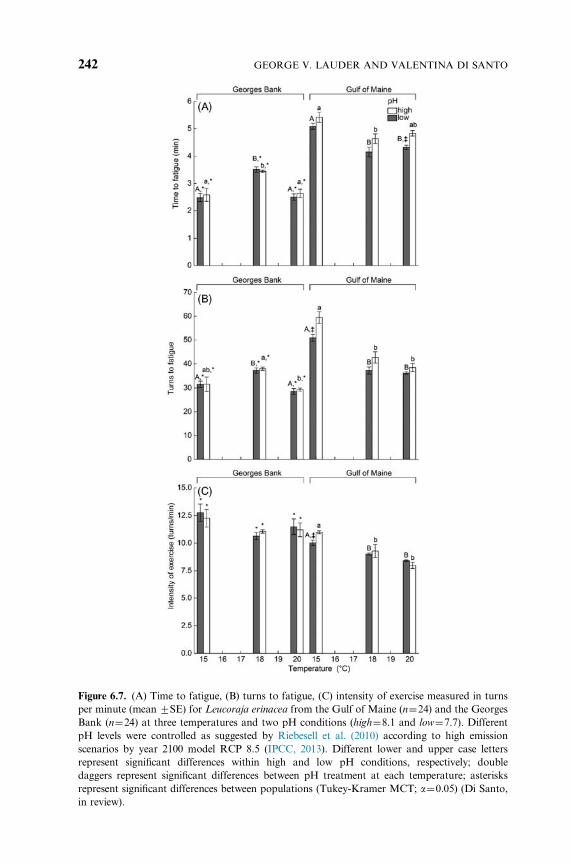
Figure 6.7. (A) Time to fatigue, (B) turns to fatigue, (C) intensity of exercise measured in turns
per minute (mean 7SE) for Leucoraja erinacea from the Gulf of Maine (n¼24) and the Georges
Bank (n¼24) at three temperatures and two pH conditions (high¼8.1 and low¼7.7). Different
pH levels were controlled as suggested by Riebesell et al. (2010) according to high emission
scenarios by year 2100 model RCP 8.5 (IPCC, 2013). Different lower and upper case letters
represent significant differences within high and low pH conditions, respectively; double
daggers represent significant differences between pH treatment at each temperature; asterisks
represent significant differences between populations (Tukey-Kramer MCT; a¼0.05) (Di Santo,
in review).
GEORGE V. LAUDER AND VALENTINA DI SANTO242
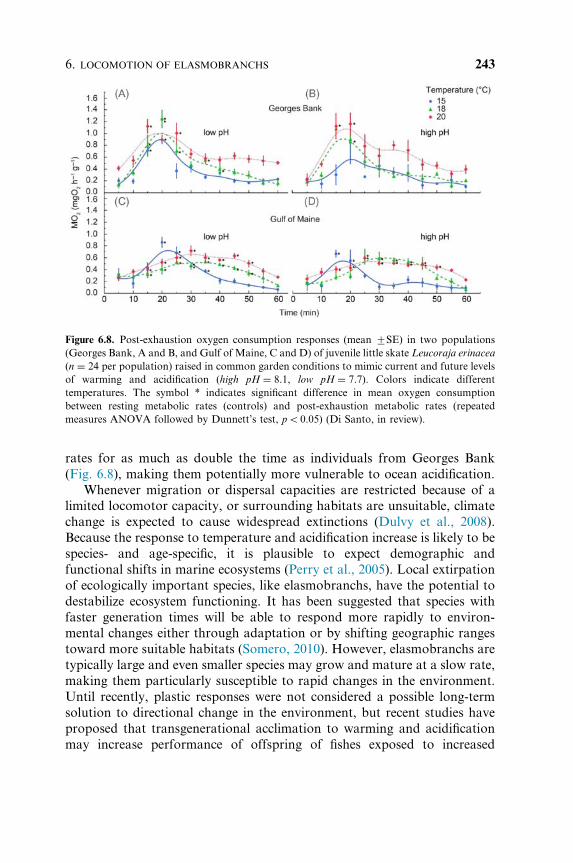
rates for as much as double the time as individuals from Georges Bank
(Fig. 6.8), making them potentially more vulnerable to ocean acidification.
Whenever migration or dispersal capacities are restricted because of a
limited locomotor capacity, or surrounding habitats are unsuitable, climate
change is expected to cause widespread extinctions (Dulvy et al., 2008).
Because the response to temperature and acidification increase is likely to be
species- and age-specific, it is plausible to expect demographic and
functional shifts in marine ecosystems (Perry et al., 2005). Local extirpation
of ecologically important species, like elasmobranchs, have the potential to
destabilize ecosystem functioning. It has been suggested that species with
faster generation times will be able to respond more rapidly to environ-
mental changes either through adaptation or by shifting geographic ranges
toward more suitable habitats (Somero, 2010). However, elasmobranchs are
typically large and even smaller species may grow and mature at a slow rate,
making them particularly susceptible to rapid changes in the environment.
Until recently, plastic responses were not considered a possible long-term
solution to directional change in the environment, but recent studies have
proposed that transgenerational acclimation to warming and acidification
may increase performance of offspring of fishes exposed to increased
Figure 6.8. Post-exhaustion oxygen consumption responses (mean 7SE) in two populations
(Georges Bank, A and B, and Gulf of Maine, C and D) of juvenile little skate Leucoraja erinacea
(n ¼ 24 per population) raised in common garden conditions to mimic current and future levels
of warming and acidification (high pH ¼ 8.1, low pH ¼ 7.7). Colors indicate different
temperatures. The symbol * indicates significant difference in mean oxygen consumption
between resting metabolic rates (controls) and post-exhaustion metabolic rates (repeated
measures ANOVA followed by Dunnett’s test, po0.05) (Di Santo, in review).
6. LOCOMOTION OF ELASMOBRANCHS 243
temperature (Ho and Burggren, 2009; Donelson et al., 2011; Grossniklaus
et al., 2013). Although a number of studies have examined the effect of
ocean warming and acidification on teleost fishes over the past five years
(e.g., Checkley et al., 2009; Baumann and Conover, 2011; Baumann et al.,
2012; Nilsson et al., 2012), our understanding of the impacts on
elasmobranch species is still surprisingly limited. To improve management
practices for elasmobranch populations, there is an urgent need for
implementation of multistressor studies on different life stages and on
individuals sampled at different locations. This will allow us to identify
critical life stages for survival, as well as the potential for acclimation and/or
adaptation in swimming performance when directional environmental
change occurs (Melzner et al., 2009; Checkley et al., 2009; Baumann and
Conover, 2011; Baumann et al., 2012; Nilsson et al., 2012). Multistressor
studies on different life stages and locally adapted populations may also
allow us to understand the mechanisms underlying resilience to warming
and acidification and ultimately can aid in determining “winning and losing”
elasmobranch physiotypes under a changing climate.
8. CONCLUSIONS
Despite considerable progress toward understanding elasmobranch
locomotion in recent years, there is still much to be done with some
surprising gaps remaining. In particular, there are relatively few data on
elasmobranch locomotor energetics and batoid swimming energetics in
particular, for which data are only available for one small skate species
(Table 6.1). Active metabolic rates of pelagic sharks are certainly extremely
challenging to measure directly, and controlled studies in a laboratory flume
setting will be difficult to achieve given the complications of swimming large
sharks in the confined space of a flume working section. Surrogate and
indirect metrics such as heart rate do not provide a complete picture of the
cost of transport, and it is likely that values of active metabolic rates for
many shark species will remain largely inferential for some time to come.
But even fundamental kinematic analyses of body and tail deformation
during swimming are limited to a few species, and three-dimensional data
are available for even fewer and mostly smaller species. A much broader
quantitative study of locomotor kinematics in three-dimensions is key to
understanding the diversity of locomotor modes and the relation between
body shape and bending patterns of the body and tail during swimming and
maneuvering. The remarkable diversity of elasmobranch shape and internal
structure is not reflected in functional studies, which to date have focused on
GEORGE V. LAUDER AND VALENTINA DI SANTO244
a very limited subset of diversity. A key challenge for the future is to extend
current work on locomotor biomechanics to encompass a variety of species
of different habits and forms.
ACKNOWLEDGMENTS
This work was supported by ONR-MURI Grant N000141410533 monitored by Dr. Bob
Brizzolara, ONR grant N00014-09-1-0352 monitored by Dr. Tom McKenna, and by National
Science Foundation grants EFRI-0938043 and CDI 0941674. Many thanks to all our collaborators
for their assistance and efforts to better understand elasmobranch locomotion over the years.
REFERENCES
Affleck, R. J. (1950). Some points in the function, development, and evolution of the tail in
fishes. Proc. Zool. Soc. Lond. 120, 349–368.
Allan, B. J., Domenici, P., McCormick, M. I., Watson, S.-A. and Munday, P. L. (2013).
Elevated CO2 affects predator-prey interactions through altered performance. PLoS One 8,
e58520.
Anderson, E. J., McGillis, W. and Grosenbaugh, M. A. (2001). The boundary layer of
swimming fish. J. Exp. Biol. 204, 81–102.
Angilletta, M. J., Oufiero, C. E. and Sears, M. W. (2004). Thermal adaptation of maternal and
embryonic phenotypes in a geographically widespread ectotherm animals and environ-
ments. In Proceedings of the Third International Conference of Comparative Physiology and
Biochemistry, KwaZulu-Natal, South Africa, 7–13 August 2004, vol. 1275. Elsevier,
pp. 258–266.
Baumann, H. and Conover, D. O. (2011). Adaptation to climate change: contrasting patterns of
thermal-reaction-norm evolution in Pacific versus Atlantic silversides. Proc. R. Soc. B Biol.
Sci. 278, 2265–2273.
Baumann, H., Talmage, S. C. and Gobler, C. J. (2012). Reduced early life growth and survival
in a fish in direct response to increased carbon dioxide. Nat. Clim. Change 2, 38–41.
Beaugrand, G., Edwards, M., Brander, K., Luczak, C. and Ibanez, F. (2008). Causes and
projections of abrupt climate-driven ecosystem shifts in the North Atlantic. Ecol. Lett. 11,
1157–1168.
Bechert, D. W. and Hage, W. (2007). Drag reduction with riblets in nature and engineering.
In Flow Phenomena in Nature. Volume 2. Inspiration, Learning, and Application, vol. 2
(ed. R. Liebe), pp. 457–469. Southampton, UK: WIT Press.
Bechert, D. W., Bartenwerfer, M., Hoppe, G. and Reif, W. E. (1986). Drag reduction
mechanisms derived from shark skin. Proc. 15th Int. Council of Aeronautical Sciences
Congress: London, UK, 7–12 September vol. 2: 1044–1068, paper no. ICAS-86-1.8.3.
AIAA.
Bernal, D., Dickson, K. A., Shadwick, R. E. and Graham, J. B. (2001). Analysis of the
evolutionary convergence for high performance swimming in lamnid sharks and tunas.
Comp. Biochem. Physiol. A 129, 695–726.
Bernal, D., Donley, J. M., Shadwick, R. E. and Syme, D. A. (2005). Mammal-like muscles
power swimming in a cold-water shark. Nature 437, 1349–1352.
Bernal, D., Sepulveda, C., Mathieu-Costello, O. and Graham, J. B. (2003a). Comparative
studies of high performance swimming in sharks I. Red muscle morphometrics,
vascularization and ultrastructure. J. Exp. Biol. 206, 2831–2843.
6. LOCOMOTION OF ELASMOBRANCHS 245
Bernal, D., Smith, D., Lopez, G., Weitz, D., Grimminger, T., Dickson, K. A., et al. (2003b).
Comparative studies of high performance swimming in sharks II. Metabolic biochemistry
of locomotor and myocardial muscle in endothermic and ectothermic sharks. J. Exp. Biol.
206, 2845–2857.
Bernal, D., Sepulveda, C., Musyl, M. and Brill, R. (2009). The eco-physiology of swimming and
movement patterns of tunas, billfishes, and large pelagic sharks. In Fish locomotion-an etho-
ecological Perspective (eds. P. Domenici and B. G. Kapoor), pp. 436–483. Enfield, NH:
Science Publishers.
Bernal, D., Carlson, J. K., Goldman, K. J. and Lowe, C. G. (2012). Energetics, Metabolism, and
Endothermy in Sharks and Rays: Biology of Sharks and Their Relatives (second ed.). Boca
Raton, FL: CRC Press.
Binning, S. A., Roche, D. G. and Layton, C. (2013). Ectoparasites increase swimming costs in a
coral reef fish. Biol. Lett. 9, 20120927.
Blevins, E. and Lauder, G. V. (2012). Rajiform locomotion: three-dimensional kinematics of the
pectoral fin surface during swimming by the freshwater stingray Potamotrygon orbignyi. J.
Exp. Biol. 215, 3231–3241.
Blevins, E. L. and Lauder, G. V. (2013). Swimming near the substrate: a simple robotic model of
stingray locomotion. Bioinsp. Biomimet. 8, 016005.
Bone, Q. (1966). On the function of the two types of myotomal muscle fibre in elasmobranch
fish. J. Mar. Biol. Ass. U.K 46, 321–349.
Bone, Q. (1978). Locomotor muscle. In Fish Physiology: Locomotion, vol. 7 (eds. W. S. Hoar
and D. J. Randall), pp. 1–100. New York, NY: Academic Press.
Bone, Q. (1988). Muscles and locomotion. In Physiology of Elasmobranch Fishes (ed. T. J.
Shuttleworth), pp. 99–141. Berlin: Springer Verlag.
Bone, Q. (1999). Muscular system: microscopical anatomy, physiology, and biochemistry of
elasmobranch muscle fibers. In Sharks, Skates, and Rays: The Biology of Elasmobranch
Fishes (ed. W. C. Hamlett), pp. 115–143. Baltimore, MD: Johns Hopkins University Press.
Bone, Q., Kiceniuk, J. and Jones, D. R. (1978). On the role of the different fibre types in fish
myotomes at intermediate swimming speeds. Fish. Bull. 76, 691–699.
Bonfil, R., Meyer, M., Scholl, M. C., Johnson, R., O’Brien, S., Oosthuizen, H., et al. (2005).
Transoceanic migration, spatial dynamics, and population linkages of white sharks. Science
310, 100–103.
Borazjani, I. and Daghooghi, M. (2013). The fish tail motion forms an attached leading edge
vortex. Proc. R. Soc. Biol. Sci. 280, 20122071.
Breder, C. M. (1926). The locomotion of fishes. Zool. N. Y. 4, 159–256.
Brett, J. and Blackburn, J. (1978). Metabolic rate and energy expenditure of the spiny dogfish,
Squalus acanthias. J. Fish. Bd. Can. 35, 816–821.
Brett, J. R. (1971). Energetic responses of salmon to temperature. A study of some thermal
relations in the physiology and freshwater ecology of sockeye salmon (Oncorhynchus
nerkd). Am. Zool. 11, 99–113.
Brill, R. W., Lai, N. C., 2015. Elasmobranch cardiovascular system. In Physiology of
Elasmobranch Fishes: Internal Processes, vol. 34B (eds. R. E. Shadwick, A. P. Farrell and
C. Brauner).
Burrows, M. T., Schoeman, D. S., Buckley, L. B., Moore, P., Poloczanska, E. S., Brander,
K. M., et al. (2011). The pace of shifting climate in marine and terrestrial ecosystems.
Science 334, 652–655.
Camhi, M. D., Fordham, S. V. and Fowler, S. L. (2008). Domestic and international
management for pelagic sharks. In Sharks of the Open Ocean: Biology, Fisheries and
Conservation (eds. M. D. Camhi, E. K. Pikitch and E. A. Babcock). Oxford, UK: Blackwell
Publishing Ltd.
GEORGE V. LAUDER AND VALENTINA DI SANTO246
Carlson, J. K. and Parsons, G. R. (2001). The effects of hypoxia on three sympatric shark
species: physiological and behavioral responses. Environ. Biol. Fish. 61, 427–433.
Carlson, J. K., Palmer, C. L. and Parsons, G. R. (1999). Oxygen consumption rate and
swimming efficiency of the blacknose shark, Carcharhinus acronotus. Copeia 1999, 34–39.
Checkley, D. M., Dickson, A. G., Takahashi, M., Radich, J. A., Eisenkolb, N. and Asch, R.
(2009). Elevated CO2 enhances otolith growth in young fish. Science 324,1683–1683
Chen, I.-C., Hill, J. K., Ohlemuller, R., Roy, D. B. and Thomas, C. D. (2011). Rapid range
shifts of species associated with high levels of climate warming. Science 333, 1024–1026.
Chin, A., Kyne, P. M., 2007. Vulnerability of chondrichthyan fishes of the Great Barrier Reef to
climate change. In Climate Change and the Great Barrier Reef (eds. J. E. Johnson and P. A.
Marshall), pp. 393–425. Townsville, Australia: Great Barrier Reef Marine Park Authority
and Australian Greenhouse Office.
Clark, E. (1963). Massive aggregations of large rays and sharks in and near Sarasota, Florida.
Zoologica 48, 61–66.
Clark, T. D., Sandblom, E. and Jutfelt, F. (2013). Aerobic scope measurements of fishes in an
era of climate change: respirometry, relevance and recommendations. J. Exp. Biol. 216,
2771–2782.
Cutts, C. J.,Metcalfe, N. B. and Taylor, A. C. (2002). Juvenile Atlantic salmon (Salmo salar) with
relatively high standard metabolic rates have small metabolic scopes. Funct. Ecol. 16, 73–78.
Daniel, T. L. (1988). Forward flapping flight from flexible fins. Can. J. Zool. 66, 630–638.
Dean, B. and Bhushan, B. (2010). Shark-skin surfaces for fluid-drag reduction in turbulent flow:
a review. Phil. Trans. R. Soc. A Math. Phys. Eng. Sci. 368, 4775–4806.
Dean, M. N. and Summers, A. P. (2006). Mineralized cartilage in the skeleton of
chondrichthyan fishes. Zoology 109, 164–168.
Dean, M. N., Mull, C. G., Gorb, S. N. and Summers, A. P. (2009). Ontogeny of the tessellated
skeleton: insight from the skeletal growth of the round stingray Urobatis halleri. J. Anat.
215, 227–239.
Dewar, H. and Graham, J. (1994). Studies of tropical tuna swimming performance in a large
water tunnel-energetics. J. Exp. Biol. 192, 13–31.
Di Santo, V. (2015). Ocean acidification exacerbates the impacts of global warming on
embryonic little skate, Leucoraja erinacea (Mitchill). J. Exp. Marine Biol. Ecol. 463, 72–78.
Di Santo, V. and Bennett, W. A. (2011a). Is post-feeding thermotaxis advantageous in
elasmobranch fishes? J. Fish Biol. 78, 195–207.
Di Santo, V. and Bennett, W. A. (2011b). Effect of rapid temperature change on resting routine
metabolic rates of two benthic elasmobranchs. Fish Physiol. Biochem. 37, 1–6.
Domenici, P., Standen, E. M. and Levine, R. P. (2004). Escape manoeuvres in the spiny dogfish
(Squalus acanthias). J. Exp. Biol. 207, 2339–2349.
Donelson, J. M., Munday, P. L., McCormick, M. I. and Nilsson, G. E. (2011). Acclimation to
predicted ocean warming through developmental plasticity in a tropical reef fish. Glob.
Change Biol. 17, 1712–1719.
Donelson, J. M., Munday, P. L., McCormick, M. I. and Pitcher, C. R. (2012). Rapid
transgenerational acclimation of a tropical reef fish to climate change. Nat. Clim. Change 2,
30–32.
Donley, J. and Shadwick, R. (2003). Steady swimming muscle dynamics in the leopard shark
Triakis semifasciata. J. Exp. Biol. 206, 1117–1126.
Donley, J., Sepulveda, C., Konstantinidis, P., Gemballa, S. and Shadwick, R. (2004).
Convergent evolution in mechanical design of lamnid sharks and tunas. Nature 429, 61–65.
Drucker, E. G. and Lauder, G. V. (1999). Locomotor forces on a swimming fish: three-
dimensional vortex wake dynamics quantified using digital particle image velocimetry.
J. Exp. Biol. 202, 2393–2412.
6. LOCOMOTION OF ELASMOBRANCHS 247
Drucker, E. G. and Lauder, G. V. (2001). Locomotor function of the dorsal fin in teleost fishes:
experimental analysis of wake forces in sunfish. J. Exp. Biol. 204, 2943–2958.
Dulvy, N. K., Rogers, S. I., Jennings, S., Stelzenmuller, V., Dye, S. R. and Skjoldal, H. R.
(2008). Climate change and deepening of the North Sea fish assemblage: a biotic indicator
of warming seas. J. Appl. Ecol. 45, 1029–1039.
Eliason,E. J.,Clark,T.D.,Hague,M. J.,Hanson,L.M.,Gallagher,Z. S., Jeffries,K.M., et al. (2011).
Differences in thermal tolerance among sockeye salmon populations. Science 332, 109–112.
Ezcurra, J. M., Lowe, C. G., Mollet, H. F., Ferry, L. A. and O’Sullivan, J. B. (2012). Captive
feeding and growth of young-of-the-year white sharks, Carcharodon carcharias, at the
Monterey Bay Aquarium. In Global Perspectives on the Biology and Life History of the
White Shark (ed. M. L. Domeier), pp. 3–15. Boca Raton, FL: CRC Press.
Farrell, A. P., Hinch, S. G., Cooke, S. J., Patterson, D. A., Crossin, G. T., Lapointe, M., et al.
(2008). Pacific salmon in hot water: applying aerobic scope models and biotelemetry to
predict the success of spawning migrations. Physiol. Biochem. Zool. 81, 697–709.
Farrell, A. P., Eliason, E. J., Sandblom, E. and Clark, T. D. (2009). Fish cardiorespiratory
physiology in an era of climate change. Can. J. Zool. 87, 835–851.
Ferry, L. A. and Lauder, G. V. (1996). Heterocercal tail function in leopard sharks: a three-
dimensional kinematic analysis of two models. J. Exp. Biol. 199, 2253–2268.
Ferry-Graham, L. A. and Gibb, A. C. (2001). Comparison of fasting and postfeeding metabolic
rates in a sedentary shark, Cephaloscyllium ventriosum. Copeia 2001, 1108–1113.
Fish, F. E. and Shannahan, L. D. (2000). The role of the pectoral fins in body trim of sharks.
J. Fish. Biol. 56, 1062–1073.
Flammang, B. E. (2010). Functional morphology of the radialis muscle in shark tails.
J. Morphol. 271, 340–352.
Flammang, B. E., Lauder, G. V., Troolin, D. R. and Strand, T. (2011). Volumetric imaging of
shark tail hydrodynamics reveals a three-dimensional dual-ring vortex wake structure.
Proc. R. Soc. Lond. B 278, 3670–3678.
Fontanella, J. E., Fish, F. E., Barchi, E. I., Campbell-Malone, R., Nichols, R. H., DiNenno,
N. K., et al. (2013). Two-and three-dimensional geometries of batoids in relation to
locomotor mode. J. Exp. Mar. Biol. Ecol. 446, 273–281.
Fry, F. E. J. (1947). Effects of the Environment on Animal Activity. Toronto: University of
Toronto Press.
Fry, F. (1971). The effect of environmental factors on the physiology of fish. In Fish Physiology:
Environmental Relations and Behavior, vol. 6 (eds. W. S. Hoar and D. J. Randall), pp. 1–98.
New York: Academic Press.
Gardner, J. L., Peters, A., Kearney, M. R., Joseph, L. and Heinsohn, R. (2011). Declining body
size: a third universal response to warming? Trends Ecol. Evol. 26, 285–291.
Genner, M. J., Sims, D. W., Southward, A. J., Budd, G. C., Masterson, P., Mchugh, M., et al.
(2010). Body size-dependent responses of a marine fish assemblage to climate change and
fishing over a century-long scale. Glob. Change Biol. 16, 517–527.
Gleiss, A. C., Dale, J. J., Holland, K. N. and Wilson, R. P. (2010). Accelerating estimates of
activity-specific metabolic rate in fishes: testing the applicability of acceleration data-
loggers. J. Exp. Mar. Biol. Ecol. 385, 85–91.
Goodwin, N. B., Dulvy, N. K. and Reynolds, J. D. (2005). Macroecology of live-bearing in fishes:
latitudinal and depth range comparisons with egg-laying relatives. Oikos 110, 209–218.
Graham, J. B., DeWar, H., Lai, N., Lowell, W. R. and Arce, S. M. (1990). Aspects of shark
swimming performance determined using a large water tunnel. J. Exp. Biol. 151, 175–192.
Graham, J. B., Dewar, H., Lai, N. C., Korsmeyer, K. E., Fields, P. A., Knower, T., et al. (1994).
Swimming physiology of pelagic fishes. InMechanics and Physiology of Animal Swimming (eds.
L.Maddock,Q.Bone and J.M.V.Rayner), pp. 63–74.Cambridge:CambridgeUniversityPress.
GEORGE V. LAUDER AND VALENTINA DI SANTO248
Graham, R. T., Witt, M. J., Castellanos, D. W., Remolina, F., Maxwell, S., Godley, B. J., et al.
(2012). Satellite tracking of manta rays highlights challenges to their conservation. PLoS
One 7, e36834.
Greenstein, B. J. and Pandolfi, J. M. (2008). Escaping the heat: range shifts of reef coral taxa in
coastal Western Australia. Glob. Change Biol. 14, 513–528.
Grossniklaus, U., Kelly, W. G., Ferguson-Smith, A. C., Pembrey, M. and Lindquist, S. (2013).
Transgenerational epigenetic inheritance: how important is it?Nat. Rev. Genet. 14, 228–235.
Ho, D. H. and Burggren, W. W. (2009). Epigenetics and transgenerational transfer: a
physiological perspective. J. Exp. Biol. 213, 3–16.
IPCC (2013). Climate Change: The Assessment Reports of the Intergovernmental Panel on
Climate Change. Cambridge, UK: Cambridge University Press.
Irschick, D. and Hammerschlag, N. (2014a). A new metric for measuring condition in large
predatory sharks. J. Fish. Biol. 85, 917–926.
Irschick, D. J. and Hammerschlag, N. (2014b). Morphological scaling of body form in four
shark species differing in ecology and life history. Biol. J. Linn. Soc. 114, 126–135.
Ishimatsu, A., Hayashi, M. and Kikkawa, T. (2008). Fishes in high-CO2, acidified oceans.
Marine Ecol. Prog. Ser. 373, 295–302.
Jorgensen, S. J., Reeb, C. A., Chapple, T. K., Anderson, S., Perle, C., Van Sommeran, S. R.,
et al. (2010). Philopatry and migration of pacific white sharks. Proc. R. Soc. B Biol. Sci.
277, 679–688.
Kajiura, S. M., Forni, J. and Summers, A. (2003). Maneuvering in juvenile carcharhinid and
sphyrnid sharks: the role of the hammerhead shark cephalofoil. Zoology 106, 19–28.
Kemp, N. E. (1999). Integumentary system and teeth. In Sharks, Skates, and Rays: The Biology
of Elasmobranch Fishes (ed. W. C. Hamlett), pp. 43–68. Baltimore, MD: Johns Hopkins
University Press.
Klausewitz, W. (1964). Der Locomotionsmodus der Flugelrochen (Myliobatoidei). Zool. Anz.
173, 111–120.
Koester, D. M. and Spirito, C. P. (2003). Punting: an unusual mode of locomotion in the little
skate, Leucoraja erinacea (Chondrichthyes: Rajidae). Copeia 2003, 553–561.
Korsmeyer, K., Steffensen, J. and Herskin, J. (2002). Energetics of median and paired fin
swimming, body and caudal fin swimming, and gait transition in parrotfish (Scarus
schlegeli) and triggerfish (Rhinecanthus aculeatus). J. Exp. Biol. 205, 1253–1263.
Lang, A. W., Motta, P., Hidalgo, P. and Westcott, M. (2008). Bristled shark skin: a
microgeometry for boundary layer control? Bioinsp. Biomimet. 3, 046005.
Lang, A. W., Bradshaw, M. T., Smith, J. A., Wheelus, J. N., Motta, P. J., Habegger, M. L.,
et al. (2014). Movable shark scales act as a passive dynamic micro-roughness to control
flow separation. Bioinsp. Biomimet. 9, 036017.
Lauder, G. V. (2006). Locomotion. In The Physiology of Fishes (eds. D. H. Evans and J. B.
Claiborne), third ed., pp. 3–46. Boca Raton, FL: CRC Press.
Lauder, G. V. (2015). Fish locomotion: recent advances and new directions. Annu. Rev. Marine
Sci. 7, 521–545.
Lauder, G. V., Anderson, E. J., Tangorra, J. and Madden, P. G. A. (2007). Fish biorobotics:
kinematics and hydrodynamics of self-propulsion. J. Exp. Biol. 210, 2767–2780.
Lauder, G. V., Lim, J., Shelton, R., Witt, C., Anderson, E. J. and Tangorra, J. (2011). Robotic
models for studying undulatory locomotion in fishes. Marine Tech. Soc. J. 45, 41–55.
Lauder, G. V., Flammang, B. E. and Alben, S. (2012). Passive robotic models of propulsion by
the bodies and caudal fins of fish. Int. Comp. Biol. 52, 576–587.
Liem, K. F., Bemis, W. E., Walker, W. F. and Grande, L. (2001). Functional Anatomy of the
Vertebrates. An Evolutionary Perspective (third ed.). Fort Worth, TX: Harcourt College
Publishers.
6. LOCOMOTION OF ELASMOBRANCHS 249

Lindsey, C. C. (1978). Form, function, and locomotory habits in fish. In Fish Physiology:
Locomotion, vol. 7 (eds. W. S. Hoar and D. J. Randall), pp. 1–100. New York,
NY: Academic Press.
Liu, X., Dean, M. N., Summers, A. P. and Earthman, J. C. (2010). Composite model of the
shark’s skeleton in bending: a novel architecture for biomimetic design of functional
compression bias. Mater. Sci. Eng. C 30, 1077–1084.
Long, J., Koob, T., Irving, K., Combie, K., Engel, V., Livingston, N., et al. (2006). Biomimetic
evolutionary analysis: testing the adaptive value of vertebrate tail stiffness in autonomous
swimming robots. J. Exp. Biol. 209, 4732–4746.
Long, J., Koob, T. J., Schaefer, J., Summers, A., Bantilan, K., Grotmol, S., et al. (2011).
Inspired by sharks: a biomimetic skeleton for the flapping, propulsive tail of an aquatic
robot. Marine Tech. Soc. J. 45, 119–129.
Lowe, C. (1996). Kinematics and critical swimming speed of juvenile scalloped hammerhead
sharks. J. Exp. Biol. 199, 2605–2610.
Lowe, C. G. (2002). Bioenergetics of free-ranging juvenile scalloped hammerhead sharks
(Sphyrna lewini) in Kane’ohe Bay, O’ahu, HI. J. Exp. Mar. Biol. Ecol. 278, 141–156.
Macesic, L. J. and Kajiura, S. M. (2010). Comparative punting kinematics and pelvic fin
musculature of benthic batoids. J. Morphol. 271, 1219–1228.
Macesic, L. J. and Summers, A. P. (2012). Flexural stiffness and composition of the batoid
propterygium as predictors of punting ability. J. Exp. Biol. 215, 2003–2012.
Macesic, L. J., Mulvaney, D. and Blevins, E. L. (2013). Synchronized swimming: coordination
of pelvic and pectoral fins during augmented punting by the freshwater stingray
Potamotrygon orbignyi. Zoology 116, 144–150.
Maia, A. and Wilga, C. A. (2013a). Function of dorsal fins in bamboo shark during steady
swimming. Zoology 116, 224–231.
Maia, A. and Wilga, C. D. (2013b). Anatomy and muscle activity of the dorsal fins in bamboo
sharks and spiny dogfish during turning maneuvers. J. Morphol. 274, 1288–1298.
Maia, A., Wilga, C. D. and Lauder, G. V. (2012). Biomechanics of locomotion in sharks, rays
and chimeras. In Biology of Sharks and Their Relatives (eds. J. C. Carrier, J. A. Musick and
M. R. Heithaus), second ed., pp. 125–151. Boca Raton, FL: CRC Press.
Martins, E. G., Hinch, S. G., Patterson, D. A., Hague, M. J., Cooke, S. J., Miller, K. M., et al.
(2011). Effects of river temperature and climate warming on stock-specific survival of adult
migrating Fraser River sockeye salmon (Oncorhynchus nerka). Glob. Change Biol. 17,
99–114.
McEachran, J. D. (2002). Skates. family Rajidae. In Bigelow and Schroeder’s Fishes of the Gulf
of Maine (eds. B. B. Collette and G. Klein-Macphee), third ed., pp. 60–75. Washington,
D.C.: Smithsonian Institution Press.
Meloni, C. J., Cech, J. J., Jr., Katzman, S. M. and Gatten, R. E., Jr. (2002). Effect of brackish
salinities on oxygen consumption of bat rays (Myliobatis californica). Copeia 2002,
462–465.
Melzner, F., Gutowska, M. A., Langenbuch, M., Dupont, S., Lucassen, M., Thorndyke, M. C.,
et al. (2009). Physiological basis for high CO2 tolerance in marine ectothermic animals:
pre-adaptation through lifestyle and ontogeny? Biogeosciences 6, 2313–2331.
Moored, K. W., Dewey, P. A., Leftwich, M., Bart-Smith, H. and Smits, A. (2011a). Bioinspired
propulsion mechanisms based on manta ray locomotion. Marine Tech. Soc. J. 45, 110–118.
Moored, K. W., Fish, F., Kemp, T. H. and Bart-Smith, H. (2011b). Batoid fishes: inspiration
for the next generation of underwater robots. Marine Tech. Soc. J. 45, 99–109.
Motta, P., Habegger, M. L., Lang, A., Hueter, R. and Davis, J. (2012). Scale morphology and
flexibility in the shortfin mako Isurus oxyrinchus and the blacktip shark Carcharhinus
limbatus. J. Morphol. 273, 1096–1110.
GEORGE V. LAUDER AND VALENTINA DI SANTO250
Motta, P. J. (1977). Anatomy and functional morphology of dermal collagen fibres in sharks.
Copeia 1977, 454–464.
Murawski, S. A. (1993). Climate change and marine fish distributions: forecasting from
historical analogy. Trans. Am. Fish. Soc. 122, 647–658.
Musick, J. A., Harbin, M. M. and Compagno, L. J. (2004). Historical zoogeography of the
Selachii. In Biology of Sharks and Their Relatives (eds. J. Carrier and J. Musick), pp. 33–78.
Boca Raton, FL: CRC Press.
Nilsson, G. E., Dixson, D. L., Domenici, P., McCormick, M. I., Sørensen, C., Watson, S.-A.,
et al. (2012). Near-future carbon dioxide levels alter fish behaviour by interfering with
neurotransmitter function. Nat. Clim. Change 2, 201–204.
Oeffner, J. and Lauder, G. V. (2012). The hydrodynamic function of shark skin and two
biomimetic applications. J. Exp. Biol. 215, 785–795.
Palm, B. D., Koester, D. M., Driggers, W. B. and Sulikowski, J. A. (2011). Seasonal variation in
fecundity, egg case viability, gestation, and neonate size for little skates, Leucoraja erinacea,
in the Gulf of Maine. Environ. Biol. Fish. 92, 585–589.
Pang, X., Cao, Z.-D. and Fu, S.-J. (2011). The effects of temperature on metabolic interaction
between digestion and locomotion in juveniles of three cyprinid fish (Carassius auratus,
Cyprinus carpio and Spinibarbus sinensis). Comp. Biochem. Physiol. Part A Mol. Integ.
Physiol. 159, 253–260.
Parmesan, C. (2006). Ecological and evolutionary responses to recent climate change. Annu.
Rev. Ecol. Evol. Syst. 637–669.
Parson, J. M., Fish, F. E. and Nicastro, A. J. (2011). Turning performance of batoids:
limitations of a rigid body. J. Exp. Mar. Biol. Ecol. 402, 12–18.
Parsons, G. and Carlson, J. (1998). Physiological and behavioral responses to hypoxia in the
bonnethead shark, Sphyrna tiburo: routine swimming and respiratory regulation. Fish
Physiol. Biochem. 19, 189–196.
Parsons, G. R. (1990). Metabolism and swimming efficiency of the bonnethead shark, Sphyrna
tiburo. Mar. Biol. 104, 363–367.
Perry, A. L., Low, P. J., Ellis, J. R. and Reynolds, J. D. (2005). Climate change and distribution
shifts in marine fishes. Science 308, 1912–1915.
Piiper, J., Meyer, M., Worth, H. and Willmer, H. (1977). Respiration and circulation during
swimming activity in the dogfish Scyliorhinus stellaris. Respir. Physiol. 30, 221–239.
Porter,M.E. andLong, J.H. (2010).Vertebrae in compression:mechanical behaviorof arches and
centra in the gray smooth-hound shark (Mustelus californicus). J. Morphol. 271, 366–375.
Porter, M. E., Beltran, J. L., Koob, T. J. and Summers, A. P. (2006). Material properties and
biochemical composition of mineralized vertebral cartilage in seven elasmobranch species
(Chondrichthyes). J. Exp. Biol. 209, 2920–2928.
Porter, M. E., Koob, T. J. and Summers, A. P. (2007). The contribution of mineral to the
material properties of vertebral cartilage from the smooth-hound shark Mustelus
californicus. J. Exp. Biol. 210, 3319–3327.
Porter, M. E., Roque, C. M. and Long, J. H. (2009). Turning maneuvers in sharks: predicting
body curvature from axial morphology. J. Morphol. 270, 954–965.
Porter, M. E., Roque, C. M. and Long, J. H., Jr. (2011). Swimming fundamentals: turning
performance of leopard sharks (Triakis semifasciata) is predicted by body shape and
postural reconfiguration. Zoology 114, 348–359.
Porter, M. E., Diaz, C., Jr, Sturm, J. J., Grotmol, S., Summers, A. P. and Long, J. H., Jr (2014).
Built for speed: strain in the cartilaginous vertebral columns of sharks. Zoology 117, 19–27.
Portner, H. O. and Farrell, A. P. (2008). Physiology and climate change. Science 322, 690–692.
Quinn, D. B., Lauder, G. V. and Smits, A. J. (2014a). Flexible propulsors in ground effect.
Bioinsp. Biomimet. 9, 1–9.
6. LOCOMOTION OF ELASMOBRANCHS 251

Quinn, D. B., Moored, K. W., Dewey, P. A. and Smits, A. J. (2014b). Unsteady propulsion near
a solid boundary. J. Fluid Mech. 742, 152–170.
Raubenheimer, D., Simpson, S. J. and Tait, A. H. (2012). Match and mismatch: conservation
physiology, nutritional ecology and the timescales of biological adaptation. Phil. Trans. R.
Soc. B 367, 1628–1646.
Rayner, J. M. V. (1991). On the aerodynamics of animal flight in ground effect. Phil. Trans. R.
Soc. Lond. B 334, 119–128.
Reif, W. (1982). Morphogenesis and function of the squamation in sharks. Neues J. Geol.
Palaon. Abh. 164, 172–183.
Reif, W.-E. (1985). Squamation and ecology of sharks. Courier Forsch.-Inst. Senckenberg 78,
1–255.
Riebesell, U., Fabry, V. J., Hansson, L. and Gattuso, J. P. (2010). Guide to Best Practices for
Ocean Acidification Research and Data Reporting. Luxembourg: Publications Office of the
European Union.
Roche, D. G., Binning, S. A., Bosiger, Y., Johansen, J. L. and Rummer, J. L. (2013). Finding
the best estimates of metabolic rates in a coral reef fish. J. Exp. Biol. 216, 2103–2110.
Rosenberger, L. (2001). Pectoral fin locomotion in batoid fishes: undulation versus oscillation.
J. Exp. Biol. 204, 379–394.
Rosenberger, L. and Westneat, M. W. (1999). Functional morphology of undulatory pectoral
fin locomotion in the stingray Taeniura lymma (Chondrichthyes: Dasyatidae). J. Exp. Biol.
202, 3523–3539.
Rummer, J. L., Couturier, C. S., Stecyk, J. A. W., Gardiner, N. M., Kinch, J. P., Nilsson, G. E.,
et al. (2014). Life on the edge: thermal optima for aerobic scope of equatorial reef fishes are
close to current day temperatures. Glob. Change Biol. 20, 1055–1066.
Scharold, J. and Gruber, S. H. (1991). Telemetered heart rate as a measure of metabolic rate in
the lemon shark, Negaprion brevirostris. Copeia 1991, 942–953.
Scharold, J., Lai, N., Lowell, W. and Graham, J. (1988). Metabolic rate, heart rate, and tailbeat
frequency during sustained swimming in the leopard shark Triakis semifasciata. Exp. Biol.
48, 223–230.
Seamone, S., Blaine, T. and Higham, T. E. (2014). Sharks modulate their escape behavior in
response to predator size, speed and approach orientation. Zoology 117, 377–382.
Sepulveda, C. A., Kohin, S., Chan, C., Vetter, R. and Graham, J. (2004). Movement patterns,
depth preferences, and stomach temperatures of free-swimming juvenile mako sharks,
Isurus oxyrinchus, in the Southern California Bight. Mar. Biol. 145, 191–199.
Sepulveda, C. A., Wegner, N. C., Bernal, D. and Graham, J. B. (2005). The red muscle
morphology of the thresher sharks (family Alopiidae). J. Exp. Biol. 208, 4255–4261.
Sepulveda, C. A., Graham, J. and Bernal, D. (2007). Aerobic metabolic rates of swimming
juvenile mako sharks, Isurus oxyrinchus. Mar. Biol. 152, 1087–1094.
Shadwick, R. (2005). How tunas and lamnid sharks swim: an evolutionary convergence.
Am. Sci. 93, 524–531.
Shadwick, R. and Gemballa, S. (2006). Structure, kinematics, and muscle dynamics in
undulatory swimming. In Fish Physiology: Fish Biomechanics, vol. 23 (eds. R. E. Shadwick
and G. V. Lauder), pp. 241–280. San Diego, CA: Academic Press.
Shadwick, R. E. and Goldbogen, J. A. (2012). Muscle function and swimming in sharks. J. Fish.
Biol. 80, 1904–1939.
Shadwick, R. E. and Lauder, G. V. (2006). Fish Physiology: Fish Biomechanics, vol. 23. San
Diego, CA: Acedemic Press.
Sims, D. W., Wearmouth, V. J., Southall, E. J., Hill, J. M., Moore, P., Rawlinson, K., et al.
(2006). Hunt warm, rest cool: bioenergetic strategy underlying diel vertical migration of a
benthic shark. J. Anim. Ecol. 75, 176–190.
GEORGE V. LAUDER AND VALENTINA DI SANTO252
Somero, G. N. (2010). The physiology of climate change: how potentials for acclimatization and
genetic adaptation will determine ‘winners’ and ‘losers.’ J. Exp. Biol. 213, 912–920.
Standen, E. M. and Lauder, G. V. (2007). Hydrodynamic function of dorsal and anal fins in
brook trout (Salvelinus fontinalis). J. Exp. Biol. 210, 325–339.
Stebbing, A. R. D., Turk, S. M. T., Wheeler, A. and Clarke, K. R. (2002). Immigration of
southern fish species to south-west England linked to warming of the North Atlantic (1960–
2001). J. Mar. Biol. Assoc. UK. 82, 177–180.
Svendsen, J. C., Steffensen, J. F., Aarestrup, K., Frisk, M., Etzerodt, A. and Jyde, M. (2011).
Excess posthypoxic oxygen consumption in rainbow trout (Oncorhynchus mykiss): recovery
in normoxia and hypoxia. Can. J. Zool. 90, 1–11.
Syme, D. A. (2006). Functional properties of skeletal muscle. In Fish Physiology: Fish
Biomechanics, vol. 23 (eds. R. E. Shadwick and G. V. Lauder), pp. 179–240. San Diego,
CA: Academic Press.
Thomson, K. S. (1976). On the heterocercal tail in sharks. Paleobiology 2, 19–38.
Thomson, K. S. and Simanek, D. E. (1977). Body form and locomotion in sharks. Am. Zool. 17,
343–354.
Trudel, M., Tremblay, A., Schetagne, R. and Rasmussen, J. B. (2001). Why are dwarf fish so
small? An energetic analysis of polymorphism in lake whitefish (Coregonus clupeaformis).
Can. J. Fish. Aquat. Sci. 58, 394–405.
van Ginneken, V., Antonissen, E., Muller, U. K., Booms, R., Eding, E., Verreth, J., et al.
(2005). Eel migration to the Sargasso: remarkably high swimming efficiency and low energy
costs. J. Exp. Biol. 208, 1329–1335.
Wallman, H. L. and Bennett, W. A. (2006). Effects of parturition and feeding on thermal
preference of atlantic stingray, Dasyatis sabina (Lesueur). Environ. Biol. Fish. 75, 259–267.
Walther, G.-R., Post, E., Convey, P., Menzel, A., Parmesan, C., Beebee, T. J. C., et al. (2002).
Ecological responses to recent climate change. Nature 416, 389–395.
Webb, P. W. and Keyes, R. S. (1982). Swimming kinematics of sharks. Fish. Bull. 80, 803–812.
Wen, L., Weaver, J. C. and Lauder, G. V. (2014). Biomimetic shark skin: design, fabrication,
and hydrodynamic function. J. Exp. Biol. 217, 1656–1666.
Weng, K. and Block, B. (2004). Diel vertical migration of the bigeye thresher shark
(Alopias superciliosus), a species possessing orbital retia mirabilia. Fish. Bull. 102, 221–229.
Weng, K., Boustany, A., Pyle, P., Anderson, S., Brown, A. and Block, B. (2007). Migration and
habitat of white sharks (Carcharodon carcharias) in the eastern Pacific Ocean. Mar. Biol.
152, 877–894.
Wilga, C. D. and Lauder, G. V. (2000). Three-dimensional kinematics and wake structure of the
pectoral fins during locomotion in leopard sharks Triakis semifasciata. J. Exp. Biol. 203,
2261–2278.
Wilga, C. D. and Lauder, G. V. (2001). Functional morphology of the pectoral fins in bamboo
sharks, Chiloscyllium plagiosum: benthic versus pelagic station holding. J. Morphol. 249,
195–209.
Wilga, C. D. and Lauder, G. V. (2002). Function of the heterocercal tail in sharks: quantitative
wake dynamics during steady horizontal swimming and vertical maneuvering. J. Exp. Biol.
205, 2365–2374.
Wilga, C. D. and Lauder, G. V. (2004a). Biomechanics of locomotion in sharks, rays and
chimeras. In Biology of Sharks and Their Relatives (eds. J. C. Carrier, J. A. Musick and
M. R. Heithaus), pp. 139–164. Boca Raton, FL: CRC Press.
Wilga,C.D. andLauder,G.V. (2004b).Hydrodynamic functionof the shark’s tail.Nature 430, 850.
Wurtsbaugh, W. and Li, H. (1985). Diel migrations of Menidia beryllina in relation to the
distribution of its prey in a large eutrophic lake. Limnol. Oceanogr. 30, 565476.
6. LOCOMOTION OF ELASMOBRANCHS 253


![ANIMAL ROBOTS Copyright © 2019 Tuna robotics: A high ...glauder/reprints_unzipped/Zhu.etal.2019[18987].pdftail, resulting directly in faster swimming speed. In this way, fish swimming](https://static.fdocuments.net/doc/165x107/60e1110c70aefb5c785877df/animal-robots-copyright-2019-tuna-robotics-a-high-glauderreprintsunzippedzhuetal201918987pdf.jpg)


