Predicting Surgical Outcome of Percutaneous Nephrolithotomy ...

Avalaible online at https://journals.ums.ac.id/index.php/biomedika, Permalink/DOI: 10.23917/biomedika.v12i2.10562
Biomedika, ISSN 2085-8345
Biomedika, Volume 12 No. 2, Agustus 2020
90
SURGICAL TREATMENT OUTCOME OF AMNIOTIC BAND
SYNDROME (ABS) INVOLVING THE FINGER AND LEG WITH
INFECTION OF A FOUR-MONTHS MALE CHILD: A CASE REPORT
LUARAN TATALAKSANA OPERASI SINDROM AMNIOTIC BAND JARI TANGAN DAN
TUNGKAI DENGAN INFEKSI PADA ANAK LAKI-LAKI USIA 4 BULAN: SEBUAH
LAPORAN KASUS
Hendra Cahya Kumara1, Pamudji Utomo1, Umar Kharisma Islami2, Adhitya Indra Pradhana2 1Staff of Orthopaedic& Traumatology Faculty of Medicine Universitas Sebelas Maret Surakarta/ Dr.
Moewardi General Hospital / Prof. Dr. R. Soeharso Orthopaedic Hospital 2Resident of Orthopaedic & Traumatology Faculty of Medicine Universitas Sebelas Maret Surakarta/
Dr. Moewardi General Hospital / Prof. Dr. R. Soeharso Orthopaedic Hospital
Corespondent : dr. Umar Kharisma. Email : [email protected]
ABSTRACT
Amniotic band syndrome is an uncommon congenital disorder without any genetic or hereditary
predisposition factor. It involves fetal entrapment in strands of amniotic tissue and causes an array of deletions
and deformations. The aim of this article was to report surgical treatment outcome of amniotic band syndrome
finger and infected intrauterine leg amputation. A case of four months male child with complaint of incomplete
formation and constricting band on his left leg was reported. On the stump of left leg initially there was a small
lump. Over time the size of the stump grew bigger and infected wound appeared. Other deformities were
constricting band on proximal phalanx of the middle finger of left hand, acrosyndactyly of first and second toe of
right foot and congenital scrotalis hernia. We performed a release surgery of constriction ring band of proximal
phalanx of the middle finger of left hand with z-plasty incision and below knee amputation for left leg. We
followed up patient in one year after operation. The patient complained no pain and no sign of infection. Patient
could walk normally and independently with good activity daily living. We concluded that procedure with z-
plasty incision had good result and avoided morbidity. Below knee amputation procedure and application of
suitable prosthesis provided satisfying outcome on patient activity daily living and ambulation.
Key words: Amniotic band syndrome, intrauterine amputation, z-plasty, infectio
ABSTRAK
Sindrom amniotic band merupakan kelainan bawaan yang jarang terjadi dan tanpa adanya
kecenderungan faktor genetik atau keturunan. Kelainan ini diakibatkan terlilitnya janin dalam untaian jaringan
amnion dan menyebabkan berbagai jenis kehilangan dan kecacatan. Tujuan dari artikel ini adalah melaporkan
luaran tatalaksana operasi sindrom amniotic band pada jari tangan dan amputasi pada tungkai yang terinfeksi
intrauterine. Pasien adalah seorang anak laki-laki empat bulan dengan amputasi tungkai bawah kiri disertai
constriction band pada bagian proksimal. Pada ujung tungkai bawah kiri didapatkan benjolan dengan ukuran
minimal yang makin lama ukuran sisi distal menjadi lebih besar dan tampak luka dengan nanah. Kelainan
bentuk lainnya adalah constricting band pada phalanx proksimal jari tengah tangan kiri, acrosyndactyly jari
kaki pertama dan kedua kaki kanan serta congenital hernia skrotalis. Kami melakukan operasi release
constriction ring band phalanx proximal jari tengah tangan kiri dengan irisan z-plasty dan below knee
amputation tungkai kiri. Follow up pasien setelah operasi satu tahun. Tidak ada keluhan nyeri, tidak ada tanda
infeksi, pasien dapat berjalan normal dan aktivitas sehari-hari dengan mandiri. Kesimpulan bahwa prosedur
dengan irisan z-plasty memberikan hasil yang baik dan menghidari morbiditas pasien. Prosedur below knee
amputation dan penggunaan prosthesis yang tepat memberikan hasil yang memuaskan pada aktivitas sehari-
hari dan ambulasi pasien.
Kata kunci: Sindrome amniotic band, amputasi intrauterine, z-plasty, infeksi
How To Cite: Kumara, H., Utomo, P., Islami, U., & Pradhana, A. (2020). SURGICAL TREATMENT
OUTCOME OF AMNIOTIC BAND SYNDROME (ABS) INVOLVING THE FINGER AND LEG WITH
INFECTION OF A FOUR-MONTHS MALE CHILD: A CASE REPORT. Biomedika, 12(2), 90-97
doi:https://doi.org/10.23917/biomedika.v12i2.10562
DOI: https://doi.org/10.23917/ biomedika.v12i2.10562
Avalaible online at https://journals.ums.ac.id/index.php/biomedika, Permalink/DOI: 10.23917/biomedika.v12i2.10562
Biomedika, ISSN 2085-8345
Biomedika, Volume 12 No. 2, Agustus 2020
91
INTODUCTION
Amniotic band syndrome is a genetic
disorder that involves fetal entrapment of the
amniotic tissue strand and causes various
deletions and deformations. This disorder rarely
occurs without genetic dissociation or heredity
(Shetty et al., 2013). It is also known as
Amniotic Deformity, Adhesions, Mutilations
(ADAM) complex, amnion ruptur sequence,
amniotic band sequence, Streeter’s dysplasia,
congenital constriction bands, congenital band
syndrome, and pseudoainhum. This entrapments
of fetal body parts by the intrauterine band in
ADAM complex causes malformations by
diminishing the blood supplies to the affected
organs (Sharma et al., 2015).
Constriction band syndrome (CBS) and
amniotic band sequence are the terms applied to
a wide range of congenital anomalies, most
typically limb and digital amputations and
constriction rings which occur in association
with fibrous bands. These classic CBS birth
defects represent disruptions, and do not occur
along the known lines of embryologic
development. CBS is also characterized by
major anomalies of the craniofacial region and
body wall complex (Koskimies et al., 2015).
Two pathogenesis theories have been
put forward. The first one is the exogenous
theory, it proposes that the early partial rupture
of the amniotic sac leads to fibrous bands; these
fibrous bands float in the amniotic fluid and can
encircle and entrap a part of the fetus. These act
as constricting bands as the fetus grows, causing
reduced blood circulation, which can lead to
autoamputation of a digit or limb in utero. In
some cases, it leads to necrosis that requires
surgical amputation following birth. The second
theory is endogenous theory that suggest the
tissue vascular supply disturbance (Shetty et al.,
2013).
Patterson developed a useful
classification for the extent of banding : first is
simple constriction ring, second is constriction
ring with deformity of the distal part, third is
constriction with fusion of distal parts
(acrosyndactyly), and fourth is complete
intrauterine amputation (Ho, 2014).
Pieces of amnion and other material can
be found in the depth of some clefts encircling
and strangulating the digits. After the initial
trauma, the defect heals and resulting cleft may
be superficial and involve only on skin and part
of the subcutaneous tissue, or it may involve
veins, nerves, and arteries. Fusions of bone and

Avalaible online at https://journals.ums.ac.id/index.php/biomedika, Permalink/DOI: 10.23917/biomedika.v12i2.10562
Biomedika, ISSN 2085-8345
Biomedika, Volume 12 No. 2, Agustus 2020
92
skin may develop. Neurologic deficits distal to
these deep clefts may be severe. Occasionally,
even the bone can be involved, and
abnormalities of the tibia may occur in
association with the band (Ho, 2014).
CASE REPORT
A four-months male child came with his
mother to pediatric orthopaedic clinic with
complaint of swollen amputation stump of left
leg and constricting band on his leg (Figure 2).
On the stump of left leg initially was a small
lump. Over time the size of the stump got bigger
than its first appearance, and wound with pus
appeared. Other deformities were constricting
band on proximal phalanx of middle finger of
left hand (Figure 1), acrosyndactyly of first and
second toes of right foot (Figure 9), and
congenital scrotalis hernia since birth. There
was no history of trauma before.
Figure 1. Constricting band on proximal phalanx of
middle finger of left hand (red arrow)
Figure 2. Amputation of left leg and constricting
band.
Physical examination of the left hand
showed constriction band on proximal phalanx
of left hand with skin intact, redness, swelling,
there was no neurovascular disturbance, CRT <
2 seconds, ROM of middle finger was limited.
The left cruris showed amputated stump of left
leg, swelling, redness, with wound size 2 cm x 4
cm, irreguler border with pus. There was
tenderness and we could not evaluate the
neurovascular disturbance. X-ray of left hand
AP and oblique showed normal bone structure
of all digits. (Figure 3).
Figure 3. X-ray of left manus AP and oblique
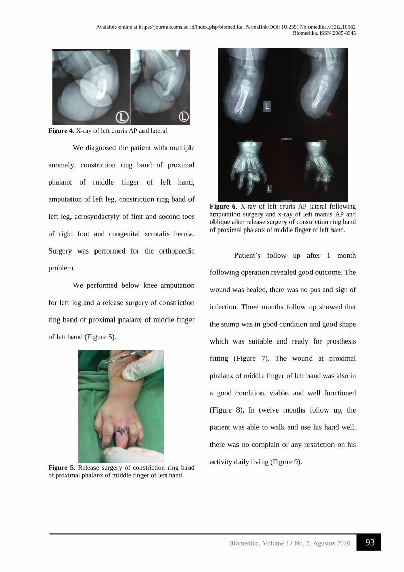
X-ray of left cruris AP lateral showed
amputated tibia one third proximal and signs of
soft tissue swelling in distal part (Figure. 4).
Avalaible online at https://journals.ums.ac.id/index.php/biomedika, Permalink/DOI: 10.23917/biomedika.v12i2.10562
Biomedika, ISSN 2085-8345
Biomedika, Volume 12 No. 2, Agustus 2020
93
Figure 4. X-ray of left cruris AP and lateral
We diagnosed the patient with multiple
anomaly, constriction ring band of proximal
phalanx of middle finger of left hand,
amputation of left leg, constriction ring band of
left leg, acrosyndactyly of first and second toes
of right foot and congenital scrotalis hernia.
Surgery was performed for the orthopaedic
problem.
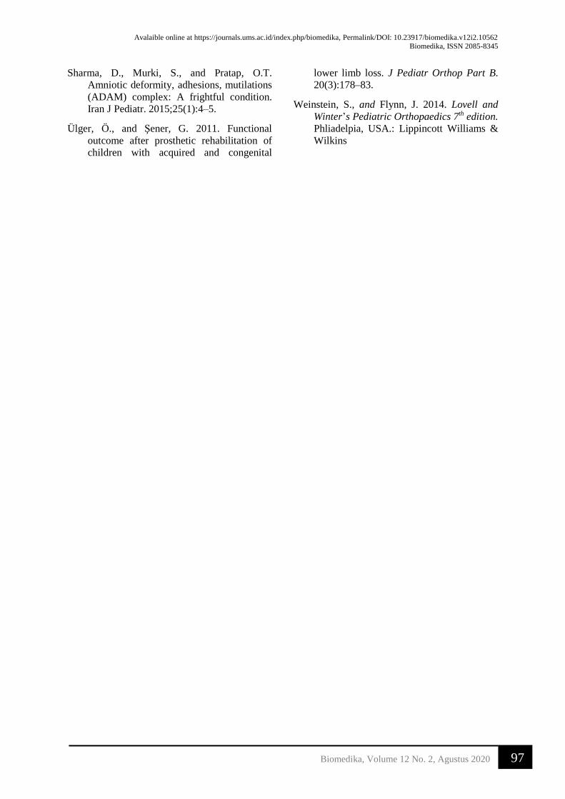
We performed below knee amputation
for left leg and a release surgery of constriction
ring band of proximal phalanx of middle finger
of left hand (Figure 5).
Figure 5. Release surgery of constriction ring band
of proximal phalanx of middle finger of left hand.
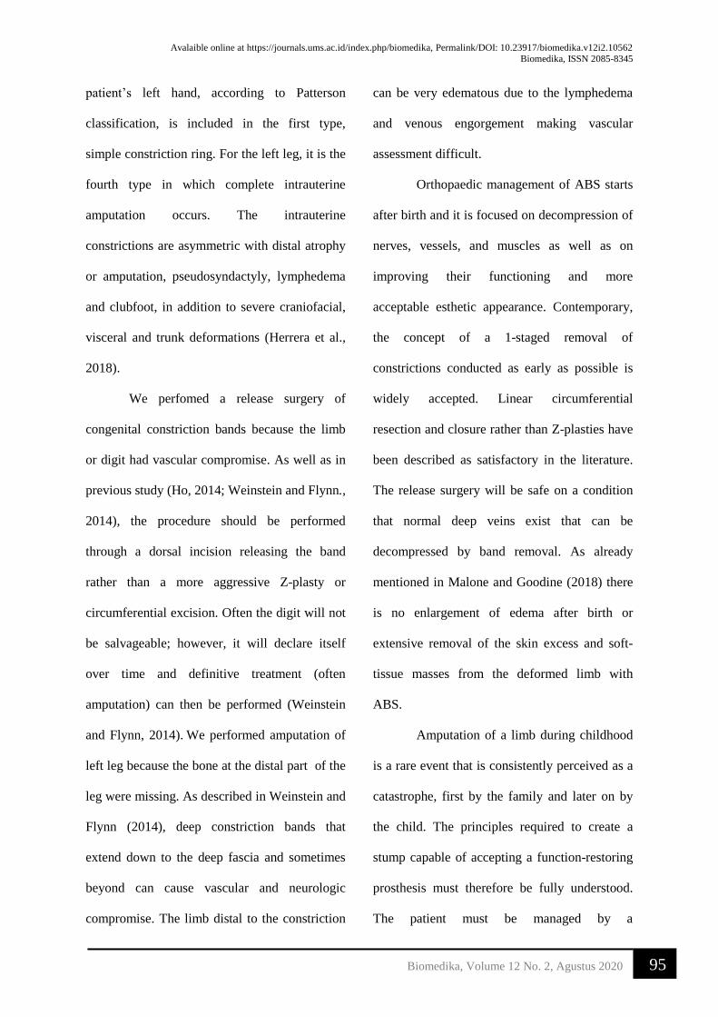
Figure 6. X-ray of left cruris AP lateral following
amputation surgery and x-ray of left manus AP and
oblique after release surgery of constriction ring band
of proximal phalanx of middle finger of left hand.
Patient’s follow up after 1 month
following operation revealed good outcome. The
wound was healed, there was no pus and sign of
infection. Three months follow up showed that
the stump was in good condition and good shape
which was suitable and ready for prosthesis
fitting (Figure 7). The wound at proximal
phalanx of middle finger of left hand was also in
a good condition, viable, and well functioned
(Figure 8). In twelve months follow up, the
patient was able to walk and use his hand well,
there was no complain or any restriction on his
activity daily living (Figure 9).

Avalaible online at https://journals.ums.ac.id/index.php/biomedika, Permalink/DOI: 10.23917/biomedika.v12i2.10562
Biomedika, ISSN 2085-8345
Biomedika, Volume 12 No. 2, Agustus 2020
94
Figure 7. the clinical picture of one year follow up of
the amputated stump..
Figure 8. The clinical picture of one year follow up
of left hand. There was the wound scar with good
viability of middle finger.
Figure 9. Follow up patient after one year, the
patient stood using prosthetic of below knee patellar
tendon bearing.
DISCUSSION
Amniotic band syndrome is a rare
condition that can present with many clinical
features, of which the major three components
are circumferential transverse bands,
acrosyndactyly, and terminal amputations
(Weinstein and Flynn., 2014).
The international standard nomenclature
for congenital limb deficiencies can help guide
the initial assessment of Congenital Limb
Deficiencies Disorders (CLLD) by dividing
limb deficiencies into two types: longitudinal
and transverse. In longitudinal deficiencies,
there is a reduction or absence within the long
axis of the limb and these deficiencies are often
part of a more complex syndrome. Transverse
limb deficiencies, as in this case, are defined as
having a developmentally normal limb until a
specific transverse plane is reached, with absent
or dysmorphic limb features distal to that plane
(Malone and Goodine, 2018).
Commonly, transverse limb deficiencies
result from amniotic banding within the
amniotic sac, which occurs when the amnion
layer of the placenta ruptures and separates from
the chorion, producing fibrous strands that
entangle and compromise blood flow in affected
extremities (Malone and Goodine, 2018). This
Avalaible online at https://journals.ums.ac.id/index.php/biomedika, Permalink/DOI: 10.23917/biomedika.v12i2.10562
Biomedika, ISSN 2085-8345
Biomedika, Volume 12 No. 2, Agustus 2020
95
patient’s left hand, according to Patterson
classification, is included in the first type,
simple constriction ring. For the left leg, it is the
fourth type in which complete intrauterine
amputation occurs. The intrauterine
constrictions are asymmetric with distal atrophy
or amputation, pseudosyndactyly, lymphedema
and clubfoot, in addition to severe craniofacial,
visceral and trunk deformations (Herrera et al.,
2018).
We perfomed a release surgery of
congenital constriction bands because the limb
or digit had vascular compromise. As well as in
previous study (Ho, 2014; Weinstein and Flynn.,
2014), the procedure should be performed
through a dorsal incision releasing the band
rather than a more aggressive Z-plasty or
circumferential excision. Often the digit will not
be salvageable; however, it will declare itself
over time and definitive treatment (often
amputation) can then be performed (Weinstein
and Flynn, 2014). We performed amputation of
left leg because the bone at the distal part of the
leg were missing. As described in Weinstein and
Flynn (2014), deep constriction bands that
extend down to the deep fascia and sometimes
beyond can cause vascular and neurologic
compromise. The limb distal to the constriction
can be very edematous due to the lymphedema
and venous engorgement making vascular
assessment difficult.
Orthopaedic management of ABS starts
after birth and it is focused on decompression of
nerves, vessels, and muscles as well as on
improving their functioning and more
acceptable esthetic appearance. Contemporary,
the concept of a 1-staged removal of
constrictions conducted as early as possible is
widely accepted. Linear circumferential
resection and closure rather than Z-plasties have
been described as satisfactory in the literature.
The release surgery will be safe on a condition
that normal deep veins exist that can be
decompressed by band removal. As already
mentioned in Malone and Goodine (2018) there
is no enlargement of edema after birth or
extensive removal of the skin excess and soft-
tissue masses from the deformed limb with
ABS.
Amputation of a limb during childhood
is a rare event that is consistently perceived as a
catastrophe, first by the family and later on by
the child. The principles required to create a
stump capable of accepting a function-restoring
prosthesis must therefore be fully understood.
The patient must be managed by a

Avalaible online at https://journals.ums.ac.id/index.php/biomedika, Permalink/DOI: 10.23917/biomedika.v12i2.10562
Biomedika, ISSN 2085-8345
Biomedika, Volume 12 No. 2, Agustus 2020
96
multidisciplinary team of professionals who
work closely together (Griffet, 2018). Prosthetic
use in children with lower limb loss fulfils the
appearance, provides early ambulation and
adapts the child to home life and social
participation through appropriate activity
training (Ülger and Şener, 2011).
Children are special. Their rehabilitation
requirements are different from those of an
adult. Children’s prostheses must allow some
kind of growth adjustments. Children are
extremely active, so the component used must
be robust; at the same time, follow-up and repair
services must be reliably available. Among the
“soft factors” of treatment, the prosthesis as well
as the fitting process must be enjoyable for the
child to get the child to use the prosthesis. The
objective of treatment here was achieved
successfully, the child could fully reintegrate
into his routine activity—schooling, cycling,
and playing football. Only then we will be able
to help the child to grow up without complexes
and inhibitions into a happy individual
(O’Keeffe and Rout , 2019).
CONCLUSION
Orthopaedic management of ABS starts
after birth and it is focused on decompression of
nerves, vessels, and muscles as well as on
improving their functioning and more
acceptable aesthetic appearance. We did an
acute releasing surgery of congenital
constriction bands with Z-plasty incision and
have a good results, also we could avoid
morbidity to the patient, provide more
acceptable aesthetic appearance. We also
performed below knee amputation procedure
and applied prosthetic of below knee patellar
tendon bearing. We achieved a good outcome in
patient activity daily living and ambulation.
REFFERENCES
Griffet, J. 2016. Amputation and prosthesis
fitting in paediatric patients. Orthop
Traumatol Surg Res. 102(1):S161–75.
Herrera, H.R.J., Martínez, P.Y.M., and
Izaguirre, E.D.M. 2015. Pseudosyndactyly
and amputation as the main features of the
amniotic band syndrome. Bol Med Hosp
Infant Mex. 68(February 2009):50–2.
Ho, C. 2014. Tachdjian’s Pediatric
Orthopaedics. V. Herring JA, editor. United
States of America: Texas Scottish Rite
Hospital for Children.
Koskimies, E., Syvänen, J., Nietosvaara, Y.,
Mäkitie, O., and Pakkasjärvi, N. 2015.
Congenital constriction band syndrome
with limb defects. J Pediatr Orthop.
35(1):100–3.
Malone, C., and Goodine, R.A. 2018.
Constriction band syndrome in a healthy
full-term newborn. Can Fam Physician.
64(8):577–8.
O’Keeffe, B., and Rout, S. 2019. Prosthetic
rehabilitation in the lower limb. Indian J
Plast Surg. 52(1):134–44.
Shetty, P., Menezes, L.T., Tauro, L.F., and
Diddigi, K.A. 2013. Amniotic Band
Syndrome. Indian J Surg. 75(5):401–2.
Avalaible online at https://journals.ums.ac.id/index.php/biomedika, Permalink/DOI: 10.23917/biomedika.v12i2.10562
Biomedika, ISSN 2085-8345
Biomedika, Volume 12 No. 2, Agustus 2020
97
Sharma, D., Murki, S., and Pratap, O.T.
Amniotic deformity, adhesions, mutilations
(ADAM) complex: A frightful condition.
Iran J Pediatr. 2015;25(1):4–5.
Ülger, Ö., and Şener, G. 2011. Functional
outcome after prosthetic rehabilitation of
children with acquired and congenital
lower limb loss. J Pediatr Orthop Part B.
20(3):178–83.
Weinstein, S., and Flynn, J. 2014. Lovell and
Winter’s Pediatric Orthopaedics 7th edition.
Phliadelpia, USA.: Lippincott Williams &
Wilkins