Surgical Treatment of Intracerebral Hemorrhage...Clinical Burden of ICH • Hypertension is the...
51
Surgical Treatment of Intracerebral Hemorrhage Regional Upstate Stroke and Health Summit 6/1/2018 Grahame C Gould, MD Assistant Professor of Neurological Surgery
Transcript of Surgical Treatment of Intracerebral Hemorrhage...Clinical Burden of ICH • Hypertension is the...

Surgical Treatment of Intracerebral Hemorrhage
Regional Upstate Stroke and Health Summit
6/1/2018
Grahame C Gould, MD
Assistant Professor of Neurological Surgery

Disclosures
No financial interests to disclose

Objectives• Brief overview of intracerebral hemorrhage (ICH)
• Current treatments / recommendations for ICH
• Role of traditional surgery in treatment of ICH
• Ongoing trials for minimally invasive ICH surgery
• Review tube-based retractor system for ICH surgery
• Discuss ENRICH trial

Hemorrhagic StrokeClinical History

Prevalence of Disease• 795,000+ strokes in United States annually
Circulation. 2017 March 07; 135(10): e146–e603. doi:10.1161/CIR.0000000000000485.
10%Intracerebral Hemorrhage
87%Ischemic Stroke
3%Subarachnoid Hemorrhage

Clinical Burden of ICH
• Hypertension is the leading cause of intracerebral hemorrhage (ICH)
• Mortality is greater than in Ischemic Stroke
• Early Mortality is up to 32-50%
• Of those that Survive:• 80% are left with significant disability• 12-39% Independent Function
• Economically, $12.7B of the $74B in direct costs for stroke care/year is attributed to ICH
Stroke, 2014;45:903-908

Current ICH Standard of Care
Neuro-ICU = Lower Mortality (I, LOE B)
• BP Control (<140, SAFE: I, LOE A - Improved Outcome:
IIa, LOE B)
• CPP optimization & ICP monitoring
• Reversal of Coagulopathy • PCCs over FFP in VKA, rFVIIa not recommended (III, LOE C)
• Protamine for Heparin (Iib, LOE C)
• PLT transfusion in PLA?
• IVH/CSF Drainage (IIa, LOE B)
• Supportive Care & Multi-system homeostasis• (Glc: I, LOE C, Fever Iib, LOE C)
No surgical intervention UNTIL/UNLESS
NEUROLOGICAL DECLINE
Stroke. 2015 Jul;46(7):2032-60
Only Cerebellar ICH surgery supported(8% of all ICHs): Class I, LOE B
Cerebellar hemorrhage >3 cm deteriorating neurologically or brain stem compression and/or hydrocephalus from ventricular
obstruction should have emergent surgical evacuation
Supratentorial spontaneous ICH (85% of all ICHs) is generally NOT a surgical event

Supratentorial vs Infratentorial?

ICH Standards for Surgery?
CLASS II RECOMMENDATIONS• For most supratentorial ICHs, usefulness of surgery NOT WELL
ESTABLISHED - (Class IIB, LOE A)
• Early evacuation Vs After deterioration NOT CLEARLY BENEFICIAL -(Class IIB, LOE A)
• For supratentorial spontaneous ICH (85% of all ICHs) evacuation MAY BE CONSIDERED in deteriorating patients as a LIFE-SAVING MEASURE - (Class IIB, LOE C)
• Decompressive craniectomy +/-hematoma evacuation might reduce mortality for supratentorial ICH with: coma/large hematoma with MLS or refractory ICP - (Class IIB, LOE C)
The effectiveness of MIS evacuation utilizing stereotactic or endoscopic aspiration +/-thrombolytics is uncertain (Class IIB, LOE A)
Stroke. 2015 Jul;46(7):2032-60

Surgical Trials in ICH1961-2004
• McKissock W, et al. Lancet. 1961;278(7196):221-226
• Auer LM, et al. J Neurosurg 1989;70(4):530-535
• Juvela S, et al. J Neurosurg. 1989;70(5):755-758
• Morgenstern LB, et al. (STICH single center) Neurology. 1998;51(5):1359-1363
• Zuccarello M, et al. Stroke. 1999;30(9):1833-1839
• Teernstra OP, et al. (SICHPA). Stroke. 2003;34(4):968-974
• Hattori N, et al. J Neurosurg. 2004;101(3):417-420
Positive for Surgical Evacuation BENEFIT!

Meta-Analyses: Surgical Trials
Meta-Analysis Studies AnalyzedOdds of Death/Dependency
with SurgeryConclusion
Hankey & Hon McKissock, Juvela, Auer, Batjer 1.23 (0.77-1.98) Insufficient Evidence
Prasad et al McKissock, Juvela, Auer, Batjer 1.99 (0.92-4.31) Insufficient Evidence
Saver et al Juvela, Auer, Batjer 0.72 (0.38-1.44)Trends Toward
Improved Outcome
Fernandes et alMcKissock, Juvela, Auer,
Batjer, Chern, Morgenstern, Zuccarello
1.2 (0.83-1.74)Trends Toward
Improved Outcome

STICHEarly surgery vs. initial conservative treatment in patients with spontaneous supratentorialintracerebral hematomas in the International Surgical Trial in intracerebral hemorrhage. Lancet. 2005;365(9457):387-397
• 1033 patients from 83 centers in 27 countries were randomized to early surgery (503) or initial conservative treatment (530)
• Favorable Outcome: Early surgery 122 (26%) Vs Initial conservative treatment 118 (24%)
• Subgroup Analysis:
• Patients with lobar hematoma and no IVH had 37% favorable outcome with conservative treatment vs. 49% with early surgery (p=0.08)
• STICH lacked sufficient power to address this subgroup
David Mendelow, MBBCh, PhD, FRCSDepartment of NeurosurgeryNewcastle General HospitalUniversity of Newcastle, UK
Failed to demonstrate superiority of surgery over medical management

STICH IIEarly surgery vs. initial conservative treatment in patients with spontaneous supratentorial lobar ICH: a randomized trial. Lancet. 2013;382(9890):397-408
• Craniotomy Vs. Best medical therapy for lobar spontaneous ICH 10-100 mL and no associated IVH (78 centers, 27 countries)
• 601 patients:
• Early surgery = 307, 6-month f/u: 298, included = 297
• Initial conservative treatment = 294, 6-month f/u: 291, included 286
•% Unfavorable Outcome: Surgery 174/297 (59%) Vs. 178/286 (62%) in conservative treatment group (absolute difference 3·7% [95% CI -4·3 to 11·6], odds ratio 0·86 [0·62 to 1·20]; p=0·367)
• 21% crossed-over to surgical group
David Mendelow, MBBCh, PhD, FRCSDepartment of NeurosurgeryNewcastle General HospitalUniversity of Newcastle, UK
No difference in intention-to-treat analysis of primary outcomes (extended GOS) at 6 months
Absolute difference of primary outcome dichotomy of 3.7% favored surgery
Other trends of a secondary shift in good disability scale strata and mortality also favored surgery with a 6% effect, trending but not reaching statistical significance

MISTIE IIMinimally invasive surgery plus rtPA for intracerebral hemorrhage evacuationISC 2013
• Purpose• To determine the safety of using a
combination of MIS and rtPA to remove ICH
• ICES arm of trial to determine safety of endoscopic surgery to remove ICH
• Study Design• Randomized, safety study, parallel
assignment, open label
Daniel Hanley, MDDepartment of Neurology, Anesthesiology & CCMJohns Hopkins University, School of MedicineBaltimore, MD

MISTIE II Outcomes
14
%
38 day LOS
Reduction$44K
savings
Hanley DF. MISTIE II Trial Results: Safety, Efficacy and Surgical Performance. Presentation at ISC 2013.

MISTIE II Outcomes
Hanley DF. MISTIE II Trial Results: Safety, Efficacy and Surgical Performance. Presentation at ISC 2013.

MISTIE III
• MISTIE III is funded by NIH/NINDS, under a cooperative agreement (U01: 1U01NS080824-01A1)• It involved over 90 centers in the US, Europe, Israel, China and
Australia• Last subject enrolled in August 2017
• MISTIE III intervention seeks to remove ICH through MIS and intermittent dosing of rt-PA for 3days • Cathflo Activase by Genentech in the US • Actilyse by Boehringer Ingelheim in Europe and Asia
• The primary endpoint is the Modified Rankin Score measured at 180-and 365-days
• Goal Improvement 12%

These Trials Have Shown:
Hanley et al, Minimally Invasive Surgery plus rt-PA for ICH Evacuation (MISTIE).Hanley, et al. Mistie Trial: 365-day Results Demonstrate Improved Outcomes and Cost Benefit.
• Benefit of MIS above other approaches
• Benefit of surgery over conservative TX
• Early surgery benefit
• Positive impact on functional outcomes
• Increased acute care cost savings
• Reduced hospital LOS

Challenges of Current MIS Approaches
• Requirement of clot stability for 6-12 hours• Prolonged clot resolution time
• Difficult to address active bleeding• Risk of rebleeding at primary and secondary sites• Clinical trials for surgical ICH Evacuation have NOT
consistently shown improved patient outcomes

Current ICH Trial Limitations
• Timing of intervention – Undefined therapeutic window
• Technique – Invasive and non standardized surgical techniques
• Patient selection – limitations, impact of associated factors

• Ideal surgery for ICH would:
• Allow for early intervention
• Minimize injury
• Maximize clot reduction
• Allow for hemostasis management
• Minimize re-bleeds
Defining a Surgical Approach for ICH

Rationale for Early Intervention
• ~ 30-40% of hematoma expansion of >1/3 of initial volume (most within 3 hours of onset)
• Targeting the spot sign (CTA)
• Rapid correction of intracranial hypertension
• Medical complications• PNA• Aspiration• Respiratory Failure• PE• Sepsis = 50% of deaths
2hr
4hr
Bross, et al. Early hemorrhage growth in patients with intracerebral hemorrhage. Stroke 1997.Kaneko et al. Surgery for hypertensive intracerebral hemorrhage. Jneurosurg 1977.

RBCs
HGB
Plasma proteins
DAMPs
Thrombin
Cytotoxic
Pro-inflammatory
Pro-oxidative
Endothel.
&
MG
Activation
BBB disruption
Vasogenic
edema
Apoptosis
Cytotoxic edema Necrosis
Mitochondrial
dysfunction
Cellular disruptionGlutamate release
Ca ++ influx
Secondary Brain Injury Cascade
Courtesy: Dr. Gustavo Pradilla

Evidence for Early Intervention
Gregson et al. Stroke 2012; 43 Zhou et al. Stroke 2012; 43
• 8 RCTs• 2186 Cases
Evidence that surgery is of benefit if undertaken early, before the patient
deteriorates
• 12 RCTs• 1955 Total Patients
Clinical outcomes showed early removal of hematoma can potentially mitigate brain injury provided surgeon can avoid damage to eloquent areas, cortical vasculature, and subcortical tracts
during surgical access

Is surgical technique important?

The Subcortical Space: Challenges with Traditional Surgical Approaches
• Deep lesions requiring access through uninvolved cortical and white matter structures
• Intraventricular lesions• Difficult access with traditional techniques
• Accurate subcortical targeting• Atraumatic Access
Subcortical Space
CHALLENGESRetraction Injury
• Brain tissue “creep”
Endoscopic Visualization
• Light & Magnification—visualization• Quality tissue collection
Access

Subcortical Injury
• Subcortical injury was found the primary cause of neurological deficits following awake craniotomy procedures
• Of cases developing new intraoperative neurological deficits, 90% occurred during subcortical dissection
• In eloquent cortex, 43% experienced worsened neurological deficits in immediate post-op period and 14% continued to have worsened deficits at 3-month follow-up
Preserving subcortical areas during cerebral resections may reduce the severity of both
immediate and late neurological deficits Trinh VT et al. Subcortical injury is an independent predictor of
worsening neurological deficits following awake craniotomy
procedures. Journal of Neurosurgery. 2013; 72(2):160-169.

Retraction Injury
• Challenges of subcortical surgery with retraction injury
• Dispersion of Pressure on Tissue
• Tissue “creep”
• Visualization at Depth
Retractor Evolution
Flat Blade Retractors
EndoscopyRadial
Retractors

Radial Retraction
• Dr. Pat Kelly (1980s)
• No inclusion of WMT analysis
• Manual Dissection Placement of Radial Retractor
• SHOWED POTENTIAL FOR LESS INJURY

Radial Retraction
• Small craniotomy • ≅30mm or smaller
• Small dural opening• ≅ Size of sheath used
• 13.5mm or 11mm
• Venting of ICP to be during cannulation
• Air-medium & bimanual microsurgical technique

Intra-operative Ultrasound
• Customized probe designed to work within retractor system:• Sulcal Identification• Vascular Flow• Lesion Location• Real Time Monitoring:
• Extent of Resection/Evacuation
• Proximity to Critical Structures
• Lesion Movement

Clot removalThe Myriad Device:• Customized aspiration system with cutting capacity
• Side-mouth aperture• Variable aspiration • Toggle between aspiration only and aspiration with cutting
• Allows for tissue collection in sterile, closed system

Less Invasive ICH Solution?
C
Labib et al. The Safety and Feasibility of Image-Guided BrainPath-Mediated Transsulcul Hematoma Evacuation: A Multicenter Study. Neurosurgery. 2016


Courtesy: Dr. Mohamed Labib
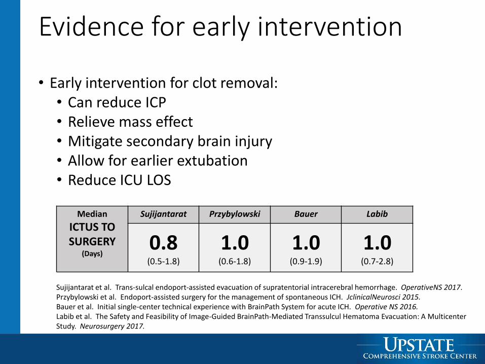
Evidence for early intervention
Median
ICTUS TO SURGERY
(Days)
Sujijantarat Przybylowski Bauer Labib
0.8(0.5-1.8)
1.0(0.6-1.8)
1.0(0.9-1.9)
1.0(0.7-2.8)
• Early intervention for clot removal:• Can reduce ICP• Relieve mass effect• Mitigate secondary brain injury• Allow for earlier extubation• Reduce ICU LOS
Sujijantarat et al. Trans-sulcal endoport-assisted evacuation of supratentorial intracerebral hemorrhage. OperativeNS 2017.Przybylowski et al. Endoport-assisted surgery for the management of spontaneous ICH. JclinicalNeurosci 2015.Bauer et al. Initial single-center technical experience with BrainPath System for acute ICH. Operative NS 2016.Labib et al. The Safety and Feasibility of Image-Guided BrainPath-Mediated Transsulcul Hematoma Evacuation: A Multicenter Study. Neurosurgery 2017.

Why Use Radial Retractor System?
• Standardize Technique
• Direct Hemostasis
• Avoid use of thrombolytics
Standard Approach MIS Port Access

• Shortest distance to lesion• Assumes all tissue is relevant• Tissue turgor favors “closure”
Kassam et al. Part II: an evaluation of an integrated systems approach using diffusion-weighted, image-guided, exoscopic-assisted, transulcal radial corridors. Innovative Neurosurgery; 2015Scranton et al. Transulcal parafascicular minimally invasive approach to deep and subcortical cavernomas: technical note. JNS 2016, Mar 4:1-7
Why Trans-sulcal?

Clot Reduction Matters
• Results from MISTIE have shown EOT volume correlated to mRS at 180-days
• Patients with <10mL of clot remaining had better mRS
• Patients with >35mL of clot remaining had worse mRS

Evidence for clot reduction
• Growing evidence to support maximal clot reduction with this approach
• Air-medium, bimanual approach for hemostasis
Sujijantarat Przybylowski Bauer Labib
Median
Pre-op Volume(cm3)
50.5cc(23.9-76.6)
78.0cc(40.5-105.5)
52.0cc(41.0-66.0)
36.0cc(27.0-65.0)
Median
Volume Clot Reduction (%)
91.6%(86.9-98.9)
87.0%(48.5-94.5)
98.2%(94.8-99.2)
NA
Sujijantarat et al. Trans-sulcal endoport-assisted evacuation of supratentorial intracerebral hemorrhage. OperativeNS2017.Przybylowski et al. Endoport-assisted surgery for the management of spontaneous ICH. JclinicalNeurosci 2015.Bauer et al. Initial single-center technical experience with BrainPath System for acute ICH. Operative NS 2016.Labib et al. The Safety and Feasibility of Image-Guided BrainPath-Mediated Transsulcul Hematoma Evacuation: A Multicenter Study. Neurosurgery 2017.

Outcomes
Sujijantarat Przybylowski Bauer Labib
Median
Pre-Op ICH Score
3 2 2.5 2Median
In-Hospital Mortality
6.3% 33.3% 0.0% 0.0%
Median
Length of Stay (Days)
24.0(16.0-33.0)
7.0(2.5-36.3)
16.7(9.7-20.9)
NA
Sujijantarat et al. Trans-sulcal endoport-assisted evacuation of supratentorial intracerebral hemorrhage. OperativeNS 2017.Przybylowski et al. Endoport-assisted surgery for the management of spontaneous ICH. JclinicalNeurosci2015.Bauer et al. Initial single-center technical experience with BrainPath System for acute ICH. Operative NS 2016.Labib et al. The Safety and Feasibility of Image-Guided BrainPath-Mediated Transsulcul Hematoma Evacuation: A Multicenter Study. Neurosurgery 2017.
• Growing evidence to support decreased mortality and LOS

Outcomes• Significant improvement in post-operative GCS
(p<0.001)
Sujijantarat Przybylowski Bauer Labib
Median
Pre-Op GCS7
(6.25-10)
NA 10(5.75-12)
10(8-14)
Median
Post-op GCS
13(10.25-14)
NA 14(9-14.25)
14(11-15)
Sujijantarat et al. Trans-sulcal endoport-assisted evacuation of supratentorial intracerebral hemorrhage. OperativeNS 2017.Przybylowski et al. Endoport-assisted surgery for the management of spontaneous ICH. JclinicalNeurosci2015.Bauer et al. Initial single-center technical experience with BrainPath System for acute ICH. Operative NS 2016.Labib et al. The Safety and Feasibility of Image-Guided BrainPath-Mediated Transsulcul Hematoma Evacuation: A Multicenter Study. Neurosurgery 2017.


ENRICH:Early MiNimally-Invasive Removal of
ICH

Study Team
• Scientific Leadership Team• Gustavo Pradilla, MD (Neurosurgery) – Emory University• Daniel Barrow, MD (Neurosurgery) – Emory University• Jonathan Ratcliff, MD (Neurocritical Care) – Emory University• Jason Allen, MD (Neuroradiology) – Emory University• David Wright, MD (Emergency Medicine) – Emory University• Michael Frankel, MD (Neurology) – Emory University• Alex Hall, MS, RN (Clinical Research Nurse) – Emory University• Victoria Phillips, DPhil (Healthcare Economist) – Emory University
• Data Safety Monitoring Board & Medical Monitor• Mark Hadley, MD (DSMB Chair) – University of Alabama• Greg Campbell, PhD (Biostatistician) – GCStat Consulting, LLC• Opeolu Adeoye, MD (Medical Monitor) – University of Cincinnati

Study Purpose
To determine if minimally invasive parafascicular surgery (MIPS) using currently available and FDA cleared
technology for early intracerebral hemorrhage evacuation results in improved functional outcome and economic
benefit when compared to standard medical management

Inclusion Criteria
• Age 18 – 80• CT showing acute spontaneous primary ICH• GCS 5-14• ICH Volume 30-80mL• Study intervention can be initiated within 24
hours of last known well (TX ≤ 8 hrs is preferred)
• Historical mRS 0 of 1
Exclusion Criteria• Aneurysm, avm, vascular anamoly etc.• NIHSS</=5• Bilateral fixed and dilated pupils• Extensor motor posturing• IVH involving >50% of the lateral ventricles• Primary thalamic ICH• Midbrain, pontine, or cerebellar ICH• Use of anticoagulants that can’t be rapidly reversed• ESRD• ESLD• Evidence of active bleeding involving the retroperitoneal,
gastrointestinal, genitourinary or respiratory tract• Uncorrected coagulopathy or known clotting disorder• Plt < 75k, INR > 1.4 after correction• Life expectancy < 6 months• Pregnant• No reasonable expectation of recovery, DNR, comfort measures
only• Mechanical heart valve• Need to resume anticoagulation < 5 days• Unable to meet follow up requirements

Objectives
• Efficacy• Determine if MIPS for ICH evacuation results in a 10% improvement in 180-day utility-
weighted modified Rankin Scale• Quantify the cost per quality-adjusted life-years (QALY) gained through MIPS
• Safety• Determine if MIPS results in increased mortality compared to standard management
• Determine if MIPS results in an increase in hemorrhage volume between index and 24 hour follow-up CT
• Secondary• To assess and quantify post-operative rebleeding associated with clinical deterioration
following MIPS
• Demonstrate that percent volume of ICH reduction is associated with improved functional outcome
• Compare UW-mRS at discharge, 30 days, and 90 days between the treatment groups.

Study Design
• Multi-center, randomized, adapative clinical trial
• Block randomization based on hemorrhage location (anterior basal ganglia vs lobar)
with subsequent enrichment on location.
• Sample size: 150 - 300 subjects
• Interim Analyses: 150 then every 25 patients up to 300
• Evaluate for stopping or enrichment at each interim
• Enrichment, and early stopping rules for futility and predicted success are predetermined

Study Update
• Trial update as of December 31, 2017 presented at 2018 International Stroke Conference• 21 Active Sites• 60 Enrolled Patients • 50mL Median Hemorrhage Volume (48%
Lobar)• Mean NIHSS and GCS reported as 18.3
and 11.3, respectively• Mean time from LKN to randomization
13:07 hours• Randomization to surgery of 2:30 hours• Follow-up completed for 29/60 enrolled
patients
Ratcliff et al. Clinical Trial Update: Early Minimally Invasive Removal of Intracerebral Hemorrhage (ENRICH) Clinical Trial. Presented at
2018 International Stroke Conference. Poster # CTP7.

Conclusion• ICH is a common, devastating form of stroke
• ICH treatment has made little progress
• Optimal clinical targets based on data• Treat early
• Remove more clot
• Protect normal brain
• Radial retractor system to hit the targets
• Could ICH be the next ELVO?


