Well-differentiated intrahepatic cholangiocarcinoma in the ...
Upload
gouda-ellabbanCategory
view
98download
3

Updates in Surgery
Alfredo Guglielmi • Andrea Ruzzenente • Calogero Iacono
Surgical Treatment of Hilar and IntrahepaticCholangiocarcinoma
In cooperation with Luigi MarchioriSilvia PacheraLuca BortolasiRiccardo ManfrediPaola Capelli
13
Alfredo GuglielmiAndrea RuzzenenteCalogero IaconoGeneral Surgery A, Department of Surgery and Gastroenterology University Hospital G.B. Rossi Verona, Italy
in cooperation withLuigi Marchiori, Silvia Pachera, Luca BortolasiGeneral Surgery A, Department of Surgery and GastroenterologyUniversity Hospital G.B. Rossi Verona, Italy
Riccardo ManfrediDepartment of RadiologyUniversity Hospital G.B. Rossi Verona, Italy
Paola CapelliDepartment of PathologyUniversity Hospital G.B. Rossi Verona, Italy
The publication and the distribution of this volume have been supported by the ItalianSociety of Surgery
Library of Congress Control Number: 2007931745
ISBN 978-88-470-0728-4 Milan Heidelberg New Yorke-ISBN 978-88-470-0729-1
Springer is a part of Springer Science+Business Mediaspringer.com© Springer-Verlag Italia 2008
This work is subject to copyright. All rights are reserved, whether the whole or part of the mate-rial is concerned, specifically the rights of translation, reprinting, re-use of illustrations, recitation,broadcasting, reproduction on microfilms or in other ways, and storage in data banks. Duplicationof this publication or parts thereof is only permitted under the provisions of the Italian CopyrightLaw in its current version, and permission for use must always be obtained from Springer.Violations are liable for prosecution under the Italian Copyright Law.
The use of general descriptive names, registered names, trademarks, etc., in this publication doesnot imply, even in the absence of a specific statement, that such names are exempt from the rele-vant protective laws and regulations and therefore free for general use.Product liability: The publisher cannot guarantee the accuracy of any information about dosageand application contained in this book. In every individual case the user must check such infor-mation by consulting the relevant literature.
Cover design: Simona Colombo, Milan, ItalyTypesetting: Graphostudio, Milan, ItalyPrinting and binding: Arti Grafiche Nidasio, Assago, Italy
Printed in ItalySpringer-Verlag Italia S.r.l. – Via Decembrio 28 – I-20137 Milan
Foreword
In the 10 years since the presentation of Gazzaniga’s excellent monograph on extra-hepatic biliary tumours at the SIC Congress in 1997, interesting developments inthe field have pressed upon us an undoubted need to reassess the complex topic ofhepatobiliary surgery. It is therefore with great pleasure that, at the proposal of theSteering Committee of the Italian Society of Surgery (SIC), we present to Italiansurgeons this monograph on the treatment of hilar and intrahepatic cholangiocarci-noma, prepared by Alfredo Guglielmi, professor and chairman of SurgicalDepartment A at the University School of Medicine of Verona, and his colleagues.
The volume is divided into two parts. The first relates to hilar cholangiocarci-nomas, about which there are a number of complex and still controversial issues;the second relates to intrahepatic cholangiocarcinomas, which are frequently treat-ed like other primary tumours of the liver. The monograph includes preliminaryinformation about molecular biology, and diagnostic and treatment methods areextensively examined in relation to type of neoplastic spread. In our opinion themost interesting part of the book concerns the treatment of these tumours with bothinterventional radiology and surgery, which can range from simple hepatectomiesto liver transplantation. We would like to point out that Professor Guglielmi carriedout the first liver transplantation in the University of Verona surgical department.
The monograph is aimed at those in clinical practice, and is written by a col-league for whom the most important objective of his work is to perform the bestsurgery in the light of the most recent developments and to achieve results in theinterests of his patients. Just as usually happens in clinical practice, pathologistsand radiologists have participated in the making of this book. This type of “diseasemanagement team” improves the quality and results of surgery.
Over the last 20 years, Alfredo Guglielmi has followed with enthusiasm andintelligence the progress made in hepatobiliary surgery. He has connections withEuropean, American and Japanese surgical institutes; he performed an in-depthstudy of the surgical treatment of cholangiocarcinoma at the University of Nagoyaunder the guidance of Yuji Nimura, a pioneer in this field. The mutuality of this pro-fessional relationship and sincere friendship is attested to by several visits of YujiNimura to the surgical department of the University School of Medicine of Verona.

It is for all these reasons that we are proud to present to Italian surgeons thismonograph, which conveys the most recent orientations of the SIC. For the firsttime it is being published in English by a publishing house with an internationalpresence. We think that this is the best way to share our experiences with colleaguesfrom other countries. We also feel that this volume will be an excellent instrumentfor the achievement of the aims of the SIC Steering Committee. We hope it willachieve wide circulation and great success.
Verona, October 2007 Claudio CordianoPast President, Italian Society of Surgery
Rome, October 2007 Roberto TersigniPresident, Italian Society of Surgery
ForewordVI
Preface
I would like to thank the Steering Committee of the Italian Society of Surgery (SIC)for giving me the opportunity of writing this book. I accepted the task enthusiasti-cally, because the surgery of cholangiocarcinoma is a fascinating and complex partof hepatobiliary surgery that has undergone numerous changes over recent decades.My aim has been to provide an update on the diagnosis, staging, preoperative man-agement and treatment of hilar and intrahepatic cholangiocarcinoma, and to pro-vide a critical review of diagnostic and therapeutic tools in the light of the publishedliterature and personal experience.
Cholangiocarcinoma is a rare neoplasm, but its incidence is increasing inWestern and Eastern countries. It was described in rare reports before the 1950s,and only in 1965 did Klatskin collect 13 cases and describe the clinical and patho-logical characteristics of the disease. Classically, cholangiocarcinoma is cate-gorised into intrahepatic and extrahepatic types according to the location of thetumour along the biliary tract. However, the frequently mixed type of growth ofthese tumours often makes this type of classification difficult to apply. This mono-graph has therefore been divided into two parts to analyse the differences betweenthe two types of cholangiocarcinoma, emphasising that the treatment of these twoneoplasms often requires combined hepatic and bile duct resection.
The modern era of surgery of hilar cholangiocarcinoma began in 1954 whenBrown performed the first bile duct resection for hilar cholangiocarcinoma. Thefirst experiences of bile duct resection associated with liver resection were pub-lished during the 1960s. From the 1970s onwards, Longmire, Fortner and Launoisreported the first surgical series with good survival results but with a high rate ofmortality and complications. After the 1980s, the clinico-anatomical study of thehepatic hilum and the segmental biliary drainage proposed by Nimura led to furtherprogress in precise preoperative diagnosis of tumour extent and surgical planning.
More recently, progress in non-invasive diagnostic tools has further improvedthe preoperative evaluation, with a reduction in invasive diagnostic techniques.During the past decade, improvements in surgical techniques and preoperative opti-misation of liver function (preoperative biliary drainage and portal vein embolisa-tion) have made it possible to perform extended liver resection combined with vas-
cular resection and reconstruction with low mortality and morbidity. The highercurative resection rate of this type of surgical approach has also improved long-term results.
The surgery of cholangiocarcinoma is still changing today, and several contro-versies in preoperative and surgical management remain. These open issues providestimulus for further research and new ideas on the treatment of this tumour.
I am extremely grateful to Professor Claudio Cordiano, who in his surgicaldepartment stimulated and supported me in the development of hepatobiliary sur-gery. I owe a debt of thanks to my “maestro”. I also thank Professor Yuji Nimurafor all the teachings he gave me, with great willingness and undisputed expertise,over the last 15 years. His thoughts are present in many places in the pages of thisbook.
Thanks also go to all the colleagues who helped me during the writing of thebook, of whom I asked dedication and diligence, and into whom I hope I haveinstilled my passion for this challenging branch of surgery.
Finally, thank you to Giovanni Paolo Pianegonda for his wonderful drawings.
Alfredo Guglielmi
PrefaceVIII
Contents
Part 1: Hilar Cholangiocarcinoma
Reporting Cholangiocarcinoma: Pathological Aspects . . . . . . . . . . . . . . . . . . 3
Definitions . . . . . . . . . . . . . . . . . . . . . . . . . . . . . . . . . . . . . . . . . . . . . . . . . . . . . . . . . 3
Clinical Information . . . . . . . . . . . . . . . . . . . . . . . . . . . . . . . . . . . . . . . . . . . . . . . . . 4
Intraoperative Consultation . . . . . . . . . . . . . . . . . . . . . . . . . . . . . . . . . . . . . . . . . . . . 5
Macroscopic Examination . . . . . . . . . . . . . . . . . . . . . . . . . . . . . . . . . . . . . . . . . . . . . 6
Pathology Findings in Non-Neoplastic Liver . . . . . . . . . . . . . . . . . . . . . . . . . . . . . . 9
Lymph Nodes (Location, Number) . . . . . . . . . . . . . . . . . . . . . . . . . . . . . . . . . . . . . . 10
Frozen Tissue (Molecular Studies) . . . . . . . . . . . . . . . . . . . . . . . . . . . . . . . . . . . . . . 10
Microscopic Examination . . . . . . . . . . . . . . . . . . . . . . . . . . . . . . . . . . . . . . . . . . . . . 10
Additional Pathology Findings . . . . . . . . . . . . . . . . . . . . . . . . . . . . . . . . . . . . . . . . . 12
Immunohistochemistry . . . . . . . . . . . . . . . . . . . . . . . . . . . . . . . . . . . . . . . . . . . . . . . 13
Diagnosis . . . . . . . . . . . . . . . . . . . . . . . . . . . . . . . . . . . . . . . . . . . . . . . . . . . . . . . . . . . . 17
Ultrasound (Endoscopic, Intraductal, Transabdominal) . . . . . . . . . . . . . . . . . . . . . . 17
Computed Tomography . . . . . . . . . . . . . . . . . . . . . . . . . . . . . . . . . . . . . . . . . . . . . . . 20
Magnetic Resonance Imaging . . . . . . . . . . . . . . . . . . . . . . . . . . . . . . . . . . . . . . . . . . 21
Positron Emission Tomography . . . . . . . . . . . . . . . . . . . . . . . . . . . . . . . . . . . . . . . . 23
Direct Cholangiography (ERCP and PTC) . . . . . . . . . . . . . . . . . . . . . . . . . . . . . . . . 23
Cholangioscopy (Peroral, Percutaneous) . . . . . . . . . . . . . . . . . . . . . . . . . . . . . . . . . 24
Angiography . . . . . . . . . . . . . . . . . . . . . . . . . . . . . . . . . . . . . . . . . . . . . . . . . . . . . . . 25
Preoperative Staging . . . . . . . . . . . . . . . . . . . . . . . . . . . . . . . . . . . . . . . . . . . . . . . . . 29
Evaluation of the Biliary Involvement (Longitudinal Extent) . . . . . . . . . . . . . . . . . 30
Evaluation of Radial Extent: Vascular Involvement,Parenchymal Involvement and Hepatic Lobar Atrophy . . . . . . . . . . . . . . . . . . . . . . 33
Preoperative Assessment of Tumour Resectability (T) . . . . . . . . . . . . . . . . . . . . . . . 35
Evaluation of Lymph Node Status (N) . . . . . . . . . . . . . . . . . . . . . . . . . . . . . . . . . . . 36
Evaluation of Metastases (M) . . . . . . . . . . . . . . . . . . . . . . . . . . . . . . . . . . . . . . . . . . 37
Conclusions . . . . . . . . . . . . . . . . . . . . . . . . . . . . . . . . . . . . . . . . . . . . . . . . . . . . . . . . 37
The Role of Laparoscopy in Preoperative Staging . . . . . . . . . . . . . . . . . . . . . . 43
Technique . . . . . . . . . . . . . . . . . . . . . . . . . . . . . . . . . . . . . . . . . . . . . . . . . . . . . . . . . 43
Results . . . . . . . . . . . . . . . . . . . . . . . . . . . . . . . . . . . . . . . . . . . . . . . . . . . . . . . . . . . . 44
Laparoscopic Ultrasound . . . . . . . . . . . . . . . . . . . . . . . . . . . . . . . . . . . . . . . . . . . . . 45
Conclusions . . . . . . . . . . . . . . . . . . . . . . . . . . . . . . . . . . . . . . . . . . . . . . . . . . . . . . . . 46
Preoperative Assessment of Liver Function . . . . . . . . . . . . . . . . . . . . . . . . . . . . 49
Preoperative Biliary Drainage . . . . . . . . . . . . . . . . . . . . . . . . . . . . . . . . . . . . . . . . 57
Drainage: Pros . . . . . . . . . . . . . . . . . . . . . . . . . . . . . . . . . . . . . . . . . . . . . . . . . . . . . . 58
Drainage: Cons . . . . . . . . . . . . . . . . . . . . . . . . . . . . . . . . . . . . . . . . . . . . . . . . . . . . . 59
Conclusions . . . . . . . . . . . . . . . . . . . . . . . . . . . . . . . . . . . . . . . . . . . . . . . . . . . . . . . . 62
Preoperative Portal Vein Embolization . . . . . . . . . . . . . . . . . . . . . . . . . . . . . . . . 67
Physiopathology of PVE . . . . . . . . . . . . . . . . . . . . . . . . . . . . . . . . . . . . . . . . . . . . . . 67
Indications . . . . . . . . . . . . . . . . . . . . . . . . . . . . . . . . . . . . . . . . . . . . . . . . . . . . . . . . . 69
Contraindications . . . . . . . . . . . . . . . . . . . . . . . . . . . . . . . . . . . . . . . . . . . . . . . . . . . 69
Technique . . . . . . . . . . . . . . . . . . . . . . . . . . . . . . . . . . . . . . . . . . . . . . . . . . . . . . . . . 69
Results . . . . . . . . . . . . . . . . . . . . . . . . . . . . . . . . . . . . . . . . . . . . . . . . . . . . . . . . . . . . 71
Post-PVE Course and Timing of Resection . . . . . . . . . . . . . . . . . . . . . . . . . . . . . . . 72
Conclusions . . . . . . . . . . . . . . . . . . . . . . . . . . . . . . . . . . . . . . . . . . . . . . . . . . . . . . . . 72
Prognostic Factors . . . . . . . . . . . . . . . . . . . . . . . . . . . . . . . . . . . . . . . . . . . . . . . . . . . 75
Gross Type . . . . . . . . . . . . . . . . . . . . . . . . . . . . . . . . . . . . . . . . . . . . . . . . . . . . . . . . . 75
Microscopic Pattern . . . . . . . . . . . . . . . . . . . . . . . . . . . . . . . . . . . . . . . . . . . . . . . . . 76
Biological and Molecular Prognostic Factors . . . . . . . . . . . . . . . . . . . . . . . . . . . . . . 76
T Category . . . . . . . . . . . . . . . . . . . . . . . . . . . . . . . . . . . . . . . . . . . . . . . . . . . . . . . . . 77
N Category . . . . . . . . . . . . . . . . . . . . . . . . . . . . . . . . . . . . . . . . . . . . . . . . . . . . . . . . 81
M Category . . . . . . . . . . . . . . . . . . . . . . . . . . . . . . . . . . . . . . . . . . . . . . . . . . . . . . . . 82
Prognostic Significance of TNM UICC/AJCC Classification . . . . . . . . . . . . . . . . . 82
Staging Systems . . . . . . . . . . . . . . . . . . . . . . . . . . . . . . . . . . . . . . . . . . . . . . . . . . . . . 87
Bismuth-Corlette Classification . . . . . . . . . . . . . . . . . . . . . . . . . . . . . . . . . . . . . . . . 87
TNM Staging System According to UICC/AJCC 6th Edition . . . . . . . . . . . . . . . . . 88
Comparison between 5th and 6th Edition of TNM UICC/AJCC . . . . . . . . . . . . . . . 91
Staging System According to JSBS . . . . . . . . . . . . . . . . . . . . . . . . . . . . . . . . . . . . . 91
Early Cancer . . . . . . . . . . . . . . . . . . . . . . . . . . . . . . . . . . . . . . . . . . . . . . . . . . . . . . . 96
Gazzaniga Staging System . . . . . . . . . . . . . . . . . . . . . . . . . . . . . . . . . . . . . . . . . . . . 96
Memorial Sloan-Kettering Cancer Center Staging . . . . . . . . . . . . . . . . . . . . . . . . . . 98
Conclusions . . . . . . . . . . . . . . . . . . . . . . . . . . . . . . . . . . . . . . . . . . . . . . . . . . . . . . . . 99
ContentsX
Surgical Anatomy of the Hepatic Hilus . . . . . . . . . . . . . . . . . . . . . . . . . . . . . . . . 101
Anatomy of the Bile Duct Branches . . . . . . . . . . . . . . . . . . . . . . . . . . . . . . . . . . . . . 102
Anatomy of the Portal Vein Branches . . . . . . . . . . . . . . . . . . . . . . . . . . . . . . . . . . . . 105
Anatomy of the Hepatic Artery Branches . . . . . . . . . . . . . . . . . . . . . . . . . . . . . . . . . 107
Surgical Anatomy of the Caudate Lobe . . . . . . . . . . . . . . . . . . . . . . . . . . . . . . . . . . 108
Surgical Treatment . . . . . . . . . . . . . . . . . . . . . . . . . . . . . . . . . . . . . . . . . . . . . . . . . . 113
General Principles . . . . . . . . . . . . . . . . . . . . . . . . . . . . . . . . . . . . . . . . . . . . . . . . . . . 113
Assessment of Resectability . . . . . . . . . . . . . . . . . . . . . . . . . . . . . . . . . . . . . . . . . . . 115
Indication for Surgical Resection . . . . . . . . . . . . . . . . . . . . . . . . . . . . . . . . . . . . . . . 120
Isolated Extrahepatic Bile Duct Resection . . . . . . . . . . . . . . . . . . . . . . . . . . . . . . . . 121
Independent Caudate Lobectomy (S1) . . . . . . . . . . . . . . . . . . . . . . . . . . . . . . . . . . . 122
Central Hepatic Resections . . . . . . . . . . . . . . . . . . . . . . . . . . . . . . . . . . . . . . . . . . . . 123
Extended Right Resections . . . . . . . . . . . . . . . . . . . . . . . . . . . . . . . . . . . . . . . . . . . . 124
Extended Left Resections . . . . . . . . . . . . . . . . . . . . . . . . . . . . . . . . . . . . . . . . . . . . . 125
Surgical Technique . . . . . . . . . . . . . . . . . . . . . . . . . . . . . . . . . . . . . . . . . . . . . . . . . . . 129
Position of the Patient . . . . . . . . . . . . . . . . . . . . . . . . . . . . . . . . . . . . . . . . . . . . . . . . 129
Incision . . . . . . . . . . . . . . . . . . . . . . . . . . . . . . . . . . . . . . . . . . . . . . . . . . . . . . . . . . . 129
Intraoperative Exploration . . . . . . . . . . . . . . . . . . . . . . . . . . . . . . . . . . . . . . . . . . . . . 130
Hepatic Pedicle Dissection and Lymphadenectomy . . . . . . . . . . . . . . . . . . . . . . . . . 130
Bile Duct Resection Alone . . . . . . . . . . . . . . . . . . . . . . . . . . . . . . . . . . . . . . . . . . . . 131
Independent Caudate Lobectomy (S1) . . . . . . . . . . . . . . . . . . . . . . . . . . . . . . . . . . . 132
Right Hepatectomy with Caudate Lobectomy (S4a, S5, S6, S7, S8 + S1) . . . . . . . 133
Right Trisectionectomy with Caudate Lobectomy (S4, S5, S6, S7, S8 + S1) . . . . . 135
Left Hepatectomy with Caudate Lobectomy (S2, S3, S4 + S1) . . . . . . . . . . . . . . . . 138
Left Trisectionectomy with Caudate Lobectomy (S2, S3, S4, S5, S8 + S1) . . . . . . 139
Central (Preserving) Hepatectomy . . . . . . . . . . . . . . . . . . . . . . . . . . . . . . . . . . . . . . 141
Hepatectomy with Portal Resection and Reconstruction . . . . . . . . . . . . . . . . . . . . . 143
Hepatectomy with Arterial Resection and Reconstruction . . . . . . . . . . . . . . . . . . . . 148
Hepatopancreatoduodenectomy (HPD) . . . . . . . . . . . . . . . . . . . . . . . . . . . . . . . . . . 149
Biliary Anastomosis . . . . . . . . . . . . . . . . . . . . . . . . . . . . . . . . . . . . . . . . . . . . . . . . . 150
Results of Surgery . . . . . . . . . . . . . . . . . . . . . . . . . . . . . . . . . . . . . . . . . . . . . . . . . . . 153
Morbidity and Mortality . . . . . . . . . . . . . . . . . . . . . . . . . . . . . . . . . . . . . . . . . . . . . . 153
Long-term Results . . . . . . . . . . . . . . . . . . . . . . . . . . . . . . . . . . . . . . . . . . . . . . . . . . . 157
Recurrence . . . . . . . . . . . . . . . . . . . . . . . . . . . . . . . . . . . . . . . . . . . . . . . . . . . . . . . . . 159
The Role of Liver Transplantation . . . . . . . . . . . . . . . . . . . . . . . . . . . . . . . . . . . . 163
Indications and Results . . . . . . . . . . . . . . . . . . . . . . . . . . . . . . . . . . . . . . . . . . . . . . . 163
Combined Transplantation . . . . . . . . . . . . . . . . . . . . . . . . . . . . . . . . . . . . . . . . . . . . 165
Contents XI
Transplantation with Adjuvant and Neoadjuvant Treatments . . . . . . . . . . . . . . . . . . 166
Conclusions . . . . . . . . . . . . . . . . . . . . . . . . . . . . . . . . . . . . . . . . . . . . . . . . . . . . . . . . 167
Adjuvant and Neoadjuvant Treatments . . . . . . . . . . . . . . . . . . . . . . . . . . . . . . . 169
Chemotherapy . . . . . . . . . . . . . . . . . . . . . . . . . . . . . . . . . . . . . . . . . . . . . . . . . . . . . . 169
Radiotherapy . . . . . . . . . . . . . . . . . . . . . . . . . . . . . . . . . . . . . . . . . . . . . . . . . . . . . . . 170
Chemoradiation Therapy . . . . . . . . . . . . . . . . . . . . . . . . . . . . . . . . . . . . . . . . . . . . . . 170
Neoadjuvant Therapy . . . . . . . . . . . . . . . . . . . . . . . . . . . . . . . . . . . . . . . . . . . . . . . . 171
Conclusions . . . . . . . . . . . . . . . . . . . . . . . . . . . . . . . . . . . . . . . . . . . . . . . . . . . . . . . . 172
Palliative Treatments . . . . . . . . . . . . . . . . . . . . . . . . . . . . . . . . . . . . . . . . . . . . . . . . . 175
Palliation of Jaundice . . . . . . . . . . . . . . . . . . . . . . . . . . . . . . . . . . . . . . . . . . . . . . . . 175
Chemotherapy, Radiotherapy and Photodynamic Therapy . . . . . . . . . . . . . . . . . . . . 179
Part 2: Intrahepatic Cholangiocarcinoma
Diagnosis . . . . . . . . . . . . . . . . . . . . . . . . . . . . . . . . . . . . . . . . . . . . . . . . . . . . . . . . . . . . 187
Ultrasound . . . . . . . . . . . . . . . . . . . . . . . . . . . . . . . . . . . . . . . . . . . . . . . . . . . . . . . . . 187
Computed Tomography . . . . . . . . . . . . . . . . . . . . . . . . . . . . . . . . . . . . . . . . . . . . . . . 188
Magnetic Resonance Imaging . . . . . . . . . . . . . . . . . . . . . . . . . . . . . . . . . . . . . . . . . . 190
Angiography . . . . . . . . . . . . . . . . . . . . . . . . . . . . . . . . . . . . . . . . . . . . . . . . . . . . . . . 192
Prognostic Factors . . . . . . . . . . . . . . . . . . . . . . . . . . . . . . . . . . . . . . . . . . . . . . . . . . . 193
Gross Type . . . . . . . . . . . . . . . . . . . . . . . . . . . . . . . . . . . . . . . . . . . . . . . . . . . . . . . . . 193
T Category . . . . . . . . . . . . . . . . . . . . . . . . . . . . . . . . . . . . . . . . . . . . . . . . . . . . . . . . . 195
N Category . . . . . . . . . . . . . . . . . . . . . . . . . . . . . . . . . . . . . . . . . . . . . . . . . . . . . . . . 196
Microscopic Pattern . . . . . . . . . . . . . . . . . . . . . . . . . . . . . . . . . . . . . . . . . . . . . . . . . 199
Staging Systems . . . . . . . . . . . . . . . . . . . . . . . . . . . . . . . . . . . . . . . . . . . . . . . . . . . . . 203
TNM Staging System According to UICC/AJCC . . . . . . . . . . . . . . . . . . . . . . . . . . 203
TNM Classification According to the LCSGJ . . . . . . . . . . . . . . . . . . . . . . . . . . . . . 206
Conclusions . . . . . . . . . . . . . . . . . . . . . . . . . . . . . . . . . . . . . . . . . . . . . . . . . . . . . . . . 210
Surgical Treatment . . . . . . . . . . . . . . . . . . . . . . . . . . . . . . . . . . . . . . . . . . . . . . . . . . 213
Intraoperative Assessment of Resectability . . . . . . . . . . . . . . . . . . . . . . . . . . . . . . . 213
Indications for Surgical Resection . . . . . . . . . . . . . . . . . . . . . . . . . . . . . . . . . . . . . . 214
Type of Surgical Resection . . . . . . . . . . . . . . . . . . . . . . . . . . . . . . . . . . . . . . . . . . . . 214
Indications for Lymphadenectomy . . . . . . . . . . . . . . . . . . . . . . . . . . . . . . . . . . . . . . 217
Extrahepatic Metastases . . . . . . . . . . . . . . . . . . . . . . . . . . . . . . . . . . . . . . . . . . . . . . 219
ContentsXII
Results of Surgery . . . . . . . . . . . . . . . . . . . . . . . . . . . . . . . . . . . . . . . . . . . . . . . . . . . 221
Morbidity and Mortality . . . . . . . . . . . . . . . . . . . . . . . . . . . . . . . . . . . . . . . . . . . . . . 221
Long-Term Survival . . . . . . . . . . . . . . . . . . . . . . . . . . . . . . . . . . . . . . . . . . . . . . . . . 222
Recurrence . . . . . . . . . . . . . . . . . . . . . . . . . . . . . . . . . . . . . . . . . . . . . . . . . . . . . . . . . 226
The Role of Liver Transplantation . . . . . . . . . . . . . . . . . . . . . . . . . . . . . . . . . . . . 229
Adjuvant and Palliative Treatments . . . . . . . . . . . . . . . . . . . . . . . . . . . . . . . . . . . 233
Adjuvant Therapy . . . . . . . . . . . . . . . . . . . . . . . . . . . . . . . . . . . . . . . . . . . . . . . . . . . 233
Palliative Therapy . . . . . . . . . . . . . . . . . . . . . . . . . . . . . . . . . . . . . . . . . . . . . . . . . . . 235
Subject Index . . . . . . . . . . . . . . . . . . . . . . . . . . . . . . . . . . . . . . . . . . . . . . . . . . . . . . . . 239
Contents XIII

Part 1Hilar Cholangiocarcinoma
Reporting Cholangiocarcinoma:Pathological Aspects
Definitions
Cholangiocarcinoma is a malignant tumour composed of cells resembling thoseof the bile ducts. According to WHO classification [1] the term cholangiocarci-noma is reserved for carcinomas arising in the intrahepatic bile ducts. For thisreason, tumours arising from extrahepatic bile ducts should be designated asextrahepatic bile duct carcinomas. However clinical and pathological differenti-ation of intrahepatic from extrahepatic bile duct cancers can be difficult. Cancersarising from the bile duct epithelium of the right and left hepatic ducts and at thebifurcation are also considered cholangiocarcinomas and are called “hilarcholangiocarcinomas”. Intrahepatic (or peripheral) cholangiocarcinoma is a pri-mary liver cancer and can arise from any portion of the intrahepatic bile ductepithelium [2].
The TNM staging system of the American Joint Committee on Cancer(AJCC) and the International Union Against Cancer (UICC) applies to all pri-mary carcinomas of the liver, including hepatocellular carcinomas, intrahepaticbile duct carcinomas and mixed tumours [3]. General Rules for the Clinical andPathological Study of Primary Liver Cancer of the Liver Cancer Study Group ofJapan also applies to all primary carcinomas of the liver [4]. Hilar cholangiocar-cinoma arises from the extrahepatic bile ducts (right and left hepatic ducts at ornear their junction) and is considered an extrahepatic carcinoma [5]. The TNMstaging system for malignant tumours of the extrahepatic bile ducts of theAmerican Joint Committee on Cancer (AJCC) and the International Unionagainst Cancer (UICC) is recommended [3]. Classification of Biliary TractCarcinoma of the Japanese Society of Biliary Surgery (JSBS) is also applied [6].
In most peripheral cholangiocarcinomas, hard, compact, and grayish-whitemassive or nodular lesions are found in the liver. They may grow inside the dilat-ed bile duct lumen or show an infiltrative growth along the portal pedicle.Usually the tumours are not big compared to the whole liver. Haemorrhage andnecrosis are infrequent, and the association with cirrhosis is only occasional.Tumour located just beneath the capsule of the liver shows umbilication, as inmetastatic liver cancer.
3A. Guglielmi, A. Ruzzenente, C. Iacono (eds.) Surgical Treatment of Hilar and ICC.© Springer 2008
In most hilar cholangiocarcinomas, the tumour infiltrates and proliferatesalong the extrahepatic bile duct, which is thickened in most cases. Mass forma-tion may be minimal and there could be thickening and enlargement of the por-tal region. The infiltration in the liver has an arborescent appearance. Extensiveparenchymal infiltration in also observed in most cases.
In the peripheral type, there is no dilatation of intrahepatic bile ducts in non-cancerous areas; in the hilar type this dilatation is often prominent. Moreover, inhilar cholangiocarcinoma there is frequently cholestasis, biliary fibrosis andcholangitis with abscess formation. These findings may also be present inperipheral cholangiocarcinoma, which involves the hepatic hilum.
Differentiation of intrahepatic from extrahepatic bile duct cancer may be dif-ficult in cases with massive tumour at the hilum of the liver. In surgical cases,cancers occurring in the hilum are often small and can be identified relativelyeasily as being intra- or extrahepatic of origin.
Maybe the pathological differentiation of intra- and extrahepatic bile ductcarcinoma will become easier thanks to morphological, immunohistochemicaland molecular studies.
Clinical outcome of intra- and extrahepatic cholangiocarcinoma will becomemore evident after studying a larger number of surgically resected cases.
However it is difficult to compare the benefits of different surgical approach-es described in many studies since there are several discrepancies. First of all,different stage classification systems are applied (Japanese vs. UICC), resultingin different tumour stages. Second, there is no consensus on the extent of thepathomorphological examination of the resection specimens; consequentlyresults can vary considerably.
In this study we used a checklist based on a standardized pathological stag-ing of specimens and resection margins for cholangiocarcinoma that closely fol-lows the surgical procedure and also includes the pathological details necessaryfor comparison with other series, both Japanese and American.
Clinical Information
– Relevant history. Family history of liver tumours; prior surgery for cancer;ulcerative colitis; viral hepatitis (HBV, HCV); haemochromatosis; cirrhosis;bile duct disease (e.g. sclerosing cholangitis); inflammatory bowel disease.
– Relevant findings. Tumoural markers, jaundice.– Relevant imaging studies. CT, MRI, US, ECPR. They should be sent to the
Pathologist, especially when there is a hilar cholangiocarcinoma, in order tocorrelate radiological and pathological findings.
– Prior diagnostic procedure. Fine needle aspiration (FNA), brushing, needlebiopsy.
– Clinical diagnosis description.– Procedure description. lobectomy, partial hepatectomy, total hepatectomy,
Surgical Treatment of Hilar and Intrahepatic Cholangiocarcinoma 4
non-neoplastic liver biopsy, needle biopsy, wedge biopsy, segmental bileduct resection.
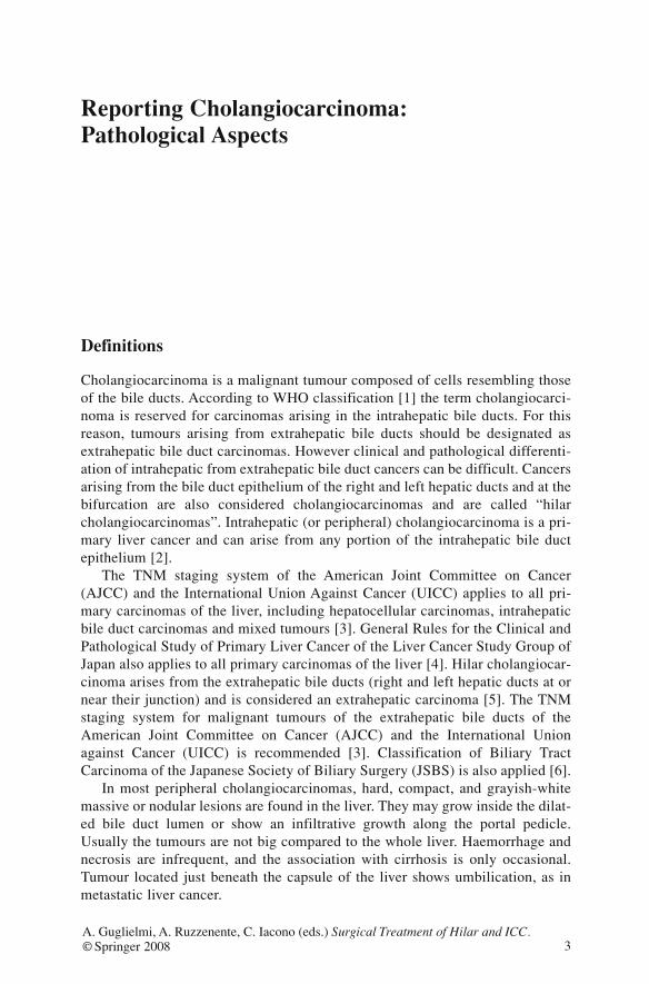
Intraoperative Consultation
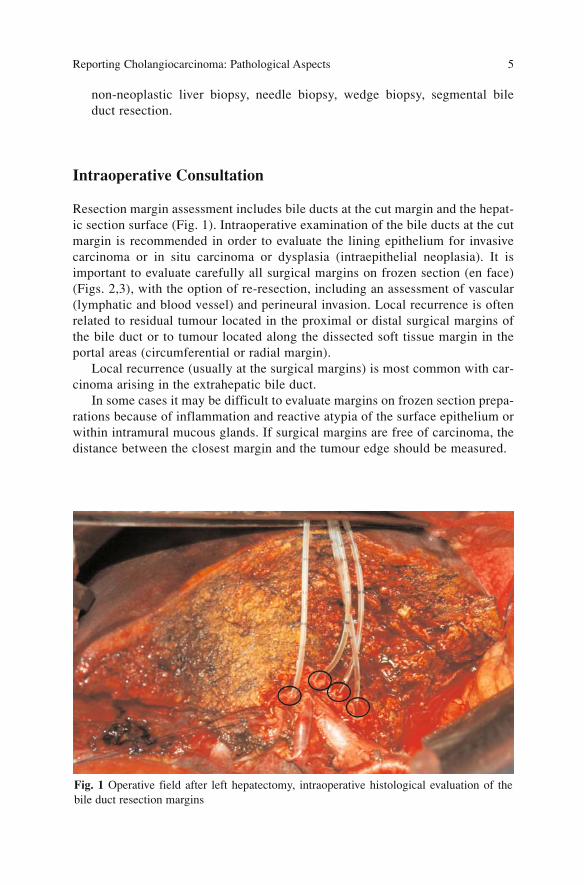
Resection margin assessment includes bile ducts at the cut margin and the hepat-ic section surface (Fig. 1). Intraoperative examination of the bile ducts at the cutmargin is recommended in order to evaluate the lining epithelium for invasivecarcinoma or in situ carcinoma or dysplasia (intraepithelial neoplasia). It isimportant to evaluate carefully all surgical margins on frozen section (en face)(Figs. 2,3), with the option of re-resection, including an assessment of vascular(lymphatic and blood vessel) and perineural invasion. Local recurrence is oftenrelated to residual tumour located in the proximal or distal surgical margins ofthe bile duct or to tumour located along the dissected soft tissue margin in theportal areas (circumferential or radial margin).
Local recurrence (usually at the surgical margins) is most common with car-cinoma arising in the extrahepatic bile duct.
In some cases it may be difficult to evaluate margins on frozen section prepa-rations because of inflammation and reactive atypia of the surface epithelium orwithin intramural mucous glands. If surgical margins are free of carcinoma, thedistance between the closest margin and the tumour edge should be measured.
Reporting Cholangiocarcinoma: Pathological Aspects 5
Fig. 1 Operative field after left hepatectomy, intraoperative histological evaluation of thebile duct resection margins
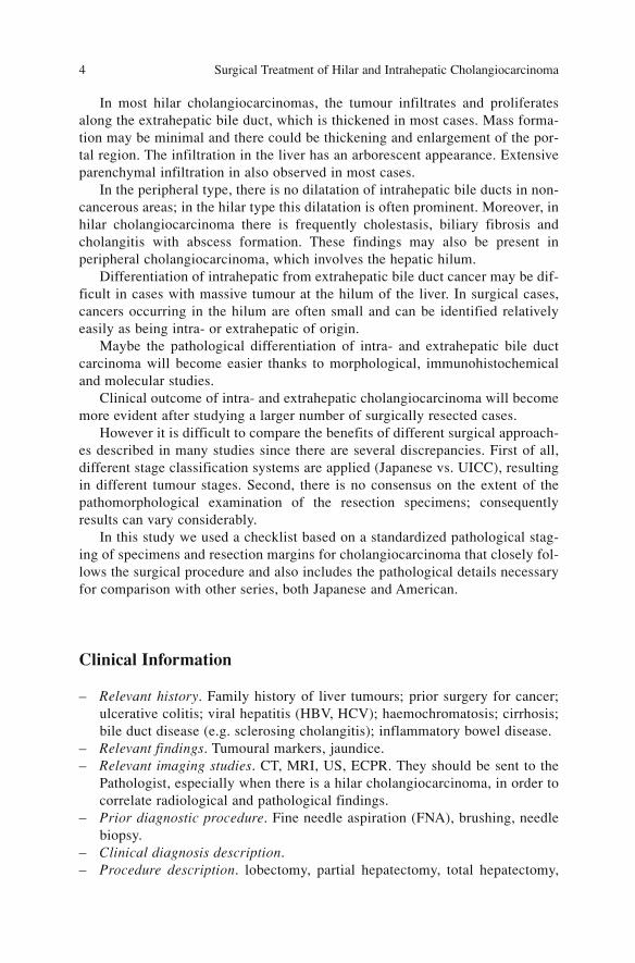
Macroscopic Examination
It is mandatory to examine the specimen in a fresh, unfixed state, in the operat-ing theatre in close cooperation with the surgeons. The bile ducts should beprobed and the site of origin of the carcinoma must be identified exactly. This ismore important for carcinoma arising in the extra hepatic bile duct, in relationto the longitudinal extension of the neoplasia (Bismuth-Corlette Classification)[7]. The tumour should then be recorded in relation to the main branches/trunkof the portal vein or hepatic artery, also using radiological data.
In case of suspected tumour adherence to the portal vein requiring vesselresection, the segment should be separated from the specimen, serially sectioned
Surgical Treatment of Hilar and Intrahepatic Cholangiocarcinoma 6
Fig. 2 Bile duct cross section for eval-uation of surgical resection margin
Fig. 3a,b a Presence of adenocarcinoma at surgical margin of the bile duct. b Negative bileduct margin
a b
and submitted “in toto” so that tumour invasion can be checked histologically.Both ends and perivascular tissue have to be considered as additional resectionmargins.
Local tumour extension and invasion of adjacent structures should also bereported.
Specimen
– Liver. Size (3 dimensions); Weight; Descriptive features (external/cut sur-faces); Bile duct / vessels on cut surface
– Extrahepatic Bile Duct. Dimension of bile ducts (length and thickness ofwall); External surface; Obstruction (partial / complete)
– Margins. Transection margin, Bile duct margin
The intraoperative examination of the bile ducts at the cut margin is recom-mended in order to evaluate the lining epithelium for carcinoma in situ or dys-plasia.
The raw surface of a hepatectomy may be large, rendering it impractical forcomplete examination. The surgeon should be consulted to determine the criti-cal foci that may require microscopic evaluation. Grossly positive marginsshould be microscopically confirmed and documented. If the margins are gross-ly free of tumour, judicious sampling of the cut surface in the region closest tothe nearest identified tumour nodule is indicated.
If the neoplasm is found near the surgical margin, the distance from the mar-gin should be reported.
The adipose tissue is very important for carcinoma arising in the extrahepaticbile: it is difficult to investigate and represents the periductal soft tissue dissectedin the portal area. The outer surface should be marked with India ink and the tis-sue, with the bile duct inside, should be sectioned perpendicularly in subsequent,numbered specimens that should be submitted for histological examination.
The hepatic section margin should also be marked with India ink, and thewhole specimen should be cut with sections made perpendicularly to the capsule(pay attention to the hepatic hilum).
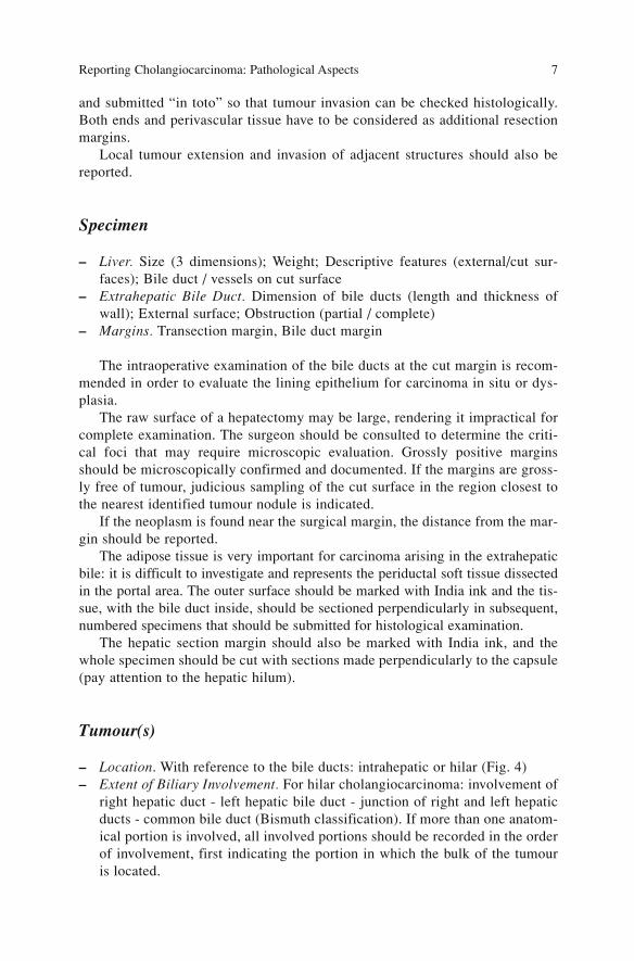
Tumour(s)
– Location. With reference to the bile ducts: intrahepatic or hilar (Fig. 4)– Extent of Biliary Involvement. For hilar cholangiocarcinoma: involvement of
right hepatic duct - left hepatic bile duct - junction of right and left hepaticducts - common bile duct (Bismuth classification). If more than one anatom-ical portion is involved, all involved portions should be recorded in the orderof involvement, first indicating the portion in which the bulk of the tumouris located.
Reporting Cholangiocarcinoma: Pathological Aspects 7
If possible specify the growth pattern in the bile duct wall (papillary, nodu-lar, flat type)
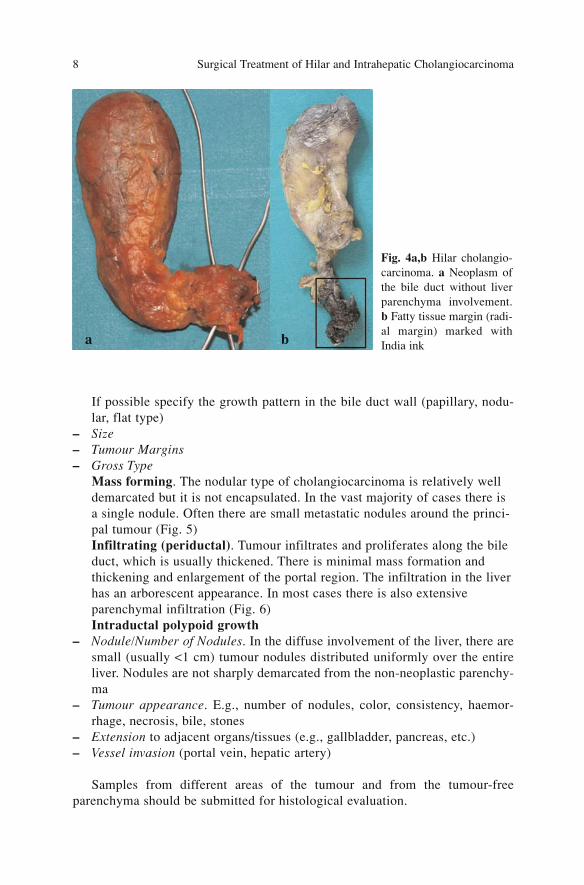
– Size– Tumour Margins – Gross Type
Mass forming. The nodular type of cholangiocarcinoma is relatively welldemarcated but it is not encapsulated. In the vast majority of cases there isa single nodule. Often there are small metastatic nodules around the princi-pal tumour (Fig. 5)Infiltrating (periductal). Tumour infiltrates and proliferates along the bileduct, which is usually thickened. There is minimal mass formation andthickening and enlargement of the portal region. The infiltration in the liverhas an arborescent appearance. In most cases there is also extensiveparenchymal infiltration (Fig. 6)Intraductal polypoid growth
– Nodule/Number of Nodules. In the diffuse involvement of the liver, there aresmall (usually <1 cm) tumour nodules distributed uniformly over the entireliver. Nodules are not sharply demarcated from the non-neoplastic parenchy-ma
– Tumour appearance. E.g., number of nodules, color, consistency, haemor-rhage, necrosis, bile, stones
– Extension to adjacent organs/tissues (e.g., gallbladder, pancreas, etc.)– Vessel invasion (portal vein, hepatic artery)
Samples from different areas of the tumour and from the tumour-freeparenchyma should be submitted for histological evaluation.
Surgical Treatment of Hilar and Intrahepatic Cholangiocarcinoma 8
Fig. 4a,b Hilar cholangio-carcinoma. a Neoplasm ofthe bile duct without liverparenchyma involvement.b Fatty tissue margin (radi-al margin) marked withIndia inka b
Pathology Findings in Non-Neoplastic Liver
Cirrhosis, atrophy, duct obstructions/dilatation, calcifications, cysts, abscess,other.
Reporting Cholangiocarcinoma: Pathological Aspects 9
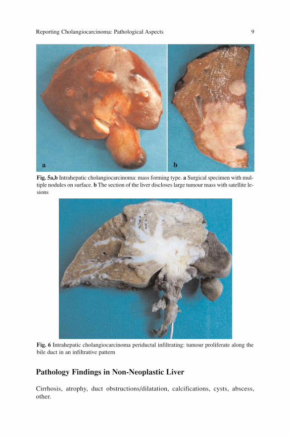
Fig. 5a,b Intrahepatic cholangiocarcinoma: mass forming type. a Surgical specimen with mul-tiple nodules on surface. b The section of the liver discloses large tumour mass with satellite le-sions
Fig. 6 Intrahepatic cholangiocarcinoma periductal infiltrating: tumour proliferate along thebile duct in an infiltrative pattern
a b
Lymph Nodes (Location, Number)
The removed lymph nodes should be classified and numbered according to theJapanese classification. Although this classification is a complex system, itshould be applied to be sure that the individual nodes are precisely defined.
According to UICC criteria the regional lymph nodes must be separated fromnon-regional lymph nodes; the involvement of non-regional lymph nodes isdefined as distant metastases. The last classification reflects the anatomical siteof larger node groups in relation to the liver and also closely follows the surgi-cal procedure, since most nodes are already dissected and submitted separately.
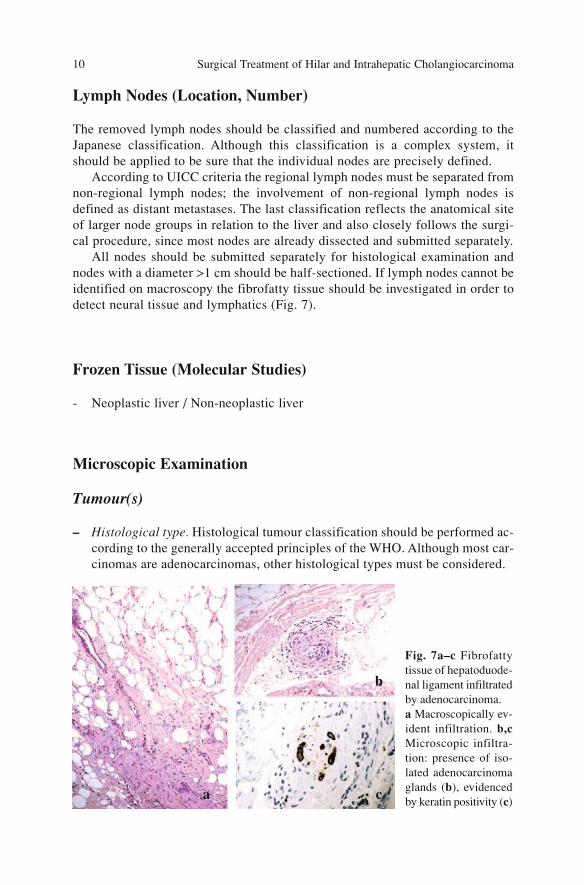
All nodes should be submitted separately for histological examination andnodes with a diameter >1 cm should be half-sectioned. If lymph nodes cannot beidentified on macroscopy the fibrofatty tissue should be investigated in order todetect neural tissue and lymphatics (Fig. 7).
Frozen Tissue (Molecular Studies)
- Neoplastic liver / Non-neoplastic liver
Microscopic Examination
Tumour(s)
– Histological type. Histological tumour classification should be performed ac-cording to the generally accepted principles of the WHO. Although most car-cinomas are adenocarcinomas, other histological types must be considered.
Surgical Treatment of Hilar and Intrahepatic Cholangiocarcinoma 10
Fig. 7a–c Fibrofattytissue of hepatoduode-nal ligament infiltratedby adenocarcinoma. a Macroscopically ev-ident infiltration. b,cMicroscopic infiltra-tion: presence of iso-lated adenocarcinomaglands (b), evidencedby keratin positivity (c)
a c
b
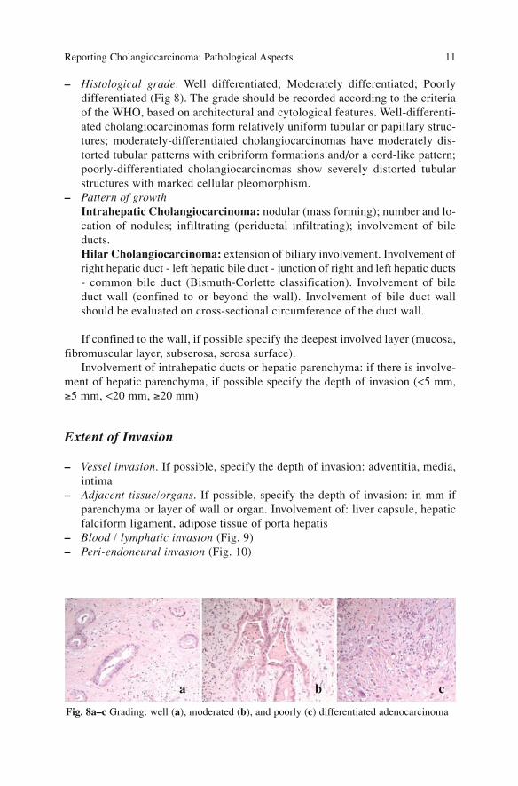
– Histological grade. Well differentiated; Moderately differentiated; Poorlydifferentiated (Fig 8). The grade should be recorded according to the criteriaof the WHO, based on architectural and cytological features. Well-differenti-ated cholangiocarcinomas form relatively uniform tubular or papillary struc-tures; moderately-differentiated cholangiocarcinomas have moderately dis-torted tubular patterns with cribriform formations and/or a cord-like pattern;poorly-differentiated cholangiocarcinomas show severely distorted tubularstructures with marked cellular pleomorphism.
– Pattern of growthIntrahepatic Cholangiocarcinoma: nodular (mass forming); number and lo-cation of nodules; infiltrating (periductal infiltrating); involvement of bileducts.Hilar Cholangiocarcinoma: extension of biliary involvement. Involvement ofright hepatic duct - left hepatic bile duct - junction of right and left hepatic ducts- common bile duct (Bismuth-Corlette classification). Involvement of bileduct wall (confined to or beyond the wall). Involvement of bile duct wallshould be evaluated on cross-sectional circumference of the duct wall.
If confined to the wall, if possible specify the deepest involved layer (mucosa,fibromuscular layer, subserosa, serosa surface).
Involvement of intrahepatic ducts or hepatic parenchyma: if there is involve-ment of hepatic parenchyma, if possible specify the depth of invasion (<5 mm,≥5 mm, <20 mm, ≥20 mm)
Extent of Invasion
– Vessel invasion. If possible, specify the depth of invasion: adventitia, media,intima
– Adjacent tissue/organs. If possible, specify the depth of invasion: in mm ifparenchyma or layer of wall or organ. Involvement of: liver capsule, hepaticfalciform ligament, adipose tissue of porta hepatis
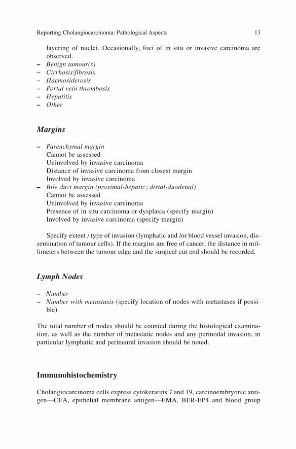
– Blood / lymphatic invasion (Fig. 9)– Peri-endoneural invasion (Fig. 10)
Reporting Cholangiocarcinoma: Pathological Aspects 11
Fig. 8a–c Grading: well (a), moderated (b), and poorly (c) differentiated adenocarcinoma
a b c
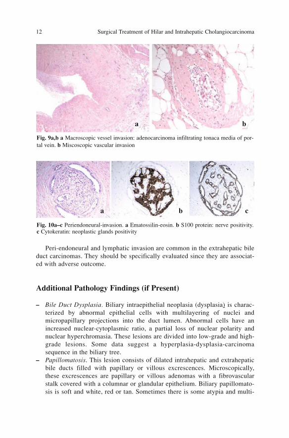
Peri-endoneural and lymphatic invasion are common in the extrahepatic bileduct carcinomas. They should be specifically evaluated since they are associat-ed with adverse outcome.
Additional Pathology Findings (if Present)
– Bile Duct Dysplasia. Biliary intraepithelial neoplasia (dysplasia) is charac-terized by abnormal epithelial cells with multilayering of nuclei andmicropapillary projections into the duct lumen. Abnormal cells have anincreased nuclear-cytoplasmic ratio, a partial loss of nuclear polarity andnuclear hyperchromasia. These lesions are divided into low-grade and high-grade lesions. Some data suggest a hyperplasia-dysplasia-carcinomasequence in the biliary tree.
– Papillomatosis. This lesion consists of dilated intrahepatic and extrahepaticbile ducts filled with papillary or villous excrescences. Microscopically,these excrescences are papillary or villous adenomas with a fibrovascularstalk covered with a columnar or glandular epithelium. Biliary papillomato-sis is soft and white, red or tan. Sometimes there is some atypia and multi-
Surgical Treatment of Hilar and Intrahepatic Cholangiocarcinoma 12
Fig. 10a–c Periendoneural-invasion. a Ematossilin-eosin. b S100 protein: nerve positivity. c Cytokeratin: neoplastic glands positivity
Fig. 9a,b a Macroscopic vessel invasion: adenocarcinoma infiltrating tonaca media of por-tal vein. b Miscoscopic vascular invasion
a b
a b c
layering of nuclei. Occasionally, foci of in situ or invasive carcinoma areobserved.
– Benign tumour(s)– Cirrhosis/fibrosis– Haemosiderosis– Portal vein thrombosis– Hepatitis– Other
Margins
– Parenchymal marginCannot be assessedUninvolved by invasive carcinomaDistance of invasive carcinoma from closest marginInvolved by invasive carcinoma
– Bile duct margin (proximal-hepatic; distal-duodenal)Cannot be assessedUninvolved by invasive carcinoma Presence of in situ carcinoma or dysplasia (specify margin)Involved by invasive carcinoma (specify margin)
Specify extent / type of invasion (lymphatic and /or blood vessel invasion, dis-semination of tumour cells). If the margins are free of cancer, the distance in mil-limeters between the tumour edge and the surgical cut end should be recorded.
Lymph Nodes
– Number– Number with metastasis (specify location of nodes with metastases if possi-
ble)
The total number of nodes should be counted during the histological examina-tion, as well as the number of metastatic nodes and any perinodal invasion, inparticular lymphatic and perineural invasion should be noted.
Immunohistochemistry
Cholangiocarcinoma cells express cytokeratins 7 and 19, carcinoembryonic anti-gen—CEA, epithelial membrane antigen—EMA, BER-EP4 and blood group
Reporting Cholangiocarcinoma: Pathological Aspects 13
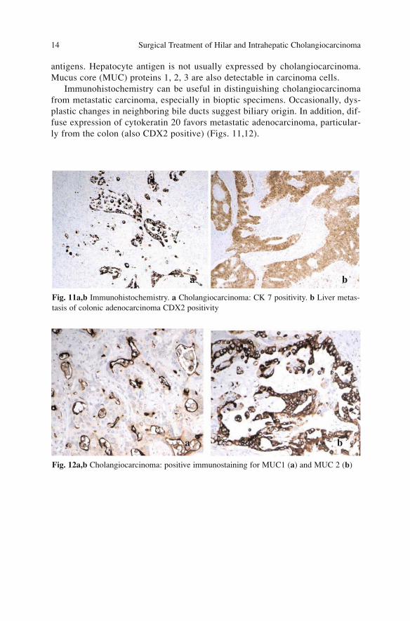
antigens. Hepatocyte antigen is not usually expressed by cholangiocarcinoma.Mucus core (MUC) proteins 1, 2, 3 are also detectable in carcinoma cells.
Immunohistochemistry can be useful in distinguishing cholangiocarcinomafrom metastatic carcinoma, especially in bioptic specimens. Occasionally, dys-plastic changes in neighboring bile ducts suggest biliary origin. In addition, dif-fuse expression of cytokeratin 20 favors metastatic adenocarcinoma, particular-ly from the colon (also CDX2 positive) (Figs. 11,12).
Surgical Treatment of Hilar and Intrahepatic Cholangiocarcinoma 14
Fig. 11a,b Immunohistochemistry. a Cholangiocarcinoma: CK 7 positivity. b Liver metas-tasis of colonic adenocarcinoma CDX2 positivity
Fig. 12a,b Cholangiocarcinoma: positive immunostaining for MUC1 (a) and MUC 2 (b)
a b
a b
References
1. Hamilton SR, Aaltonen LA (2000) Pathology and genetics of tumours of the digestive sys-tem:WHO classification of tumours. IARC Press, Lyon
2. Ishak KG, Goodman ZD, Stocker JT (2001) Tumours of the liver and intrahepatic bile ducts.Atlas of tumour pathology, 3rd Series, Fascicle 31. Armed Forces Institute of Pathology,Washington DC
3. Greene FL, Page DL, Fleming ID et al (eds) (2003) AJCC Cancer staging manual, 6th edi-tion. Springer, New York
4. Liver Cancer Study Group of Japan (2003) General rules for clinical and pathological studyof primary liver cancer, 2nd English edition. Kanehara, Tokyo.
5. Albores-Saavedra J, Henson DE, Klimstra DS (2000) Tumours of the gallbladder, extrahep-atic bile ducts, and ampulla of vater. Atlas of tumour pathology, 3rd Series, Fascicle 27.Armed Forces Institute of Pathology, Washington DC
6. Japanese Society of Biliary Surgery (2004) Classification of biliary tract carcinoma, 2ndEnglish edition. Kanehara, Tokyo
7. Bismuth H, Corlette MB (1975) Intrahepatic cholangioenteric anastomosis in carcinoma ofthe hilus of the liver. Surg Gynecol Obstet 140(2):170–178
Reporting Cholangiocarcinoma: Pathological Aspects 15
Diagnosis
Diagnosis of cholangiocarcinoma is usually suspected in presence of obstructivejaundice or high cholestasis in blood test values. Currently, many preoperativestudies can be used to achieve a correct diagnosis; they can be direct or indirect,invasive or non-invasive (Table 1).
In absence of previous operations a stenosis of the biliary tree associatedwith the aforesaid symptoms arouses suspicions of a probable hilar cholangio-carcinoma. However, it must be emphasisized that not all stenoses are neoplas-tic; in fact until now from 5 to 15% [1–3] of patients resected due to suspectedcholangiocarcinoma turn out to be non-neoplastic or suffering from other neo-plasms at the definitive pathology examination [4]. Since treatment of theselesions is surgical in any case, in patients with a resectable hilar lesion a patho-logical diagnosis prior to surgical exploration is not mandatory [4].
Ultrasound (Endoscopic, Intraductal, Transabdominal)
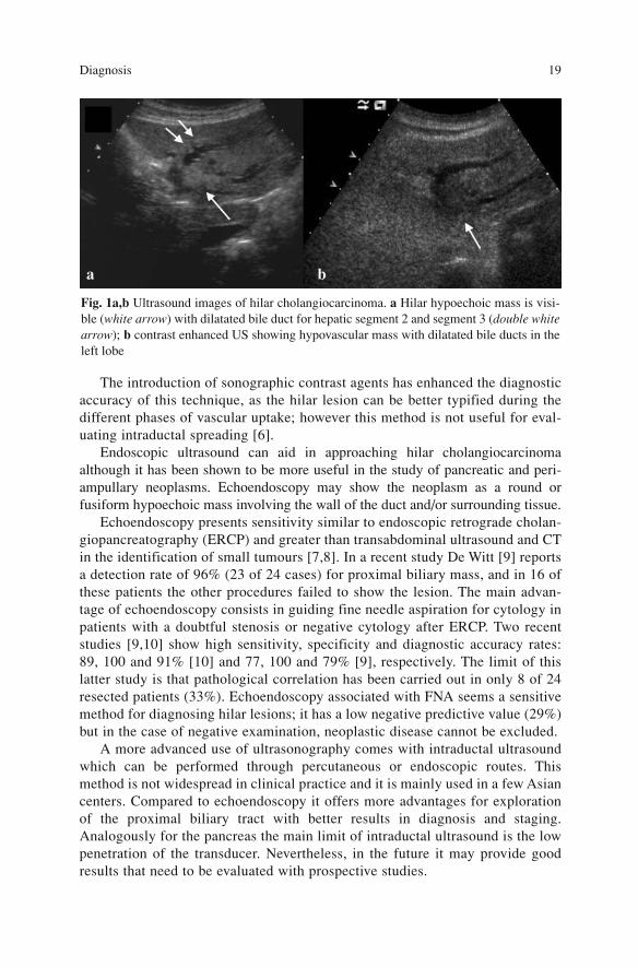
Ultrasound represents the technique of choice for confirming obstructive jaun-dice. In fact it allows easy detection of the dilatation of the intra- and extrahep-atic biliary systems. However, although it shows high diagnostic reliability(80–94%) in detecting biliary dilatation and the level of obstruction, its reliabil-ity in diagnosing the cause of obstruction decreases.
Hilar cholangiocarcinoma may be presumed at ultrasound in presence of ahilar hypoechoic mass that has spread along the biliary tract, determining dilata-tion of the upstream biliary system. Sometimes the lesion may be surrounded byhyperechoic tissue resulting from fibrosis [5]; other indirect signs suggestive ofa correct diagnosis are the ductal abnormalities, ductal obstruction, or vascularinvolvement of hilar structures that this technique can reveal (Fig. 1).
17A. Guglielmi, A. Ruzzenente, C. Iacono (eds.) Surgical Treatment of Hilar and ICC.© Springer 2008
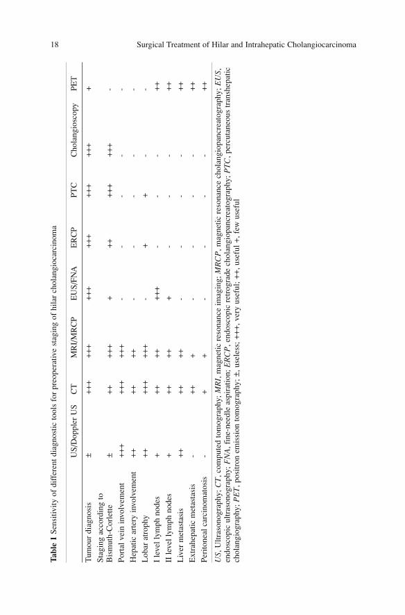
Surgical Treatment of Hilar and Intrahepatic Cholangiocarcinoma 18Ta
ble
1 Se
nsiti
vity
of
diff
eren
t dia
gnos
tic to
ols
for
preo
pera
tive
stag
ing
of h
ilar
chol
angi
ocar
cino
ma
US/
Dop
pler
US
CT
MR
I/M
RC
PE
US/
FNA
ER
CP
PTC
Cho
lang
iosc
opy
PET
Tum
our
diag
nosi
s±
++
++
++
++
++
++
++
++
++
+
Stag
ing
acco
rdin
g to
Bis
mut
h-C
orle
tte±
++
++
++
++
++
++
++
-
Port
al v
ein
invo
lvem
ent
++
++
++
++
+-
--
--
Hep
atic
art
ery
invo
lvem
ent
++
++
++
--
--
-
Lob
ar a
trop
hy+
++
++
++
+-
++
--
I le
vel l
ymph
nod
es+
++
++
++
+-
--
++
II le
vel l
ymph
nod
es+
++
++
+-
--
++
Liv
er m
etas
tasi
s+
++
++
+-
--
-+
+
Ext
rahe
patic
met
asta
sis
-+
++
--
--
++
Peri
tone
al c
arci
nom
atos
is-
++
--
--
++
US ,
Ultr
ason
ogra
phy;
CT
,com
pute
d to
mog
raph
y; M
RI,
mag
netic
res
onan
ce im
agin
g; M
RC
P,m
agne
tic r
eson
ance
cho
lang
iopa
ncre
atog
raph
y; E
US ,
endo
scop
ic u
ltras
onog
raph
y; F
NA
,fin
e-ne
edle
asp
irat
ion;
ER
CP
,end
osco
pic
retr
ogra
de c
hola
ngio
panc
reat
ogra
phy;
PT
C,p
ercu
tane
ous
tran
shep
atic
chol
angi
ogra
phy;
PE
T,p
ositr
on e
mis
sion
tom
ogra
phy;
±,u
sele
ss; +
++
,ver
y us
eful
; ++
,use
ful +
,few
use
ful
The introduction of sonographic contrast agents has enhanced the diagnosticaccuracy of this technique, as the hilar lesion can be better typified during thedifferent phases of vascular uptake; however this method is not useful for eval-uating intraductal spreading [6].
Endoscopic ultrasound can aid in approaching hilar cholangiocarcinomaalthough it has been shown to be more useful in the study of pancreatic and peri-ampullary neoplasms. Echoendoscopy may show the neoplasm as a round orfusiform hypoechoic mass involving the wall of the duct and/or surrounding tissue.
Echoendoscopy presents sensitivity similar to endoscopic retrograde cholan-giopancreatography (ERCP) and greater than transabdominal ultrasound and CTin the identification of small tumours [7,8]. In a recent study De Witt [9] reportsa detection rate of 96% (23 of 24 cases) for proximal biliary mass, and in 16 ofthese patients the other procedures failed to show the lesion. The main advan-tage of echoendoscopy consists in guiding fine needle aspiration for cytology inpatients with a doubtful stenosis or negative cytology after ERCP. Two recentstudies [9,10] show high sensitivity, specificity and diagnostic accuracy rates:89, 100 and 91% [10] and 77, 100 and 79% [9], respectively. The limit of thislatter study is that pathological correlation has been carried out in only 8 of 24resected patients (33%). Echoendoscopy associated with FNA seems a sensitivemethod for diagnosing hilar lesions; it has a low negative predictive value (29%)but in the case of negative examination, neoplastic disease cannot be excluded.
A more advanced use of ultrasonography comes with intraductal ultrasoundwhich can be performed through percutaneous or endoscopic routes. Thismethod is not widespread in clinical practice and it is mainly used in a few Asiancenters. Compared to echoendoscopy it offers more advantages for explorationof the proximal biliary tract with better results in diagnosis and staging.Analogously for the pancreas the main limit of intraductal ultrasound is the lowpenetration of the transducer. Nevertheless, in the future it may provide goodresults that need to be evaluated with prospective studies.
Diagnosis 19
Fig. 1a,b Ultrasound images of hilar cholangiocarcinoma. a Hilar hypoechoic mass is visi-ble (white arrow) with dilatated bile duct for hepatic segment 2 and segment 3 (double whitearrow); b contrast enhanced US showing hypovascular mass with dilatated bile ducts in theleft lobe
a b
In 2005 Stavropoulos et al. [11] reported the results in 61 patients with jaun-dice without mass at preoperative workup, with a biliary tract stenosis docu-mented by ERCP (malignant obstruction in 43 cases and benign in 18); subjectsunderwent ultrasound with a high frequency probe (20 MHz). While ERCPshowed 25 false negative cases, 22 of whom had malignant stenosis, intraductalultrasound showed only 7 false-negative cases and 3 false-positive. The percent-age of patients with positive diagnosis for malignant disease was 2.06 timeshigher for intraductal echography than ERCP. The diagnostic accuracy haschanged from 58% in ERCP alone, to 90% in ERCP combined with intraductalultrasound. Recently Japanese authors [12] introduced intraductal tridimension-al ultrasonography, that would have a role of great magnitude compared to thestandard technique of evaluating the extent of the neoplasm and its relationshipto the portal axis. In addition, this novel technique would allow performing thevolumetric assessment of the tumour, which appear to have significant prognos-tic value, and allow evaluation of the efficacy of palliative therapy such as laserand photodynamic therapy.
Computed Tomography
As to pathological findings, hilar cholangiocarcinoma shows three differentaspects at CT scan [13,14]:1. Infiltrative: determines a focal stenosis of biliary ducts and comprises more
than 70% of cases; 2. Nodular: shows a hilar mass resembling peripheral cholangiocarcinoma and
therefore it is difficult to differentiate a main duct neoplasm from advancedperipheral neoplasm invading the biliary ducts at the confluence;
3. Papillary: rare, appears as an intraductal polypoid lesion.The infiltrative pattern on enhanced CT scan with contrast agents is demon-
strated as a focal area of ductal involvement with lumen occlusion; the neoplas-tic area shows high attenuation compared to normal parenchyma in 80% ofcases. The nodular pattern on CT appears as a large area of low attenuation witha hypervascular peripheral rim as intrahepatic cholangiocarcinoma [13]. Thenodular variant presents as an intraductal lesion with low attenuation of theintensity compared with surrounding parenchyma and biliary dilatation. Thoserare forms are often multiple and disseminated to the entire biliary tree.
Multislice spiral CT allows diagnostic accuracy of 86% [15] which in the expe-rience of Tillich et al. reaches 100% [16]. Arterial contrast phase has allowed adiagnostic rate of 100% for both infiltrating and nodular types. Multislice tech-nique allows better acquisition of information and a complete visualization of thebiliary tree during the different phases of contrast injection. The infiltrating typelesion is showed as a nodular or round mass with high attenuation during arterialcontrast phase and subsequently it presents attenuation features similar to hepaticparenchyma in the dominant portal phase. These characteristics allow differentiat-
Surgical Treatment of Hilar and Intrahepatic Cholangiocarcinoma 20
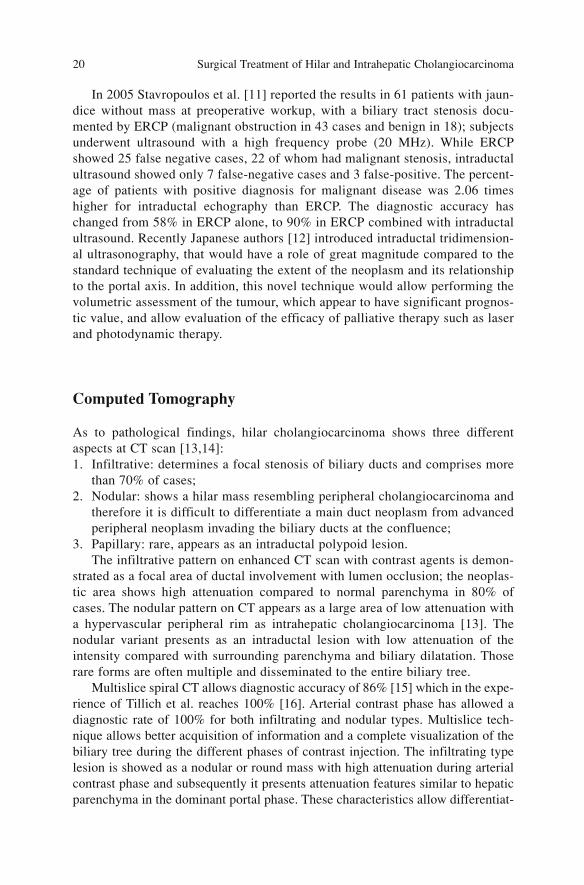
ing infiltrative cholangiocarcinoma from other benign lesions or lymph nodes thatdo not usually form a mass or result hypodense. The nodular variant is hypodensecompared to normal liver during both the dominant arterial and portal phases, asin intrahepatic cholangiocarcinoma (Fig. 2).
A recent study considered the hypothesis of differentiating cholangiocarcino-ma from periductal fibrosis in patients with hepatolithiasis through CT scan; theparameters considered are density of periductal tissue, presence of ascites, por-tal vein occlusion, lymph node enlargement and biliary stones.
Magnetic Resonance Imaging
The use of T2-weighted sequences permits acquisition of images that present alow signal in solid tissue and circulating blood and a high signal in static fluidssuch as bile or pancreatic juice. This provides a MR-cholangiopancreatographywithout using specific contrast, an invasive maneuver that can cause complica-tions. Another advantage is that it permits cholangiographic study even inpatients who had previously undergone upper digestive tract surgery.
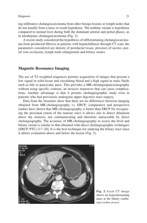
Data from the literature show that there are no differences between imagingobtained from MR-cholangiography vs. ERCP; comparative and prospectivestudies have shown that MR-cholangiography is better than ERCP for recogniz-ing the proximal extent of the tumour since it allows one to detect dilatationabove the stenosis, not communicating and therefore untraceable by directcholangiography. The accuracy of MR-cholangiography to assess the level andbiliary extent is similar to that obtained with direct cholangiographic techniques(ERCP, PTC) [17–20]. It is the best technique for studying the biliary tract sinceit allows evaluation above and below the lesion (Fig. 3).
Diagnosis 21
Fig. 2 Axial CT imageshows an hyperattenuatingmass at the biliary conflu-ence (white arrow)
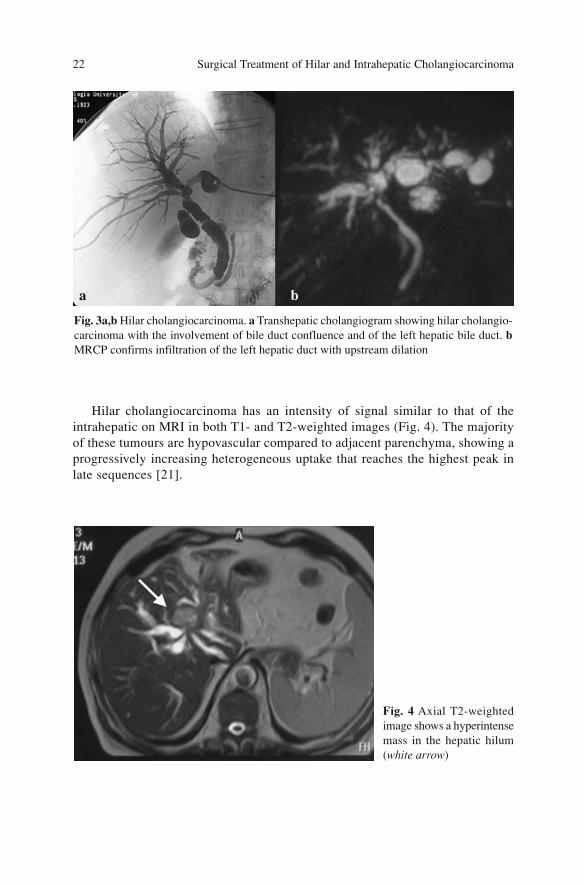
Hilar cholangiocarcinoma has an intensity of signal similar to that of theintrahepatic on MRI in both T1- and T2-weighted images (Fig. 4). The majorityof these tumours are hypovascular compared to adjacent parenchyma, showing aprogressively increasing heterogeneous uptake that reaches the highest peak inlate sequences [21].
Surgical Treatment of Hilar and Intrahepatic Cholangiocarcinoma 22
Fig. 3a,b Hilar cholangiocarcinoma. a Transhepatic cholangiogram showing hilar cholangio-carcinoma with the involvement of bile duct confluence and of the left hepatic bile duct. bMRCP confirms infiltration of the left hepatic duct with upstream dilation
Fig. 4 Axial T2-weightedimage shows a hyperintensemass in the hepatic hilum(white arrow)
a b
Positron Emission Tomography
At the end of the 1990s Delbeke [22] foresaw a potential role for this techniquein the diagnosis of these neoplasms; nevertheless today this diagnostic techniqueseems more useful in preoperative staging than in the diagnosis of hilar cholan-giocarcinoma. In the identification of peripheral cholangiocarcinoma it shows anaccuracy of 95 vs 69% of the extrahepatic one. This difference is due to the dif-ferent size of the presenting tumours, the former being larger. In hilar cholangio-carcinoma it is useful to recognize the nodular type even in lesions larger than 1cm (sensitivity 85%) while its utility is low for the infiltrative type that is morefrequent (sensitivity 18%) [23]. It could have a role in the diagnosis of cholan-giocarcinoma presenting with atypical radiological aspects or in absence of his-tologic malignant diagnosis [23]. Conversely, in the experience of Kluge et al.[24] in 2001, PET presented a high sensitivity (92.3%) and specificity (92.9%)in diagnosing and localizing cholangiocarcinoma while it only played a minorrole in staging, particularly lymph node staging; in fact it has identified metasta-tic lymph nodes in only 2 of 15 cases while for distant metastases it has an accu-racy of 70% (7 of 10 cases).
In a comparative study between CT vs. PET FDG, in 30 cases with extrahep-atic cholangiocarcinoma CT resulted reliable in 80% of the cases while PET wasreliable in 60% of cases [25].
Direct Cholangiography (ERCP and PTC)
Direct visualization of the biliary tree is achieved by means of endoscopic retro-grade cholangiography and percutaneous transhepatic cholangiography that pro-vide a precise and complete opacification with contrast material.
Since the introduction of ultrasound (CT and especially MRCP) in clinicalpractice, indication of these techniques for diagnosis has decreased and they aremainly applied for operative purposes. The application of these direct and inva-sive techniques has to be proposed by the hepatobiliary team since the choice ofmethod, either ERCP or PTC, is fundamental. If the latter is chosen, it is neces-sary to specify the right or left approach; in fact the choice of the exact methodmust be tailored to the needs of the patient, considering the diagnostic purposeas well as the therapeutic possibility (surgical resection, surgical palliation, non-surgical palliation).
The techniques show noticeable complication rates; PTC has a morbidity rateof 3–5% and the main complications are cholangitis, biliary leakage with poten-tial bile peritonitis or perihepatic biliary collection (biloma), haemobilia, bil-hemia, and subcapsular or intrahepatic haematoma. The complications of ERCP
Diagnosis 23
are cholangitis whose risk is enhanced by severe stenosis, since the introduction ofthe contrast that is not drained increases infective risk; to prevent infection, biliarydecompression after the diagnostic procedure of the stenosis is mandatory.
Success rates of PTC range from 95 to 100% of jaundiced patients with biliaryobstruction, while for ERCP with a recognized papilla, the rate is about 90%.
Of the two procedures PTC allows a better evaluation of cholangiocarcino-ma, especially hilar compared to ERCP since it can better visualize the proximalbiliary tract above the stenosis. The advantage of ERCP compared to PTC andMRCP is the possibility of performing brushing cytology or intraductal biopsyfor pathological evaluation; however the success rate of these technique is low,about 50–60% [26].
On direct cholangiography, cholangiocarcinoma shows as an annular stric-ture since most of the tumour is infiltrative. Polypoid type is rare and someforms producing mucin present intraluminal defects.
PTC performed by experienced personnel [27] and in large series correctlyshows the site of stenosis in a range between 96 and 99% and reveals the natureof the lesion at a rate of between 93 to 99%, respectively.
Cholangioscopy (Peroral, Percutaneous)
Cholangioscopy associated with biopsy has an important role in the differentialdiagnosis of biliary stenosis [28–32]. It can be performed through a peroralendoscopic or a percutaneous approach [30–33]; the former is the less invasiveroute and does not necessarily require sphincterectomy, with the advantage thatit can be performed at the same time as ERCP, reducing the time of diagnosisand preoperative hospitalization. Conversely percutaneous transhepatic cholan-gioscopy requires a gradual dilatation of the PTBD path.
Although both techniques are useful diagnostic tools, peroral cholangioscopyis less efficacious compared to percutaneous transhepatic for evaluating the lon-gitudinal extent of the tumour due to the technical and mechanical limitations ofthis approach [32,33].
As mentioned above, the cytological and histological results obtainedthrough endoscopic trans-papillary approach or percutaneously under fluo-roscopy are not good: Ponchon reports a sensitivity rate in malignant lesions of36% with cytology and 43.5% with pinch biopsy (47% in cholangiocarcinoma)[31]. On the other hand, when cyto-histological samples are taken under cholan-gioscopy results improve as the sample is taken on direct vision of the suspectarea; this improvement is reflected in a sensitivity rate arriving at 78% for diag-nosis of malignancy and 82.4% for cholangiocarcinoma [31]. In Nimura’s expe-rience with 257 cholangioscopies the sensitivity rate was 81% in malignantstenosis and 96% in cholangiocarcinoma [34]. Neuhaus reports sensitivity val-ues above 75%, as well [35].
Surgical Treatment of Hilar and Intrahepatic Cholangiocarcinoma 24
Fokuda [32] reports similar results in the peroral approach: he identifies withERCP/tissue sampling 22/38 cases of malignant stenosis and 35/38 benignlesions (in three cases the samples were inadequate) with an accuracy of 78%, asensitivity of 57.9% and a specificity of 100%; the results change significantlyafter the application of peroral cholangioscopy, which allows identifying 38 of38 malignant lesions and 33 of the 38 benign lesions with an accuracy of 93.4%,sensitivity of 100% and specificity of 86.8%.
The recent introduction of the Narrow Band Imaging technique increasesreliability compared to White Light Imaging. The Nagoya group, which is oneof the supporters of percutaneous cholangioscopy, has introduced this techniqueinto the workup of cholangiocarcinoma [36].
Angiography
In the last 20 years the role of angiography has diminished after the introductionof new imaging techniques. Once believed fundamental for assessing locoregional diffusion in hilar neoplasms, especially when evaluating portal and arte-rial involvement, today these evaluations are carried out by spiral CT, MRI andcolor-Doppler sonography. Angiography is no longer performed and nowbelongs to the history of these tumours.
References
1. Gerhards MF, Vos P, van Gulik TM et al (2001) Incidence of benign lesions in patientsresected for suspicious hilar obstruction. Br J Surg 88(1):48–51
2. Knoefel WT, Prenzel KL, Peiper M et al (2003) Klatskin tumours and Klatskin mimickinglesions of the biliary tree. Eur J Surg Oncol 29(8):658–661
3. Nakayama A, Imamura H, Shimada R et al(1999) Proximal bile duct stricture disguised asmalignant neoplasm. Surgery 125(5):514–521
4. Jarnagin WR, D’Angelica M, Blumgart LH (2006) Intrahepatic and extrahepatic biliary can-cer. In: Blumgart LH (ed) Surgery of the liver, biliary tract, and pancreas. 4th edn. SaundersElsevier, Philadelphia
5. Dancygier H, Nattermann C (1994) The role of endoscopic ultrasonography in biliary tractdisease: obstructive jaundice. Endoscopy 26(9):800–802
6. Schuessler G, Ignee A, Hirche T, Dietrich CF (2003) [Improved detection and characterisa-tion of liver tumours with echo-enhanced ultrasound]. Gastroenterol Z 41(12):1167-1176(German)
7. Sugiyama M, Atomi Y, Wada N et al (1996) Endoscopic transpapillary bile duct biopsy with-out sphincterotomy for diagnosing biliary strictures: a prospective comparative study withbile and brush cytology. Am J Gastroenterol 91(3):465–467
8. Tio TL, Reeders JW, Sie LH et al(1993) Endosonography in the clinical staging of Klatskintumour. Endoscopy 25(1):81–85
9. DeWitt J, Misra VL, Leblanc JK et al (2006) EUS-guided FNA of proximal biliary stricturesafter negative ERCP brush cytology results. Gastrointest Endosc 64(3):325–333
Diagnosis 25
10. Fritscher-Ravens A, Broering DC, Knoefel WT et al (2004) EUS-guided fine-needle aspira-tion of suspected hilar cholangiocarcinoma in potentially operable patients with negativebrush cytology. Am J Gastroentero 99(1):45–51
11. Stavropoulos S, Larghi A, Verna E (2005) Intraductal ultrasound for the evaluation ofpatients with biliary strictures and no abdominal mass on computed tomography. Endoscopy37(8):715–721
12. Inui K, Miyoshi H (2005) Cholangiocarcinoma and intraductal sonography. GastrointestEndosc Clin N Am 15(1):143–155
13. Han JK, Choi BI, Kim AY et al (2002) Cholangiocarcinoma: pictorial essay of CT andcholangiographic findings. Radiographics 22(1):173–187
14. Lim JH (2003) Cholangiocarcinoma: morphologic classification according to growth patternand imaging findings. AJR Am J Roentgenol 181(3):819–827
15. Zandrino F, Benzi L, Ferretti ML et al (2002) Multislice CT cholangiography without bil-iary contrast agent: technique and initial clinical results in the assessment of patients withbiliary obstruction. Eur Radiol 12(5):1155-1161
16. Tillich M, Mischinger HJ, Preisegger KH et al (1998) Multiphasic helical CT in diagnosisand staging of hilar cholangiocarcinoma. AJR Am J Roentgenol 171:651–658
17. Manfredi R, Masselli G, Maresca G et al (2003) MR imaging and MRCP of hilar cholan-giocarcinoma. Abdom Imaging 28:319–325
18. Manfredi R, Barbaro B, Masselli G et al (2004) Magnetic resonance imaging of cholangio-carcinoma. Semin Liver Dis 24(2):155–164
19. Lee WJ, Lim HK, Jang KM et al (2001) Radiologic spectrum of cholangiocarcinoma:emphasis on unusual manifestations and differential diagnoses. Radiographics 21: S97-S116
20. Lopera JE, Soto JA, Munera F (2001) Malignant hilar and perihilar biliary obstruction: useof MR cholangiography to define the extent of biliary ductal involvement and plan percuta-neous interventions. Radiology 220:90–96
21. Slattery JM, Sahani DV (2006) What is the current state-of-the-art imaging for detection andstaging of cholangiocarcinoma? Oncologist 11(8):913–922
22. Delbeke D, Martin WH, Sandler MP et al (1998) Evaluation of benign vs. malignant hepat-ic lesions with positron emission tomography. Arch Surg 133(5):510–515; discussion515–516
23. Anderson CD, Rice MH, Pinson CW et al (2004) Fluorodeoxyglucose PET imaging in theevaluation of gallbladder carcinoma and cholangiocarcinoma. J Gastrointest Surg8(1):90–97
24. Kluge R, Schimdt F, Caca K et al ( 2001) Positron emission tomography with (18F)fluoro-2-deoxy-D-glucose for diagnosis and staging of bile duct cancer. Hepatology 33:1029–1035
25. Kato T, Tsukamoto E, Kuge Y et al ( 2002) Clinical role of (18)F-FDG PET for initial stag-ing of patients with extrahepatic bile duct cancer. Eur J Nucl Med Mol Imaging29(8):1047–1054
26. Rustgi AK (1989) Malignant tumours of the bile ducts: diagnosis by biopsy during endo-scopic cannulation. Gastroinst Endosc 35:248–251
27. Gazzaniga GM, Faggioni A, Bondanza G et al (1990) Percutaneous transhepatic biliarydrainage twelve years’ experience. Hepatogastroenterology 37(5):517–523
28. Kim DI, Kim MH, Lee SK et al ( 2001) Risk factors for recurrence of primary bile ductstones after endoscopic biliary sphincterotomy. Gastrointest Endosc 54(1):42–48
29. Lee SS, Kim MH, Lee SK et al (2002) MR cholangiography versus cholangioscopy for eval-uation of longitudinal extension of hilar cholangiocarcinoma. Gastrointest Endosc56(1):25–32
30. Nimura Y, Kamiya J, Hayakawa N, Shionoya S (1989) Cholangioscopic differentiation ofbiliary strictures and polyps. Endoscopy 21(Suppl 1):351–356
31. Ponchon T, Genin G, Mitchell R et al (1996) Methods, indications, and results of percuta-neous choledochoscopy. A series of 161 procedures. Ann Surg 223(1):26–36
32. Fukuda Y, Tsuyuguchi T, Sakai Y et al (2005) Diagnostic utility of peroral cholangioscopyfor various bile-duct lesions. Gastrointest Endosc 62(3):374–382
Surgical Treatment of Hilar and Intrahepatic Cholangiocarcinoma 26
33. Nagino M, Nimura Y (2006) Perihilar cholangiocarcinoma with emphasis on presurgicalmanagement. In: Blumgart LH (ed) Surgery of the liver, biliary tract, and pancreas. 4th edn.Saunders Elsevier, Philadelphia, pp 804–814
34. Nimura Y, Kamiya J (1998) Cholangioscopy. Endoscopy 30(2):182–18835. Neuhaus H (1994) Cholangioscopy. Endoscopy 26(1):120–12536. Nimura Y (2007) Cholangiocarcinoma- Diagnostic Work up. 7th Congress of EHPBA,
Verona, Italy, June 6–9 2007
Diagnosis 27
Preoperative Staging
The only successful treatment of cholangiocarcinoma is curative resection; eval-uation of biliary involvement, either longitudinal or radial, loco-regional diffu-sion and presence of distant metastases are important for planning the therapeu-tic strategy.
The introduction of imaging techniques, especially spiral CT and MRI asso-ciated with MRCP, have changed evaluation of preoperative staging of cholan-giocarcinoma; these procedures allow to obtain high definition images, andmake possible to elaborate and perform tridimensional reconstructions in orderto obtain a better definition of the tumour.
The required parameters for the two most widespread classifications of hilarcholangiocarcinoma [1,2] are obtained nowadays with non-invasive imagingtechniques. Nevertheless the majority of series report underestimation rates ofpreoperative staging due to undiagnosed hepatic and peritoneal metastases,lymph-node metastases, major involvement of vascular structures and greaterdiffusion of the disease along the biliary ducts [2]. On the other hand, only aGerman study [3] reports errors of overstatement that risk excluding patientsfrom the only treatment that can ensure a good outcome.
The questions that a hepatobiliary surgeon asks to the imaging in order todetermine the correct therapeutic approach concern: definition of hilar anatomy;extent of the tumour along the biliary ducts, especially towards second orderconfluences; vascular relationship of the tumour with hepatic artery, portal veinand their left and right branches; presence or absence of parenchymal atrophysecondary to long-term portal branch involvement; volume and pattern of resid-ual liver in order to plan surgical resection, preoperative biliary decompressionand/or vascular embolisation.
29A. Guglielmi, A. Ruzzenente, C. Iacono (eds.) Surgical Treatment of Hilar and ICC.© Springer 2008
Evaluation of the Biliary Involvement (Longitudinal Extent)
Ultrasound
Transabdominal ultrasound does not contribute any information to the evalua-tion of this parameter. However echoendoscopy appears to be useful in stagingcholangiocarcinoma; the lesion is detected as a hypoechoic mass within thelumen of the duct, frequently with infiltration of surrounding tissue. Sometimesthe lesion is encased by hyperechoic tissue that is the expression of peritumoralfibrosis [4]. Tio et al. [5] reported a correct evaluation of T-stage in 85% of 43cases with carcinoma of hilar and of the common hepatic duct. The limit ofechoendoscopy is that exploration of the hilum is not always possible; in factsome authors report fewer data in proximal cholangiocarcinoma compared todistal cholangiocarcinoma [4].
MRCP
MRCP has an accuracy similar to direct cholangiography and is more powerfulthan CT in evaluating ductal involvement; it also allows detection of isolatedobstructed ducts that are not communicating and thus are not identified byERCP and/or PTC. In a comparative study between ERCP and MRCP in 40patients, both techniques permitted diagnosis of biliary obstruction in 100% ofcases, but MRCP was superior to ERCP in evaluating the extent of the tumour[6]. In another study comparing the two techniques, MRCP permitted correctBismuth-Corlette classification of 78% of the patients and underestimated 22%.The authors noted that if management of the patients had relied only on MRCP,the definitive treatment would have been modified in 28% of the cases (five of18 patients); this is mainly due to a mistaken assessment of second order duc-tal involvement. As a matter of fact, in Bismuth-Corlette stages I and II MRCPshowed an accuracy of 90%, whereas in stage III and IV it presented errors ofunderestimation in three of seven cases. The authors concluded that MRCPmust be considered a useful technique in the therapeutic planning for malignanthilar neoplasms: “accurate depiction of high grade strictures for which endo-scopic drainage is not the option of choice can preclude unnecessary invasiveimaging” [7]. Manfredi has shown that MRCP gives similar results of directcholangiographic techniques although it is less effective in identifying theextent of small hilar lesions [8]; evaluating biliary involvement according toBismuth-Corlette, he obtained accuracy in 84% of the cases (10 of 12).
Surgical Treatment of Hilar and Intrahepatic Cholangiocarcinoma 30
Spiral CT
The diagnostic accuracy of CT in evaluating longitudinal diffusion is not partic-ularly satisfying; in fact it ranges between 54 and 64% [13,14]. These low per-centages are related to the type of diffusion of cholangiocarcinoma. It occursunder the epithelium along the wall of the biliary duct, and in the periductal tis-sue without affecting the epithelium, and therefore are difficult to assess on CT[15,16]. The use of biliary contrast materials administered orally or intravenous-ly, which could have provided advantages, present some limitations since theyare not correctly excreted in the patients with an obstructed biliary tract [17]. Toincrease diagnostic accuracy in evaluating longitudinal diffusion, Kim et al. [18]have proposed the use of spiral CT associated with direct cholangiographyobtained by injection of contrast material in the nasobiliary or in the percuta-neous transhepatic drainage with this technique Kim et al. have correctlyassessed the involvement of primary confluence in 11 of 11 patients, and of sec-ond level confluence in 18 of 19 (95%). The correct extent of the neoplasm wasachieved in 10 of 11 patients; the procedure showed a false positive case in theevaluation of second order confluence involvement [18]. Sensitivity, specificity,positive predictive value and negative predictive value rates were 100, 90, 90 and100%, respectively.
In Lee’s study [19], the association of CT and direct cholangiographyallowed identifying the correct extent of the disease along the ducts in 46 of 55patients (accuracy 84%), underestimation in seven of 55 patients, and overesti-mation in only two patients.
ERCP/PTC
If the majority of authors report underestimation errors in the evaluation of lon-gitudinal diffusion, others [3] report a high rate of overestimation due to peritu-moral fibrosis. Comparing the data of ERCP, PTC, and MRCP with surgicalspecimens the German authors report a correct evaluation with ERCP in 29% ofthe cases, with MRCP in 36%, and with PTC in 53%, an overestimation rate of42, 41 and 31%, and underestimation rate of 31, 23 and 16%, respectively. In theexperience of these authors the most reliable technique for studying hilarcholangiocarcinoma is PTC: significantly better than ERCP (p<0.008), slightlysuperior to MRCP (p=0.06), while ERCP and MRCP have similar results. Thelimit of ERCP is the evaluation of proximal extension of the tumour, determinedby the absent or incomplete opacification of the intrahepatic biliary tree. MRCP,conversely, allows a correct image of both the hepatic lobes in about 90% ofcases, while it has a lower ability than PTC (p<0.019) to evaluate the edges ofthe neoplasm. The main diagnostic problems concern stages III and IV ofBismuth-Corlette, which are better studied with PTC than ERCP or MRCP.
Preoperative Staging 31
Rotational Cine-cholangiography
To overcome the limits of direct cholangiography a rotational cholangiographictechnique has been proposed that allows correct evaluation of ductal anatomyand a better evaluation of intraductal diffusion of the tumour [9–11]. Miura [10]performed rotational cine-cholangiography in 60 patients with obstructive jaun-dice; he evaluated the biliary anatomy and the ability to visualise the main con-fluence as well as second order ductal confluences, and he compared the radio-logical findings with the pathological reports in 26 resected patients.
The percentage of primary confluence visualization was 97.6% with rota-tional cine-cholangiography, while it was 87% for right anterior-posterior con-fluence and 93.1% for medial left duct confluent. The diagnostic accuracy ofneoplastic diffusion was 91.7% for common hepatic duct, 100% for right hepat-ic duct, 91.7% for left hepatic duct, 100% for right anterior duct, 83.3% for rightposterior duct, and 100% for left medial duct. Nevertheless, the author conclud-ed that rotational cine-cholangiography is a useful method for evaluating theanatomy of first and second order confluences and also for identifying the intra-ductal spread of the carcinoma [10].
Recently a new technique of rotational three-dimensional cholangiographywas suggested [12] that is obtained by injecting contrast material through percu-taneous transhepatic drainage and using the technique of digital angiographysubtraction of images and 3-D reconstruction; this method would allow an accu-rate anatomical study of the biliary tree.
Cholangioscopy
Different authors, especially Japanese, believe that percutaneous transhepaticcholangioscopy is unrivalled for identifying superficial diffusion of cholangio-carcinoma [20–24].
Lee compared MRCP with percutaneous transhepatic cholangioscopy in theevaluation of longitudinal extent of cholangiocarcinoma and reported in theinfiltrative form an optimal correlation between the two techniques: in 16/18cases for type I of Bismuth-Corlette (88.9%), in 14/16 for type II (87.5%), in19/23 for type IIIa (82.6%), in 14/14 for type IIIb (100%), and in 24/28 for typeIV (85.7%), so that it is believed that MRCP must replace cholangioscopy; onthe other hand cholangioscopy was more reliable for assessing polypoid and dif-fuse sclerosing types [25].
Nimura [24] confirmed these results and suggested that cholangioscopyaimed at preoperative diagnosis of cancer extent is indicated in patients withpapillary type or nodular type cholangiocarcinoma since the mucosal extensionof the tumour is easily observable. The evaluation of tumoral extension must beperformed not only in the intrahepatic portion but also in the distal bile duct inorder to tailor the resection (hepatobiliary or hepatopancreatoduodenectomy).
Surgical Treatment of Hilar and Intrahepatic Cholangiocarcinoma 32
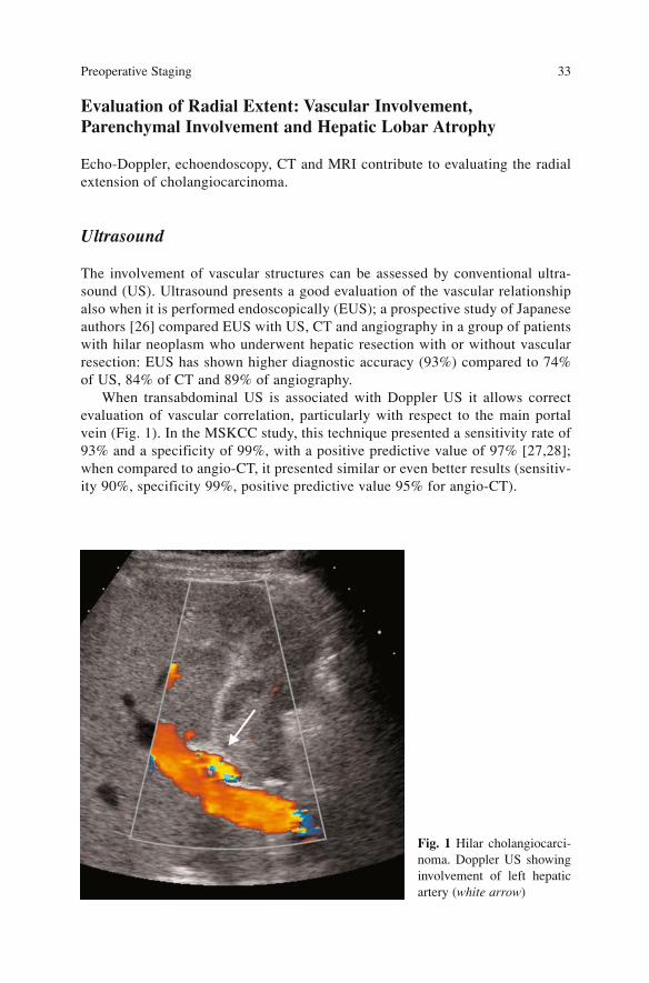
Evaluation of Radial Extent: Vascular Involvement,Parenchymal Involvement and Hepatic Lobar Atrophy
Echo-Doppler, echoendoscopy, CT and MRI contribute to evaluating the radialextension of cholangiocarcinoma.
Ultrasound
The involvement of vascular structures can be assessed by conventional ultra-sound (US). Ultrasound presents a good evaluation of the vascular relationshipalso when it is performed endoscopically (EUS); a prospective study of Japaneseauthors [26] compared EUS with US, CT and angiography in a group of patientswith hilar neoplasm who underwent hepatic resection with or without vascularresection: EUS has shown higher diagnostic accuracy (93%) compared to 74%of US, 84% of CT and 89% of angiography.
When transabdominal US is associated with Doppler US it allows correctevaluation of vascular correlation, particularly with respect to the main portalvein (Fig. 1). In the MSKCC study, this technique presented a sensitivity rate of93% and a specificity of 99%, with a positive predictive value of 97% [27,28];when compared to angio-CT, it presented similar or even better results (sensitiv-ity 90%, specificity 99%, positive predictive value 95% for angio-CT).
Preoperative Staging 33
Fig. 1 Hilar cholangiocarci-noma. Doppler US showinginvolvement of left hepaticartery (white arrow)
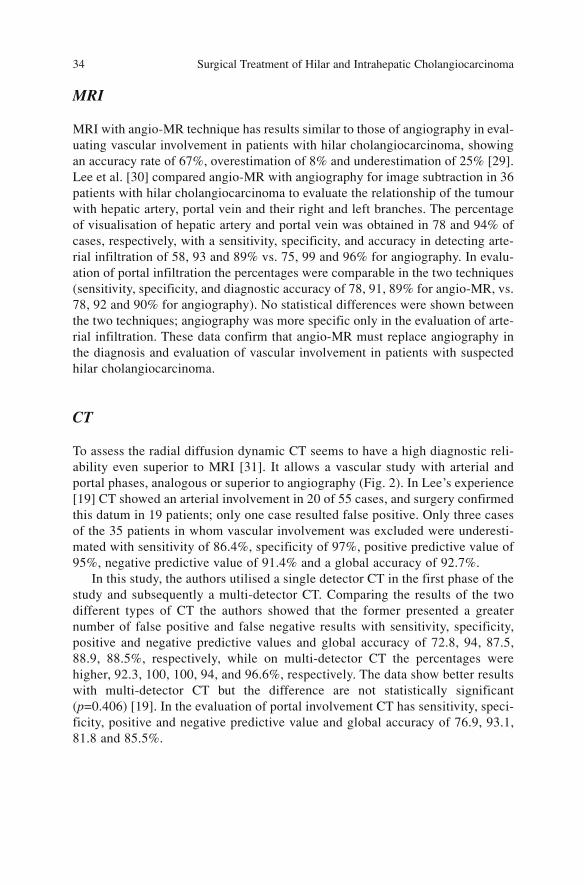
MRI
MRI with angio-MR technique has results similar to those of angiography in eval-uating vascular involvement in patients with hilar cholangiocarcinoma, showingan accuracy rate of 67%, overestimation of 8% and underestimation of 25% [29].Lee et al. [30] compared angio-MR with angiography for image subtraction in 36patients with hilar cholangiocarcinoma to evaluate the relationship of the tumourwith hepatic artery, portal vein and their right and left branches. The percentageof visualisation of hepatic artery and portal vein was obtained in 78 and 94% ofcases, respectively, with a sensitivity, specificity, and accuracy in detecting arte-rial infiltration of 58, 93 and 89% vs. 75, 99 and 96% for angiography. In evalu-ation of portal infiltration the percentages were comparable in the two techniques(sensitivity, specificity, and diagnostic accuracy of 78, 91, 89% for angio-MR, vs.78, 92 and 90% for angiography). No statistical differences were shown betweenthe two techniques; angiography was more specific only in the evaluation of arte-rial infiltration. These data confirm that angio-MR must replace angiography inthe diagnosis and evaluation of vascular involvement in patients with suspectedhilar cholangiocarcinoma.
CT
To assess the radial diffusion dynamic CT seems to have a high diagnostic reli-ability even superior to MRI [31]. It allows a vascular study with arterial andportal phases, analogous or superior to angiography (Fig. 2). In Lee’s experience[19] CT showed an arterial involvement in 20 of 55 cases, and surgery confirmedthis datum in 19 patients; only one case resulted false positive. Only three casesof the 35 patients in whom vascular involvement was excluded were underesti-mated with sensitivity of 86.4%, specificity of 97%, positive predictive value of95%, negative predictive value of 91.4% and a global accuracy of 92.7%.
In this study, the authors utilised a single detector CT in the first phase of thestudy and subsequently a multi-detector CT. Comparing the results of the twodifferent types of CT the authors showed that the former presented a greaternumber of false positive and false negative results with sensitivity, specificity,positive and negative predictive values and global accuracy of 72.8, 94, 87.5,88.9, 88.5%, respectively, while on multi-detector CT the percentages werehigher, 92.3, 100, 100, 94, and 96.6%, respectively. The data show better resultswith multi-detector CT but the difference are not statistically significant(p=0.406) [19]. In the evaluation of portal involvement CT has sensitivity, speci-ficity, positive and negative predictive value and global accuracy of 76.9, 93.1,81.8 and 85.5%.
Surgical Treatment of Hilar and Intrahepatic Cholangiocarcinoma 34
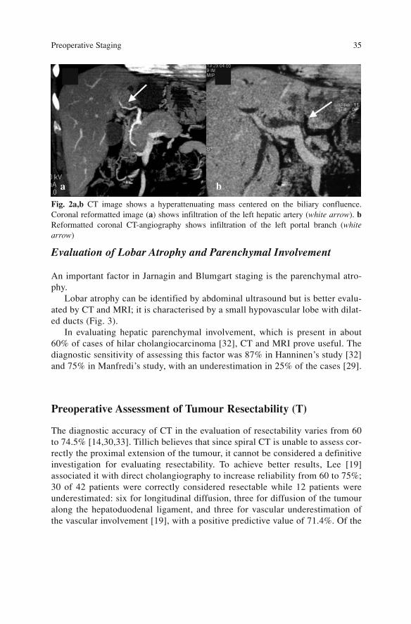
Evaluation of Lobar Atrophy and Parenchymal Involvement
An important factor in Jarnagin and Blumgart staging is the parenchymal atro-phy.
Lobar atrophy can be identified by abdominal ultrasound but is better evalu-ated by CT and MRI; it is characterised by a small hypovascular lobe with dilat-ed ducts (Fig. 3).
In evaluating hepatic parenchymal involvement, which is present in about60% of cases of hilar cholangiocarcinoma [32], CT and MRI prove useful. Thediagnostic sensitivity of assessing this factor was 87% in Hanninen’s study [32]and 75% in Manfredi’s study, with an underestimation in 25% of the cases [29].
Preoperative Assessment of Tumour Resectability (T)
The diagnostic accuracy of CT in the evaluation of resectability varies from 60to 74.5% [14,30,33]. Tillich believes that since spiral CT is unable to assess cor-rectly the proximal extension of the tumour, it cannot be considered a definitiveinvestigation for evaluating resectability. To achieve better results, Lee [19]associated it with direct cholangiography to increase reliability from 60 to 75%;30 of 42 patients were correctly considered resectable while 12 patients wereunderestimated: six for longitudinal diffusion, three for diffusion of the tumouralong the hepatoduodenal ligament, and three for vascular underestimation ofthe vascular involvement [19], with a positive predictive value of 71.4%. Of the
Preoperative Staging 35
Fig. 2a,b CT image shows a hyperattenuating mass centered on the biliary confluence.Coronal reformatted image (a) shows infiltration of the left hepatic artery (white arrow). bReformatted coronal CT-angiography shows infiltration of the left portal branch (whitearrow)
a b
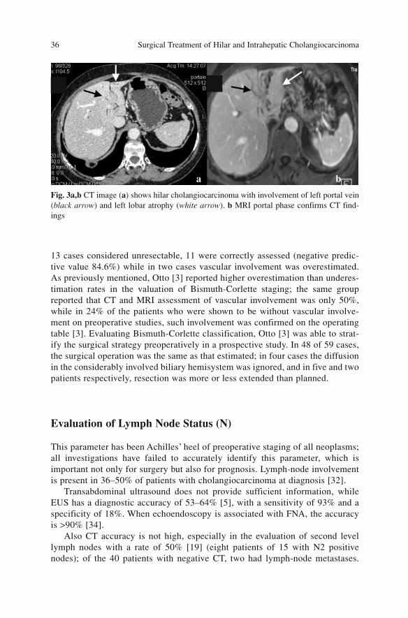
13 cases considered unresectable, 11 were correctly assessed (negative predic-tive value 84.6%) while in two cases vascular involvement was overestimated.As previously mentioned, Otto [3] reported higher overestimation than underes-timation rates in the valuation of Bismuth-Corlette staging; the same groupreported that CT and MRI assessment of vascular involvement was only 50%,while in 24% of the patients who were shown to be without vascular involve-ment on preoperative studies, such involvement was confirmed on the operatingtable [3]. Evaluating Bismuth-Corlette classification, Otto [3] was able to strat-ify the surgical strategy preoperatively in a prospective study. In 48 of 59 cases,the surgical operation was the same as that estimated; in four cases the diffusionin the considerably involved biliary hemisystem was ignored, and in five and twopatients respectively, resection was more or less extended than planned.
Evaluation of Lymph Node Status (N)
This parameter has been Achilles’ heel of preoperative staging of all neoplasms;all investigations have failed to accurately identify this parameter, which isimportant not only for surgery but also for prognosis. Lymph-node involvementis present in 36–50% of patients with cholangiocarcinoma at diagnosis [32].
Transabdominal ultrasound does not provide sufficient information, whileEUS has a diagnostic accuracy of 53–64% [5], with a sensitivity of 93% and aspecificity of 18%. When echoendoscopy is associated with FNA, the accuracyis >90% [34].
Also CT accuracy is not high, especially in the evaluation of second levellymph nodes with a rate of 50% [19] (eight patients of 15 with N2 positivenodes); of the 40 patients with negative CT, two had lymph-node metastases.
Surgical Treatment of Hilar and Intrahepatic Cholangiocarcinoma 36
Fig. 3a,b CT image (a) shows hilar cholangiocarcinoma with involvement of left portal vein(black arrow) and left lobar atrophy (white arrow). b MRI portal phase confirms CT find-ings
a b
Correct lymph node staging was obtained in 46/55 cases (83.6%).MRI presents a value of reliability comparable to EUS. Hanninen [32]
reported an accuracy of 60%. The limit is that size is not a reliable parameter; infact lymph nodes of normal size may be metastatic; instead enlarged nodes maybe inflammatory and non-metastatic [32].
A recent study [35] showed the utility of a lymph node-specific contrastagent (ultrasmall particles of iron oxide) used in the detection of lymph nodes inpatients with prostate cancer. The results regarding the accuracy of this study areexcellent but this agent has not yet been introduced in clinical practice.
PET results are discordant: some authors report a low rate of lymph-nodemetastasis detection (two of 15 cases in Kluge’s experience [36]), while others[37] report an 86% accuracy rate; sensitivity is similar to CT, but specificity ofPET is 100% vs. 59% of CT (p<0.01).
Evaluation of Metastases (M)
Abdominal ultrasound enhanced with contrast agent can better identify and typifyhepatic lesions than conventional US; CT and MRI are also useful to focalise thisparameter. PET is effective for staging due to its ability to detect distant metastases.Anderson [38] reported that 30% of the patients with distant metastases were notdiagnosed with other radiological tests, while Kim [39] identified distant metas-tases in four of 21 patients with peripheral cholangiocarcinoma, not detected byother diagnostic techniques. In Kluge’s study, the reliability rate in diagnosing dis-tant metastases was 70% (peritoneum, lung) [36].
Preoperative workup often fails to ascertain the presence of peritoneal andhepatic metastases that are shown only by preliminary laparoscopy or laparoto-my, since the small size of these lesions cannot be assessed by preoperativeimaging.
Conclusions
In our opinion, the diagnostic flow chart reported in Figure 4 allows a correctevaluation of resectability without performing direct cholangiography.
Further diagnostic investigation, such as the probable use of invasive tech-nique, biliary drainage, or restoring hepatic function, should be decided by thehepatobiliary team based on the performance status of the patient(presence/absence of sepsis, alteration of hepatic function, malnutrition) and thefeasible therapeutic approach.
Optimising management allows to avoid invasive, risky and useless manoeu-vres, long hospitalisation and delay of definitive treatment, and can facilitateselection of the patients for different therapeutic options.
Preoperative Staging 37
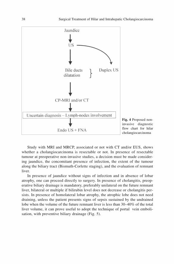
Study with MRI and MRCP, associated or not with CT and/or EUS, showswhether a cholangiocarcinoma is resectable or not. In presence of resectabletumour at preoperative non-invasive studies, a decision must be made consider-ing jaundice, the concomitant presence of infection, the extent of the tumouralong the biliary tract (Bismuth-Corlette staging), and the evaluation of remnantliver.
In presence of jaundice without signs of infection and in absence of lobaratrophy, one can proceed directly to surgery. In presence of cholangitis, preop-erative biliary drainage is mandatory, preferably unilateral on the future remnantliver, bilateral or multiple if bilirubin level does not decrease or cholangitis per-sists. In presence of homolateral lobar atrophy, the atrophic lobe does not needdraining, unless the patient presents signs of sepsis sustained by the undrainedlobe when the volume of the future remnant liver is less than 30–40% of the totalliver volume, it can prove useful to adopt the technique of portal vein emboli-sation, with preventive biliary drainage (Fig. 5).
Surgical Treatment of Hilar and Intrahepatic Cholangiocarcinoma 38
Fig. 4 Proposed non-invasive diagnosticflow chart for hilarcholangiocarcinoma
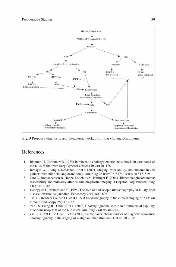
References
1. Bismuth H, Corlette MB (1975) Intrahepatic cholangioenteric anastomosis in carcinoma ofthe hilus of the liver. Surg Gynecol Obstet 140(2):170–178
2. Jarnagin WR, Fong Y, DeMatteo RP et al (2001) Staging, resectability, and outcome in 225patients with hilar cholangiocarcinoma. Ann Surg 234(4):507–517; discussion 517–519
3. Otto G, Romaneehsen B, Hoppe-Lotichius M, Bittinger F (2004) Hilar cholangiocarcinoma:resectability and radicality after routine diagnostic imaging. J Hepatobiliary Pancreat Surg11(5):310–318
4. Dancygier H, Nattermann C (1994) The role of endoscopic ultrasonography in biliary tractdisease: obstructive jaundice. Endoscopy 26(9):800–802
5. Tio TL, Reeders JW, Sie LH et al (1993) Endosonography in the clinical staging of Klatskintumour. Endoscopy 25(1):81–85
6. Yeh TS, Tseng JH, Chiu CT et al (2006) Cholangiographic spectrum of intraductal papillarymucinous neoplasm of the bile ducts. Ann Surg 244(2):248–253
7. Zidi SH, Prat F, Le Guen L et al (2000) Performance characteristics of magnetic resonancecholangiography in the staging of malignant hilar strictures. Gut 46:103–106
Preoperative Staging 39
Fig. 5 Proposed diagnostic and therapeutic workup for hilar cholangiocarcinoma
8. Manfredi R, Brizi MG, Masselli G et al (2001) Malignant biliary hilar stenosis: MR cholan-giopancreatography compared with direct cholangiography. Radiol Med 102:48–54
9. Furukawa H, Sano K, Kosuge T et al (1997) Analysis of biliary drainage in the caudate lobeof the liver: comparison of three-dimensional CT cholangiography and rotating cine cholan-giography. Radiology 204(1):113–117
10. Miura F, Asano T, Okazumi S et al (1999) Rotational cine cholangiography: evaluation foruse in diagnosing bile duct carcinoma. AJR Am J Roentgenol 173(4):1043–1048
11. Furukawa H, Sano K, Kosuge T et al (2000) Hilar cholangiocarcinoma evaluated by three-dimensional CT cholangiography and rotating cine cholangiography.Hepatogastroenterology 47(33):615–620
12. Uchida M, Abe T, Nishimura K et al (2002) Rotational three-dimensional cholangiography:initial clinical experience. Radiat Med 20(4):213–215
13. Nesbit GM, Johnson CD, James EM et al (1988) Cholangiocarcinoma: diagnosis and eval-uation of resectability by CT and sonography as procedures complementary to cholangiog-raphy. AJR Am J Roentgenol 151(5):933–938
14. Tillich M, Mischinger HJ, Preisegger KH et al (1998) Multiphasic Helical CT in diagnosisand staging of hilar cholangiocarcinoma. AJR Am J Roentgenol 171:651–658
15. Wiston C, Teitcher J (2006) Computer tomography of the liver, biliary tract, and pancreas.Blumgarth H (ed) Surgery of the liver, biliary tract, and pancreas, 4th edn. SaundersElsevier, Philadelphia, pp 266–305
16. Feydy A, Vilgrain V, Denys A et al (1999) Helical CT assessment in hilar cholangiocarcino-ma: correlation with surgical and pathologic findings. AJR Am J Roentgenol 172(1):73–77
17. Fleischmann D, Ringl H, Schofl R et al (1996) Three-dimensional spiral CT cholangiogra-phy in patients with suspected obstructive biliary disease: comparison with endoscopic ret-rograde cholangiography. Radiology 198(3):861–868
18. Kim JH, Kim TK, Eun HW et al (2006) CT findings of cholangiocarcinoma associated withrecurrent pyogenic cholangitis. AJR Am J Roentgenol 187(6):1571–1577
19. Lee HY, Kim SH, Lee JM et al (2006) Preoperative assessment of resectability of hepatichilar cholangiocarcinoma: combined CT and cholangiography with revised criteria.Radiology 239(1):113–121
20. Iwahashi N, Hayakawa N, Yamamoto H et al (1998) Mucosal bile duct carcinoma withsuperficial spread. J Hepatobiliary Pancreat Surg 5(2):221–225
21. Kim DI, Kim MH, Lee SK et al (2001) Risk factors for recurrence of primary bile ductstones after endoscopic biliary sphincterotomy. Gastrointest Endosc 54(1):42–48
22. Sano T, Nimura Y, Hayakawa N et al (1997) Clinical utility of percutaneous transhepaticcholangioscopy in defining tumour extent: a case of mucin-producing bile duct carcinomaoriginating in the left caudate lobe. Gastrointest Endosc 46(5):455–458
23. Ponchon T, Genin G, Mitchell R et al (1996) Methods, indications, and results of percuta-neous choledochoscopy. A series of 161 procedures. Ann Surg 223(1):26–36
24. Nagino M, Nimura Y (2006) Perihilar cholangiocarcinoma with emphasis on presurgicalmanagement. In: Blumgart H (ed) Surgery of the liver, biliary tract, and pancreas. 4th edn.Saunders Elsevier, Philadelphia, pp 804–814
25. Lee SS, Kim MH, Lee SK et al (2002) MR cholangiography versus cholangioscopy for eval-uation of longitudinal extension of hilar cholangiocarcinoma. Gastrointest Endosc56(1):25–32
26. Sugiyama M, Hagi H, Atomi Y, Saito M (1997) Diagnosis of portal venous invasion by pan-creatobiliary carcinoma: value of endoscopic ultrasonography. Abdom Imaging22(4):434–438
27. Hann LE, Greatrex KV, Bach AM et al (1997) Cholangiocarcinoma at the hepatic hilus:sonographic findings. AJR Am J Roentgenol 168(4):985–989
28. Jarnagin WR, D’Angelica M, Blumgart LH (2006) Intrahepatic and extrahepatic biliary can-cer. In: Blumgart H (ed) Surgery of the liver, biliary tract, and pancreas, 4th edn. SaundersElsevier, Philadelphia
Surgical Treatment of Hilar and Intrahepatic Cholangiocarcinoma 40
29. Manfredi R, Barbaro B, Masselli G et al (2004) Magnetic resonance imaging of cholangio-carcinoma. Semin Liver Dis 24(2):155–164
30. Lee MG, Park KB, Shin YM et al (2003) Preoperative evaluation of hilar cholangiocarcino-ma with contrast-enhanced three-dimensional fast imaging with steady-state precessionmagnetic resonance angiography: comparison with intraarterial digital subtraction angiogra-phy. World J Surg 27(3):278–83
31. Zhang Y, Uchida M, Abe T et al (1999) Intrahepatic peripheral cholangiocarcinoma: com-parison of dynamic CT and dynamic MRI. J Comput Assist Tomogr 23(5):670–677
32. Hanninen EL, Pech M, Jonas S et al (2005) Magnetic resonance imaging including magnet-ic resonance cholangiopancreatography for tumour localization and therapy planning inmalignant hilar obstructions. Acta Radiol 46(5):462–470
33. Cha JH, Han JK, Kim TK et al (2000) Preoperative evaluation of Klatskin tumour: accura-cy of spiral CT in determining vascular invasion as a sign of unresectability. Abdom Imaging25(5):500–507
34. Hoffman BJ, Hawes RH (1995) Endoscopic ultrasonography-guided puncture of the lymphnodes: first experience and clinical consequences. Gastrointest Endosc Clin N Am5(3):587–593
35. Harisinghani MG, Barentsz J, Hahn PF et al (2003) Noninvasive detection of clinicallyoccult lymph-node metastases in prostate cancer. N Engl J Med 348(25):24912499
36. Kluge R, Schimdt F, Caca K et al (2001) Positron emission tomography with (18F)fluoro-2-deoxy-D-glucose for diagnosis and staging of bile duct concer. Hepatology 33:1029–1035
37. Kato T, Tsukamoto E, Kuge Y et al (2002) Clinical role of (18)F-FDG PET for initial stag-ing of patients with extrahepatic bile duct cancer. Eur J Nucl Med Mol Imaging29(8):1047–1054
38. Anderson CD, Rice MH, Pinson CW et al (2004) Fluorodeoxyglucose PET imaging in theevaluation of gallbladder carcinoma and cholangiocarcinoma. J Gastrointest Surg8(1):90–97
39. Kim YJ, Yun M, Lee WJ et al (2003) Usefulness of 18F-FDG PET in intrahepatic cholan-giocarcinoma. Eur J Nucl Med Mol Imaging 30(11):1467–1472
Preoperative Staging 41

The Role of Laparoscopy in PreoperativeStaging
Hilar cholangiocarcinoma often presents in an advanced stage when the diseaseis not resectable. Despite the evolution of imaging techniques such as CT, MRI,ultrasonography and PET scan, it is often difficult to identify small liver andperitoneal metastases at preoperative staging, and the precise extent of the dis-ease along the hepato-duodenal ligament is difficult to determine. In about25–46% of patients the condition of non-resectability is confirmed only atlaparotomy [1,2].
Diagnostic laparoscopy has been proposed as a staging tool in hepatobiliaryneoplasms [3]; its main aim is to identify the patients with disseminated diseasenot diagnosed at preoperative workup, in whom the disadvantages of an unnec-essary laparotomy can be avoided. In non-resectable patients the laparoscopicapproach significantly reduces the length of stay, cost and the time of initiationof palliative therapies [4].
In the literature, reports on the use of laparoscopy in cholangiocarcinomastaging are limited. Some authors suggest carrying out laparoscopic explorationimmediately before laparotomy; others use it as a staging method preliminary toinvasive procedures such as portal vein embolisation and/or biliary drainage [5].
Technique
Laparoscopic examination is generally performed under general anaesthesia.The laparoscopic access is done in the line of the planned laparotomy incision[6]. Usually two or more port sites are used in order to more easily manipulatethe structures and obtain biopsy samples of suspect lesions. A 30-degree scopeis usually used to provide an optimal view from the periumbilical or right upperquadrant access. Laparoscopic examination entails careful inspection of theentire abdominal cavity, with particular attention to the parietal peritoneumwhich is often the site of carcinomatosis. The next exploration evaluates thesuperior and inferior surfaces of right and left lobes, hepatoduodenal ligament
43A. Guglielmi, A. Ruzzenente, C. Iacono (eds.) Surgical Treatment of Hilar and ICC.© Springer 2008
and porta hepatis; examination allows a direct view of the aspect of hepaticparenchyma, and the possible presence of cirrhosis or lobar atrophy not seen atpreoperative studies. The occurrence of evident parietal and visceral peritoneallocalisations must be confirmed by frozen-section histology, which is not indi-cated, due to the risk of neoplastic seeding for lesions that will not change thecriteria of resectability (i.e. regional lymph-node metastases) [7]. The dissectionof hilar structures is not indicated due to the highly demanding laparoscopicmanoeuvre and its limited utility in evaluating the local extent of the disease.
Results
Diagnostic laparoscopy has a low morbidity and almost no mortality. In the lit-erature, the complication rate of laparoscopy for hepatobiliary neoplasms variesfrom 0 to 4% [5,7–10]. Major and more frequently observed complications arepostoperative bleeding, intestinal perforation and intra-abdominal infection.Minor complications are wound infection, abdominal pain and pulmonary com-plications.
Neoplastic seeding in the port sites is described in the laparoscopy studies forhepatobiliary neoplasms. Nieveen reported an incidence of 2% in his study of420 diagnostic laparoscopies for upper gastrointestinal neoplasms [7]. Otherreports confirmed the occurrence of port site metastases (Shoup observed anincidence of 0.8% in a series of 1,650 diagnostic laparoscopies [11]).
In the literature, the yield of laparoscopy is variable and the frequency ofidentification of preoperative misdiagnosed lesions ranges from 20 to 40%[5,6,9,12]. Many factors contribute to this variability: limited series, differenttype of cholangiocarcinoma (i.e. hilar cholangiocarcinoma and gallbladder neo-plasm), long observation time during which the new preoperative imaging tech-niques are not employed.
The most frequent causes of unresectability are peritoneal carcinomatosis,vascular invasion, hepatic metastases, and involvement of non-regional lymphnodes. The accuracy of laparoscopy in detecting the causes of non-resectabilityis highly variable; while it is very useful for identifying hepatic and peritonealmetastases, it is of little value in evaluating vascular invasion and lymph-nodemetastases. Laparoscopic sensitivity in defining peritoneal carcinomatosis isover 80% in some series; it permits identification of more than 60% of patientswith hepatic metastases, while detection of metastatic lymph nodes is lower than30% and it rarely allows identification of patients who are non-resectable due tolocal advanced disease [5,6]. The usefulness of laparoscopy is also determinedby the stage of the disease; in a series of 84 patients Connor observed that theaccuracy of laparoscopy for T3 lesions according to the MSKCC preoperativestaging system was significantly higher compared to stage T1-2 patients. Thecause of non-resectability was metastatic disease, which was 68% in T3, 37% inT2, and 26% in T1 (p<0.05) [12].
Surgical Treatment of Hilar and Intrahepatic Cholangiocarcinoma 44
Other authors have proposed the use of peritoneal lavage with instillation of100–200 cc saline solution to enhance the accuracy of laparoscopy; however, inthe literature the only experience with hilar cholangiocarcinoma shows a lowaccuracy. In a series of 26 patients Martin [13] reported a positive peritonealcytology in only 2 of 6 cases with peritoneal carcinomatosis.
Laparoscopic Ultrasound
Laparoscopic ultrasound can be useful for increasing the sensitivity and accura-cy of laparoscopic staging. Laparoscopic ultrasound probe is linear at high fre-quency (8–10 MHz) and gives high-resolution images; besides Doppler ultra-sound evaluation it permits identification of vascular structure infiltration. Anultrasound probe is inserted through the 10-mm umbilical or right upper quad-rant port and a complete examination of hepatic parenchyma is performed, sinceit can detect hepatic lesions smaller than a few millimeters. The initial ultra-sound examination evaluates the left and right lobes; subsequently positioningthe probe transversely to the hepato-duodenal ligament, the common hepaticduct, hepatic artery and portal vein are inspected, as are the main lymph nodestations. Laparoscopic ultrasound can guide biopsy of suspect lesions.
In the literature, many experiences have been reported on the use of laparo-scopic ultrasound, but are limited to small series of patients [7,12,14–16].Although laparoscopic ultrasound allows precise visualisation of the hepaticparenchyma and hilar structures its utility in the definition of resectability ofhilar cholangiocarcinoma is not supported by the literature.
Van Delden was the first to propose the technique; in his series of 31 patientswith unresectable hilar cholangiocarcinoma, only 10 patients (28%) were iden-tified by traditional laparoscopy and the diagnosis was obtained by ultrasound inonly one patient [16]. This fact was confirmed by more recent observationswhich reported that the procedure was useful in less than 20% of cases [6,8,9].Only one author showed a significant increase in laparoscopic examinationthrough ultrasound; in his experience, in 84 patients the yield of laparoscopyalone was 24%, while it was 41% in the case of laparoscopy associated withultrasound [12]. The causes of the limited usefulness of laparoscopic ultrasoundare correlated with the difficulties in correctly defining the local neoplasticextent at the hepatic hilum, due to inflammatory alterations and the presence ofbiliary stents [6]. Sensitivity of ultrasound in diagnosing hepatic metastases isvery high; however, these patients often have hepatic lesions that emerge fromthe hepatic surface and are easily detected by direct vision. Ultrasound evalua-tion of regional and non-regional lymph nodes does not distinguish betweenmetastatic and inflammatory lymph nodes; moreover lymph node biopsy underultrasound control is very demanding. Tillman underlined the fact that very oftenthe ultrasound suspicion of unresectability is not confirmed at subsequentlaparotomy; in his study of 110 patients, ultrasound revealed a non-resectable
The Role of Laparoscopy in Preoperative Staging 45
disease in 19 cases, but at subsequent laparotomy the diagnosis was confirmedin only one patient.
Despite a better sensitivity in diagnosing hepatic metastases, ultrasound eval-uation does not offer any useful elements for evaluating lymph-node involve-ment at the hepatic hilum.
Conclusions
Diagnostic laparoscopy in hilar cholangiocarcinoma has a low yield since theneoplasm is often unresectable because locally advanced. The accuracy of theprocedure is high in the identification of peritoneal and hepatic metastases butits sensibility is low even with ultrasound support, as it does not allow identifi-cation of patients with local advanced disease or metastatic lymph nodes. Inadvanced disease, when the percentage of metastatic disease is high, the method-ical use of laparoscopy could be helpful.
References
1. Hemming AW, Reed AI, Fujita S et al (2005) Surgical management of hilar cholangiocarci-noma. Ann Surg 241(5):693–699; discussion 699–702
2. Lang H, Sotiropoulos GC, Fruhauf NR et al (2005) Extended hepatectomy for intrahepaticcholangiocellular carcinoma (ICC): when is it worthwhile? Single center experience with 27resections in 50 patients over a 5-year period. Ann Surg 241(1):134–143
3. D’Angelica M, Jarnagin W, Dematteo R et al (2002) Staging laparoscopy for potentiallyresectable noncolorectal, non-neuroendocrine liver metastases. Ann Surg Oncol9(2):204–209
4. Cuschieri A (2001) Role of video-laparoscopy in the staging of intra-abdominal lymphomasand gastrointestinal cancer. Semin Surg Oncol 20(2):167–172
5. Goere D, Wagholikar GD, Pessaux P et al (2006) Utility of staging laparoscopy in subsetsof biliary cancers: laparoscopy is a powerful diagnostic tool in patients with intrahepatic andgallbladder carcinoma. Surg Endosc 20(5):721–725
6. Weber SM, DeMatteo RP, Fong Y et al (2002) Staging laparoscopy in patients with extra-hepatic biliary carcinoma. Analysis of 100 patients. Ann Surg 235(3):392–399
7. Nieveen van Dijkum EJ, de Wit LT (1999) Staging laparoscopy and laparoscopic ultrasonog-raphy in more than 400 patients with upper gastrointestinal carcinoma. J Am Coll Surg189(5):459–465
8. Vollmer CM, Drebin JA, Middleton WD et al (2002) Utility of staging laparoscopy in sub-sets of peripancreatic and biliary malignancies. Ann Surg 235(1):1–7
9. Tilleman EH, de Castro SM, Busch OR et al (2002) Diagnostic laparoscopy and laparoscop-ic ultrasound for staging of patients with malignant proximal bile duct obstruction. JGastrointest Surg 6(3):426–430; discussion 430–431
10. Jarnagin WR, Bodniewicz J, Dougherty E et al (2000) A prospective analysis of staginglaparoscopy in patients with primary and secondary hepatobiliary malignancies. JGastrointest Surg 4(1):34–43
11. Shoup M, Brennan MF, Karpeh MS et al (2002) Port site metastasis after diagnostic
Surgical Treatment of Hilar and Intrahepatic Cholangiocarcinoma 46
laparoscopy for upper gastrointestinal tract malignancies: an uncommon entity. Ann SurgOncol 9(7):632–636
12. Connor S, Barron E, Wigmore SJ et al (2005) The utility of laparoscopic assessment in thepreoperative staging of suspected hilar cholangiocarcinoma. J Gastrointest Surg9(4):476–480
13. Martin RC 2nd, Fong Y, DeMatteo RP et al (2001) Peritoneal washings are not predictive ofoccult peritoneal disease in patients with hilar cholangiocarcinoma. J Am Coll Surg193(6):620–625
14. Callery MP, Strasberg SM, Doherty GM et al (1997) Staging laparoscopy with laparoscop-ic ultrasonography: optimizing resectability in hepatobiliary and pancreatic malignancy. JAm Coll Surg 185(1):33–39
15. Lo CM, Lai EC, Liu CL et al (1998) Laparoscopy and laparoscopic ultrasonography avoidexploratory laparotomy in patients with hepatocellular carcinoma. Ann Surg227(4):527–532
16. van Delden OM, de Wit LT, Nieveen van Dijkum EJ et al (1997) Value of laparoscopic ultra-sonography in staging of proximal bile duct tumours. J Ultrasound Med 16(1):7–12
The Role of Laparoscopy in Preoperative Staging 47

Preoperative Assessment of Liver Function
Many elements contribute to the optimal selection of patients who will undergosurgery for hilar cholangiocarcinoma, including:– Improvement of the preoperative diagnosis in the definition of size, site and
tumour extent related to intrahepatic vascularisation– Findings of the intraoperative workup– Knowledge of the biological behavior of the tumour– Effects of biliary obstruction and jaundice– Effects of parenchymal inflammatory process and of cholangitis on clinical
course and prognosis– Nutritional state– Adjuvant therapy
The choice of resection must take into consideration the need to remove thetumour completely as well as the need to maintain residual volume of parenchy-ma to carry on hepatic function.
In surgery of hilar cholangiocarcinoma, which also requires extended resec-tion, attention must be focused not only on the amount of hepatic parenchymaremoved but mainly to the residual quantity and its ability to guarantee accept-able postoperative residual liver function. Depending on the type of growth,cholangiocarcinoma can bring about complete or selective obstruction of one ormore segments of the intrahepatic biliary tree. The obstacle to bile flow isresponsible for worsened global hepatic function with a major risk of septic phe-nomena and relevant systemic effects. In presence of biliary obstruction, biliarypressure increases from 5–10 to 30 cm of H2O with destruction of the junctionsbetween hepatocytes and biliary cells. The results are: (1) compression by dilat-ed biliary tract on capacity portal vessels with reduction of portal flow; (2) neg-ative influence on humoral factors involved in initiating and maintaining theregeneration with a decrease in growth-related factors; (3) activation of hepato-cellular apoptosis process by accumulation of toxic bile salts; (4) increasedcholangiole permeability with direct bile reflux in sinusoids and beginning ofinflammatory response; (5) decreased bile excretion by hepatocytes; (6)
49A. Guglielmi, A. Ruzzenente, C. Iacono (eds.) Surgical Treatment of Hilar and ICC.© Springer 2008
decreased excretory activity of hepatocytes with a direct reflux of metabolites incirculatory system and consequent systemic toxicity; (7) alteration of micro- andmacro-vascular perfusion.
Protide synthesis is altered with albumin level reduction, alteration of coag-ulation factors and a fall in immunoglobulins. The effects of obstruction extendto detoxification activity of the liver with decreased excretion of the substancesmetabolised in the liver. The systemic effects of biliary obstruction are evidentwith regard to cardiovascular activity, renal function, and the coagulationprocess. Jaundiced patients are more susceptible to developing postoperativeshock consequent to depression of left ventricular activity and decreased periph-eral vascular resistance as well as plasmatic volume. Renal function is impairedby hyperbilirubinemia, due to the reduction of renal perfusion related to cardiacpump impairment and to renal causes themselves, as natriuretic effects of bilesalts and direct parenchymal toxicity of endotoxaemia; renal failure in jaundicepatients has a mortality that can reach 70% [1]. Deprivation of bile at the intes-tinal level interferes with vitamin K absorption, with prolongation of prothrom-bin time. The effects of bile deprivation on the intestine and the interruption ofenterohepatic circulation determine the loss of emulsive and antitoxic activity ofbile salts, permitting the abundant amount of endotoxins present in the intestin-al lumen to be absorbed by the portal circulation [2]. With a normal biliary pres-sure (7–14 cm of H2O) bacteremia in portal circulation is cleared by Kuppfercells; in presence of biliary obstruction their activity is impaired with decreasedbacterial and endotoxin clearance and antigen presentation. High concentrationof bacteria in the bile and biliary hypertension are the causes of cholangitis andbiliary sepsis. Decompression of the biliary system, with a normalisation ofbilirubinemia values, produces such improvement of biochemical circulatingparameters and hepatic function that it allows performance of extended hepate-ctomies with 70% hepatic volume resection without or with very low postoper-ative hepatic insufficiency [3–6]. Jaundice resolution is considered able torestore a hepatic reserve similar to that of patients with normal hepatic function[4]. However, external drainage does not restore enterohepatic circulation anddoes not affect bacterial translocation. Bile replacement has been proposed injaundiced patients to repair intestinal mucosa integrity and reduce septic compli-cations [7]. The need for preoperative drainage in patients with hilar cholangio-carcinoma is debated; in patients with cholangitis, long-term jaundice, severemalnutrition, and bilirubinemia >5 mg/ml, who require major hepatectomy(more than 60%), preoperative drainage is considered efficacious [8–11]. In thesame way, biliary drainage of the future remnant liver is considered mandatoryprior to performing PVE since expected hypertrophy requires normal values ofbilirubinemia [12,13]. In all other cases of biliary obstruction there are two dif-ferent schools of thought: Western thinking supports early resection in jaundicedpatients with hilar cholangiocarcinoma, considering that biliary drainage pres-ents the risk of complications (peritonitis, cholangitis, bleeding) from 3 to 5%[14,15]; also, the potential risk of neoplastic seeding, although rare, is consid-
Surgical Treatment of Hilar and Intrahepatic Cholangiocarcinoma 50
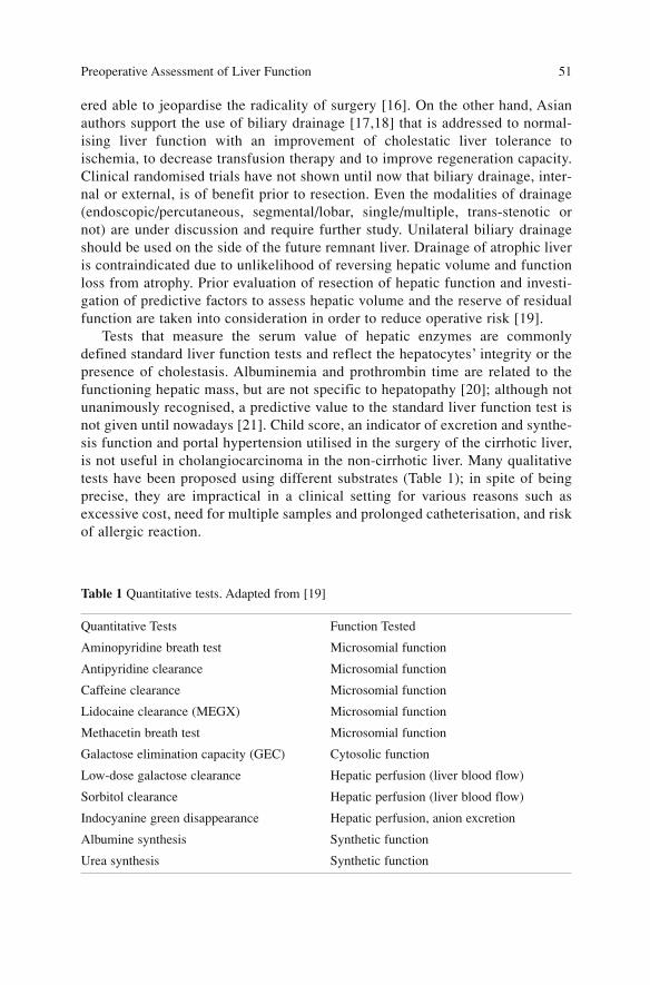
ered able to jeopardise the radicality of surgery [16]. On the other hand, Asianauthors support the use of biliary drainage [17,18] that is addressed to normal-ising liver function with an improvement of cholestatic liver tolerance toischemia, to decrease transfusion therapy and to improve regeneration capacity.Clinical randomised trials have not shown until now that biliary drainage, inter-nal or external, is of benefit prior to resection. Even the modalities of drainage(endoscopic/percutaneous, segmental/lobar, single/multiple, trans-stenotic ornot) are under discussion and require further study. Unilateral biliary drainageshould be used on the side of the future remnant liver. Drainage of atrophic liveris contraindicated due to unlikelihood of reversing hepatic volume and functionloss from atrophy. Prior evaluation of resection of hepatic function and investi-gation of predictive factors to assess hepatic volume and the reserve of residualfunction are taken into consideration in order to reduce operative risk [19].
Tests that measure the serum value of hepatic enzymes are commonlydefined standard liver function tests and reflect the hepatocytes’ integrity or thepresence of cholestasis. Albuminemia and prothrombin time are related to thefunctioning hepatic mass, but are not specific to hepatopathy [20]; although notunanimously recognised, a predictive value to the standard liver function test isnot given until nowadays [21]. Child score, an indicator of excretion and synthe-sis function and portal hypertension utilised in the surgery of the cirrhotic liver,is not useful in cholangiocarcinoma in the non-cirrhotic liver. Many qualitativetests have been proposed using different substrates (Table 1); in spite of beingprecise, they are impractical in a clinical setting for various reasons such asexcessive cost, need for multiple samples and prolonged catheterisation, and riskof allergic reaction.
Preoperative Assessment of Liver Function 51
Table 1 Quantitative tests. Adapted from [19]
Quantitative Tests Function Tested
Aminopyridine breath test Microsomial function
Antipyridine clearance Microsomial function
Caffeine clearance Microsomial function
Lidocaine clearance (MEGX) Microsomial function
Methacetin breath test Microsomial function
Galactose elimination capacity (GEC) Cytosolic function
Low-dose galactose clearance Hepatic perfusion (liver blood flow)
Sorbitol clearance Hepatic perfusion (liver blood flow)
Indocyanine green disappearance Hepatic perfusion, anion excretion
Albumine synthesis Synthetic function
Urea synthesis Synthetic function
The maximal enzymatic liver function capacity has been re-proposed basedon the C-methacetin breath test (LiMAx test) [22]; it shows a significant corre-lation with remnant liver volume, but its recent introduction requires further val-idation. Of the proposed tests for anticipating the postoperative residual liverfunction, ICG clearance is considered the most powerful predictive test of oper-ative mortality after hepatectomy if compared to other tests such as theaminoacid clearance test or aminopyrine breath test [23,24]. The model of reten-tion rate (ICG-15) is the one most frequently used. The percentage of retentioncan be measured by pulsed spectrophotometry using an optical sensor [25].There is no unanimous consensus on the cut-off value of IGC retention with apredictive value of postoperative hepatic failure, but it is believed that IGC-15equal to or greater than 15% is indicative of inadequate clearance with limitedhepatic reserve; therefore major hepatectomy is unwise. ICG and bilirubin bindto the same carrier in the transport phase in hepatocytes, determining a compet-itive inhibition. In patients with obstructive jaundice hyperbilirubinemia is inde-pendent of the reserve of hepatic function and ICG retention is therefore notvalid. In these cases 99-m TC-GSA scintigraphy is proposed, which assumes therole of a quantitative test of hepatic function [19]. Scintigraphy with 99-m TC-GSA (diethylenetriamine-pentaacetic acid with galactosyl human serum albu-min) is a dynamic technique that provides information on the density of specif-ic receptors on the plasma membrane of hepatocytes, whose density directlyreflects the functioning hepatic mass. The liver can tolerate considerable reduc-tion of its volume since mechanisms of compensatory hypertrophy are activated,but inadequate hepatic volume after resection (small remnant liver volume) canhave a negative influence on the postoperative course with the risk of hepaticfailure. Two events can occur: (1) the residual volume of a normo-functioningliver can be insufficient for providing an adequate hepatic reserve; (2) the hepat-ic reserve can be reduced due to a concomitant pre-existing hepatocellularimpairment (in the case of hilar cholangiocarcinoma, obstructed biliary tractwith jaundice, steatosis, fibrosis, cirrhosis) even in presence of an apparentlysufficient volume. Techniques of preoperative measurement of hepatic volumeon CT scans have been introduced [26]. The volumetric findings of hepaticresection and transplantation have shown a close correlation between actual andCT-calculated volumes allowing a precise evaluation of each hepatic segment[27–29]. The minimal volume of residual liver after resection that can guaranteea normal function is at least 25–30% of the initial functioning liver. This per-centage must be increased to 40% and more, in patients with chronic hepatic dis-ease or who have undergone previous chemotherapy [28,30,31]. The acquisitionof hepatic volumetry allows evaluation of the efficacy of the procedure as PVEaddressed to initiate preoperatively the compensatory hypertrophy of the futureremnant liver.
The presence of cirrhosis greatly affects hepatic regeneration, and as anindication for hepatic resection in the cirrhotic patient, is subject to debate.
Surgical Treatment of Hilar and Intrahepatic Cholangiocarcinoma 52
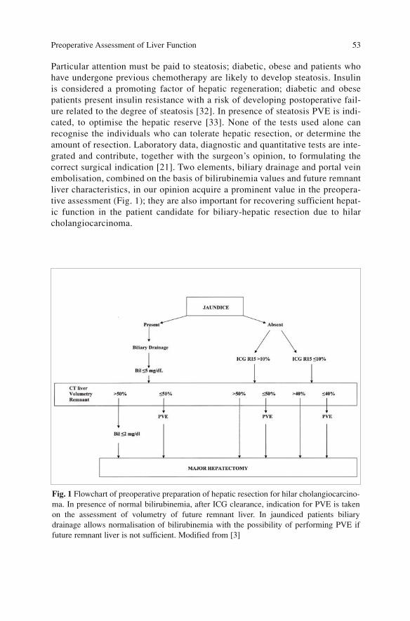
Particular attention must be paid to steatosis; diabetic, obese and patients whohave undergone previous chemotherapy are likely to develop steatosis. Insulinis considered a promoting factor of hepatic regeneration; diabetic and obesepatients present insulin resistance with a risk of developing postoperative fail-ure related to the degree of steatosis [32]. In presence of steatosis PVE is indi-cated, to optimise the hepatic reserve [33]. None of the tests used alone canrecognise the individuals who can tolerate hepatic resection, or determine theamount of resection. Laboratory data, diagnostic and quantitative tests are inte-grated and contribute, together with the surgeon’s opinion, to formulating thecorrect surgical indication [21]. Two elements, biliary drainage and portal veinembolisation, combined on the basis of bilirubinemia values and future remnantliver characteristics, in our opinion acquire a prominent value in the preopera-tive assessment (Fig. 1); they are also important for recovering sufficient hepat-ic function in the patient candidate for biliary-hepatic resection due to hilarcholangiocarcinoma.
Preoperative Assessment of Liver Function 53
Fig. 1 Flowchart of preoperative preparation of hepatic resection for hilar cholangiocarcino-ma. In presence of normal bilirubinemia, after ICG clearance, indication for PVE is takenon the assessment of volumetry of future remnant liver. In jaundiced patients biliarydrainage allows normalisation of bilirubinemia with the possibility of performing PVE iffuture remnant liver is not sufficient. Modified from [3]

References
1. Fogarty BJ, Parks RW, Rowlands BJ, Diamond T (1995) Renal dysfunction in obstructivejaundice. Br J Surg 82(7):877–884
2. Yokoyama Y, Nagino M, Nimura Y (2007) Mechanism of impaired hepatic regeneration incholestatic liver. J Hepatobiliary Pancreat Surg 14(2):159–166
3. Seyama Y, Makuuchi M (2007) Current surgical treatment for bile duct cancer. World J.Gastroenterol 13(10):1505–1515
4. Takahashi T, Togo S, Tanaka K et al (2004) Safe and permissible limits of hepatectomy inobstructive jaundice patients. World J Surg 28(5):475–481
5. Nimura Y, Kamiya J, Kondo S et al (1995) Technique of inserting multiple biliary drains andmanagement. Hepatogastroenterology 42(4):323–331
6. Kawasaki S, Imamura H, Kobayashi A et al (2003) Results of surgical resection for patientswith hilar bile duct cancer: application of extended hepatectomy after biliary drainage andhemihepatic portal vein embolization. Ann Surg 238(1):84–92
7. Kamiya S, Nagino M, Kanazawa H et al (2004) The value of bile replacement during exter-nal biliary drainage: an analysis of intestinal permeability, integrity, and microflora. AnnSurg 239(4):510–517
8. Bismuth H, Nakache R, Diamond T (1992) Management strategies in resection for hilarcholangiocarcinoma. Ann Surg 215(1):31–38
9. Vauthey JN, Blumgart LH (1994) Recent advances in the management of cholangiocarcino-mas. Semin Liver Dis 14(2):109–114
10. Nakeeb A, Pitt HA, Sohn TA et al (1996) Cholangiocarcinoma. A spectrum of intrahepatic,perihilar, and distal tumours. Ann Surg 224(4):463–473; discussion 473–475
11. Cherqui D, Benoist S, Malassagne B et al (2000) Major liver resection for carcinoma injaundiced patients without preoperative biliary drainage. Arch Surg 135(3):302–308
12. Makuuchi M, Thai BL, Takayasu K et al (1990) Preoperative portal embolization to increasesafety of major hepatectomy for hilar bile duct carcinoma: a preliminary report. Surgery107(5):521–527
13. Nagino M, Nimura Y, Hayakawa N et al (1993) Logistic regression and discriminant analy-ses of hepatic failure after liver resection for carcinoma of the biliary tract. World J Surg17(2):250–255
14. Lai EC, Mok FP, Fan ST et al (1994) Preoperative endoscopic drainage for malignantobstructive jaundice. Br J Surg 81(8):1195–1198
15. Liu CL, Lo CM, Lai EC, Fan ST (1998) Endoscopic retrograde cholangiopancreatographyand endoscopic endoprosthesis insertion in patients with Klatskin tumours. Arch Surg133(3):293–296
16. Chapman WC, Sharp KW, Weaver F, Sawyers JL (1989) Tumour seeding from percutaneousbiliary catheters. Ann Surg 209(6):708–713; discussion 713–715
17. Nimura Y, Hayakawa N, Kamiya J et al (1990) Hepatic segmentectomy with caudate loberesection for bile duct carcinoma of the hepatic hilus. World J Surg 14(4):535–543; discus-sion 544
18. Miyagawa S, Makuuchi M, Kawasaki S (1995) Outcome of extended right hepatectomyafter biliary drainage in hilar bile duct cancer. Arch Surg 130(7):759–763
19. Imamura H, Sano K, Sugawara Y et al (2005) Assessment of hepatic reserve for indicationof hepatic resection: decision tree incorporating indocyanine green test. J HepatobiliaryPancreat Surg 12(1):16–22
20. Giannini EG, Testa R, Savarino V (2005) Liver enzyme alteration: a guide for clinicians.CMAJ. Feb 1;172(3):367–379
21. Mullin EJ, Metcalfe MS, Maddern GJ (2005) How much liver resection is too much? Am JSurg 190(1):87–97
22. Stockmann M, Schwabauer E, Videv N et al (2006) The new intravenous LiMax test (max-imal enzymatic liver function capacity based on 13-c-methacetin kinetics) for mentoring of
Surgical Treatment of Hilar and Intrahepatic Cholangiocarcinoma 54
liver function after liver transplantation.Am J Transplant 6(Suppl 2):72523. Nonami T, Nakao A, Kurokawa T et al (1999) Blood loss and ICG clearance as best prog-
nostic markers of post-hepatectomy liver failure. Hepatogastroenterology46(27):1669–1672
24. Hemming AW, Scudamore CH, Shackleton CR et al (1992) Indocyanine green clearance asa predictor of successful hepatic resection in cirrhotic patients. Am J Surg 163(5):515–518
25. Okochi O, Kaneko T, Sugimoto H et al (2002) ICG pulse spectrophotometry for periopera-tive liver function in hepatectomy. J Surg Res 103(1):109–113
26. Vauthey JN, Abdalla EK, Doherty DA et al (2002) Body surface area and body weight pre-dict total liver volume in Western adults. Liver Transpl 8(3):233–240
27. Heymsfield SB, Fulenwider T, Nordlinger B et al (1979) Accurate measurement of liver, kid-ney, and spleen volume and mass by computerized axial tomography. Ann Intern Med90(2):185–187
28. Kubota K, Makuuchi M, Kusaka K et al (1997) Measurement of liver volume and hepaticfunctional reserve as a guide to decision-making in resectional surgery for hepatic tumours.Hepatology 26(5):1176–1181
29. Madoff DC, Abdalla EK, Vauthey JN (2005) Portal vein embolization in preparation formajor hepatic resection: evolution of a new standard of care. J Vasc Interv Radiol16(6):779–790
30. Abdalla EK, Hicks ME, Vauthey JN (2001) Portal vein embolization: rationale, techniqueand future prospects. Br J Surg 88(2):165–175
31. de Baere T, Roche A, Elias D et al (1996) Preoperative portal vein embolization for exten-sion of hepatectomy indications. Hepatology 24(6):1386–1391
32. Behrns KE, Tsiotos GG, DeSouza NF et al (1998) Hepatic steatosis as a potential risk fac-tor for major hepatic resection. J Gastrointest Surg 2(3):292–298
33. Bennett JJ, Blumgart LH (2005) Assessment of hepatic reserve prior to hepatic resection. JHepatobiliary Pancreat Surg 12(1):10–15
Preoperative Assessment of Liver Function 55
Preoperative Biliary Drainage
One of the most important factors of morbidity in hilar cholangiocarcinoma isthe presence of jaundice as a consequence of physiopathologic changes follow-ing cholestasis, described in Chap. “Preoperative Assessment of LiverFunction”. The close association of this complex disease with high operativerisk, determined by increased postoperative mortality and morbidity, has led tothe belief that preoperative drainage of jaundice would have decreased the riskof a major resection (i.e. pancreaticoduodenectomy or hepatic resection).
In fact, the first retrospective [1–3] and randomized studies [4] after percuta-neous drainage and endoscopic stent [5] showed decreased mortality and insome cases morbidity in jaundiced patients who had been drained. Nevertheless,controlled randomized studies [6–8] and more recent studies [9–11] showed notonly a lack of significant advantages but even an increased mortality in drainedpatients [12]. However, it is important to underline that in all these studiespatients who underwent hepatic resection were few since most underwent pan-creaticoduodenectomy for pancreatic or periampullary disease.
Recent prospective studies with larger series [12–16] offer variable resultsregarding the comparison drained/not drained: from the absence of significantdifferences in mortality and morbidity [15] to an increased rate of wound infec-tion with unchanged mortality [13,14,16] to a significant increase in complica-tions (specifically, infective and abscess) and fourfold increased mortality indrained patients [12]. Even a recent published meta-analysis on the worth of pre-operative biliary drainage shows that percutaneous transhepatic biliary drainage(PTBD) for neoplastic jaundice would not provide clear benefits and should notbe carried out routinely [17]. The probable advantages of PTBD regarding per-centage of mortality and morbidity do not appear to balance the procedure’s dis-advantages. The metanalysis concludes that controlled randomized studies and abetter drain technique are necessary in order to correctly define the problem;another point is that jaundice in patients with hilar cholangiocarcinoma who arecandidates for major liver resection presents different problems than jaundice inpatients who are candidates for pancreaticoduodenectomy; for this reason spe-cific randomized studies are required.
57A. Guglielmi, A. Ruzzenente, C. Iacono (eds.) Surgical Treatment of Hilar and ICC.© Springer 2008
In the cited meta-analysis the number of patients with hilar cholangiocarci-noma were 34 of 312 patients (11%) in randomized studies, while the numberwas 113 of 372 patients (30%) in non-randomized studies [17]. Only one ran-domized study [10] and two non-randomized studies [18,19] consider the role ofPTBD in patients with proximal cholangiocarcinoma. All three studies do notshow advantages of PTBD; however the authors of the meta-analysis underlinethat the data reported are not sufficient for a correct analysis [17]. Figueras [19]reports a morbidity rate of 100% in the 11 drained patients vs. 66% in the 9 non-drained patients (p=0.8) and a postoperative hospitalization time that is longerin drained patients (25 vs. 13 days, p=0.009).
Drainage: Pros
Considerations derived from these studies lead to the hypothesis of a change intrend that will determine a radical reduction in preoperative drainage [12] or atleast a restriction based on the following precise indications [13–17,20,21]:– Need to resolve cholangitis– Need to avoid exacerbation of severe jaundice while awaiting completion of
the workup– To correct severe malnutrition– To improve jaundice-related renal or hepatic insufficiency– To plan neoadjuvant treatment– To improve hypertrophy of the remnant liver after PVE
Mean duration of preoperative biliary drainage is 2 weeks and in the litera-ture it varies from 12 to 26 days in randomized studies and from 10 to 32 daysin non-randomized studies [17]; at the end of this period the values of bilirubine-mia are reduced on average to one-fourth of the initial value. An absent or slowdecrease in bilirubinemia suggests a malfunction of the drainage or impairedliver function. The abovementioned timing and bilirubinemia values in patientcandidates for pancreaticoduodenectomy can be considered adequate; however,in patients who are destined for hepatobiliary resection it is mandatory to re-establish bilirubinemia levels under 2 mg/dL as well as hepatic function, whichrequires 4–6 weeks as previously shown.
In literature many studies show that hepatic resection in jaundiced patients isassociated with significant morbidity and mortality rates consequent to haemor-rhage, subphrenic abscesses secondary to biliary leak, sepsis and hepatic insuf-ficiency [22–25]. For these reasons and to increase resectability and feasibilityof major hepatectomy many authors (mainly Asian) prefer to perform PTBDroutinely [26–29]. Even Belghiti [30], following Japanese indications, hasobserved that preoperative PTBD increases resectability rate; in fact dividing hisseries in two different time periods he noticed that in the first period(1992–1995) 39% of 31 patients underwent PTBD and resectability rate was32% (10 of 31 patients), vascular resection/reconstruction was performed in
Surgical Treatment of Hilar and Intrahepatic Cholangiocarcinoma 58
only 2 of the 10 resected patients (20%) while in the second time period(1995–2001) 70% of 37 patients with hilar cholangiocarcinoma underwentPTBD with a resectability rate of 59% (2 cases) and vascular resection/recon-struction in 8 of 22 resected patients (36%) [30]. Extensive use of this procedurehas determined a smaller rate of septic complications in the French study that theauthor also ascribes to improved skill (60% in the first period vs. 20% in the sec-ond) [30].
Drainage: Cons
A critical evaluation of the indiscriminate use of preoperative biliary drainagehas revealed functional limits and undeniable side effects:– After biliary drainage bilirubinemia normalizes in only two-thirds of the
treated cases [5,31] and 4–8 weeks are required for complete disappearanceof jaundice [32]. Watanapa [33] has shown in preoperative and postoperativefunctional studies that the liver resumes its normal functions 6 weeks aftersurgical operation entailing jaundice drainage with decompression of bothhepatic lobes. It has been shown experimentally that normalization ofimpaired mitochondrial function caused by jaundice requires at least 6 weeksafter biliary decompression [34] and pathological alterations become com-pletely reversible 8 weeks after restoration of normal bile drainage [35].
– Retrograde endoscopic drainage and most frequently percutaneous drains arethe causes of biliary tract, peritoneal and parietal infection with increasedoperative mortality [11,12,18,36].
– Periductal and hepatoduodenal fibrosis caused by stents or drains increaseoperative difficulties during dissection; moreover, reduction of duct size fol-lowing biliary decompression can be the reason for a demanding bilio-diges-tive anastomosis.
– Seeding along the site of percutaneous drainage [19,29,37–39].Conversely, Cherqui [25] plans a non-invasive work-up to stage the disease
and perform an early operation, believing that the utility of preoperativedrainage has not been proved. Two groups of neoplastic patients similar for age,size of tumour, type of resection and vascular occlusion were compared; 20jaundiced patients who underwent a major hepatic resection and 27 non-jaun-diced patients were selected from the institutional database of 261 patients.Haemotransfusion was performed in 85% of patients in the jaundiced group vs.48% in the non-jaundiced group, morbidity rate, which was determined mainlyby subphrenic abscess (p=0.02) and biliary leak (p=0.04) observed only in thejaundiced group, was 50 vs. 15% (p=0.006). Mortality rate (5 vs. 0%), hepaticinsufficiency rate (5 vs. 0%) and postoperative alteration of hepatic functionwere similar in the two groups. Eventually the authors concluded that: (1) evenmajor hepatic resection can be performed safely in jaundiced patients withoutpreventive biliary drainage; however the increased rate of complications and
Preoperative Biliary Drainage 59
blood transfusion is similar to that observed in patients who underwent PTBD;(2) no evidence supports the concept that preoperative PTBD can decrease thesecomplications.
An experimental study on rats [40] shows that cholestatic liver is largelyexposed to complications related to post-ischemic reperfusion, probably due todecreased antioxidant activity and increased inflammatory response; on theother hand these alterations are lessened after biliary decompression. The resultsof the study support a high risk of inflammatory events after the Pringle maneu-ver during hepatic resection in presence of cholestasis.
Endoscopic or Percutaneous Drainage
The endoscopic route is currently the procedure of choice for treating obstruc-tion of middle and distal biliary tract [41,42] with a 90–95% success rate; how-ever this percentage is lower in proximal obstruction when the procedure is com-pared to percutaneous drainage [43] although one study reports a higher successrate for endoscopic route (81–87%) vs. PTBD (57–83%) [44]. Considering themajor risks of transhepatic biliary drainage and longer preoperative hospitaliza-tion required, endoscopic drainage can be indicated in stage I and possibly stageII of Bismuth-Corlette classification, planning a fortnightly replacement to pre-vent occlusion [45]. PTBD is instead the treatment of choice in stages III e IV[42,44,46] as the endoscopic route has a success rate of only 15% in thesepatients; only one study in the literature [47] reports a success rate of 100% inthis group of patients after placement of an endoscopic plastic stent. In the futurethe high quality of MRCP imaging and the use of better endoscopic stents willincrease the endoscopic approach even for proximal obstruction [30].
Percutaneous Drainage Technique
Transhepatic percutaneous drainage can be performed either under fluoroscopy[26,48,49] or sonographic guidance [50,51], external or internal, unilateral,bilateral or multiple. Success rate for experienced personnel is high at firstattempt: 99.2% of 501 patients in the Nagoya group, arriving at 100% when afurther pass was performed, with no mortality and 9.2% morbidity rate [49].Takada [51] reports a complication rate of 6% in 603 cases of PTBD under flu-oroscopy and 0.7% in 409 cases of PTBD under sonography. To prevent cholan-gitis in case of obstructed confluence cholangiogram does not need to be per-formed, but according to Makuuchi would be carried out only to place the drainor the evening prior to surgery [20].
Surgical Treatment of Hilar and Intrahepatic Cholangiocarcinoma 60
Uni- vs. Bilateral PTBD
The initial approach must be unilateral on the future hepatic remnant lobe[26,29,30,32] but if the decision is not made about which hepatic lobe will beresected Nimura suggests draining the left hepatic duct [26]. In case the intra-hepatic biliary ducts are separated because involved by the neoplasm (as inBismuth-Corlette stages II, III, and IV) the indication changes according to theinstitution: if the contralateral ductal system is not contrast-injected it is enoughto drain a single hepatic system [43], while other authors [30] believe that bilat-eral drainage is required only in presence of cholangitis not resolved by mono-lateral PTBD. In the Nimura group’s experience bilateral drainage can beobtained by means of a single percutaneous catheter [26] and when this is notfeasible, a second PTBD is always required; in advanced cases when secondorder confluences are involved, the placement of multiple drains is needed, aswell [52]; conversely the Watanapa study [53] does not report any significantdifferences in hepatic function after complete vs. monolateral hepatic drainage6 weeks after biliary decompression; the only difference is that monolateraldrainage cannot prevent cholangitis in the non-drained lobe. The choice of mul-tiple drains is determined by segmental cholangitis [26], which represents themajor prognostic factor for outcome in major hepatic resection for hilar cholan-giocarcinoma [49,53]. In contrast, Kawasaki [52] does not consider useful themeticulous evaluation of intrahepatic diffusion of the tumour through directcholangiography due to the limits of the procedure; moreover, as the infectionrate increases the number of required drains, he supports monolateral drainageof the hepatic remnant even in the patient candidate for preliminary PVE. Thesame opinion is corroborated by the Makuuchi group that in recent reports[20,21] sustains the superiority of selective drainage of the future hepatic rem-nant since it does not increase cholangitis compared with complete liverdrainage and associated with PVE, and eases residual hepatic hypertrophy,assuring a good hepatic function.
Internal vs. External Drainage
Another unresolved and much-debated problem is that of deciding between aninternal or external drainage; when the biliary tract is drained through the endo-scopic route the drainage is always internal and it is aggravated by a noticeablerate of biliary tract infection (up to 50% and more of the cases [54]), whereastranshepatic percutaneous drainage is either internal or external: internaldrainage re-institutes bile flow in the duodenum and presents some advantagesin comparison with an external drain, above all in stimulating hepatic regenera-tion [55,56], and maintaining the intestinal barrier [57,58]. External drainage,
Preoperative Biliary Drainage 61
preferred by the Nimura group, permits decompression of the biliary systemproximally to the stenosis reducing the risk of ascending contamination asoccurs in internal drainage placed in the duodenum; although other authors [59]utilize PTBD or trans-tumoral stents, they prefer to place the tip of the catheterin the common hepatic duct to preserve sphincter function. In presence of exter-nal drainage the Nagoya group [26], and the Makuuchi as well [20,21], consid-er it important to reintroduce the drained bile per os through a nasogastric tubeor jejunostomy before and after hepatic resection to increase hepatic regenera-tion [53,56,60,61] and to maintain the barrier function of the small bowel.However other studies are needed to evaluate if intestinal bile restoration pre-vents bacterial translocation and therefore the incidence of postoperative septiccomplications [58].
Conclusions
Currently there are two schools of thought, the Western and the Eastern pointsof view, concerning preoperative biliary drainage in jaundiced patients who arecandidates for major hepatectomy. The Asian school [9,25,62,63] believes thatthe theoretical advantages of preoperative drainage are annulled by the high per-centages of procedural complications, and the risk of recurrence along the pathof the drain that can compromise the results of curative resection. For these rea-sons the procedure is not applied systematically prior to hepatic resection butonly in a subgroup of malnourished patients with hypoalbuminemia, cholangitisor long-lasting jaundice. The Asian school [20,21,26], also followed by someEuropean groups [30], regularly utilize single or multiple PTBD. However thesedifferences reveal a diverse philosophy regarding the surgical approach to thistype of disease: the Euro-American being less aggressive and the Asian moreaggressive. This idea is supported by the evaluation of the series of patients andresectability rate, hepatobiliary resection associated with vascular resection andreconstruction, percentage of trisegmentectomies and of patients who underwentPVE to determine hypertrophy of the residual hepatic segments.
Makuuchi [21] suggests the following guidelines in managing patients withcholangiocarcinoma:– In patient candidates for hepatobiliary resection, biliary drainage is required– Cholangiogram must be performed only when the drain is introduced on the
evening prior to surgery– Selective and external PTBD of the remnant liver is the treatment of choice– Drained bile must be reintroduced per os– Bile cultures with antibiogram need to be performed routinely– In stage II–IV the endoscopic route must not be used for biliary drainage– Whenever a stent is positioned endoscopically it must be substituted every 15
days– Hepatic resection must be carried out only when hepatic function is restored
Surgical Treatment of Hilar and Intrahepatic Cholangiocarcinoma 62
References
1. Denning DA, Ellison EC, Carey LC (1981) Preoperative percutaneous transhepatic biliarydecompression lowers operative morbidity in patients with obstructive jaundice. Am J Surg141(1):61–65
2. Norlander A, Kalin B, Sundblad R (1982) Effect of percutaneous transhepatic drainage uponliver function and postoperative mortality. Surg Gynecol Obstet 155(2):161–166
3. Gundry SR, Strodel WE, Knol JA et al (1984) Efficacy of preoperative biliary tract decom-pression in patients with obstructive jaundice. Arch Surg 119(6):703–708
4. Smith RC, Pooley M, George CR, Faithful GR (1985) Preoperative percutaneous transhep-atic internal drainage in obstructive jaundice: a randomized, controlled trial examining renalfunction. Surgery 97(6):641–648
5. Neff R, Fantochen E, Cooperman AM et al (1987) The radiologic management of malignantbiliary obstruction. Clin Radiol 34:143–146
6. Hatfield AR, Tobias R, Terblanche J et al (1982) Preoperative external biliary drainage inobstructive jaundice. A prospective controlled clinical trial. Lancet 2(8304):896–899
7. McPherson GA, Benjamin IS, Hodgson HJ (1984) Pre-operative percutaneous transhepaticbiliary drainage: the results of a controlled trial. Br J Surg 71(5):371–375
8. Pitt HA, Gomes AS, Lois JF et al (1985) Does preoperative percutaneous biliary drainagereduce operative risk or increase hospital cost? Ann Surg 201(5):545–553
9. Nakeeb A, Pitt HA (1995) The role of preoperative biliary decompression in obstructivejaundice. Hepatogastroenterology 42(4):332–337
10. Lai EC, Mok FP, Fan ST et al (1994) Preoperative endoscopic drainage for malignantobstructive jaundice. Br J Surg 81(8):1195–1198
11. Heslin MJ, Brooks AD, Hochwald SN et al (1998) A preoperative biliary stent is associatedwith increased complications after pancreatoduodenectomy. Arch Surg 133(2):149–154
12. Povoski SP, Karpeh MS Jr, Conlon KC et al (1999) Association of preoperative biliarydrainage with postoperative outcome following pancreaticoduodenectomy. Ann Surg230(2):131–142
13. Sohn TA, Yeo CJ, Cameron J et al (2000) Do preoperative biliary stents increase postpan-creaticoduodenectomy complications? J Gastrointest Surg 4(3):258–267; discussion267–268
14. Pisters PW, Hudec WA, Hess KR et al (2001) Effect of preoperative biliary decompressionon pancreaticoduodenectomy-associated morbidity in 300 consecutive patients. Ann Surg234(1):47–55
15. Martignoni ME, Wagner M, Krahenbuhl L et al (2001) Effect of preoperative biliarydrainage on surgical outcome after pancreatoduodenectomy. Am J Surg 181(1):52–59; dis-cussion 87
16. Srivastava S, Sikora SS, Kumar A et al (2001) Outcome following pancreaticoduodenecto-my in patients undergoing preoperative biliary drainage. Dig Surg 18(5):381–387
17. Sewnath ME, Karsten TM, Prins MH et al (2002) A meta-analysis on the efficacy of preop-erative biliary drainage for tumours causing obstructive jaundice. Ann Surg 236(1):17–27
18. Hochwald SN, Burke EC, Jarnagin W al (1999) Association of preoperative biliary stentingwith increased postoperative infectious complications in proximal cholangiocarcinoma.Arch Surg 134(3):261–266
19. Figueras J, Llado L, Valls C et al (2000) Changing strategies in diagnosis and managementof hilar cholangiocarcinoma. Liver Transpl 6(6):786–794
20. Ishizawa T, Hasegawa K, Sano K et al (2007) Selective vs. total biliary drainage for obstruc-tive jaundice caused by a hepatobiliary malignancy. Am J Surg 193(2):149–154
21. Seyama Y, Makuuchi M (2007) Current surgical treatment for bile duct cancer. World JGastroenterol 13(10):1505–1515
22. Su CH, P’eng FK, Lui WY (1992) Factors affecting morbidity and mortality in biliary tractsurgery. World J Surg 16(3):536–540
Preoperative Biliary Drainage 63
23. Clements WD, Diamond T, McCrory DC, Rowlands BJ (1993) Biliary drainage in obstruc-tive jaundice: experimental and clinical aspects. Br J Surg 80(7):834–842
24. Su CH, Tsay SH, Wu CC et al (1996) Factors influencing postoperative morbidity, mortali-ty, and survival after resection for hilar cholangiocarcinoma. Ann Surg 223(4):384–394
25. Cherqui D, Benoist S, Malassagne B et al (2000) Major liver resection for carcinoma injaundiced patients without preoperative biliary drainage. Arch Surg 135(3):302–308
26. Nagino M, Nimura Y (2006) Perihilar cholangiocarcinoma with emphasis on presurgicalmanagement. In: Blumgart LH (ed) Surgery of the liver, biliary tract, and pancreas. 4th edn.Saunders Elsevier, Philadelphia, pp 804–814
27. Miyagawa S, Makuuchi M, Kawasaki S (1995) Outcome of extended right hepatectomyafter biliary drainage in hilar bile duct cancer. Arch Surg 130(7):759–763
28. Ebata T, Nagino M, Kamiya J (2003) Hepatectomy with portal vein resection for hilarcholangiocarcinoma: audit of 52 consecutive cases. Ann Surg 238(5):720–727
29. Seyama Y, Kubota K, Sano K et al (2003) Long-term outcome of extended hemihepatecto-my for hilar bile duct cancer with no mortality and high survival rate. Ann Surg238(1):73–83
30. Belghiti J, Ogata S (2005) Preoperative optimization of the liver for resection in patientswith hilar cholangiocarcinoma. HPB Surg 7:252–253
31. Pellegrini CA, Thomas MJ, Way LW (1982) Bilirubin and alkaline phosphatase valuesbefore and after surgery for biliary obstruction. Am J Surg 143(1):67–73
32. Howard JH, Jordan GL, Reber HA(1987) Surgical disease of the pancreas. Lea&Febriger,Philadelphia
33. Watanapa P (1996) Recovery patterns of liver function after complete and partial surgicalbiliary decompression. Am J Surg 171(2):230–234
34. Koyama K, Takagi Y, Ito K, Sato T (1981) Experimental and clinical studies on the effect ofbiliary drainage in obstructive jaundice. Am J Surg 142(2):293–299
35. Aronson DC, Chamuleau RA, Frederiks WM (1993) Reversibility of cholestatic changesfollowing experimental common bile duct obstruction: fact or fantasy? J Hepatol18(1):85–95
36. Temudom T, Sarr MG, Douglas MG, Farnell MB (1995) An argument against routine per-cutaneous biopsy, ERCP, or biliary stent placement in patients with clinically resectableperiampullary masses: a surgical perspective. Pancreas 11(3):283–288
37. Jarnagin WR, Burke E, Powers C et al (1998) Intrahepatic biliary enteric bypass provideseffective palliation in selected patients with malignant obstruction at the hepatic duct con-fluence. Am J Surg 175(6):453–460
38. Soyer P, Pelage JP, Dufresne AC et al (1998) CT of abdominal wall implantation metastasesafter abdominal percutaneous procedures. J Comput Assist Tomogr 22(6):889–893
39. Sakata J, Shirai Y, Wakai T et al (2005) Catheter tract implantation metastases associatedwith percutaneous biliary drainage for extrahepatic cholangiocarcinoma. World JGastroenterol 11(44):7024–7027
40. Kloek JJ, Marsman HA, van Vliet AK et al (2007) Biliary drainage attenuates post-ischemicreperfusion injury in the cholestatic rat liver. J Hepatobiliary Pancreat Surg 9(Suppl 2):11,abs 34
41. Sherman S (2001) Current status of endoscopic pancreaticobiliary interventions. J VascInterv Radiol 12:120–155
42. England RE, Martin DF (1996) Endoscopic and percutaneous intervention in malignantobstructive jaundice. Cardiovasc Intervent Radiol 19(6):381–387
43. Hatzidakis A, Adam A (2003) The interventional radiological management of cholangiocar-cinoma. Clin Radiol 58(2):91–96
44. Nelsen KM, Kastan DJ, Shetty PC et al (1996) Utilization pattern and efficacy of nonsurgi-cal techniques to establish drainage for high biliary obstruction. J Vasc Interv Radiol7(5):751–756
45. Jagannath P, Dhir V, Shrikhande S et al (2005) Effect of preoperative biliary stenting onimmediate outcome after pancreaticoduodenectomy. Br J Surg 92(3):356–361
Surgical Treatment of Hilar and Intrahepatic Cholangiocarcinoma 64
46. Cowling MG, Adam A (2001) Internal stenting in malignant biliary obstruction. World JSurg 25:355–361
47. Gerhards MF, den Hartog D, Rauws EA et al (2001) Palliative treatment in patients withunresectable hilar cholangiocarcinoma: results of endoscopic drainage in patients with typeIII and IV hilar cholangiocarcinoma. Eur J Surg 167(4):274–280
48. Takada T, Hanyu F, Kobayashi S, Uchida Y (1976) Percutaneous transhepatic cholangialdrainage: direct approach under fluoroscopic control. J Surg Oncol 8(1):83–97
50. Kanai M, Nimura Y, Kamiya J et al (1996) Preoperative intrahepatic segmental cholangitisin patients with advanced carcinoma involving the hepatic hilus. Surgery 119(5):498–504
50. Singhal D, van Gulik TM, Gouma DJ (2005) Palliative management of hilar cholangiocar-cinoma. Surg Oncol 14(2):59–74
51. Takada T, Yasuda H, Hanyu F (1995) Technique and management of percutaneous transhep-atic cholangial drainage for treating an obstructive jaundice. Hepatogastroenterology42(4):317–322
52. Kawasaki S, Imamura H, Kobayashi A (2003) Results of surgical resection for patients withhilar bile duct cancer: application of extended hepatectomy after biliary drainage and hemi-hepatic portal vein embolization. Ann Surg 238(1):84–92
53. Nimura Y, Kamiya J, Kondo S (2000) Aggressive preoperative management and extendedsurgery for hilar cholangiocarcinoma: Nagoya experience. J Hepatobiliary Pancreat Surg7(2):155–162
54. Rerknimitr R, Attasaranya S, Kladchareon N et al (2002) Feasibility and complications ofendoscopic biliary drainage in patients with malignant biliary obstruction at KingChulalongkorn Memorial Hospital. J Med Assoc Thai 85(Suppl 1):S48-S53
55. Saiki S, Chijiiwa K, Komura M et al (1999) Preoperative internal biliary drainage is superi-or to external biliary drainage in liver regeneration and function after hepatectomy inobstructive jaundiced rats. Ann Surg 230(5):655–662
56. Suzuki H, Iyomasa S, Nimura Y, Yoshida S (1994) Internal biliary drainage, unlike externaldrainage, does not suppress the regeneration of cholestatic rat liver after partial hepatecto-my. Hepatology 20(5):1318–1322
57. Ogata Y, Nishi M, Nakayama H et al (2003) Role of bile in intestinal barrier function and itsinhibitory effect on bacterial translocation in obstructive jaundice in rats. J Surg Res115(1):18–23
58. Kamiya S, Nagino M, Kanazawa H et al (2004) The value of bile replacement during exter-nal biliary drainage: an analysis of intestinal permeability, integrity, and microflora. AnnSurg 239(4):510–517
59. Brown KT (2006) Interventional radiologic techniques in hilar and intrahepatic biliarytumours. In: Blumgart LH (ed) Surgery of the liver, biliary tract, and pancreas. 4 edn.Saunders Elsevier, Philadelphia, pp 814–822
60. Iyomasa S, Terasaki M, Kuriki H et al (1992) Decrease in regeneration capacity of rat liverafter external biliary drainage. Eur Surg Res 24(5):265–272
61. Takeuchi E, Nimura Y, Nagino M et al (1997) Human hepatocyte growth factor in bile: anindicator of posthepatectomy liver function in patients with biliary tract carcinoma.Hepatology 26(5):1092–1099
62. Bismuth H, Nakache R, Diamond T (1992) Management strategies in resection for hilarcholangiocarcinoma. Ann Surg 215(1):31–38
63. Parc Y, Frileux P, Vaillant JC et al (1999) Postoperative peritonitis originating from the duo-denum: operative management by intubation and continuous intraluminal irrigation. Br JSurg 86(9):1207–1212
Preoperative Biliary Drainage 65
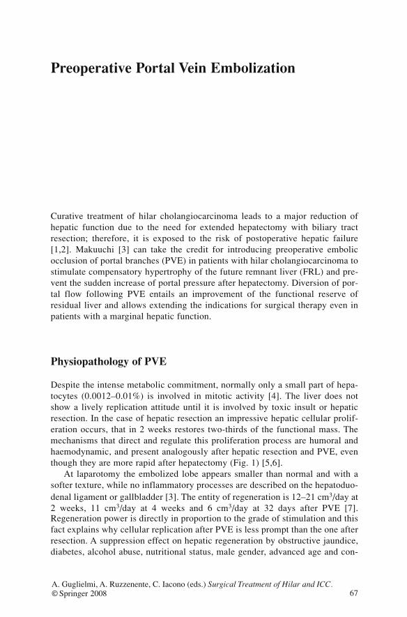
Preoperative Portal Vein Embolization
Curative treatment of hilar cholangiocarcinoma leads to a major reduction ofhepatic function due to the need for extended hepatectomy with biliary tractresection; therefore, it is exposed to the risk of postoperative hepatic failure[1,2]. Makuuchi [3] can take the credit for introducing preoperative embolicocclusion of portal branches (PVE) in patients with hilar cholangiocarcinoma tostimulate compensatory hypertrophy of the future remnant liver (FRL) and pre-vent the sudden increase of portal pressure after hepatectomy. Diversion of por-tal flow following PVE entails an improvement of the functional reserve ofresidual liver and allows extending the indications for surgical therapy even inpatients with a marginal hepatic function.
Physiopathology of PVE
Despite the intense metabolic commitment, normally only a small part of hepa-tocytes (0.0012–0.01%) is involved in mitotic activity [4]. The liver does notshow a lively replication attitude until it is involved by toxic insult or hepaticresection. In the case of hepatic resection an impressive hepatic cellular prolif-eration occurs, that in 2 weeks restores two-thirds of the functional mass. Themechanisms that direct and regulate this proliferation process are humoral andhaemodynamic, and present analogously after hepatic resection and PVE, eventhough they are more rapid after hepatectomy (Fig. 1) [5,6].
At laparotomy the embolized lobe appears smaller than normal and with asofter texture, while no inflammatory processes are described on the hepatoduo-denal ligament or gallbladder [3]. The entity of regeneration is 12–21 cm3/day at2 weeks, 11 cm3/day at 4 weeks and 6 cm3/day at 32 days after PVE [7].Regeneration power is directly in proportion to the grade of stimulation and thisfact explains why cellular replication after PVE is less prompt than the one afterresection. A suppression effect on hepatic regeneration by obstructive jaundice,diabetes, alcohol abuse, nutritional status, male gender, advanced age and con-
67A. Guglielmi, A. Ruzzenente, C. Iacono (eds.) Surgical Treatment of Hilar and ICC.© Springer 2008
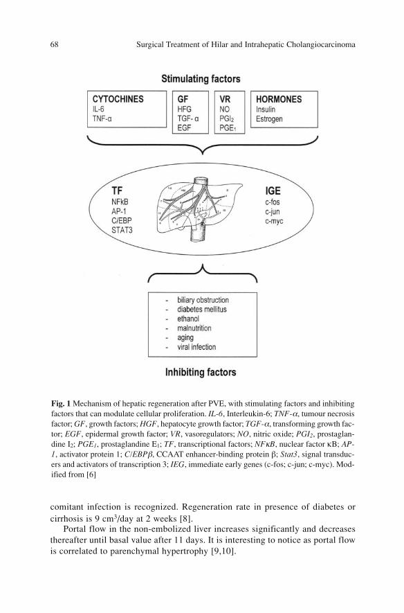
comitant infection is recognized. Regeneration rate in presence of diabetes orcirrhosis is 9 cm3/day at 2 weeks [8].
Portal flow in the non-embolized liver increases significantly and decreasesthereafter until basal value after 11 days. It is interesting to notice as portal flowis correlated to parenchymal hypertrophy [9,10].
Surgical Treatment of Hilar and Intrahepatic Cholangiocarcinoma 68
Fig. 1 Mechanism of hepatic regeneration after PVE, with stimulating factors and inhibitingfactors that can modulate cellular proliferation. IL-6, Interleukin-6; TNF-α, tumour necrosisfactor; GF, growth factors; HGF, hepatocyte growth factor; TGF-α, transforming growth fac-tor; EGF, epidermal growth factor; VR, vasoregulators; NO, nitric oxide; PGI2, prostaglan-dine I2; PGE1, prostaglandine E1; TF, transcriptional factors; NFκB, nuclear factor κB; AP-1, activator protein 1; C/EBPβ, CCAAT enhancer-binding protein β; Stat3, signal transduc-ers and activators of transcription 3; IEG, immediate early genes (c-fos; c-jun; c-myc). Mod-ified from [6]
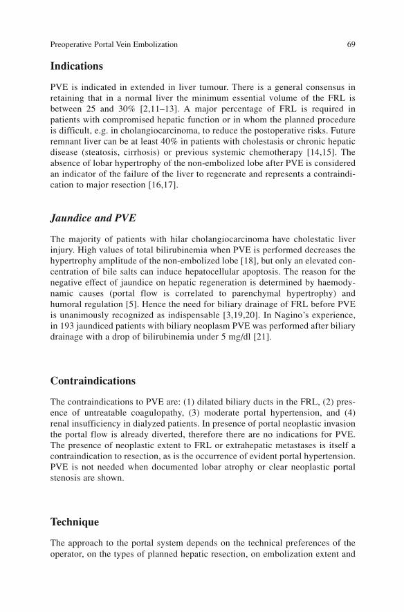
Indications
PVE is indicated in extended in liver tumour. There is a general consensus inretaining that in a normal liver the minimum essential volume of the FRL isbetween 25 and 30% [2,11–13]. A major percentage of FRL is required inpatients with compromised hepatic function or in whom the planned procedureis difficult, e.g. in cholangiocarcinoma, to reduce the postoperative risks. Futureremnant liver can be at least 40% in patients with cholestasis or chronic hepaticdisease (steatosis, cirrhosis) or previous systemic chemotherapy [14,15]. Theabsence of lobar hypertrophy of the non-embolized lobe after PVE is consideredan indicator of the failure of the liver to regenerate and represents a contraindi-cation to major resection [16,17].
Jaundice and PVE
The majority of patients with hilar cholangiocarcinoma have cholestatic liverinjury. High values of total bilirubinemia when PVE is performed decreases thehypertrophy amplitude of the non-embolized lobe [18], but only an elevated con-centration of bile salts can induce hepatocellular apoptosis. The reason for thenegative effect of jaundice on hepatic regeneration is determined by haemody-namic causes (portal flow is correlated to parenchymal hypertrophy) andhumoral regulation [5]. Hence the need for biliary drainage of FRL before PVEis unanimously recognized as indispensable [3,19,20]. In Nagino’s experience,in 193 jaundiced patients with biliary neoplasm PVE was performed after biliarydrainage with a drop of bilirubinemia under 5 mg/dl [21].
Contraindications
The contraindications to PVE are: (1) dilated biliary ducts in the FRL, (2) pres-ence of untreatable coagulopathy, (3) moderate portal hypertension, and (4)renal insufficiency in dialyzed patients. In presence of portal neoplastic invasionthe portal flow is already diverted, therefore there are no indications for PVE.The presence of neoplastic extent to FRL or extrahepatic metastases is itself acontraindication to resection, as is the occurrence of evident portal hypertension.PVE is not needed when documented lobar atrophy or clear neoplastic portalstenosis are shown.
Technique
The approach to the portal system depends on the technical preferences of theoperator, on the types of planned hepatic resection, on embolization extent and
Preoperative Portal Vein Embolization 69

the embolizing agent. Independently of the chosen route, the aim is to occludecompletely the portal branches of the portion of hepatic parenchyma that isgoing to be resected; this prevents the development of porto-portal collateralsthat can negatively condition the volumetric increase of the residual part of theliver [22]. The type of planned resection regulates the choice of the segments tobe embolized. If right hepatectomy is planned, segments V–VIII are embolized,with extension to IVa and IVb if right extended hepatectomy is required to allowsurgical radicality and amplify hypertrophy of the FRL. The extension ofembolization to segment IV (PVE “trisegment”) in patients with hilar cholangio-carcinoma has been shown to decrease the technical difficulties of trisectionec-tomy and to increase hypertrophy of segments II and III. On percentage, the useof left extended hepatectomy (4 of 240 extended hepatectomies for biliary neo-plasm in Nagino’s experience [21]) with preventive embolization of left andright anterior portal branches results lower. The technique requires antibioticprophylaxis and even if general anesthesia may be needed, PVE can be per-formed under local anesthesia with intravenous sedative. Under ultrasound con-trol the most suitable access to the portal system is identified. Access to the por-tal system is achieved under ultrasound or fluoroscopic guidance in a preparedantiseptic field. Transparenchymal portal puncture is carried out by Chiba nee-dle and the porta vein is cannulated by means of the Seldinger technique. Theangiographic study with right and left selective injections in different projec-tions allow definition of the portal anatomy.
The standard procedures of PVE are: contralateral transhepatic, ipsilateraltranshepatic and laparotomic transileocolic. In the contralateral transhepaticapproach, portal access is achieved through the parenchyma of the FRL [23]; inthe case that right hepatectomy is planned, the catheter is introduced in theumbilical portion of the portal vein and advanced until the branches of the rightportal system. This technique has the advantage of being simple since thecatheterization of right portal branches is direct; its disadvantages are repre-sented by adverse events in 12.8%: migration of embolic material in the FLR,portal thrombosis, bleeding, (haemobilia, haemoperitoneum, subcapsularhaematoma) and transient hepatic failure [24]. The complications of the con-tralateral approach, on the part of non-embolized liver that will remain, may beso significant that the planned resection may be more difficult or be abandoned[7,25]. The ipsilateral transhepatic approach described by Nagino [25] with theuse of a dedicated two-way catheter and subsequently with traditional catheters[26] requires the puncture and catheterization of a peripheral portal branch ofthe lobe that will be resected. It presents the advantage that the FLR is not trau-matized, the risk of bleeding at catheter withdrawal is lower, but migration ofembolic material into the portal system is still possible. The direct cannulationof the ileocolic vein and progression up to the portal branch for the emboliza-tion is the transileocolic approach [27]; it requires general anesthesia, laparoto-my, and is indicated whenever a staging operation or associated surgery areneeded. It has the disadvantages of laparotomy risks and cannot take advantageof the assistance of the technical tools of an equipped radiologic room. The
Surgical Treatment of Hilar and Intrahepatic Cholangiocarcinoma 70
experience in performing transjugular intrahepatic portosistemic shunts hasallowed proposing the transjugular approach for PVE. The procedure requiresthe puncture of the right or left branch of portal vein from right, middle or lefthepatic vein under ultrasound control; the positioning of the catheter to the por-tal bifurcation allows the right PVE. It is an initial experience that requires val-idation [28]. There are many agents with embolizing action that are utilized,sometimes associated with coils, and each one has its own characteristics.Development of new agents is very dynamic in the effort to guarantee a com-plete and permanent occlusion that can reduce recanalization after PVE andpromote a prompt and adequate hypertrophy.
Results
Independently of the disease, technique, type of embolizing agents, and intervalPVE-resection the results of the different series are similar. From Abdalla’sreview of 345 patients who underwent PVE, mean value in percentage of totalliver volume resulted 26.6% before the procedure; after PVE the mean volumewas 39.1% with a mean volume increase of 12.4%. From 2 to 20% of the casesPVE has been unable to stimulate a satisfactory hypertrophy [9]. The reasons forthe unsuccessful augment have been vascular abnormalities due to recanaliza-tion of embolized segments and the presence of significant portal hypertensionwith porto-systemic shunts [19,29,30]. As abovementioned, diabetes and chron-ic hepatopathy reduce regenerative stimulus and can explain the ineffectivenessof PVE.
Preoperative Portal Vein Embolization 71
Fig. 2a,b Portal vein embolization: a Transparenchymal portography: the angiographicstudy allows defining the portal anatomy. b Radiological control after PVE shows embolicmaterial (fibrin glue) in the right portal vein branches
a b
Post-PVE Course and Timing of Resection
PVE is usually followed by alteration of the hepatic functional parameters (par-ticularly increased ALT, AST and bilirubinemia), leukocytosis and fever. Thesealterations are slight, transient and self-limiting. The entity of tissue necrosisafter PVE is insignificant; therefore it is not related to considerable systemicsymptoms as after arterial embolization. From 14 to 63 days after PVE a newhepatic volumetry is performed. If the expected hypertrophy of the FRL isnoticed the patient is prepared for resection, when bilirubinemia returns withinnormal range [31]. The risk of portal recanalization induces the surgeon not todefer the operation.
Complications
Complications of the procedure (6–15%) [32] are represented by: (1) haemoperi-toneum, haemobilia and subcapsular haematoma in the site of the hepatic access;(2) portal thrombosis; (3) migration of embolizing particles in the hepatic por-tion to be preserved; (4) intestinal obstruction in the transileocolic approach; (5)need for re-embolization. The planned hepatic resection was not performed dueto complications (mainly hepatic thrombosis) of PVE in 0.5% of the cases [24].
Conclusions
Right extended hepatectomy or (less frequently) left extended hepatectomy areindicated in hilar cholangiocarcinoma. In case of right extended hepatectomyPVE is performed because the volume of segments II and III is often inadequate.PVE has an insignificant incidence of complications and gives a measurablehypertrophy of the non-embolized lobe. The preoperative increase in the volumeand hepatic function of the FRL enlarges the pool of patient candidates for a cur-ative resection and makes resection itself safer. Many surgical series confirm thelow complication rate and postoperative failure after major hepatic resection[15,33–36]. PVE does not change long-term survival: 5-year survival was 40%in the resected group with previous PVE vs. 38% in the group that underwentresection alone [15]. The benefits of the procedure are validated by controlledrandomized trials that are considered unethical. Indications for PVE are stillunclear because of the few data regarding the minimal hepatic volume requiredto tolerate surgery without serious complications [21]. There are no evident datathat define the minimal volume of hepatic parenchyma required for tolerating anextended hepatic resection; therefore it is possible that at present, PVE is over-used [9,21].
Surgical Treatment of Hilar and Intrahepatic Cholangiocarcinoma 72
References
1. Shirabe K, Shimada M, Gion T et al (1999) Postoperative liver failure after major hepaticresection for hepatocellular carcinoma in the modern era with special reference to remnantliver volume. J Am Coll Surg 188(3):304–309
2. Abdalla EK, Barnett CC, Doherty D et al (2002) Extended hepatectomy in patients withhepatobiliary malignancies with and without preoperative portal vein embolization. ArchSurg 137(6):675–680; discussion 680–681
3. Makuuchi M, Thai BL, Takayasu K et al (1990) Preoperative portal embolization to increasesafety of major hepatectomy for hilar bile duct carcinoma: a preliminary report. Surgery107(5):521–527
4. Black DM, Behrns KE (2002) A scientist revisits the atrophy-hypertrophy complex: hepat-ic apoptosis and regeneration. Surg Oncol Clin N Am 11(4):849–864
5. Yokoyama Y, Nagino M, Nimura Y (2007) Mechanisms of hepatic regeneration followingportal vein embolization and partial hepatectomy: a review. World J Surg 31(2):367–374
6. Komori K, Nagino M, Nimura Y (2006) Hepatocyte morphology and kinetics after portalvein embolization. Br J Surg 93(6):745–751
7. Madoff DC, Abdalla EK, Vauthey JN (2005) Portal vein embolization in preparation formajor hepatic resection: evolution of a new standard of care. J Vasc Interv Radiol16(6):779–790
8. Nagino M, Nimura Y, Kamiya J (1995) Changes in hepatic lobe volume in biliary tract can-cer patients after right portal vein embolization. Hepatology 21(2):434–439
9. Abdalla EK, Hicks ME, Vauthey JN (2001) Portal vein embolization: rationale, techniqueand future prospects. Br J Surg 88(2):165–175
10. Goto Y, Nagino M, Nimura Y (1998) Doppler estimation of portal blood flow after percuta-neous transhepatic portal vein embolization. Ann Surg 228(2):209–213
11. Hemming AW, Reed AI, Howard RJ et al (2003) Preoperative portal vein embolization forextended hepatectomy. Ann Surg 237(5):686–691; discussion 691–693
12. Yigitler C, Farges O, Kianmanesh R et al (2003) The small remnant liver after major liverresection: how common and how relevant? Liver Transpl 9(9):S18-S25
13. Vauthey JN, Chaoui A, Do KA et al (2000) Standardized measurement of the future liverremnant prior to extended liver resection: methodology and clinical associations. Surgery127(5):512–519
14. Kubota K, Makuuchi M, Kusaka K et al (1997) Measurement of liver volume and hepaticfunctional reserve as a guide to decision-making in resectional surgery for hepatic tumours.Hepatology 26(5):1176–1181
15. Azoulay D, Castaing D, Krissat J et al (2000) Percutaneous portal vein embolization increas-es the feasibility and safety of major liver resection for hepatocellular carcinoma in injuredliver. Ann Surg 232(5):665–672
16. Farges O, Belghiti J, Kianmanesh R et al (2003) Portal vein embolization before right hepa-tectomy: prospective clinical trial. Ann Surg 237(2):208–217
17. Belghiti J (2004) Arguments for a selective approach of preoperative portal vein emboliza-tion before major hepatic resection. J Hepatobiliary Pancreat Surg 11(1):21–24
18. Imamura H, Shimada R, Kubota M et al (1999) Preoperative portal vein embolization: anaudit of 84 patients. Hepatology 29(4):1099–1105
19. Cherqui D, Benoist S, Malassagne B et al (2000) Major liver resection for carcinoma injaundiced patients without preoperative biliary drainage. Arch Surg 135(3):302–308
20. Nagino M, Nimura Y, Hayakawa N (1993) Percutaneous transhepatic portal embolizationusing newly devised catheters: preliminary report. World J Surg 17(4):520–524
21. Nagino M, Kamiya J, Nishio H et al (2006) Two hundred forty consecutive portal veinembolizations before extended hepatectomy for biliary cancer: surgical outcome and long-term follow-up. Ann Surg 243(3):364–372
Preoperative Portal Vein Embolization 73
22. Denys AL, Abehsera M, Sauvanet A et al (1999) Failure of right portal vein ligation toinduce left lobe hypertrophy due to intrahepatic portoportal collaterals: successful treatmentwith portal vein embolization. AJR Am J Roentgenol 173(3):633–635
23. Kinoshita H, Sakai K, Hirohashi K et al (1986) Preoperative portal vein embolization forhepatocellular carcinoma. World J Surg 10(5):803–808
24. Di Stefano DR, de Baere T, Denys A et al (2005) Preoperative percutaneous portal veinembolization: evaluation of adverse events in 188 patients. Radiology 234(2):625–630
25. Nagino M, Nimura Y, Kamiya J et al (1996) Selective percutaneous transhepatic emboliza-tion of the portal vein in preparation for extensive liver resection: the ipsilateral approach.Radiology 200(2):559–563
26. Madoff DC, Hicks ME, Abdalla EK et al (2003) Portal vein embolization with polyvinylalcohol particles and coils in preparation for major liver resection for hepatobiliary malig-nancy: safety and effectiveness study in 26 patients. Radiology 227(1):251–260
27. Liem MS, Liu CL, Tso WK et al (2005) Portal vein embolisation prior to extended right-sided hepatic resection. Hong Kong Med J 11(5):366–372
28. Perarnau JM, Daradkeh S, Johann M et al (2003) Transjugular preoperative portal emboliza-tion (TJPE) a pilot study. Hepatogastroenterology 50(51):610–613
29. Bruix J, Castells A, Bosch J et al (1996) Surgical resection of hepatocellular carcinoma incirrhotic patients: prognostic value of preoperative portal pressure. Gastroenterology111(4):1018–1022
30. Tanaka H, Hirohashi K, Kubo S et al (1999) Influence of histological inflammatory activityon regenerative capacity of liver after percutaneous transhepatic portal vein embolization. JGastroenterol 34(1):100–104
31. Seyama Y, Kubota K, Sano K et al (2003) Long-term outcome of extended hemihepatecto-my for hilar bile duct cancer with no mortality and high survival rate. Ann Surg238(1):73–83
32. Kodama Y, Shimizu T, Endo H et al (2002) Complications of percutaneous transhepatic por-tal vein embolization. J Vasc Interv Radiol 13(12):1233–1237
33. Lee KC, Kinoshita H, Hirohashi K et al (1993) Extension of surgical indications for hepa-tocellular carcinoma by portal vein embolization. World J Surg 17(1):109–115
34. Shimamura T, Nakajima Y, Une Y et al (1997) Efficacy and safety of preoperative percuta-neous transhepatic portal embolization with absolute ethanol: a clinical study. Surgery121(2):135–141
35. Wakabayashi H, Yachida S, Maeba T, Maeta H (2000) Indications for portal vein emboliza-tion combined with major hepatic resection for advanced-stage hepatocellular carcinomas.A preliminary clinical study. Dig Surg 17(6):587–594
36. Tanaka H, Hirohashi K, Kubo S et al (2000)Preoperative portal vein embolization improvesprognosis after right hepatectomy for hepatocellular carcinoma in patients with impairedhepatic function. Br J Surg 87(7):879–882
Surgical Treatment of Hilar and Intrahepatic Cholangiocarcinoma 74
Prognostic Factors
Radical surgery is the only therapeutic option that can ensure a long-term sur-vival; until now many aspects of this disease have not been well-known, such aspathogenesis, and clinical and histopathologic factors that determine survival. Inthis chapter we will analyze the main prognostic factors that influence survival:macroscopic aspect, local extent, lymph-node involvement and distant metas-tases. Histological characteristics and molecular factors related to prognosis willalso be analysed.
Gross Type
Three different macroscopic forms have been described: sclerosing, nodular, andpapillary. The sclerosing type represents about 70% of all hilar cholangiocarci-nomas; it presents as a circumferential thickening of the biliary wall and isaccompanied by important fibrotic and desmoplastic phenomena. The sclerosingtype is characterized by compact masses that protrude into the lumens and areoften associated with the infiltrative type. The papillary type represents 4–5 %[1,2] of all cholangiocarcinomas and is characterized by soft and friable lesionthat occupies the bile duct lumen. In about 40% of papillary neoplasms a super-ficial spread is seen that extends up to 35 mm from the main lesion; this findingis defined as “bile duct carcinoma with superficial spread” [3].
Papillary neoplasms are usually associated with the early stage of the dis-ease, TNM UICC/AJCC Stage <IIa, in more than 70% of the cases, with 4% oflymph-node metastases [4]. In general papillary forms are associated with a bet-ter prognosis than nodular-infiltrative, with median survival of 55 vs. 33 months[4]. Even when they are associated with advanced stages with lymph-nodeinvolvement, prognosis is better than nodular-infiltrative forms with 10-year sur-vival rate of 12 vs. 5%, respectively [1].
75A. Guglielmi, A. Ruzzenente, C. Iacono (eds.) Surgical Treatment of Hilar and ICC.© Springer 2008
Microscopic Pattern
The main microscopic factors of hilar cholangiocarcinoma, that affect progno-sis, are: cellular differentiation, perineural infiltration, lymphatic and microvas-cular infiltration.
Cellular differentiation is an important prognostic factor, and is directly asso-ciated with the stage of disease, percentage of curative resections and long-termprognosis.
Survival is significantly related to cellular differentiation: 34–41 months forG1–G2 and 14–20 months for G3 [5,6].
Jarnagin [4] has observed that 64% of well-differentiated hilar cholangiocar-cinoma shows an early stage (TNM UICC/AJCC IIa), compared to only 25% ofmoderately or poorly differentiated types. Moreover, the percentage of curativeR0 resection is related to cellular differentiation; Neuhaus reported [7] a radicalresection in 75, 63 and 48% for well, moderately and poorly differentiated neo-plasms, respectively.
Perineural infiltration is present in 60–80% of cases and is a further prognos-tic factor associated with stage and long-term survival [7,8]. Five-year survivalrate in patients with perineural infiltration is significantly lower than that inpatients without infiltration, 32 vs. 67%, respectively [8]. In addition, it is asso-ciated with a more advanced stage and a lower rate of R0 resections than inpatients without perineural infiltration, 58 vs. 66% respectively [7].
Lymphangiosis carcinomatosa occurs in more than 70% of the neoplasmsand survival is significantly higher in patients without this condition, 48 vs. 30%at 5 years. It is also associated with a larger number of non-curative resectionsfor advanced stage of disease, 45 vs. 21% [7].
Microvascular invasion determines long-term survival, with 5-year survivalrate of 40% in absence of microvascular invasion vs. 16% when it is present [9].
Biological and Molecular Prognostic Factors
In extrahepatic cholangiocarcinoma biological and molecular prognostic factorsand pathological associations have not yet been analyzed in prospective studies.
More frequent genetic alterations comprehend oncogene and antioncogenemutations such as K-ras, p53, p27, of TGF-β, of HGF, of MDM2, of NMT, andthe presence of microsatellite instability (MSI).
Of these, alteration of K-ras is highly variable, from 0 to 39%, and its prog-nostic significance has not been clarified.
The frequency of alteration of p53 expression in extrahepatic cholangiocar-cinoma varies from 38 to 66% in the literature: these alterations are rare in earlypapillary tumours but their frequency increases progressively with the degree ofcellular atypia grade. The expression of p53 correlates with cellular differentia-tion, stage and presence of N+ [10].
Surgical Treatment of Hilar and Intrahepatic Cholangiocarcinoma 76

Overexpression of mdm2 is present in 79% of cases and it is correlated to aworse prognosis, with a median survival of 33 and 75 months in patients who aremdm2 positive and negative, respectively [11].
TGF-β is involved in alteration of cellular growth in the tumoral cell lines ofthe biliary extrahepatic tract, but its prognostic role has not yet been established.
Presence of MSI and TGFBR2 have been identified as potential prognosticfactors but their role has not yet been clarified (Table 1).
T Category
The wall of the biliary duct is composed of biliary epithelium, submucosa sur-rounded by a dense fibromuscular layer (dense fibrous tissue with sparse smoothmuscle fibers) organized around the lumen. Contiguously with the ductal wallthere are the adipose tissue and the vessels of the hepatoduodenal ligament.TNM UICC/AJCC classification defines as T1 the neoplasms confined to thebile duct, whereas T2 neoplasm invades beyond the wall of the bile duct, T3 neo-plasm invades the liver, gallbladder, unilateral branch of portal vein and hepaticartery, while in T4 the tumour invades other adjacent structures. According toTNM UICC/AJCC criteria the radial extent is not well defined, so some authorshave proposed assessing the depth of invasion of the neoplasm in mm. T1 neo-plasm shows a depth of invasion less than 5 mm in 87% of cases, while this rate
Prognostic Factors 77
Table 1 Biological and molecular prognostic factors in hilar cholangiocarcinoma. Adaptedfrom [11–17]
Poor diffe- Increased Increased Increased Distant Increased Decreased rentiation T stage lymphatic nodal metastases likelihood survival
invasion metastases of recurrence
– p53 – – NMT+p53 – NMT+p53
– MDM2 – – NMD2 – NMD2+p53
– – Cyclin D1 Cyclin D1 – – –
– – CD44 CD44 – – CD44
↓p27Kip1 ↓p27Kip1 ↓p27Kip1 ↓p27Kip1 – ↓p27Kip1 ↓p27Kip1
– ↓p27WAF/CIP1 – – – ↑or↓p27WAF/CIP1↑or↓p27WAF/CIP1
– – – – – – KRas
p27Kip1, Cyclin-dependent kinase inhibitor; p27WAF/CIP1, cyclin-dependent kinase inhibitor; p53,tumour suppressor gene; Cyclin D1, cell-cycle modulator; CD44, transmembrane protein withendothelial cell binding function; NMD2, oncoprotein that binds Tp53 and inhibits p53-me-diated transactivation; NMT, N-myristoyltransferase, intracellular signal transduction modu-lator
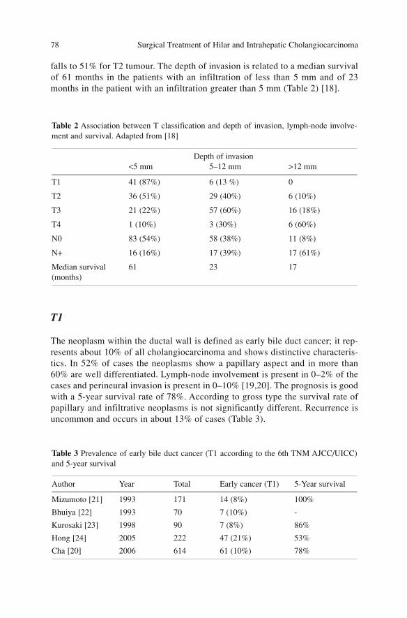
falls to 51% for T2 tumour. The depth of invasion is related to a median survivalof 61 months in the patients with an infiltration of less than 5 mm and of 23months in the patient with an infiltration greater than 5 mm (Table 2) [18].
T1
The neoplasm within the ductal wall is defined as early bile duct cancer; it rep-resents about 10% of all cholangiocarcinoma and shows distinctive characteris-tics. In 52% of cases the neoplasms show a papillary aspect and in more than60% are well differentiated. Lymph-node involvement is present in 0–2% of thecases and perineural invasion is present in 0–10% [19,20]. The prognosis is goodwith a 5-year survival rate of 78%. According to gross type the survival rate ofpapillary and infiltrative neoplasms is not significantly different. Recurrence isuncommon and occurs in about 13% of cases (Table 3).
Surgical Treatment of Hilar and Intrahepatic Cholangiocarcinoma 78
Table 2 Association between T classification and depth of invasion, lymph-node involve-ment and survival. Adapted from [18]
Depth of invasion<5 mm 5–12 mm >12 mm
T1 41 (87%) 6 (13 %) 0
T2 36 (51%) 29 (40%) 6 (10%)
T3 21 (22%) 57 (60%) 16 (18%)
T4 1 (10%) 3 (30%) 6 (60%)
N0 83 (54%) 58 (38%) 11 (8%)
N+ 16 (16%) 17 (39%) 17 (61%)
Median survival 61 23 17(months)
Table 3 Prevalence of early bile duct cancer (T1 according to the 6th TNM AJCC/UICC)and 5-year survival
Author Year Total Early cancer (T1) 5-Year survival
Mizumoto [21] 1993 171 14 (8%) 100%
Bhuiya [22] 1993 70 7 (10%) -
Kurosaki [23] 1998 90 7 (8%) 86%
Hong [24] 2005 222 47 (21%) 53%
Cha [20] 2006 614 61 (10%) 78%
T2
The depth of the neoplastic invasion of the ductal wall is associated with rele-vant prognostic aspects such as vascular, lymphatic and perineural invasion. InT1 neoplasms Tabata did not observe vascular, perineural or lymphatic invasion,while in T2 neoplasms the invasion rate increased to 40, 56 and 31%, respective-ly [19] (Table 4).
In T2 neoplasms the survival is significantly worse than in T1 neoplasms;Hong noticed a 5-year survival rate of 30 and 53%, respectively [24]. Instead thedifference between T2 and T3 neoplasms is not significant: 30–37 and 25–32%,respectively (Table 5) [24,25].
T3
According UICC/AJCC classification, T3-stage neoplasm spreads beyond theductal wall and involves adjacent structures: hepatic parenchyma, gallbladder,and diramations of portal vein and/or hepatic artery. As mentioned previously,the depth of ductal invasion is related with more aggressive neoplasms, with anincreased frequency of lymph-node metastases (64.7% in T3 neoplasm; 33.3%in T2 neoplasm) [26].
The patients with T3 (6th edition TNM UICC/AJCC) neoplasms have a sig-nificantly worse prognosis than T1, with a 5-year survival rate of 32 vs. 69%,respectively, while the difference is not significant if compared with T2 neo-plasms (survival rate 37%) [25].
Prognostic Factors 79
Table 4 Association between T stage and pathological factors. Adapted from [19]
T stage Patients Lymph nodes Lymphatic Venous Perineuralinfiltration metastasis permeation invasion(5th edn)
T1 4 (5.3%) 0% 0% 0% 0%
T2 32 (42%) 31% 71% 40% 56%
T3 39 (52%) 61% 87% 59% 84%
Table 5 5-Year survival according to T-stage
Author Patients T1 T2 T3 T4
Nishio 2005 [25] 166 69% 37% 32% 10%
Hong 2005 [24] 222 53% 30% 25% 0%
Invasion of hepatic parenchyma is very frequent in hilar cholangiocarcinoma,but in the patients who undergo radical resection (R0) it does not represent anegative prognostic factor [6,24,32].
In the past, invasion of the portal vein was a major obstacle to resection ofadvanced cholangiocarcinoma; however, recent studies reported in the literatureshow that resection and reconstruction of the portal vein offer better long-termsurvivals compared to non-resected patients, with 5-year survival rate of 9.9 vs.0%, respectively. However, portal invasion represents an important prognosticfactor; 5-year survival rate in the patients with macroscopic infiltration of theportal vein is 9.9 vs. 36.8% of the patients without [9]. Other authors confirmthe prognostic significance of portal vein involvement (Table 6).
Infiltration of the hepatic artery is considered a criterion for unresectabilityin the majority of the cases; it is a negative prognostic factor and only recentlysome studies in the literature report the results of hepatic resection combinedwith resection and reconstruction of the hepatic artery [39,40]. Invasion of thehepatic artery is associated with an advanced stage of disease, lymph-nodemetastases in more than 70% of the cases, and perineural invasion in all of them[33]. Miyazaki reports a 3-year survival rate of 11% and no 5-year survival in agroup of patients who underwent arterial resection and reconstruction [33].
According to Kosuge, transmural extension to gallbladder is a negative prog-nostic factor (p=0.0023). Neoplasms involving the gallbladder are usually asso-ciated with diffuse infiltration of the connective tissue of the hepatoduodenal lig-ament [32].
T4
According to the 6th edition of TNM UICC/AJCC, stage T4 is determined by theinvasion of surrounding organs, or portal vein, or infiltration of the hepaticartery proper. These elements imply unresectability in the majority of the cases.
Surgical Treatment of Hilar and Intrahepatic Cholangiocarcinoma 80
Table 6 Survival in patients with portal vein involvement subjected to portal vein resection
Author Patients Portal involvement 5-Year survival Median survival (months)
Launois 1999 [35] 40 17.5% 0% –
Kosuge 1999 [32] 65 27.7% – 23.9
Kondo 2004 [6] 40 30% – 34.7
Nishio 2005 [25] 301 32% 11% –
Stage T4 is correlated with poor prognosis, with a median survival of 13–17months [24,25].
N Category
Lymph-node involvement is an important prognostic factor in hilar cholangio-carcinoma and it is present in 30–50% of patients who have undergone surgicalresection [7,26–31].
The percentage of lymph-node involvement is closely related to T stage inaccordance with UICC/AJCC criteria and the depth of invasion estimated in mil-limeters. Lymph-node involvement is present in 17.1% of T1 neoplasms, in28.4% of T2, in 39.6% of T3 and in 60% of T4 [24].
Based on radial extent expressed in millimeters, the frequency of N+ was16% in the neoplasms under 5 mm, 39% in neoplasms between 5 and 12 mm,and 61% in neoplasms over 12 mm [1].
Lymph-node metastases are also associated with vascular invasion: the ratesare 55% and 39% in patients with and without vascular invasion [9].
Five-year survival rate is 45% in N0 patients, 16% in N1, 14% in non-region-al node involvement defined as M1 according to the criteria of TNMUICC/AJCC. In the literature, different series show that the 5-year survival ratein patients with lymph-node metastases does not exceed 25% [7,25–29,32–34](Table 7).
The prevalence of non-regional lymph-node metastases, defined as M1, isnot well known in hilar cholangiocarcinoma since few authors performed a sys-tematic dissection of non-regional lymph nodes; in Kitagawa’s study [26], theprevalence of positive para-aortic lymph nodes (M1) was 17% [26]. The pres-ence of non-regional lymph-node metastases is an important prognostic factor,
Prognostic Factors 81
Table 7 Prevalence and survival for patients with positive lymph-node metastases (N+)
Author Patients N+ patients Survival for N+ patientsMedian (months) Rate (%)
Launois 1999 [35] 40 15% - 16.7%
Kosuge 1999 [32] 65 44% 26.4 -
Nagakawa 2002 [36] 1183 45% - 15%
Kitagawa [26] 110 53% - 16%
Kondo 2004 [6] 40 37.5% 26.6 -
Hemming 2005 [34] 80 - - 21%
Lai 2005 [37] 36 35% - 0%
Nishio 2005 [16] 301 49% - 10%
Ramacciato 2006 [38] 23 34% 21 -
Dinant 2006 [5] 99 19% 18 -
with survival of less than 15% at 5 years [17]. The analysis of the characteristicsof the patients with para-aortic lymph-node metastases showed that survival inpatients with M1 nodes with a normal macroscopic aspect is significantly betterthan the group of patients with macroscopically positive nodes, with a 5-yearsurvival rate of 28.6 vs. 0%, respectively [26].
Even the number of metastatic nodes represents an independent prognosticfactor that is statistically significant (p=0.017). Patients with more than 5 posi-tive lymph nodes have shown a shorter survival compared to patients with lessthan 4 metastatic lymph nodes, with a HR at univariate analysis of 2.95 and 1.7,respectively [1].
In a recent study, Schwartz [41] observed a correlation between survival andnumber of removed nodes in N0 patients. Median survival was 21 months in thepatients with 1 to 2 total lymph nodes examinated, and 34 months in patientswith more than 10 lymph nodes examinated, respectively. Also in N1 patients adifference in survival has been recognized in the patients with 1 to 2 lymphnodes examinated vs. more than 10 nodes, with median values of 13 and 16months, respectively.
Immunohistochemical techniques that utilize antibodies against cytokeratincan identify micrometastases that are not detected by haematoxylin-eosin stain-ing. Micrometastases are detected in 24–39% of the patients with negative nodes[42,43]. The prognostic significance of micrometastases is still controversial.Yuchiro has not found significant differences in patients with or without micro-scopic lymph-node metastases, with 43.6 and 42.1% 5-year survival rates,respectively [42]. Conversely, in 28 patients in N0 stage Taniguchi identified asignificant correlation between micrometastases, stage of disease and prognosis,with 5-year survival rates of 21 and 66%, respectively [43].
M Category
The presence of distant metastases is the most frequent cause of unresectability:about 30% of patients are excluded from surgical treatment during preoperativeworkup or at laparotomy [44].
The more common sites of metastases are: liver, peritoneum, lung and bone; themedian survival of the patients with metastases is no longer than 11 months [35].
In presence of hepatic metastases, the resection has very few satisfactoryresults with a median survival of 6–12 months [32].
Prognostic Significance of TNM UICC/AJCC Classification
Until now, few reports have valued the prognostic significance of TNM classifi-cation for hilar cholangiocarcinoma. The new edition of TNM staging (6th edi-tion of UICC/AJCC) focuses on two aspects: vascular invasion and the presenceof lymph-node metastases [45].
Surgical Treatment of Hilar and Intrahepatic Cholangiocarcinoma 82
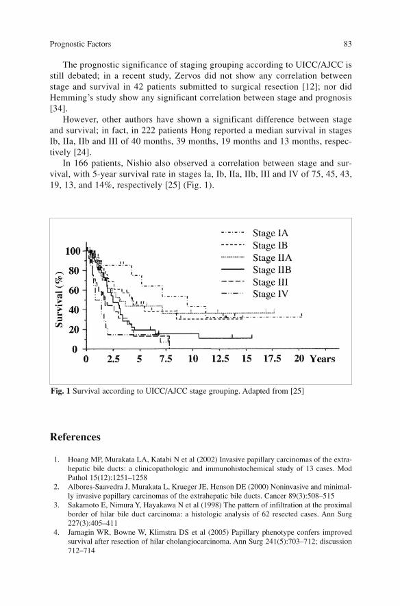
The prognostic significance of staging grouping according to UICC/AJCC isstill debated; in a recent study, Zervos did not show any correlation betweenstage and survival in 42 patients submitted to surgical resection [12]; nor didHemming’s study show any significant correlation between stage and prognosis[34].
However, other authors have shown a significant difference between stageand survival; in fact, in 222 patients Hong reported a median survival in stagesIb, IIa, IIb and III of 40 months, 39 months, 19 months and 13 months, respec-tively [24].
In 166 patients, Nishio also observed a correlation between stage and sur-vival, with 5-year survival rate in stages Ia, Ib, IIa, IIb, III and IV of 75, 45, 43,19, 13, and 14%, respectively [25] (Fig. 1).
References
1. Hoang MP, Murakata LA, Katabi N et al (2002) Invasive papillary carcinomas of the extra-hepatic bile ducts: a clinicopathologic and immunohistochemical study of 13 cases. ModPathol 15(12):1251–1258
2. Albores-Saavedra J, Murakata L, Krueger JE, Henson DE (2000) Noninvasive and minimal-ly invasive papillary carcinomas of the extrahepatic bile ducts. Cancer 89(3):508–515
3. Sakamoto E, Nimura Y, Hayakawa N et al (1998) The pattern of infiltration at the proximalborder of hilar bile duct carcinoma: a histologic analysis of 62 resected cases. Ann Surg227(3):405–411
4. Jarnagin WR, Bowne W, Klimstra DS et al (2005) Papillary phenotype confers improvedsurvival after resection of hilar cholangiocarcinoma. Ann Surg 241(5):703–712; discussion712–714
Prognostic Factors 83
Fig. 1 Survival according to UICC/AJCC stage grouping. Adapted from [25]
5. Dinant S, Gerhards MF, Rauws EA et al (2006) Improved outcome of resection of hilarcholangiocarcinoma (Klatskin tumour). Ann Surg Oncol 13(6):872–80
6. Kondo S, Hirano S, Ambo Y et al (2004) Forty consecutive resections of hilar cholangiocar-cinoma with no postoperative mortality and no positive ductal margins: results of a prospec-tive study. Ann Surg 240(1):95–101
7. Neuhaus P, Jonas S, Bechstein WO et al (1999) Extended resections for hilar cholangiocar-cinoma. Ann Surg 230(6):808–818; discussion 819
8. Bhuiya MR, Nimura Y, Kamiya J et al (1992) Clinicopathologic studies on perineural inva-sion of bile duct carcinoma. Ann Surg 215(4):344–349
9. Ebata T, Nagino M, Kamiya J et al (2003) Hepatectomy with portal vein resection for hilarcholangiocarcinoma: audit of 52 consecutive cases. Ann Surg 238(5):720–727
10. Liu XF, Zhang H, Zhu SG et al (2006) Correlation of p53 gene mutation and expression ofP53 protein in cholangiocarcinoma. World J Gastroenterol 12(29):4706–4709
11. Jarnagin WR, Klimstra DS, Hezel M et al (2006) Differential cell cycle-regulatory proteinexpression in biliary tract adenocarcinoma: correlation with anatomic site, pathologic vari-ables, and clinical outcome. J Clin Oncol 24(7):1152–1160
12. Zervos EE, Osborne D, Goldin SB et al (2005) Stage does not predict survival after resec-tion of hilar cholangiocarcinomas promoting an aggressive operative approach. Am J Surg190(5):810–815
13. Itoi T, Shinohara Y, Takeda K et al (2000) Detection of telomerase activity in biopsy speci-mens for diagnosis of biliary tract cancers. Gastrointest Endosc 52(3):380–386
14. Abdalla EK, Vauthey JN (2001) Biliary tract cancer. Curr Opin Gastroenterol 17(5):450–45715. Rajala RV, Radhi JM, Kakkar R et al (2000) Increased expression of N-myristoyltransferase
in gallbladder carcinomas. Cancer 88(9):1992–199916. Cormier JN, Vauthey JN (2000) Biliary tract cancer. Curr Opin Gastroenterol 16(5):437–44317. Rashid A, Ueki T, Gao YT et al (2002) K-ras mutation, p53 overexpression, and microsatel-
lite instability in biliary tract cancers: a population-based study in China. Clin Cancer Res8(10):3156–3163
18. Hong SM, Cho H, Moskaluk CA, Yu E (2007) Measurement of the invasion depth of extra-hepatic bile duct carcinoma: an alternative method overcoming the current T classificationproblems of the AJCC staging system. Am J Surg Pathol 31(2):199–206
19. Tabata M, Kawarada Y, Yokoi H et al (2000) Surgical treatment for hilar cholangiocarcino-ma. J Hepatobiliary Pancreat Surg 7(2):148–154
20. Cha JM, Kim MH, Lee SK et al (2006) Clinicopathological review of 61 patients with earlybile duct cancer. Clin Oncol (R Coll Radiol) 18(9):669–677
21. Mizumoto R, Ogura Y, Kusuda T (1993) Definition and diagnosis of early cancer of the bil-iary tract. Hepatogastroenterology 40(1):69–77
22. Kurosaki I, Tsukada K, Watanabe H, Hatakeyama K (1998) Prognostic determinants inextrahepatic bile duct cancer. Hepatogastroenterology 45(22):905–909
23. Bhuiya MR, Nimura Y, Kamiya J et al (1993) Clinicopathologic factors influencing survivalof patients with bile duct carcinoma: multivariate statistical analysis. World J Surg17(5):653–657
24. Hong SM, Kim MJ, Pi DY et al (2005) Analysis of extrahepatic bile duct carcinomas accord-ing to the New American Joint Committee on Cancer staging system focused on tumourclassification problems in 222 patients. Cancer 104(4):802–810
25. Nishio H, Nagino M, Oda K et al (2005) TNM classification for perihilar cholangiocarcino-ma: comparison between 5th and 6th editions of the AJCC/UICC staging system.Langenbecks Arch Surg 390(4):319–327
26. Kitagawa Y, Nagino M, Kamiya J et al (2001) Lymph-node metastasis from hilar cholangio-carcinoma: audit of 110 patients who underwent regional and paraaortic node dissection.Ann Surg 233(3):385–392
27. Nakeeb A, Pitt HA, Sohn TA et al (1996) Cholangiocarcinoma. A spectrum of intrahepatic,perihilar, and distal tumours. Ann Surg 224(4):463–473; discussion 473–475
Surgical Treatment of Hilar and Intrahepatic Cholangiocarcinoma 84
28. Sugiura Y, Nakamura S, Iida S et al (1994) Extensive resection of the bile ducts combinedwith liver resection for cancer of the main hepatic duct junction: a cooperative study of theKeio Bile Duct Cancer Study Group. Surgery 115(4):445–451
29. Iwatsuki S, Todo S, Marsh JW et al (1998) Treatment of hilar cholangiocarcinoma (Klatskintumours) with hepatic resection or transplantation. J Am Coll Surg 187(4):358–364
30. Ogura Y, Kawarada Y (1998) Surgical strategies for carcinoma of the hepatic duct conflu-ence. Br J Surg 85(1):20–24
31. Miyazaki M, Ito H, Nakagawa K et al (1999) Parenchyma-preserving hepatectomy in thesurgical treatment of hilar cholangiocarcinoma. J Am Coll Surg 189(6):575–583
32. Kosuge T, Yamamoto J, Shimada K et al (1999) Improved surgical results for hilar cholan-giocarcinoma with procedures including major hepatic resection. Ann Surg 230(5):663-671
33. Miyazaki M, Kato A, Ito H et al (2007) Combined vascular resection in operative resectionfor hilar cholangiocarcinoma: does it work or not? Surgery 141(5):581–588
34. Hemming AW, Reed AI, Fujita S et al (2005) Surgical management of hilar cholangiocarci-noma. Ann Surg 241(5):693–699; discussion 699–702
35. Launois B, Terblanche J, Lakehal M et al (1999) Proximal bile duct cancer: high resectabil-ity rate and 5-year survival. Ann Surg 230(2):266–275
36. Nagakawa T, Kayahara M, Ikeda S et al () Biliary tract cancer treatment: results from theBiliary Tract Cancer Statistics Registry in Japan. J Hepatobiliary Pancreat Surg9(5):569–575
37. Lai EC, Lau WY (2005) Aggressive surgical resection for hilar cholangiocarcinoma. ANZ JSurg 75(11):981–985
39. Miyazaki M, Ito H, Nakagawa K et al (1998) Aggressive surgical approaches to hilar cholan-giocarcinoma: hepatic or local resection? Surgery 123(2):131–136
40. Gerhards MF, van Gulik TM, de Wit LT et al (2000) Evaluation of morbidity and mortalityafter resection for hilar cholangiocarcinoma—a single center experience. Surgery127(4):395–404
41. Schwarz RE, Smith DD (2007) Lymph node dissection impact on staging and survival ofextrahepatic cholangiocarcinomas, based on U.S. population data. J Gastrointest Surg11(2):158–165
42. Tojima Y, Nagino M, Ebata T et al (2003) Immunohistochemically demonstrated lymphnode micrometastasis and prognosis in patients with otherwise node-negative hilar cholan-giocarcinoma. Ann Surg 237(2):201–207
43. Taniguchi K, Tabata M, Iida T et al (2006) Significance of lymph node micrometastasis inpN0 hilar bile duct carcinoma. Eur J Surg Oncol 32(2):208–212
44. Jarnagin WR, Shoup M (2004) Surgical management of cholangiocarcinoma. Semin LiverDis 24(2):189–199
45. Ramacciato G, Corigliano N, Mercantini P et al (2006) [Prognostic factors after surgicalresection for hilar cholangiocarcinoma] Ann Chir 131(6-7):379–85, French
45. Sobin LH, Wittekind C (eds) (2002) TNM classification of malignant tumours, 6th edn.Wiley, New York
Prognostic Factors 85
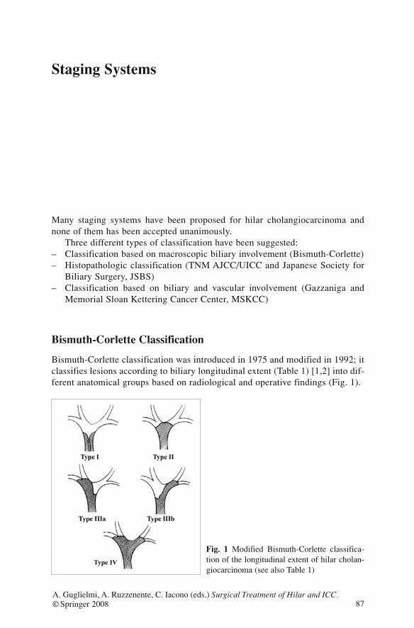
Staging Systems
Many staging systems have been proposed for hilar cholangiocarcinoma andnone of them has been accepted unanimously.
Three different types of classification have been suggested:– Classification based on macroscopic biliary involvement (Bismuth-Corlette)– Histopathologic classification (TNM AJCC/UICC and Japanese Society for
Biliary Surgery, JSBS)– Classification based on biliary and vascular involvement (Gazzaniga and
Memorial Sloan Kettering Cancer Center, MSKCC)
Bismuth-Corlette Classification
Bismuth-Corlette classification was introduced in 1975 and modified in 1992; itclassifies lesions according to biliary longitudinal extent (Table 1) [1,2] into dif-ferent anatomical groups based on radiological and operative findings (Fig. 1).
87A. Guglielmi, A. Ruzzenente, C. Iacono (eds.) Surgical Treatment of Hilar and ICC.© Springer 2008
Fig. 1 Modified Bismuth-Corlette classifica-tion of the longitudinal extent of hilar cholan-giocarcinoma (see also Table 1)
This classification considers only the extent of biliary involvement at preop-erative workup. Bismuth’s validation experience in 1992 [2] on 23 resectedpatients showed that this system can be used to tailor the surgical operation. Intype I patients, resection of the biliary tract alone provides a curative resection.In type II the neoplasm involves the confluence, dividing the left and right ducts,and frequently the biliary ducts of the caudate lobe. In order to obtain a radicalresection (R0) Bismuth suggests biliary resection associated with caudate loberesection. In type IIIa and IIIb, resection of the biliary tract does not guaranteea curative resection and therefore major hepatic resection must be associated. Intype IV the bilateral involvement of secondary ducts frequently precludes a rad-ical resection despite extended hepatectomy. This staging system is still used todefine the extent of the disease; nevertheless its use in therapeutic choice andprognostic evaluation is limited since important factors such as vascular invasionand lymph-node metastases are not considered. In a recent comparative study on42 resected patients with hilar cholangiocarcinoma. Bismuth-Corlette classifica-tion failed to show a correlation with long-term survival [3].
TNM Staging System According to UICC/AJCC 6th Edition
The sixth edition of TNM classification by Internal Union Against Cancer(UICC) and the sixth edition of the Manual for Staging of Cancer edited byAmerican Joint Committee on Cancer were published in 2002 and used the sameTNM classification and subdivision in stages [4]. Local extent of disease (T),nodal involvement (N) and presence of distant organ involvement (M) are deter-mined by preoperative, intraoperative and pathological findings.
Surgical Treatment of Hilar and Intrahepatic Cholangiocarcinoma 88
Table 1 Bismuth-Corlette classification [1]
Type I Neoplasm arises near the biliary confluence without involvement of the left andright biliary ducts
Type II Neoplasm arises at the biliary confluence with extension to left and right ducts
Type IIIa Neoplasm arises at the biliary confluence with extension to the right hepaticduct up to the second order ducts
Type IIIb Neoplasm arises at the biliary confluence with extension to left biliary duct upto the second order ducts
Type IV Neoplasm arises at the biliary confluence with bilateral extension to the secondorder biliary ducts
T Category
T category evaluates local extent based on the degree of ductal wall involvement,portal vein and hepatic artery involvement and invasion of adjacent structures. T-category is divided into four types (Table 2).
This staging system has been validated by a clinical study on 166 resectedpatients where T category showed a correlation with long-term survival: 5-yearsurvival was 69% in T1, 37% in T2, 32% in T3 and 10% in T4 [5].
N Category
According to UICC/AJCC criteria, the regional lymph nodes are: cyst duct, peri-choledochal, hilar, peripancreatic (head only), periduodenal, periportal, celiac,and superior mesenteric lymph nodes. The involvement of non-regional lymphnodes (aorto-caval and inferior peripancreatic) is defined as distant metastases(M1).
N category is defined in two stages (Table 3).
Many reports in the literature confirm the prognostic value of regional nodalinvolvement with 5-year survival of 30.5% in N0, and 14.7% in N1 (p=0.09). Kitagawa et al. have also proved the poor prognosis in non-regionalnode diffusion (M1) with a 5-year survival of 12.3% [6].
Staging Systems 89
Table 2 Evaluation of local extent according to UICC/AJCC [4]
T Carcinoma in situ
T1 Tumour is confined to biliary duct
T2 Tumour invades through entire biliary duct wall
T3 Tumour invades liver, gallbladder, main branches of portal vein or hepatic artery(right or left)
T4 Tumour invades portal vein trunk, proper hepatic artery or other surrounding organs(colon, stomach, duodenum)
Table 3 Evaluation of lymph-node involvement according to UICC/AJCC [4]
N0 No lymph-node metastases
N1 Presence of regional lymph-node involvement
M Category
M category is based on the presence of metastases to other organs or non-region-al lymph nodes. It is classified in Table 4.
Stage Grouping
The three categories T, N and M are arranged to classify patients in homoge-neous groups. In the sixth edition of TNM classification of the UICC/AJCCseven groups are listed (Table 5).
The grouping in stages defines neoplasms at the early stage as limited to thebiliary duct (stage I), neoplasms at the intermediate stage involving hepaticparenchyma, vascular structures (hepatic artery or portal vein) or regional lymphnodes (stage II). Advanced tumours in stages III (diffusion to adjacent structuresand/or regional lymph nodes) and IV (presence of metastases) rarely benefitfrom a surgical operation with intent to cure. Prognostic significance of this clas-sification has been evaluated by several clinical studies; nevertheless there areno conclusive data available as yet. In a study of 40 patients who underwentresection for hilar cholangiocarcinoma, Liu et al. showed no significant differ-ence in terms of survival between the groups of patients with stages I and II vsstages III and IV, with median survival of 17.6 and 21.2 months (p=0.25),respectively [7]. Hong reported a significant difference in survival for stages Ia,Ib, IIa, IIb and III, with a 5-year survival rate of 54, 35, 31, 7, 5 and 0%, respec-tively [8].
Surgical Treatment of Hilar and Intrahepatic Cholangiocarcinoma 90
Table 4 Evaluation of presence of metastases according to UICC/AJCC [4]
M0 No metastases
M1 Presence of metastases
Table 5 Staging system according to UICC/AJCC [4]
Stage 0 T N0 M0
Stage IA T1 N0 M0
Stage IB T2 N0 M0
Stage IIA T3 N0 M0
Stage IIB T1, T2, T3 N1 M0
Stage III T4 Any N M0
Stage IV Any T Any N M1
Comparison between 5th and 6th Edition of TNM UICC/AJCC
The last two staging editions of TNM, the 5th and 6th editions, present differentcriteria for classification of T and N categories. In the T category of the 5th edi-tion all neoplasms involving adjacent structures were classified as T3; instead inthe last edition T3 has been divided based on the structures involved: T3 forlesions that invade per contiguity the liver, gallbladder, pancreas, or the vesselsunilaterally and T4 for the lesions invading other adjacent organs (colon, stom-ach and duodenum) or the main portal vein.
This new distinction has shown better accuracy in evaluating prognosis ofpatients who had undergone surgical resection. A comparative study between the5th and 6th editions in 166 patients shows 5-year survival for T3 stage (5th edi-tion) of 24% while for T3 of the 6th edition survival is 32% and 10% for T4, witha statistically significant difference [5]. The invasion of stomach, colon and duo-denum is rare in hilar cholangiocarcinoma and the majority of T4 indicates inva-sion of main portal vein. The new formulation of T4 category emphasizes thevalue of vascular invasion which many reports have shown to be an importantnegative prognostic factor [9]. N category has been modified as well; in fact inthe 5th edition lymph-node metastases were divided in N1 (hepatoduodenal lig-ament lymph nodes) and N2 (other regional lymph nodes); in the updated edi-tion N category has been simplified in a single group of patients with metastat-ic regional lymph nodes. This new group of regional lymph nodes has gainedvalidation in a clinical study on 110 patients in whom significant differences inlong-term survival were not shown between patients with positive N1 versus N2with median survival of 29 and 25 months respectively [6]. Both editions defineas M1 the presence of non-regional lymph-node metastases.
Staging System According to JSBS
Classification of hilar neoplasm formulated by JSBS was published in Englishin its 2nd edition in 2004 [10]. As TNM of UICC/AJCC this classification isbased on anatomic and pathological findings and analyzes the local extent of theneoplasm (T), its nodal extension, and involvement of other organs (M). BothTNM UICC/AJCC and JSBS classify hilar cholangiocarcinoma along with theother neoplasms of the extrahepatic biliary tract.
Macroscopic Growth Pattern
On macroscopic assessment neoplasms would be classified on the basis of thetype of growth of the tumour inside the biliary duct:
Staging Systems 91
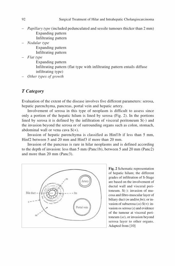
– Papillary type (included pedunculated and sessile tumours thicker than 2 mm)Expanding patternInfiltrating pattern
– Nodular typeExpanding patternInfiltrating pattern
– Flat typeExpanding patternInfiltrating pattern (flat type with infiltrating pattern entails diffuseinfiltrating type)
– Other types of growth
T Category
Evaluation of the extent of the disease involves five different parameters: serosa,hepatic parenchyma, pancreas, portal vein and hepatic artery.
Involvement of serosa in this type of neoplasm is difficult to assess sinceonly a portion of the hepatic hilum is lined by serosa (Fig. 2). In the portionslined by serosa it is defined by the infiltration of visceral peritoneum S(-) andthe invasion beyond the serosa or of surrounding organs such as colon, stomach,abdominal wall or vena cava S(+).
Invasion of hepatic parenchyma is classified as Hinf1b if less than 5 mm,Hinf2 between 5 and 20 mm and Hinf3 if more than 20 mm.
Invasion of the pancreas is rare in hilar neoplasms and is defined accordingto the depth of invasion: less than 5 mm (Panc1b), between 5 and 20 mm (Panc2)and more than 20 mm (Panc3).
Surgical Treatment of Hilar and Intrahepatic Cholangiocarcinoma 92
Fig. 2 Schematic representationof hepatic hilum; the differentgrades of infiltration of S-Stageare based on the involvement ofductal wall and visceral peri-toneum. S(-): invasion of mu-cosa and fibro-muscular layer ofbiliary duct (m and/or fm); or in-vasion of subserosa (ss) S(+): in-vasion os serosa (s) and evidenceof the tumour at visceral peri-toneum (se), or invasion beyondserosa layer to other organs.Adapted from [10]

The invasions of portal and arterial structures are divided into adventitialinfiltration (PV1 or A1), tunica media infiltration (PV2 or A2) or extended totunica intima with stenosis and/or obstruction (PV3 or A3).
The division of T category on the basis of involvement of adjacent structuresallows us to distinguish four categories (Table 6).
Classification of T according to JSBS includes prognostic factors thatbecome evident from many clinical studies and permit precise classification ofthe extent of the disease. In literature no reported studies have validated this cat-egorization of T category and due to its complexity it is not much used in clini-cal practice.
N Category
According to the Japanese classification, the lymph nodes for hilar hepatic neo-plasms are divided into three levels: N1, N2 and N3. Lymph nodes of the hepa-toduodenal ligament (station 12) belong to the first level (N1) and are catego-rized for their relationship with the biliary tract (12b), portal vein (12p) andproper hepatic artery (12a). Retropancreatic lymph nodes (13a) and those alongthe common hepatic artery (8) belong to the second level (N2). Para-aortic (16),celiac (9), mesenteric (14) or anterior-pancreatic (17) and inferior retropancreat-ic (13b) lymph node stations pertain to the third level (N3) (Figs. 3,4).
The division of lymph nodes for hilar hepatic neoplasms is shown in Table 7.
Staging Systems 93
Table 6 Classification of T according to JSBS (Japanese Society of Biliary Surgery) [10]
T-stage Serosa Hepatic Pancreatic Portal Arterialinfiltration infiltration infiltration infiltration
T1 m, fm Hinf0 Panc0 PV0 A0
T2 ss Hinf1a Panc1a PV0 A0
T3 se Hinf1b Panc1b PV0 A0
T4 any Hinf2,3 Panc2,3 PV1–3 A1–3
Hinf, direct extent to hepatic parenchyma (0: no invasion; 1a: invasion beyond fibromuscu-lar layer but not of parenchyma; 1b: <5 mm; 2: 5–20 mm; 3 invasion more than 20 mm);Panc, extent to pancreas (0: no invasion; 1a: invasion beyond fibromuscolar layer but not ofparenchyma; 1b: <5 mm; 2: 5–20 mm; 3 more than 20 mm); PV, extent to portal vein (0: noinvasion; 1: invasion of adventitia; 2: up to tunica media; 3: up to tunica intima with steno-sis or obstruction); A, extent to hepatic artery (0: no invasion; 1: invasion of adventitia; 2: upto tunica media; 3: up to tunica intima with stenosis or obstruction). For serosa invasion, seeFig. 2
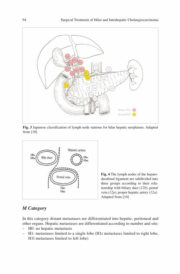
M Category
In this category distant metastases are differentiated into hepatic, peritoneal andother organs. Hepatic metastases are differentiated according to number and site:– H0: no hepatic metastasis– H1: metastases limited to a single lobe (H1r metastases limited to right lobe,
H1l metastases limited to left lobe)
Surgical Treatment of Hilar and Intrahepatic Cholangiocarcinoma 94
Fig. 3 Japanese classification of lymph node stations for hilar hepatic neoplasms. Adaptedfrom [10]
Fig. 4 The lymph nodes of the hepato-duodenal ligament are subdivided intothree groups according to their rela-tionship with biliary duct (12b), portalvein (12p), proper hepatic artery (12a).Adapted from [10]
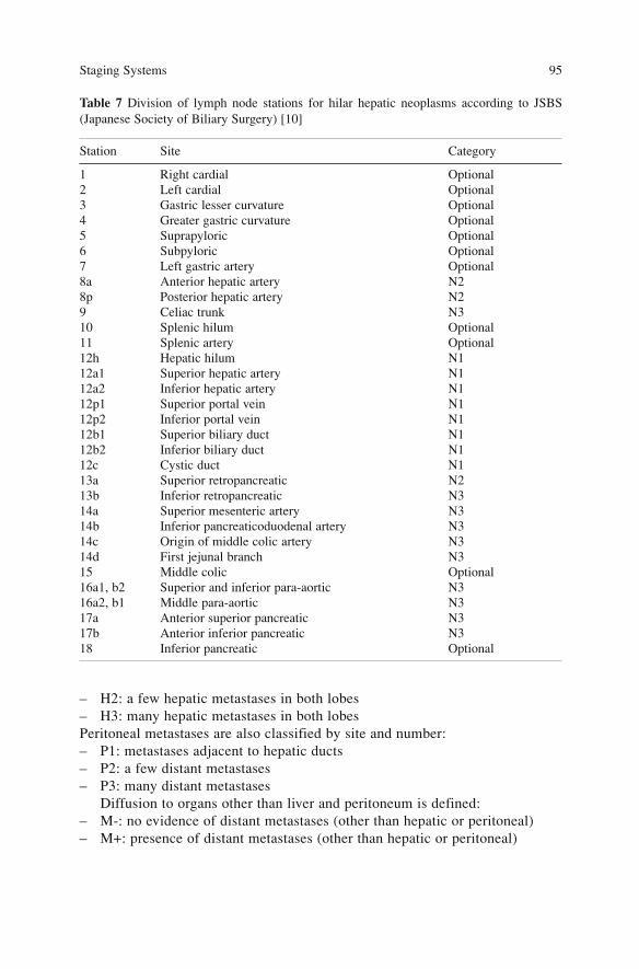
– H2: a few hepatic metastases in both lobes– H3: many hepatic metastases in both lobesPeritoneal metastases are also classified by site and number:– P1: metastases adjacent to hepatic ducts– P2: a few distant metastases– P3: many distant metastases
Diffusion to organs other than liver and peritoneum is defined:– M-: no evidence of distant metastases (other than hepatic or peritoneal)– M+: presence of distant metastases (other than hepatic or peritoneal)
Staging Systems 95
Table 7 Division of lymph node stations for hilar hepatic neoplasms according to JSBS(Japanese Society of Biliary Surgery) [10]
Station Site Category
1 Right cardial Optional2 Left cardial Optional3 Gastric lesser curvature Optional4 Greater gastric curvature Optional5 Suprapyloric Optional6 Subpyloric Optional7 Left gastric artery Optional8a Anterior hepatic artery N28p Posterior hepatic artery N29 Celiac trunk N310 Splenic hilum Optional11 Splenic artery Optional12h Hepatic hilum N112a1 Superior hepatic artery N112a2 Inferior hepatic artery N112p1 Superior portal vein N112p2 Inferior portal vein N112b1 Superior biliary duct N112b2 Inferior biliary duct N112c Cystic duct N113a Superior retropancreatic N213b Inferior retropancreatic N314a Superior mesenteric artery N314b Inferior pancreaticoduodenal artery N314c Origin of middle colic artery N314d First jejunal branch N315 Middle colic Optional16a1, b2 Superior and inferior para-aortic N316a2, b1 Middle para-aortic N317a Anterior superior pancreatic N317b Anterior inferior pancreatic N318 Inferior pancreatic Optional
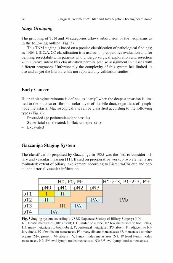
Stage Grouping
The grouping of T, N and M categories allows subdivision of the neoplasms asin the following outline (Fig. 5).
This TNM staging is based on a precise classification of pathological findings;as TNM UICC/AJCC classification it is useless in preoperative evaluation and fordefining resectability. In patients who undergo surgical exploration and resectionwith curative intent this classification permits precise assignment to classes withdifferent prognoses. Unfortunately the complexity of this system has limited itsuse and as yet the literature has not reported any validation studies.
Early Cancer
Hilar cholangiocarcinoma is defined as “early” when the deepest invasion is lim-ited to the mucosa or fibromuscolar layer of the bile duct, regardless of lymph-node metastasis. Macroscopically it can be classified according to the followingtypes (Fig. 6):– Protruded (p: pedunculated; s: sessile)– Superficial (a: elevated; b: flat; c: depressed)– Excavated
Gazzaniga Staging System
The classification proposed by Gazzaniga in 1985 was the first to consider bil-iary and vascular invasion [11]. Based on preoperative workup two elements areevaluated: extent of biliary involvement according to Bismuth-Corlette and por-tal and arterial vascular infiltration.
Surgical Treatment of Hilar and Intrahepatic Cholangiocarcinoma 96
Fig. 5 Staging system according to JSBS (Japanese Society of Biliary Surgery) [10]H, Hepatic metastases (H0: absent; H1: limited to a lobe; H2 few metastases to both lobes,H3: many metastases to both lobes); P, peritoneal metastases (P0: absent, P1 adjacent to bil-iary ducts, P2: few distant metastases, P3: many distant metastases); M, metastases to otherorgans (M+: present, M- absent); N, lymph nodes metastases (N1: 1st level lymph nodesmetastases, N2: 2nd level lymph nodes metastases, N3: 3rd level lymph nodes metastases
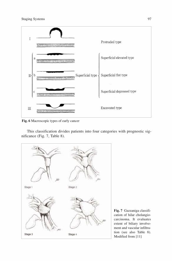
This classification divides patients into four categories with prognostic sig-nificance (Fig. 7, Table 8).
Staging Systems 97
Fig. 6 Macroscopic types of early cancer
Fig. 7 Gazzaniga classifi-cation of hilar cholangio-carcinoma. It evaluatesextent of biliary involve-ment and vascular infiltra-tion (see also Table 8).Modified from [11]
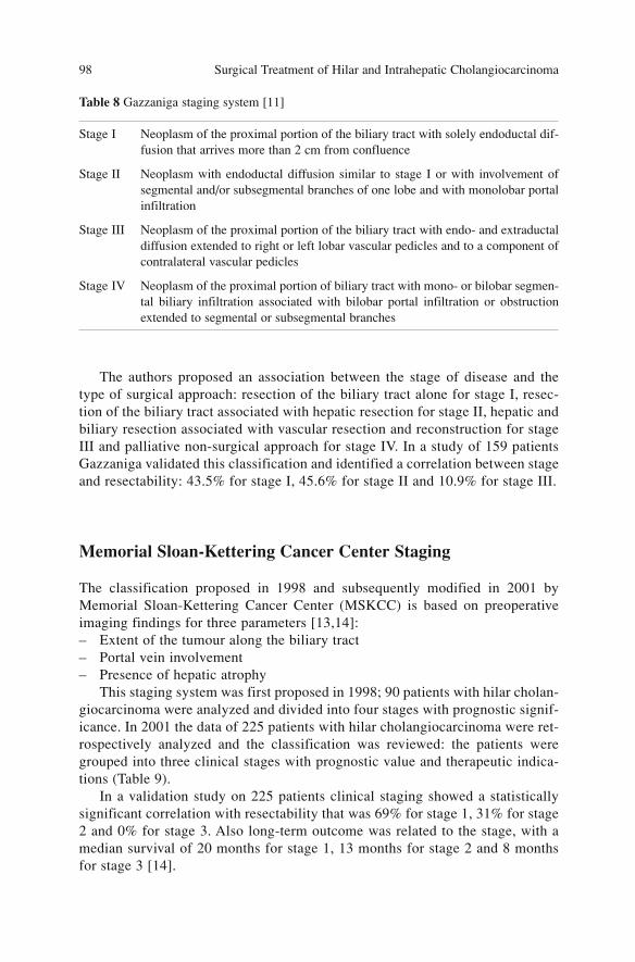
The authors proposed an association between the stage of disease and thetype of surgical approach: resection of the biliary tract alone for stage I, resec-tion of the biliary tract associated with hepatic resection for stage II, hepatic andbiliary resection associated with vascular resection and reconstruction for stageIII and palliative non-surgical approach for stage IV. In a study of 159 patientsGazzaniga validated this classification and identified a correlation between stageand resectability: 43.5% for stage I, 45.6% for stage II and 10.9% for stage III.
Memorial Sloan-Kettering Cancer Center Staging
The classification proposed in 1998 and subsequently modified in 2001 byMemorial Sloan-Kettering Cancer Center (MSKCC) is based on preoperativeimaging findings for three parameters [13,14]:– Extent of the tumour along the biliary tract– Portal vein involvement– Presence of hepatic atrophy
This staging system was first proposed in 1998; 90 patients with hilar cholan-giocarcinoma were analyzed and divided into four stages with prognostic signif-icance. In 2001 the data of 225 patients with hilar cholangiocarcinoma were ret-rospectively analyzed and the classification was reviewed: the patients weregrouped into three clinical stages with prognostic value and therapeutic indica-tions (Table 9).
In a validation study on 225 patients clinical staging showed a statisticallysignificant correlation with resectability that was 69% for stage 1, 31% for stage2 and 0% for stage 3. Also long-term outcome was related to the stage, with amedian survival of 20 months for stage 1, 13 months for stage 2 and 8 monthsfor stage 3 [14].
Surgical Treatment of Hilar and Intrahepatic Cholangiocarcinoma 98
Table 8 Gazzaniga staging system [11]
Stage I Neoplasm of the proximal portion of the biliary tract with solely endoductal dif-fusion that arrives more than 2 cm from confluence
Stage II Neoplasm with endoductal diffusion similar to stage I or with involvement ofsegmental and/or subsegmental branches of one lobe and with monolobar portalinfiltration
Stage III Neoplasm of the proximal portion of the biliary tract with endo- and extraductaldiffusion extended to right or left lobar vascular pedicles and to a component ofcontralateral vascular pedicles
Stage IV Neoplasm of the proximal portion of biliary tract with mono- or bilobar segmen-tal biliary infiltration associated with bilobar portal infiltration or obstructionextended to segmental or subsegmental branches

In a recent study Zervos et al. did not confirm the correlation between the clas-sification of MSKCC with resectability and long-term survival with a median sur-vival of 45 months for T1, 90 months for T2, 43 months for T3 and 33 months forT4 [3].
This clinical classification offers an approach that has proved to be useful indefining resectability and prognosis. Unfortunately prognostic evaluation doesnot consider some factors such as lymph-node involvement which has beenshown to be prognostically relevant.
Conclusions
There are various staging systems proposed for hilar cholangiocarcinoma and atpresent the choice of the best system is still under debate.
As noted previously, the classifications listed show different features and canbe divided into two categories: clinical and pathological staging systems.
The clinical classifications [1,11,14] serve to define criteria of resectability,indicate the type of operation and estimate the prognosis of the disease.
Bismuth-Corlette classification divides patients only based on extent of bil-iary involvement and does not consider other important elements of the preoper-ative evaluation such as vascular involvement and lobar atrophy; therefore itcannot be used to assess resectability [1,2].
The Gazzaniga classification proposed in 1985 has added the degree of vas-cular involvement [11] to evaluation of extent of biliary involvement; howeverits diffusion in clinical practice is very limited.
The clinical classification proposed by MSKCC adds vascular involvementand hepatic atrophy evaluation of extent of biliary involvement according to theBismuth-Corlette classification [13,14].
TNM UICC/AJCC [4] and JSBS [10] staging systems are based onhistopathologic criteria and evaluate the local and distant extent after surgicaloperation. These classifications have mainly prognostic significance but are notuseful for assessing resectability. The complexity of JSBS classification limitsits use in clinical practice.
Staging Systems 99
Table 9 Memorial Sloan-Kettering Center staging system [13]
T1 Tumour involves biliary confluence ± unilateral extent to 2nd order ducts
T2 Tumour involves biliary confluence ± unilateral extent to 2nd order ducts and homo-lateral portal infiltration ± homolateral lobar atrophy
T3 Tumour involves biliary confluence + bilateral extent to 2nd order ducts, unilateralextent to 2nd order ducts with contralateral portal vein infiltration, unilateral extentto 2nd order biliary ducts with contralateral hepatic atrophy, or involvement of mainportal trunk
A classification of this disease that permits a complete evaluation ofresectability and outcome is not available at present. It would be useful to havea staging system that combine biliary involvement, vascular invasion, localextent and lymph-node involvement.
References
1. Bismuth H, Corlette MB (1975) Intrahepatic cholangioenteric anastomosis in carcinoma ofthe hilus of the liver. Surg Gynecol Obstet 140(2):170–178
2. Bismuth H, Nakache R, Diamond T (1992) Management strategies in resection for hilarcholangiocarcinoma. Ann Surg 199 215(1):31–38
3. Zervos EE, Osborne D, Goldin SB et al (2005) Stage does not predict survival after resec-tion of hilar cholangiocarcinomas promoting an aggressive operative approach. Am J Surg190(5):810–815
4. Sobin LH, Wittekind C (eds) (2002) TNM classification of malignant tumours, 6th edn.Wiley, New York
5. Nishio H, Nagino M, Oda K et al (2005) TNM classification for perihilar cholangiocarcino-ma: comparison between 5th and 6th editions of the AJCC/UICC staging system.Langenbecks Arch Surg 390(4):319–327
6. Kitagawa Y, Nagino M, Kamiya J et al ( 2001) Lymph-node metastasis from hilar cholan-giocarcinoma: audit of 110 patients who underwent regional and paraaortic node dissection.Ann Surg 233(3):385–392
7. Liu CL, Fan ST, Lo CM et al (2006 ) Improved operative and survival outcomes of surgicaltreatment for hilar cholangiocarcinoma. Br J Surg 93(12):1488–1494
8. Hong S-M, Kim M-J, Pi DY et al (2005) Analysis of extrahepatic bile duct carcinomasaccording to the new American Joint Committee on Cancer Staging System focused ontumour classification problems in 222 patients. Cancer 104(4):802–810
9. Ebata T, Nagino M, Kamiya J et al (2003) Hepatectomy with portal vein resection for hilarcholangiocarcinoma: audit of 52 consecutive cases. Ann Surg 238(5):720–727
10. Japanese Society of Biliary Surgery (2004) Classification of biliary tract carcinoma. 2ndEnglish edn. Kanehara, Tokyo
11. Gazzaniga GM, Faggioni A, Bonanza G et al (1984) Classificazione anatomo-chirurgica deitumori dell’ilo epatico. Notiz Chir 5:128–129
12. Gazzaniga GM, Filauro M, Faggioni A et al (1986) Neoplasie primitive dell’ilo epatico: trat-tamento e risultati. Chir Epatobil 5:59–63
13. Burke EC, Jarnagin WR, Hochwald SN et al (1998) Hilar Cholangiocarcinoma: patterns ofspread, the importance of hepatic resection for curative operation, and a presurgical clinicalstaging system. Ann Surg 228(3):385–394
14. Jarnagin WR, Fong Y, DeMatteo RP et al (2001) Staging, resectability, and outcome in 225patients with hilar cholangiocarcinoma. Ann Surg 234(4):507–517; discussion 517–519
Surgical Treatment of Hilar and Intrahepatic Cholangiocarcinoma 100
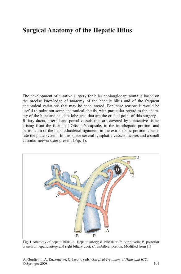
Surgical Anatomy of the Hepatic Hilus
The development of curative surgery for hilar cholangiocarcinoma is based onthe precise knowledge of anatomy of the hepatic hilus and of the frequentanatomical variations that may be encountered. For these reasons it would beuseful to point out some anatomical details, with particular regard to the anato-my of the hilar and caudate lobe area that are the crucial point of this surgery.Biliary ducts, arterial and portal vessels that are covered by connective tissuearising from the fusion of Glisson’s capsule, in the intrahepatic portion, andperitoneum of the hepatoduodenal ligament, in the extrahepatic portion, consti-tute the plate system. In this space several lymphatic vessels, nerves and a smallvascular network are present (Fig. 1).
101A. Guglielmi, A. Ruzzenente, C. Iacono (eds.) Surgical Treatment of Hilar and ICC.© Springer 2008
Fig. 1 Anatomy of hepatic hilus. A, Hepatic artery; B, bile duct; P, portal vein; P, posteriorbranch of hepatic artery and right biliary duct; U, umbilical portion. Modified from [1]
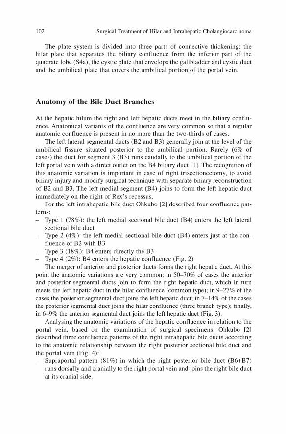
The plate system is divided into three parts of connective thickening: thehilar plate that separates the biliary confluence from the inferior part of thequadrate lobe (S4a), the cystic plate that envelops the gallbladder and cystic ductand the umbilical plate that covers the umbilical portion of the portal vein.
Anatomy of the Bile Duct Branches
At the hepatic hilum the right and left hepatic ducts meet in the biliary conflu-ence. Anatomical variants of the confluence are very common so that a regularanatomic confluence is present in no more than the two-thirds of cases.
The left lateral segmental ducts (B2 and B3) generally join at the level of theumbilical fissure situated posterior to the umbilical portion. Rarely (6% ofcases) the duct for segment 3 (B3) runs caudally to the umbilical portion of theleft portal vein with a direct outlet on the B4 biliary duct [1]. The recognition ofthis anatomic variation is important in case of right trisectionectomy, to avoidbiliary injury and modify surgical technique with separate biliary reconstructionof B2 and B3. The left medial segment (B4) joins to form the left hepatic ductimmediately on the right of Rex’s recessus.
For the left intrahepatic bile duct Ohkubo [2] described four confluence pat-terns:– Type 1 (78%): the left medial sectional bile duct (B4) enters the left lateral
sectional bile duct– Type 2 (4%): the left medial sectional bile duct (B4) enters just at the con-
fluence of B2 with B3– Type 3 (18%): B4 enters directly the B3– Type 4 (2%): B4 enters the hepatic confluence (Fig. 2)
The merger of anterior and posterior ducts forms the right hepatic duct. At thispoint the anatomic variations are very common: in 50–70% of cases the anteriorand posterior segmental ducts join to form the right hepatic duct, which in turnmeets the left hepatic duct in the hilar confluence (common type); in 9–27% of thecases the posterior segmental duct joins the left hepatic duct; in 7–14% of the casesthe posterior segmental duct joins the hilar confluence (three branch type); finally,in 6–9% the anterior segmental duct joins the left hepatic duct (Fig. 3).
Analysing the anatomic variations of the hepatic confluence in relation to theportal vein, based on the examination of surgical specimens, Ohkubo [2]described three confluence patterns of the right intrahepatic bile ducts accordingto the anatomic relationship between the right posterior sectional bile duct andthe portal vein (Fig. 4):– Supraportal pattern (81%) in which the right posterior bile duct (B6+B7)
runs dorsally and cranially to the right portal vein and joins the right bile ductat its cranial side.
Surgical Treatment of Hilar and Intrahepatic Cholangiocarcinoma 102
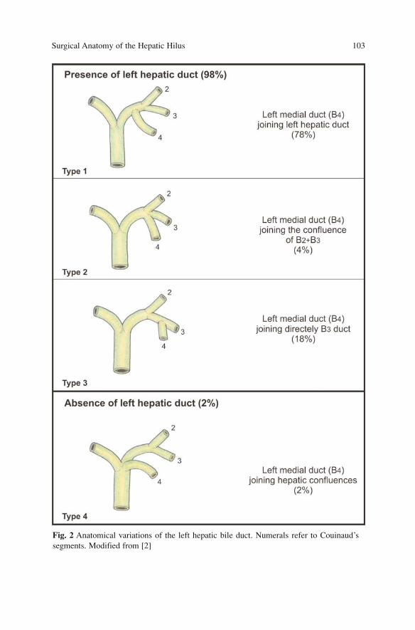
Surgical Anatomy of the Hepatic Hilus 103
Fig. 2 Anatomical variations of the left hepatic bile duct. Numerals refer to Couinaud’ssegments. Modified from [2]
Surgical Treatment of Hilar and Intrahepatic Cholangiocarcinoma 104
Fig. 3 Anatomical variation of the right hepatic bile duct. A, Anterior branch; P, posteriorbranch; BC, bile duct confluence. Modified from [2]
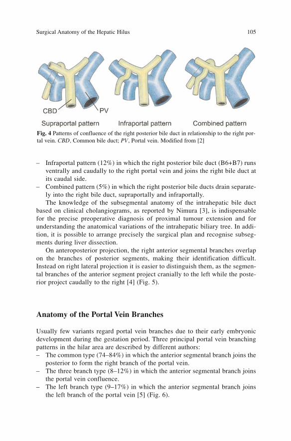
– Infraportal pattern (12%) in which the right posterior bile duct (B6+B7) runsventrally and caudally to the right portal vein and joins the right bile duct atits caudal side.
– Combined pattern (5%) in which the right posterior bile ducts drain separate-ly into the right bile duct, supraportally and infraportally.The knowledge of the subsegmental anatomy of the intrahepatic bile duct
based on clinical cholangiograms, as reported by Nimura [3], is indispensablefor the precise preoperative diagnosis of proximal tumour extension and forunderstanding the anatomical variations of the intrahepatic biliary tree. In addi-tion, it is possible to arrange precisely the surgical plan and recognise subseg-ments during liver dissection.
On anteroposterior projection, the right anterior segmental branches overlapon the branches of posterior segments, making their identification difficult.Instead on right lateral projection it is easier to distinguish them, as the segmen-tal branches of the anterior segment project cranially to the left while the poste-rior project caudally to the right [4] (Fig. 5).
Anatomy of the Portal Vein Branches
Usually few variants regard portal vein branches due to their early embryonicdevelopment during the gestation period. Three principal portal vein branchingpatterns in the hilar area are described by different authors:– The common type (74–84%) in which the anterior segmental branch joins the
posterior to form the right branch of the portal vein.– The three branch type (8–12%) in which the anterior segmental branch joins
the portal vein confluence.– The left branch type (9–17%) in which the anterior segmental branch joins
the left branch of the portal vein [5] (Fig. 6).
Surgical Anatomy of the Hepatic Hilus 105
Fig. 4 Patterns of confluence of the right posterior bile duct in relationship to the right por-tal vein. CBD, Common bile duct; PV, Portal vein. Modified from [2]
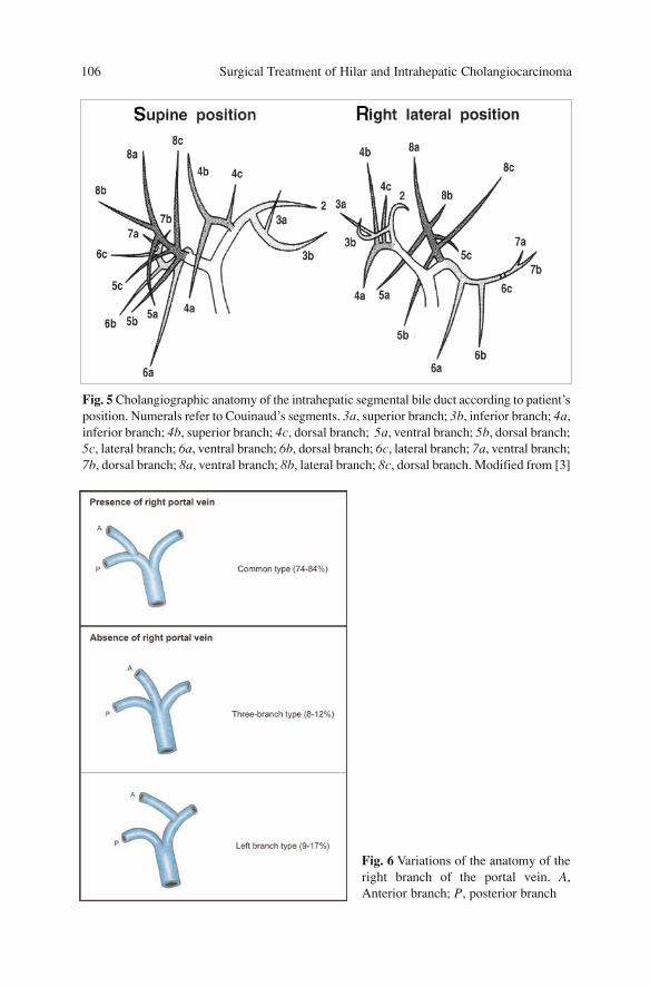
Surgical Treatment of Hilar and Intrahepatic Cholangiocarcinoma 106
Fig. 5 Cholangiographic anatomy of the intrahepatic segmental bile duct according to patient’sposition. Numerals refer to Couinaud’s segments. 3a, superior branch; 3b, inferior branch; 4a,inferior branch; 4b, superior branch; 4c, dorsal branch; 5a, ventral branch; 5b, dorsal branch;5c, lateral branch; 6a, ventral branch; 6b, dorsal branch; 6c, lateral branch; 7a, ventral branch;7b, dorsal branch; 8a, ventral branch; 8b, lateral branch; 8c, dorsal branch. Modified from [3]
Fig. 6 Variations of the anatomy of theright branch of the portal vein. A,Anterior branch; P, posterior branch
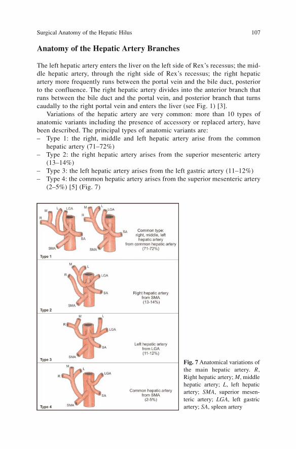
Anatomy of the Hepatic Artery Branches
The left hepatic artery enters the liver on the left side of Rex’s recessus; the mid-dle hepatic artery, through the right side of Rex’s recessus; the right hepaticartery more frequently runs between the portal vein and the bile duct, posteriorto the confluence. The right hepatic artery divides into the anterior branch thatruns between the bile duct and the portal vein, and posterior branch that turnscaudally to the right portal vein and enters the liver (see Fig. 1) [3].
Variations of the hepatic artery are very common: more than 10 types ofanatomic variants including the presence of accessory or replaced artery, havebeen described. The principal types of anatomic variants are:– Type 1: the right, middle and left hepatic artery arise from the common
hepatic artery (71–72%)– Type 2: the right hepatic artery arises from the superior mesenteric artery
(13–14%)– Type 3: the left hepatic artery arises from the left gastric artery (11–12%)– Type 4: the common hepatic artery arises from the superior mesenteric artery
(2–5%) [5] (Fig. 7)
Surgical Anatomy of the Hepatic Hilus 107
Fig. 7 Anatomical variations ofthe main hepatic artery. R,Right hepatic artery; M, middlehepatic artery; L, left hepaticartery; SMA, superior mesen-teric artery; LGA, left gastricartery; SA, spleen artery
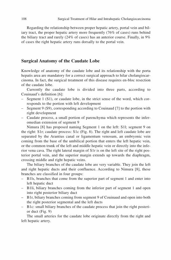
Regarding the relationship between proper hepatic artery, portal vein and bil-iary tract, the proper hepatic artery more frequently (76% of cases) runs behindthe biliary tract and rarely (24% of cases) has an anterior course. Finally, in 9%of cases the right hepatic artery runs dorsally to the portal vein.
Surgical Anatomy of the Caudate Lobe
Knowledge of anatomy of the caudate lobe and its relationship with the portahepatis area are mandatory for a correct surgical approach to hilar cholangiocar-cinoma. In fact, the surgical treatment of this disease requires en-bloc resectionof the caudate lobe.
Currently the caudate lobe is divided into three parts, according toCouinaud’s definition [6]:– Segment 1 (S1), or caudate lobe, in the strict sense of the word, which cor-
responds to the portion with left development– Segment 9 (S9), corresponding according to Couinaud [7] to the portion with
right development– Caudate process, a small portion of parenchyma which represents the infer-
omedian extension of segment 9Nimura [8] has proposed naming Segment 1 on the left: S1l; segment 9 on
the right: S1r; caudate process: S1c (Fig. 8). The right and left caudate lobe areseparated by the Arantius canal or ligamentum venosum, an embryonic veincoming from the base of the umbilical portion that enters the left hepatic vein,or the common trunk of the left and middle hepatic vein or directly into the infe-rior vena cava. The right lateral margin of S1r is on the left site of the right pos-terior portal vein, and the superior margin extends up towards the diaphragm,crossing middle and right hepatic veins.
The biliary branches of the caudate lobe are very variable. They join the leftand right hepatic ducts and their confluence. According to Nimura [8], thesebranches are classified in four groups:– B1ls, branches that come from the superior part of segment 1 and enter into
left hepatic duct– B1li, biliary branches coming from the inferior part of segment 1 and open
into right posterior biliary duct– B1r, biliary branches coming from segment 9 of Couinaud and open into both
the right posterior segmental and the left ducts– B1c: small biliary branches of the caudate process that join the right posteri-
or duct (Fig. 9)The small arteries for the caudate lobe originate directly from the right and
left hepatic artery.
Surgical Treatment of Hilar and Intrahepatic Cholangiocarcinoma 108
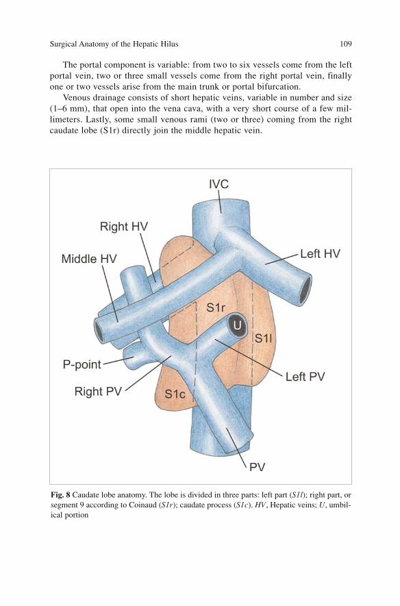
The portal component is variable: from two to six vessels come from the leftportal vein, two or three small vessels come from the right portal vein, finallyone or two vessels arise from the main trunk or portal bifurcation.
Venous drainage consists of short hepatic veins, variable in number and size(1–6 mm), that open into the vena cava, with a very short course of a few mil-limeters. Lastly, some small venous rami (two or three) coming from the rightcaudate lobe (S1r) directly join the middle hepatic vein.
Surgical Anatomy of the Hepatic Hilus 109
Fig. 8 Caudate lobe anatomy. The lobe is divided in three parts: left part (S1l); right part, orsegment 9 according to Coinaud (S1r); caudate process (S1c). HV, Hepatic veins; U, umbil-ical portion
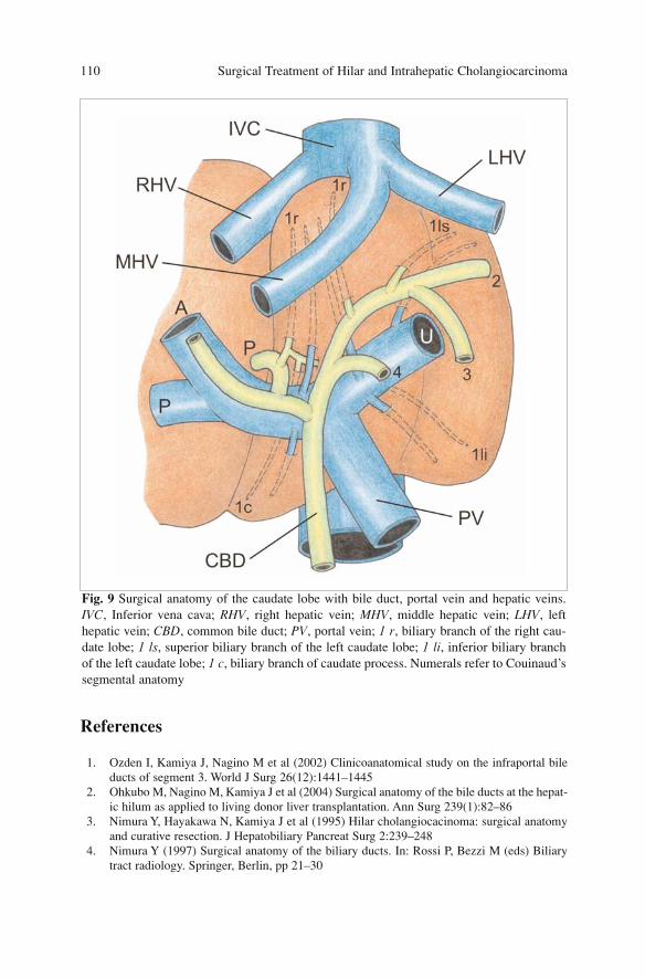
References
1. Ozden I, Kamiya J, Nagino M et al (2002) Clinicoanatomical study on the infraportal bileducts of segment 3. World J Surg 26(12):1441–1445
2. Ohkubo M, Nagino M, Kamiya J et al (2004) Surgical anatomy of the bile ducts at the hepat-ic hilum as applied to living donor liver transplantation. Ann Surg 239(1):82–86
3. Nimura Y, Hayakawa N, Kamiya J et al (1995) Hilar cholangiocacinoma: surgical anatomyand curative resection. J Hepatobiliary Pancreat Surg 2:239–248
4. Nimura Y (1997) Surgical anatomy of the biliary ducts. In: Rossi P, Bezzi M (eds) Biliarytract radiology. Springer, Berlin, pp 21–30
Surgical Treatment of Hilar and Intrahepatic Cholangiocarcinoma 110
Fig. 9 Surgical anatomy of the caudate lobe with bile duct, portal vein and hepatic veins.IVC, Inferior vena cava; RHV, right hepatic vein; MHV, middle hepatic vein; LHV, lefthepatic vein; CBD, common bile duct; PV, portal vein; 1 r, biliary branch of the right cau-date lobe; 1 ls, superior biliary branch of the left caudate lobe; 1 li, inferior biliary branchof the left caudate lobe; 1 c, biliary branch of caudate process. Numerals refer to Couinaud’ssegmental anatomy
5. Kawarada Y, Das BC, Taoka H (2000) Anatomy of the hepatic hilar area: the plate system.J Hepatobiliary Pancreat Surg 7(6):580–586
6. Couinaud C (1989) Surgical anatomy of the liver revisited. Couinaud, Paris 7. Couinaud C (1994) The paracaval segment of the liver. J Hep Bil Panc Surg 2:145–1518. Nimura Y, Hayakawa N, Kamiya J et al (1990) Hepatic segmentectomy with caudate lobe
resection for bile duct carcinoma of the hepatic hilus. World J Surg 14(4):535–543;Discussion 544
Surgical Anatomy of the Hepatic Hilus 111
Surgical Treatment
The rationale for surgical treatment requires the knowledge provided by T-stageof radial and longitudinal neoplastic diffusion, and of N and M stages. Asdescribed in Chap. “Preoperative Assessment of Liver Function”, a correct pre-operative assessment is often difficult to achieve.
Significant data on lymph node diffusion and correlated prognosis are stilllacking in the literature, except for few reports of Japanese institutions. Surgicalchoices are now determined mainly by local extension of disease. Nowadaysresectability criteria differ from those of the past: portal and/or arterial infiltra-tion are no longer an absolute contraindication to resection.
In the past, hilar cholangiocarcinoma was treated by biliary tract resectionand limited hepatic resection. Recent data in the literature have shown that asso-ciated hepatic resection significantly increases the rate of R0 resection, with abetter long-term outcome. Instead, the need to resect the caudate lobe systemat-ically is still under debate.
Postoperative morbidity and mortality rates after major resection havedecreased thanks to improvement of preoperative hepatic function (biliarydrainage, portal embolisation), a better selection of patients and an upgrading ofsurgical techniques.
Surgical option is determined by stage and neoplastic diffusion, tailoring thetype of operation that guarantees a better probability of curative resection andtherefore a better prognosis.
General Principles
Hilar cholangiocarcinoma remains one of the most difficult management prob-lems in terms of staging and radical treatment. It has long been recognised thatsurgical resection with complete removal of all cancer tissues offers the onlychance for cure and long-term survival.
Several studies have confirmed the importance of hepatic resection as ameans of achieving a margin negative resection (Table 1).
113A. Guglielmi, A. Ruzzenente, C. Iacono (eds.) Surgical Treatment of Hilar and ICC.© Springer 2008
There appears to be controversy regarding the selection of patients for whomextensive hepatic resection is indicated, the type of hepatectomy indicated, i.e.right- or left- sided hepatectomy, and whether routine caudate lobe resection isnecessary.
The lack of consensus largely arises from the difficulty of diagnosing pre-cisely the proximal tumour extension before surgical resection or even duringlaparotomy. The main purpose of surgical therapy of hilar cholangiocarcinomais to achieve not only curative resection but also to apply the general rules ofoncological surgery which must accomplish the following:1. R0 resection of biliary tract and liver preserving a residual parenchyma,
well-functioning not less than 30–40%2. Broad tumour-free margin not less than 1 cm on proximal and distal biliary
tract confirmed by frozen section3. Caudate lobe resection, isolated or associated with extended hepatectomy4. Dissection of the lymph nodes and connective tissues in the hepatoduodenal
ligament (12a, 12p, 12b), posterior to the upper portion of the pancreatichead (superior retropancreatic 13a), and around the common hepatic artery(8a)
5. The decision of whether right- or left-sided hepatectomy is indicated, ismade according to the predominant site of the lesion (longitudinal tumourextension)
6. No-touch technique to prevent neoplastic seeding7. Avoid intra-operative open tumour biopsy8. Keep the operative field clear of biliary contamination due to the risk of neo-
plastic cell spreading
Surgical Treatment of Hilar and Intrahepatic Cholangiocarcinoma 114
Table 1 Resection rate of curative resection in patients with hilar cholangiocarcinoma
Author Year Patients (number) Resection rate Curative resections
Jarnagin [1] 2001 225 36% 78%
Launois [2] 2000 552 32% –
Lee [3] 2000 151 85% –
Neuhaus [4] 2003 133 – 60%
Nimura [5] 2000 177 80% 70%
Puhalla [6] 2003 88 42% 33%
Tsao [7] (Lahey) 2000 100 25% 28%
Tsao [7] (Nagoya) 2000 155 79% 78%
Uchiyama [8] 2003 57 58% 64%
Yi [9] 2004 197 61% 41%
Otto [10] 2007 99 71% 75%
The close relationship of the neoplasm with portal bifurcation, right hepaticartery and hepatic parenchyma makes it difficult to address the aforementionedprinciples without a careful evaluation of tumour extent.
Radial spread of the lesion to surrounding structures is facilitated by delayeddiagnosis and by the absence of a muscular layer in the biliary duct; besides,infiltrative growth pattern is the most frequent in this neoplasm and determinesa rapid periductal and perineural diffusion along the ducts and Glisson spaces.These are the reasons why surgical choice must follow the rule of obtaining anadequate free margin at least of 1 cm rather than limited resection.
Assessment of Resectability
Evaluation of patients with hilar cholangiocarcinoma is mainly an assessment ofresectability, since resection is the only effective therapy. As a general rule, inpresence of a resectable tumour an R0 resection must be carried out preservinga well-functioning and vascularised future remnant liver with adequate biliarydrainage.
In the last 20 years the definition of resectability has changed and a standard-ised opinion has not yet been agreed upon, especially between Japanese andWestern authors [7].
The following must be taken into consideration:1. Physical status of the patient and liver function2. Biliary extent of neoplasm3. Vascular involvement4. Presence of lobe atrophy5. Lymph-node involvement6. Presence of distant metastases
Patient Factors
First of all the surgeon must assess the patient’s general condition and fitness fora major operation that usually includes hepatectomy. Resection is generally pre-cluded if the patient is medically unfit or otherwise unable to tolerate a majoroperation, or in the presence of significant co-morbidity, chronic liver disease,hepatic cirrhosis and/or portal hypertension.
Biliary Extent of Neoplasm
The infiltration growth of hilar cholangiocarcinoma includes longitudinal andradial diffusion. Longitudinal diffusion is either mucosal or submucosal and iscorrelated to the macroscopic aspect of the neoplasm [11]. The former is more
Surgical Treatment 115
frequent in the papillary or nodular types and can spread 20 mm beyond themacroscopic margin of the lesion in 40% of cases; the latter is typical of inva-sive types and spreads on average 6 mm. Ebata [12] analysed 80 cases of resect-ed specimens with microscopically positive margin and observed that the sub-mucosal invasive type spread less than 10 mm in all the cases while the non-invasive superficial type (carcinoma in situ) is limited to within 20 mm in 90%of the cases. Considering these data, a macroscopic margin of 10 mm can beconsidered accurate for eradication of invasive extra-hepatic bile duct carcino-ma. However this requires additional removal of any non-invasive component.
The presence of invasive carcinoma on the resected margin worsens the out-come while the presence of carcinoma in situ on the margin does not have a sta-tistical significance on survival compared to negative margin cases [13]. When amargin is positive on frozen section it is advisable to extend biliary resection, iftechnically feasible; however, discriminating between carcinoma in situ andinvasive carcinoma is clinically important.
If tumour extension is definitely thought to extend peripherally to the secondsegmental ramification of the second order intrahepatic biliary radicles bilater-ally the patient is considered to be unresectable. In selected cases with Bismuthtype IV neoplasm a left or right trisectionectomy can be performed if hepaticfunction and residual volume after PVE are at least 40%.
Radial diffusion extends to the hepatoduodenal ligament and to the perivas-cular connective tissue of the hepatic artery and portal vein that are close to thebiliary tract. This aspect determines the choice of type of hepatectomy and like-ly associated vascular resection.
Vascular Involvement
Portal Vein
Portal invasion is generally associated with homolateral lobar atrophy but it isnot a contraindication to surgical resection (Fig. 1). However, the results are dif-ferent if we consider portal resection in advanced cases or in “en-bloc” portalresection during extended right hepatectomy.
Ebata [14] reported a study of 52 patients who underwent portal resection,and classified histological grades of portal infiltration in three levels:– Grade 0: no involvement– Grade I: cancer invasion limited to the tunica adventitia or media– Grade II: cancer invasion arriving at the tunica intima.
Portal resection is a negative prognostic factor but it improves outcome com-pared with non-resected patients. Three and 5-year survival were 26 and 10%,respectively, in patients with hepatic and portal combined resection, and 54 and37% in patients who underwent hepatic resection alone (p<0.0001), respectively.
Moreover, survival in patients with grade I or II portal invasion was similarto that of the group of patients without microscopic portal invasion (grade 0)
Surgical Treatment of Hilar and Intrahepatic Cholangiocarcinoma 116
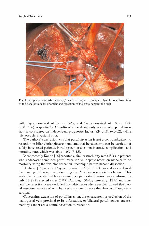
with 3-year survival of 22 vs. 36%, and 5-year survival of 10 vs. 18%(p=0.1506), respectively. At multivariate analysis, only macroscopic portal inva-sion is considered an independent prognostic factor (RR 2.18; p<0.02), whilemicroscopic invasion is not.
The authors’ conclusion was that portal invasion is not a contraindication toresection in hilar cholangiocarcinoma and that hepatectomy can be carried outsafely in selected patients. Portal resection does not increase complications andmortality rate, which was about 10% [5,15].
More recently Kondo [16] reported a similar morbidity rate (48%) in patientswho underwent combined portal resection vs. hepatic resection alone with nomortality using the “en-bloc resection” technique before hepatic dissection.
Neuhaus [15] reported 5-year survival of 65% in R0 cases after combinedliver and portal vein resection using the “en-bloc resection” technique. Thiswork has been criticised because microscopic portal invasion was confirmed inonly 12% of resected cases (2/17). Although 60-day mortality (17%) and non-curative resection were excluded from this series, these results showed that por-tal resection associated with hepatectomy can improve the chances of long-termsurvival.
Concerning extension of portal invasion, the encasement or occlusion of themain portal vein proximal to its bifurcation, or bilateral portal venous encase-ment by cancer are a contraindication to resection.
Surgical Treatment 117
Fig. 1 Left portal vein infiltration (left white arrow) after complete lymph node dissectionof the hepatoduodenal ligament and resection of the extra-hepatic bile duct
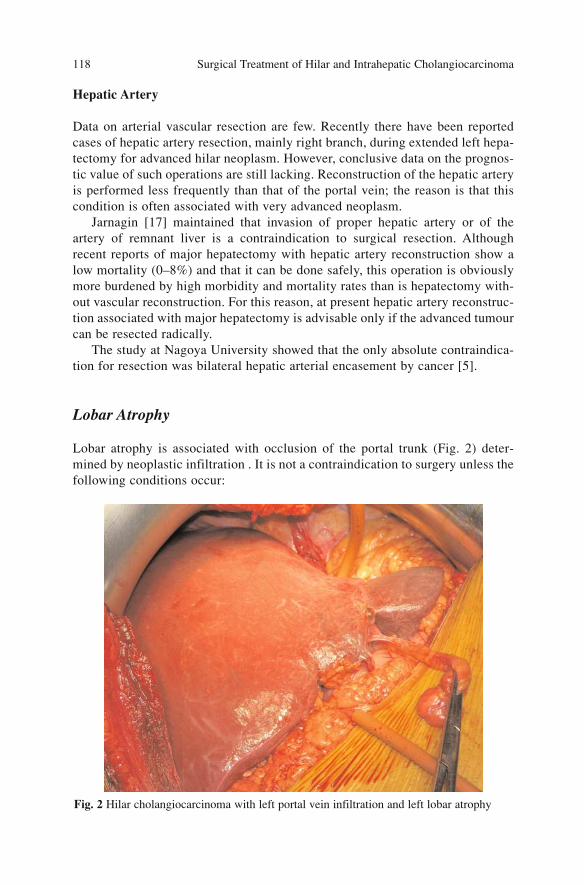
Hepatic Artery
Data on arterial vascular resection are few. Recently there have been reportedcases of hepatic artery resection, mainly right branch, during extended left hepa-tectomy for advanced hilar neoplasm. However, conclusive data on the prognos-tic value of such operations are still lacking. Reconstruction of the hepatic arteryis performed less frequently than that of the portal vein; the reason is that thiscondition is often associated with very advanced neoplasm.
Jarnagin [17] maintained that invasion of proper hepatic artery or of theartery of remnant liver is a contraindication to surgical resection. Althoughrecent reports of major hepatectomy with hepatic artery reconstruction show alow mortality (0–8%) and that it can be done safely, this operation is obviouslymore burdened by high morbidity and mortality rates than is hepatectomy with-out vascular reconstruction. For this reason, at present hepatic artery reconstruc-tion associated with major hepatectomy is advisable only if the advanced tumourcan be resected radically.
The study at Nagoya University showed that the only absolute contraindica-tion for resection was bilateral hepatic arterial encasement by cancer [5].
Lobar Atrophy
Lobar atrophy is associated with occlusion of the portal trunk (Fig. 2) deter-mined by neoplastic infiltration . It is not a contraindication to surgery unless thefollowing conditions occur:
Surgical Treatment of Hilar and Intrahepatic Cholangiocarcinoma 118
Fig. 2 Hilar cholangiocarcinoma with left portal vein infiltration and left lobar atrophy
– Atrophy of one hepatic lobe with contralateral portal vein branch encasementor occlusion
– Atrophy of one hepatic lobe with contralateral tumour extension to second-ary biliary radicles
– Unilateral tumour extension to secondary biliary radicles with contralateralportal vein branch encasement or occlusion
Lymph-Node Involvement
The presence of lymph-node metastases is not rare in hilar cholangiocarcinomapatients. In a recent review, Kitagawa [18] reported N0 in 47% cases, regionalN+ in 35% and para-aortic N+ in 17%. Survival was closely related to nodalinvolvement: 5-year survival in N0 patients was 31%, 15% in regional N+ and12% in para-aortic N+ [18]. Considering carefully the outcome of patients withpara-aortic positive nodes the author identified that survival was significantlybetter in patients with “macroscopically negative” nodes than in patients with“macroscopically positive” nodes, with a 5-year survival of 29 vs. 0%, respec-tively.
Kosuge [19] reported a mean and median survival of 79 months and 39months, respectively, in N0 patients, 52 and 26 months in regional N+, and 15and 14 months in N+ beyond regional. Five-year survival was 38% in N0, 30%in regional N+ (similar survival, not statistically significant) and 0% at 3-yearsurvival in N+ beyond regional nodes.
To date, performing surgical resection in patients with wide and macroscop-ic involvement of non-regional lymph nodes does not appear justified. It has notyet been proved that extended lymphadenectomy guarantees improvement insurvival in hilar cholangiocarcinoma but we believe a methodical regional nodaldissection must always be performed associated with R0 resection, at least untildata are more consistent and conclusive.
According to Jarnagin at the MSKCC [20], pathological proven lymph-nodemetastases beyond the hepatoduodenal ligament (peripancreatic, periduodenal,celiac, superior mesenteric or posterior pancreaticoduodenal lymph nodes) wasconsidered to represent disease not amenable to a potentially curative resection. Incontrast, metastatic disease to cystic duct, pericholedochal, hilar or portal lymphnodes (i.e. within the hepatoduodenal ligament) did not necessary constitute aunresectable condition. At this time we perform lymphadenectomy of hepatoduo-denal ligament and of the hepatic hilum (periportal, proper and common hepaticartery and retroduodenal lymph nodes) while peripancreatic, periduodenal, para-aortic and periceliac lymph nodes are sampled, if macroscopically suspect forstaging.
Surgical Treatment 119
Extrahepatic Disease
Adjacent extrahepatic organ invasion is not an absolute contraindication toresection. A concomitant pancreatic resection (hepatopancreatoduodenectomyHPD) is indicated if the tumour’s longitudinal spread was considered to be in thelower intrapancreatic bile duct and/or massive peripancreatic head and peric-holedochal lymph nodes metastases were suspected.
In presence of wide nodal infiltration, HPD indication is not the optimum asdistant nodal metastases represent a negative prognostic factor. Nimura [21] firstreported the results of this operation with an operative mortality of 35% and 5-year survival of 6%. Main causes of mortality were postoperative hepatic insuf-ficiency and biliary leak. To decrease the risk of pancreatic fistula a two-stageoperation with a delayed pancreo-jejunostomy was proposed [22].
Miyagawa [23] reported 12 cases treated with HPD and delayed pancreaticreconstruction, with 0% mortality rate.
Metastatic Disease
The presence of distant metastases (peritoneal, hepatic, pulmonary etc,) diag-nosed preoperatively or intraoperatively stands for an absolute contraindicationto surgical resection. The only exception can be the occurrence of hepatic metas-tases limited in the lobe to be resected; however, these cases there is no 5-yearsurvival.
Indication for Surgical Resection
The indication of the type of surgical procedure is planned after a careful eval-uation of the patient’s physical status, hepatic function and neoplastic diffusionof the disease. The strategy of hepatic resection must be defined preoperativelybased on exact knowledge of longitudinal and radial extension of the neoplasm.
Currently, there is no clinical staging system available that stratifies patientspreoperatively into subgroups based on potential for resection. The modifiedBismuth-Corlette classification [24] stratifies patients based only on the extentof biliary involvement by tumour, and the AJCC staging system is based largelyon pathological criteria and has little applicability for preoperative staging orpredicting resectability.
The preoperative staging system proposed recently by Jarnagin [1] is based onlocal tumour-related factors that determine resectability: biliary ductal involvement,vascular involvement, and lobar atrophy. This last staging system seems related toresectability (60% for T1, 30% for T2 and 0% for T3) and to the presence of ex-trahepatic disease (21% for T1, 43% for T2 and 41% for T3) [1]. Unfortunately itdoes not aid in establishing the type of resection to perform.
Surgical Treatment of Hilar and Intrahepatic Cholangiocarcinoma 120
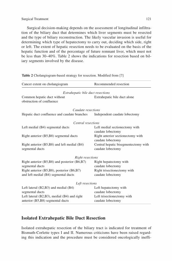
Surgical decision-making depends on the assessment of longitudinal infiltra-tion of the biliary duct that determines which liver segments must be resectedand the type of biliary reconstruction. The likely vascular invasion is useful fordetermining which type of hepatectomy to carry out, deciding which side, rightor left. The extent of hepatic resection needs to be evaluated on the basis of thehepatic function and of the percentage of future remnant liver, which must notbe less than 30–40%. Table 2 shows the indications for resection based on bil-iary segments involved by the disease.
Isolated Extrahepatic Bile Duct Resection
Isolated extrahepatic resection of the biliary tract is indicated for treatment ofBismuth-Corlette types I and II. Numerous criticisms have been raised regard-ing this indication and the procedure must be considered oncologically ineffi-
Surgical Treatment 121
Table 2 Cholangiogram-based strategy for resection. Modified from [7]
Cancer extent on cholangiogram Recommended resection
Extrahepatic bile duct resectionsCommon hepatic duct without Extrahepatic bile duct aloneobstruction of confluence
Caudate resectionsHepatic duct confluence and caudate branches Independent caudate lobectomy
Central resectionsLeft medial (B4) segmental ducts Left medial sectionectomy with
caudate lobectomyRight anterior (B5,B8) segmental ducts Right anterior sectionectomy with
caudate lobectomyRight anterior (B5,B8) and left medial (B4) Central hepatic bisegmentectomy with segmental ducts caudate lobectomy
Right resectionsRight anterior (B5,B8) and posterior (B6,B7) Right hepatectomy with segmental ducts caudate lobectomyRight anterior (B5,B8), posterior (B6,B7) Right trisectionectomy with and left medial (B4) segmental ducts caudate lobectomy
Left resectionsLeft lateral (B2,B3) and medial (B4) Left hepatectomy with segmental ducts caudate lobectomyLeft lateral (B2,B3), medial (B4) and right Left trisectionectomy with anterior (B5,B8) segmental ducts caudate lobectomy
cient concerning long-term results, based on the following considerations:– Cholangiography is not accurate enough to provide precise information
about longitudinal cancer extension– Hilar bile duct cancers often show submucosal tumour extension at their
proximal margins, which might not be visualised by cholangiography– Skip type lesions render assessment by imaging and biopsy modalities diffi-
cultMoreover, in the group of patients who undergo margin-negative resections,
hepatic resection is the only independent predictor of improved survival on mul-tivariate analysis, suggesting that a more limited resection is ineffective for com-plete tumour clearance [1].
In the series reported by MSKCC there were no 5-year survival rates amongpatients who had undergone biliary resection alone [20]. Analogous results havebeen reported by Miyazaki [25] with 5-year survival of 0% in patients with bil-iary tract resection alone vs. 27% in patients with additional hepatic resection.A larger series is described by Nimura [5] who reported 8 cases of isolated R0biliary tract resection, compared with 100 cases of associated hepatic resection.All the patients who had undergone isolated biliary tract resection died within65 months. These data are confirmed by Neuhaus [4] who reported 5-year sur-vival of 0 vs. 35% in patients who had undergone R0 biliary tract resection alonevs. additional hepatic resection, respectively.
Moreover, this type of operation has a high percentage of recurrence, up to76%, mainly on the proximal resection margin [26].
The indications are therefore limited to surgical high-risk patients withtumour in an early stage (T1 and T2) and accompanied by poor hepatic function.It is no longer a good indication even as palliative treatment, since endoscopicand percutaneous drainages are preferred.
Independent Caudate Lobectomy (S1)
Resection of caudate lobe is always indicated in association with hepatectomywhen the tumour involves biliary confluence, due to the high frequency ofinvolvement of biliary branches varying from 48% [27] to 96% [28]. Neoplasticinfiltration follows three pathways:– Infiltration along the epithelium of biliary duct– Direct infiltration of caudate parenchyma– Periductal diffusion in interstitial tissue of caudate lobe
The importance of caudate lobe resection associated with hepatectomy wasfirst underlined by Nimura [28] and it is now largely accepted. The clinicalworth of caudate lobe resection was reported for the first time by Sugiura [29],who presented a 5-year survival of 46% with additional hepatectomy to caudateresection, and 12% without caudate resection.
Surgical Treatment of Hilar and Intrahepatic Cholangiocarcinoma 122
Isolated resection of caudate lobe associated with resection of extrahepaticbiliary tract finds few indications and is limited to the cases with neoplasm thatgrows at the biliary bifurcation and is confined between the right and left hepat-ic ducts with the sole involvement of the branches to caudate lobe. The resultsof isolated caudate lobectomy seem worse than those of more extended hepaticresections: Kondo [30] reported a 3-year survival of 30% after isolated caudatelobectomy vs. 75% after right hepatectomy (p=0.013).
Central Hepatic Resections
Central hepatectomies consisting of left medial sectionectomy with caudatelobectomy (S1+S4), right anterior sectionectomy with caudate lobectomy(S1+S5, S8) and central hepatic bisegmentectomy with caudate lobectomy(S1+S4, S5, S8), are proposed mainly by Japanese authors [5,31,32].
These “parenchyma-preserving” hepatectomies find their premise in limitedhepatic resection to minimise postoperative risk of hepatic failure in high-riskpatients, still maintaining a potentially curative R0 resection. These procedureshave been criticised at times: resection is generally difficult since it required twoparenchymal section lines and caudate resection, with increased postoperativemorbidity (bile leakage); it is not possible to obtain an adequate length of mar-gin on the biliary tract. Moreover, the need to maintain the portal bifurcation,and especially the right and left hepatic arteries, leads to oncological problemsof radicality, above all regarding the right hepatic artery which runs just ventral-ly and very close to the biliary confluence. The reconstruction of bilio-digestiveanastomosis is more complex and requires a greater number of anastomoses.
Shimada [32] compared the results of major hepatectomies with those ofparenchyma-preserving hepatectomy. Curability rate (R0:R1) was 77% for theformer and 54% for the latter, very similar to that of biliary resection alone(50%). The percentage of complications was comparable (47 vs. 54%), but bil-iary leaks were more frequent in parenchyma-preserving hepatectomy (27 vs.3%), caused either by the wider raw surface of resection or by the greater num-ber of bilio-jejunostomies (4.8±1.8). Mortality rate was nil in parenchyma-pre-serving hepatectomy and 13% in major hepatectomy, and 5-year survival were15 and 25%, respectively.
The reasons for non-curative resection in parenchyma-preserving hepatecto-my are almost always determined by microscopical infiltration of proximal bil-iary margin [33] or by residual tumour on portal bifurcation or hepatic artery [7].
Therefore parenchyma-preserving hepatectomy should be limited strictly tohigh-risk patients in whom the tumour is confined longitudinally to the right orleft hepatic duct, does not invade any of segmental hepatic ducts or transverselyinvades an adjacent organ.
Surgical Treatment 123
Extended Right Resections
If the predominant site of involvement is the right hepatic duct or when bothhepatic ducts are invaded equally (Bismuth-Corlette types I, II, IIIa, IV), extend-ed right hepatectomy or trisectionectomy are indicated.
In cases with hilar infiltration involving right intrahepatic ducts withoutextension to segment 4, an extended right hepatectomy with caudate lobectomy(S1+S5, S6, S7, S8) is indicated; instead, in presence of infiltration for contigu-ity of segment 4 a right trisectionectomy with caudate lobectomy (S1+S4, S5,S6, S7, S8) is indicated.
These operations are believed by most authors to guarantee the higher radi-cality rate and the best long-term results, especially if the principles of “no-touch technique” and portal resection “de principio” are applied [4].
Right hepatectomy is more likely to be associated with a negative resection mar-gin than is left hepatectomy, based on the following anatomic considerations:– The common bile duct is on the right side of the hepatoduodenal ligament,
with the right hepatic artery passing behind its proximal portion. Thereforethe right hepatic artery is frequently invaded by cancer at this site, whereasthe left and middle hepatic artery runs along the left side of the hepatoduo-denal ligament and is not associated with the bile duct until the end of thetransverse portion (extratumoural course at the left margin of the hepatoduo-denal ligament).
– It is not necessary to dissect any structure near the tumour, the portal bifur-cation may be resected and thus does not need to be dissected. An end-to-endanastomosis of the portal trunk to the left portal vein branch will result in amore stretched course of this vessel and may avoid the kinking that some-times occurs after right-sided resections.
– Generally liver dissection between the left lateral or left medial section willresult in a small parenchymal surface.
– The extrahepatic part of the left hepatic duct is longer, with a more distantsegmental ramification, than that of the right hepatic duct.
– Systematic caudate lobectomy can be carried out more easily in patientsundergoing right-sided rather than left-sided hepatectomy.
– When portal vein resection is necessary for tumour invasion, it is easier toperform venous reconstruction with the left than with the right portal vein,due to the long extrahepatic portion of the transverse portion of the left part.Neuhaus [4], utilising the principle of “no-touch technique” with right trisec-
tionectomy and portal resection, reported a 5-year survival of 72% in R0 resect-ed patients with portal vein resection vs. 52% in R0 patients who had undergoneright trisectionectomy but without portal resection, and 23% in patients treatedwith simple right hepatectomy.
Surgical Treatment of Hilar and Intrahepatic Cholangiocarcinoma 124
Extended Left Resections
The “extended left resections” are indicated when the neoplasm involves thehilar bifurcation and spreads preferentially towards left intrahepatic ducts(extended left hepatectomy with caudate lobectomy S1+S2, S3, S4) or when theneoplasm involves the entire left lobe and extends to the anterior segment (lefttrisectionectomy with caudate lobectomy S1+S2, S3, S4, S5, S8).
Left resections require a greater parenchymal dissection compared to rightones. Furthermore right portal pedicles are shorter, determining a more demand-ing portal resection. Ultimately the right hepatic artery that lies close to hilarneoplasm, may facilitate a microscopic local seeding during its mobilisation.The right biliary duct has a shorter course (about 5 mm) before its segmentalbifurcation; this may make it difficult to obtain resection with an adequate mar-gin. For all these reasons, many authors report 5-year survival that is worse inleft hepatectomies than right ones (28 vs. 50% [7] and 34 vs. 44% [28]). Kondo[24] confirmed these results with significantly better survivals in right than lefthepatectomies (p>0.0013).
References
1. Jarnagin WR, Fong Y, DeMatteo RP et al (2001) Staging, resectability, and outcome in 225patients with hilar cholangiocarcinoma. Ann Surg 234(4):507–517; discussion 517–519
2. Launois B, Reding R, Lebeau G, Buard JL (2000) Surgery for hilar cholangiocarcinoma:French experience in a collective survey of 552 extrahepatic bile duct cancers. JHepatobiliary Pancreat Surg 7(2):128–134
3. Lee SG, Lee YJ, Park KM et al (2000) One hundred and eleven liver resections for hilar bileduct cancer. J Hepatobiliary Pancreat Surg 7(2):135–141
4. Neuhaus P, Jonas S, Settmacher U et al (2003) Surgical management of proximal bile ductcancer: extended right lobe resection increases resectability and radicality. LangenbecksArch Surg 388(3):194–200
5. Nimura Y, Kamiya J, Kondo S et al (2000) Aggressive preoperative management and extend-ed surgery for hilar cholangiocarcinoma: Nagoya experience. J Hepatobiliary Pancreat Surg7(2):155–162
6. Puhalla H, Gruenberger T, Pokorny H et al (2003) Resection of hilar cholangiocarcinomas:pivotal prognostic factors and impact of tumour sclerosis. World J Surg 27(6):680–684
7. Tsao JI, Nimura Y, Kamiya J et al (2000) Management of hilar cholangiocarcinoma: com-parison of an American and a Japanese experience. Ann Surg 232(2):166–174
8. Uchiyama K, Nakai T, Tani M et al (2003) Indications for extended hepatectomy in the man-agement of stage IV hilar cholangiocarcinoma. Arch Surg 138(9):1012–1016
9. Yi B, Zhang BH, Zhang YJ et al (2004) Surgical procedure and prognosis of hilar cholan-giocarcinoma. Hepatobiliary Pancreat Dis Int 3(3):453–457
Surgical Treatment 125
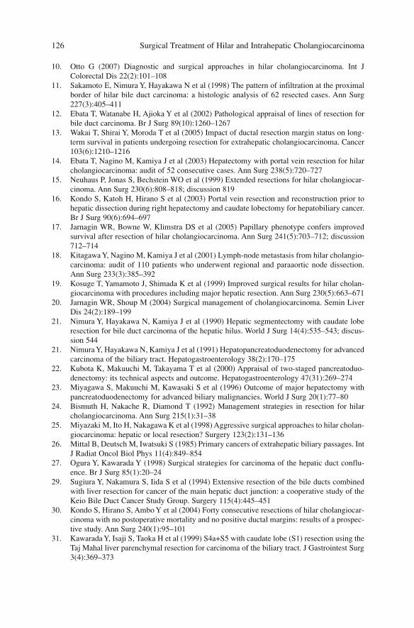
10. Otto G (2007) Diagnostic and surgical approaches in hilar cholangiocarcinoma. Int JColorectal Dis 22(2):101–108
11. Sakamoto E, Nimura Y, Hayakawa N et al (1998) The pattern of infiltration at the proximalborder of hilar bile duct carcinoma: a histologic analysis of 62 resected cases. Ann Surg227(3):405–411
12. Ebata T, Watanabe H, Ajioka Y et al (2002) Pathological appraisal of lines of resection forbile duct carcinoma. Br J Surg 89(10):1260–1267
13. Wakai T, Shirai Y, Moroda T et al (2005) Impact of ductal resection margin status on long-term survival in patients undergoing resection for extrahepatic cholangiocarcinoma. Cancer103(6):1210–1216
14. Ebata T, Nagino M, Kamiya J et al (2003) Hepatectomy with portal vein resection for hilarcholangiocarcinoma: audit of 52 consecutive cases. Ann Surg 238(5):720–727
15. Neuhaus P, Jonas S, Bechstein WO et al (1999) Extended resections for hilar cholangiocar-cinoma. Ann Surg 230(6):808–818; discussion 819
16. Kondo S, Katoh H, Hirano S et al (2003) Portal vein resection and reconstruction prior tohepatic dissection during right hepatectomy and caudate lobectomy for hepatobiliary cancer.Br J Surg 90(6):694–697
17. Jarnagin WR, Bowne W, Klimstra DS et al (2005) Papillary phenotype confers improvedsurvival after resection of hilar cholangiocarcinoma. Ann Surg 241(5):703–712; discussion712–714
18. Kitagawa Y, Nagino M, Kamiya J et al (2001) Lymph-node metastasis from hilar cholangio-carcinoma: audit of 110 patients who underwent regional and paraaortic node dissection.Ann Surg 233(3):385–392
19. Kosuge T, Yamamoto J, Shimada K et al (1999) Improved surgical results for hilar cholan-giocarcinoma with procedures including major hepatic resection. Ann Surg 230(5):663–671
20. Jarnagin WR, Shoup M (2004) Surgical management of cholangiocarcinoma. Semin LiverDis 24(2):189–199
21. Nimura Y, Hayakawa N, Kamiya J et al (1990) Hepatic segmentectomy with caudate loberesection for bile duct carcinoma of the hepatic hilus. World J Surg 14(4):535–543; discus-sion 544
21. Nimura Y, Hayakawa N, Kamiya J et al (1991) Hepatopancreatoduodenectomy for advancedcarcinoma of the biliary tract. Hepatogastroenterology 38(2):170–175
22. Kubota K, Makuuchi M, Takayama T et al (2000) Appraisal of two-staged pancreatoduo-denectomy: its technical aspects and outcome. Hepatogastroenterology 47(31):269–274
23. Miyagawa S, Makuuchi M, Kawasaki S et al (1996) Outcome of major hepatectomy withpancreatoduodenectomy for advanced biliary malignancies. World J Surg 20(1):77–80
24. Bismuth H, Nakache R, Diamond T (1992) Management strategies in resection for hilarcholangiocarcinoma. Ann Surg 215(1):31–38
25. Miyazaki M, Ito H, Nakagawa K et al (1998) Aggressive surgical approaches to hilar cholan-giocarcinoma: hepatic or local resection? Surgery 123(2):131–136
26. Mittal B, Deutsch M, Iwatsuki S (1985) Primary cancers of extrahepatic biliary passages. IntJ Radiat Oncol Biol Phys 11(4):849–854
27. Ogura Y, Kawarada Y (1998) Surgical strategies for carcinoma of the hepatic duct conflu-ence. Br J Surg 85(1):20–24
29. Sugiura Y, Nakamura S, Iida S et al (1994) Extensive resection of the bile ducts combinedwith liver resection for cancer of the main hepatic duct junction: a cooperative study of theKeio Bile Duct Cancer Study Group. Surgery 115(4):445–451
30. Kondo S, Hirano S, Ambo Y et al (2004) Forty consecutive resections of hilar cholangiocar-cinoma with no postoperative mortality and no positive ductal margins: results of a prospec-tive study. Ann Surg 240(1):95–101
31. Kawarada Y, Isaji S, Taoka H et al (1999) S4a+S5 with caudate lobe (S1) resection using theTaj Mahal liver parenchymal resection for carcinoma of the biliary tract. J Gastrointest Surg3(4):369–373
Surgical Treatment of Hilar and Intrahepatic Cholangiocarcinoma 126
32. Shimada H, Endo I, Sugita M et al (2003) Is parenchyma-preserving hepatectomy a nobleoption in the surgical treatment for high-risk patients with hilar bile duct cancer?Langenbecks Arch Surg 388(1):33–41
33. Tashiro S, Tsuji T, Kanemitsu K et al (1993) Prolongation of survival for carcinoma at thehepatic duct confluence. Surgery 113(3):270–278
Surgical Treatment 127
Surgical Technique
Surgical resection for hilar cholangiocarcinoma is usually a challenging opera-tion. Correct accomplishment of a R0 resection must be planned with a carefulpreoperative evaluation that considers longitudinal and radial extent of neo-plasms, the presence of lymph-node involvement, vascular infiltration andanatomic variants.
Surgical resection entails exeresis of the biliary tract, associated with hepat-ic resection and regional lymphadenectomy. Reconstructive phase follows withbilioenteric anastomosis with Roux-en-Y jejunal loop.
Position of the Patient
The patient is placed on the table in supine position, with left arm secured atright angles and right arm at the site. It may be helpful to place an inflatable sup-port at the base of the thorax at D9-D10 level to expose the liver optimally. Theskin is cleansed higher than the nipples and down to the pubis, and laterally tomiddle axillary lines. It may be useful to apply a crossing bar or similar devicefixed on the table, which later holds a self-retaining retractor to elevate the costalmargin.
Incision
In most cases a right subcostal incision which can be extended to the left and inthe midline toward the xiphoid provides a good exposure. It is advisable to beginwith a small right subcostal incision that permits one to explore the abdominalcavity and judge the resectability of the neoplasm.
In particular cases requiring biliary tract resection alone or limited left hepat-ic resection, a midline incision is made.
129A. Guglielmi, A. Ruzzenente, C. Iacono (eds.) Surgical Treatment of Hilar and ICC.© Springer 2008
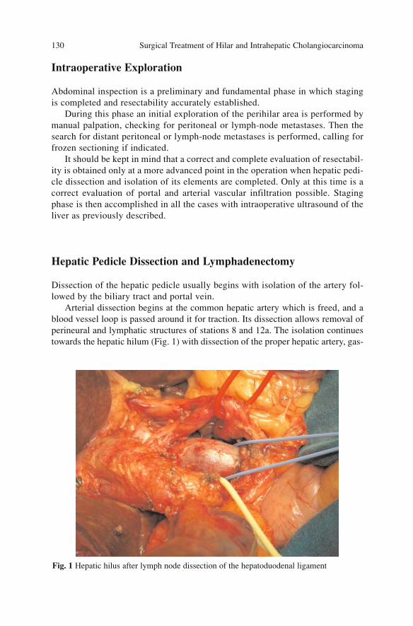
Intraoperative Exploration
Abdominal inspection is a preliminary and fundamental phase in which stagingis completed and resectability accurately established.
During this phase an initial exploration of the perihilar area is performed bymanual palpation, checking for peritoneal or lymph-node metastases. Then thesearch for distant peritoneal or lymph-node metastases is performed, calling forfrozen sectioning if indicated.
It should be kept in mind that a correct and complete evaluation of resectabil-ity is obtained only at a more advanced point in the operation when hepatic pedi-cle dissection and isolation of its elements are completed. Only at this time is acorrect evaluation of portal and arterial vascular infiltration possible. Stagingphase is then accomplished in all the cases with intraoperative ultrasound of theliver as previously described.
Hepatic Pedicle Dissection and Lymphadenectomy
Dissection of the hepatic pedicle usually begins with isolation of the artery fol-lowed by the biliary tract and portal vein.
Arterial dissection begins at the common hepatic artery which is freed, and ablood vessel loop is passed around it for traction. Its dissection allows removal ofperineural and lymphatic structures of stations 8 and 12a. The isolation continuestowards the hepatic hilum (Fig. 1) with dissection of the proper hepatic artery, gas-
Surgical Treatment of Hilar and Intrahepatic Cholangiocarcinoma 130
Fig. 1 Hepatic hilus after lymph node dissection of the hepatoduodenal ligament
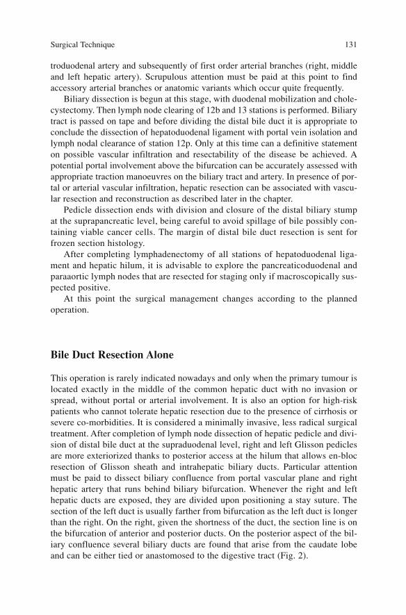
troduodenal artery and subsequently of first order arterial branches (right, middleand left hepatic artery). Scrupulous attention must be paid at this point to findaccessory arterial branches or anatomic variants which occur quite frequently.
Biliary dissection is begun at this stage, with duodenal mobilization and chole-cystectomy. Then lymph node clearing of 12b and 13 stations is performed. Biliarytract is passed on tape and before dividing the distal bile duct it is appropriate toconclude the dissection of hepatoduodenal ligament with portal vein isolation andlymph nodal clearance of station 12p. Only at this time can a definitive statementon possible vascular infiltration and resectability of the disease be achieved. Apotential portal involvement above the bifurcation can be accurately assessed withappropriate traction manoeuvres on the biliary tract and artery. In presence of por-tal or arterial vascular infiltration, hepatic resection can be associated with vascu-lar resection and reconstruction as described later in the chapter.
Pedicle dissection ends with division and closure of the distal biliary stumpat the suprapancreatic level, being careful to avoid spillage of bile possibly con-taining viable cancer cells. The margin of distal bile duct resection is sent forfrozen section histology.
After completing lymphadenectomy of all stations of hepatoduodenal liga-ment and hepatic hilum, it is advisable to explore the pancreaticoduodenal andparaaortic lymph nodes that are resected for staging only if macroscopically sus-pected positive.
At this point the surgical management changes according to the plannedoperation.
Bile Duct Resection Alone
This operation is rarely indicated nowadays and only when the primary tumour islocated exactly in the middle of the common hepatic duct with no invasion orspread, without portal or arterial involvement. It is also an option for high-riskpatients who cannot tolerate hepatic resection due to the presence of cirrhosis orsevere co-morbidities. It is considered a minimally invasive, less radical surgicaltreatment. After completion of lymph node dissection of hepatic pedicle and divi-sion of distal bile duct at the supraduodenal level, right and left Glisson pediclesare more exteriorized thanks to posterior access at the hilum that allows en-blocresection of Glisson sheath and intrahepatic biliary ducts. Particular attentionmust be paid to dissect biliary confluence from portal vascular plane and righthepatic artery that runs behind biliary bifurcation. Whenever the right and lefthepatic ducts are exposed, they are divided upon positioning a stay suture. Thesection of the left duct is usually farther from bifurcation as the left duct is longerthan the right. On the right, given the shortness of the duct, the section line is onthe bifurcation of anterior and posterior ducts. On the posterior aspect of the bil-iary confluence several biliary ducts are found that arise from the caudate lobeand can be either tied or anastomosed to the digestive tract (Fig. 2).
Surgical Technique 131
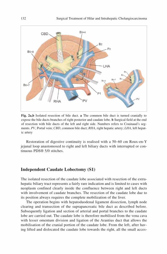
Restoration of digestive continuity is realised with a 50–60 cm Roux-en-Yjejunal loop anastomosed to right and left biliary ducts with interrupted or con-tinuous PDS® 5/0 stitches.
Independent Caudate Lobectomy (S1)
The isolated resection of the caudate lobe associated with resection of the extra-hepatic biliary tract represents a fairly rare indication and is limited to cases withneoplasm confined clearly inside the confluence between right and left ductswith involvement of caudate branches. The resection of the caudate lobe due toits position always requires the complete mobilization of the liver.
The operation begins with hepatoduodenal ligament dissection, lymph nodeclearing and transection of the suprapancreatic bile duct as described before.Subsequently ligation and section of arterial and portal branches to the caudatelobe are carried out. The caudate lobe is therefore mobilised from the vena cavawith lesser omentum division and ligation of the Arantius duct that allows themobilisation of the cranial portion of the caudate lobe. From the left, after hav-ing lifted and dislocated the caudate lobe towards the right, all the small acces-
Surgical Treatment of Hilar and Intrahepatic Cholangiocarcinoma 132
Fig. 2a,b Isolated resection of bile duct. a The common bile duct is turned cranially toexpose the bile ducts branches of right posterior and caudate lobe. b Surgical field at the endof resection with bile ducts of the left and right side. Numbers refers to Couinaud’s seg-ments. PV, Portal vein; CBD, common bile duct; RHA, right hepatic artery; LHA, left hepat-ic artery

sory hepatic veins are tied and divided; in this way the anterior surface of thevena cava is exposed up to the confluence with the left hepatic vein that repre-sents the upper limit of the caudate lobe.
The right portion of caudate lobe is still connected to segment 7 by the cau-date process, a thin parenchymal bridge which is easily divided. The biliary tractis transected on the left at level of S2, S3, and S4 ducts and on the right at thelevel of the anterior and posterior ducts (Fig. 3).
Bilio-digestive continuity is difficult and challenging due to the large num-ber of anastomoses to be performed and the depth of operative field.
Right Hepatectomy with Caudate Lobectomy (S4a, S5, S6, S7,S8 + S1)
Right hepatectomy with caudate lobectomy is indicated for tumours involvingthe right anterior and posterior sectorial bile ducts with sparing of the left medi-al segmental bile duct to segment 4.
The resection entails the right hepatic lobe, caudate lobe and, if indicated,the caudal portion of segment 4 (4a) to obtain an adequate margin of biliaryducts.
Surgical Technique 133
Fig. 3 Independent caudate lobectomy. The left bile duct is resected on the right side of theumbilical plate (B4-B3-B2); on the right side, at the origin of right anterior and posterior bileduct
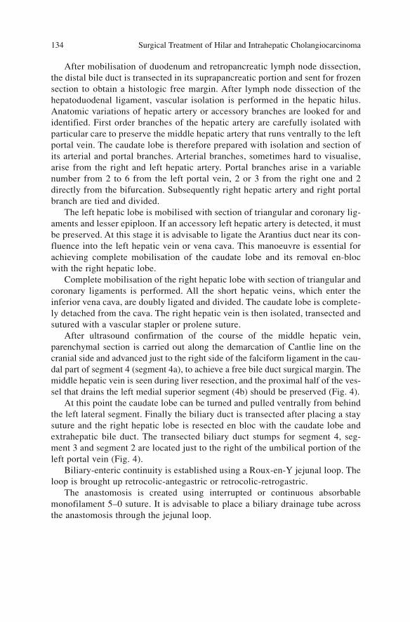
After mobilisation of duodenum and retropancreatic lymph node dissection,the distal bile duct is transected in its suprapancreatic portion and sent for frozensection to obtain a histologic free margin. After lymph node dissection of thehepatoduodenal ligament, vascular isolation is performed in the hepatic hilus.Anatomic variations of hepatic artery or accessory branches are looked for andidentified. First order branches of the hepatic artery are carefully isolated withparticular care to preserve the middle hepatic artery that runs ventrally to the leftportal vein. The caudate lobe is therefore prepared with isolation and section ofits arterial and portal branches. Arterial branches, sometimes hard to visualise,arise from the right and left hepatic artery. Portal branches arise in a variablenumber from 2 to 6 from the left portal vein, 2 or 3 from the right one and 2directly from the bifurcation. Subsequently right hepatic artery and right portalbranch are tied and divided.
The left hepatic lobe is mobilised with section of triangular and coronary lig-aments and lesser epiploon. If an accessory left hepatic artery is detected, it mustbe preserved. At this stage it is advisable to ligate the Arantius duct near its con-fluence into the left hepatic vein or vena cava. This manoeuvre is essential forachieving complete mobilisation of the caudate lobe and its removal en-blocwith the right hepatic lobe.
Complete mobilisation of the right hepatic lobe with section of triangular andcoronary ligaments is performed. All the short hepatic veins, which enter theinferior vena cava, are doubly ligated and divided. The caudate lobe is complete-ly detached from the cava. The right hepatic vein is then isolated, transected andsutured with a vascular stapler or prolene suture.
After ultrasound confirmation of the course of the middle hepatic vein,parenchymal section is carried out along the demarcation of Cantlie line on thecranial side and advanced just to the right side of the falciform ligament in the cau-dal part of segment 4 (segment 4a), to achieve a free bile duct surgical margin. Themiddle hepatic vein is seen during liver resection, and the proximal half of the ves-sel that drains the left medial superior segment (4b) should be preserved (Fig. 4).
At this point the caudate lobe can be turned and pulled ventrally from behindthe left lateral segment. Finally the biliary duct is transected after placing a staysuture and the right hepatic lobe is resected en bloc with the caudate lobe andextrahepatic bile duct. The transected biliary duct stumps for segment 4, seg-ment 3 and segment 2 are located just to the right of the umbilical portion of theleft portal vein (Fig. 4).
Biliary-enteric continuity is established using a Roux-en-Y jejunal loop. Theloop is brought up retrocolic-antegastric or retrocolic-retrogastric.
The anastomosis is created using interrupted or continuous absorbablemonofilament 5–0 suture. It is advisable to place a biliary drainage tube acrossthe anastomosis through the jejunal loop.
Surgical Treatment of Hilar and Intrahepatic Cholangiocarcinoma 134
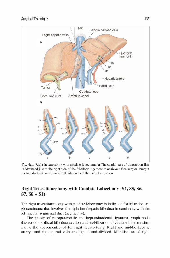
Right Trisectionectomy with Caudate Lobectomy (S4, S5, S6,S7, S8 + S1)
The right trisectionectomy with caudate lobectomy is indicated for hilar cholan-giocarcinoma that involves the right intrahepatic bile duct in continuity with theleft medial segmental duct (segment 4).
The phases of retropancreatic and hepatoduodenal ligament lymph nodedissection, of distal bile duct section and mobilization of caudate lobe are sim-ilar to the abovementioned for right hepatectomy. Right and middle hepaticartery and right portal vein are ligated and divided. Mobilisation of right
Surgical Technique 135
Fig. 4a,b Right hepatectomy with caudate lobectomy. a The caudal part of transaction lineis advanced just to the right side of the falciform ligament to achieve a free surgical marginon bile ducts. b Variation of left bile ducts at the end of resection
a
b
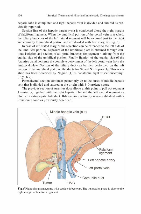
hepatic lobe is completed and right hepatic vein is divided and sutured as pre-viously reported.
Section line of the hepatic parenchyma is conducted along the right marginof falciform ligament. When the umbilical portion of the portal vein is reached,the biliary branches of the left lateral segment will be exposed just to the rightand cranially to umbilical portion and are divided with free margins (Fig. 5).
In case of infiltrated margins the resection can be extended to the left side ofthe umbilical portion. Exposure of the umbilical plate is obtained through cau-tious isolation and section of all portal branches for segment 4 arising from thecranial side of the umbilical portion. Finally ligation of the cranial side of theArantius canal consents the complete detachment of the left portal vein from theumbilical plate. Section of the biliary duct can be then performed on the leftmargin of the umbilical plate, on the ducts for S2 and S3, separately. This oper-ation has been described by Nagino [1] as “anatomic right trisectionectomy”(Figs. 6,7).
Parenchymal section continues posteriorly up to the onset of middle hepaticvein that is divided and sutured at the origin with 4–0 prolene suture.
The previous section of Arantius duct allows at this point to pull out segment1 ventrally, together with the right hepatic lobe and the left medial segment enbloc with extrahepatic bile duct. Bilioenteric continuity is re-established with aRoux-en-Y loop as previously described.
Surgical Treatment of Hilar and Intrahepatic Cholangiocarcinoma 136
Fig. 5 Right trisegmentectomy with caudate lobectomy. The transaction plane is close to theright margin of falciform ligament

Surgical Technique 137
Fig. 6a,b a Complete dissection of the umbilical plate with ligation of the small branches ofsegment 2 (P4 dor). The resection line can be performed on segment B2 and B3 separately.The three lines indicate resection line of the left bile duct in right hepatectomy (A), right tri-sectionectomy (B), and anotomic right trisectionectomy (C). b Numerals indicate the seg-ment bile duct (B) and portal branch (P) according to Couinaud’s hepatic segment. UP,Umbilical portion
Fig. 7 Right trisectionectomy with portal resection (white arrow). B2,B3, bile duct for seg-ment 2 and segment 3
a b
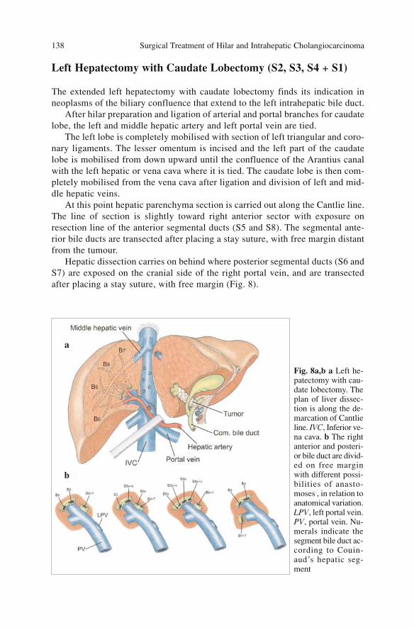
Left Hepatectomy with Caudate Lobectomy (S2, S3, S4 + S1)
The extended left hepatectomy with caudate lobectomy finds its indication inneoplasms of the biliary confluence that extend to the left intrahepatic bile duct.
After hilar preparation and ligation of arterial and portal branches for caudatelobe, the left and middle hepatic artery and left portal vein are tied.
The left lobe is completely mobilised with section of left triangular and coro-nary ligaments. The lesser omentum is incised and the left part of the caudatelobe is mobilised from down upward until the confluence of the Arantius canalwith the left hepatic or vena cava where it is tied. The caudate lobe is then com-pletely mobilised from the vena cava after ligation and division of left and mid-dle hepatic veins.
At this point hepatic parenchyma section is carried out along the Cantlie line.The line of section is slightly toward right anterior sector with exposure onresection line of the anterior segmental ducts (S5 and S8). The segmental ante-rior bile ducts are transected after placing a stay suture, with free margin distantfrom the tumour.
Hepatic dissection carries on behind where posterior segmental ducts (S6 andS7) are exposed on the cranial side of the right portal vein, and are transectedafter placing a stay suture, with free margin (Fig. 8).
Surgical Treatment of Hilar and Intrahepatic Cholangiocarcinoma 138
Fig. 8a,b a Left he-patectomy with cau-date lobectomy. Theplan of liver dissec-tion is along the de-marcation of Cantlieline. IVC, Inferior ve-na cava. b The rightanterior and posteri-or bile duct are divid-ed on free marginwith different possi-bilities of anasto-moses , in relation toanatomical variation.LPV, left portal vein.PV, portal vein. Nu-merals indicate thesegment bile duct ac-cording to Couin-aud’s hepatic seg-ment
a
b

Parenchymal section is completed with the section between right posteriorsegment and caudate lobe that begins down along the ischemic demarcation lineof the caudate process and advances upward along the right margin of the venacava. Dissection and division of caudate from segment 7 is completed.
The left hepatic lobe is then removed together with the caudate lobe andextrahepatic bile duct.
Bilioenteric continuity is re-established with a Roux-en-Y jejunal limb anas-tomosed with biliary ducts (Fig. 9).
Left Trisectionectomy with Caudate Lobectomy (S2, S3, S4, S5,S8 + S1)
The left trisectionectomy with caudate lobectomy is indicated for hilar cholan-giocarcinoma that involves the left intrahepatic bile ducts in continuity with theduct to the right anterior sector (S5 and S8).
After concluding dissection of hepatoduodenal ligament as previouslydescribed, the left and middle hepatic arteries are tied and divided at their ori-gins. The right artery is isolated very gently up to its second order bifurcationfor anterior and posterior sectors. The branch for anterior sector and the small
Surgical Technique 139
Fig. 9 Left hepatectomy. At the end of liver resection three different bile ducts for segmentsB5, B8 and B6+7 are identified
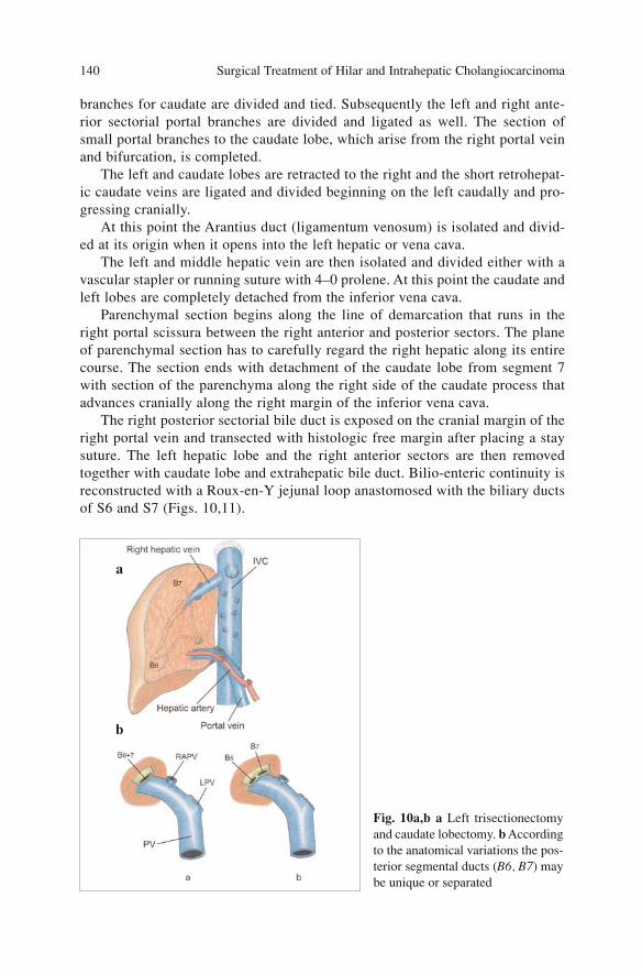
branches for caudate are divided and tied. Subsequently the left and right ante-rior sectorial portal branches are divided and ligated as well. The section ofsmall portal branches to the caudate lobe, which arise from the right portal veinand bifurcation, is completed.
The left and caudate lobes are retracted to the right and the short retrohepat-ic caudate veins are ligated and divided beginning on the left caudally and pro-gressing cranially.
At this point the Arantius duct (ligamentum venosum) is isolated and divid-ed at its origin when it opens into the left hepatic or vena cava.
The left and middle hepatic vein are then isolated and divided either with avascular stapler or running suture with 4–0 prolene. At this point the caudate andleft lobes are completely detached from the inferior vena cava.
Parenchymal section begins along the line of demarcation that runs in theright portal scissura between the right anterior and posterior sectors. The planeof parenchymal section has to carefully regard the right hepatic along its entirecourse. The section ends with detachment of the caudate lobe from segment 7with section of the parenchyma along the right side of the caudate process thatadvances cranially along the right margin of the inferior vena cava.
The right posterior sectorial bile duct is exposed on the cranial margin of theright portal vein and transected with histologic free margin after placing a staysuture. The left hepatic lobe and the right anterior sectors are then removedtogether with caudate lobe and extrahepatic bile duct. Bilio-enteric continuity isreconstructed with a Roux-en-Y jejunal loop anastomosed with the biliary ductsof S6 and S7 (Figs. 10,11).
Surgical Treatment of Hilar and Intrahepatic Cholangiocarcinoma 140
Fig. 10a,b a Left trisectionectomyand caudate lobectomy. b Accordingto the anatomical variations the pos-terior segmental ducts (B6, B7) maybe unique or separated
a
b
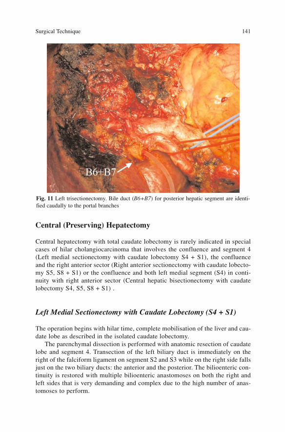
Central (Preserving) Hepatectomy
Central hepatectomy with total caudate lobectomy is rarely indicated in specialcases of hilar cholangiocarcinoma that involves the confluence and segment 4(Left medial sectionectomy with caudate lobectomy S4 + S1), the confluenceand the right anterior sector (Right anterior sectionectomy with caudate lobecto-my S5, S8 + S1) or the confluence and both left medial segment (S4) in conti-nuity with right anterior sector (Central hepatic bisectionectomy with caudatelobectomy S4, S5, S8 + S1) .
Left Medial Sectionectomy with Caudate Lobectomy (S4 + S1)
The operation begins with hilar time, complete mobilisation of the liver and cau-date lobe as described in the isolated caudate lobectomy.
The parenchymal dissection is performed with anatomic resection of caudatelobe and segment 4. Transection of the left biliary duct is immediately on theright of the falciform ligament on segment S2 and S3 while on the right side fallsjust on the two biliary ducts: the anterior and the posterior. The bilioenteric con-tinuity is restored with multiple bilioenteric anastomoses on both the right andleft sides that is very demanding and complex due to the high number of anas-tomoses to perform.
Surgical Technique 141
Fig. 11 Left trisectionectomy. Bile duct (B6+B7) for posterior hepatic segment are identi-fied caudally to the portal branches
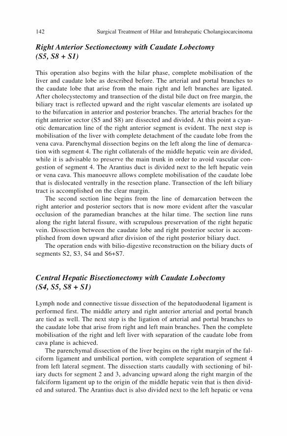
Right Anterior Sectionectomy with Caudate Lobectomy (S5, S8 + S1)
This operation also begins with the hilar phase, complete mobilisation of theliver and caudate lobe as described before. The arterial and portal branches tothe caudate lobe that arise from the main right and left branches are ligated.After cholecystectomy and transection of the distal bile duct on free margin, thebiliary tract is reflected upward and the right vascular elements are isolated upto the bifurcation in anterior and posterior branches. The arterial braches for theright anterior sector (S5 and S8) are dissected and divided. At this point a cyan-otic demarcation line of the right anterior segment is evident. The next step ismobilisation of the liver with complete detachment of the caudate lobe from thevena cava. Parenchymal dissection begins on the left along the line of demarca-tion with segment 4. The right collaterals of the middle hepatic vein are divided,while it is advisable to preserve the main trunk in order to avoid vascular con-gestion of segment 4. The Arantius duct is divided next to the left hepatic veinor vena cava. This manoeuvre allows complete mobilisation of the caudate lobethat is dislocated ventrally in the resection plane. Transection of the left biliarytract is accomplished on the clear margin.
The second section line begins from the line of demarcation between theright anterior and posterior sectors that is now more evident after the vascularocclusion of the paramedian branches at the hilar time. The section line runsalong the right lateral fissure, with scrupulous preservation of the right hepaticvein. Dissection between the caudate lobe and right posterior sector is accom-plished from down upward after division of the right posterior biliary duct.
The operation ends with bilio-digestive reconstruction on the biliary ducts ofsegments S2, S3, S4 and S6+S7.
Central Hepatic Bisectionectomy with Caudate Lobectomy (S4, S5, S8 + S1)
Lymph node and connective tissue dissection of the hepatoduodenal ligament isperformed first. The middle artery and right anterior arterial and portal branchare tied as well. The next step is the ligation of arterial and portal branches tothe caudate lobe that arise from right and left main branches. Then the completemobilisation of the right and left liver with separation of the caudate lobe fromcava plane is achieved.
The parenchymal dissection of the liver begins on the right margin of the fal-ciform ligament and umbilical portion, with complete separation of segment 4from left lateral segment. The dissection starts caudally with sectioning of bil-iary ducts for segment 2 and 3, advancing upward along the right margin of thefalciform ligament up to the origin of the middle hepatic vein that is then divid-ed and sutured. The Arantius duct is also divided next to the left hepatic or vena
Surgical Treatment of Hilar and Intrahepatic Cholangiocarcinoma 142
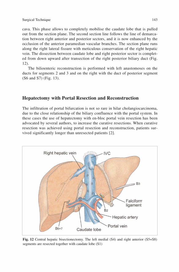
cava. This phase allows to completely mobilise the caudate lobe that is pulledout from the section plane. The second section line follows the line of demarca-tion between right anterior and posterior sectors, and it is now enhanced by theocclusion of the anterior paramedian vascular branches. The section plane runsalong the right lateral fissure with meticulous conservation of the right hepaticvein. The dissection between caudate lobe and right posterior sector is complet-ed from down upward after transection of the right posterior biliary duct (Fig.12).
The bilioenteric reconstruction is performed with left anastomoses on theducts for segments 2 and 3 and on the right with the duct of posterior segment(S6 and S7) (Fig. 13).
Hepatectomy with Portal Resection and Reconstruction
The infiltration of portal bifurcation is not so rare in hilar cholangiocarcinoma,due to the close relationship of the biliary confluence with the portal system. Inthese cases the use of hepatectomy with en-bloc portal vein resection has beenadvocated by several authors, to increase the curative resections. When curativeresection was achieved using portal resection and reconstruction, patients sur-vived significantly longer than unresected patients [2].
Surgical Technique 143
Fig. 12 Central hepatic bisectionectomy. The left medial (S4) and right anterior (S5+S8)segments are resected together with caudate lobe (S1)
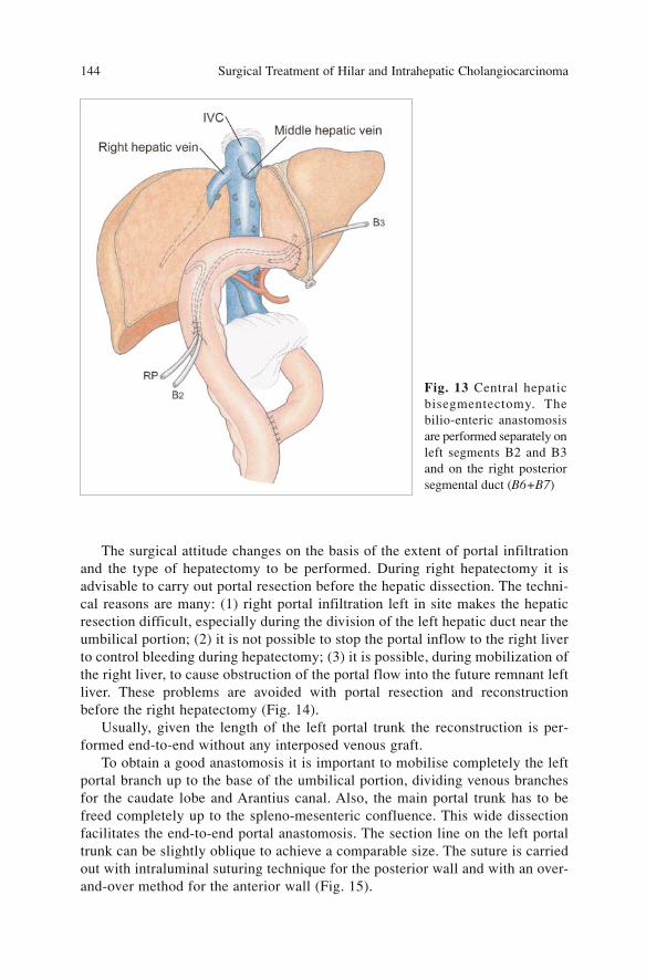
The surgical attitude changes on the basis of the extent of portal infiltrationand the type of hepatectomy to be performed. During right hepatectomy it isadvisable to carry out portal resection before the hepatic dissection. The techni-cal reasons are many: (1) right portal infiltration left in site makes the hepaticresection difficult, especially during the division of the left hepatic duct near theumbilical portion; (2) it is not possible to stop the portal inflow to the right liverto control bleeding during hepatectomy; (3) it is possible, during mobilization ofthe right liver, to cause obstruction of the portal flow into the future remnant leftliver. These problems are avoided with portal resection and reconstructionbefore the right hepatectomy (Fig. 14).
Usually, given the length of the left portal trunk the reconstruction is per-formed end-to-end without any interposed venous graft.
To obtain a good anastomosis it is important to mobilise completely the leftportal branch up to the base of the umbilical portion, dividing venous branchesfor the caudate lobe and Arantius canal. Also, the main portal trunk has to befreed completely up to the spleno-mesenteric confluence. This wide dissectionfacilitates the end-to-end portal anastomosis. The section line on the left portaltrunk can be slightly oblique to achieve a comparable size. The suture is carriedout with intraluminal suturing technique for the posterior wall and with an over-and-over method for the anterior wall (Fig. 15).
Surgical Treatment of Hilar and Intrahepatic Cholangiocarcinoma 144
Fig. 13 Central hepaticbisegmentectomy. Thebilio-enteric anastomosisare performed separately onleft segments B2 and B3and on the right posteriorsegmental duct (B6+B7)
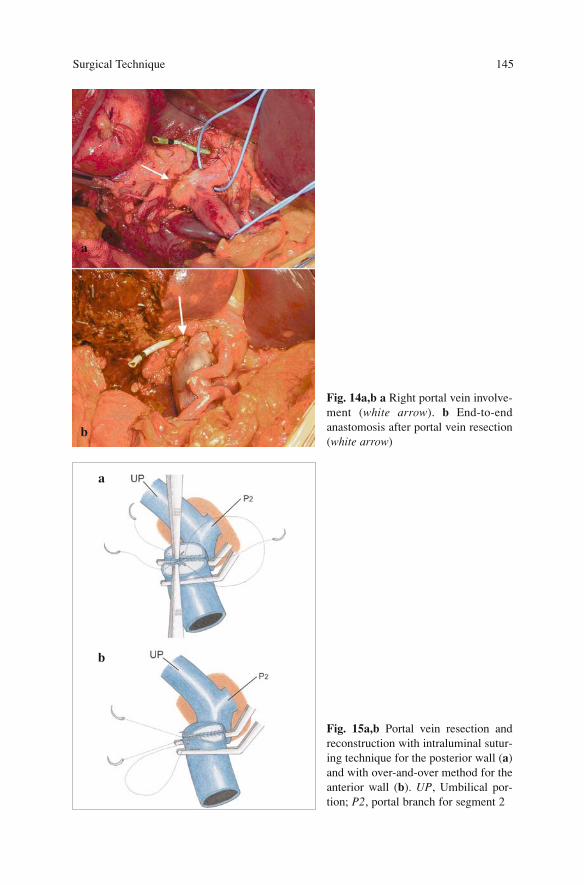
Surgical Technique 145
Fig. 14a,b a Right portal vein involve-ment (white arrow). b End-to-endanastomosis after portal vein resection(white arrow)
Fig. 15a,b Portal vein resection andreconstruction with intraluminal sutur-ing technique for the posterior wall (a)and with over-and-over method for theanterior wall (b). UP, Umbilical por-tion; P2, portal branch for segment 2
a
a
b
b
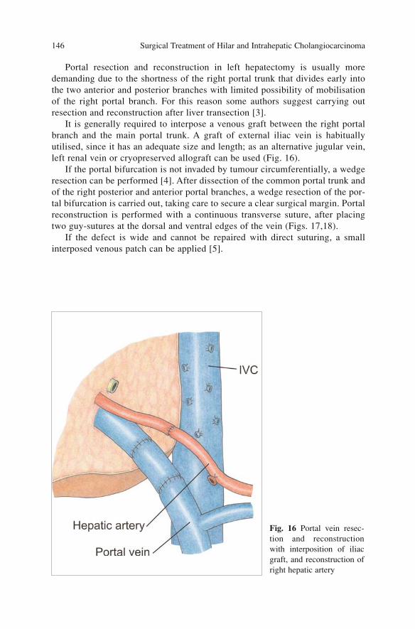
Portal resection and reconstruction in left hepatectomy is usually moredemanding due to the shortness of the right portal trunk that divides early intothe two anterior and posterior branches with limited possibility of mobilisationof the right portal branch. For this reason some authors suggest carrying outresection and reconstruction after liver transection [3].
It is generally required to interpose a venous graft between the right portalbranch and the main portal trunk. A graft of external iliac vein is habituallyutilised, since it has an adequate size and length; as an alternative jugular vein,left renal vein or cryopreserved allograft can be used (Fig. 16).
If the portal bifurcation is not invaded by tumour circumferentially, a wedgeresection can be performed [4]. After dissection of the common portal trunk andof the right posterior and anterior portal branches, a wedge resection of the por-tal bifurcation is carried out, taking care to secure a clear surgical margin. Portalreconstruction is performed with a continuous transverse suture, after placingtwo guy-sutures at the dorsal and ventral edges of the vein (Figs. 17,18).
If the defect is wide and cannot be repaired with direct suturing, a smallinterposed venous patch can be applied [5].
Surgical Treatment of Hilar and Intrahepatic Cholangiocarcinoma 146
Fig. 16 Portal vein resec-tion and reconstructionwith interposition of iliacgraft, and reconstruction ofright hepatic artery
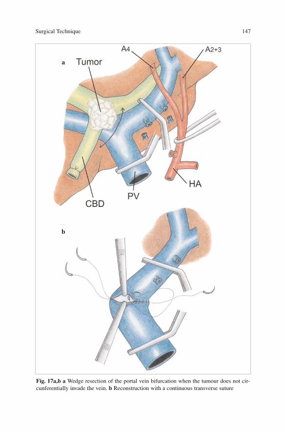
Surgical Technique 147
Fig. 17a,b a Wedge resection of the portal vein bifurcation when the tumour does not cir-cunferentially invade the vein. b Reconstruction with a continuous transverse suture
a
b
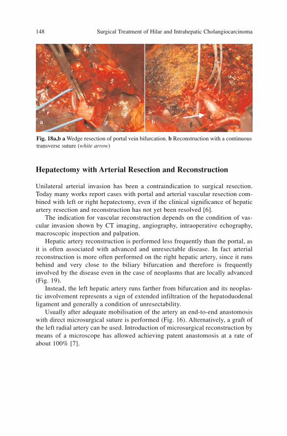
Hepatectomy with Arterial Resection and Reconstruction
Unilateral arterial invasion has been a contraindication to surgical resection.Today many works report cases with portal and arterial vascular resection com-bined with left or right hepatectomy, even if the clinical significance of hepaticartery resection and reconstruction has not yet been resolved [6].
The indication for vascular reconstruction depends on the condition of vas-cular invasion shown by CT imaging, angiography, intraoperative echography,macroscopic inspection and palpation.
Hepatic artery reconstruction is performed less frequently than the portal, asit is often associated with advanced and unresectable disease. In fact arterialreconstruction is more often performed on the right hepatic artery, since it runsbehind and very close to the biliary bifurcation and therefore is frequentlyinvolved by the disease even in the case of neoplasms that are locally advanced(Fig. 19).
Instead, the left hepatic artery runs farther from bifurcation and its neoplas-tic involvement represents a sign of extended infiltration of the hepatoduodenalligament and generally a condition of unresectability.
Usually after adequate mobilisation of the artery an end-to-end anastomosiswith direct microsurgical suture is performed (Fig. 16). Alternatively, a graft ofthe left radial artery can be used. Introduction of microsurgical reconstruction bymeans of a microscope has allowed achieving patent anastomosis at a rate ofabout 100% [7].
Surgical Treatment of Hilar and Intrahepatic Cholangiocarcinoma 148
Fig. 18a,b a Wedge resection of portal vein bifurcation. b Reconstruction with a continuoustransverse suture (white arrow)
a b
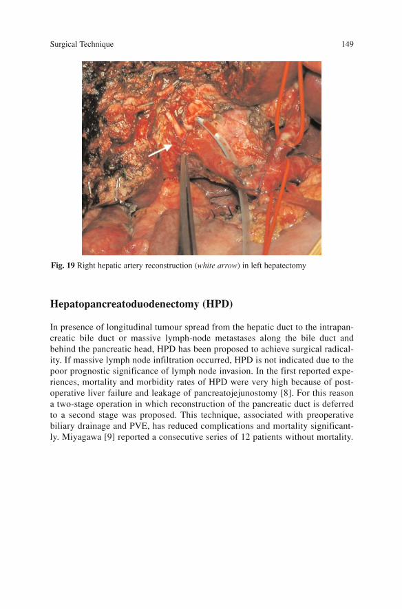
Hepatopancreatoduodenectomy (HPD)
In presence of longitudinal tumour spread from the hepatic duct to the intrapan-creatic bile duct or massive lymph-node metastases along the bile duct andbehind the pancreatic head, HPD has been proposed to achieve surgical radical-ity. If massive lymph node infiltration occurred, HPD is not indicated due to thepoor prognostic significance of lymph node invasion. In the first reported expe-riences, mortality and morbidity rates of HPD were very high because of post-operative liver failure and leakage of pancreatojejunostomy [8]. For this reasona two-stage operation in which reconstruction of the pancreatic duct is deferredto a second stage was proposed. This technique, associated with preoperativebiliary drainage and PVE, has reduced complications and mortality significant-ly. Miyagawa [9] reported a consecutive series of 12 patients without mortality.
Surgical Technique 149
Fig. 19 Right hepatic artery reconstruction (white arrow) in left hepatectomy
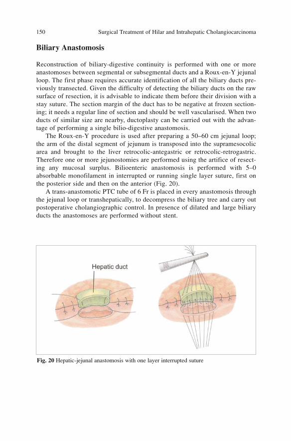
Biliary Anastomosis
Reconstruction of biliary-digestive continuity is performed with one or moreanastomoses between segmental or subsegmental ducts and a Roux-en-Y jejunalloop. The first phase requires accurate identification of all the biliary ducts pre-viously transected. Given the difficulty of detecting the biliary ducts on the rawsurface of resection, it is advisable to indicate them before their division with astay suture. The section margin of the duct has to be negative at frozen section-ing; it needs a regular line of section and should be well vascularised. When twoducts of similar size are nearby, ductoplasty can be carried out with the advan-tage of performing a single bilio-digestive anastomosis.
The Roux-en-Y procedure is used after preparing a 50–60 cm jejunal loop;the arm of the distal segment of jejunum is transposed into the supramesocolicarea and brought to the liver retrocolic-antegastric or retrocolic-retrogastric.Therefore one or more jejunostomies are performed using the artifice of resect-ing any mucosal surplus. Bilioenteric anastomosis is performed with 5–0absorbable monofilament in interrupted or running single layer suture, first onthe posterior side and then on the anterior (Fig. 20).
A trans-anastomotic PTC tube of 6 Fr is placed in every anastomosis throughthe jejunal loop or transhepatically, to decompress the biliary tree and carry outpostoperative cholangiographic control. In presence of dilated and large biliaryducts the anastomoses are performed without stent.
Surgical Treatment of Hilar and Intrahepatic Cholangiocarcinoma 150
Fig. 20 Hepatic-jejunal anastomosis with one layer interrupted suture
References
1. Nagino M, Kamiya J, Arai T et al (2006) “Anatomic” right hepatic trisectionectomy (extend-ed right hepatectomy) with caudate lobectomy for hilar cholangiocarcinoma. Ann Surg243(1):28–32
2. Kondo S, Nimura Y, Hayakawa N et al (2002) Extensive surgery for carcinoma of the gall-bladder. Br J Surg 89(2):179–184
3. Nimura Y, Kamiya J, Kondo S et al (2000) Aggressive preoperative management and extend-ed surgery for hilar cholangiocarcinoma: Nagoya experience. J Hepatobiliary Pancreat Surg7(2):155–162
3. Nagino M, Nimura Y (2006) Perihilar cholangiocarcinoma with emphasis on presurgicalmanagement. In: Blumgart LH (ed) Surgery of the liver, biliary tract, and pancreas, 4th edn.Saunders Elsevier, Philadelphia, pp 804–814
4. Kondo S, Katoh H, Hirano S et al (2002) Wedge resection of the portal bifurcation concomi-tant with left hepatectomy plus biliary reconstruction for hepatobiliary cancer. JHepatobiliary Pancreat Surg 9(5):603–606
5. Nimura Y, Hayakawa N, Kamiya J et al (1991) Combined portal vein and liver resection forcarcinoma of the biliary tract. Br J Surg 78(6):727–731
6. Seyama Y, Makuuchi M (2007) Current surgical treatment for bile duct cancer. World JGastroenterol 13(10):1505–1515
7. Shimada H, Endo I, Sugita M et al (2003) Hepatic resection combined with portal vein orhepatic artery reconstruction for advanced carcinoma of the hilar bile duct and gallbladder.World J Surg 27(10):1137–1142
8. Nimura Y, Hayakawa N, Kamiya J et al (1991) Hepatopancreatoduodenectomy for advancedcarcinoma of the biliary tract. Hepatogastroenterology 38(2):170–175
9. Miyagawa S, Makuuchi M, Kawasaki S et al (1996) Outcome of major hepatectomy withpancreatoduodenectomy for advanced biliary malignancies. World J Surg 20(1):77–80
Surgical Technique 151
Results of Surgery
Morbidity and Mortality
Long-term survival after surgery of hilar cholangiocarcinoma is related topatient status, tumour stage and proper treatment selection.
Principles of surgery for hilar cholangiocarcinoma have changed over the last20 years. Early surgical experiences in the treatment of this disease were limit-ed to isolated bile duct resection. The high recurrence rate (>70%) after thisoperation forced a change in the principles of the surgical management [1].
Current management of hilar cholangiocarcinoma includes resection of theextrahepatic biliary tract associated with major hepatic resection, and the resultshave improved during the last 20 years with a significant decrease in morbidityand mortality (Table 1).
The main reasons for these improvements are: accurate patient selection,optimization of liver function before surgery with preoperative biliary drainageand portal vein embolization, better surgical and anaesthesiological techniquesand postoperative management.
Morbidity after surgical resection of hilar cholangiocarcinoma is still high;major complications occur in about 50% of patients and perioperative mortalityranges from 5 to 18%. Moreover, in recent series no mortality has been report-ed [2,3] (Table 1).
In a retrospective study in 105 patients Nagino observed 81% morbidity rateand 9.4% mortality rate after an aggressive surgical approach (including extend-ed hepatectomies, vascular resections and associated pancreaticoduodenenc-tomies). Minor complications occurred in 37.1% of patients and 43.8% ofpatients suffered from major complications (Table 2).
Several factors are related to postoperative morbidity: jaundice, malnutrition,extent of hepatectomy, complexity of surgical procedure, associated procedures(pancreaticoduodenectomy), blood loss and operative time [2–6].
The relationship between jaundice and postoperative morbidity and mortalityis still matter of debate as previously described, but several authors reported ahigher incidence of postoperative complications in patients with jaundice and/orcholangitis [3,7,8]. Cherqui observed that the postoperative complication rate was50% in jaundiced patients and 15% in the control group [7].
153A. Guglielmi, A. Ruzzenente, C. Iacono (eds.) Surgical Treatment of Hilar and ICC.© Springer 2008
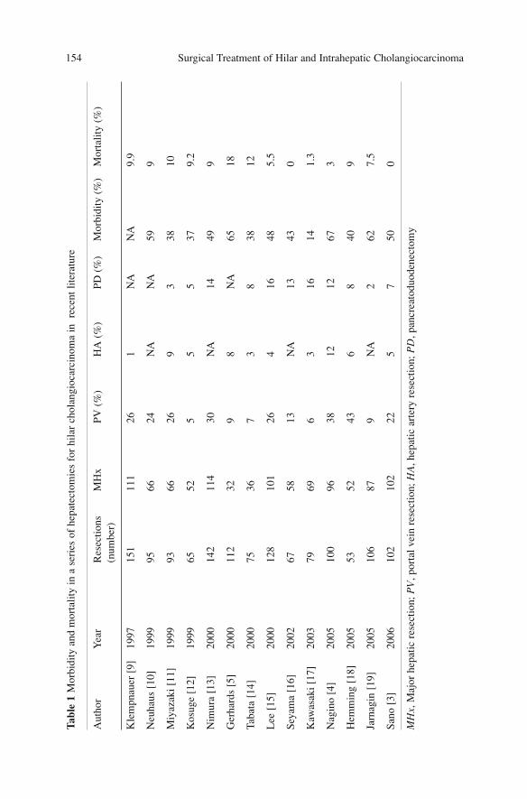
Surgical Treatment of Hilar and Intrahepatic Cholangiocarcinoma 154Ta
ble
1 M
orbi
dity
and
mor
talit
y in
a s
erie
s of
hep
atec
tom
ies
for
hila
r ch
olan
gioc
arci
nom
a in
rec
ent l
itera
ture
Aut
hor
Yea
rR
esec
tions
MH
xPV
(%
)H
A (
%)
PD (
%)
Mor
bidi
ty (
%)
Mor
talit
y (%
)(n
umbe
r)
Kle
mpn
auer
[9]
1997
151
111
261
NA
NA
9.9
Neu
haus
[10
]19
9995
6624
NA
NA
599
Miy
azak
i [11
]19
9993
6626
93
3810
Kos
uge
[12]
1999
6552
55
537
9.2
Nim
ura
[13]
2000
142
114
30N
A14
499
Ger
hard
s [5
]20
0011
232
98
NA
6518
Taba
ta [
14]
2000
7536
73
838
12
Lee
[15
]20
0012
810
126
416
485.
5
Seya
ma
[16]
2002
6758
13N
A13
430
Kaw
asak
i [17
]20
0379
696
316
141.
3
Nag
ino
[4]
2005
100
9638
1212
673
Hem
min
g [1
8]20
0553
5243
68
409
Jarn
agin
[19
]20
0510
687
9N
A2
627.
5
Sano
[3]
2006
102
102
225
750
0
MH
x,M
ajor
hep
atic
res
ectio
n; P
V,p
orta
l vei
n re
sect
ion;
HA
,hep
atic
art
ery
rese
ctio
n; P
D,p
ancr
eato
duod
enec
tom
y
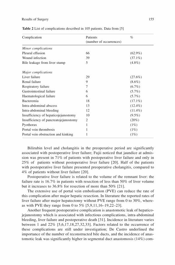
Bilirubin level and cholangitis in the preoperative period are significantlyassociated with postoperative liver failure; Fujii noticed that jaundice at admis-sion was present in 71% of patients with postoperative liver failure and only in25% of patients without postoperative liver failure [20]. Half of the patientswith postoperative liver failure presented preoperative cholangitis, compared to4% of patients without liver failure [20].
Postoperative liver failure is related to the volume of the remnant liver: thefailure rate is 16.7% in patients with resection of less than 50% of liver volumebut it increases to 36.8% for resection of more than 50% [21].
The extensive use of portal vein embolisation (PVE) can reduce the rate ofthis complication after major hepatic resection. In literature the reported rates ofliver failure after major hepatectomy without PVE range from 0 to 30%, where-as with PVE they range from 0 to 5% [5,9,11,16–19,22–23].
Another frequent postoperative complication is anastomotic leak of hepatico-jejunostomy which is associated with infectious complications, intra-abdominalbleeding, liver failure and postoperative death [31]. Incidence in literature variesbetween 1 and 22% [5,6,17,18,27,32,33]. Factors related to the occurrence ofthese complications are still under investigation; De Castro underlined theimportance of the number of reconstructed bile ducts, and the incidence of anas-tomotic leak was significantly higher in segmental duct anastomosis (14%) com-
Results of Surgery 155
Table 2 List of complications described in 105 patients. Data from [5]
Complication Patients %(number of occurrences)
Minor complicationsPleural effusion 66 (62.9%)Wound infection 39 (37.1%)Bile leakage from liver stump 5 (4.8%)
Major complicationsLiver failure 29 (27.6%)Renal failure 9 (8.6%)Respiratory failure 7 (6.7%)Gastrointestinal failure 6 (5.7%)Haematological failure 6 (5.7%)Bacteremia 18 (17.1%)Intra-abdominal abscess 13 (12.4%)Intra-abdominal bleeding 12 (11.4%)Insufficiency of hepaticojejunostomy 10 (9.5%)Insufficiency of pancreatojejunostomy 2 (20%)Pyothorax 1 (1%)Portal vein thrombosis 1 (1%)Portal vein obstruction and kinking 1 (1%)
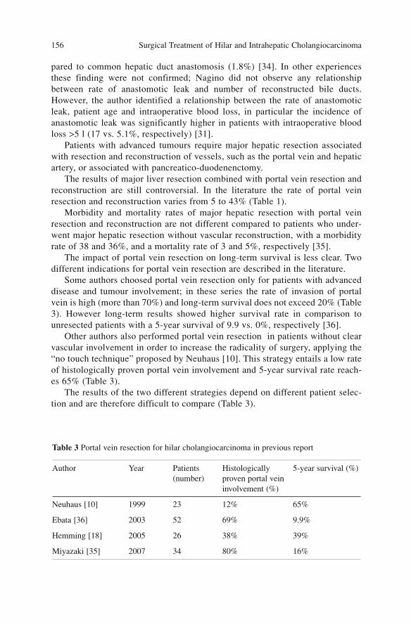
pared to common hepatic duct anastomosis (1.8%) [34]. In other experiencesthese finding were not confirmed; Nagino did not observe any relationshipbetween rate of anastomotic leak and number of reconstructed bile ducts.However, the author identified a relationship between the rate of anastomoticleak, patient age and intraoperative blood loss, in particular the incidence ofanastomotic leak was significantly higher in patients with intraoperative bloodloss >5 l (17 vs. 5.1%, respectively) [31].
Patients with advanced tumours require major hepatic resection associatedwith resection and reconstruction of vessels, such as the portal vein and hepaticartery, or associated with pancreatico-duodenenctomy.
The results of major liver resection combined with portal vein resection andreconstruction are still controversial. In the literature the rate of portal veinresection and reconstruction varies from 5 to 43% (Table 1).
Morbidity and mortality rates of major hepatic resection with portal veinresection and reconstruction are not different compared to patients who under-went major hepatic resection without vascular reconstruction, with a morbidityrate of 38 and 36%, and a mortality rate of 3 and 5%, respectively [35].
The impact of portal vein resection on long-term survival is less clear. Twodifferent indications for portal vein resection are described in the literature.
Some authors choosed portal vein resection only for patients with advanceddisease and tumour involvement; in these series the rate of invasion of portalvein is high (more than 70%) and long-term survival does not exceed 20% (Table3). However long-term results showed higher survival rate in comparison tounresected patients with a 5-year survival of 9.9 vs. 0%, respectively [36].
Other authors also performed portal vein resection in patients without clearvascular involvement in order to increase the radicality of surgery, applying the“no touch technique” proposed by Neuhaus [10]. This strategy entails a low rateof histologically proven portal vein involvement and 5-year survival rate reach-es 65% (Table 3).
The results of the two different strategies depend on different patient selec-tion and are therefore difficult to compare (Table 3).
Surgical Treatment of Hilar and Intrahepatic Cholangiocarcinoma 156
Table 3 Portal vein resection for hilar cholangiocarcinoma in previous report
Author Year Patients Histologically 5-year survival (%)(number) proven portal vein
involvement (%)
Neuhaus [10] 1999 23 12% 65%
Ebata [36] 2003 52 69% 9.9%
Hemming [18] 2005 26 38% 39%
Miyazaki [35] 2007 34 80% 16%
Few studies in the literature evaluate the role of hepatic artery resection andreconstruction. In surgical series less than 10% of patients were subjected to thistype of vascular reconstruction and there is no consensus regarding the criteriaof resectability for patients requiring hepatic artery resection or combined hepat-ic artery and portal vein resection and reconstruction. The postoperative morbid-ity and mortality for patients submitted to hepatic artery reconstruction is high.Miyazaki observed a 78% morbidity rate in the hepatic artery resection groupcompared to 38% in patients with portal vein resection alone and 36% in patientswithout vascular resection [35]. In this series, 77% of the patients who under-went hepatic artery resection had combined resection of the portal vein.
The high operative mortality rate after hepatic artery resection (30–50%) canbe justified by the advanced stage of the tumours and the complexity of surgicalinterventions [5,33,35]. However, in other recent studies hepatic artery resectionand reconstruction were carried out with postoperative mortality lower than 10%[37–39]. Long-term survival after hepatic artery resection for hilar cholangiocar-cinoma is still under evaluation. In 9 patients Miyazaki reported 1- and 3- yearsurvival rates of 11 and 0% [35].
The association of liver resection with pancreaticoduodenectomy (PD)increases the rate of surgical complications; Sano observed postoperative com-plications in more than 85% of patients with associated PD, and Nagino report-ed an incidence of liver failure of 50% in patients with combined PD in compar-ison to 25% in patients without PD [3,10]. Two stage strategies with delayedpancreaticoduodenectomy have been shown to reduce significantly the morbidi-ty of this procedure with 0% postoperative mortality [40].
Long-term Results
Survival after resection of hilar cholangiocarcinoma has improved in the lastdecades with a 5-year survival rate ranging from 12 to 40% (Table 4).
Several prognostic factors have been identified after surgical resection ofhilar cholangiocarcinoma: gross type, local extent, lymph-node involvement,distant metastases, vascular invasion, perineural invasion and radicality of sur-gery. Among these, R0 resection is the strongest factor identified in both univari-ate and multivariate analyses [10,12,19,27].
Neuhaus reported 5-year survival rate in R0 resections of 39%, comparedwith 9, and 0% for R1 and R2 resections. In R2 resection no patients survivedmore than 22 months. In a large series of 400 patients, Nishio confirmed thesedata and reported a 5-year survival rate of 27 and 2% for curative and non-cur-ative resection, respectively [41].
To achieve radicality with safe margin, resection of the bile duct associatedwith hepatectomy, caudate lobectomy and regional lymphadenectomy arerequired.
Results of Surgery 157
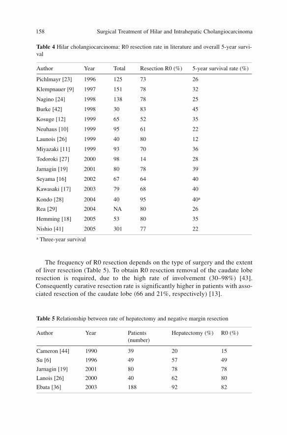
The frequency of R0 resection depends on the type of surgery and the extentof liver resection (Table 5). To obtain R0 resection removal of the caudate loberesection is required, due to the high rate of involvement (30–98%) [43].Consequently curative resection rate is significantly higher in patients with asso-ciated resection of the caudate lobe (66 and 21%, respectively) [13].
Surgical Treatment of Hilar and Intrahepatic Cholangiocarcinoma 158
Table 4 Hilar cholangiocarcinoma: R0 resection rate in literature and overall 5-year survi-val
Author Year Total Resection R0 (%) 5-year survival rate (%)
Pichlmayr [23] 1996 125 73 26
Klempnauer [9] 1997 151 78 32
Nagino [24] 1998 138 78 25
Burke [42] 1998 30 83 45
Kosuge [12] 1999 65 52 35
Neuhaus [10] 1999 95 61 22
Launois [26] 1999 40 80 12
Miyazaki [11] 1999 93 70 36
Todoroki [27] 2000 98 14 28
Jarnagin [19] 2001 80 78 39
Seyama [16] 2002 67 64 40
Kawasaki [17] 2003 79 68 40
Kondo [28] 2004 40 95 40a
Rea [29] 2004 NA 80 26
Hemming [18] 2005 53 80 35
Nishio [41] 2005 301 77 22
a Three-year survival
Table 5 Relationship between rate of hepatectomy and negative margin resection
Author Year Patients Hepatectomy (%) R0 (%)(number)
Cameron [44] 1990 39 20 15
Su [6] 1996 49 57 49
Jarnagin [19] 2001 80 78 78
Lanois [26] 2000 40 62 80
Ebata [36] 2003 188 92 82
The type of hepatic resection contributes to determining curative rate andlong-term outcome; Neuhaus reported a significantly higher rate of curativeresection in patients submitted to right-sided hepatectomy in comparison withleft hepatectomy, 71 vs. 33% [10]. Moreover, Kondo confirmed these resultswith a significantly higher survival rate for patients submitted to right hepatec-tomy in comparison to left hepatectomy, isolated caudate lobe resection and iso-lated bile duct resection [28].
Evaluation of the margin of the biliary duct is an important issue of curativesurgery for hilar cholangiocarcinoma. A positive ductal margin influences thesurvival; in presence of negative margin 5-year survival rate is 46–56% vs. no 5-year survivors in presence of positive margin [45,46]. However, the presence ofnon-invasive carcinoma at the surgical margin does not influence long-term out-come, with 5- and 10-year survival rates of 69 and 23% [45].
Diffusion of the disease to regional lymph nodes is frequent in hilar cholan-giocarcinoma (30–50%) and is one of the main determinants of prognosis aftersurgical resection [10,11,25,47–50].
Node-negative patients show 5-year survival rate of 31%, compared with10% for those with regional lymph-node metastases [41]. Metastases to para-aortic lymph nodes can be found in 17% of patients; survival for these patientsis significantly lower than for negative-lymph node patients, with 5-year survivalrate of 12 and 30%, respectively.
Recurrence
Recurrence of the disease is frequent (22–60%) and is usually the cause of death[3,13,17,28]. The most frequent sites of recurrence are the peritoneum (50%),the remnant liver (22%) and, less frequently, local lymph nodes [28,51]. Themedian disease-free survival time is short (19 months).
The recurrence rate after curative surgery is higher in patients subjected toisolated resection of the bile duct (75%) in comparison to associated biliary andliver resection (52%) [13] (Table 6).
Anastomotic recurrence occurs in 8% of cases after curative resection and isrelated to the length of biliary tumour-free resection margin: Sakamotodescribed a recurrence rate of 18% for a tumour-free resection margin <2.5 mm,and of 10% for margins 2.5–5 mm; furthermore, he did not observe anastomot-ic recurrences when the margin was >5 mm [52].
Results of Surgery 159
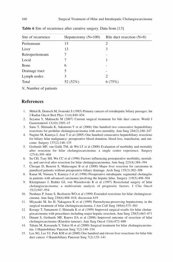
References
1. Mittal B, Deutsch M, Iwatsuki S (1985) Primary cancers of extrahepatic biliary passages. IntJ Radiat Oncol Biol Phys 11(4):849–854
2. Seyama Y, Makuuchi M (2007) Current surgical treatment for bile duct cancer. World JGastroenterol 13(10):1505–15
3. Sano T, Shimada K, Sakamoto Y et al (2006) One hundred two consecutive hepatobiliaryresections for perihilar cholangiocarcinoma with zero mortality. Ann Surg 244(2):240–247
4. Nagino M, Kamiya J, Arai T et al (2005) One hundred consecutive hepatobiliary resectionsfor biliary hilar malignancy: preoperative blood donation, blood loss, transfusion, and out-come. Surgery 137(2):148–155
5. Gerhards MF, van Gulik TM, de Wit LT et al (2000) Evaluation of morbidity and mortalityafter resection for hilar cholangiocarcinoma: a single center experience. Surgery127(4):395–404
6. Su CH, Tsay SH, Wu CC et al (1996) Factors influencing postoperative morbidity, mortali-ty, and survival after resection for hilar cholangiocarcinoma. Ann Surg 223(4):384–394
7. Cherqui D, Benoist S, Malassagne B et al (2000) Major liver resection for carcinoma injaundiced patients without preoperative biliary drainage. Arch Surg 135(3):302–308
8. Kanai M, Nimura Y, Kamiya J et al (1996) Preoperative intrahepatic segmental cholangitisin patients with advanced carcinoma involving the hepatic hilus. Surgery 119(5):498–504
9. Klempnauer J, Ridder GJ, von Wasielewski R et al (1997) Resectional surgery of hilarcholangiocarcinoma: a multivariate analysis of prognostic factors. J Clin Oncol15(3):947–954
10. Neuhaus P, Jonas S, Bechstein WO et al (1999) Extended resections for hilar cholangiocar-cinoma. Ann Surg 230(6):808–818; discussion 819
11. Miyazaki M, Ito H, Nakagawa K et al (1999) Parenchyma-preserving hepatectomy in thesurgical treatment of hilar cholangiocarcinoma. J Am Coll Surg 189(6):575–583
12. Kosuge T, Yamamoto J, Shimada K et al (1999) Improved surgical results for hilar cholan-giocarcinoma with procedures including major hepatic resection. Ann Surg 230(5):663–671
13. Dinant S, Gerhards MF, Rauws EA et al (2006) Improved outcome of resection of hilarcholangiocarcinoma (Klatskin tumour). Ann Surg Oncol 13(6):872–880
14. Tabata M, Kawarada Y, Yokoi H et al (2000) Surgical treatment for hilar cholangiocarcino-ma. J Hepatobiliary Pancreat Surg 7(2):148–154
15. Lee SG, Lee YJ, Park KM et al (2000) One hundred and eleven liver resections for hilar bileduct cancer. J Hepatobiliary Pancreat Surg 7(2):135–141
Surgical Treatment of Hilar and Intrahepatic Cholangiocarcinoma 160
Table 6 Site of recurrence after curative surgery. Data from [13]
Site of recurrence Hepatectomy (N=100) Bile duct resection (N=8)
Peritoneum 15 2Liver 13 3Retroperitoneum 7 -Local 7 1Bone 6 -Drainage tract 5 -Lymph nodes 3 2Total 52 (52%) 6 (75%)
N, Number of patients
16. Seyama Y, Makuuchi M, Sano K et al (2002) Intermittent total vascular exclusion in remov-ing caudate lobe tumour with tumour thrombus in the vena cava. Surgery 131(5):574–576
17. Kawasaki S, Imamura H, Kobayashi A et al (2003) Results of surgical resection for patientswith hilar bile duct cancer: application of extended hepatectomy after biliary drainage andhemihepatic portal vein embolization. Ann Surg 238(1):84–92
18. Hemming AW, Reed AI, Fujita S et al (2005) Surgical management of hilar cholangiocarci-noma. Ann Surg 241(5):693–699; discussion 699–702
19. Jarnagin WR, Fong Y, DeMatteo RP et al (2001) Staging, resectability, and outcome in 225patients with hilar cholangiocarcinoma. Ann Surg 234(4):507–517; discussion 517–519
20. Fujii Y, Shimada H, Endo I et al (2003) Risk factors of posthepatectomy liver failure afterportal vein embolization. J Hepatobiliary Pancreat Surg 10(3):226–232
21. Nagino M, Kamiya J, Uesaka K et al (2001)Complications of hepatectomy for hilar cholan-giocarcinoma. World J Surg 25(10):1277–1283
22. Miyagawa S, Makuuchi M, Kawasaki S (1995) Outcome of extended right hepatectomyafter biliary drainage in hilar bile duct cancer. Arch Surg 130(7):759–763
23. Pichlmayr R, Weimann A, Klempnauer J et al (1996) Surgical treatment in proximal bileduct cancer. A single-center experience. Ann Surg 224(5):628–638
24. Nagino M, Nimura Y, Kamiya J et al (1998) Segmental liver resections for hilar cholangio-carcinoma. Hepatogastroenterology 45(19):7–13
25. Ogura Y, Kawarada Y (1998) Surgical strategies for carcinoma of the hepatic duct conflu-ence. Br J Surg 85(1):20–24
26. Launois B, Terblanche J, Lakehal M et al (1999) Proximal bile duct cancer: high resectabil-ity rate and 5-year survival. Ann Surg 230:266–275
27. Todoroki T, Kawamoto T, Koike N et al (2000) Radical resection of hilar bile duct carcino-ma and predictors of survival. Br J Surg 87(3):306–313
28. Kondo S, Hirano S, Ambo Y et al (2004) Forty consecutive resections of hilar cholangiocar-cinoma with no postoperative mortality and no positive ductal margins: results of a prospec-tive study. Ann Surg 240(1):95–101
29. Rea DJ, Munoz-Juarez M, Farnell MB et al (2004) Major hepatic resection for hilar cholan-giocarcinoma: analysis of 46 patients. Arch Surg 139(5):514–523; discussion 523–525
30. Seyama Y, Makuuchi M (2007) Current surgical treatment for bile duct cancer. World JGastroenterol 13(10):1505–1515
31. Nagino M, Nishio H, Ebata T et al (2007) Intrahepatic cholangiojejunostomy followinghepatobiliary resection. Br J Surg 94(1):70–77
32. Miyazaki M, Ito H, Nakagawa K et al (1998) Aggressive surgical approaches to hilar cholan-giocarcinoma: hepatic or local resection? Surgery 123(2):131–136
33. Madariaga JR, Iwatsuki S, Todo S et al (1998) Liver resection for hilar and peripheralcholangiocarcinomas: a study of 62 cases. Ann Surg 227(1):70–79
34. de Castro SM, Kuhlmann KF, Busch OR et al (2005) Incidence and management of biliaryleakage after hepaticojejunostomy. J Gastrointest Surg 9(8):1163–1171; discussion1171–1173
35. Miyazaki M, Kato A, Ito H et al (2007) Combined vascular resection in operative resectionfor hilar cholangiocarcinoma: does it work or not? Surgery 141(5):581–588
36. Ebata T, Nagino M, Kamiya J et al (2003) Hepatectomy with portal vein resection for hilarcholangiocarcinoma: audit of 52 consecutive cases. Ann Surg 238(5):720–727
37. Yamanaka N, Yasui C, Yamanaka J et al (2001) Left hemihepatectomy with microsurgicalreconstruction of the right-sided hepatic vasculature. A strategy for preserving hepatic func-tion in patients with proximal bile duct cancer. Langenbecks Arch Surg 386(5):364–368
38. Sakamoto Y, Sano T, Shimada K et al (2006) Clinical significance of reconstruction of theright hepatic artery for biliary malignancy. Langenbecks Arch Surg 391(3):203–208
39. Shimada M, Hamatsu T, Yamashita Y et al (2001) Characteristics of multicentric hepatocel-lular carcinomas: comparison with intrahepatic metastasis. World J Surg 25(8):991–995
40. Miyagawa S, Makuuchi M, Kawasaki S et al (1996) Outcome of major hepatectomy withpancreatoduodenectomy for advanced biliary malignancies. World J Surg 20(1):77–80
Results of Surgery 161
41. Nishio H, Nagino M, Nimura Y (2005) Surgical management of hilar cholangiocarcinoma:the Nagoya experience. HPB 7:259–262
42. Burke EC, Jarnagin WR, Hochwald SN et al (1998) Hilar cholangiocarcinoma: patterns ofspread, the importance of hepatic resection for curative operation, and a presurgical clinicalstaging system. Ann Surg 228: 385–394
43. Dinant S, Gerhards MF, Busch OR et al (2005) The importance of complete excision of thecaudate lobe in resection of hilar cholangiocarcinoma. HPB 7:263–267
44. Cameron JL, Pitt HA, Zinner MJ et al (1990) Management of proximal cholangiocarcino-mas by surgical resection and radiotherapy. Am J Surg 159(1):91–97; discussion 97–98
45. Nimura Y, Kamiya J, Kondo S et al (2000) Aggressive preoperative management and extend-ed surgery for hilar cholangiocarcinoma: Nagoya experience. J Hepatobiliary Pancreat Surg7(2):155–162
46. Wakai T, Shirai Y, Moroda T et al (2005) Impact of ductal resection margin status on long-term survival in patients undergoing resection for extrahepatic cholangiocarcinoma. Cancer103:1210–1216
47. Nakeeb A, Pitt HA, Sohn TA et al (1996) Cholangiocarcinoma. A spectrum of intrahepatic,perihilar, and distal tumours. Ann Surg 224(4):463–473; discussion 473–475
48. Sugiura Y, Nakamura S, Iida S et al (1994) Extensive resection of the bile ducts combinedwith liver resection for cancer of the main hepatic duct junction: a cooperative study of theKeio Bile Duct Cancer Study Group. Surgery 115(4):445–451
49. Iwatsuki S, Todo S, Marsh JW et al (1998) Treatment of hilar cholangiocarcinoma (Klatskintumours) with hepatic resection or transplantation. J Am Coll Surg 187(4):358–364
50. Kitagawa Y, Nagino M, Kamiya J et al (2001) Lymph-node metastasis from hilar cholangio-carcinoma: audit of 110 patients who underwent regional and paraaortic node dissection.Ann Surg 233(3):385–392
51. Liu CL, Fan ST, Lo CM et al (2006) Improved operative and survival outcomes of surgicaltreatment for hilar cholangiocarcinoma. Br J Surg 93(12):1488–1494
52. Sakamoto E, Nimura Y, Hayakawa N et al (1998) The pattern of infiltration at the proximalborder of hilar bile duct carcinoma: a histologic analysis of 62 resected cases. Ann Surg227(3):405–411
Surgical Treatment of Hilar and Intrahepatic Cholangiocarcinoma 162
The Role of Liver Transplantation
Indications and Results
Curative resection is the only treatment that produces good results in hilarcholangiocarcinoma. Hepatic transplantation has the theoretical premise ofincreasing surgical radicality and offers a treatment with curative intent forpatients who are excluded from surgical treatment due to advanced stages of thedisease or the presence of hepatic disease that contraindicates hepatic resection.
The indications for transplantation are not clearly defined in the literaturesince the studies are limited to a few cases in each institution and at present noabsolute indications for this disease are ascertained (Table 1).
In the early studies, transplantation was performed in very advanced neo-plasms with poor results: 3-year survival was 20% and recurrence rate was 57%[1]. Even more recent experiences report similar results: at the CincinnatiTransplant Tumour Registry, in 207 patients Meyer observed 5-year survival of23% with a recurrence rate of more than 50% of the transplanted patients [2]. Ina Spanish multicenter survey Robles reported 3-year survival of 30% with a 5-year disease-free survival of 30% [3].
Incidental cholangiocarcinoma revealed during transplantation for primarysclerosing cholangitis (PSC) represents about 8% of all the transplanted casesfor PSC [4]. The localization is in the extrahepatic biliary tract in more than 80%of the cases [5].
The results of transplantation for patients with incidental hilar cholangiocar-cinoma seem to be better than those for patients with a preoperative diagnosis ofcholangiocarcinoma. Goss has described 10 cases with 1-year, 3-year and 5-yearsurvival of 100, 83 and 83%, respectively, while none of the three patients withpreoperative diagnosis survived more than 3 years [4]; other studies confirmthese reports. Abu-Elmagd shows a 2-year survival of 55% for incidental cholan-giocarcinoma, compared to 29% of the cases ascertained preoperatively [6].
Many prognostic survival factors have been identified in the literature. Thedepth of invasion of the biliary duct, determined by T-stage according to theUICC (5th edition), is related to prognosis; Iwatsuki reports a median survival
163A. Guglielmi, A. Ruzzenente, C. Iacono (eds.) Surgical Treatment of Hilar and ICC.© Springer 2008
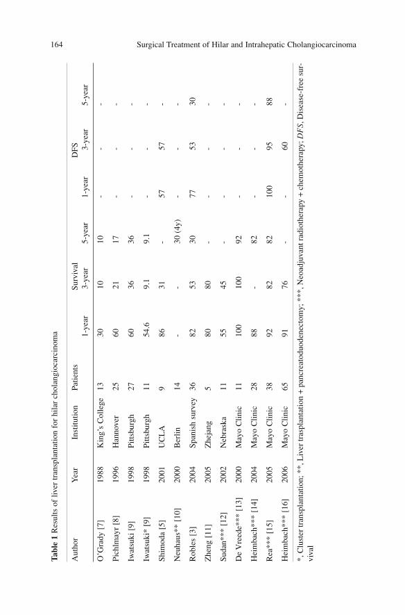
Surgical Treatment of Hilar and Intrahepatic Cholangiocarcinoma 164Ta
ble
1 R
esul
ts o
f liv
er tr
ansp
lant
atio
n fo
r hi
lar
chol
angi
ocar
cino
ma
Aut
hor
Yea
rIn
stitu
tion
Patie
nts
Surv
ival
DFS
1-ye
ar3-
year
5-ye
ar1-
year
3-ye
ar5-
year
O’G
rady
[7]
1988
Kin
g’s
Col
lege
1330
1010
--
-
Pich
lmay
r [8
]19
96H
anno
ver
2560
2117
--
-
Iwat
suki
[9]
1998
Pitts
burg
h27
6036
36-
--
Iwat
suki
* [9
]19
98Pi
ttsbu
rgh
1154
.69.
19.
1-
--
Shim
oda
[5]
2001
UC
LA
986
31-
5757
-
Neu
haus
** [
10]
2000
Ber
lin14
--
30 (
4y)
--
-
Rob
les
[3]
2004
Span
ish
surv
ey
3682
5330
7753
30
Zhe
ng [
11]
2005
Zhe
jang
5
8080
--
--
Suda
n***
[12
]20
02N
ebra
ska
1155
45-
--
-
De
Vre
ede*
** [
13]
2000
May
o C
linic
1110
010
092
--
-
Hei
mba
ch**
* [1
4]20
04M
ayo
Clin
ic28
88-
82-
--
Rea
***
[15]
2005
May
o C
linic
3892
8282
100
9588
Hei
mba
ch**
* [1
6]20
06M
ayo
Clin
ic65
9176
--
60-
*,C
lust
er tr
ansp
lant
atio
n; *
*,L
iver
tras
plan
tatio
n +
pan
crea
todu
oden
ecto
my;
***
, Neo
adju
vant
rad
ioth
erap
y +
che
mot
hera
py; D
FS ,
Dis
ease
-fre
e su
r-vi
val
of 99 months for patients in stage T1 and T2, and of 12 months in T3 [9]. In hisexperience, all the patients with a survival longer than 5 years had early stagedisease (T1 and T2) without lymph-node involvement (Stage I and II accordingto TNM UICC). In 25 patients, Pichlmayr observed a median survival of 37months for stage I and II patients, of 20 months for stage III patients and only 5.8months for stages IVa and IVb. In his experience, only the patients in stages I andII had a survival longer than 3 years. The univariate and multivariate analyses haveconfirmed the prognostic value of TNM staging system with a RR of 4.5 [8].Another prognostic factor is vascular invasion; Robles reports a survival of 0 and63% in the patients with and without vascular invasion, respectively [3]. The sameauthor has also considered the prognostic value of perineural invasion, with amedian survival of 37 months in the presence and 78 months in absence of inva-sion, and lymphatic invasion with a median survival of 28 months in the presenceand 65 months in absence of invasion, respectively. Other authors have underlinedthe importance of lymphatic invasion as prognostic factor as well; Iwatsuki reportsa 5-year survival of 0% in N+ patients vs. 27% in N- patients [9]. Recurrence ofdisease is quite common, with frequency that varies from 40 to 60%[2,3,7,9,17–19]. Recurrences are usually early; Meyer observed 84% of recur-rences in the first two years, localized in the hepatic hilum, transplanted liver andperipancreatic area [2,3,9]. The prognosis of the patients with recurrence is fairlypoor, with a median survival after recurrence not longer than 3 months [3].
Combined Transplantation
As recurrence is more frequent at the hepatic and pancreatic area, some authorshave proposed alternative surgical strategies, extending resection to the entirebiliary tract and combining hepatic transplantation with pancreatoduodenectomy[20]. This approach increases the margins of resection, does not necessitate dis-secting the structures adjacent to the neoplasm and allows an extended lym-phadenectomy. In 14 patients, Neuhaus obtained a curative resection in morethan 90% of the cases, with an operative mortality of 14%. Unfortunately, thelong-term results are not satisfactory, with a 4-year survival of 30%. The mostfrequent cause of death is neoplastic recurrence, with implantation metastases inhalf of the cases [10].
Other authors have described similar approaches, but despites the significantincrease of surgical radicality compared to resective surgery, long-term outcomeis unsatisfactory [21,22].
Starzl has proposed a more aggressive operation combining the resection ofall the organs derived from the foregut (liver, stomach, spleen, pancreaticoduo-denal complex) with subsequent cluster transplantation (liver, pancreas, duode-num) or with isolated hepatic transplantation [1]. This type of operation has beendefined as upper abdominal exenteration. The experience of the Pittsburg groupin 11 patients showed a high postoperative mortality (18%); in spite of the high-
The Role of Liver Transplantation 165
er oncologic radicality of cluster transplantation, with clear margins in morethan 90% of the operations, the long-term results were poor, with 3-year and 5-year survival of 9.1% [9].
Transplantation with Adjuvant and Neoadjuvant Treatments
In hopes of improving the poor results of transplantation different protocols ofadjuvant and neoadjuvant therapy with chemo- and radiation therapy have beenproposed.
In 1998 Iwatsuki reported the results in 18 patients of adjuvant therapy withdifferent protocols combining 5-FU chemotherapy with external radiotherapy;he showed a better long-term survival in the patients who underwent adjuvanttreatment with median survival of 16.7 months vs 12.3 months in the group with-out adjuvant therapy [9].
The studies in the literature fail to show a clear utility of adjuvant treatmentand do not support evidence that the treatment is useful to modify the prognosisafter transplantation [19,23].
Even the experiences of intraoperative radiotherapy (IORT) are limited toisolated cases; Sotiropoulos reports a survival longer than 10 years in a patientwho underwent hepatic transplantation with IORT for hilar cholangiocarcinoma.In literature no other significant experiences of such treatment associated withtransplantation are reported.
More recently two protocols that combined neoadjuvant chemotherapy withradiotherapy in transplantation have been proposed.
The protocol proposed by the University of Nebraska implies neoadjuvanttreatment with Ir-192 biliary catheters brachytherapy for a total dose of 6000cGyassociated with chemotherapy with continuous infusion of 5-FU (daily dose 300mg/m2). The treatment is continued until transplantation. In this initial experienceonly patients who were excluded from surgical resection due to the stage of thedisease or presence of cirrhosis were included in this study. Six of the 17 recruit-ed patients (35%) were excluded from transplantation since the onset of compli-cations or neoplasm progression while on the waiting list. The 11 patients whounderwent transplantation showed a mortality rate of 27% and 3-year survival of45%; two patients (18%) presented recurrence of disease during follow-up [12].
The second trial proposed by the Mayo Clinic includes patients who wereunresectable due to the stage of the disease or concomitant cirrhosis, and isbased on external and intraluminal radiation therapy associated with chemother-apy. External beam-irradiation is delivered in a total dose of 4000–4500 cGy in27–30 fractions and to complete the radiation therapy a boost with trans-catheterirradiation is performed after 2–3 weeks. Brachytherapy is delivered using irid-ium-192 wire with a total dose of 2000–3000 cGy. During radiotherapy andbefore transplantation 5-FU chemotherapy is administered (daily dose of225/500 mg/m2); more recently chemotherapy in patients on the waiting list has
Surgical Treatment of Hilar and Intrahepatic Cholangiocarcinoma 166
been modified using capecitabine orally (2000 mg/m2/die, twice every threeweeks). All the patients included in the study undergo exploratory laparotomy toverify the absence of local contraindications (peritoneal carcinomatosis, lymph-node metastases) for transplantation after completion of radiation therapy andjust before admittance in the waiting list [13–15]. In a recent experience in 106patients, only 65 (61%) underwent transplantation while 11 patients wereexcluded from the protocol during neoadjuvant treatment because of progressionof disease or onset of severe complications, and 18 were excluded during laparo-tomy due to the advanced stage of the disease [16]. In the group of patients whounderwent operations the prognosis is good, with a 5-year survival of 76% andrecurrence rate of 17% of the cases. Many factors have been identified as relat-ed to relapse: preoperative serum Ca19-9 level >100, size of residual tumour >2cm, and presence of perineural invasion [16].
The results of both the series are encouraging and are the only two examplesof good long-term results with a low recurrence rate in the literature. On theother hand, some authors have underlined that these experiences are not compa-rable with the data on surgical resections. In the two series, the frequency ofpatients with PSC is very high (65% in the Mayo Clinic series [16]). It has beenhypothesized that patients with PSC who routinely undergo an accurate follow-up would have an earlier stage of disease, compared to the surgical resectionseries [24].
Conclusions
At present, studies on transplantation for hilar cholangiocarcinoma are limitedand there are no current indications regarding the use of this therapeutic optionin clinical practice; only the preliminary experiences on neoadjuvant aggressivetreatment seem to improve long-term outcome significantly.
References
1. Alessiani M, Tzakis A, Todo S et al (1995) Assessment of five-year experience with abdom-inal organ cluster transplantation. J Am Coll Surg 180(1):1–9
2. Meyer CG, Penn I, James L (2000) Liver transplantation for cholangiocarcinoma: results in207 patients. Transplantation 69(8):1633–1637
3. Robles R, Figueras J, Turrion VS et al (2004) Spanish experience in liver transplantation forhilar and peripheral cholangiocarcinoma. Ann Surg 239(2):265–271
4. Goss JA, Shackleton CR, Farmer DG et al (1997) Orthotopic liver transplantation for pri-mary sclerosing cholangitis. A 12-year single center experience. Ann Surg 225(5):472–481;discussion 481–483
5. Shimoda M, Farmer DG, Colquhoun SD et al (2001) Liver transplantation for cholangiocel-lular carcinoma: analysis of a single-center experience and review of the literature. LiverTranspl 7(12):1023–1033
The Role of Liver Transplantation 167
6. Abu-Elmagd KM, Selby R, Iwatsuki S et al (1993) Cholangiocarcinoma and sclerosingcholangitis: clinical characteristics and effect on survival after liver transplantation.Transplant Proc 25(1 Pt 2):1124–1125
7. O’Grady JG, Polson RJ, Rolles K et al (1988) Liver transplantation for malignant disease.Results in 93 consecutive patients. Ann Surg 207(4):373–379
8. Pichlmayr R, Weimann A, Klempnauer J et al (1996) Surgical treatment in proximal bileduct cancer. A single-center experience. Ann Surg 224(5):628–638
9. Iwatsuki S, Todo S, Marsh JW et al (1998) Treatment of hilar cholangiocarcinoma (Klatskintumours) with hepatic resection or transplantation. J Am Coll Surg 187(4):358–364
10. Neuhaus P, Jonas S (2000) Surgery for hilar cholangiocarcinoma—the German experience.J Hepatobiliary Pancreat Surg 7(2):142–147
11. Zheng SS, Huang DS, Wang WL et al (2002) Orthotopic liver transplantation in treatmentof 77 patients with end-stage hepatic disease. Hepatobiliary Pancreat Dis Int 1(1):8–13
12. Sudan D, DeRoover A, Chinnakotla S et al (2002) Radiochemotherapy and transplantationallow long-term survival for nonresectable hilar cholangiocarcinoma. Am J Transplant2(8):774–779
13. De Vreede I, Steers JL, Burch PA et al (2000) Prolonged disease-free survival after ortho-topic liver transplantation plus adjuvant chemoirradiation for cholangiocarcinoma. LiverTranspl 6(3):309–316
14. Heimbach JK, Gores GJ, Haddock MG et al (2004) Liver transplantation for unresectableperihilar cholangiocarcinoma. Semin Liver Dis 24(2):201–207
15. Rea DJ, Heimbach JK, Rosen CB et al (2005) Liver transplantation with neoadjuvantchemoradiation is more effective than resection for hilar cholangiocarcinoma. Ann Surg242(3):451–458; discussion 458–461
16. Heimbach JK, Gores GJ, Haddock MG et al (2006) Predictors of disease recurrence follow-ing neoadjuvant chemoradiotherapy and liver transplantation for unresectable perihilarcholangiocarcinoma. Transplantation 82(12):1703–1707
17. Ringe B, Wittekind C, Bechstein WO et al (1989) The role of liver transplantation in hepa-tobiliary malignancy. A retrospective analysis of 95 patients with particular regard to tumourstage and recurrence. Ann Surg 209(1):88–98
18. Penn I (1991) Hepatic transplantation for primary and metastatic cancers of the liver.Surgery 110(4):726–734; discussion 734–735
19. Goldstein RM, Stone M, Tillery GW et al (1993) Is liver transplantation indicated forcholangiocarcinoma? Am J Surg 166(6):768–771; discussion 771–772
20. Neuhaus P, Blumhardt G (1994) Extended bile duct resection—a new oncological approachto the treatment of central bile duct carcinomas? Description of method and early results.Langenbecks Arch Chir 379(2):123–128
21. Cherqui D, Alon R, Piedbois P et al (1995) Combined liver transplantation and pancreato-duodenectomy for irresectable hilar bile duct carcinoma. Br J Surg 82(3):397–398
22. Anthuber M, Schauer R, Jauch KW et al (1996) Experiences with liver transplantation andliver transplantation combined with Whipple’s operation in Klatskin tumour. LangenbecksArch Chir Suppl kongressbd 113:413–415
23. Pitt HA, Nakeeb A, Abrams RA et al (1995) Perihilar cholangiocarcinoma. Postoperativeradiotherapy does not improve survival. Ann Surg 221(6):788–797; discussion 797–798
24. Lang H, Sotiropoulos GC, Kaiser GM et al (2005) The role of liver transplantation in thetreatment of hilar cholangiocarcinoma. J Hepatobiliary Pancreat Surg 7:268–272
Surgical Treatment of Hilar and Intrahepatic Cholangiocarcinoma 168
Adjuvant and Neoadjuvant Therapies
Local recurrence is a frequent event after surgical resection for cholangiocarci-noma; to achieve better control radiotherapy has been proposed, alone or asso-ciated with chemotherapy [1], although other authors have suggested applyingchemotherapy alone after surgical resection. However, the role of these treat-ments is not standardized throughout the various centers that manage biliaryneoplasms.
Pitt has conducted a study among the members of the IHPBA, AHPBA and theOncology Group of the American College of Surgeons, and has found out thatradiotherapy is applied as adjuvant therapy by 70% of American surgeons com-pared to 40% of Asian/Pacific surgeons and 29% of European surgeons, thatchemotherapy is employed with a similar distribution between different geograph-ic areas (66%, 79% and 68%, respectively) and that the combination radiotherapy-chemotherapy is applied more by American and Asian/Pacific than European sur-geons (71%, 55% and 29%, respectively) [2].
Chemotherapy
The role of adjuvant chemotherapy alone after resection for hilar cholangiocar-cinoma is still unclear and, according to some authors, it is indicated in resect-ed patients with positive lymph nodes [3]. The most commonly used drug is 5-FU, alone or in combination with others such as methotrexate, leucovorin, cis-platin, mitomycin C or IFN-α. Injection routes aside from the systemic are loco-regional, intra-arterial and intraductal. However, most of the studies report smallseries, retrospective single group experiences with data that are not comparable.
A recent controlled randomized phase-III study by Takada [4] reports data onadjuvant treatment after resection of bilio-pancreatic tumours in 158 pancreaticcancers, 118 extrahepatic biliary tract neoplasms, 112 gallbladder cancers, and48 carcinomas of the papilla of Vater. Comparison between a group of patientswho underwent adjuvant chemotherapy with two cycles of mitomycin C plus 5-FU, followed by oral long-term uptake of 5-FU until the likely recurrence, vs. a
169A. Guglielmi, A. Ruzzenente, C. Iacono (eds.) Surgical Treatment of Hilar and ICC.© Springer 2008
group of patients treated by surgical resection alone did not show significant dif-ferences considering stage and positive lymph nodes (84 vs. 88%), curativeresection (59 vs. 63%) and 5-year survival (26.7 vs. 24%), respectively. Theauthor has observed analogous results in pancreatic and papilla of Vater cancers,while in patients with carcinoma of the gallbladder survival was significantlybetter in treated vs. non-treated patients (5-year survival: 26 vs. 14.4%;p=0.0367).
Data on the use of new drugs such as gemcitabine and oxaliplatin have notyet been reported in literature. At present chemotherapy does not have any roleas adjuvant therapy in these tumours apart from controlled clinical trials [5].
Radiotherapy
In the 1990s, non-randomized studies at John Hopkins [6,7] did not show anybenefits with radiotherapy alone, external or intramural; conversely, Kamada [8]reports a better survival in resected patients and with positive microscopic mar-gin of resection (R1 resection).
More recently, two studies demonstrated the efficacy of radiotherapy inincreasing survival in patients with hilar cholangiocarcinoma. The Japanesestudy compared four groups of patients who underwent: (1) only surgery (21cases); (2) surgery associated with external radiotherapy (8 cases, mean dose43.6 Gy); (3) surgery associated with intraoperative radiotheraphy (IORT) (12cases, mean dose 21 Gy); (4) surgery associated with external and intraoperativeradiotherapy (22 cases). The data showed a better local control of the disease (in80% of the combined treatment vs. 31% of surgery alone) and a better survivalin patients who underwent double radiotherapy (External Beam + IORT) (5-yearsurvival 39.2 vs. 13.5% in patients who underwent surgery alone) [9].
The European study [10] compared a group of patients who underwent sur-gical treatment alone (20 cases) vs. a group of patients who were subjected totwo different types of adjuvant radiotherapy, the former only external (30 cases,mean dose 46 Gy) and the latter (41 cases) external radiotherapy (mean dose42.3 Gy) associated with brachytherapy with iridium (mean dose 10.4 Gy). Alsothis study shows a better survival in the patients who underwent adjuvant treat-ment, with a median survival of 24 months vs. 8 months; but no significant dif-ferences were observed between the two types of radiotherapy treatment (medi-an survival 21 vs. 30 months).
Chemoradiation Therapy
Considering the radiosensitivity of some drugs such as 5-FU and gemcitabine,combining both therapies would appear hypothetically more efficient than sin-
Surgical Treatment of Hilar and Intrahepatic Cholangiocarcinoma 170
gle treatment. However the data in the literature are divergent even for this ther-apeutic combination. In a group of 84 patients with cholangiocarcinoma (30stages I and II, 54 stage III) who submitted to surgery alone and surgery associ-ated with chemoradiation therapy (40 Gy + bolus 5-FU) Kim et al. [11] showeda 5-year survival of 36% in the 47 patients with R0 resection, 35% in the 25patients with R1 resection and 0% in the 12 patients with R2 resection, showingthe value of this combined therapy in the R1 resected patients; similar resultshave been reported by others [12,13], principally in patients with microscopicinfiltrated margins [11,14]. On the other hand Figueras did not show any advan-tages of chemoradiation vs. radiation therapy alone [15].
By comparing retrospectively two groups with different types of chemoradi-ation (RT + 5-FU vs. RT + 5-FU + gemcitabine) Nakeeb showed a significantlyhigher survival in the group with the association of 5-FU + gemcitabine vs. 5-FU alone (p<0.05) [16].
Neoadjuvant Therapy
This approach, which in other oncological fields has changed the therapeuticstrategy and long-term results, is not indicated in patients with cholangiocarci-noma since nearly all of them present jaundice.
At the end of the 1990s a study reported encouraging data regarding this typeof approach, even though the main limit of this paper was the small number(only 9) of treated patients, 5 with hilar and 4 with peripheral cholangiocarcino-ma [17]. The authors used an association of chemo-radiation therapy with thefollowing scheme: 5-FU 300mg/m2/day from Monday to Friday associated withexternal radiotherapy (45–50 Gy). The results showed a complete pathologicalresponse in the specimen in three cases (33%), negative margin of resection in100% of the cases compared to 54% of non-treated cases; moreover the treat-ment was well tolerated, without postoperative major complications.
With the exception of this study no other papers report the application ofneoadjuvant therapy; however at present there are two ongoing trials regardingneoadjuvant chemoradiation and liver transplantation.
The protocol of the University of Nebraska entails the use of brachytherapy(60 Gy) with iridium 192, through a bilateral PTBD, associated with infusion of5-FU in a dose of 300 mg/m2/day until transplantation. The authors observed in5 of 17 patients a progression of disease that precludes transplantation and inanother 5 catheter-related complications (4 perforation of biliary ducts and 1bilio-portal fistula) that led to death in all 5 patients [18].
The Mayo Clinic group in Rochester, Minnesota, utilizes a chemoradiationprotocol similar to the previous one: external RT (45 Gy) associated with infu-sion of 5-FU in a dose of 500 mg/m2/day for 3 days, followed by intraductaltreatment with brachytherapy (20-30 Gy) associated with infusion of 5-FU in adose of 225 mg/m2/day; the patients then undergo explorative laparotomy to
Adjuvant and Neoadjuvant Therapies 171
evaluate the extension of the disease. Patients who fulfill the criteria of inclusionfor transplantation continue the infusion of 5-FU until transplantation. In a morerecent study 5-FU was replaced with capecitabine [19,20].
The authors report catheter-related complications in four patients (cholangi-tis, hepatic abscess, sepsis) and mortality in one patient due to hepatic abscess.
Recently Wiedmann et al. [21] have investigated photodynamic therapy in aphase-II pilot study as a neoadjuvant treatment in seven patients with advancedcholangiocarcinoma. The study, although limited by the low number of patients,has shown a good local control of disease allowing resection with negative mar-gin; nevertheless, at 1 year 17% of the patients had relapsed.
Conclusions
At present no data support the efficacy of adjuvant and neoadjuvant treatment.Data in the literature in fact are somewhat contradictory: despite studies show-ing an acceptable effectiveness, others do not report any advantages. Howeverall these studies are limited by the small number of cases, and by the fact thatthe disease is not very frequent; each center recruits small series with few casestreated and long-term surveillance, and often the series includes heterogeneousdisease. Therefore, at the present time these therapies have no role in the treat-ment of cholangiocarcinoma and should be used only in controlled studies andreferral centers.
References
1. Chari RS, Anderson CA, Bavarese DMF (2003) Treatment of cholangiocarcinoma. I. In:Rose BD (ed) UpToDate. UpToDate, Wellesley, MA, pp [AQ3]
2. Pitt HA, Broelsch C, Fong Y et al (2003) Adjuvant therapy for biliary malignancies: inter-national trends and possibilities. J Gastrointest Surg 7:309A
3. Nagino M, Nimura Y (2006) Perihilar cholangiocarcinoma with emphasis on presurgicalmanagement. In: Blumgart LH (ed) Surgery of the liver, biliary tract, and pancreas. 4 edn.Saunders Elsevier, Philadelphia, pp 804–814
4. Takada T, Amano H, Yasuda H et al; Study Group of Surgical Adjuvant Therapy forCarcinomas of the Pancreas and Biliary Tract (2002) Is postoperative adjuvant chemothera-py useful for gallbladder carcinoma? A phase III multicenter prospective randomized con-trolled trial in patients with resected pancreaticobiliary carcinoma. Cancer 95(8):1685–1695
5. Khan SA, Thomas HC, Davidson BR, Taylor-Robinson SD (2005) Cholangiocarcinoma.Lancet 366(9493):1303–1314
6. Cameron JL, Pitt HA, Zinner MJ et al (1990) Management of proximal cholangiocarcino-mas by surgical resection and radiotherapy. Am J Surg 159(1):91–97; discussion 97–98
7. Pitt HA, Nakeeb A, Abrams RA et al (1995) Perihilar cholangiocarcinoma. Postoperativeradiotherapy does not improbe survival. Ann Surg 221(6):788–797; discussion 797–798
Surgical Treatment of Hilar and Intrahepatic Cholangiocarcinoma 172

8. Kamada T, Saitou H, Takamura A et al (1996) The role of radiotherapy in the managementof extrahepatic bile duct cancer: an analysis of 145 consecutive patients treated with intra-luminal and/or external beam radiotherapy. Int J Radiat Oncol Biol Phys 34(4):767–774
9. Todoroki T, Ohara K, Kawamoto T et al (2000) Benefits of adjuvant radiotherapy after rad-ical resection of locally advanced main hepatic duct carcinoma. Int J Radiat Oncol Biol Phys46(3):581–587
10. Gerhards MF, van Gulik TM, Gonzalez D et al (2003) Results of postoperative radiothera-py for resectable hilar cholangiocarcinoma. World J Surg 27(2):173–179
11. Kim S, Kim SW, Bang YJ et al (2002) Role of postoperative radiotherapy in the manage-ment of extrahepatic bile duct cancer. Int J Radiat Oncol Biol Phys 54(2):414–419
12. Robertson JM, Lawrence TS, Andrews JC et al (1997) Long-term results of hepatic arteryfluorodeoxyuridine and conformal radiation therapy for primary hepatobiliary cancers. Int JRadiat Oncol Biol Phys 37(2):325–330
13. Whittington R, Neuberg D, Tester WJ et al (1995) Protracted intravenous fluorouracil infu-sion with radiation therapy in the management of localized pancreaticobiliary carcinoma: aphase I Eastern Cooperative Oncology Group Trial. J Clin Oncol 13:227–232
14. Serafini FM, Sachs D, Bloomston M et al (2001) Location, not staging, of cholangiocarci-noma determines the role for adjuvant chemoradiation therapy. Am Surg 67(9):839–843;discussion 843–844
15. Figueras J, Llado L, Valls C et al (2000) Changing strategies in diagnosis and managementof hilar cholangiocarcinoma. Liver Transpl 6(6):786–794
16. Nakeeb A, Tran KQ, Black MJ et al (2002) Improved survival in resected biliary malignan-cies. Surgery 132(4):555–563; discussion 563–564
17. McMasters KM, Tuttle TM, Leach SD et al (1997) Neoadjuvant chemoradiation for extra-hepatic cholangiocarcinoma. Am J Surg 174(6):605–608; discussion 608–609
18. Sudan D, DeRoover A, Chinnakotla S et al (2002) Radiochemotherapy and transplantationallow long-term survival for nonresectable hilar cholangiocarcinoma. Am J Transplant2(8):774–779
19. De Vreede I, Steers JL, Burch PA et al (2000) Prolonged disease-free survival after ortho-topic liver transplantation plus adjuvant chemoirradiation for cholangiocarcinoma. LiverTranspl 6(3):309–316
20. Heimbach JK, Gores GJ, Nagorney DM, Rosen CB (2006) Liver transplantation for perihi-lar cholangiocarcinoma after aggressive neoadjuvant therapy: a new paradigm for liver andbiliary malignancies? Surgery 140(3):331–334
21. Wiedmann M, Caca K, Berr F et al (2003) Neoadjuvant photodynamic therapy as a newapproach to treating hilar cholangiocarcinoma: a phase II pilot study. Cancer97(11):2783–2790
Adjuvant and Neoadjuvant Therapies 173
Palliative Treatments
Palliation of Jaundice
The principal end-point of treatment of cholangiocarcinoma is resection withnegative margins, but this goal fails in one-half to two-thirds of cases [1,2]. OnlyNimura’s group reports a resectability rate of 80% [1–3]. Therefore mostpatients with cholangiocarcinoma need palliative treatment of jaundice; more-over, local recurrence with relapse of jaundice, which is the most frequent prob-lem after surgery, also requires palliation.
Indications for jaundice palliation are:– Intractable pruritus– Cholangitis– Access for intramural radiotherapy (brachytherapy)– To increase hepatic function in patient candidates for chemotherapy and/or
radiotherapy– Treatment of recurrence after surgical resection
Although histological diagnosis is not indispensable in presence of resectableneoplasm, it is essential for all patients who undergo palliative treatment [4].
Choice of Treatment
The palliation strategy depends on when the diagnosis of unresectability ismade; in fact, when it is obtained preoperatively or at preliminary laparoscopythere are two options: the endoscopic and the percutaneous route. Instead, whenthe condition of unresectability is verified intraoperatively there are differentoptions: surgical management, maintenance of the biliary drainage, if alreadypositioned, intraoperative trans-tumoral catheterization or postoperative posi-tioning of endoscopic or percutaneous drainage.
The choice of palliative treatment must consider the cost/benefit of each pallia-tion modality and the patient’s life expectancy (more or less than 6 months) (Fig. 1).
As previously stated it is sufficient to drain a hepatic volume of 25–50% toresolve jaundice and pruritus [5–7]. However, to plan a correct therapeutic
175A. Guglielmi, A. Ruzzenente, C. Iacono (eds.) Surgical Treatment of Hilar and ICC.© Springer 2008
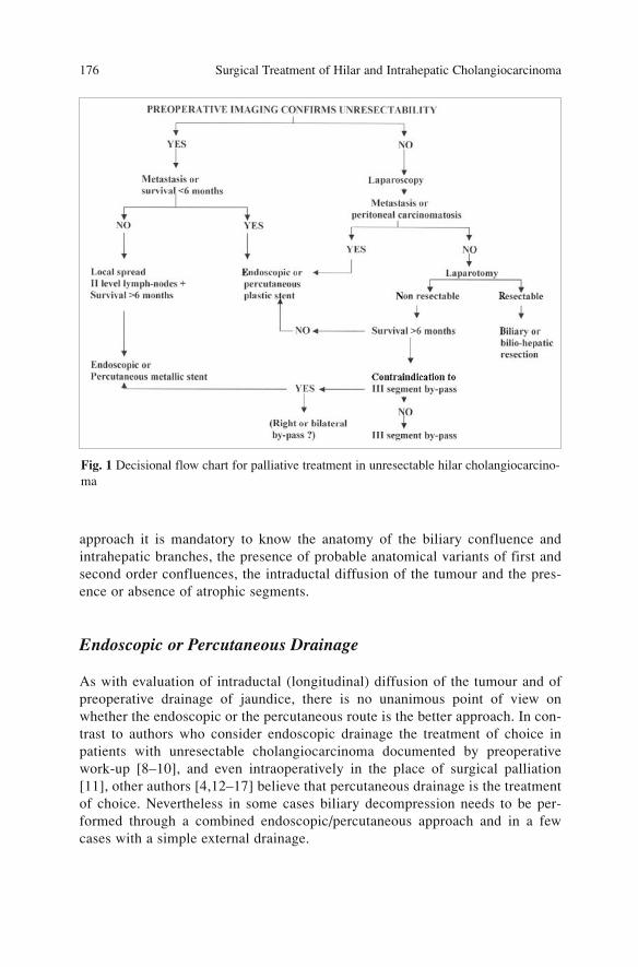
approach it is mandatory to know the anatomy of the biliary confluence andintrahepatic branches, the presence of probable anatomical variants of first andsecond order confluences, the intraductal diffusion of the tumour and the pres-ence or absence of atrophic segments.
Endoscopic or Percutaneous Drainage
As with evaluation of intraductal (longitudinal) diffusion of the tumour and ofpreoperative drainage of jaundice, there is no unanimous point of view onwhether the endoscopic or the percutaneous route is the better approach. In con-trast to authors who consider endoscopic drainage the treatment of choice inpatients with unresectable cholangiocarcinoma documented by preoperativework-up [8–10], and even intraoperatively in the place of surgical palliation[11], other authors [4,12–17] believe that percutaneous drainage is the treatmentof choice. Nevertheless in some cases biliary decompression needs to be per-formed through a combined endoscopic/percutaneous approach and in a fewcases with a simple external drainage.
Surgical Treatment of Hilar and Intrahepatic Cholangiocarcinoma 176
Fig. 1 Decisional flow chart for palliative treatment in unresectable hilar cholangiocarcino-ma
Plastic or Metallic Stents
Regarding the type of drainage there are two possibilities: a plastic or a metallicstent. The advantages of a plastic stent are low cost, easy placement and medium-lasting patency (about 3 months); disadvantages are the small caliber of the lumen(10 F) which often leads to occlusion by biliary sludge and the risk of determin-ing obstruction of the second order ducts. Both factors can provoke cholangitis,which occurs in 20–40% of cases after placement of these stents [11,18,19].
Metallic stents have a larger caliber (30 F) and this allows long-lasting func-tioning, sometimes for the rest of the patient’s life, and it does not present therisk of obstructing the adjacent segmental ducts since it is fenestrated; disadvan-tages are the high initial cost and the impossibility of removing the stent itself[20]. Controlled studies comparing the two types of stents [21–24] have con-firmed that metallic stents present a longer functionality (median duration 6months) [19,25,26], a percentage of cholangitis of 4.9–6% [19,26], and lateocclusion of the stent in 23% of the cases. All these factors determine less needto substitute the stent, rehospitalization and reduced antibiotic use, with an over-all lowering of final costs and a better quality of life for the patients. However,no significant differences on survival have been noted.
Based on previous considerations, for choosing the right stent it is essentialto consider the life expectancy of the patient. Patients with unresectable carcino-ma without signs of distant metastases and with a life expectancy of 6 months ormore benefit more from a metallic stent; conversely, in patients with unre-sectable disease and hepatic or distant metastases with a short prognosis (<3months) the placement of a plastic stent is sufficient [22].
Another point still under debate is the number of stents required to obtainadequate palliation: if the disease is in stage I according to Bismuth-Corletteclassification a single drain is clearly sufficient; in stage II–IV there is no con-sensus on how many drains must be employed.
Various studies in the literature have compared unilateral vs. bilateraldrainage; Deviere [27] and Cheng [26] show better survival (178 vs. 118 days)and a smaller incidence of complications in patients who underwent doubledrainage; Abu-Hamda also demonstrates better results compared to unilateral,particularly when the contrast medium is distributed in both hemisystems [9].De Palma’s controlled prospective randomized study [19] which compares thedifferent treatment modalities, unilateral vs. bilateral, shows a greater successrate in placement of the stent (89 vs. 77%), biliary drainage (81 vs. 73%), andfewer complications (19 vs. 27%) in unilateral vs. bilateral stents; related mor-tality, late complications and survival are similar. The study by Inal et al. [28]does not show significant differences in clinical response to treatment or stentpatency rate with unilobar vs. bilateral drainage.
Watanapa, studying hepatic alterations in jaundiced patients who underwentmonolateral or complete surgical decompression of jaundice, concludes thatunilobar drainage allows resumption of hepatic function after 6 weeks, as withbilateral drainage [29].
Palliative Treatments 177
The better preoperative classification with non-invasive techniques providedby MRCP will permit more frequent use of selective monolateral drainage; infact the excellent staging possibility of this imaging technique allows reducedmedium contrast injection, decreasing the risk of contaminating biliary segmentswhich otherwise would need to be drained.
Bilateral or multiple drainages are indicated exclusively for patients withcholangitis of the contralateral lobe or of segmental ducts.
Surgical Palliation
When unresectability is determined intraoperatively different therapeuticchances are achievable, represented by surgical palliation by means of bilio-enteric bypasses with peripheral intrahepatic anastomosis.
The commonest operation is segment 3 intrahepatic hepato-jejunostomy,called B3 cholangio-jejunostomy. The technique was described originally byHepp-Couinaud, and requires complete or partial resection of the third hepaticsegment, identification of the biliary duct and latero-lateral bilio-enteric anasto-mosis performed with a Roux-en-Y jejunal limb. Subsequently, in 1957,Soupault and Couinaud changed this technique using a direct approach to theduct of the three segments through a hepatotomy, avoiding hepatic resection aswell. The isolation of the biliary duct is performed through the umbilical scis-sura. This maneuver permits identification of the portal branch for segment 3,which can reach a diameter of 4–6 mm in the jaundiced patient; cholangio-jejunostomy is then performed on a Roux-en-Y loop with interrupted stitches.An anastomotic stent can be positioned with the egress site on the loop withVitzel technique or on the liver (Volker) for postoperative control of the anasto-mosis. This technique was diffused in the 1970s by Bismuth and Corlette [30]and in the 1980s by Blumgart and Kelly [31].
Contraindications to intrahepatic B3 cholangio-jejunostomy are the presenceof an atrophic left lobe, the presence of a PTBD on the right lobe, the presenceof cholangitis on the right lobe, a percentage of hepatic parenchyma to be drainedless than 30% or less than two segments and presence of portal hypertension [8,32].
The surgical decompression on the right lobe can be performed on the ductsof segments 5 and 6 by means of wedge resections or hepatotomies; differentlyfrom intrahepatic B3 cholangio-jejunostomy, the isolation of the duct is moredifficult and demanding with major risk of complications (fistula) and lesserefficacy of the biliary decompression.
Mortality after segment 3 intrahepatic cholangio-jejunostomy ranges from 3to 12% [7,32–36] and morbidity from 17 to 51%. In the experience of MSKCC[33] no mortality after intrahepatic B3 cholangio-jejunostomy has been reportedin patients with cholangiocarcinoma while it was 21.4% in patients with carci-noma of the gallbladder; mortality rate after right segmental duct anastomosis inpatients with cholangiocarcinoma was 14.3% [33].
The most frequent complication of this type of surgery is biliary leak and its
Surgical Treatment of Hilar and Intrahepatic Cholangiocarcinoma 178
related complications, and it ranges from 6 to 21% [33,34,36]; the frequency ishigher for right surgical decompression as is the percentage of patients whorequire re-operation due to failure of the procedure (55 vs. 15%, p=0.06) [33].
Also, the patency rate of the anastomosis at 1 year is higher in segment IIIintrahepatic cholangio-jejunostomy than right ducts (80 vs. 60%) even if the dif-ference is not statistically significant (p=0.1) [33].
There are no controlled prospective randomized studies that compare surgicalvs. endoscopic or percutaneous palliation for hilar cholangiocarcinoma in the lit-erature; for this reason, based on our previous considerations, we can concludethat bypass on segment 3 is an operation that entails acceptable risks of mortalityand morbidity, similar to those observed after percutaneous and endoscopic stents,and allows an excellent palliation of jaundice, with a good duration over time andfew later complications. Conversely the right intrahepatic cholangio-jejunostomyis associated with high percentage of morbidity and mortality both early and late,and therefore is not competitive vs. non-surgical biliary decompression.
Other possibilities for surgical palliation are:– Transtumoral intubation. This procedure is associated with a high mortality
rate (27%) [37] and the disadvantages of an external biliary catheter (disloca-tion, obstruction, cholangitis); consequently their application is not attractive.
– Palliative resection of extrahepatic biliary tract. This operation has the samemortality and morbidity rates as surgical by-pass [38] and allows bilateraldrainage [39]; however recurrence rate is very high and for this reason itsindication is limited.
Chemotherapy, Radiotherapy and Photodynamic Therapy
The indications for palliative treatment with chemotherapy and/or radiotherapyare limited to patients with unresectable hilar cholangiocarcinoma, with recur-rence after resection or with metastatic disease.
Radiotherapy alone or associated with chemotherapy is indicated in patientswith local advanced disease without distant metastasis; instead, in presence ofthis condition chemotherapy is the only possible treatment.
Chemotherapy
Response to chemotherapy is very poor [10,40–43]; in fact at present nochemotherapeutic agent is effective enough to change the natural history of thedisease. The most frequently utilized and studied agent is 5-FU alone or associ-ated with other agents. Some studies do not report benefits in the treated patientscompared to patients who underwent biliary drainage alone [44,45]. The per-centage of response varies in literature from 0% (obtained with 5-FU alone [41])to 40% (in a group of patients treated with a combination of 5-FU, epirubicine
Palliative Treatments 179
and cisplatin [46]). This last datum shows that the combination of 5-FU withother drugs gives better results. The agents used were leucovorin, otoposide,streptozocin, mitomycin C, doxorubicin and IFN-α. The number of treatedpatients, however, is low in all the series and ranges from a minimum of 18 to amaximum of 37 patients; median survival ranges from 8 weeks to 12 months.Another important limit of these studies is that most report data on heteroge-neous groups of patients with intra- and extrahepatic cholangiocarcinoma, car-cinoma of the gallbladder and in some cases pancreatic carcinoma.
Nevertheless there is a study [47] that reports improvement and a positivetrend in quality of life in a group of patients with a Karnosfy index greater than70% and treated with 5-FU, leucovorin and etoposide, compared with patientstreated with supportive care alone (6.5 months vs. 2.5 months, p=0.10).
A recent phase-II study [48] has utilized intra-arterial infusion, withSeldinger technique, of epirubicine and cisplatin in combination to systemic 5-FU administration in a group of 30 patients with unresectable carcinoma of bil-iary tract; a positive response was observed in 40% of the patients (12/30)–onecase of complete response and 11 cases of partial response. The median progres-sion-free and overall survival time were 7.1 and 13.2 months, respectively; 1-and 2-year survival were 54 and 20%, respectively. The improvement of per-formance status was observed in 30% of the patients.
Recently gemcitabine was introduced in clinical practice for treatment of bil-iary tract neoplasm; it can be used alone in different doses with a response ratethat ranges from 8% [49] to 60% [50], with median survival 6.3–16 months, orin combination with other agents such as 5-FU, cisplatin, oxaliplatin, docetaxel,irinotecan and capecitabine, with response rate varying from 9% (combinationgemcitabine–docetaxel) [51] to 50% in the combination gemcitabine–cisplatin[52] and with median survival between 20 weeks [53] and 15.4 months [54],with an acceptable toxicity.
The more efficient combinations are those that utilized gemcitabine and cis-platin in the patients with good performance status and hepatic function [43](response from 27.5 to 50% with median survival 5–11.3 months) and with gem-citabine and oxaliplatin (response in 35% of the patients with good performancestatus and 22% of the patients with poor performance status). Conversely, com-binations with capecitabine, irinotecan and docetaxel do not seem effective [43].
In conclusion, until now there has been no evidence of the utility of thesetreatments in the palliation of cholangiocarcinoma but results are more encour-aging than the past; controlled randomized phase-III multicentric studies areadvisable to recruit larger series that compare more promising agents.
Radiotherapy
The types of radiation therapy are different: external, brachytherapy and theassociation of both; the positioning of the radioactive source can be either endo-scopic or percutaneous.
Surgical Treatment of Hilar and Intrahepatic Cholangiocarcinoma 180
The results of radiation therapy in studies in the literature are contradictory;compared with authors [55–57] who consider this kind of treatment useful forbiliary decompression and pain control with a better quality of life, others [58]do not report advantages with external radiotherapy, even using different dosesof radiation (from 30 to 85 Gy). Even the results of brachytherapy alone or asso-ciated with external radiotherapy are divergent; in fact some authors reportfavorable results [1,56,59–61] while others do not recognize its usefulness [11]but report a high rate of complications, especially cholangitis (40–50%), andgastroduodenal toxicity ranging from 8 to 42% [11,56,60,61].
Photodynamic Therapy
Photodynamic therapy is indicated for local control of unresectable disease inabsence of distant metastases; similarly to other types of tumour such as oesoph-agus, colon, stomach, bronchi, urinary bladder and brain, photodynamic therapyhas been introduced in the treatment of cholangiocarcinoma [62]. The procedurerequires the infusion of a photosensitizing drug (sodium porfimer) that accumu-lates electively in tumoral cells 24–48 h after administration; subsequently cyto-toxic free radicals such as singlet oxygen, develop through cholangioscopic pho-toactivation and determine the death of neoplastic cells. Different phase-II stud-ies report median of survival of 330–439 days with 1-year survival that variesfrom 45% to 78% [39,62]. A recent prospective randomized study by Ortner’sgroup [63] performed on 39 patients with cholangiocarcinoma stages III and IV,and treated with biliary stent with or without photodynamic therapy, showed abetter survival of the treated group (493 vs. 98 days), lower cholestasis rate, anda better quality of life and performance status. The authors believe that the realdifference is due to a better biliary decompression following the reduction of theneoplastic mass that the procedure provides.
Witzigmann [64] reports a median survival of 12 months vs. 6.4 months(p<0.01), lower bilirubinemia values (p<0.05) and higher Karnofsky index(p<0.01) in the patients treated with stent and photodynamic therapy vs. patientswith biliary stent alone when evaluating the results of his own experience of sur-gical resection and palliative treatment in 184 cases of hilar cholangiocarcinoma(60 resected: 42 R0, 18 R1-2; 56 treated with biliary stent; 68 with biliary stentcombined with photodynamic therapy); in addition, the median survival ofpatients who underwent photodynamic therapy is similar to that of R1-2 resect-ed patients.
Evaluating the 150 patients reported in the literature, photodynamic therapyassociated with biliary stent is shown to improve survival with a range from 9.3to 16.3 months.
These promising results require further verification and new studies to eval-uate the worth of photodynamic therapy with novel chemotherapeutic agentswhich show more encouraging possibilities as well.
Palliative Treatments 181
References
1. Chari RS, Anderson CA, Saverese DMF (2003) Treatment of cholangiocarcinoma II. In:Rose BD (ed) UpToDate. UpToDate, Wellesley, MA
2. Vauthey JN, Blumgart LH (1994) Recent advances in the management of cholangiocarcino-mas. Semin Liver Dis 14(2):109–114
3. Nimura Y, Kamiya J, Kondo S et al (2000) Aggressive preoperative management and extend-ed surgery for hilar cholangiocarcinoma: Nagoya experience. J Hepatobiliary Pancreat Surg7(2):155–162
4. Abraham NS, Barkun JS, Barkun AN (2002) Palliation of malignant biliary obstruction: aprospective trial examining impact on quality of life. Gastrointest Endosc 56(6):835–841
5. Bismuth H, Malt RA (1979) Current concepts in cancer: carcinoma of the biliary tract. NEngl J Med 301(13):704–706
6. Dowsett JF, Vaira D, Hatfield AR et al (1989) Endoscopic biliary therapy using the com-bined percutaneous and endoscopic technique. Gastroenterology 96(4):1180–1186
7. Traynor O, Castaing D, Bismuth H (1987) Left intrahepatic cholangio-enteric anastomosis(round ligament approach): an effective palliative treatment for hilar cancers. Br J Surg74(10):952–954
8. Singhal D, van Gulik TM, Gouma DJ (2005) Palliative management of hilar cholangiocar-cinoma. Surg Oncol 14(2):59–74
9. Abu-Hamda EM, Baron TH (2004) Endoscopic management of cholangiocarcinoma. SeminLiver Dis 24(2):165–175
10. Anderson CD, Pinson CW, Berlin J, Chari RS (2004) Diagnosis and treatment of cholangio-carcinoma. Oncologist 9(1):43–57
11. Gerhards MF, van Gulik TM, Gonzalez Gonzalez D et al (2003) Results of postoperativeradiotherapy for resectable hilar cholangiocarcinoma. World J Surg 27(2):173–179
12. Khan SA, Thomas HC, Davidson BR, Taylor-Robinson SD (2005) Cholangiocarcinoma.Lancet 366(9493):1303–1314
13. Shapiro MJ (1995) Management of malignant biliary obstruction: nonoperative and pallia-tive techniques. Oncology 9(6):493–496, discussion 499–500, 503
14. England RE, Martin DF (1996) Endoscopic and percutaneous intervention in malignantobstructive jaundice. Cardiovasc Intervent Radiol 19(6):381–387
15. Nelsen KM, Kastan DJ, Shetty PC et al (1996) Utilization pattern and efficacy of nonsurgi-cal techniques to establish drainage for high biliary obstruction. J Vasc Interv Radiol7(5):751–756
16. Cowling MG, Adam AN (2001) Internal stenting in malignant biliary obstruction. World JSurg 25(3):355–359; discussion 359–361
17. Lillemoe KD, Cameron JL (2000) Surgery for hilar cholangiocarcinoma: the Johns Hopkinsapproach. J Hepatobiliary Pancreat Surg 7(2):115–121
18. Liu CL, Lo CM, Lai EC, Fan ST (1998) Endoscopic retrograde cholangiopancreatographyand endoscopic endoprosthesis insertion in patients with Klatskin tumours. Arch Surg133(3):293–296
19. De Palma GD, Galloro G, Siciliano S et al (2001) Unilateral versus bilateral endoscopichepatic duct drainage in patients with malignant hilar biliary obstruction: results of aprospective, randomized, and controlled study. Gastrointest Endosc 53(6):547–553
20. Venbrux AC (2001) Current status of endoscopic pancreaticobiliary interventions. J VascInterv Radiol 12:155–162
21. Davids PH, Groen AK, Rauws EA et al (1992) Randomised trial of self-expanding metalstents versus polyethylene stents for distal malignant biliary obstruction. Lancet340(8834–8835):1488–1492
22. Kaassis M, Boyer J, Dumas R et al (2003) Plastic or metal stents for malignant stricture ofthe common bile duct? Results of a randomized prospective study. Gastrointest Endosc57(2):178–182
Surgical Treatment of Hilar and Intrahepatic Cholangiocarcinoma 182
23. Prat F, Chapat O, Ducot B et al (1998) A randomized trial of endoscopic drainage methodsfor inoperable malignant strictures of the common bile duct. Gastrointest Endosc 47(1):1–7
24. Wagner HJ, Knyrim K, Vakil N, Klose KJ (1993) Plastic endoprostheses versus metal stentsin the palliative treatment of malignant hilar biliary obstruction. A prospective and random-ized trial. Endoscopy 25(3):213–218
25. Glattli A, Stain SC, Baer HU et al (1993) Unresectable malignant biliary obstruction: treat-ment by self-expandable biliary endoprostheses. HPB Surg 6(3):175–184
26. Cheng JL, Bruno MJ, Bergman JJ et al (2002) Endoscopic palliation of patients with biliaryobstruction caused by nonresectable hilar cholangiocarcinoma: efficacy of self-expandablemetallic Wallstents. Gastrointest Endosc 56(1):33–39
27. Deviere J, Baize M, de Toeuf J, Cremer M (1988) Long-term follow-up of patients with hilarmalignant stricture treated by endoscopic internal biliary drainage. Gastrointest Endosc34(2):95–101
28. Inal M, Akgul E, Aksungur E, Seydaoglu G (2003) Percutaneous placement of biliary metal-lic stents in patients with malignant hilar obstruction: unilobar versus bilobar drainage. JVasc Interv Radiol 14(11):1409–1416
29. Watanapa P (1996) Recovery patterns of liver function after complete and partial surgicalbiliary decompression. Am J Surg 171(2):230–234
30. Bismuth H, Corlette MB (1975) Intrahepatic cholangioenteric anastomosis in carcinoma ofthe hilus of the liver. Surg Gynecol Obstet 140(2):170–178
31. Blumgart LH, Kelley CJ (1984) Hepaticojejunostomy in benign and malignant high bileduct stricture: approaches to the left hepatic ducts. Br J Surg 71(4):257–261
32. Connors S, Wigmore SJ, Madhavan KK et al (2005) Surgical palliation for unresectable hilarcholangiocarcinoma. HPB Surg 7:273–277
33. Jarnagin WR, Burke E, Powers C et al (1998) Intrahepatic biliary enteric bypass provideseffective palliation in selected patients with malignant obstruction at the hepatic duct con-fluence. Am J Surg 175(6):453–460
34. Chaudhary A, Dhar P, Tomey S et al (1997) Segment III cholangiojejunostomy for carcino-ma of the gallbladder. World J Surg 21(8):866–870; discussion 870–871
35. Guthrie CM, Banting SW, Garden OJ, Carter DC (1994) Segment III cholangiojejunostomyfor palliation of malignant hilar obstruction. Br J Surg 81(11):1639–1641
36. Kapoor VK, Pradeep R, Haribhakti SP et al (1996) Intrahepatic segment III cholangioje-junostomy in advanced carcinoma of the gallbladder. Br J Surg 83(12):1709–1711
37. Launois B, Reding R, Lebeau G, Buard JL (2000) Surgery for hilar cholangiocarcinoma:French experience in a collective survey of 552 extrahepatic bile duct cancers. JHepatobiliary Pancreat Surg 7(2):128–134
38. Jarnagin WR, Fong Y, DeMatteo RP et al (2001) Staging, resectability, and outcome in 225patients with hilar cholangiocarcinoma. Ann Surg 234(4):507–517; discussion 517–519
39. Berr F, Wiedmann M, Tannapfel A et al (2000) Photodynamic therapy for advanced bile ductcancer: evidence for improved palliation and extended survival. Hepatology 31(2):291–298
40. Kajanti M, Pyrhonen S (1994) Epirubicin-sequential methotrexate-5-fluorouracil-leucovorintreatment in advanced cancer of the extrahepatic biliary system. A phase II study. Am J ClinOncol 17(3):223–226
41. Takada T, Kato H, Matsushiro T et al (1994) Comparison of 5-fluorouracil, doxorubicin andmitomycin C with 5-fluorouracil alone in the treatment of pancreatic-biliary carcinomas.Oncology 51(5):396–400
42. Mazhar D, Stebbing J, Bower M (2006) Chemotherapy for advanced cholangiocarcinoma:what is standard treatment? Future Oncol 2(4):509–514
43. Thongprasert S (2005) The role of chemotherapy in cholangiocarcinoma. Ann Oncol16(Suppl 2):ii93-ii96
44. Patt YZ, Hassan MM, Lozano RD et al (2001) Phase II trial of cisplatin, interferon alpha-2b, doxorubicin, and 5-fluorouracil for biliary tract cancer. Clin Cancer Res7(11):3375–3380
45. Patt YZ, Jones DV Jr, Hoque A et al (1996) Phase II trial of intravenous flourouracil and sub-
Palliative Treatments 183
cutaneous interferon alfa-2b for biliary tract cancer. J Clin Oncol 14(8):2311–231546. Ellis PA, Norman A, Hill A et al (1995) Epirubicin, cisplatin and infusional 5-fluorouracil
(5-FU) (ECF) in hepatobiliary tumours. Eur J Cancer 31A(10):1594–159847. Glimelius B, Hoffman K, Sjoden PO et al (1996) Chemotherapy improves survival and qual-
ity of life in advanced pancreatic and biliary cancer. Ann Oncol 7(6):593–60048. Cantore M, Mambrini A, Fiorentini G et al (2005) Phase II study of hepatic intraarterial
epirubicin and cisplatin, with systemic 5-fluorouracil in patients with unresectable biliarytract tumours. Cancer 103(7):1402–1407
49. Mezger J, Sauerbruch T, Ko Y et al (1998) Phase II trial of gemcitabine in gallbladder andbiliary tract carcinomas. Onkologie 21:232–234
50. Dobrila-Dintinjana R, Kovac D, Depolo A et al (2000) Gemcitabine in patients with nonre-sectable cancer of the biliary system or advanced gallbladder cancer. Am J Gastroenterol95:2476
51. Kuhn R, Hribaschek A, Eichelmann K et al (2002) Outpatient therapy with gemcitabine anddocetaxel for gallbladder, biliary, and cholangio-carcinomas. Invest New Drugs20(3):351–356
52. Carraro S, Servienti PJ, Bruno MF (2001) Gemcitabine and cisplatin in locally advanced ormetastatic gallbladder and bile duct adenocancer. Proc Am Soc Clin Oncol 20:146B (abs2333)
53. Doval DC, Sekhon JS, Gupta SK et al (2004) A phase II study of gemcitabine and cisplatinin chemotherapy-naive, unresectable gall bladder cancer. Br J Cancer 90(8):1516–1520
54. Andre T, Tournigand C, Rosmorduc O et al; GERCOR Group (2004) Gemcitabine combinedwith oxaliplatin (GEMOX) in advanced biliary tract adenocarcinoma: a GERCOR study.Ann Oncol 15(9):1339–1343
55. Shinchi H, Takao S, Nishida H, Aikou T (2000) Length and quality of survival followingexternal beam radiotherapy combined with expandable metallic stent for unresectable hilarcholangiocarcinoma. J Surg Oncol 75(2):89–94
56. Ishii H, Furuse J, Nagase M et al (2004) Relief of jaundice by external beam radiotherapyand intraluminal brachytherapy in patients with extrahepatic cholangiocarcinoma: resultswithout stenting. Hepatogastroenterology 51(58):954–957
57. Ohnishi H, Asada M, Shichijo Y et al (1995) External radiotherapy for biliary decompres-sion of hilar cholangiocarcinoma. Hepatogastroenterology 42(3):265–268
58. Crane CH, Macdonald KO, Vauthey JN et al (2002) Limitations of conventional doses ofchemoradiation for unresectable biliary cancer. Int J Radiat Oncol Biol Phys 53(4):969–974
59. Kuvshinoff BW, Armstrong JG, Fong Y et al (1995) Palliation of irresectable hilar cholan-giocarcinoma with biliary drainage and radiotherapy. Br J Surg 82(11):1522–1525
60. Vallis KA, Benjamin IS, Munro AJ et al (1996) External beam and intraluminal radiothera-py for locally advanced bile duct cancer: role and tolerability. Radiother Oncol 41(1):61–66
61. Foo ML, Gunderson LL, Bender CE, Buskirk SJ (1997) External radiation therapy and tran-scatheter iridium in the treatment of extrahepatic bile duct carcinoma. Int J Radiat OncolBiol Phys 39(4):929–935
62. Ortner MA, Liebetruth J, Schreiber S et al (1998) Photodynamic therapy of nonresectablecholangiocarcinoma. Gastroenterology 114(3):536–542
63. Ortner ME, Caca K, Berr F et al (2003) Successful photodynamic therapy for nonresectablecholangiocarcinoma: a randomized prospective study. Gastroenterology 125(5):1355–1363
64. Witzigmann H, Berr F, Ringel U et al (2006) Surgical and palliative management and out-come in 184 patients with hilar cholangiocarcinoma: palliative photodynamic therapy plusstenting is comparable to r1/r2 resection. Ann Surg 244(2):230–239
Surgical Treatment of Hilar and Intrahepatic Cholangiocarcinoma 184

Part 2Intrahepatic
Cholangiocarcinoma
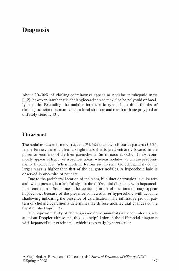
Diagnosis
About 20–30% of cholangiocarcinomas appear as nodular intrahepatic mass[1,2]; however, intrahepatic cholangiocarcinomas may also be polypoid or focal-ly stenotic. Excluding the nodular intrahepatic type, about three-fourths ofcholangiocarcinomas manifest as a focal stricture and one-fourth are polypoid ordiffusely stenotic [3].
Ultrasound
The nodular pattern is more frequent (94.4%) than the infiltrative pattern (5.6%).In the former, there is often a single mass that is predominantly located in theposterior segments of the liver parenchyma. Small nodules (<3 cm) most com-monly appear as hypo- or isoechoic areas, whereas nodules >3 cm are predomi-nantly hyperechoic. When multiple lesions are present, the echogenicity of thelarger mass is higher than that of the daughter nodules. A hypoechoic halo isobserved in one-third of patients.
Due to the peripheral location of the mass, bile-duct obstruction is quite rareand, when present, is a helpful sign in the differential diagnosis with hepatocel-lular carcinoma. Sometimes, the central portion of the tumour may appearhypoechoic, because of the presence of necrosis, or hyperechoic with acousticshadowing indicating the presence of calcification. The infiltrative growth pat-tern of cholangiocarcinoma determines the diffuse architectural changes of thehepatic lobe (Figs. 1,2).
The hypovascularity of cholangiocarcinoma manifests as scant color signalsat colour Doppler ultrasound; this is a helpful sign in the differential diagnosiswith hepatocellular carcinoma, which is typically hypervascular.
187A. Guglielmi, A. Ruzzenente, C. Iacono (eds.) Surgical Treatment of Hilar and ICC.© Springer 2008
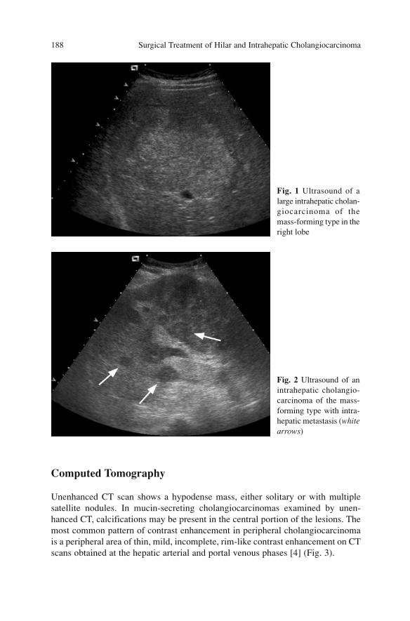
Computed Tomography
Unenhanced CT scan shows a hypodense mass, either solitary or with multiplesatellite nodules. In mucin-secreting cholangiocarcinomas examined by unen-hanced CT, calcifications may be present in the central portion of the lesions. Themost common pattern of contrast enhancement in peripheral cholangiocarcinomais a peripheral area of thin, mild, incomplete, rim-like contrast enhancement on CTscans obtained at the hepatic arterial and portal venous phases [4] (Fig. 3).
Surgical Treatment of Hilar and Intrahepatic Cholangiocarcinoma 188
Fig. 1 Ultrasound of alarge intrahepatic cholan-giocarcinoma of themass-forming type in theright lobe
Fig. 2 Ultrasound of anintrahepatic cholangio-carcinoma of the mass-forming type with intra-hepatic metastasis (whitearrows)
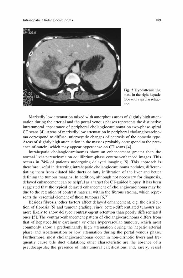
Markedly low attenuation mixed with amorphous areas of slightly high atten-uation during the arterial and the portal venous phases represents the distinctiveintratumoral appearance of peripheral cholangiocarcinoma on two-phase spiralCT scans [4]. Areas of markedly low attenuation in peripheral cholangiocarcino-ma correspond to diffuse, microcystic changes of necrosis of the comedo type.Areas of slightly high attenuation in the masses probably correspond to the pres-ence of mucin, which may appear hyperdense on CT scans [4].
Intrahepatic cholangiocarcinomas show an enhancement greater than thenormal liver parenchyma on equilibrium-phase contrast-enhanced images. Thisoccurs in 74% of patients undergoing delayed imaging [5]. This approach istherefore useful in detecting intrahepatic cholangiocarcinoma nodules, differen-tiating them from dilated bile ducts or fatty infiltration of the liver and betterdefining the tumour margins. In addition, although not necessary for diagnosis,delayed enhancement can be helpful as a target for CT-guided biopsy. It has beensuggested that the typical delayed enhancement of cholangiocarcinoma may bedue to the retention of contrast material within the fibrous stroma, which repre-sents the essential element of these tumours [6,7].
Besides fibrosis, other factors affect delayed enhancement, e.g. the distribu-tion of fibrosis [5] and tumour grading, since better-differentiated tumours aremore likely to show delayed contrast-agent retention than poorly differentiatedones [5]. The contrast-enhancement pattern of cholangiocarcinoma differs fromthat of hepatocellular carcinoma or other hypervascular tumours, which mostcommonly show a predominantly high attenuation during the hepatic arterialphase and isoattenuation or low attenuation during the portal venous phase.Furthermore, most cholangiocarcinomas occur in non-cirrhotic livers and fre-quently cause bile duct dilatation; other characteristic are the absence of apseudocapsule, the presence of intratumoral calcifications and, rarely, vessel
Intrahepatic Cholangiocarcinoma 189
Fig. 3 Hypoattenuatingmass in the right hepaticlobe with capsular retrac-tion
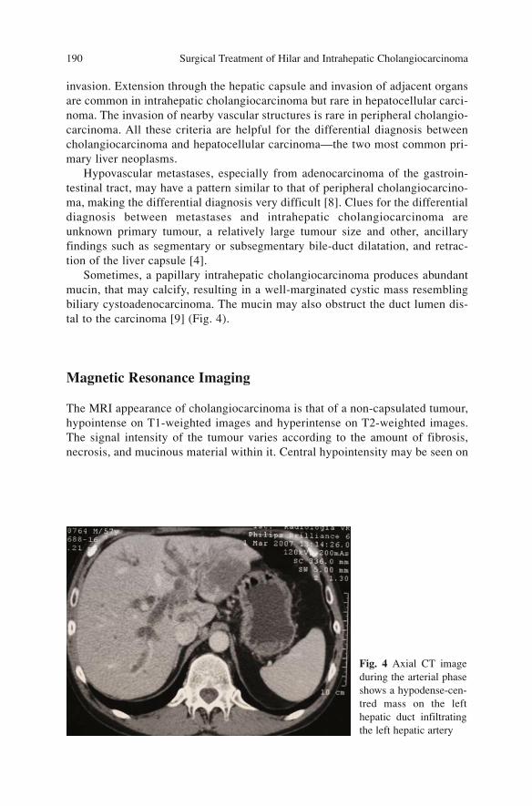
invasion. Extension through the hepatic capsule and invasion of adjacent organsare common in intrahepatic cholangiocarcinoma but rare in hepatocellular carci-noma. The invasion of nearby vascular structures is rare in peripheral cholangio-carcinoma. All these criteria are helpful for the differential diagnosis betweencholangiocarcinoma and hepatocellular carcinoma—the two most common pri-mary liver neoplasms.
Hypovascular metastases, especially from adenocarcinoma of the gastroin-testinal tract, may have a pattern similar to that of peripheral cholangiocarcino-ma, making the differential diagnosis very difficult [8]. Clues for the differentialdiagnosis between metastases and intrahepatic cholangiocarcinoma areunknown primary tumour, a relatively large tumour size and other, ancillaryfindings such as segmentary or subsegmentary bile-duct dilatation, and retrac-tion of the liver capsule [4].
Sometimes, a papillary intrahepatic cholangiocarcinoma produces abundantmucin, that may calcify, resulting in a well-marginated cystic mass resemblingbiliary cystoadenocarcinoma. The mucin may also obstruct the duct lumen dis-tal to the carcinoma [9] (Fig. 4).
Magnetic Resonance Imaging
The MRI appearance of cholangiocarcinoma is that of a non-capsulated tumour,hypointense on T1-weighted images and hyperintense on T2-weighted images.The signal intensity of the tumour varies according to the amount of fibrosis,necrosis, and mucinous material within it. Central hypointensity may be seen on
Surgical Treatment of Hilar and Intrahepatic Cholangiocarcinoma 190
Fig. 4 Axial CT imageduring the arterial phaseshows a hypodense-cen-tred mass on the lefthepatic duct infiltratingthe left hepatic artery
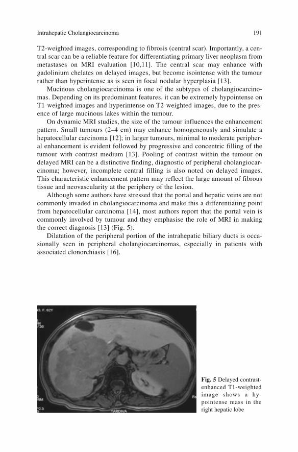
T2-weighted images, corresponding to fibrosis (central scar). Importantly, a cen-tral scar can be a reliable feature for differentiating primary liver neoplasm frommetastases on MRI evaluation [10,11]. The central scar may enhance withgadolinium chelates on delayed images, but become isointense with the tumourrather than hyperintense as is seen in focal nodular hyperplasia [13].
Mucinous cholangiocarcinoma is one of the subtypes of cholangiocarcino-mas. Depending on its predominant features, it can be extremely hypointense onT1-weighted images and hyperintense on T2-weighted images, due to the pres-ence of large mucinous lakes within the tumour.
On dynamic MRI studies, the size of the tumour influences the enhancementpattern. Small tumours (2–4 cm) may enhance homogeneously and simulate ahepatocellular carcinoma [12]; in larger tumours, minimal to moderate peripher-al enhancement is evident followed by progressive and concentric filling of thetumour with contrast medium [13]. Pooling of contrast within the tumour ondelayed MRI can be a distinctive finding, diagnostic of peripheral cholangiocar-cinoma; however, incomplete central filling is also noted on delayed images.This characteristic enhancement pattern may reflect the large amount of fibroustissue and neovascularity at the periphery of the lesion.
Although some authors have stressed that the portal and hepatic veins are notcommonly invaded in cholangiocarcinoma and make this a differentiating pointfrom hepatocellular carcinoma [14], most authors report that the portal vein iscommonly involved by tumour and they emphasise the role of MRI in makingthe correct diagnosis [13] (Fig. 5).
Dilatation of the peripheral portion of the intrahepatic biliary ducts is occa-sionally seen in peripheral cholangiocarcinomas, especially in patients withassociated clonorchiasis [16].
Intrahepatic Cholangiocarcinoma 191
Fig. 5 Delayed contrast-enhanced T1-weightedimage shows a hy-pointense mass in theright hepatic lobe
Angiography
Angiographically, intrahepatic cholangiocarcinoma is predominantly hypovas-cular, and the appearance of thin vessels is pathognomonic of its fibrous nature[17]. Encasement of hepatic arteries and other major vessels is associated withthe degree of sclerosis resulting from the tumour.
References
1. Thorsen MK, Quiroz F, Lawson TL et al (1984) Primary biliary carcinoma: CT evaluation.Radiology 152(2):479–483
2. Nesbit GM, Johnson CD, James EM et al (1988) Cholangiocarcinoma: diagnosis and eval-uation of resectability by CT and sonography as procedures complementary to cholangiog-raphy. AJR Am J Roentgenol 151(5):933–938
3. Kokubo T, Itai Y, Ohtomo K et al (1988) Mucin-hypersecreting intrahepatic biliary neo-plasms. Radiology 168(3):609–614
4. Kim TK, Choi BI, Han JK et al (1997) Peripheral cholangiocarcinoma of the liver: two-phase spiral CT findings. Radiology 204(2):539–543
5. Lacomis JM, Baron RL, Oliver JH 3rd et al (1997) Cholangiocarcinoma: delayed CT con-trast enhancement patterns. Radiology 203(1):98–104
6. Takayasu K, Ikeya S, Mukai K et al (1990) CT of hilar cholangiocarcinoma: late contrastenhancement in six patients. AJR Am J Roentgenol 154(6):1203–1206
7. Honda H, Onitsuka H, Yasumori K et al (1993) Intrahepatic peripheral cholangiocarcinoma:two-phased dynamic incremental CT and pathologic correlation. J Comput Assist Tomogr17(3):397–402
8. Choi BI, Han JK, Shin YM et al (1995) Peripheral cholangiocarcinoma: comparison of MRIwith CT. Abdom Imaging 20(4):357–360
9. Itai Y, Araki T, Furui S et al (1983) Computed tomography of primary intrahepatic biliarymalignancy. Radiology 147(2):485–490
10. Ishak KG, Sesterhenn IA, Goodman ZD et al (1984) Epithelioid hemangioendothelioma ofthe liver: a clinicopathologic and follow-up study of 32 cases. Hum Pathol 15(9):839–852
11. Rummeny E, Weissleder R, Stark DD et al (1989) Primary liver tumours: diagnosis by MRimaging. AJR Am J Roentgenol 152(1):63–72
12. Adjei ON, Tamura S, Sugimura H et al (1995) Contrast-enhanced MR imaging of intrahep-atic cholangiocarcinoma. Clin Radiol 50(1):6–10
13. Soyer P, Bluemke DA, Reichle R et al (1995) Imaging of intrahepatic cholangiocarcinoma:1. Peripheral cholangiocarcinoma. AJR Am J Roentgenol 165(6):1427–1431
14. Ros PR, Buck JL, Goodman ZD et al (1988) Intrahepatic cholangiocarcinoma: radiologic-pathologic correlation. Radiology 167(3):689–693
15. Tani K, Kubota Y, Yamaguchi T et al (1991) MR imaging of peripheral cholangiocarcinoma.J Comput Assist Tomogr 15(6):975–978
16. Choi BI, Kim TK, Han JK (1998) MRI of clonorchiasis and cholangiocarcinoma. J MagnReson Imaging 8(2):359–366
17. Kaude J, Rian R (1971) Cholangiocarcinoma. Radiology 100(3):573–580
Surgical Treatment of Hilar and Intrahepatic Cholangiocarcinoma 192
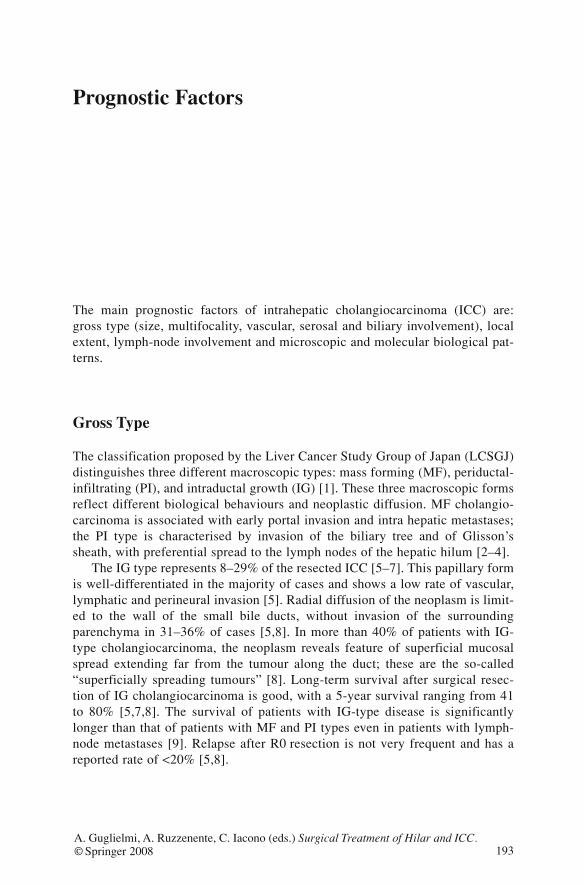
Prognostic Factors
The main prognostic factors of intrahepatic cholangiocarcinoma (ICC) are:gross type (size, multifocality, vascular, serosal and biliary involvement), localextent, lymph-node involvement and microscopic and molecular biological pat-terns.
Gross Type
The classification proposed by the Liver Cancer Study Group of Japan (LCSGJ)distinguishes three different macroscopic types: mass forming (MF), periductal-infiltrating (PI), and intraductal growth (IG) [1]. These three macroscopic formsreflect different biological behaviours and neoplastic diffusion. MF cholangio-carcinoma is associated with early portal invasion and intra hepatic metastases;the PI type is characterised by invasion of the biliary tree and of Glisson’ssheath, with preferential spread to the lymph nodes of the hepatic hilum [2–4].
The IG type represents 8–29% of the resected ICC [5–7]. This papillary formis well-differentiated in the majority of cases and shows a low rate of vascular,lymphatic and perineural invasion [5]. Radial diffusion of the neoplasm is limit-ed to the wall of the small bile ducts, without invasion of the surroundingparenchyma in 31–36% of cases [5,8]. In more than 40% of patients with IG-type cholangiocarcinoma, the neoplasm reveals feature of superficial mucosalspread extending far from the tumour along the duct; these are the so-called“superficially spreading tumours” [8]. Long-term survival after surgical resec-tion of IG cholangiocarcinoma is good, with a 5-year survival ranging from 41to 80% [5,7,8]. The survival of patients with IG-type disease is significantlylonger than that of patients with MF and PI types even in patients with lymph-node metastases [9]. Relapse after R0 resection is not very frequent and has areported rate of <20% [5,8].
193A. Guglielmi, A. Ruzzenente, C. Iacono (eds.) Surgical Treatment of Hilar and ICC.© Springer 2008
MF cholangiocarcinoma is the most frequent type and represents 60–70% ofall cases [10,11]; vascular invasion occurs in 47% of patients, intrahepaticmetastases in 36% and lymph-node metastases in 31% [12]. The 5-year survivalrate reported in the literature varies from 25 to 47% (Table 1). In 63%, patientswith MF-type disease have histological evidence of bile-duct invasion. Theselesions more frequently have a microscopic pattern that is prognostically nega-tive, associated with an increased rate of vascular, perineural and lymphaticinvasion (65, 69 and 80%) compared to neoplasms without ductal infiltration(33, 6 and 33%) [13]. The long-term prognosis is significantly worse for patientswith ductal-invasive MF than for those without ductal invasion with a 5-yearssurvival rate of 11 vs. 62%, respectively [13].
The prognosis of PI-type cholangiocarcinoma is worse than that of the othertwo types. The prevalence of this form ranges between 15 and 35% [9,11,14].PI-type disease is associated with biliary-duct infiltration at the hepatic hilum;vascular and lymphatic involvement are frequent at this same site. Conversely,the rates of portal invasion and intrahepatic metastases are lower [2]. Five-yearsurvival in patients with PI-type cholangiocarcinoma ranges from 0 to 49%[4,9,15].
The MF+PI mixed type of cholangiocarcinoma is observed in 25–46% ofpatients and has the worst prognosis [4,16,17]. This form correlates with a moreadvanced stage of disease, with a higher frequency of lymph-node metastases,gross vascular invasion and intrahepatic metastases (80, 80 and 46%, respec-tively) [17]. The long-term outcome is poor, with a 5-year survival rate <10%(Table 1).
Surgical Treatment of Hilar and Intrahepatic Cholangiocarcinoma 194
Table 1 Intrahepatic cholangiocarcinoma, survival according to gross type
Author Year 5-Year survival 5-Year survival 5-Year survival
MF MF PI PI MF MF(%) (%) +PI +PI (%)
Nozaki [16] 1998 30 36 2 0 13 7Yamamoto [4] 1998 28 39 14 17 18 0Isaji [17] 1999 15 25 3 33 15 7Isa [9] 2001 12 33 7 0 - -Suzuki [15] 2002 12 47 - - 7 0
Morimoto [14] 2003 29a 0 13 49 - -Nakagawa [18] 2005 17 51c 11b 36c - -Miwa [19] 2006 25 44 - - 16 7
MF, Mass forming; PI, periductal infiltratingaPI and MF+PIbEight cases of MF+PI were included in this groupcThree-years survival
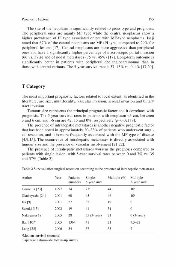
The site of the neoplasm is significantly related to gross type and prognosis.The peripheral ones are mainly MF type while the central neoplasms show ahigher prevalence of PI type associated or not with MF-type neoplasms. Isajinoted that 67% of the central neoplasms are MF+PI type, compared to 29% forperipheral lesions [17]. Central neoplasms are more aggressive than peripheralones and have a significantly higher percentage of macroscopic portal invasion(66 vs. 37%) and of nodal metastases (75 vs. 45%) [17]. Long-term outcome issignificantly better in patients with peripheral cholangiocarcinomas than inthose with central variants. The 5-year survival rate is 37–43% vs. 0–4% [17,20].
T Category
The most important prognostic factors related to local extent, as identified in theliterature, are size, multifocality, vascular invasion, serosal invasion and biliarytract invasion.
Tumour size represents the principal prognostic factor and it correlates withprognosis. The 5-year survival rates in patients with neoplasm <3 cm, between3 and 6 cm, and >6 cm are 42, 15 and 0%, respectively (p=0.02) [9].
The presence of intrahepatic metastases is another negative prognostic factorthat has been noted in approximately 20–33% of patients who underwent surgi-cal resection, and it is more frequently associated with the MF type of disease[4,9,15]. The occurrence of intrahepatic metastases is directly associated withtumour size and the presence of vascular involvement [21,22].
The presence of intrahepatic metastases worsens the prognosis compared topatients with single lesion, with 5-year survival rates between 0 and 7% vs. 35and 57% (Table 2).
Prognostic Factors 195
Table 2 Survival after surgical resection according to the presence of intrahepatic metastases
Author Year Patients Single Multiple (%) Multiple numbers 5-year surv. 5-year surv.
Casavilla [23] 1997 34 77a 44 10a
Okabayashi [24] 2001 60 45 46 10a
Isa [9] 2001 27 35 19 0
Suzuki [15] 2002 19 41 31 0
Nakagawa 18] 2005 28 55 (3-year) 21 0 (3-year)
Ikai [10]b 2005 1364 41 21 7.5–22
Lang [25] 2006 54 57 53 7
aMedian survival (months) bJapanese nationwide follow-up survey

Portal invasion is an important prognostic factor for peripheral ICC. Survivalis significantly longer in patients without portal-vein involvement than in thosewith macroscopic portal infiltration (3-year survival rates of 46.1 and 0%,respectively). The type of vascular involvement has a prognostic significancetoo: 5-year survival of patients with invasion of the peripheral portal branches issignificantly higher than that of patients with invasion of the major branches orthe main portal trunk (5-year survival of 25 vs. 0%, respectively [24]).
The prognostic significance of serosal invasion is still unclear. Uenishireported a 5-year survival rate of 24 vs. 39% in patients with or without serosalinvasion—a difference that was not statistically significant [12]. Conversely,other authors found a relationship between prognosis and serosal involvement,with a relative risk for survival of 3.94 at univariate analysis [26].
N Category
The presence of lymph-node metastases in ICC is a major negative prognosticfactor. The prevalence of nodal metastases is high and varies from 7 to 73%[2,17,18,27,28]. Long-term prognosis in patients with lymph-node metastases ispoor (5-year survival of 0–17%; Table 3).
Surgical Treatment of Hilar and Intrahepatic Cholangiocarcinoma 196
Table 3 Survival according to lymph-node status
Author Year Patients Frequency 5-Year survival 5-Year survivalnumbers N+ (%) N+ N-
Casavilla [23] 1997 34 17 6.5 (months) 22.4*(months)Nozaki [16] 1998 47 31 7 38*Yamamoto [29] 1999 51 45 0 51*Valverde [22] 1999 30 27 0 46*Isa [9] 2001 27 55 7 45*Okabayashi [24] 2001 60 36 0 40*Suzuki [15] 2002 19 73 (33 single, 0 ≥2) 80 Morimoto [14] 2003 51 32 0 40* Huang [30] 2004 31 22 0 35 (overall)
Ikai [10]a 2005 1364 31 17 43 Nakagawa [18] 2005 30 46 25 (3-year)b 62 (3-year)*Uenishi [12] 2005 63 33 5 47* Miwa [19] 2006 41 39 0 49* Lang [25] 2006 54 37 22 (3-year) 47 (3-year)*
* p<0.05aJapanese nationwide follow-up surveybSurvival for patients with <3 positive lymph nodes was 50%, whereas it was 0% for >3 pos-itive lymph nodes

The distinction between regional and non-regional lymph nodes and their prog-nostic significance in ICC are still debated. Moreover, as already underlined, thesubdivision into regional and non-regional lymph nodes according to UICC/AJCCand LCSGJ definitions does not seem to have prognostic value [10,29].
The routes of diffusion of ICC to lymph nodes also remain matters of studyand are influenced by gross type, stage and site of the neoplasm.
The frequency of lymph-node involvement in IG neoplasm is significantlylower than in the other macroscopic types and does not exceed 20%. The differ-ence in the occurrence of lymph-node involvement in MF and PI types is lessclear; however, it is evident that the rate of lymph-node metastases of the com-bined type (PF+MF), is significantly higher than the rate of the other gross types(Table 4).
The stage of the neoplasm significantly determines the occurrence of lymph-node metastases (Table 5). In particular, Miwa observed that N+ patients morefrequently presented with larger tumours and involvement of hepatic hilus [19].
The location of the neoplasms determines both the frequency and the distri-bution of nodal involvement. Central cholangiocarcinomas have a higher fre-quency of nodal metastases than peripheral ones (75 vs. 46%) [17].
Studies on nodal diffusion of ICC have evidenced that the major lymphaticspreading routes of intrahepatic cholangiocarcinoma are (1) through the hepato-duodenal ligament, (2) through the paracardial, lesser-curvature or left-gastricnodes and (3) through the inferior phrenic artery or directly from the right liverto the lateral para-aortic group. In addition, neoplasms of the left lobe exhibitnodal spreading not only to the hepatoduodenal ligament but also to right parac-ardial stations, along the lesser curvature and left gastric artery [16].
Prognostic Factors 197
Table 4 Gross type and frequency of lymph-node involvement
Author Year Patients IG MF PI MF+PI (number) (% N+) (% N+) (% N+) (% N+)
Yamamoto [29] 1999 51 20 42 33 73*
Isaji [17] 1999 36 0 33 100 80
Tsuji [31] 2001 39 0 69 0 -*
Hirohashi [13] 2002 41 - 33 - 80*
Morimoto [14] 2003 51 14 41 15 -*
Miwa [19] 2006 41 - 42 - 50
*p<0.05; IG, intraductal growth; MF, mass forming; PI, periductal infiltrating
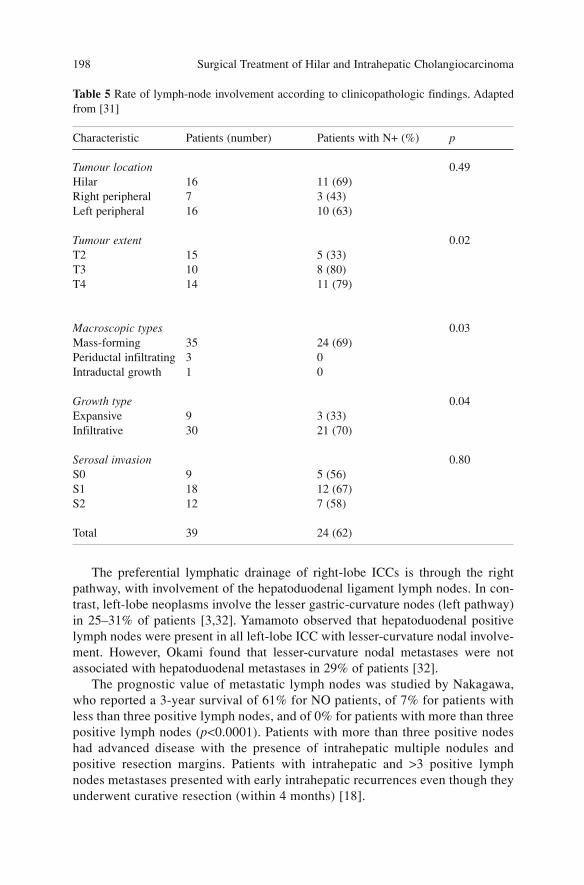
The preferential lymphatic drainage of right-lobe ICCs is through the rightpathway, with involvement of the hepatoduodenal ligament lymph nodes. In con-trast, left-lobe neoplasms involve the lesser gastric-curvature nodes (left pathway)in 25–31% of patients [3,32]. Yamamoto observed that hepatoduodenal positivelymph nodes were present in all left-lobe ICC with lesser-curvature nodal involve-ment. However, Okami found that lesser-curvature nodal metastases were notassociated with hepatoduodenal metastases in 29% of patients [32].
The prognostic value of metastatic lymph nodes was studied by Nakagawa,who reported a 3-year survival of 61% for NO patients, of 7% for patients withless than three positive lymph nodes, and of 0% for patients with more than threepositive lymph nodes (p<0.0001). Patients with more than three positive nodeshad advanced disease with the presence of intrahepatic multiple nodules andpositive resection margins. Patients with intrahepatic and >3 positive lymphnodes metastases presented with early intrahepatic recurrences even though theyunderwent curative resection (within 4 months) [18].
Surgical Treatment of Hilar and Intrahepatic Cholangiocarcinoma 198
Table 5 Rate of lymph-node involvement according to clinicopathologic findings. Adaptedfrom [31]
Characteristic Patients (number) Patients with N+ (%) p
Tumour location 0.49Hilar 16 11 (69) Right peripheral 7 3 (43)Left peripheral 16 10 (63)
Tumour extent 0.02T2 15 5 (33)T3 10 8 (80)T4 14 11 (79)
Macroscopic types 0.03Mass-forming 35 24 (69)Periductal infiltrating 3 0Intraductal growth 1 0
Growth type 0.04Expansive 9 3 (33)Infiltrative 30 21 (70)
Serosal invasion 0.80S0 9 5 (56)S1 18 12 (67)S2 12 7 (58)
Total 39 24 (62)
Microscopic Pattern
The main histologic patterns related to prognosis are: cellular differentiation,perineural, vascular and lymphatic invasion.
Concerning cellular differentiation, well or moderately differentiated neo-plasms have a better prognosis than poorly differentiated ones. Five-year sur-vival rates of 50, 39 and 0% respectively, were reported in patients with well,moderate and poorly differentiated tumours [16].
Perineural invasion is a negative prognostic factor and is associated with ahigh frequency of nodal metastases and vascular invasion. Suzuki reported 5-year survival rate of 83% in patients with MF cholangiocarcinoma without per-ineural infiltration vs. 8% in patients with perineural infiltration [13]. Nakagohriconfirmed these data, reporting a 5-year survival rate of 50 vs. 7% [20].
Lymphatic invasion represents a further negative independent prognostic fac-tor for survival. In one study, patients without lymphatic invasion had a 5-yearsurvival rate of 71% while none of the patients with lymphatic invasion survivedmore than 3 years [33].
Biological and Molecular Factors
Many biological and molecular prognostic factors have been identified in ICCincluding interleukin (IL)-6, hepatocyte growth factors (HGFs), c-met, trans-forming growth factor (TGF)-β, epidermal growth factor (EGF), c-erb-2, lym-phocyte inhibitory factor, k-ras and p53 [34–37].
The expression of IL-6 is inversely related with cellular proliferation anddirectly correlated with cholangiocarcinoma differentiation. Increase levels ofIL-6 are frequently found in well-differentiated cholangiocarcinoma whereasdecreased levels of this cytokine are observed in moderately or poorly differen-tiated ICC.
A mutation in the oncogene k-ras is a frequent finding in ICC; the prevalenceis related to the gross type: MF types rarely show k-ras mutations in contrast toperiductal infiltrating cholangiocarcinoma. According to Isa, such mutations arerelated to a poor prognosis, although the difference did not reach statistical sig-nificance (p=0.0898). Nevertheless in that study none of the patients with a sur-vival longer than 5 years had tumours with k-ras mutations.
The incidence of p53 mutation in intrahepatic cholangiocarcinoma rangesbetween 3 and 35%, and occurs especially in the MF type. MDM2 (inhibitoryoncoprotein of p53) expression increases in intrahepatic cholangiocarcinoma.This amplified expression of p53 is related to tumour stage and to the presenceof metastases. It represents a late event in tumour progression and is related witha poor prognosis.
Cholangiocarcinomas with increased expression of N-myristoyltransferase(NMT), a signal modulator of intracellular transduction, and p53 are moreaggressive and have a higher number of metastases.
Prognostic Factors 199
Cholangiocarcinomas with a decrease in the expression of the cycle-depend-ent kinase inhibitor p27kip1 are poorly differentiated. These tumours are char-acterised by increased T-stage as well as lymphatic and lymph-node invasion.Patients with this type of cholangiocarcinoma have a lower survival.
Alterations of E-caderin, α-catenin and β-catenin regulation in intrahepaticcholangiocarcinomas are associated with high tumour grade and stage, and rapiddisease progression.
Vascular endothelial growth factor (VEGF-C) is an important lympho-angio-genic factor. Park reported that the association of VEGF-C expression with thedevelopment of lymph-node metastases is statistically significant (p=0.032).This is also the case for positive resection margins (p=0.03). The increasedexpression of VEGF-C represented a negative independent prognostic factor alsofor survival [38].
Many studies have demostrated a role for mucins in complex biologicalprocesses, such as differentiation, epithelial cell renewal, cellular adhesion andcarcinogenesis. Tissue expression of the mucin proteins MUC 1 and 2 in ICC isrelated with gross type; high-level expression of MUC 1 is observed in MF andPI types compared to IG type, in which MUC 1 expression is usually low [39].
Recent studies have suggested that the serum concentration of MUC 5AC rep-resents negative prognostic marker in patients with ICC. Conclusive data on biolog-ical and molecular prognostic factors are still lacking and under evalutaion.
References
1. The Liver Cancer Study Group of Japan (2003). General rules for the clinical and patholog-ical study of primary liver cancer, 2nd edn. Kanehara, Tokyo
2. Sasaki A, Aramaki M, Kawano K et al (1998) Intrahepatic peripheral cholangiocarcinoma:mode of spread and choice of surgical treatment. Br J Surg 85(9):1206–1209
3. Yamamoto M, Takasaki K, Yoshikawa T (1999) Extended resection for intrahepatic cholan-giocarcinoma in Japan. J Hepatobiliary Pancreat Surg 6(2):117–121
4. Yamamoto M, Takasaki K, Yoshikawa T et al (1998) Does gross appearance indicate prog-nosis in intrahepatic cholangiocarcinoma? J Surg Oncol 69(3):162-167
5. Suh KS, Roh HR, Koh YT et al (2000) Clinicopathologic features of the intraductal growthtype of peripheral cholangiocarcinoma. Hepatology 31(1):12–17
6. Ohashi K, Nakajima Y, Kanehiro H et al (1995) Ki-ras mutations and p53 protein expres-sions in intrahepatic cholangiocarcinomas: relation to gross tumour morphology.Gastroenterology 109(5):1612–1617
7. Yeh CN, Jan YY, Yeh TS et al (2004) Hepatic resection of the intraductal papillary type ofperipheral cholangiocarcinoma. Ann Surg Oncol 11(6):606–611
8. Sakamoto E, Hayakawa N, Kamiya J et al (1999) Treatment strategy for mucin-producingintrahepatic cholangiocarcinoma: value of percutaneous transhepatic biliary drainage andcholangioscopy. World J Surg 23(10):1038–1043
9. Isa T, Kusano T, Shimoji H et al (2001) Predictive factors for long-term survival in patientswith intrahepatic cholangiocarcinoma. Am J Surg 181(6):507–511
10. Ikai I, Arii S, Ichida T et al (2005) Report of the 16th follow-up survey of primary liver can-cer. Hepatol Res 32(3):163–172
Surgical Treatment of Hilar and Intrahepatic Cholangiocarcinoma 200
11. Yamasaki S (2003) Intrahepatic cholangiocarcinoma: macroscopic type and stage classifica-tion. J Hepatobiliary Pancreat Surg 10(4):288–291
12. Uenishi T, Yamazaki O, Yamamoto T et al (2005) Serosal invasion in TNM staging of mass-forming intrahepatic cholangiocarcinoma. J Hepatobiliary Pancreat Surg 12(6):479–483
13. Hirohashi K, Uenishi T, Kubo S et al (2002) Histologic bile duct invasion by a mass-form-ing intrahepatic cholangiocarcinoma. J Hepatobiliary Pancreat Surg 9(2):233–236
14. Morimoto Y, Tanaka Y, Ito T et al (2003) Long-term survival and prognostic factors in thesurgical treatment for intrahepatic cholangiocarcinoma. J Hepatobiliary Pancreat Surg10(6):432–440
15. Suzuki S, Sakaguchi T, Yokoi Y et al (2002) Clinicopathological prognostic factors andimpact of surgical treatment of mass-forming intrahepatic cholangiocarcinoma. World JSurg 26(6):687–693
16. Nozaki Y, Yamamoto M, Ikai I et al (1998) Reconsideration of the lymph-node metastasispattern (N factor) from intrahepatic cholangiocarcinoma using the International UnionAgainst Cancer TNM staging system for primary liver carcinoma. Cancer 83(9):1923–1929
17. Isaji S, Kawarada Y, Taoka H et al (1999) Clinicopathological features and outcome ofhepatic resection for intrahepatic cholangiocarcinoma in Japan. J Hepatobiliary PancreatSurg 6(2):108–116
18. Nakagawa T, Kamiyama T, Kurauchi N et al (2005) Number of lymph-node metastases is asignificant prognostic factor in intrahepatic cholangiocarcinoma. World J Surg29(6):728–733
19. Miwa S, Miyagawa S, Kobayashi A et al (2006) Predictive factors for intrahepatic cholan-giocarcinoma recurrence in the liver following surgery. J Gastroenterol 41(9):893–900
20. Nakagohri T, Asano T, Kinoshita H et al (2003) Aggressive surgical resection for hilar-inva-sive and peripheral intrahepatic cholangiocarcinoma. World J Surg 27(3):289–293
21. Nakajima T, Kondo Y, Miyazaki M, Okui K (1988) A histopathologic study of 102 cases ofintrahepatic cholangiocarcinoma: histologic classification and modes of spreading. HumPathol 19(10):1228–1234
22. Valverde A, Bonhomme N, Farges O et al (1999) Resection of intrahepatic cholangiocarci-noma: a Western experience. J Hepatobiliary Pancreat Surg 6(2):122–127
23. Casavilla FA, Marsh JW, Iwatsuki S et al (1997) Hepatic resection and transplantation forperipheral cholangiocarcinoma. J Am Coll Surg 185(5):429–436
24. Okabayashi T, Yamamoto J, Kosuge T et al (2001) A new staging system for mass-formingintrahepatic cholangiocarcinoma: analysis of preoperative and postoperative variables.Cancer 92(9):2374–2383
25 Lang H, Kaiser GM, Zopf T et al (2006) Surgical therapy of hilar cholangiocarcinoma.Chirurg 77(4):325–334
26. Ohtsuka M, Ito H, Kimura F et al (2002) Results of surgical treatment for intrahepaticcholangiocarcinoma and clinicopathological factors influencing survival. Br J Surg89(12):1525–1531
27. Yamanaka N, Okamoto E, Ando T et al (1995) Clinicopathologic spectrum of resected extra-ductal mass-forming intrahepatic cholangiocarcinoma. Cancer 76(12):2449–2456
28. Cherqui D, Tantawi B, Alon R et al (1995) Intrahepatic cholangiocarcinoma. Results ofaggressive surgical management. Arch Surg 130(10):1073–1078
29. Yamamoto M, Takasaki K, Yoshikawa T (1999) Lymph-node metastasis in intrahepaticcholangiocarcinoma. Jpn J Clin Oncol 29(3):147–150
30. Huang JL, Biehl TR, Lee FT et al (2004) Outcomes after resection of cholangiocellular car-cinoma. Am J Surg 187(5):612–617
31. Tsuji T, Hiraoka T, Kanemitsu K et al (2001) Lymphatic spreading pattern of intrahepaticcholangiocarcinoma. Surgery 129(4):401–407
32. Okami J, Dono K, Sakon M et al (2003) Patterns of regional lymph-node involvement inintrahepatic cholangiocarcinoma of the left lobe. J Gastrointest Surg 7(7):850–856
33. Uenishi T, Hirohashi K, Kubo S et al (2001) Clinicopathological factors predicting outcomeafter resection of mass-forming intrahepatic cholangiocarcinoma. Br J Surg 88(7):969–974
Prognostic Factors 201
34. Abdalla EK, Vauthey JN (2001) Biliary tract cancer. Curr Opin Gastroenterol 17(5):450–45735. Rashid A, Ueki T, Gao YT et al (2002) K-ras mutation, p53 overexpression, and microsatel-
lite instability in biliary tract cancers: a population-based study in China. Clin Cancer Res8(10):3156–3163
36. Malhi H, Gores GJ (2006) Cholangiocarcinoma: modern advances in understanding a dead-ly old disease. J Hepatol 45(6):856–867
37. Nakanuma Y, Harada K, Ishikawa A et al (2003) Anatomic and molecular pathology of intra-hepatic cholangiocarcinoma. J Hepatobiliary Pancreat Surg 10(4):265–281
38. Park BK, Paik YH, Park JY et al (2006) The clinicopathologic significance of the expressionof vascular endothelial growth factor-C in intrahepatic cholangiocarcinoma. Am J ClinOncol 29(2):138–142
39. Andrianifahanana M, Moniaux N, Batra SK (2006) Regulation of mucin expression: mech-anistic aspects and implications for cancer and inflammatory diseases. Biochim BiophysActa 1765(2):189–222
Surgical Treatment of Hilar and Intrahepatic Cholangiocarcinoma 202
Staging Systems
The staging systems of intrahepatic cholangiocarcinoma (ICC) are based onpathological findings. The most common classification systems are the TNMUICC/AJCC classification (6th edn.) 2002 [1] and the TNM classification of theLiver Cancer Study Group of Japan (LCSGJ) (2nd English edn.) 2003 [2].
TNM Staging System According to UICC/AJCC
The sixth edition of TNM defined by International Union Against Cancer(UICC) is the same as that of the American Joint Committee on Cancer (AJCC).This edition, same as the previous one, uses the classification criteria developedfor hepatocellular carcinoma. Subdivision in stages is obtained through defini-tion of T, N, and M categories.
T Category
T category values the extent of disease based on size of neoplasm, focality, vas-cular invasion and extension to adjacent organs. It divides the neoplasms intofour categories (Table 1).
Although there are few reports on the validation of T-staging, the compo-nents that define T category (size, number, vascular invasion) have shown a closecorrelation with prognosis in several studies [3–8]. Miwa et al. have observedthat the size of the tumour significantly determined the prognosis; in 41 patients,5-year survival was 47% for those with lesions <4.5 cm vs. 16% for patientswhose lesions were larger (p=0.01). The same author underlined that portalinvolvement represents a negative prognostic factor: 5-year survival was 24% inpatients with portal involvement vs. 47% in those without (p=0.07) [7].
In a series of 1,364 surgical resections for ICC, the 16th national follow-upsurvey of Japan reported a 5-year survival of 66% for tumours <2 cm, 36% for
203A. Guglielmi, A. Ruzzenente, C. Iacono (eds.) Surgical Treatment of Hilar and ICC.© Springer 2008
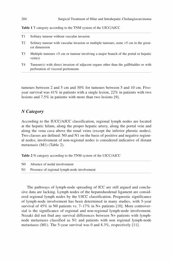
tumours between 2 and 5 cm and 30% for tumours between 5 and 10 cm. Five-year survival was 41% in patients with a single lesion, 22% in patients with twolesions and 7.5% in patients with more than two lesions [9].
N Category
According to the IUCC/AJCC classification, regional lymph nodes are locatedat the hepatic hilum, along the proper hepatic artery, along the portal vein andalong the vena cava above the renal veins (except the inferior phrenic nodes).Two classes are defined: N0 and N1 on the basis of positive and negative region-al nodes; involvement of non-regional nodes is considered indicative of distantmetastasis (M1) (Table 2).
The pathways of lymph-node spreading of ICC are still argued and conclu-sive data are lacking. Lymph nodes of the hepatoduodenal ligament are consid-ered regional lymph nodes by the UICC classification. Prognostic significanceof lymph-node involvement has been determined in many studies, with 5-yearsurvival of 45% in N0 patients vs. 7–17% in N+ patients [10]. More controver-sial is the significance of regional and non-regional lymph-node involvement.Nozaki did not find any survival differences between N+ patients with lymph-node metastases classified as N1 and patients with non regional lymph-nodemetastases (M1). The 5-year survival was 0 and 8.3%, respectively [11].
Surgical Treatment of Hilar and Intrahepatic Cholangiocarcinoma 204
Table 1 T category according to the TNM system of the UICC/AJCC
T1 Solitary tumour without vascular invasion
T2 Solitary tumour with vascular invasion or multiple tumours, none >5 cm in the great-est dimension
T3 Multiple tumours >5 cm or tumour involving a major branch of the portal or hepaticvein(s)
T4 Tumour(s) with direct invasion of adjacent organs other than the gallbladder or withperforation of visceral peritoneum
Table 2 N category according to the TNM system of the UICC/AJCC
N0 Absence of nodal involvement
N1 Presence of regional lymph-node involvement
M Category
Evaluation of M category is based on the presence of metastases to other organsor non-regional lymph nodes. Two categories are therefore identified: M0 andM1 (Table 3).
Stage Grouping
The three categories T, N and M, are combined to subdivide patients in homoge-neous prognostic groups. In the sixth edition of the UICC/AJCC classification,six stages have been defined (Table 4).
The staging system proposed by UICC/AJCC has been formulated for hepa-tocellular carcinoma, as previously stated. This is the main limit of this classifi-cation in that two diseases with completely different biological behaviours (i.e.vascular invasion, limph-nodes involvement) are joined.
The literature contains very few validation experiences regarding the 6thTNM UICC/AJCC classification. Lang identified a correlation between TNMstages, curative resection survival and recurrence. In this clinical study of 27patients who underwent surgical resection all stage I and II patients underwent
Staging Systems 205
Table 3 M category according to the TNM system of the UICC/AJCC
M0 Absence of metastases
M1 Presence of metastases
Table 4 Stage grouping according to the TNM system of the UICC/AJCC
T N M
Stage I T1 N0 M0
Stage II T2 N0 M0
Stage IIIA T3 N0 M0
Stage IIIB T4 N0 M0
Stage IIIC Any T N1 M0
Stage IV Any T Any N M1
a R0 resection vs. 42% of stage III patients [12]. Several published studies did,however, confirm the 5th edition of TNM UICC. These demonstrated that TNMstage is related to long-term outcome. In 27 patients, Isa reported a 5-year sur-vival of 42% for stage I and II patients, 25% for those with stage III and 0% forthose with stage IV (p=0.001) [11].
Similar results were obtained by Fu in 79 patients in whom median survivalafter surgical resection was 20 months in stage I, 16 months in stage II, 15months in stage IIIa, 9 months in stage IIIb and 5 months in stage IVa [5].
TNM Classification According to the LCSGJ
In 1992, the LCSGJ convened a research group to formulate a new staging sys-tem specific for ICC. This group carried out a multicentric study with nineJapanese surgical institutions and analysed 173 patients who underwent resec-tion with curative intent for ICC. In 1997, the first edition of this new classifica-tion was published and it was revised in 2003 [2].
The Japanese classification of cholangiocarcinoma defines intrahepatic neo-plasm as originating from the peripheral branches of biliary ducts (beyond sec-ond-order divisions). Based on the gross appearance of the tumour, three pat-terns of growth are defined. that can be present alone or in combination: mass-forming type (MF), periductal infiltrating type (PI) and intraductal growth (IG)type cholangiocarcinoma (Fig. 1).
Surgical Treatment of Hilar and Intrahepatic Cholangiocarcinoma 206
Fig. 1. Growth patterns of cholagiocarcinoma: mass-forming (MF), periductal infiltrating(PI) and intraductal growth (IG) types. a Intraductal papillary growth; b intraduct, growthforming a tumour thrombus
a b
The MF type is characterised by a distinct, round-shaped mass localisedinside the liver, with a well-defined margin between cancer and the surroundingparenchyma.
The PI type is characterised by diffuse infiltration along the major axis of theportal tract, with involvement of the biliary tract, vessels and periductal connec-tive tissue.
The IG type is characterised by a papillary and/or granular growth within adilated duct; sporadically a superficial spread or the formation of intraductaltumoral nodules is seen. Dilatation of the cystic duct due to mucin, produced byan intraductal tumour, that collects inside the ducts should not be misdiagnosedas cystadenocarcinoma of the biliary duct.
Macroscopic type is determined by the most representative type inside thelesion; thus, when more than one type is present the neoplasm, is classified bythe more prevalent form and the other types are described with a + symbol (i.e.MF+PI).
A LCSGJ staging system for MF type or forms in which the MF type is preva-lent had been formulated. A staging classification for the PI and IG forms has notbeen formulated yet, as there are very few data. The staging system considers thelocal extent of disease (T), nodal diffusion (N) and distant metastases (M).
T Category
Valuation of the extent of neoplasm (T) is assessed by one or more of three dif-ferent parameters: (1) focality (single or multifocal), (2) size of the lesion (≤2 or>2 cm) and (3) vascular (portal vein or hepatic vein) and/or serosal involvement.
Four T categories with prognostic value are defined (Table 5).This division inT categories comes from the multicentric experience of the LCSGJ. In that studyof 136 patients with MF cholangiocarcinoma, univariate analysis identified the fol-lowing prognostic factors correlated with survival: size ≥2 cm (HR 2.39), lymphnodes metastases (HR 2.36), serosal invasion (HR 2.19), portal-vein invasion(HR 1.68) multiple nodules (HR 1.95) and hepatic-vein invasion (HR 1.18) [6].These factors have been identified also in other clinical studies that highlight among
Staging Systems 207
Table 5 T category according to the LCSGJ
Criteria1: Unifocal lesion2: Size ≤2 cm3: No vascular or serosal involvement
T1 All criteria are satisfied
T2 Two out of three criteria are satisfied
T3 One out of three criteria is satisfied
T4 No criterion is satisfied
the most important prognostic factors size, focality and vascular invasion [13–17].More controversial is the prognostic significance of serosal invasion; some authorsemphasised its value, such as Ohtsuka [8] who identified the prognostic value ofthis factor in univariate analysis, whereas other authors, such as Uenishi, did notfind any significant differences in survival between patients with or without seros-al invasion, with a 5-year survival of 24 and 39% (p=0.16), respectively [8,13].
N Category
The lymphatic system of the liver can be divided into a superficial and a deepsystem. The lymph of the upper portions of the liver (diaphragmatic side) drainsto lymphatic stations situated at the confluence of the hepatic veins and near thefalciform, coronary and triangular ligaments. Then the lymph passes through thediaphragm and enters into the mediastinal lymphatic system. Inferior portions ofthe liver (visceral side), in contrast, drain to lymphatic stations of the hepatichilum, lesser omentum and subsequently to abdominal lymphatic system.
According to the Japanese classification, lymph nodes are numbered asshown in Table 6.
Unlike the first English edition, in the second classification of the LCSGJ Ncategory is no longer subdivided as a function of the site of the neoplasm.
Surgical Treatment of Hilar and Intrahepatic Cholangiocarcinoma 208
Table 6 Japanese classification of lymph-nodes stations
Number Site
1 Right cardial2 Left cardial3 Lesser gastric curvature4 Greater curvature5 Suprapyloric6 Infrapyloric7 Left gastric artery8 Hepatic artery9 Celiac trunk10 Splenic hilum11 Splenic artery12 Hepatoduodenal ligament13 Retropancreatic 14 Superior mesenteric artery15 Middle colic16 Para-aortic17 Superior pancreatic18 Inferior pancreatic19 Subdiaphragmatic20 Para-oesophageal110 Intrathoracic para-oesophageal111 Supradiaphragmatic
Clinical studies in patients with ICC have found different lymphatic drainagepatterns depending on the site of the tumour. Nozaki observed that neoplasmssituated in the left lobe show lymph-node involvement along the lesser gastriccurvature in up to 30% of N+ patients [11]. In left lobe cholangiocarcinoma, dif-fusion to the lymph nodes of the lesser curvature can be present even in theabsence of a hepatoduodenal ligament localisation in up to 29% of patients [18].
Since these observations in the first English edition of the LCSGJ classifica-tion, lymphatic stations for cholangiocarcinoma have been differentiateddepending on the site of the tumour. Regional lymph nodes for lesions of theright lobe are those of the hepatoduodenal ligament (12), whereas for left-lobetumours hepatoduodenal ligament (12), lesser gastric curvature (3) and right car-dial (1) nodes are involved. Other lymph-node localisations have been defined asnon-regional (N2 and N3).
The simplification of the second English edition is the product of many clini-cal studies that did not show a significant difference of prognosis in patients withregional (N1) positive lymph nodes versus those with non-regional positive lymphnodes (N2 and N3). Yamamoto, in a study of 51 patients with ICC, did not observesignificant differences in survival between patients with N1, N2 and N3 positivelymph nodes according to the first LCSGJ classification [19]. These data wereconfirmed in a nation-wide survey comprising 791 Japanese institutions. Theresults of 1,087 patients with ICC did not show significant differences in the 3-year survival of N1, N2 and N3 patients: 29, 22 and 12%, respectively [20].
The second edition of the LCGSJ classification subdivides N category onlyaccording to lymph-node involvement, without providing a proposal for furtherlymph-node grouping at the first or second level for this type of neoplasm. N cat-egory is therefore divided in two categories: N1 and N0 (Table 7).
M Category
Evaluation of M category is based on the presence of metastases to other organsor non-regional lymph nodes. Two categories are defined: M0 and M1 (Table 8).
Staging Systems 209
Table 7 N category according to the LCSGJ
N0 No lymph-node metastases
N1 Lymph-node metastases
Table 8 M category according to the LCSGJ
M0 Absence of metastases
M1 Presence of metastases

Stage Grouping
As noted previously, the staging system proposed by the LCSGJ can be appliedonly to the MF type of ICC and to tumours in which the MF type is prevalent. Itcannot be applied to PI and IG neoplasms. The stage defined by the T, N and Mcategories determines four subdivisions having different prognoses (Table 9).
The survival of patients is significantly related to different stages: the 5-yearsurvival for stage I was 100%, for stage II it was 70%, for stage III 40% and forstage IV 10% [6].
Other experiences in the literature have confirmed the prognostic signifi-cance of the LCGSJ staging system. Uenishi reported that the 5-year survival of63 patients was 100% for those with stage I, 54% for patients with stage II, 44%for those with stage III and 7% for stage IV patients [13].
Conclusions
Correct staging of ICC is still a subject of debate. However, the differencesbetween ICC and hepatocellular carcinoma are clear. For these reasons, the stag-ing system proposed by the UICC/AJCC, which was mainly developed to stagehepatocarcinoma, is quite limited when it is applied to ICC. The UICC/AJCCclassification does not differentiate neoplasms on the basis of type of tumoralgrowth and provides a limited differentiation of the criteria of local disease anddisease involving lymph-node diffusion. In contrast, the classification proposedby the LCSGJ is specific for ICC. In addition, it differentiates these neoplasmsas a function of tumoral growth. Unfortunately LCSGJ classification refers onlyto MF-type, whereas there is not staging system for PI- and IG-types.Nonetheless the classification proposed by the LCSGJ will eventually becomethe most useful to assess the prognosis of ICC.
Surgical Treatment of Hilar and Intrahepatic Cholangiocarcinoma 210
Table 9 Stage grouping according to the LCSGJ
T N M
Stage I T1 N0 M0
Stage II T2 N0 M0
Stage III T3 N0 M0
Stage IVA T4 N0 M0
Stage IVB T1–T4 N1 M0
T1–T4 N0, N1 M1
References
1. Sobin LH, Wittekind C (eds) (2002) TNM classification of malignant tumours, 6th edn.Wiley, New York
2. Liver Cancer Study Group of Japan (2003) General rules for clinical and pathological studyof primary liver cancer, 2nd English Edition. Kanehara, Tokyo
3. Roayaie S, Guarrera JV, Ye MQ et al (1998) Aggressive surgical treatment of intrahepaticcholangiocarcinoma: predictors of outcomes. J Am Coll Surg 187(4):365–372
4. Kinoshita H, Tanimura H, Uchiyama K et al (2002) Prognostic factors of intrahepaticcholangiocarcinoma after surgical treatment. Oncol Rep 9(1):97–101
5. Fu XH, Tang ZH, Zong M et al (2004) Clinicopathologic features, diagnosis and surgicaltreatment of intrahepatic cholangiocarcinoma in 104 patients. Hepatobiliary Pancreat Dis Int3(2):279–283
6. Yamasaki S (2003) Intrahepatic cholangiocarcinoma: macroscopic type and stage classifica-tion. J Hepatobiliary Pancreat Surg 10(4):288–91
7. Miwa S, Miyagawa S, Kobayashi A et al (2006) Predictive factors for intrahepatic cholan-giocarcinoma recurrence in the liver following surgery. J Gastroenterol 41(9):893–900
8. Ohtsuka M, Ito H, Kimura F et al (2002) Results of surgical treatment for intrahepaticcholangiocarcinoma and clinicopathological factors influencing survival. Br J Surg89(12):1525–1531
9. Ikai I, Arii S, Ichida T et al (2005) Report of the 16th follow-up survey of primary liver can-cer. Hepatol Res 32(3):163–172
10. Isa T, Kusano T, Shimoji H et al (2001) Predictive factors for long-term survival in patientswith intrahepatic cholangiocarcinoma. Am J Surg 181(6):507–511
11. Nozaki Y, Yamamoto M, Ikai I et al (1998) Reconsideration of the lymph-node metastasispattern (N factor) from intrahepatic cholangiocarcinoma using the International UnionAgainst Cancer TNM staging system for primary liver carcinoma. Cancer 83(9):1923–1929
12. Lang H, Sotiropoulos GC, Fruhauf NR et al (2005) Extended hepatectomy for intrahepaticcholangiocellular carcinoma (ICC): when is it worthwhile? Single center experience with 27resections in 50 patients over a 5-year period. Ann Surg 241(1):134–143
13. Uenishi T, Yamazaki O, Yamamoto T et al (2005) Serosal invasion in TNM staging of mass-forming intrahepatic cholangiocarcinoma. J Hepatobiliary Pancreat Surg 12(6):479–483
14. Uenishi T, Hirohashi K, Kubo S et al (2001) Clinicopathological factors predicting outcomeafter resection of mass-forming intrahepatic cholangiocarcinoma. Br J Surg 88(7):969–974
15. Morimoto Y, Tanaka Y, Ito T et al (2003) Long-term survival and prognostic factors in thesurgical treatment for intrahepatic cholangiocarcinoma. J Hepatobiliary Pancreat Surg10(6):432–440
16. Inoue K, Makuuchi M, Takayama T et al (2000) Long-term survival and prognostic factorsin the surgical treatment of mass-forming type cholangiocarcinoma. Surgery127(5):498–505
17. Shimada M, Yamashita Y, Aishima S et al (2001) Value of lymph node dissection duringresection of intrahepatic cholangiocarcinoma. Br J Surg 88(11):1463–1466
18. Okami J, Dono K, Sakon M et al (2003) Patterns of regional lymph-node involvement inintrahepatic cholangiocarcinoma of the left lobe. J Gastrointest Surg 7(7):850–856
19. Yamamoto M, Takasaki K, Yoshikawa T (1999) Lymph-node metastasis in intrahepaticcholangiocarcinoma. Jpn J Clin Oncol 29(3):147–150
20. Ikai J, Itai Y, Okita K et al (2004) Report of the 15th follow-up survey of primary liver can-cer. Hepatol Res 28:21–29
Staging Systems 211
Surgical Treatment
Intraoperative Assessment of Resectability
At the time of diagnosis, patients with intrahepatic cholangiocarcinoma (ICC)are frequently found to have disease beyond the limits of surgical therapy, suchthe presence of intrahepatic satellite nodules, vascular invasion, or regionallymph-nodes metastases. In such patients, the resectability rate varies from 19 to74% [1–4]. Moreover, exploratory laparotomy or R1 resection has a poor prog-nosis, with median patient survival of 5 months and postoperative complicationsof 17% [4]. Consequently, palliative resection is not justified; instead, there isclearly a need to improve the assessment of resectability, in order to avoidunnecessary laparotomy and to develop an aggressive approach to obtain com-plete resection (R0).
Both of these aims can be achieved by laparoscopy with laparoscopic ultra-sound that showed a reduction in exploratory laparotomy from 27 to 36% [5,6].
As previously described for hilar cholangiocarcinoma, in ICC after laparoto-my the abdominal cavity has to be carefully inspected to identify peritonealmetastases and loco-regional or distantly involved lymph nodes. The liver isexplored with the aid of intraoperative ultrasound to determine the extent andsite of the neoplasm as well as the number and localisation of metastatic nod-ules. All doubtful lesions, either peritoneal or intrahepatic, and lymph nodes thatappear increased in size or consistency must be sent for frozen sectioning.
In a considerable number of patients (36–41% [4,7]), hepatic resection, con-sidered curative, showed involvement of the liver margins, mostly because ofinfiltration by small satellite lesions. Intraoperative ultrasound allows the detec-tion of intrahepatic metastases <1 cm. It remains to be investigated whetherintraoperative ultrasound with contrast agent can reduce the number of incom-plete resections by providing better visualisation of small, otherwise unde-tectable lesions [8,9].
It is also important to carefully evaluate the condition of the hepaticparenchyma, especially in the presence of chronic hepatopathy or cirrhosis,
213A. Guglielmi, A. Ruzzenente, C. Iacono (eds.) Surgical Treatment of Hilar and ICC.© Springer 2008

either of which could limit or contraindicate surgical resection. This situation isnot exceptional: Isaji [10] reported that 30% of patients with peripheral ICChave HBsAg- or HCV-positive hepatitis, and this rate reaches 42% in patientswith mass-forming ICC.
Indications for Surgical Resection
As with extrahepatic cholangiocarcinoma, R0 of ICC is the most effective treat-ment and the only therapy associated with prolonged disease-free survival.Nonetheless, there is currently little agreement on the indications for surgicalresection. According to some authors curative resection (R0) of ICC is feasibleonly in patients with a single lesion, negative lymph nodes and resectable hepat-ic margins of >1 cm [7], or in patients without gross intrahepatic biliary infiltra-tion; all patients with stage III and IV disease are therefore excluded from sur-gical resection [11]. Following these indications the results are good, with 2- and5-year survival rates of 100 and 42%, respectively [7,11].
Unfortunately, according to these highly selective criteria, patients who pres-ent with one or more negative prognostic factors will not undergo surgical resec-tion but only palliative treatment. The survival rate of such patients is between 6and 12 months. It has also been suggested that, even in the presence of negativeprognostic factors, better results can be achieved with surgical resection thanwith palliative therapy alone, with some reports of long-term survival of thesepatients. It is therefore evident that the precise indications for surgical resectionrequire an analysis of the value of each prognostic factor, and a comparison ofthe results with the limited benefits of palliative therapy with respect to morbid-ity and mortality.
Type of Surgical Resection
Surgical resection of cholangiocarcinoma is represented by anatomic hepaticresection. Advanced-stage neoplasms must often be treated by extended hepate-ctomy, with extension of the resection to the extrahepatic biliary tract, vascularhilar structures, vena cava and diaphragm. Usually, this type of neoplasm devel-ops in a non-cirrhotic liver, which allows the surgeon to perform extended resec-tion without the need of portal-vein embolisation. The mortality and morbidityrates in these cases vary from 3–9% and 30–40%, respectively [4,12–15].
The surgical approach has to be tailored in consideration of the gross type ofneoplasm, the presence of multifocal lesions (perilesional satellitosis and distantmetastases), vascular, biliary and serosal involvement and lymph-nodes metas-tases. However, even after aggressive resections the prognosis remains unsatis-factory, with a 5-year survival rate of 21–42% [11,15–19].
Surgical Treatment of Hilar and Intrahepatic Cholangiocarcinoma 214

The intraductal growth (IG) type of ICC shows intraductal and/or granulargrowth and is sometimes associated with carcinoma that spreads over the super-ficial mucosa or with intraductal tumour thrombus. This type of biliary epithe-lial neoplasia is frequently associated with gastrointestinal metaplasia and over-production of mucin and mucobilia, i.e. biliary intraductal growth mucin-pro-ducing cholangiocarcinoma [20]. In these patients, in order to obtain correct pre-operative staging of the tumour Sakamoto [20] recommended preoperative per-cutaneous biliary drainage and percutaneous cholangioscopy with biopsy inorder to accurately assess the extent of the tumour into the intrahepatic segmen-tal duct. This type of staging can be also achieved non invasively through theendoscopic route with peroral cholangioscopy.
After anatomic hepatic resection, the biliary duct margins should be evaluat-ed by frozen sectioning. If neoplastic infiltration of the proximal biliary marginis diagnosed, the resection must be extended to the biliary confluence (Fig. 1).If the distal intrapancreatic margin is positive, pancreaticoduodenectomy is indi-cated.
Because of the high percentage of satellite nodules in mass-forming-type(MF) neoplasms, from 26 to 58% of patients treated surgically [1,2,7,10,21], anaccurate intraoperative sonographic study is mandatory, both near the mainlesion and of all the liver in order to identify metastatic nodules and verify theplane and radicality of resection. All suspicious nodules should be verified withintraoperative frozen sectioning. Anatomic resection with a section parenchymalmargin at least 5–10 mm (R0) is adequate for MF neoplasms located peripheral-ly (Fig. 2).
In cases of MF lesions either with biliary duct infiltration or located central-ly near the biliary confluence, frozen section is always indicated to assess themargins of the biliary resection; if the results are positive, the resection has to beextended.
Surgical Treatment 215
Fig. 1a,b A centrally located, intraductal-growth type of cholangiocarcinoma , treated withright portal-vein embolisation (white arrow) and right trisectionectomy with extrahepaticbile duct resection, a CT scan; b surgical specimen
a b

The presence of intrahepatic metastases represents a negative independentfactor that is always associated with very low survival similar to those of pallia-tive treatment: Madariaga [1] reported a 3-year survival that is nil and no sur-vivors after 14 months; Isa [22] found a median survival of 19 months; in theseries of Nakagawa [15], none of the patients with multiple lesions and extend-ed lymph-node involvement reached a survival of 3 years, although Uenishi [23]published a 3-year survival rate in such cases of 6%.
In the light of these results, surgical indications for multifocal lesions are thesubject of debate. We believe that multifocal lesions with unilobar satellite nod-ules or located in the same segmental area of the main lesion can be resected ifthe programmed operation is of low risk and no other negative prognostic fac-tors, such as nodal metastases, are present.
Multiple bilobar lesions or lymph-node metastases contraindicate surgicalresection, since R0 resection is not possible. In these patients with lymph-nodeand intrahepatic metastases, the prognosis is as poor as that of non-resectedpatients, and adjuvant or neoadjuvant therapies must be considered.
Periductal-infiltrating-type (PI) neoplasms that spread along Glisson’s sheaththrough lymphatic vessels, with a low incidence of spreading in the portal sys-tem, frequently show nodal involvement as well as perineural and vascular inva-sion. In these cases, the surgical approach is the same as for MF neoplasms andnecessitates anatomic hepatic resection always in association with biliary-ductsampling and extrahepatic biliary resection, if the margins are positive. Lymph-node dissection is also indicated with the modalities described below.
Ultimately, mixed forms (MF+PI) of neoplasms are characterised by consid-erable biological aggressiveness, with vascular and lymph-node infiltration in80% of the cases, intrahepatic metastases in 46% [10] and a worse prognosisthan either MF or PI neoplasms. The 5-year survival rate is between 0 and 7%[16,22]. These poor results are the consequences of early intrahepatic and extra-hepatic diffusion of the neoplasm, which makes it difficult to achieve R0 resec-tion (Fig. 3).
Surgical Treatment of Hilar and Intrahepatic Cholangiocarcinoma 216
Fig. 2a,b Mass-forming type of cholangiocarcinoma with satellite lesions (a) treated withright hepatectomy and lymph-node dissection. Intraoperative field after resection (b)
a b

In these cases, the surgical approach is the same as for the PI type, i.e.anatomic hepatic resection and biliary-duct sampling with likely extrahepaticbiliary resection. It must be kept in mind that patients with advanced PI or mixedform (MF+PI) of cholangiocarcinoma have not benefited from extended surgery(hepatic and biliary resection, extended lymphadenectomy) in terms of improvedsurgical outcome compared to conventional resection (hepatic resection alone),with increased rates of mortality and morbidity. The surgical indications are con-troversial in these cases and must be limited to patients without other negativeprognostic factors (intrahepatic metastases and positive lymph nodes).
The size of the neoplasm does not represent a prognostic factor that limitssurgical indication, even if small tumours show a better prognosis than largeones. This suggests that if radical removal of the tumour is technically feasibleand can be accomplished safely, it should be performed independently of thesize of the intrahepatic tumour [17].
Vascular involvement is present in 27–85% of patients [24] and represents anegative prognostic factor. Casavilla [25], in a series of 39 patients, found thatthose with vascular infiltration did not reach 5-year survival. However, Inoue[17] reported 5-year survival rates of 60 and 22% in patients without and withvascular infiltration, respectively; survival in the latter group was significantlybetter than in non-resected patients. Therefore vascular invasion, even if it is anegative prognostic factor, does not represent an absolute contraindication tosurgery.
Indications for Lymphadenectomy
The rational extent of radical lymphadenectomy for ICC has not been clearlydefined and there is no consensus on the role of lymph-node dissection [18,26].The incidence of lymph-node metastasis in ICC is 43–62% [1,2,7,10,15,21].
Surgical Treatment 217
Fig. 3a,b Intrahepatic cholangiocarcinoma of the periductal infiltrating type (a).Intrahepatic cholangiocarcinoma of the combined mass-forming and periductal-infiltratingtypes (MF+PI) (b)
a b

Several series have shown that one of the strongest prognostic factors in ICCis lymph-node involvement [17,25,27,28]. All of these studies reported that nopatients with lymph-node involvement survived for more than 3 years after sur-gery. Inoue [17] stated that lymph-node metastasis in MF neoplasms is a sign ofnon-curative disseminated disease, and therefore hepatectomy is contraindicatedif metastatic involvement is observed at the time of lymph-node sampling.Nonetheless, Murakami [29] and Weber [5] reported long-term survivors (morethan 5 years) with lymph-node metastasis. Ohtsuka [12] did not consider lymph-node involvement to be a significant prognostic factor in patients who underwentlymph-node dissection, with a few patients who survived more than 3 years.
Isa [22] reported 3-year survival rate in N+ patients of 6.7%, with a mediansurvival of 10 months. Some authors have underlined the prognostic importanceof the number of positive lymph nodes, analogous to gastric and colonic cancers:Suzuki [30] reported that 5-year survival in patients with a single lymph-nodemetastasis is 33%, and that patients with two or more lymph-node metastases donot survive beyond 2 years; Nakagawa [15] series had 3-year survival rates of62, 50 and 0% in patients with N0 tumours, with one or two nodes and with morethan three nodes, respectively.
These results suggest that, although lymph-node involvement may generallybe associated with an unfavourable prognosis, long-term survival might beexpected following aggressive surgical resection, including lymph-node dissec-tion, in highly selected patients, providing that nodal diffusion, as confirmed byintraoperative sampling, is not beyond regional station.
In the presence of extensive nodal diffusion or in association with other neg-ative prognostic factors, such as multifocal involvement, resection does notguarantee better results than palliative therapy and is therefore not recommend-ed.
Major lymphatic spreading of ICC follows three routes: (1) through the hepa-toduodenal ligament; (2) through the paracardial, lesser curvature and left gas-tric artery and (3) through the inferior phrenic artery or directly from the rightliver to the lateral para-aortic group. Neoplasms located in the left lobe presenteither a right pathway of diffusion to the hepatoduodenal ligament (with N+ in38% of patients) or a left pathway to the right paracardial stations, along thelesser curvature and left gastric artery (with N+ in 31% of patients) [31].Consequently, the rational extent of radical lymphadenectomy should includenot only the hepatoduodenal ligament with the hepatic artery, para-aortic, retro-pancreatic and mesenteric nodes but also the left gastric nodes, the perigastricnodes along the lesser curvature, and the right paracardial nodes, especially inleft-located lesions [26].
Before surgical resection we always submit hepatoduodenal ligament nodesor grossly suspected nodes to frozen section. If more than three nodes or distant-station nodes are positive, the indication for hepatic resection becomes contro-versial and might be considered only in selected patients with low surgical riskand single lesion. In these cases, combination with adjuvant therapy must beconsidered.
Surgical Treatment of Hilar and Intrahepatic Cholangiocarcinoma 218
Extrahepatic Metastases
The presence of peritoneal metastases detected with laparoscopy or laparotomyis an absolute contraindication to surgical operation. Isaji [10] reported that theoutcome in 17 patients who underwent resection for stage IV B neoplasms wasas poor as that in 12 patients who did not undergo resection, with no survivorsafter 2 years. In these cases, adjuvant therapy may be considered, even if satis-factory results have not been reported until now.
References
1. Madariaga JR, Iwatsuki S, Todo S et al (1998) Liver resection for hilar and peripheralcholangiocarcinomas: a study of 62 cases. Ann Surg 227(1):70–79
2. Yamanaka N, Okamoto E, Ando T et al (1995) Clinicopathologic spectrum of resected extra-ductal mass-forming intrahepatic cholangiocarcinoma. Cancer 76(12):2449–2456
3. Lieser MJ, Barry MK, Rowland C et al (1998) Surgical management of intrahepatic cholan-giocarcinoma: a 31-year experience. J Hepatobiliary Pancreat Surg 5(1):41–47
4. Lang H, Sotiropoulos GC, Fruhauf NR (2005) Extended hepatectomy for intrahepaticcholangiocellular carcinoma (ICC): when is it worthwhile? Single center experience with 27resections in 50 patients over a 5-year period. Ann Surg. Jan 241(1):134–143
5. Weber SM, Jarnagin WR, Klimstra D (2001) Intrahepatic cholangiocarcinoma: resectabili-ty, recurrence pattern, and outcomes. J Am Coll Surg 193(4):384–391
6. Goere D, Wagholikar GD, Pessaux P et al (2006) Utility of staging laparoscopy in subsetsof biliary cancers : laparoscopy is a powerful diagnostic tool in patients with intrahepaticand gallbladder carcinoma. Surg Endosc 20(5):721–725
7. Cherqui D, Tantawi B, Alon R et al (1995) Intrahepatic cholangiocarcinoma. Results ofaggressive surgical management. Arch Surg 130(10):1073–1078
8. Harvey CJ, Blomley MJ, Eckersley RJ et al (2000) Pulse-inversion mode imaging of liverspecific microbubbles: improved detection of subcentimetre metastases. Lancet355(9206):807–808
9. Skjoldbye B, Pedersen MH, Struckmann J et al (2002) Improved detection and biopsy ofsolid liver lesions using pulse-inversion ultrasound scanning and contrast agent infusion.Ultrasound Med Biol 28(4):439–444
10. Isaji S, Kawarada Y, Taoka H et al (1999) Clinicopathological features and outcome ofhepatic resection for intrahepatic cholangiocarcinoma in Japan. J Hepatobiliary PancreatSurg 6(2):108–116
11. Harrison LE, Fong Y, Klimstra DS et al (1998) Surgical treatment of 32 patients with periph-eral intrahepatic cholangiocarcinoma. Br J Surg 85(8):1068–1070
12. Ohtsuka M, Ito H, Kimura F et al (2002) Results of surgical treatment for intrahepaticcholangiocarcinoma and clinicopathological factors influencing survival. Br J Surg89(12):1525–1531
13. Morimoto Y, Tanaka Y, Ito T et al (2003) Long-term survival and prognostic factors in thesurgical treatment for intrahepatic cholangiocarcinoma. J Hepatobiliary Pancreat Surg10(6):432–440
14. Huang JL, Biehl TR, Lee FT et al (2004) Outcomes after resection of cholangiocellular car-cinoma. Am J Surg 187(5):612–617
15. Nakagawa T, Kamiyama T, Kurauchi N et al (2005) Number of lymph-node metastases is asignificant prognostic factor in intrahepatic cholangiocarcinoma. World J Surg29(6):728–733
Surgical Treatment 219

16. Yamamoto M, Takasaki K, Yoshikawa T et al (1998) Does gross appearance indicate prog-nosis in intrahepatic cholangiocarcinoma? J Surg Oncol 69(3):162–167
17. Inoue K, Makuuchi M, Takayama T et al (2000) Long-term survival and prognostic factorsin the surgical treatment of mass-forming type cholangiocarcinoma. Surgery127(5):498–505
18. Shimada M, Yamashita Y, Aishima S et al (2001) Value of lymph node dissection duringresection of intrahepatic cholangiocarcinoma. Br J Surg 88(11):1463–1466
19. Shirabe K, Shimada M, Harimoto N et al (2002) Intrahepatic cholangiocarcinoma: its modeof spreading and therapeutic modalities. Surgery 131(1 Suppl):S159-S164
20. Sakamoto E, Hayakawa N, Kamiya J et al (1999) Treatment strategy for mucin-producingintrahepatic cholangiocarcinoma: value of percutaneous transhepatic biliary drainage andcholangioscopy. World J Surg 23(10):1038–1043; discussion 1043–1044
21. Sasaki A, Aramaki M, Kawano K et al (1998) Intrahepatic peripheral cholangiocarcinoma:mode of spread and choice of surgical treatment. Br J Surg 85(9):1206–1209
22. Isa T, Kusano T, Shimoji H et al (2001) Predictive factors for long-term survival in patientswith intrahepatic cholangiocarcinoma. Am. J. Surg 181(6):507–511
23. Uenishi T, Yamazaki O, Yamamoto T et al (2005) Serosal invasion in TNM staging of mass-forming intrahepatic cholangiocarcinoma. J Hepatobiliary Pancreat Surg 12(6):479–483
24. Weinbren K, Mutum SS (1983) Pathological aspects of cholangiocarcinoma. J Pathol139(2):217–238
25. Casavilla FA, Marsh JW, Iwatsuki S et al (1997) Hepatic resection and transplantation forperipheral cholangiocarcinoma. J Am Coll Surg 185(5):429–436
26. Tsuji T, Hiraoka T, Kanemitsu K et al (2001) Lymphatic spreading pattern of intrahepaticcholangiocarcinoma. Surgery 129(4):401–407
27. Uenishi T, Hirohashi K, Kubo S et al (2001) Clinicopathological factors predicting outcomeafter resection of mass-forming intrahepatic cholangiocarcinoma. Br J Surg 88(7):969–974
28. Chu KM, Lai EC, Al-Hadeedi S et al (1997) Intrahepatic cholangiocarcinoma. World J Surg21(3):301–305
29. Murakami Y, Yokoyama T, Takesue Y (2000) Long-term survival of peripheral intrahepaticcholangiocarcinoma with metastasis to the para-aortic lymph nodes. Surgery127(1):105–106
30. Suzuki S, Sakaguchi T, Yokoi Y et al (2002) Clinicopathological prognostic factors andimpact of surgical treatment of mass-forming intrahepatic cholangiocarcinoma. World JSurg 26(6):687–693
31. Okami J, Dono K, Sakon M et al (2003) Patterns of regional lymph-node involvement inintrahepatic cholangiocarcinoma of the left lobe. J Gastrointest Surg 7(7):850–856
Surgical Treatment of Hilar and Intrahepatic Cholangiocarcinoma 220

Surgical Treatment
Intraoperative Assessment of Resectability
At the time of diagnosis, patients with intrahepatic cholangiocarcinoma (ICC)are frequently found to have disease beyond the limits of surgical therapy, suchthe presence of intrahepatic satellite nodules, vascular invasion, or regionallymph-nodes metastases. In such patients, the resectability rate varies from 19 to74% [1–4]. Moreover, exploratory laparotomy or R1 resection has a poor prog-nosis, with median patient survival of 5 months and postoperative complicationsof 17% [4]. Consequently, palliative resection is not justified; instead, there isclearly a need to improve the assessment of resectability, in order to avoidunnecessary laparotomy and to develop an aggressive approach to obtain com-plete resection (R0).
Both of these aims can be achieved by laparoscopy with laparoscopic ultra-sound that showed a reduction in exploratory laparotomy from 27 to 36% [5,6].
As previously described for hilar cholangiocarcinoma, in ICC after laparoto-my the abdominal cavity has to be carefully inspected to identify peritonealmetastases and loco-regional or distantly involved lymph nodes. The liver isexplored with the aid of intraoperative ultrasound to determine the extent andsite of the neoplasm as well as the number and localisation of metastatic nod-ules. All doubtful lesions, either peritoneal or intrahepatic, and lymph nodes thatappear increased in size or consistency must be sent for frozen sectioning.
In a considerable number of patients (36–41% [4,7]), hepatic resection, con-sidered curative, showed involvement of the liver margins, mostly because ofinfiltration by small satellite lesions. Intraoperative ultrasound allows the detec-tion of intrahepatic metastases <1 cm. It remains to be investigated whetherintraoperative ultrasound with contrast agent can reduce the number of incom-plete resections by providing better visualisation of small, otherwise unde-tectable lesions [8,9].
It is also important to carefully evaluate the condition of the hepaticparenchyma, especially in the presence of chronic hepatopathy or cirrhosis,
213A. Guglielmi, A. Ruzzenente, C. Iacono (eds.) Surgical Treatment of Hilar and ICC.© Springer 2008
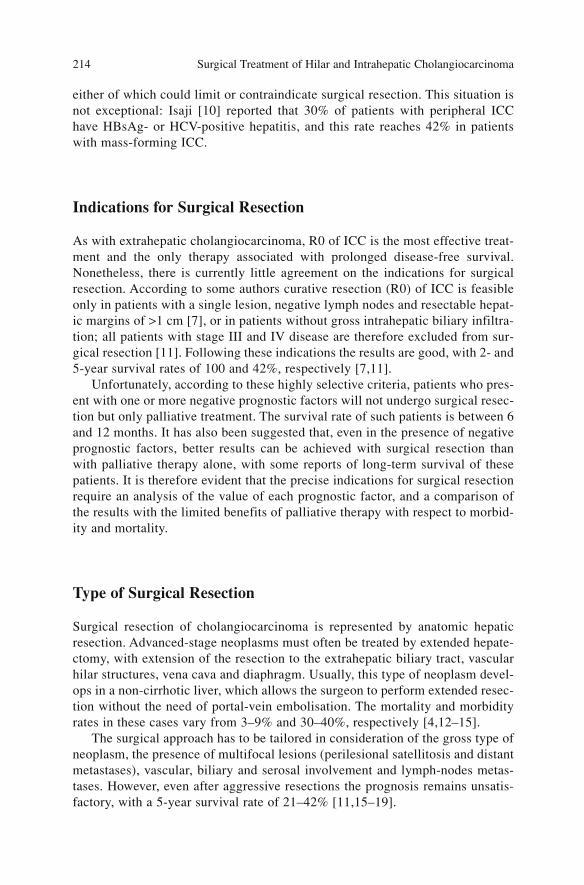
either of which could limit or contraindicate surgical resection. This situation isnot exceptional: Isaji [10] reported that 30% of patients with peripheral ICChave HBsAg- or HCV-positive hepatitis, and this rate reaches 42% in patientswith mass-forming ICC.
Indications for Surgical Resection
As with extrahepatic cholangiocarcinoma, R0 of ICC is the most effective treat-ment and the only therapy associated with prolonged disease-free survival.Nonetheless, there is currently little agreement on the indications for surgicalresection. According to some authors curative resection (R0) of ICC is feasibleonly in patients with a single lesion, negative lymph nodes and resectable hepat-ic margins of >1 cm [7], or in patients without gross intrahepatic biliary infiltra-tion; all patients with stage III and IV disease are therefore excluded from sur-gical resection [11]. Following these indications the results are good, with 2- and5-year survival rates of 100 and 42%, respectively [7,11].
Unfortunately, according to these highly selective criteria, patients who pres-ent with one or more negative prognostic factors will not undergo surgical resec-tion but only palliative treatment. The survival rate of such patients is between 6and 12 months. It has also been suggested that, even in the presence of negativeprognostic factors, better results can be achieved with surgical resection thanwith palliative therapy alone, with some reports of long-term survival of thesepatients. It is therefore evident that the precise indications for surgical resectionrequire an analysis of the value of each prognostic factor, and a comparison ofthe results with the limited benefits of palliative therapy with respect to morbid-ity and mortality.
Type of Surgical Resection
Surgical resection of cholangiocarcinoma is represented by anatomic hepaticresection. Advanced-stage neoplasms must often be treated by extended hepate-ctomy, with extension of the resection to the extrahepatic biliary tract, vascularhilar structures, vena cava and diaphragm. Usually, this type of neoplasm devel-ops in a non-cirrhotic liver, which allows the surgeon to perform extended resec-tion without the need of portal-vein embolisation. The mortality and morbidityrates in these cases vary from 3–9% and 30–40%, respectively [4,12–15].
The surgical approach has to be tailored in consideration of the gross type ofneoplasm, the presence of multifocal lesions (perilesional satellitosis and distantmetastases), vascular, biliary and serosal involvement and lymph-nodes metas-tases. However, even after aggressive resections the prognosis remains unsatis-factory, with a 5-year survival rate of 21–42% [11,15–19].
Surgical Treatment of Hilar and Intrahepatic Cholangiocarcinoma 214
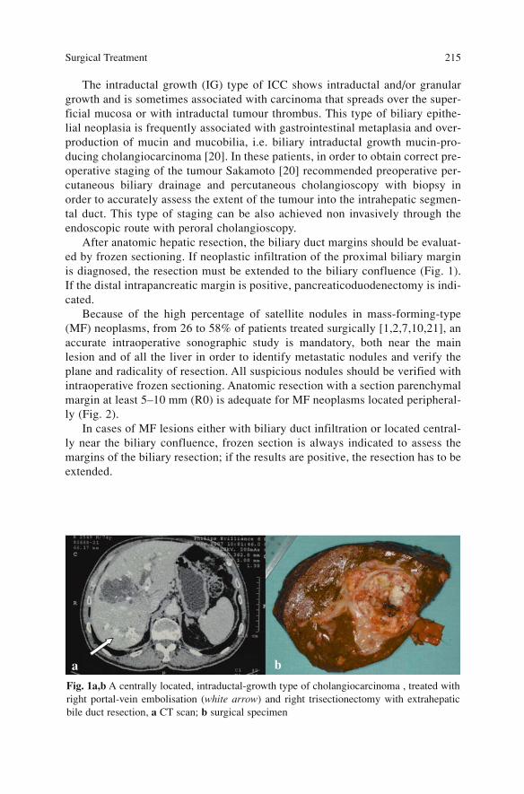
The intraductal growth (IG) type of ICC shows intraductal and/or granulargrowth and is sometimes associated with carcinoma that spreads over the super-ficial mucosa or with intraductal tumour thrombus. This type of biliary epithe-lial neoplasia is frequently associated with gastrointestinal metaplasia and over-production of mucin and mucobilia, i.e. biliary intraductal growth mucin-pro-ducing cholangiocarcinoma [20]. In these patients, in order to obtain correct pre-operative staging of the tumour Sakamoto [20] recommended preoperative per-cutaneous biliary drainage and percutaneous cholangioscopy with biopsy inorder to accurately assess the extent of the tumour into the intrahepatic segmen-tal duct. This type of staging can be also achieved non invasively through theendoscopic route with peroral cholangioscopy.
After anatomic hepatic resection, the biliary duct margins should be evaluat-ed by frozen sectioning. If neoplastic infiltration of the proximal biliary marginis diagnosed, the resection must be extended to the biliary confluence (Fig. 1).If the distal intrapancreatic margin is positive, pancreaticoduodenectomy is indi-cated.
Because of the high percentage of satellite nodules in mass-forming-type(MF) neoplasms, from 26 to 58% of patients treated surgically [1,2,7,10,21], anaccurate intraoperative sonographic study is mandatory, both near the mainlesion and of all the liver in order to identify metastatic nodules and verify theplane and radicality of resection. All suspicious nodules should be verified withintraoperative frozen sectioning. Anatomic resection with a section parenchymalmargin at least 5–10 mm (R0) is adequate for MF neoplasms located peripheral-ly (Fig. 2).
In cases of MF lesions either with biliary duct infiltration or located central-ly near the biliary confluence, frozen section is always indicated to assess themargins of the biliary resection; if the results are positive, the resection has to beextended.
Surgical Treatment 215
Fig. 1a,b A centrally located, intraductal-growth type of cholangiocarcinoma , treated withright portal-vein embolisation (white arrow) and right trisectionectomy with extrahepaticbile duct resection, a CT scan; b surgical specimen
a b
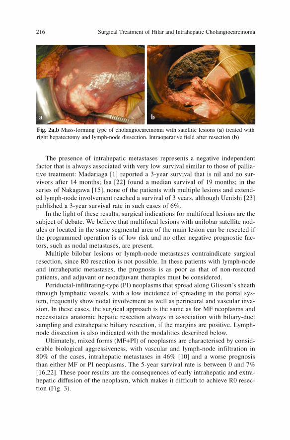
The presence of intrahepatic metastases represents a negative independentfactor that is always associated with very low survival similar to those of pallia-tive treatment: Madariaga [1] reported a 3-year survival that is nil and no sur-vivors after 14 months; Isa [22] found a median survival of 19 months; in theseries of Nakagawa [15], none of the patients with multiple lesions and extend-ed lymph-node involvement reached a survival of 3 years, although Uenishi [23]published a 3-year survival rate in such cases of 6%.
In the light of these results, surgical indications for multifocal lesions are thesubject of debate. We believe that multifocal lesions with unilobar satellite nod-ules or located in the same segmental area of the main lesion can be resected ifthe programmed operation is of low risk and no other negative prognostic fac-tors, such as nodal metastases, are present.
Multiple bilobar lesions or lymph-node metastases contraindicate surgicalresection, since R0 resection is not possible. In these patients with lymph-nodeand intrahepatic metastases, the prognosis is as poor as that of non-resectedpatients, and adjuvant or neoadjuvant therapies must be considered.
Periductal-infiltrating-type (PI) neoplasms that spread along Glisson’s sheaththrough lymphatic vessels, with a low incidence of spreading in the portal sys-tem, frequently show nodal involvement as well as perineural and vascular inva-sion. In these cases, the surgical approach is the same as for MF neoplasms andnecessitates anatomic hepatic resection always in association with biliary-ductsampling and extrahepatic biliary resection, if the margins are positive. Lymph-node dissection is also indicated with the modalities described below.
Ultimately, mixed forms (MF+PI) of neoplasms are characterised by consid-erable biological aggressiveness, with vascular and lymph-node infiltration in80% of the cases, intrahepatic metastases in 46% [10] and a worse prognosisthan either MF or PI neoplasms. The 5-year survival rate is between 0 and 7%[16,22]. These poor results are the consequences of early intrahepatic and extra-hepatic diffusion of the neoplasm, which makes it difficult to achieve R0 resec-tion (Fig. 3).
Surgical Treatment of Hilar and Intrahepatic Cholangiocarcinoma 216
Fig. 2a,b Mass-forming type of cholangiocarcinoma with satellite lesions (a) treated withright hepatectomy and lymph-node dissection. Intraoperative field after resection (b)
a b
In these cases, the surgical approach is the same as for the PI type, i.e.anatomic hepatic resection and biliary-duct sampling with likely extrahepaticbiliary resection. It must be kept in mind that patients with advanced PI or mixedform (MF+PI) of cholangiocarcinoma have not benefited from extended surgery(hepatic and biliary resection, extended lymphadenectomy) in terms of improvedsurgical outcome compared to conventional resection (hepatic resection alone),with increased rates of mortality and morbidity. The surgical indications are con-troversial in these cases and must be limited to patients without other negativeprognostic factors (intrahepatic metastases and positive lymph nodes).
The size of the neoplasm does not represent a prognostic factor that limitssurgical indication, even if small tumours show a better prognosis than largeones. This suggests that if radical removal of the tumour is technically feasibleand can be accomplished safely, it should be performed independently of thesize of the intrahepatic tumour [17].
Vascular involvement is present in 27–85% of patients [24] and represents anegative prognostic factor. Casavilla [25], in a series of 39 patients, found thatthose with vascular infiltration did not reach 5-year survival. However, Inoue[17] reported 5-year survival rates of 60 and 22% in patients without and withvascular infiltration, respectively; survival in the latter group was significantlybetter than in non-resected patients. Therefore vascular invasion, even if it is anegative prognostic factor, does not represent an absolute contraindication tosurgery.
Indications for Lymphadenectomy
The rational extent of radical lymphadenectomy for ICC has not been clearlydefined and there is no consensus on the role of lymph-node dissection [18,26].The incidence of lymph-node metastasis in ICC is 43–62% [1,2,7,10,15,21].
Surgical Treatment 217
Fig. 3a,b Intrahepatic cholangiocarcinoma of the periductal infiltrating type (a).Intrahepatic cholangiocarcinoma of the combined mass-forming and periductal-infiltratingtypes (MF+PI) (b)
a b
Several series have shown that one of the strongest prognostic factors in ICCis lymph-node involvement [17,25,27,28]. All of these studies reported that nopatients with lymph-node involvement survived for more than 3 years after sur-gery. Inoue [17] stated that lymph-node metastasis in MF neoplasms is a sign ofnon-curative disseminated disease, and therefore hepatectomy is contraindicatedif metastatic involvement is observed at the time of lymph-node sampling.Nonetheless, Murakami [29] and Weber [5] reported long-term survivors (morethan 5 years) with lymph-node metastasis. Ohtsuka [12] did not consider lymph-node involvement to be a significant prognostic factor in patients who underwentlymph-node dissection, with a few patients who survived more than 3 years.
Isa [22] reported 3-year survival rate in N+ patients of 6.7%, with a mediansurvival of 10 months. Some authors have underlined the prognostic importanceof the number of positive lymph nodes, analogous to gastric and colonic cancers:Suzuki [30] reported that 5-year survival in patients with a single lymph-nodemetastasis is 33%, and that patients with two or more lymph-node metastases donot survive beyond 2 years; Nakagawa [15] series had 3-year survival rates of62, 50 and 0% in patients with N0 tumours, with one or two nodes and with morethan three nodes, respectively.
These results suggest that, although lymph-node involvement may generallybe associated with an unfavourable prognosis, long-term survival might beexpected following aggressive surgical resection, including lymph-node dissec-tion, in highly selected patients, providing that nodal diffusion, as confirmed byintraoperative sampling, is not beyond regional station.
In the presence of extensive nodal diffusion or in association with other neg-ative prognostic factors, such as multifocal involvement, resection does notguarantee better results than palliative therapy and is therefore not recommend-ed.
Major lymphatic spreading of ICC follows three routes: (1) through the hepa-toduodenal ligament; (2) through the paracardial, lesser curvature and left gas-tric artery and (3) through the inferior phrenic artery or directly from the rightliver to the lateral para-aortic group. Neoplasms located in the left lobe presenteither a right pathway of diffusion to the hepatoduodenal ligament (with N+ in38% of patients) or a left pathway to the right paracardial stations, along thelesser curvature and left gastric artery (with N+ in 31% of patients) [31].Consequently, the rational extent of radical lymphadenectomy should includenot only the hepatoduodenal ligament with the hepatic artery, para-aortic, retro-pancreatic and mesenteric nodes but also the left gastric nodes, the perigastricnodes along the lesser curvature, and the right paracardial nodes, especially inleft-located lesions [26].
Before surgical resection we always submit hepatoduodenal ligament nodesor grossly suspected nodes to frozen section. If more than three nodes or distant-station nodes are positive, the indication for hepatic resection becomes contro-versial and might be considered only in selected patients with low surgical riskand single lesion. In these cases, combination with adjuvant therapy must beconsidered.
Surgical Treatment of Hilar and Intrahepatic Cholangiocarcinoma 218

Extrahepatic Metastases
The presence of peritoneal metastases detected with laparoscopy or laparotomyis an absolute contraindication to surgical operation. Isaji [10] reported that theoutcome in 17 patients who underwent resection for stage IV B neoplasms wasas poor as that in 12 patients who did not undergo resection, with no survivorsafter 2 years. In these cases, adjuvant therapy may be considered, even if satis-factory results have not been reported until now.
References
1. Madariaga JR, Iwatsuki S, Todo S et al (1998) Liver resection for hilar and peripheralcholangiocarcinomas: a study of 62 cases. Ann Surg 227(1):70–79
2. Yamanaka N, Okamoto E, Ando T et al (1995) Clinicopathologic spectrum of resected extra-ductal mass-forming intrahepatic cholangiocarcinoma. Cancer 76(12):2449–2456
3. Lieser MJ, Barry MK, Rowland C et al (1998) Surgical management of intrahepatic cholan-giocarcinoma: a 31-year experience. J Hepatobiliary Pancreat Surg 5(1):41–47
4. Lang H, Sotiropoulos GC, Fruhauf NR (2005) Extended hepatectomy for intrahepaticcholangiocellular carcinoma (ICC): when is it worthwhile? Single center experience with 27resections in 50 patients over a 5-year period. Ann Surg. Jan 241(1):134–143
5. Weber SM, Jarnagin WR, Klimstra D (2001) Intrahepatic cholangiocarcinoma: resectabili-ty, recurrence pattern, and outcomes. J Am Coll Surg 193(4):384–391
6. Goere D, Wagholikar GD, Pessaux P et al (2006) Utility of staging laparoscopy in subsetsof biliary cancers : laparoscopy is a powerful diagnostic tool in patients with intrahepaticand gallbladder carcinoma. Surg Endosc 20(5):721–725
7. Cherqui D, Tantawi B, Alon R et al (1995) Intrahepatic cholangiocarcinoma. Results ofaggressive surgical management. Arch Surg 130(10):1073–1078
8. Harvey CJ, Blomley MJ, Eckersley RJ et al (2000) Pulse-inversion mode imaging of liverspecific microbubbles: improved detection of subcentimetre metastases. Lancet355(9206):807–808
9. Skjoldbye B, Pedersen MH, Struckmann J et al (2002) Improved detection and biopsy ofsolid liver lesions using pulse-inversion ultrasound scanning and contrast agent infusion.Ultrasound Med Biol 28(4):439–444
10. Isaji S, Kawarada Y, Taoka H et al (1999) Clinicopathological features and outcome ofhepatic resection for intrahepatic cholangiocarcinoma in Japan. J Hepatobiliary PancreatSurg 6(2):108–116
11. Harrison LE, Fong Y, Klimstra DS et al (1998) Surgical treatment of 32 patients with periph-eral intrahepatic cholangiocarcinoma. Br J Surg 85(8):1068–1070
12. Ohtsuka M, Ito H, Kimura F et al (2002) Results of surgical treatment for intrahepaticcholangiocarcinoma and clinicopathological factors influencing survival. Br J Surg89(12):1525–1531
13. Morimoto Y, Tanaka Y, Ito T et al (2003) Long-term survival and prognostic factors in thesurgical treatment for intrahepatic cholangiocarcinoma. J Hepatobiliary Pancreat Surg10(6):432–440
14. Huang JL, Biehl TR, Lee FT et al (2004) Outcomes after resection of cholangiocellular car-cinoma. Am J Surg 187(5):612–617
15. Nakagawa T, Kamiyama T, Kurauchi N et al (2005) Number of lymph-node metastases is asignificant prognostic factor in intrahepatic cholangiocarcinoma. World J Surg29(6):728–733
Surgical Treatment 219

16. Yamamoto M, Takasaki K, Yoshikawa T et al (1998) Does gross appearance indicate prog-nosis in intrahepatic cholangiocarcinoma? J Surg Oncol 69(3):162–167
17. Inoue K, Makuuchi M, Takayama T et al (2000) Long-term survival and prognostic factorsin the surgical treatment of mass-forming type cholangiocarcinoma. Surgery127(5):498–505
18. Shimada M, Yamashita Y, Aishima S et al (2001) Value of lymph node dissection duringresection of intrahepatic cholangiocarcinoma. Br J Surg 88(11):1463–1466
19. Shirabe K, Shimada M, Harimoto N et al (2002) Intrahepatic cholangiocarcinoma: its modeof spreading and therapeutic modalities. Surgery 131(1 Suppl):S159-S164
20. Sakamoto E, Hayakawa N, Kamiya J et al (1999) Treatment strategy for mucin-producingintrahepatic cholangiocarcinoma: value of percutaneous transhepatic biliary drainage andcholangioscopy. World J Surg 23(10):1038–1043; discussion 1043–1044
21. Sasaki A, Aramaki M, Kawano K et al (1998) Intrahepatic peripheral cholangiocarcinoma:mode of spread and choice of surgical treatment. Br J Surg 85(9):1206–1209
22. Isa T, Kusano T, Shimoji H et al (2001) Predictive factors for long-term survival in patientswith intrahepatic cholangiocarcinoma. Am. J. Surg 181(6):507–511
23. Uenishi T, Yamazaki O, Yamamoto T et al (2005) Serosal invasion in TNM staging of mass-forming intrahepatic cholangiocarcinoma. J Hepatobiliary Pancreat Surg 12(6):479–483
24. Weinbren K, Mutum SS (1983) Pathological aspects of cholangiocarcinoma. J Pathol139(2):217–238
25. Casavilla FA, Marsh JW, Iwatsuki S et al (1997) Hepatic resection and transplantation forperipheral cholangiocarcinoma. J Am Coll Surg 185(5):429–436
26. Tsuji T, Hiraoka T, Kanemitsu K et al (2001) Lymphatic spreading pattern of intrahepaticcholangiocarcinoma. Surgery 129(4):401–407
27. Uenishi T, Hirohashi K, Kubo S et al (2001) Clinicopathological factors predicting outcomeafter resection of mass-forming intrahepatic cholangiocarcinoma. Br J Surg 88(7):969–974
28. Chu KM, Lai EC, Al-Hadeedi S et al (1997) Intrahepatic cholangiocarcinoma. World J Surg21(3):301–305
29. Murakami Y, Yokoyama T, Takesue Y (2000) Long-term survival of peripheral intrahepaticcholangiocarcinoma with metastasis to the para-aortic lymph nodes. Surgery127(1):105–106
30. Suzuki S, Sakaguchi T, Yokoi Y et al (2002) Clinicopathological prognostic factors andimpact of surgical treatment of mass-forming intrahepatic cholangiocarcinoma. World JSurg 26(6):687–693
31. Okami J, Dono K, Sakon M et al (2003) Patterns of regional lymph-node involvement inintrahepatic cholangiocarcinoma of the left lobe. J Gastrointest Surg 7(7):850–856
Surgical Treatment of Hilar and Intrahepatic Cholangiocarcinoma 220
Results of Surgery
An analysis of the results of surgery for intrahepatic cholangiocarcinoma (ICC),as reported in the literature is difficult, because the patient-selection criteria arenot homogeneous, and the series of cases are limited by the rarity of the diseaseand the prolonged amount of time needed for data collection. Prognostic factorsrelated to the characteristics of the neoplasm were analysed previously. Herein,only the early and late results of surgical operation are considered.
Morbidity and Mortality
Surgery of ICC is often carried out in advanced neoplasms, with the need to per-form extended hepatic resection in 21–100% of such patients [1]. Hepatic resec-tion is associated with extrahepatic biliary resection in 60–74% of the cases andwith portal (4–26%) or arterial (0–10%) resection and reconstruction. Resectionof the diaphragm (10%) and vein cava (7%) occur less frequently [2–6]. Evenwith improvements in surgical technique and perioperative management, exten-ded hepatectomies still entail significant mortality and morbidity. In the majo-rity of studies, mortality was <5% and some authors reported no mortality at all(Table 1). Mortality is often related to extended hepatic resection or to associa-ted procedures, such as vascular and biliary reconstruction or hepato-pancreatec-tomy: in these cases, mortality increases to 7–9.5% [1,3].
The complications after surgical exeresis of ICC range from 20 to 50%(Table 1) and are comparable to those following common hepatic resection of anon-cirrhotic liver. Postoperative hepatic failure is less frequent than after resec-tion for hilar neoplasms and occurs in about 10% of patients. Other complica-tions are subphrenic collections (4–8%), biliary leakage (8–12%), pulmonaryembolism (6%) and respiratory complications (4%) [1,4,7,8].
The complication rate is also related to the type of operation: 45% in simplehepatectomy and 56% in patients who underwent extended hepatectomy withvascular, diaphragmatic or hilar resection. Exploratory laparotomy has a relevantcomplication rate of 17% [1]
221A. Guglielmi, A. Ruzzenente, C. Iacono (eds.) Surgical Treatment of Hilar and ICC.© Springer 2008
.
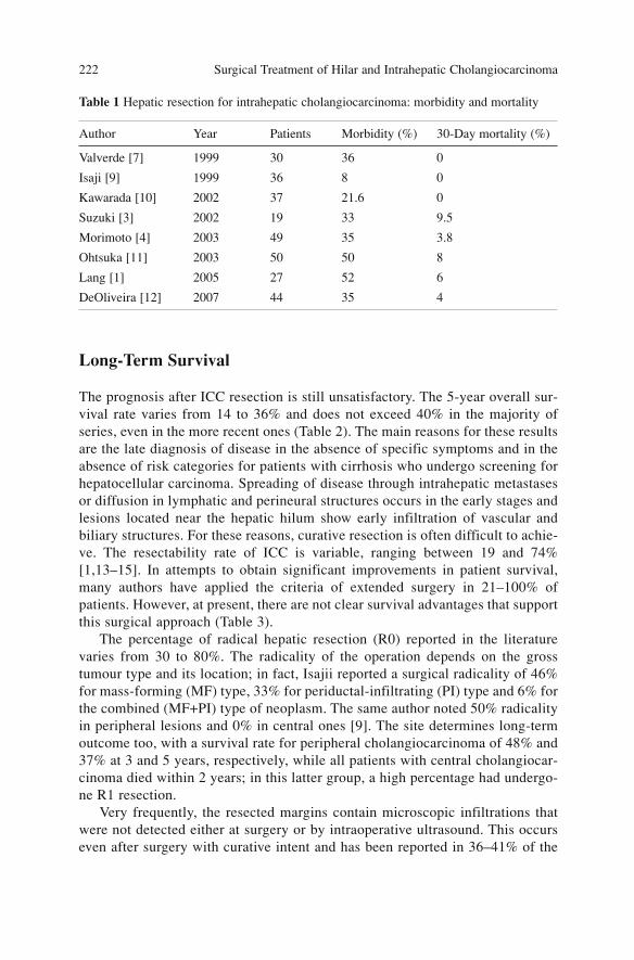
Long-Term Survival
The prognosis after ICC resection is still unsatisfactory. The 5-year overall sur-vival rate varies from 14 to 36% and does not exceed 40% in the majority ofseries, even in the more recent ones (Table 2). The main reasons for these resultsare the late diagnosis of disease in the absence of specific symptoms and in theabsence of risk categories for patients with cirrhosis who undergo screening forhepatocellular carcinoma. Spreading of disease through intrahepatic metastasesor diffusion in lymphatic and perineural structures occurs in the early stages andlesions located near the hepatic hilum show early infiltration of vascular andbiliary structures. For these reasons, curative resection is often difficult to achie-ve. The resectability rate of ICC is variable, ranging between 19 and 74%[1,13–15]. In attempts to obtain significant improvements in patient survival,many authors have applied the criteria of extended surgery in 21–100% ofpatients. However, at present, there are not clear survival advantages that supportthis surgical approach (Table 3).
The percentage of radical hepatic resection (R0) reported in the literaturevaries from 30 to 80%. The radicality of the operation depends on the grosstumour type and its location; in fact, Isajii reported a surgical radicality of 46%for mass-forming (MF) type, 33% for periductal-infiltrating (PI) type and 6% forthe combined (MF+PI) type of neoplasm. The same author noted 50% radicalityin peripheral lesions and 0% in central ones [9]. The site determines long-termoutcome too, with a survival rate for peripheral cholangiocarcinoma of 48% and37% at 3 and 5 years, respectively, while all patients with central cholangiocar-cinoma died within 2 years; in this latter group, a high percentage had undergo-ne R1 resection.
Very frequently, the resected margins contain microscopic infiltrations thatwere not detected either at surgery or by intraoperative ultrasound. This occurseven after surgery with curative intent and has been reported in 36–41% of the
Surgical Treatment of Hilar and Intrahepatic Cholangiocarcinoma 222
Table 1 Hepatic resection for intrahepatic cholangiocarcinoma: morbidity and mortality
Author Year Patients Morbidity (%) 30-Day mortality (%)
Valverde [7] 1999 30 36 0
Isaji [9] 1999 36 8 0
Kawarada [10] 2002 37 21.6 0
Suzuki [3] 2002 19 33 9.5
Morimoto [4] 2003 49 35 3.8
Ohtsuka [11] 2003 50 50 8
Lang [1] 2005 27 52 6
DeOliveira [12] 2007 44 35 4
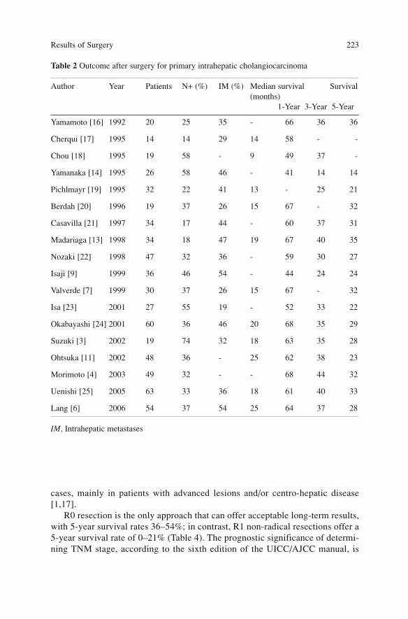
cases, mainly in patients with advanced lesions and/or centro-hepatic disease[1,17].
R0 resection is the only approach that can offer acceptable long-term results,with 5-year survival rates 36–54%; in contrast, R1 non-radical resections offer a5-year survival rate of 0–21% (Table 4). The prognostic significance of determi-ning TNM stage, according to the sixth edition of the UICC/AJCC manual, is
Results of Surgery 223
Table 2 Outcome after surgery for primary intrahepatic cholangiocarcinoma
Author Year Patients N+ (%) IM (%) Median survival Survival(months)
1-Year 3-Year 5-Year
Yamamoto [16] 1992 20 25 35 - 66 36 36
Cherqui [17] 1995 14 14 29 14 58 - -
Chou [18] 1995 19 58 - 9 49 37 -
Yamanaka [14] 1995 26 58 46 - 41 14 14
Pichlmayr [19] 1995 32 22 41 13 - 25 21
Berdah [20] 1996 19 37 26 15 67 - 32
Casavilla [21] 1997 34 17 44 - 60 37 31
Madariaga [13] 1998 34 18 47 19 67 40 35
Nozaki [22] 1998 47 32 36 - 59 30 27
Isaji [9] 1999 36 46 54 - 44 24 24
Valverde [7] 1999 30 37 26 15 67 - 32
Isa [23] 2001 27 55 19 - 52 33 22
Okabayashi [24] 2001 60 36 46 20 68 35 29
Suzuki [3] 2002 19 74 32 18 63 35 28
Ohtsuka [11] 2002 48 36 - 25 62 38 23
Morimoto [4] 2003 49 32 - - 68 44 32
Uenishi [25] 2005 63 33 36 18 61 40 33
Lang [6] 2006 54 37 54 25 64 37 28
IM, Intrahepatic metastases
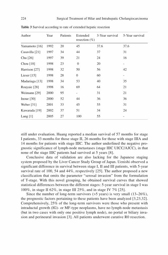
still under evaluation. Huang reported a median survival of 57 months for stageI patients, 33 months for those stage II, 26 months for those with stage IIIA and14 months for patients with stage IIIC. The author underlined the negative pro-gnostic significance of lymph-node metastases (stage IIIC UICC/AJCC), in thatnone of the stage IIIC patients had survived at 5 years [8].
Conclusive data of validation are also lacking for the Japanese stagingsystem proposed by the Liver Cancer Study Group of Japan. Uenishi observed asignificant difference in survival between stage I, II and III patients, with 5-yearsurvival rate of 100, 54 and 44%, respectively [25]. The author proposed a newclassification that omits the parameter “serosal invasion” from the formulationof T-stage. With this novel grouping, he obtained survival curves that showedstatistical differences between the different stages: 5-year survival in stage I was100%, in stage II 62%, in stage III 25%, and in stage IV 7% [25].
Since the number of long-term survivors (>5 years) is very small (13–26%),the prognostic factors pertaining to these patients have been analyzed [3,23,32].Comprehensively, 25% of the long-term survivors were those who present withintraductal growth (IG)- or MF-type neoplasms, have no lymph-node metastasis(but in two cases with only one positive lymph node), no portal or biliary inva-sion and perineural invasion [3]. All patients underwent curative R0 resection.
Surgical Treatment of Hilar and Intrahepatic Cholangiocarcinoma 224
Table 3 Survival according to rate of extended hepatic resection
Author Year Patients Extended 3-Year survival 5-Year survivalresection (%)
Yamamoto [16] 1992 20 45 37.6 37.6
Casavilla [21] 1997 34 44 37 31
Chu [26] 1997 39 21 24 16
Chou [18] 1998 23 0 20 -
Harrison [27] 1998 32 50 56 42
Lieser [15] 1998 28 0 60 -
Madariaga [13] 1998 34 53 40 35
Roayaie [28] 1998 16 69 64 21
Weimann [29] 2000 95 - 31 21
Inoue [30] 2000 52 44 36 36
Weber [31] 2001 33 45 55 31
Kawarada [19] 2002 37 51 34 24
Lang [1] 2005 27 100 55 -
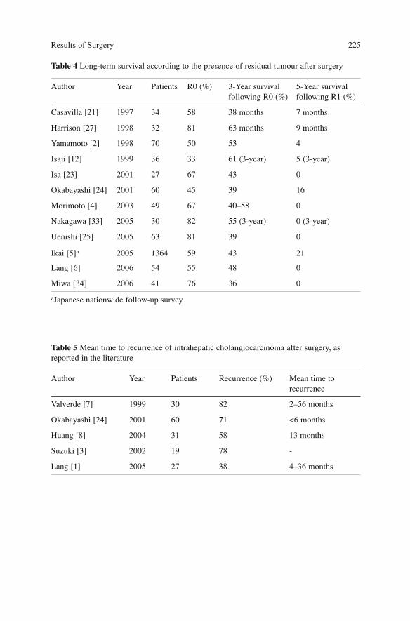
Results of Surgery 225
Table 4 Long-term survival according to the presence of residual tumour after surgery
Author Year Patients R0 (%) 3-Year survival 5-Year survivalfollowing R0 (%) following R1 (%)
Casavilla [21] 1997 34 58 38 months 7 months
Harrison [27] 1998 32 81 63 months 9 months
Yamamoto [2] 1998 70 50 53 4
Isaji [12] 1999 36 33 61 (3-year) 5 (3-year)
Isa [23] 2001 27 67 43 0
Okabayashi [24] 2001 60 45 39 16
Morimoto [4] 2003 49 67 40–58 0
Nakagawa [33] 2005 30 82 55 (3-year) 0 (3-year)
Uenishi [25] 2005 63 81 39 0
Ikai [5]a 2005 1364 59 43 21
Lang [6] 2006 54 55 48 0
Miwa [34] 2006 41 76 36 0
aJapanese nationwide follow-up survey
Table 5 Mean time to recurrence of intrahepatic cholangiocarcinoma after surgery, asreported in the literature
Author Year Patients Recurrence (%) Mean time to recurrence
Valverde [7] 1999 30 82 2–56 months
Okabayashi [24] 2001 60 71 <6 months
Huang [8] 2004 31 58 13 months
Suzuki [3] 2002 19 78 -
Lang [1] 2005 27 38 4–36 months
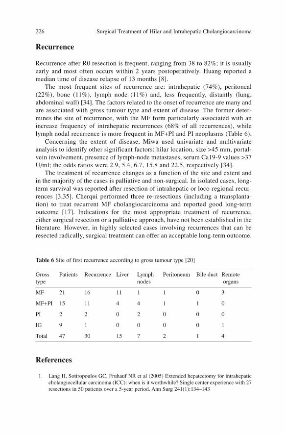
Recurrence
Recurrence after R0 resection is frequent, ranging from 38 to 82%; it is usuallyearly and most often occurs within 2 years postoperatively. Huang reported amedian time of disease relapse of 13 months [8].
The most frequent sites of recurrence are: intrahepatic (74%), peritoneal(22%), bone (11%), lymph node (11%) and, less frequently, distantly (lung,abdominal wall) [34]. The factors related to the onset of recurrence are many andare associated with gross tumour type and extent of disease. The former deter-mines the site of recurrence, with the MF form particularly associated with anincrease frequency of intrahepatic recurrences (68% of all recurrences), whilelymph nodal recurrence is more frequent in MF+PI and PI neoplasms (Table 6).
Concerning the extent of disease, Miwa used univariate and multivariateanalysis to identify other significant factors: hilar location, size >45 mm, portal-vein involvement, presence of lymph-node metastases, serum Ca19-9 values >37U/ml; the odds ratios were 2.9, 5.4, 6.7, 15.8 and 22.5, respectively [34].
The treatment of recurrence changes as a function of the site and extent andin the majority of the cases is palliative and non-surgical. In isolated cases, long-term survival was reported after resection of intrahepatic or loco-regional recur-rences [3,35]. Cherqui performed three re-resections (including a transplanta-tion) to treat recurrent MF cholangiocarcinoma and reported good long-termoutcome [17]. Indications for the most appropriate treatment of recurrence,either surgical resection or a palliative approach, have not been established in theliterature. However, in highly selected cases involving recurrences that can beresected radically, surgical treatment can offer an acceptable long-term outcome.
References
1. Lang H, Sotiropoulos GC, Fruhauf NR et al (2005) Extended hepatectomy for intrahepaticcholangiocellular carcinoma (ICC): when is it worthwhile? Single center experience with 27resections in 50 patients over a 5-year period. Ann Surg 241(1):134–143
Surgical Treatment of Hilar and Intrahepatic Cholangiocarcinoma 226
Table 6 Site of first recurrence according to gross tumour type [20]
Gross Patients Recurrence Liver Lymph Peritoneum Bile duct Remotetype nodes organs
MF 21 16 11 1 1 0 3
MF+PI 15 11 4 4 1 1 0
PI 2 2 0 2 0 0 0
IG 9 1 0 0 0 0 1
Total 47 30 15 7 2 1 4
2. Yamamoto M, Takasaki K, Yoshikawa T et al (1998) Does gross appearance indicate prog-nosis in intrahepatic cholangiocarcinoma? J Surg Oncol 69(3):162–167
3. Suzuki S, Sakaguchi T, Yokoi Y et al (2002) Clinicopathological prognostic factors andimpact of surgical treatment of mass-forming intrahepatic cholangiocarcinoma. World JSurg 26(6):687–693
4. Morimoto Y, Tanaka Y, Ito T et al (2003) Long-term survival and prognostic factors in thesurgical treatment for intrahepatic cholangiocarcinoma. J Hepatobiliary Pancreat Surg10(6):432–440
5. Ikai I, Arii S, Ichida T et al (2005) Report of the 16th follow-up survey of primary liver can-cer. Hepatol Res 32(3):163–172
6. Lang H, Kaiser GM, Zopf T et al (2006) Surgical therapy of hilar cholangiocarcinoma.Chirurg 77(4):325–334
7. Valverde A, Bonhomme N, Farges O et al (1999) Resection of intrahepatic cholangiocarci-noma: a Western experience. J Hepatobiliary Pancreat Surg 6(2):122–127
8. Huang JL, Biehl TR, Lee FT et al (2004) Outcomes after resection of cholangiocellular car-cinoma. Am J Surg 187(5):612–617
9. Isaji S, Kawarada Y, Taoka H et al (1999) Clinicopathological features and outcome ofhepatic resection for intrahepatic cholangiocarcinoma in Japan. J Hepatobiliary PancreatSurg 6(2):108–116
10. Kawarada Y, Yamagiwa K, Das BC (2002) Analysis of the relationships between clinico-pathologic factors and survival time in intrahepatic cholangiocarcinoma. Am J Surg183(6):679–685
11. Ohtsuka M, Ito H, Kimura F et al (2002) Results of surgical treatment for intrahepaticcholangiocarcinoma and clinicopathological factors influencing survival. Br J Surg89(12):1525–1531
12. DeOliveira ML, Cunningham SC, Cameron JL et al (2007) Cholangiocarcinoma: thirty-one-year experience with 564 patients at a single institution. Ann Surg 245(5):755–762
13. Madariaga JR, Iwatsuki S, Todo S et al (1998) Liver resection for hilar and peripheralcholangiocarcinomas: a study of 62 cases. Ann Surg 227(1):70–79
14. Yamanaka N, Okamoto E, Ando T et al (1995) Clinicopathologic spectrum of resected extra-ductal mass-forming intrahepatic cholangiocarcinoma. Cancer 76(12):2449–2456
15. Lieser MJ, Barry MK, Rowland C et al (1998) Surgical management of intrahepatic cholan-giocarcinoma: a 31-year experience. J Hepatobiliary Pancreat Surg 5(1):41–47
16. Yamamoto J, Kosuge T, Takayama T et al (1992) Surgical treatment of intrahepatic cholan-giocarcinoma: four patients surviving more than five years. Surgery 111(6):617–622
17. Cherqui D, Tantawi B, Alon R et al (1995) Intrahepatic cholangiocarcinoma. Results ofaggressive surgical management. Arch Surg 130(10):1073–1078
18. Chou FF, Sheen-Chen SM, Chen CL et al (1995) Prognostic factors of resectable intrahep-atic cholangiocarcinoma. J Surg Oncol 59(1):40–44
19. Pichlmayr R, Lamesch P, Weimann A et al (1995) Surgical treatment of cholangiocellularcarcinoma. World J Surg 19(1):83–88
20. Berdah SV, Delpero JR, Garcia S et al (1996) A western surgical experience of peripheralcholangiocarcinoma. Br J Surg 83(11):1517–1521
21. Casavilla FA, Marsh JW, Iwatsuki S et al (1997) Hepatic resection and transplantation forperipheral cholangiocarcinoma. J Am Coll Surg 185(5):429–436
22. Nozaki Y, Yamamoto M, Ikai I et al (1998) Reconsideration of the lymph-node metastasispattern (N factor) from intrahepatic cholangiocarcinoma using the International UnionAgainst Cancer TNM staging system for primary liver carcinoma. Cancer 83(9):1923–1929
23. Isa T, Kusano T, Shimoji H et al (2001) Predictive factors for long-term survival in patientswith intrahepatic cholangiocarcinoma. Am J Surg 181(6):507–511
24. Okabayashi T, Yamamoto J, Kosuge T et al (2001) A new staging system for mass-formingintrahepatic cholangiocarcinoma: analysis of preoperative and postoperative variables.Cancer 92(9):2374–2383
Results of Surgery 227
25. Uenishi T, Yamazaki O, Yamamoto T et al (2005) Serosal invasion in TNM staging of mass-forming intrahepatic cholangiocarcinoma. J Hepatobiliary Pancreat Surg 12(6):479–483
26. Chu KM, Lai EC, Al-Hadeedi S et al (1997) Intrahepatic cholangiocarcinoma. World J Surg21(3):301–305
27. Harrison LE, Fong Y, Klimstra DS et al (1998) Surgical treatment of 32 patients with periph-eral intrahepatic cholangiocarcinoma. Br J Surg 85(8):1068–1070
28. Roayaie S, Guarrera JV, Ye MQ et al (1998) Aggressive surgical treatment of intrahepaticcholangiocarcinoma: predictors of outcomes. J Am Coll Surg 187(4):365–372
29. Weimann A, Varnholt H, Schlitt HJ et al (2000) Retrospective analysis of prognostic factorsafter liver resection and transplantation for cholangiocellular carcinoma. Br J Surg87(9):1182–1187
30. Inoue K, Makuuchi M, Takayama T et al (2000) Long-term survival and prognostic factorsin the surgical treatment of mass-forming type cholangiocarcinoma. Surgery127(5):498–505
31. Weber SM, Jarnagin WR, Klimstra D et al (2001) Intrahepatic cholangiocarcinoma:resectability, recurrence pattern, and outcomes. J Am Coll Surg 193(4):384–391
32. Jan YY, Yeh CN, Yeh TS et al (2005) Clinicopathological factors predicting long-term over-all survival after hepatectomy for peripheral cholangiocarcinoma. World J Surg29(7):894–898
33. Nakagawa T, Kamiyama T, Kurauchi N et al (2005) Number of lymph-node metastases is asignificant prognostic factor in intrahepatic cholangiocarcinoma. World J Surg29(6):728–733
34. Miwa S, Miyagawa S, Kobayashi A et al (2006) Predictive factors for intrahepatic cholan-giocarcinoma recurrence in the liver following surgery. J Gastroenterol 41(9):893–900
35. Kurosaki I, Hatakeyama K (2005) Repeated hepatectomy for recurrent intrahepatic cholan-giocarcinoma: report of two cases. Eur J Gastroenterol Hepatol 17(1):125–130
Surgical Treatment of Hilar and Intrahepatic Cholangiocarcinoma 228
The Role of Liver Transplantation
Surgical resection is the only therapeutic approach that produces good survivalresults for patients with intrahepatic cholangiocarcinoma. However, advanced dis-ease stage and infiltration to vascular structures often require extended and compli-cated hepatic resections, such that curative operation is not always possible.
From an oncologic point of view, transplantation should allow a remarkableincrease of surgical radicality and the possibility to obtain curative treatmenteven in patients who cannot undergo hepatic resection because of the extent ofdisease and concomitant chronic liver disease. Unfortunately, the results ofhepatic transplantation of patients with intrahepatic cholangiocarcinoma are stillpoor and limited to few tens of cases without homogeneous indications. Someauthors reported that the results are even worse than those for extrahepaticcholangiocarcinoma [1,2], whereas others reported a similar outcome [3–5].
Initially, patients who underwent transplantation for intrahepatic cholangio-carcinoma was restricted to those with advanced disease who were excludedfrom resection. At the end of the 1980s, Ringe reported on 10 patients who hada median survival of 4 months and a 90% recurrence rate [6]. The Pichlmayrgroup, with a more extensive follow-up time confirmed these results and report-ed a 3-year survival rate of 0% [7]. More recent experience suggests a slightincrease of overall survival and disease-free survival; Casavilla reported 3 and 5-year survival rates of 29 and 18% in 20 patients with unresectable cholangiocar-cinoma; nine patients underwent cluster transplantation [5].
Weimann, adopting similar indications in 23 patients, obtained 1- and 3-yearsurvival rates of 21 and 4% and no survivors after 5 years. Robles, in a multi-centre Spanish study, reported more promising data, with 3- and 5-year survivalrates of 65 and 42%, respectively [8]. Currently, there are no precise indicationsreported in the literature.
Recurrence after transplantation is quite frequent. In the series of Casavilla,the recurrence rate was 55%, with a disease-free survival rate of 31% at 3- and5-year [5]. Other authors have reported similar results. Robles determined 3- and5-year disease-free survival rates of 45 and 27%, with 23% of the patients dis-ease-free at 10 years [8] (Table 1).
229A. Guglielmi, A. Ruzzenente, C. Iacono (eds.) Surgical Treatment of Hilar and ICC.© Springer 2008
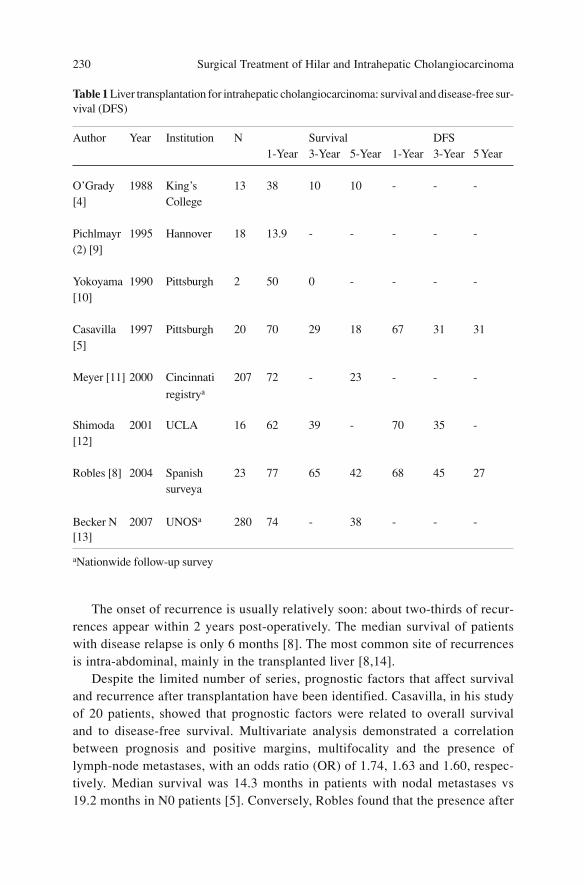
The onset of recurrence is usually relatively soon: about two-thirds of recur-rences appear within 2 years post-operatively. The median survival of patientswith disease relapse is only 6 months [8]. The most common site of recurrencesis intra-abdominal, mainly in the transplanted liver [8,14].
Despite the limited number of series, prognostic factors that affect survivaland recurrence after transplantation have been identified. Casavilla, in his studyof 20 patients, showed that prognostic factors were related to overall survivaland to disease-free survival. Multivariate analysis demonstrated a correlationbetween prognosis and positive margins, multifocality and the presence oflymph-node metastases, with an odds ratio (OR) of 1.74, 1.63 and 1.60, respec-tively. Median survival was 14.3 months in patients with nodal metastases vs19.2 months in N0 patients [5]. Conversely, Robles found that the presence after
Surgical Treatment of Hilar and Intrahepatic Cholangiocarcinoma 230
Table 1 Liver transplantation for intrahepatic cholangiocarcinoma: survival and disease-free sur-vival (DFS)
Author Year Institution N Survival DFS 1-Year 3-Year 5-Year 1-Year 3-Year 5 Year
O’Grady 1988 King’s 13 38 10 10 - - -[4] College
Pichlmayr 1995 Hannover 18 13.9 - - - - -(2) [9]
Yokoyama 1990 Pittsburgh 2 50 0 - - - -[10]
Casavilla 1997 Pittsburgh 20 70 29 18 67 31 31[5]
Meyer [11] 2000 Cincinnati 207 72 - 23 - - -
registrya
Shimoda 2001 UCLA 16 62 39 - 70 35 -[12]
Robles [8] 2004 Spanish 23 77 65 42 68 45 27surveya
Becker N 2007 UNOSa 280 74 - 38 - - -[13]
aNationwide follow-up survey
perineural invasion and TNM UICC stage are related to prognosis of transplan-tation for cholangiocarcinoma. Patients in stage I–II disease had 3- and 5-yearsurvival rates of 80%and 40%, while the rates of those in stage III–IVa were 46and 31%, respectively (p<0.05) [8].
Intrahepatic as well as extrahepatic cholangiocarcinoma can be identifiedincidentally during transplantation for primary sclerosing cholangitis. Even ifdefinitive results regarding the prognostic advantages of transplantation afterincidental cholangiocarcinoma vs preoperatively diagnosed cholangiocarcinomahave not been published, it seems clear that an earlier stage of disease detectionensures better outcome [12].
Hepato-cholangiocarcinoma is a rare variant form. Its histologic patternsresemble both hepatocarcinoma and cholangiocarcinoma. Reports in the litera-ture are few but the prognosis in patients who underwent transplantation is notdifferent from the disappointing results of intrahepatic cholangiocarcinoma[1,11,12,15].
Some authors have reported their experience with cluster transplantation(liver, duodenum-pancreas, part of jejunum) in patients with advanced cholan-giocarcinoma infiltrating other organs; however, the results are inadequate, thenumber of patients is small and the follow-up time is too short to be able to drawconclusions. The University of Pittsburgh reported a 5-year survival of 30% innine patients with advanced and unresectable cholangiocarcinomas (stage IVand IVa) who underwent to cluster transplantation [5,16].
There are no data in the literature on the results of adjuvant and neoadjuvanttreatments in transplantation for intrahepatic cholangiocarcinoma. In the light ofthe current data, transplantation does not represent a treatment option for intra-hepatic cholangiocarcinoma and its use must be restricted to clinical trials.
References
1. Pichlmayr R, Weimann A, Oldhafer KJ et al (1995) Role of liver transplantation in the treat-ment of unresectable liver cancer. World J Surg 19(6):807–813
2. Penn I (1991) Hepatic transplantation for primary and metastatic cancers of the liver.Surgery 110(4):726–34; discussion 734–735
3. Nakeeb A, Pitt HA, Sohn TA et al (1996) Cholangiocarcinoma. A spectrum of intrahepatic,perihilar, and distal tumours. Ann Surg 224(4):463–473; discussion 473–475
4. O’Grady JG, Polson RJ, Rolles K et al (1988) Liver transplantation for malignant disease.Results in 93 consecutive patients. Ann Surg 207(4):373–379
5. Casavilla FA, Marsh JW, Iwatsuki S et al (1997) Hepatic resection and transplantation forperipheral cholangiocarcinoma. J Am Coll Surg 185(5):429–436
6. Ringe B, Wittekind C, Bechstein WO et al (1989) The role of liver transplantation in hepa-tobiliary malignancy. A retrospective analysis of 95 patients with particular regard to tumourstage and recurrence. Ann Surg 209(1):88–98
7. Pichlmayr R, Weimann A, Ringe B (1994)Indications for liver transplantation in hepatobil-iary malignancy. Hepatology 20(1 Pt 2):33S-40S
8. Robles R, Figueras J, Turrion VS et al (2004) Spanish experience in liver transplantation forhilar and peripheral cholangiocarcinoma. Ann Surg 239(2):265–271
The Role of Liver Transplantation 231
9. Pichlmayr R, Weimann A, Oldhafer KJ et al (1995) Role of liver transplantation in the treat-ment of unresectable liver cancer. World J Surg 19(6):807–813
10. Yokoyama I, Todo S, Iwatsuki S, Starzl TE (1990) Liver transplantation in the treatment ofprimary liver cancer. Hepatogastroenterology 37(2):188–193
11. Meyer CG, Penn I, James L (2000) Liver transplantation for cholangiocarcinoma: results in207 patients. Transplantation 69(8):1633–1637
12. Shimoda M, Farmer DG, Colquhoun SD et al (2001) Liver transplantation for cholangiocel-lular carcinoma: analysis of a single-center experience and review of the literature. LiverTranspl 7(12):1023–1033
13. Becker N, Rodriguez J, Barshes N et al (2007) Outcome analysis for 280 patients withcholangiocarcinoma treated with liver transplantation over 18-year period. HPB 9(1): 74–75(abs)
14. Weimann A, Varnholt H, Schlitt HJ et al (2000) Retrospective analysis of prognostic factorsafter liver resection and transplantation for cholangiocellular carcinoma. Br J Surg87(9):1182–1187
15. Pichlmayr R, Lamesch P, Weimann A et al (1995) Surgical treatment of cholangiocellularcarcinoma. World J Surg 19(1):83–88
16. Alessiani M, Tzakis A, Todo S et al (1995) Assessment of five-year experience with abdom-inal organ cluster transplantation. J Am Coll Surg 180(1):1–9
Surgical Treatment of Hilar and Intrahepatic Cholangiocarcinoma 232
Adjuvant and Palliative Treatments
The role of adjuvant and palliative chemo- and radiotherapy in the treatment ofintrahepatic cholangiocarcinoma (ICC) is controversial. There are no prospec-tive and randomised studies or large series that provide conclusive indicationsregarding the efficacy of these therapies in ICC. Moreover, the majority of onco-logic papers in the literature that address this topic include intrahepatic andextrahepatic (hilar, middle and distal) cancers and even gallbladder carcinoma.Consequently, specific data for each localisation concerning treatment indica-tion, dosage, type of approach and results are lacking.
Adjuvant Therapy
The different forms of adjuvant therapy are systemic and loco-regionalchemotherapy, radiotherapy, transcatheter hepatic arterial chemoembolisation(TACE), immunotherapy and monoclonal-antibody therapies. There is not com-plete agreement on the parameters that should be used to select those patientswho will benefit from adjuvant treatment after surgery. Some investigators havestated that positive margins or local recurrence represent indications to adjuvanttreatment [1], others consider positive lymph nodes, particularly second-orderand para-aortic [2] nodes, and others base their decision on the presence ofUICC stage III and IV disease [3]. Miwa concluded that patients who shouldreceive adjuvant therapy are those with elevated serum Ca19-9 levels and preop-erative jaundice [4].
Due to the numerous therapeutic protocols that have been proposed, it isalmost impossible to compare data and to evaluate the real efficacy of adjuvanttreatment.
233A. Guglielmi, A. Ruzzenente, C. Iacono (eds.) Surgical Treatment of Hilar and ICC.© Springer 2008
Chemotherapy
The chemotherapeutic agents most commonly used to treat ICC are 5-FU andgemcitabine alone or in chemotherapeutic association with epirubicin, cisplatin,mitomycin C, doxorubicin, leucovorin, methotrexate and interferon.
If the reports that analysed only intrahepatic cholangiocarcinoma are evalu-ated [1,5–8], then it can be stated that adjuvant treatment seems to provideadvantages for long-term survival compared to patients treated by surgery alone.Yi-Yin Jan reported a relative risk for survival of 1.823 (95% CI: 1.414, 2.353;p<0.001) for patients with positive section margins or local recurrence whounderwent surgery alone vs. postoperative chemotherapy with a 5-FU-based reg-imen [1]. Nevertheless other authors have stated that there is insufficient evi-dence supporting the use of post surgical adjuvant therapy [9–12].
Radiotherapy
Adjuvant radiotherapy is usually indicated in the presence of positive resectionmargins or local recurrence [1], with better results in treated vs. non-treatedpatients. Zeng [13] considers adjuvant radiotherapy to be indicated also inpatients who undergo resection of ICC with synchronous or metachronouslymph-node metastases. The median total dose of external radiation therapy usedby this group was 50 Gy (range 30–60 Gy), administered in daily doses of 2Gy/fraction, five times a week. A comparison of the results of 16 treated patientsvs. 14 patients who underwent hepatectomy alone showed better survival in theradiotherapy group (median survival 468 vs. 211 days, respectively, p=0.075).However other authors [10] did not show any patient benefits after adjuvantradiotherapy.
Transcatheter Hepatic Arterial Chemoembolisation
This type of adjuvant approach was used by Xiao-Hui Fu [5], who subsequent-ly evaluated the results in 79 patients with mass-forming (MF) cholangiocarci-noma. Adjuvant therapy with TACE was administered (1–3 times postoperative-ly) to 29 patients and 50 underwent surgery alone. Median survival was 16months in the former group vs. 8 months in the latter (p=0.0389).
Immunotherapy
Recently, immunotherapy with CD3-activated T cells and tumour lysate- or pep-tide-pulse dendrite cells has been introduced as adjuvant post-surgical treatmentin patients with advanced cholangiocarcinoma. This approach seems to decreasethe percentage of recurrences [14].
Surgical Treatment of Hilar and Intrahepatic Cholangiocarcinoma 234
Radiofrequency Ablation
As for other malignant lesions of the liver, different types of treatments, includ-ing those involving chemo/radiotherapy, have been used to treat ICC. In the lit-erature, there are reports describing the treatment of small intrahepatic cholan-giocarcinomas [15,16] and of recurrence after resection [17] by means ofradiofrequency. Chiou reported data on 10 patients with cholangiocarcinomawho were treated with RFA. The tumour ranged in size between 1.9 and 6.8 cm;complete necrosis was obtained in eight tumours (5 out of 5 tumours with <3 cmin size, 2 out of 3 tumours with sizes between 3.1 and 5 cm, and 1 out of 3tumours >5 cm) [16]. There are no data on the efficacy of the treatment or on thelong-term results regarding local recurrence and survival.
Palliative Therapy
Chemotherapy
Khan [9], in the consensus document of guidelines for the diagnosis and treat-ment of cholangiocarcinoma, reported: “to date, a review of over 65 disparatestudies using chemotherapy and/or radiations suggests that was no strong evi-dence of survival benefit. However, most studies were small, lacked controlgroups (phase II) and were difficult to interpret.”
Data from a phase II study suggested that gemcitabine monotherapy is anactive and well-tolerated treatment. Combinations of gemcitabine with cisplatin,gemcitabine and oxaliplatin have demonstrated promising activity [18].
Other palliative treatments are obtained by intra-arterial injection ofchemotherapeutic agents (5-FU, gemcitabine, or cisplatin and epirubicin) intothe hepatic artery with a surgically implanted pump, or with percutaneousapproach [11,19,20]. This strategy seems to improve the prognosis of patientswith unresectable ICC [11]. Mambrini treated 12 patients with unresectablecholangiocarcinoma by bolus infusion in the hepatic artery of epirubicin (50mg/m2) plus cisplatin (60 mg/m2) and capecitabine (1000 mg/m2) given orallybid from day 2 to day 15. This combined intra-arterial and oral approach wasfound to be active and safe and produced encouraging survival results [21].
Recently, a German group tested a dose of intra-arterial gemcitabine >1000mg/m2 for patients with unresectable ICC who did not respond to systemicchemotherapy. The drug was administered with and without starch microspheresinto the hepatic artery [20], with good tolerance and respectable results.
Cholangiocarcinoma cells express the epidermal growth factor receptor(EGFR), which plays an important role in the pathogenesis of these tumours.Thus, an anti-EGFR antibody (cetuximab) was used successfully in combinationwith radiotherapy [22] or with gemcitabine [23]. The papers about the use of this
Adjuvant and Palliative Treatments 235
therapy are case reports on unresectable cholangiocarcinoma [23] or with distantmetastases [22]. Other immunotherapeutic agents are a monoclonal antibodyagainst vascular endothelial growth factor receptor (bevacizumab) and tyrosinekinase inhibitors, either alone or in combination with chemotherapeutic agents[18]. The potential benefit of combining regional chemo-embolisation with sys-temic chemotherapy for the treatment of unresectable ICC was recently suggest-ed [24].
Transcatheter Hepatic Arterial Chemoembolisation
The unresectable ICC has also been treated with TACE [25]. Burger reported 17patients who underwent one or more cycles of TACE between 1995 and 2004.This form of therapy showed a good tolerance rate (82% of the patients) andmedian survival was 23 months, with down-staging in two patients who wereable to undergo resection after TACE.
Radiotherapy
Radiotherapy has also been used in the palliative treatment of ICC. A retrospec-tive Chinese study [13] reported the results of radiation therapy in 22 patientswith unresectable ICC compared with a non-treated group of patients. One- and2-year survival rates in the treated group were 36.1 and 5.2% vs. 19 and 4.7% inthe non-treated group (p=0.021).
At present, there is not evidence on the efficacy of these treatments, either asadjuvant or palliative therapy. Nevertheless, recent results are more encouragingthan those obtained in the past. Controlled multicentric randomised phase-IIIstudies are still needed to recruit a larger number of patients, which wouldenable the study of homogeneous patient groups in order to compare the mostpromising agents.
References
1. Jan YY, Yeh CN, Yeh TS, Chen TC (2005) Prognostic analysis of surgical treatment ofperipheral cholangiocarcinoma: two decades of experience at Chang Gung MemorialHospital. World J Gastroenterol 11(12):1779–1784
2. Asakura H, Ohtsuka M, Ito H et al (2005) Long-term survival after extended surgical resec-tion of intrahepatic cholangiocarcinoma with extensive lymph-node metastasis.Hepatogastroenterology 52(63):722–724
3. Puhalla H, Schuell B, Pokorny H et al (2005) Treatment and outcome of intrahepatic cholan-giocellular carcinoma. Am J Surg 189(2):173–177
4. Miwa S, Miyagawa S, Kobayashi A et al (2006) Predictive factors for intrahepatic cholan-giocarcinoma recurrence in the liver following surgery. J Gastroenterol 41(9):893–900
5. Fu XH, Tang ZH, Zong M et al (2004) Clinicopathologic features, diagnosis and surgical
Surgical Treatment of Hilar and Intrahepatic Cholangiocarcinoma 236
treatment of intrahepatic cholangiocarcinoma in 104 patients. Hepatobiliary Pancreat Dis Int3(2):279–283
6. Knox JJ, Hedley D, Oza A et al (2005) Combining gemcitabine and capecitabine in patientswith advanced biliary cancer: a phase II trial. J Clin Oncol 23(10):2332–2338
7. Thongprasert S (2005) The role of chemotherapy in cholangiocarcinoma. Ann Oncol16(Suppl 2):ii93-ii96
8. Kelley ST, Bloomston M, Serafini F et al (2004) Cholangiocarcinoma: advocate an aggres-sive operative approach with adjuvant chemotherapy. Am Surg 70(9):743–748; discussion748–749
9. Khan SA, Davidson BR, Goldin R et al; British Society of Gastroenterology (2002)Guidelines for the diagnosis and treatment of cholangiocarcinoma: consensus document.Gut 51(Suppl 6):VI1-VI9
10. Casavilla FA, Marsh JW, Iwatsuki S et al (1997) Hepatic resection and transplantation forperipheral cholangiocarcinoma. J Am Coll Surg 185(5):429–436
11. Tanaka N, Yamakado K, Nakatsuka A et al (2002) Arterial chemoinfusion therapy throughan implanted port system for patients with unresectable intrahepaticcholangiocarcinoma–initial experience. Eur J Radiol 41(1):42–48
12. Valverde A, Bonhomme N, Farges O et al (1999) J Hepatobiliary Pancreat Surg6(2):122–127
13. Zeng ZC, Tang ZY, Fan J et al (2006) Consideration of the role of radiotherapy for unre-sectable intrahepatic cholangiocarcinoma: a retrospective analysis of 75 patients. Cancer J12(2):113–122
14. Higuchi R, Yamamoto M, Hatori T et al (2006) Intrahepatic cholangiocarcinoma withlymph-node metastasis successfully treated by immunotherapy with CD3-activated T cellsand dendritic cells after surgery: report of a case. Surg Today 36(6):559–562
15. Zgodzinski W, Espat NJ (2005) Radiofrequency ablation for incidentally identified primaryintrahepatic cholangiocarcinoma. World J Gastroenterol 11(33):5239–5240
16. Chiou YY, Hwang JI, Chou YH et al (2005) Percutaneous ultrasound-guided radiofrequen-cy ablation of intrahepatic cholangiocarcinoma. Kaohsiung J Med Sci 21(7):304–309
17. Slakey DP (2002) Radiofrequency ablation of recurrent cholangiocarcinoma. Am Surg68(4):395–397
18. Mazhar D, Stebbing J, Bower M (2006) Chemotherapy for advanced cholangiocarcinoma:what is standard treatment? Future Oncol 2(4):509–514
19. Cantore M, Mambrini A, Fiorentini G et al (2005) Phase II study of hepatic intraarterialepirubicin and cisplatin, with systemic 5-fluorouracil in patients with unresectable biliarytract tumours. Cancer 103(7):1402–1407
20. Vogl TJ, Schwarz W, Eichler K et al (2006) Hepatic intraarterial chemotherapy with gemc-itabine in patients with unresectable cholangiocarcinomas and liver metastases of pancreat-ic cancer: a clinical study on maximum tolerable dose and treatment efficacy. J Cancer ResClin Oncol 132(11):745–755
21. Mambrini A, Guglielmi A, Pacetti P et al (2007) Capecitabine plus hepatic intraarterialepirubicin and cisplatin in unresectable biliary cancer: a phase II study. Anticancer Res (inpress)
22. Huang TW, Wang CH, Hsieh CB (2007) Effects of the anti-epidermal growth factor recep-tor antibody cetuximab on cholangiocarcinoma of the liver. Onkologie 30(3):129–131
23. Sprinzl MF, Schimanski CC, Moehler M et al (2006) Gemcitabine in combination withEGF-Receptor antibody (Cetuximab) as a treatment of cholangiocarcinoma: a case report.BMC Cancer 6:190
24. Kirchhoff T, Zender L, Merkesdal S et al (2005) Initial experience from a combination ofsystemic and regional chemotherapy in the treatment of patients with nonresectable cholan-giocellular carcinoma in the liver. World J Gastroenterol 11(8):1091–1095
25. Burger I, Hong K, Schulick R et al (2005) Transcatheter arterial chemoembolization in unre-sectable cholangiocarcinoma: initial experience in a single institution. J Vasc Interv Radiol16(3):353–361
Adjuvant and Palliative Treatments 237
Anastomotic leak 155, 156Anatomic right trisectionectomy 136, 137Angiography 25, 32, 34Arterial resection 80, 148Assessment of resectability 35, 115, 116
Bile duct dilatation 189, 190Bile duct dysplasia 12Bile duct margins 5, 7, 12, 122, 136, 159,
215, 216Bile duct resection 5, 121, 131, 153, 159,
160Biliary anastomosis 59, 123, 129, 134, 144,
145, 148, 150, 155, 156, 178, 179Biliary dissection 131Biliary involvement 7, 11, 29, 30, 87, 88,
96, 97, 99, 100, 120, 193Biliary obstruction 24, 30, 49, 50Biliary stent 181Biological prognostic factors 76, 77, 199Bisectionectomy 141–143Bismuth-Corlette classification 6, 11, 30,
36, 60, 87, 88, 99, 120, 177Blood invasion 11Brachytherapy 166, 170, 171, 175, 180, 181Brushing 4, 24
Caudate lobectomy 121–125, 132, 133,135–142, 157
Central hepatectomy 141Chemoradiation therapy 170, 171Chemotherapy 52, 53, 69, 166, 169, 170,
175, 179, 233–235Cholangioscopy 18, 24, 25, 32, 215
Cholangitis 4, 23, 24, 38, 49, 50, 58, 60–62,153, 155, 163, 172, 175, 177–179, 181,213
Combined transplantation 165Computed tomography (CT) 4, 18–25,
29–38, 43, 52, 148, 188–190Curative resection 29, 62, 72, 88, 113, 114,
119, 143, 158, 159, 163, 165, 170, 198,205, 214, 222
Cholangiography 18, 21, 23, 24, 30–32, 35,37, 61, 122
Early cancer 78, 96, 97Endoscopic percutaneous drainage 60, 175,
176Endoscopic retrograde cholangiopancreato-
graphy (ERCP)18–21, 23–25, 30, 31External drainage 50, 61, 62, 176Extrahepatic metastases 69, 219
Fine needle aspiration (FNA) 4, 18, 19, 36Frozen section 5, 44, 114, 116, 131, 134,
215, 218
Gazzaniga staging system 96, 98Gross type 8, 75, 78, 157, 193–195, 197,
199, 200
Hepatic failure 52, 67, 70, 123, 221Hepatic function 37, 49, 50–53, 58, 59, 61,
62, 67, 69, 72, 113, 116, 120–122, 175,177, 180
Hepatic lobar atrophy 33Hepatic pedicle dissection 130
Subject Index
Hepatic regeneration 52, 53, 61, 62, 67–69Hepatopancreatoduodenectomy (HPD) 32,
120, 149Histological grade 11Histological type 10
Immunohistochemistry 13, 14Immunotherapy 233, 234Infiltrative type 23, 75Internal drainage 61–63Intraductal polypoid growth 8Intraductal ultrasound 19, 20Intraoperative exploration 130Intraoperative radiotherapy (IORT) 166, 170
JSBS staging system 91, 93, 95, 96, 99
K-Ras 76, 199
Laparoscopic ultrasound 45, 213Laparoscopy 37, 43–46, 175, 213, 219Left hepatectomy 5, 118, 121, 124, 125,
138, 139, 146, 149, 159Left trisectionectomy 121, 125, 139–141Lymphatic invasion 11, 12, 79, 165, 194,
199Lobar atrophy18, 33, 35, 36, 38, 44, 69, 99,
115, 116, 118, 120Lobar hypertrophy 61Lymph-node involvement 36, 46, 75, 78, 81,
89, 99, 100, 115, 119, 129, 157, 165, 193,197, 198–205, 209, 216, 218
Lymph node stations 45, 93–95Lymphadenectomy 119, 129–131, 157, 165,
217, 218
Magnetic resonance imaging (MRI) 4, 18,22, 25, 29, 33, 34–38, 43, 190, 191
Mass forming type 8, 9, 11, 193, 194, 197,206, 215, 217, 222, 234
Microsatellite instability (MSI) 76, 77Microscopic pattern 76, 194, 199Minor complications 44, 153, 155Molecular prognostic factors 76, 77, 199MR-cholangiography 21MSKCC staging system 44, 87, 98, 99, 119
Needle biopsy 4, 5Nodular type 8, 23, 32, 92
No-touch technique 114, 124
Obstructive jaundice 17, 32, 52, 67, 53 76,77, 199
Pancreaticoduodenectomy (PD) 57, 58, 154,164, 165, 215
Papillary type 32, 75, 92Papillomatosis 12Parenchymal margins 13, 215Percutaneous drainage 57, 59–61, 122, 175,
176Perineural invasion 5, 13, 78–80, 157, 165,
167, 193, 199, 224, 231Peritoneal carcinomatosis 18, 44, 45, 167Photodynamic therapy 20, 172, 179, 181Portal resection 116, 117, 124, 125, 137,
143, 144, 146Portal thrombosis 70, 72Positron emission tomography (PET) 18,
23, 37, 43Postoperative mortality 57, 157, 165Percutaneous transhepatic biliari drainage
(PTBD) 24, 57–62, 171, 178
Radiofrequency ablation 235Radiotherapy 164, 166, 169–171, 175,
179–181, 233–236Rational cine-cholangiography 32Recurrence 5, 62, 77, 78, 122, 153, 159,
160, 163, 165, 167, 169, 175, 179, 205,225, 226, 29, 230, 233–235
Regional lymph nodes 10, 90, 91, 159, 204,209, 213
Resection rate 114, 158Right hepatectomy 70, 116, 121, 123, 124,
133, 135, 144, 148, 159, 216Right trisectionectomy 102, 116, 121, 124,
135, 137, 215Roux-en-Y jejunal loop 129, 132, 134, 136,
139, 140, 150, 178
Sectionectomy 121, 123, 141, 142Serosal involvement 196, 207, 214Sonographic contrast agents 19Steatosis 52, 53, 69Surgical indication 53, 217Surgical margin 6, 7, 134, 135, 146, 159Surgical resection 23, 29, 81
Subject Index 240
TNM AJCC/UICC staging system 3, 75–83,87–91, 96, 99, 130, 197, 203–205, 210,223, 224
Transabdominal ultrasound 17, 19, 30, 36Transcatheter hepatic arterial chemoemboli-
sation (TACE) 233, 234, 236Transparenchymal portography 71
Trasplantation 52, 163–167, 171, 172, 226,229–231
Vascular involvement 17, 33–36, 87, 99,115–116, 120, 56, 95, 96, 217
Vessel invasion 8, 11–13
Subject Index 241

Printed in Italy in October 2007