Surgical techniques and adjuvants for the management of ... · Surgical techniques and adjuvants...
15
REVIEW/UPDATE Surgical techniques and adjuvants for the management of primary and recurrent pterygia John A. Hovanesian, MD, Christopher E. Starr, MD, David T. Vroman, MD, Francis S. Mah, MD, Jose A.P. Gomes, MD, PhD, Marjan Farid, MD, Neda Shamie, MD, Richard S. Davidson, MD, Thomas John, MD, Edward J. Holland, MD, Terry Kim, MD, the ASCRS Cornea Clinical Committee The removal and rate of recurrence of pterygium have been discussed for years. The disorder is highly associated with environmental factors, and recurrence rates can be unacceptably high and cannot be suc- cessfully predicted. New techniques and graft preparations and post- operative management strategies are helping to reduce the recurrence rates and provide an ocular surface that is near ideal for future cataract or refractive surgery. This review discusses the advan- tages and disadvantages of various treatment strategies. J Cataract Refract Surg 2017; 43:405–419 Q 2017 ASCRS and ESCRS P terygium is a noncancerous elastotic degeneration of the conjunctiva that extends past the limbus and eventually grows over the cornea. It is usually nasal, but it can grow temporally or in both directions and may or may not occur bilaterally (Figure 1). Recurrence following simple surgical removal is common. Further, environ- mental factors play a significant role, especially in people who work in direct sunlight or under windy or extremely bright conditions or live in regions with high snowfall levels because of the reflective nature of fresh snow. 1–8 It is now well accepted that ultraviolet (UV) light has a substantial role in the pathogenesis of pterygium, 8–12 even if the exact causes remain unknown. A better understanding of the disorder, including the molecular biology and patho- genesis, may lead to better medical and/or surgical treat- ments and a lower rate of recurrence. This is of particular importance as the aging population develops cataracts. Pte- rygia are known to distort keratometry readings and may induce astigmatism 13,14 ; treatment in those cases should involve pterygia removal separately from cataract extraction. The American Society of Cataract & Refractive Surgery Cornea Clinical Committee evaluated the current litera- ture and reviewed the current options for pterygium treatment. This review summarizes the current literature on this topic and the experience of the authors and our colleagues. Prevalence and Geographic Distribution Prevalence rates of pterygia range from 7% to 15%, 9,15 with reports of pinguecula higher than 70% in some popula- tions. 15 Lifetime sun exposure (UV radiation) is thought to be the primary causative factor for pterygium, 16,17 but other risks include increasing age, male sex, and rural resi- dency as in China, Pakistan, the Amazon region in Brazil, and Barbados. 8 The prevalence rate is generally much high- er in countries closer to the equator. 18,19 Conversely, wear- ing sunglasses or hats to avoid direct sun exposure to the eyes seems to have a protective effect. Some occupations (including welders and outdoor la- borers) have higher rates of pterygia, supporting the role of environmental exposure. 20–22 The UV radiation causes focal limbal defects, which are the main pathogenic factor in pterygia. 23 Although the environment may be the leading causative factor, it is far from the only cause. Detorakis and Spandi- dos 17 suggest a 2-fold cause and effect with viral infections (such as human papilloma virus or herpes simplex virus) secondary to genetic predisposition for pterygia. Submitted: July 18, 2016 | Accepted: August 3, 2016 From the University of California-Los Angeles, Jules Stein Eye Institute (Hovanesian), Laguana Hills, California, Weill Cornell Medicine, New York-Presbyterian Hospital (Starr), New York, New York, Carolina Cataract and Laser Center (Vroman), Ladson, South Carolina, Division of Ophthalmology, Department of Surgery, Scripps Clinic Medical Group (Mah), La Jolla, California, Herbert Eye Institute, University of California-Irvine (Farid), Irvine, California, Advanced Vision Care (Shamie), Los Angeles, California, University of Colorado Eye Center, University of Colorado School of Medicine (Davidson), Aurora, Colorado, Department of Ophthalmology, Loyola University at Chicago (John), Maywood, Illinois, Cincinnati Eye Institute, University of Cincinnati (Holland), Cincinnati, Ohio, Duke University School of Medicine, Duke University Eye Center (Kim), Durham, North Carolina, USA; Department of Ophthalmology and Visual Sciences, Paulista Medical School/Federal University of S~ ao Paulo (Gomes), S~ ao Paulo, Brazil. Corresponding author: John A. Hovanesian, MD, UCLA Jules Stein Eye Institute, 24401 Calle De La Louisa, number 300, Laguna Hills, California 92653, USA. E-mail: [email protected]. Q 2017 ASCRS and ESCRS Published by Elsevier Inc. 0886-3350/$ - see frontmatter http://dx.doi.org/10.1016/j.jcrs.2017.03.002 405
Transcript of Surgical techniques and adjuvants for the management of ... · Surgical techniques and adjuvants...

405
REVIEW/UPDATE
Submitted:
From the U(Starr), NewMedical GroCalifornia, Uat ChicagoEye CenterS~ao Paulo,
CorresponE-mail: jho
Q 2017 ASPublished b
Surgical techniques and adjuvants for themanagement of primary and recurrent
pterygia
John A. Hovanesian, MD, Christopher E. Starr, MD, David T. Vroman, MD, Francis S. Mah, MD,Jose A.P. Gomes, MD, PhD, Marjan Farid, MD, Neda Shamie, MD, Richard S. Davidson, MD,Thomas John, MD, Edward J. Holland, MD, Terry Kim, MD, the ASCRS Cornea Clinical Committee
J Cataract Refract Surg 2017; 43:405–419 Q 2017 ASCRS and ESCRS
The removal and rate of recurrence of pterygium have been discussedfor years. The disorder is highly associatedwith environmental factors,and recurrence rates can be unacceptably high and cannot be suc-cessfully predicted. New techniques and graft preparations and post-operative management strategies are helping to reduce the
July 18, 2016 | Accepted: August 3, 2016
niversity of California-Los Angeles, Jules Stein Eye Institute (Hovanesian), LYork, New York, Carolina Cataract and Laser Center (Vroman), Ladson, Sup (Mah), La Jolla, California, Herbert Eye Institute, University of Californiniversity of Colorado Eye Center, University of Colorado School of Medicine(John), Maywood, Illinois, Cincinnati Eye Institute, University of Cincinnati ((Kim), Durham, North Carolina, USA; Department of Ophthalmology and VisBrazil.
ding author: John A. Hovanesian, MD, UCLA Jules Stein Eye Institute, [email protected].
CRS and ESCRSy Elsevier Inc.
recurrence rates and provide an ocular surface that is near ideal forfuture cataract or refractive surgery. This review discusses the advan-tages and disadvantages of various treatment strategies.
Pterygium is a noncancerous elastotic degeneration ofthe conjunctiva that extends past the limbus andeventually grows over the cornea. It is usually nasal,
but it can grow temporally or in both directions and mayor may not occur bilaterally (Figure 1). Recurrence followingsimple surgical removal is common. Further, environ-mental factors play a significant role, especially in peoplewho work in direct sunlight or under windy or extremelybright conditions or live in regions with high snowfalllevels because of the reflective nature of fresh snow.1–8
It is now well accepted that ultraviolet (UV) light has asubstantial role in the pathogenesis of pterygium,8–12 evenif the exact causes remain unknown. A better understandingof the disorder, including the molecular biology and patho-genesis, may lead to better medical and/or surgical treat-ments and a lower rate of recurrence. This is of particularimportance as the aging population develops cataracts. Pte-rygia are known to distort keratometry readings and mayinduce astigmatism13,14; treatment in those cases shouldinvolve pterygia removal separately from cataract extraction.The American Society of Cataract & Refractive Surgery
Cornea Clinical Committee evaluated the current litera-ture and reviewed the current options for pterygiumtreatment. This review summarizes the current literature
on this topic and the experience of the authors and ourcolleagues.
Prevalence and Geographic DistributionPrevalence rates of pterygia range from 7% to 15%,9,15 withreports of pinguecula higher than 70% in some popula-tions.15 Lifetime sun exposure (UV radiation) is thoughtto be the primary causative factor for pterygium,16,17 butother risks include increasing age, male sex, and rural resi-dency as in China, Pakistan, the Amazon region in Brazil,and Barbados.8 The prevalence rate is generally much high-er in countries closer to the equator.18,19 Conversely, wear-ing sunglasses or hats to avoid direct sun exposure to theeyes seems to have a protective effect.Some occupations (including welders and outdoor la-
borers) have higher rates of pterygia, supporting the roleof environmental exposure.20–22 The UV radiation causesfocal limbal defects, which are the main pathogenic factorin pterygia.23
Although the environment may be the leading causativefactor, it is far from the only cause. Detorakis and Spandi-dos17 suggest a 2-fold cause and effect with viral infections(such as human papilloma virus or herpes simplex virus)secondary to genetic predisposition for pterygia.
aguana Hills, California, Weill Cornell Medicine, New York-Presbyterian Hospitalouth Carolina, Division of Ophthalmology, Department of Surgery, Scripps Clinica-Irvine (Farid), Irvine, California, Advanced Vision Care (Shamie), Los Angeles,(Davidson), Aurora, Colorado, Department of Ophthalmology, Loyola University
Holland), Cincinnati, Ohio, Duke University School of Medicine, Duke Universityual Sciences, Paulista Medical School/Federal University of S~ao Paulo (Gomes),
4401 Calle De La Louisa, number 300, Laguna Hills, California 92653, USA.
0886-3350/$ - see frontmatterhttp://dx.doi.org/10.1016/j.jcrs.2017.03.002

Figure 1. Examples of pterygia.Photo credit: Terry Kim, MD.
406 REVIEW/UPDATE: MANAGEMENT OF PRIMARY AND RECURRENT PTERYGIA
Some studies suggest a genetic factor, noting some genesassociated with DNA repair are crucial for pterygium devel-opment.24 Chronic irritation coupled with actinic damageare likely responsible for the fibrovascular reaction typicalof pterygium.16 Cytokines, growth factors, and matrix met-alloproteinase are involved in the pathogenesis of pteryg-ium,12 and UV exposure is known to induce theirproinflammatory aspects. However, studies of the geneticvariant contributions are limited in patient numbers andshould be interpreted with caution.As might be expected with a disorder that has a large
environmental component, direct medical and surgicalcosts to a nation's healthcare budget can be significant.25
Actinic Damage and ManifestationsPterygia have histologic components similar to those asso-ciated with other photoaged disorders, including epidermalproliferation, inflammatory infiltrates, activated fibroblasts,accumulation of elastin, glycosaminoglycans, and extracel-lular matrix remodeling.8 Hill and Maske16 report 2 epide-miologic surveys in South Africa showing that pterygium isnot closely linked to other actinic disorders such as pingue-cula and climatic droplet keratopathy. They suggest the dif-ferences are a result of the chronic inflammation associatedwith pterygia.Similar to pterygia, conjunctival epithelial malignancies
are thought to be related to UV exposure. However, it is un-usual for a malignancy to be mistaken for a pterygium giventhe distinct clinical difference in their appearances. For thisreason, clinicians who are treating a typical-appearing pte-rygium surgically need not routinely submit the excisedspecimen for pathologic evaluation because this practicegenerally represents a significant and unnecessary cost tothe healthcare system.
Recurrence and Its PredictorsPterygium recurrence can be as high as 89% even afterinitial surgical management.26 Pterygium removal with aconjunctival or amniotic graft is associated with a decreasedrecurrence risk of 5% to 10%. Biomarkers and predictors ofrecurrence of this disorder are neither well understood norwell defined.Recurrence is likely multifactorial, with patient-attributed
factors (genetics, environment) and/or surgeon-attributed
Volume 43 Issue 3 March 2017
factors (treatment methods).27 Race is also likely to play arole in the recurrence of pterygia, with 1 study and muchclinical experience suggesting higher rates among Hispanicsand other darker-skinned groups.28
Tan et al.29 suggest a grading system based on the visibilityof episcleral vessels to indicate lesion thickness. Basedon categorization, the degree to which pterygia were non-translucent was a significant risk factor in recurrence whenthe pterygia were managed initially by scleral excision. Arecent review found bare sclera excision resulted in substan-tially higher recurrence rates than excision accompanied byadjuvants.30 Some authors think that excising the Tenon tis-sue under the edge of the conjunctiva during dissection canreduce recurrence rates; techniques that involve more thansimply baring the sclera involve removal of the subconjunc-tival fibrosis and Tenon tissue.Pterygium recurrence cannot be successfully predicted
based on histologic or immunohistologic parametersalone.31 However, several biologic characteristics are associ-ated with recurrence.8We believe that biomarkers must un-dergo further evaluation before a uniform statement ontheir usefulness in a clinical setting as a predictor of recur-rence can be made.Treatment strategies for pterygium will probably continue
to target multiple pathways rather than a single pathway.There are several visually relevant reasons to treat pterygia,among them the potential that the advancing cap (or thebody) may encroach on the visual axis and reduce vision,that the pterygium can induce major against-the-rule (ATR)astigmatism, and that it can induce diplopia by restricting ex-traocular movement. In the cataract population, a pterygiumcan distort keratometry readings and the induced astigmatismit causes may be along the axis of the pterygium.13,14 In bothcases, we recommend removing pterygia larger than 2.0 mmfirst and waiting for the cornea to stabilize before performingcataract surgery. Simultaneous cataract and pterygium sur-gery is generally safe and effective, but accuracy can be bestdescribed as moderately predictable.32 Kamiya et al.32 sug-gested that a significant myopic shift can occur postoperativelyand postulate steepening of the cornea after pterygiumremoval as the cause and that the degree of myopic shiftis correlated to the size of the pterygium. Gulani andDastur33 reported 63% of 30 patients achieved a correcteddistance visual acuity of 6/12 postoperatively, but both
407REVIEW/UPDATE: MANAGEMENT OF PRIMARY AND RECURRENT PTERYGIA
with-the-rule and ATR astigmatism were well over 1.0diopter (D) at 6 months. Kamiya et al.32 reported only48% of eyes withinG0.5 D (82% withinG1.0 D) of the tar-geted correction.
HISTORICAL APPROACHES TO SURGERYThemanagement of pterygium dates back to ancient Greece,where both the disorder and the management werefrequently mentioned in the medical literature. One of theearliest mentions of surgical approaches to pterygium man-agement dates to 25 AD, when Celsus described a needle andthread passing under and raising the pterygium, which wasthen excised with a knife.34 In the 1500s, surgery was advisedin rare cases only and recurrence was common even after“you have done everything in your power to cure it.”35,36
Until the 1930s, numerous surgical techniques were sug-gested but none had overwhelming success or efficacy; thesetechniques included excision, incision, cauterization, trans-plantation, redirection, surgical division or splitting, inver-sion, irradiation, coagulation, rotation, and chemicaltreatment.37 Excision with simple conjunctival closurewas the most common; the pterygium was shaved off thecornea, affected conjunctiva removed from the limbus tothe caruncle, and the defect closed with sutures. Redirec-tion, in which surgeons would transplant the pterygiumhead away from the cornea, was widely used in the earlierparts of the 20th century, and a modification first describedby McReynolds in 190238 became the most popular op-tion.36 As with other techniques, results were sometimesunsatisfactory and recurrence rates high.39
Recent HistoryBy the mid-1900s, the bare sclera technique had evolved. Inthis method, the pterygium head is completely excisedalong with some of the adjacent abnormal nasal bulbar con-junctiva and Tenon tissue that was under the abnormalnasal bulbar conjunctiva. The surrounding normal bulbarconjunctiva was then sutured directly to the sclera, leavingan area of bare sclera several millimeters in width adjacentto the limbus.37 The initial procedure was fairly simple andhad few complications, but recurrence was high.29,40
Although the technique is still used today, most surgeonsopt to combine it with adjunctive therapies (radiation orantimetabolite treatment) to reduce recurrence rates.Beta radiation was popularized in the 1970s, predomi-
nantly in the United States and Australia, and typically per-formed with strontium-90 after the bare sclera procedure.37
The recurrence rates for pterygium excision with beta irradi-ation ranged from 0.5% to 52%, and complications includedconjunctivitis, punctate keratitis, cataract, scleromalacia, in-fectious scleritis, and (rarely) endophthalmitis.41,42
Triethylene thiophosphoramide (thiotepa), a radiomi-metic alkylating agent, was also used as an early adjunctivetreatment for pterygium. In this treatment, multiple dailydoses were necessary over a 4- to 6-week period and recur-rence rates were low (ranging from 3% to 28%).43,44
Although reported complications were low, thiotepa usediminished as other therapies gained support.
Sliding or Pedicle GraftsWhen not using simple conjunctival closure, surgeons haveused a variety of techniques, including sliding conjunctivalflaps, pedicle flaps or grafts, conjunctival Z-plasty, conjunc-tivoplasty, and conjunctival relaxing incisions.37 Slidingflap techniques minimize the defect by mobilizing and re-positioning the surrounding normal conjunctiva. In somecases, a conjunctival autograft or amniotic membrane isplaced in the remaining bulbar conjunctival defect, butthis has been most useful if an insufficient amount of donorgraft tissue is available to completely cover and close thedefect after pterygium removal.37
Historical Approaches to GraftingGraft tissue use during pterygium surgery was first reportedin the late 1800s, but it was not until Thoft's45 landmark studyof the use of conjunctival autografts for various corneal andconjunctival surface disorders that graft surgery became pop-ular.46 Kenyon et al.’s technique47 combining pterygium sur-gery with conjunctival autograft is still in widespread usetoday, 30 years after it was first reported. Other graftmethods,including split-thickness skin graft, lamellar corneal graft, andamnioticmembrane graft, have been described but not widelyaccepted (although recent techniques involving amnioticmembrane grafts are changing that perception).Techniques for conjunctival autograft transplantation
have remained unaltered since Kenyon et al.47 and Kenyonand Fava48 first described it, with the notable exception ofusing fibrin tissue adhesive in lieu of sutures. Specific surgi-cal recommendations are presented below.Postoperative management typically includes a fluoroqui-
nolone antibiotic and corticosteroid and nonsteroidal antiin-flammatory drug (NSAID) treatment. Surgeons must ensurethe steroid, antibiotic, and NSAID are dosed according totheir labeled indications to control inflammation and painand prevent infection. Dosing specifics will depend on whichcommercial product physicians choose, and the Cornea Clin-ical Committee does not endorse any specific product. Theantibiotic and NSAID are generally discontinued at 1 week;however, the steroid must be continued and titrated to avoidpotential rebound inflammation. At 1 month, if excessiveinflammation persists, subconjunctival injection of triamcin-olone 0.3 mL into the grafted area can be considered.48
SURGICAL ADHESIVESFibrin sealants have been used for decades. The first re-ported human use was for skin graft fixation in the 1940s.More recently, fibrin sealants have been used as an adjunctto hemostasis in cardiovascular surgery and trauma.49–52
United States regulators approved fibrin sealant in 1988,although not for ophthalmology. Because sutures are knownto be a cause of tissue reaction and inflammation, numerousophthalmic surgeons use fibrin sealant in an off-label use.With fibrin sealant, fibrinogen is activated by thrombin.
The adhesive capacity of the fibrin glue mimics the coagu-lation cascade.53–55 During the coagulation cascade, factorX is activated and selectively hydrolyzes prothrombin to
Volume 43 Issue 3 March 2017
408 REVIEW/UPDATE: MANAGEMENT OF PRIMARY AND RECURRENT PTERYGIA
thrombin. In the presence of thrombin, fibrinogen is con-verted to fibrin.Whenmixed, the individual components of fibrin sealant
dfibrinogen sealer protein, fibrinolysis inhibitor, and athrombin and calcium chloride solutiondmimic the hu-man clotting cascade.56 After the solutions come in contactwith each other, the surgeon has 10 to 60 seconds to manip-ulate the glue, depending on the thrombin concentration.
Available ProductsCurrently, in the U.S., 2 companies have commerciallyavailable fibrin sealant products: Baxter Healthcare (Tisseeland Artiss) and Johnson & Johnson (Evicel). The chemicaldifferences between the products were described by Hardt-en.57 Tisseel fibrin sealant can be refrigerated in the freeze-dried form or can be purchased as frozen prefilled syringes.Chemically, Tisseel is composed of clottable protein (75 to115 mg), fibrinogen (70 to 110 mg), plasma fibronectin(2 to 9 mg), factor XIII (10 to 50 IU), and plasminogen(40 to 120 mg). The small blue bottle contains bovine-derived aprotinin solution (3000 KIU/mL). The white bot-tle has thrombin 4 from a bovine source and is freeze dried(500 IU/mL). The small black bottle contains a calciumchloride solution of 40 mmol/L. After the vials are warmed,aprotinin (a fibrinolysis inhibitor) is added to the sealerconcentrate vial, followed by warming and stirring. The sec-ond component is prepared by injecting the calcium chlo-ride into the vial of thrombin. Those preparing the vialsmust use separate syringes to avoid premature clotting.The vapor-heated form has a bovine fibrinolysis inhibitorsolution, whereas the Tisseel kit has a synthetic fibrinolysisinhibitor solution. Adhesion occurs when the 2 solutionsare merged. Generally, the more concentrated the thrombinsolution, the faster the clot forms.Artiss fibrin sealant was first approved as an adherent of
autologous skin grafts to surgically prepared wound beds re-sulting from burns. It is also available as a freeze-dried kit oras a prefilled frozen syringe. Once Artiss is reconstituted,time is critical: The product must be used within 4 hours,and the surgeon has approximately 60 seconds tomanipulatethe tissue prior to polymerization after a typical dilution. Theamount of fibrinogen in the sealer protein solution is 100mg/mL, and the synthetic fibrinolysis inhibitor is 3000 KIU/mL.Human albumin is also in this solution. In the thrombin so-lution, approximately 4 units/mL of human thrombin arepresent along with 40 mmol/mL of calcium chloride. Vaporheating and solvent/detergent treatment processes are usedto reduce the chances of viral transmission.Evicel fibrin sealant was initially approved for hemostasis
in patients having surgery. One vial of the frozen solutioncontains fibrinogen at a concentration around 70 mg/mL,and the other has thrombin at a concentration around1000 IU/mL. The sealant is stored at temperatures below�18�C. Once thawed, the vials should be used within24 hours if kept at room temperature. Evicel contains nobovine protein components.Fibrin glue was first used in conjunctival surgery in the
mid-1980s. Its major use today in conjunctival surgery is
Volume 43 Issue 3 March 2017
after pterygium removal, although it has been used in stra-bismus surgery, corneal surgery (for corneal perforations,melts, corneal ulcers), glaucoma surgery (sealing leakingblebs, closing scleral flaps), cataract surgery (sealing inci-sions or securing haptics to the sclera), and vitreoretinalsurgery (sealing full-thickness macular holes or for woundclosure after retinal detachment surgery); as a fixation forlamellar grafts; and in the management of recurrent epithe-lial ingrowth after laser in situ keratomileusis surgery.57
Reports of inflammation are substantially fewer withfibrin sealant than with suture closures; the glue shortenssurgical times; and fibrin sealant purportedly providesgreater postoperative comfort than sutures. Anecdotally,members of the committee have noted the presence ofmore pyogenic granulomas after the use of sutures thanwith fibrin sealant, although granuloma can occur afterboth closure techniques. Farid and Pirnazar58 comparedpterygium excision with conjunctival autograft using tissueadhesive versus polyglactin (Vicryl) sutures and found adecrease in recurrence rates in those who had tissue adhe-sive closure. The authors suggested that the proinflamma-tory degradation of absorbable sutures incited a greaterrecurrence risk.
Risks of Fibrin AdhesivesFibrin adhesives are prepared from pooled donor sources,and there is an inherent risk for transmitted infection. How-ever, numerous studies indicate this is a rare occurrence,with a low risk.59–65 At the time of donation, donors aretested for viral markers and are tested again after 6 months.The fibrin products are typically sterilized by gamma irradi-ation or with solvent and/or detergent treatments. Anaphy-lactic reactions have been reported and attributed to thepresence of aprotinin in the compounds.66–68
Informed Consent ElementsSurgeons may wish to inform patients that fibrin sealantsare blood products produced from human blood. As partof the informed consent, surgeons may also disclose thatalthough fibrin sealants are blood products, there are no re-ports of their causing or transmitting human immunodefi-ciency virus, viral hepatitis, or prion-mediated disease.69,70
A similar informed consent is necessary when using amni-otic membrane, as that is also biologic tissue.
MITOMYCIN, 5-FLUOROURACIL, AND OTHERADJUVANTS TO PTERYGIUM SURGERYBecause the recurrence rate after pterygium surgery is high(reportedly as high as 89%),41,71 numerous adjunctive ther-apies have been proposed to reduce the recurrence rate.Among these, mitomycin-C (MMC) and 5-fluorouracil(5-FU) are commonly used.
Background and ChemistryFirst synthesized by Dushinski et al. in 1957,72
5-fluorouracil (5-FU) is a fluorinated pyrimidine antime-tabolite. Exposing the cornea to 5-FU impedes the prolifer-ation of conjunctival and Tenon capsule fibroblasts and also

409REVIEW/UPDATE: MANAGEMENT OF PRIMARY AND RECURRENT PTERYGIA
impedes proliferation of corneal epithelial cells.73 Thisinhibitory action is thought to decrease recurrence rates,but recurrence rates between 11.4% and 60.0% have beenreported.74
Application and Surgical UseIn the largest reported series of 125 consecutive eyes withintraoperative 5-FU (25 mg/mL), pterygia recurred in36%.75 Higher doses of 5-FU (50 mg/mL) decreased recur-rence rates to around 11%, but recurrence rates withconjunctival autograft are around 12%, giving no statisticaladvantage to one over the other.74 Although there is little inthe literature to support routine intraoperative 5-FU use,routine use seems to have a role in treating recurrent le-sions.76–78 Prabhasawat et al.79 reported the results of anunmasked randomized prospective controlled clinical trialof 5-FU injection for “impending recurrent pterygium.”The respective recurrence rates were 31.4% in controls,7.7% with 5-FU, and 14.3% with triamcinolone.Singh et al.80 were the first to describe MMC as an
adjunct to pterygium surgery. Mitomycin-C is anantibiotic and antineoplastic initially isolated fromStreptomyces caespitosus. In the early 1960s, some sug-gested its adjuvant use for pterygium surgery, but theuse of MMC for pterygium surgery did not gain popu-larity until the late 1980s.74 Studies have shown favorableresults with doses as low as 0.02% (decreasing recurrencerates from 32% to 7% in primary pterygia and from 45%to 9% in recurrent pterygia).81
Numerous comparative studies have indicated MMC isas efficacious as conjunctival autograft in preventing recur-rence.74 The bare sclera approach with MMC 0.02% for5 minutes has been reported to decrease recurrence ratesfrom 45% to 5% with no reported complications.82 Othershave reported recurrence rates of 4% to 6% using 0.02%to 0.04% MMC with the only complication being mild su-perficial keratitis; this led to the recommendation to usethe lowest dose intraoperatively given the similar efficacyoutcomes.83 In that study, the surgical technique includedrotation of a conjunctival flap over the excision site at theconclusion of each case.
Risks and Management of ComplicationsComplications have been reported with 5-FU use, but theyhave been minor and transient in pterygium surgery.However, in the 1980s, cicatricial ectropion with topical5-FU and punctal-canalicular stenosis with systemic 5-FU were reported.84,85 Complications with 5-FU aremore commonly reported when 5-FU is used in glaucomatreatment and include persistent epithelial defects, sponta-neous bleb rupture, and development of a bacterial ulcerleading to perforation. However, the doses used in glau-coma management (up to 105 mg over the course of2 weeks) are 5 to 10 times higher than those suggestedfor pterygia (usually 10 to 20 mg). There may be toxicityeven in lower doses, and the use of 5-FU is somewhat con-traindicated in patients with previous or current cornealdisorders.74,86,87
Mitomycin-C complications include subjective com-plaints of tearing, photophobia, and pain. In 1 study of pte-rygium surgery at very low doses (0.01%), no complicationswere reported beyond postoperative irritation.88 However,several studies indicate severe complications can occurmonths after the initial treatment; the complications includecataract formation, anterior uveitis, scleral plaque and necro-sis, corneal edema and ulceration, protracted pain, anteriorchamber inflammation, and nonhealing conjunctival,corneal, and scleral defects.74 Complication theories includethe high cumulative dosing and predisposing conditions(such as acne rosacea, ichthyosis, and dry-eye syndrome).Hayasaka et al.89 described late complications (18 to
25 years after pterygium excision) in which all cases pre-sented with calcified plaques at the excision site; at thetime of surgical removal of the plaques, scleral thinningwas discovered, necessitating scleral patch grafts. Dough-erty et al.90 described a case of severe corneoscleral meltnecessitating lamellar graft placement in a patient whohad received a 3-minute application of MMC 0.02% fol-lowed by a sliding conjunctival flap closure.
AMNIOTIC MEMBRANEAmniotic membrane is the third innermost layer of the fetalmembrane and is essentially a basement membrane graftwith numerous growth factors.91 It can facilitate the prolif-eration and differentiation of epithelial cells and has beenshown to be nonimmunogenic.91 The physical features ofhuman amnion make it useful in various potentialophthalmic surgeries, including pterygium and otherconjunctival reconstructive surgery.92,93
Principles for Use in Ocular Surface SurgeryThe cytoskeleton of amniotic membrane cells containsintracellular filaments that are crucial in maintaining thestructural integrity of the membrane and its junctionalpermeability. There is evidence that the enzymes respon-sible for prostaglandin synthesis are also in the amnion.92
Corneal stem cells are concentrated in the limbus and candivide and move in a centripetal fashion when stimulatedby trauma; conversely, central corneal cells are unable todivide when injured.94 The amniotic basement membranehas tight junctions, whereas the surface is covered with acombination of amorphous and microfibrillar structureswith microprocesses that bind the membrane to the epithe-lial cells. A connective tissue layer lies below the basementmembrane. This ultrastructure mimics the surface ofnormal conjunctival tissues, allowing amniotic membraneto be considered a viable substitute for conjunctival tissue.95
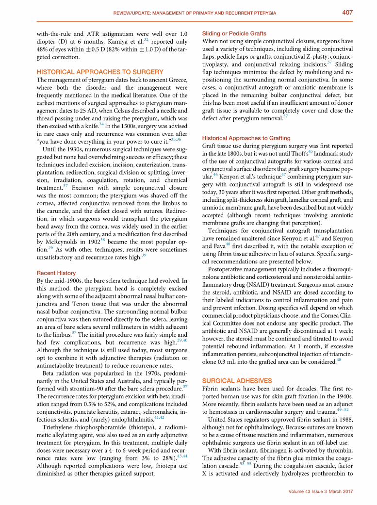
The tight junctions maintain hydration, and the loose con-nective tissue covers the nerve endings that were exposedduring surgery, thereby reducing postoperative swelling,pain, and recurrence of the pterygium (Figure 2).92
Amniotic membranes were first investigated for use inophthalmic surgery during the early 1900s, but resultswere not encouraging and use dwindled until the latterpart of the century when studies were carried out in the So-viet Union and Latin America.92 Since then, amniotic
Volume 43 Issue 3 March 2017

Figure 2. A post-pterygia eye after amniotic membrane transplantand Tisseel glue closure. Photo credit: Terry Kim, MD.
410 REVIEW/UPDATE: MANAGEMENT OF PRIMARY AND RECURRENT PTERYGIA
membrane has been shown to be beneficial during surgicalreconstruction of the limbus after pterygium surgery.Covering the nerve endings reduces the patient's postoper-ative pain. Further, the basal lamina and stromal architec-ture of the amniotic membrane resembles that of humanconjunctiva. This similarity makes amniotic membrane aparticularly attractive replacement. It features an imperme-able basal lamina that prevents evaporation and loss of elec-trolytes from the scleral bed; the basal lamina itself serves asa platform to grow healthy conjunctival and corneal epithe-lial cells.92 The membranes act as a barrier, sometimesreferred to as a “substrate that allows restoration of thenormal limbal stem cells,” while preventing abnormalconjunctival stem cells from flourishing.93
PropertiesIn the early iterations, conventional preparation used glyc-erol, tissue culture media, and freezing temperatures. In the
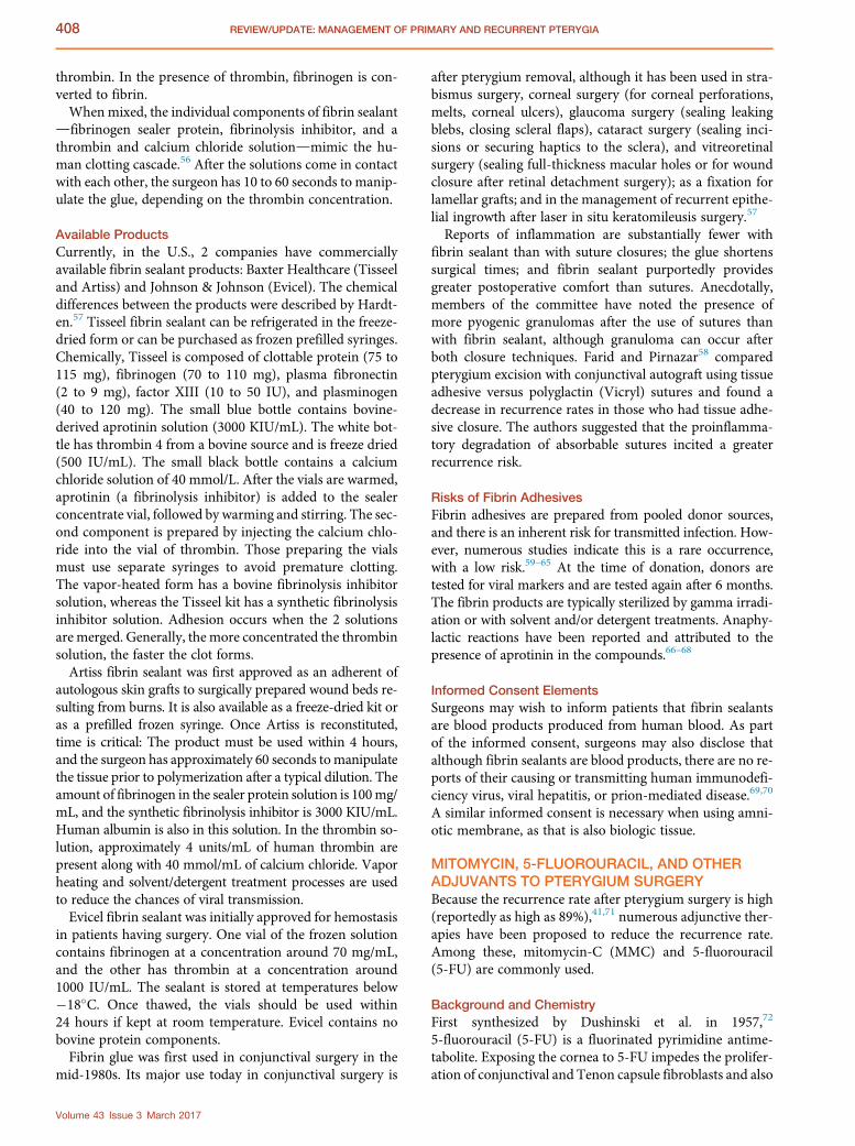
Figure 3. Pterygium before excision and conjunctival autograph.Photo credit: John A. Hovanesian, MD.
Volume 43 Issue 3 March 2017
1980s, Muldachev used a conjunctival substitute in pteryg-ium surgery and described less inflammatory response, lessredness and edema, reduced incidence of recurrence, lesspain, and faster recovery in patients who were operatedon with this particular allotransplant. The allotransplantwas later identified as amniotic membrane.92 Initial resultsin 23 patients with recurrent pterygia, bullous keratopathy,corneal ulcers, dermoid reconstruction, reconstruction ofsymblepharon, and corneal dellen were promising.96 Amni-otic membrane in both frozen and dehydrated forms arenow commercially available. Although surgeons have pref-erences as to the type of amniotic membrane, no consensusexists on which form is associated with lower rates of pte-rygium recurrence.
Available Types of Amniotic MembranesBiotissue, which provides Amniograft, removes and sepa-rates the amniotic membrane from the chorionic mem-brane and preserves it in a solution of glycerol and tissueculture medium in a 50% solution prior to freezing at�80
�C. The tissue is brought to room temperature in the
operating room and rehydrated before use.Innovative Ophthalmic Products (now part of Katena
Surgical) developed Ambiodry, which does not requirerefrigeration or special devices for transportation. It is pre-served inside a sterile envelope and boasts a shelf life of2 years. Serologic testing excludes contaminated tissue,and dehydration preserves the tissue. A drying fixture em-bosses texture onto the final graft to identify the stromaland basement membrane sides. After the membrane iscut and double packaged, electron beam sterilization isapplied. After being moistened with a balanced salt solu-tion or human tears, the membrane behaves like a natu-rally occurring biologic membrane. Thickness variesdepending on which version is used (Ambiodry2 versusAmbio5). BioD, LLC, also offers a dehydrated extracellular
Figure 4. Pterygium after excision with conjunctival autograft. Photocredit: John A. Hovanesian, MD.

Figure 5. The graft donor site at the superior limbus with mild chemo-sis, demonstrating the presence of retained Tenon fascia, evidencethat a thin graft had been harvested from this location. Photo credit:John A. Hovanesian, MD.
Figure 6. The free conjunctival autograft positioned adjacent to thepterygium excision site with the stromal side facing the surgeon andthe limbal sign adjacent to the limbus at the excision site. Photocredit: John A. Hovanesian, MD.
411REVIEW/UPDATE: MANAGEMENT OF PRIMARY AND RECURRENT PTERYGIA
membrane allograft derived from human amniotic tissueunder the trade name BioDOptix.
PTERYGIUM EXCISION WITH CONJUNCTIVALAUTOGRAFTPterygium excision with conjunctival autograft uses the pa-tient's adjacent conjunctiva to fill the void left by the excisedpterygium (Figures 3 and 4).47,97When a conjunctival auto-graft is used for pterygium excision, the preferred surgicaltechnique is to remove the pterygium body from the bulbarconjunctival using a scissors and a forceps, excising most ofthe Tenon fascia down to bare sclera without excessivemanipulation of the rectus muscle.A Particular care shouldbe taken to excise the Tenon fascia under the free edges ofthe conjunctiva as this is the source of fibroblastic prolifer-ation leading to recurrence. The leading edge or head of thepterygium can be avulsed or carefully dissected from thecorneal surface, preserving Bowman membrane to main-tain a healthy, intact corneal surface. The pterygium spec-imen can then be placed in formalin and submitted forpathology to confirm the diagnosis. (This may be necessaryonly in cases in which the clinical history and examinationdo not clearly support the diagnosis of a pterygium.) A dia-mond pterygium burr (not the type that is manufacturedfor corneal foreign-body and rust-ring removal) may behelpful in removing residual attachments of pterygium toleave a smooth corneoscleral junction. A sharp blade canalso be held perpendicular to the surface and scraped paral-lel to the limbus to restore a surface that will provide anacceptable cosmetic appearance and functional surface tograft conjunctiva. In cases of recurrence or extensive scar-ring, it is particularly important to identify and isolate therectus muscle; a muscle hook can be used to avoid damage.The resulting conjunctival defect can be measured to
determine the appropriate size for a graft, which is usuallyharvested from the superior or inferior bulbar conjunctiva.Graft edges can be marked with ink or small marks madewith low-temperature cautery. This ensures the intended
graft size is achieved and provides a mark to prevent unin-tentional graft inversion.Use of a thin conjunctival graft is recommended as this
avoids excessive chemosis in the graft and minimizes scar-ring in the donor site (Figure 5). To obtain a thin graft, theeye can be ducted to expose the full donor site and a smallsnip made with a blunt Westcott scissors at a graft edgeaway from the limbus. A spring-action Westcott scissorscan be inserted through this small opening to bluntlydissect the conjunctival surface from Tenon fascia overthe entire area of the graft. One blade of the scissors thenplaced through the small opening can extend the openingto the limbus to define the graft's lateral margins, leavingthe limbal edge attached to the cornea. The donor sitecan be closed with a suture or fibrin adhesive or left opento heal without grafting or wound closure.The graft can be placed directly on the bare sclera site
and sutured in place. Most surgeons use 5 to 8 interruptedsutures for this. Fibrin sealant can also be used to securethe graft. In the latter case, the graft is placed on thecornea with the stromal side exposed and the epitheliumtouching the corneal epithelium. The limbal side of thegraft should be adjacent to the limbal side of the pteryg-ium excision site (Figure 6). A drop of the thrombinportion of fibrin sealant is applied to the bare sclera ofthe excision site, and a small portion (less than a dropgenerally) of the fibrinogen component is applied tocoat the stromal side of the autograft. Two smooth-tipped forceps are used to grasp the autograft and invertit onto the excision site. This mixes the 2 componentsof the fibrin adhesive and aligns the limbus of the auto-graft to the limbus of the excision site.Dilution of thrombin has been widely used by
ophthalmic surgeons to slow the polymerization offibrinogen to fibrin, allowing greater time to manipulatetissues. Dilution to 1:10 or 1:100 with a balanced salt so-lution is commonly practiced. To dilute to 1:10 concen-tration, 0.1 cc of thrombin and 0.9 cc of a balanced salt
Volume 43 Issue 3 March 2017

Figure 7. Excising a pterygium in preparation for placement of anamniotic membrane graft is facilitated by excising an additional1.0 to 2.0 mm margin of conjunctiva to expose bare sclera. Photocredit: John A. Hovanesian, MD.
412 REVIEW/UPDATE: MANAGEMENT OF PRIMARY AND RECURRENT PTERYGIA
solution are drawn into a single syringe, which is agitatedto mix the components. To dilute to 1:100 concentration,0.9 cc of the thrombin solution just described is dis-carded and another 0.9 cc of a balanced salt solution isdrawn into the syringe and mixed. Using dilutedthrombin does not reduce the tensile or shear strengthof the polymerized fibrin sealant.27,98 Undiluted fibrinsealant will polymerize within approximately 20 seconds,so it is important to align and stretch the edges of thegraft to approximate the edges of the conjunctiva defectin a short period.A therapeutic soft contact lens can be used to cover a
large corneal epithelial defect. Alternatively, some surgeonsplace a small portion of fibrin adhesive on the deepithelial-ized surfaces to serve as a biologic bandage. The eye can bepatched and shielded over a steroid and NSAID eyedrops.Al Fayez99 compared limbal and conjunctival autograft
transplantation in advanced and recurrent pterygia. Limbaltransplantation appeared more effective than free conjunc-tival transplantation in recurrent pterygia, but no statisti-cally significant differences were found in advancedpterygia. Al Fayez100 found the same outcomes in a muchlarger prospective parallel group study in 224 eyes that pre-sented with recurrent pterygia.Conjunctival autograft transplantation is not without po-
tential downsides. It canmake closing the large conjunctivaldefects difficult, and the technique mandates that conjunc-tiva be reserved for the possibility of glaucoma filtering sur-gery.101 In patients who present with an increasedlikelihood of needing glaucoma filtration surgery in thefuture, harvesting the conjunctival autograft from an infe-rior bulbar location might be better.Because of graft suturing, this method has the disadvantage
of a relatively longer surgery than the bare sclera technique. Italso carries the risk for complications such as pyogenic gran-uloma formation and giant papillary conjunctivitis, as well assignificant patient discomfort after surgery.102
Finally, although rare, the graft can mechanically displaceor become lost. Avascular stromalysis of the sclera is directlyrelated to excessive or misapplied MMC use.48 However,since the recurrence rate is only 5%, even in recurrent casesinvolving cicatricial strabismus, the technique should be atthe forefront of management strategies.48
PTERYGIUM EXCISION WITH AMNIOTICMEMBRANE GRAFTINGThe less-than-stellar outcomes with previous techniquesand the high recurrence rates have necessitated explorationof other surgical techniques to remove pterygium. TheAmerican Medical Association's Current Procedural Ter-minology code for amniotic membrane transplantationfor ocular surface reconstruction is 65780. As of February2015, more than 800 peer-reviewed articles on the ocularuse of amniotic membrane, highlighting novel and thera-peutic applications, had been published. Because amnioticmembrane grafts have virtually no size limitation, fibrotictissue can be extensively excised, which may contribute tothe favorable surgical outcomes.101
Volume 43 Issue 3 March 2017
Anatomic ConsiderationsBetween 20 and 30 ophthalmic transplants can be performedwith 1 placenta. The 2 most commonly used types of amni-otic membrane are described above. However, outside theU.S. and other western countries, fresh and unpreservedmembrane is often used within days or weeks of donation.The anatomic makeup of amniotic membrane seems con-
tradictory as amniotic membrane contains both inhibitoryand proinflammatory cytokines; that is, interleukin (IL)-6and IL-8 are proinflammatory and IL-10 and IL-1ra antiin-flammatory, yet both are present in the amniotic mem-brane.103 Different donors will likely yield differences in theamniotic membrane itself. The thickness and morphologyof the donor membrane is affected by its original location.The variation in thickness may affect the integration of themembrane with other ocular surface tissues. Age, race, parity,gravidity, and duration of gestation may contribute to vari-ability of specimens.101
Preparation of TissueAfter the pterygium head is undermined and separated atthe limbus and dissected toward the central cornea with aWestcott scissors, an additional 1.0 to 2.0 mm margin ofconjunctival tissue must be dissected to expose the baresclera (Figure 7). The amniotic membrane is then cutwithin its surgical packaging to the appropriate size.When the membrane is prepared, the basement membraneshould be on top, away from the sclera.When using Ambio-dry2, for example, this is easily achieved; if the surgeon canread the letters “IOP,” the membrane is in the correct posi-tion. When frozen amniotic membrane is used, the stromalside is stickier than the basement membrane side.B
Use of Fibrin Adhesive and Best Practices for FixationFibrin glue can be prepared by the surgical assistant. If sur-geons use Tisseel VH, the surgical assistant prepares the2 components while the pterygium is being surgicallyremoved. The adhesive can be administered to the ocularsurface in 1 of 2 ways to form the fibrin clot. In the first

Figure 8. A strip of amniotic membrane placed in the subconjuncti-val space surrounding the pterygium excision site in the locationhighlighted in red. Photo credit: John A. Hovanesian, MD.
413REVIEW/UPDATE: MANAGEMENT OF PRIMARY AND RECURRENT PTERYGIA
method, the 2 components are combined in theY-connectorof a Duploject syringe (supplied in the kit), and 10 drops arepurposely wasted to ensure both components of adhesiveare flowing at the tip. One drop is then injected under theamniotic membrane. A smooth instrument is used torapidly position and smooth the membrane. The coagulum(fibrin clot) starts forming in 5 to 7 seconds, achieves 70% ofthe final tensile strength in 10minutes, and is at full strengthin 2 hours.A Altering the concentrations of the individualcomponents will provide flexibility in setting times.In the secondmethod, 1 drop of the fibrinogen component
of the sealant is placed on the scleral bed. The membranemust be placed several millimeters below the surroundingconjunctival tissue as the fibroblasts that can lead to recur-rence are typically found here. Thrombin is placed on topand around the edges of the membrane to activate it. Thisshould keep the membrane in position for weeks.104
In both methods, the speculum is removed and anti-biotic–steroid ointment applied over the treated eye, whichis then patched and shielded.
Postoperative Management and ResultsCompared with a suturing technique, the use of fibrin gluecauses less discomfort postoperatively (see above). Compli-cations can arise if surgeons use too much glue or if excessproduct is left under the graft inadvertently. If the glue wasnot distributed evenly, the graft will appear edematous dur-ing the early postoperative period. The graft may retract ifparts of the underlying sclera did not receive fibrin glue.
PTERYGIUM EXCISION WITH CONJUNCTIVALAUTOGRAFT AND AMNIOTIC MEMBRANETo further reduce the recurrence rate, amniotic membranehas been used in a simple adjunctive procedure performedwith pterygium excision and conjunctival autograft.27 Thecombined use of a conjunctival autograft and placementof an amniotic membrane is usually reserved for high-riskcases that involve large or inflamed pterygia or recurrencesof pterygia.101
The combined technique places a small strip of amnioticmembrane in the subconjunctival space surrounding the pte-rygium excision followed by placement of a conjunctivalautograft (Figure 8). In this subconjunctival location, the am-niotic membrane is thought to function as a biologic depot ofantifibrotic and antiinflammatorymaterial that prevents pte-rygium recurrence. Fibrin adhesive is used to secure theconjunctival autograft and the amniotic membrane.27
Surgical TechniqueIn the combination surgery, the pterygium is excised withan additional 1.0 mm margin of conjunctival tissue toexpose the bare sclera. A space is created beneath the con-junctiva surrounding the excision site, tunneling approxi-mately 5.0 mm into the tissue on 3 sides, ensuring thespace remains superficial to most of the Tenon fascia, themedial rectus muscle, and tendon.C The conjunctival auto-graft is prepared from the superior bulbar conjunctiva ofthe same eye and remains attached at the limbus superiorly.
A potential advantage to this technique is that either cry-opreserved or freeze-dried amniotic membrane can beused. In both cases, the membrane is cut into a C-shapedgraft large enough to surround the conjunctival defect.In this technique, fibrin tissue adhesive preparation dif-
fers from the standard preparation.27 The components arenot transferred to the syringe after preparation; rather, ascrub nurse draws approximately 0.1 cc of each componentinto 2 separate sterile 1.0 cc syringes through a sterile18-gauge needle, minimizing the air entry into the syringe.Next, 0.9 cc of a balanced salt solution is added to thethrombin-filled syringe and the syringe is inverted severaltimes to facilitate mixing.A 0.12 forceps is recommended for lifting the conjunc-
tiva, and a small amount of fibrinogen is applied to the un-dersurface of the conjunctiva (to keep the amnioticmembrane in place); thrombin is not applied. In thefreeze-dried amniotic membrane method, a balanced saltsolution or fibrinogen solution can be placed on a sectionof surgical drape to facilitate hydration of the amnioticmembrane graft; this step can be eliminated with the wetform of amniotic membrane. The hydrated amniotic mem-brane graft is placed directly on the bare sclera and broughtinto the subconjunctival space. The concept is to create asubconjunctival amniotic membrane area that surroundsthe bare sclera but does not cover it. Edges of the overlyingconjunctiva are lifted and advanced toward the limbus tocover any exposed remnants of the transplanted amnioticmembrane. The presence of the fibrinogen, mixed withsmall amounts of thrombin in the patient's own blood,will ensure the edges stay in place.27
The conjunctival autograft, which remains attached tothe superior limbus, is now reflected onto the cornea(epithelium to epithelium) and cut free from the superiorlimbus. The limbal side of the graft has to be oriented to
Volume 43 Issue 3 March 2017

414 REVIEW/UPDATE: MANAGEMENT OF PRIMARY AND RECURRENT PTERYGIA
the limbus; a small droplet from the diluted thrombin sy-ringe is applied to the bare sclera, and a small dropletfrom the fibrinogen syringe is applied to the underside ofthe graft. The graft is then flipped onto the bare sclera,which allows the 2 adhesive components to mix.27
After surgery, a topical fluoroquinolone antibiotic isapplied 4 times a day for 1 week. Prednisolone acetate1.0% is started 4 times a day and tapered over 4 weeks.A topical NSAID to control pain is recommended for 1 to3 days immediately postoperatively.27
It is possible for patients to present with 2-headed pteryg-ium. These cases can be treated with a conjunctival auto-graft obtained from the superior and/or inferior fornix,using a subconjunctival implant of amniotic membranefor 1 or both sites.
Postoperative Complications and ManagementA retrospective analysis of 100 patients who had the com-bined procedure found a 1% recurrence rate after a6-month follow-up, and all the conjunctival and amnioticmembrane grafts remained viable and in place.105
COMPLICATIONS OF PTERYGIUM SURGERYPostoperative complications are not common in pteryg-ium removal and can be attributed to the surgical tech-nique itself or to any of the possible adjuvants used.106
It has been generally accepted that pterygium recurrencerates are substantially less with conjunctival or limbal au-tografts than with the bare sclera technique, but compli-cations persist.30 Topical antibiotics, lubricants, andanalgesics are routinely used in the immediate postoper-ative period, and all patients should be examinedpostoperatively.107,108
Early ComplicationsVarious early postoperative complications have been re-ported after pterygium removal relative to the surgicalremoval technique and postoperative medication used.The use of MMC postoperatively has led to reports ofwound dehiscence and corneal epithelial defects.109 Thishas been traditionally noted when MMC has been usedtopically. The ASCRS Cornea Clinical Committee thereforecurrently supports intraoperative rather than topical post-operative use of MMC to treat pterygium.Although several other immediate complications are
associated with pterygium surgery (including reactionaryhemorrhage leading to excessive bleeding, graft edema, he-matoma below the graft, or corneal scarring), they are usu-ally not vision threatening and resolve quickly. Excessivebleeding can be managed via pressure bandages, graftedema resolves with topical treatment, and hematomasshould be examined under the microscope and then evacu-ated under a block. Depending on the depth of cornealinvolvement, scars can be observed or may require lamellarkeratoplasty later. Localized epithelial defects are noted innearly every initial postoperative examination and healwithin 24 hours.106 Similarly, conjunctival chemosis maybe noticed but resolves spontaneously.106
Volume 43 Issue 3 March 2017
Suture-related complications can be reduced by usingmonofilament nylon in lieu of polyglactin sutures. Fibrinadhesives can eliminate suture-related complications.Improperly harvesting or securing the conjunctival auto-
graft may be a risk factor for the development of a Tenoncyst, especially if the graft was harvested with a thick Tenonlayer or if the wound edges are imbricated after graft place-ment. Treatment/resolution occurs via excision of the cystfollowed by conjunctival closure.106
It should be noted that conjunctival donor site can beused for subsequent surgeries (recurrent pterygia) providedthe first dissection was performed carefully, with minimaldisruption of Tenon fascia, and there is minimal fibrosis.
Late ComplicationsRecurrence is considered a significant late complication ofinitial pterygium surgery and often happens within a fewmonths. The more repeatedly a recurrence happens in aparticular eye, the more difficult the treatment, as the recur-rence is often accompanied by increased conjunctivalinflammation and faster corneal involvement.40,106,110
Scleral ulceration is a potential long-term complicationafter the bare sclera technique.111 In the same report, theuse of beta irradiation to prevent recurrence of pterygiawas a significant cause of iatrogenic ocular disease. Ptosis,symblepharon, and iris atrophy have been reported afterirradiation use in pterygium excision.106 Ptosis often re-solves spontaneously but does take several months. If symp-tomatic, the symblepharon release can be performed andthe patient should be put on topical lubricants.106
Antimetabolite-Related ComplicationsIt is well documented that MMC can reduce the recurrencerate to less than 10%, but severe postoperative complica-tions include corneal edema, perforation, scleral calcifica-tion, corectopia, iritis, sudden-onset mature cataract,severe secondary glaucoma, incapacitating photophobia,and pain, although the complications have been associatedwith poor patient selection and overuse of MMC.80,88,112 Ifstrict patient-inclusion criteria are applied, stringent patientmonitoring until reepithelialization can reduce MMC-related postoperative complications.82,88,113
Scleral dellen as a result of delayed conjunctival woundclosure has been reported after bare sclera excision withMMC.114 Other reported complications include temporaryand prolonged discomfort, tearing, hyperemia, subconjunc-tival hemorrhage, wound dehiscence, and pigment accu-mulation after a single dose of intraoperative MMC.106 Itis now recommended that only patients with high-risk pte-rygia receive intraoperative MMC.106
Overuse of antimetabolites can also lead to scleral com-plications. These include necrotizing scleritis and scleralthinning with perforation.
MANAGING RECURRENCEMinimizing pterygium recurrence is crucial to preventvision loss, as advanced or recurrent pterygia eventuallyresult in loss of corneal transparency in the visual axis.

415REVIEW/UPDATE: MANAGEMENT OF PRIMARY AND RECURRENT PTERYGIA
High recurrence rates have led to the abandonment ofseveral surgical techniques, and debate on the optimaltechnique is ongoing. Postoperative alterations in periph-eral corneal topography, persistent wetting defects, andchronic irritation and inflammation after surgery mayalso increase the likelihood of pterygium recurrence.115
Inflammation coupled with persistent vascularity is oftenan indication of recurrence. Recurrent pterygia continueto move toward the limbus, eventually moving onto thecorneal surface.Recurrent pterygia differ histopathologically fromprimary
pterygia,116 have higher rates of recurrence with subsequentsurgical interventions, and must be considered a differententity than primary pterygia. Recurrent pterygia are alsoconsidered more aggressive clinically and therefore morelikely to cause visual deficits and other complications.115
Simple RecurrenceOnce a patient has been treated for primary pterygia, vigi-lant monitoring during the first postoperative months iscritical to minimize the risk for recurrence: While 50% ofrecurrences appear in the first 4 months, 97% appear withinthe first 12 months.117 Although conjunctival autograftshave been shown to reduce recurrence rates in both primaryand recurrent pterygia to less than 5%, recurrence can be aresult of inadequate peripheral dissection, insufficient graftsize, thick graft with Tenon tissue, and graft retraction dueto inadequate fixation.36 Similarly, recurrence rates afterpterygia surgery using an amniotic membrane graft maybe slightly higher than surgery using a conjunctival auto-graft, but recurrence rates with the 2 graft types are similarwhen appropriate dissection and/or other adjunctive thera-pies are performed.115
If persistent inflammation remains during the early post-operative period, reinitiation of topical steroids iscommonly used to deter recurrence. Topical steroids canbe used to quiet the inflammation and tapered slowly,and side effects of continued steroid use (intraocular pres-sure increases, steroid-induced cataract formation) mustbe discussed. Adjunctive use of commercially availabletopical NSAIDs should also be considered as studies haveshown they can slow epithelial migration and are as effec-tive as some topical steroids in treating inflamed pterygiaand pinguecula.118 Although the topical NSAIDs rarelyinduce side effects, their efficacy in recurrent pterygiumhas not been proven.115
Topical cyclosporine 0.05% has been used in the manage-ment of pterygium excision patients and early recur-rences119 and has been evaluated in its native form orcompounded at a higher concentration (1.0% or 2.0%).115
Topical tacrolimus and interferon alpha-2b and subcon-junctival 5-FU have also been advocated for early pterygiarecurrence77,120,121; the use of topical MMC has been dis-cussed and the potential risks in the treatment of recurrentpterygia and of primary pterygium are the same.Local steroid injections have shown efficacy in about 50%
of cases of recurrence, and they are typically delivered sub-conjunctivally adjacent to the pterygium recurrence in the
oblique quadrants, carefully avoiding the interpalpebralfissure.115 (Patients may complain about the cosmeticappearance of a white depot.) However, there are no largerandomized controlled clinical trials to confirm the efficacyof local steroid treatment for recurrent pterygium. The off-label use of topical and subconjunctival bevacizumab hasbeen evaluated successfully in the treatment of recurrentpterygia, but further study is warranted.115
It is not uncommon for patients with recurrent pterygiato have concomitant lid disease or ocular rosacea. Medica-tions that inhibit matrix metalloproteases should beconsidered, including oral doxycycline and/or topicaltetracycline.Because there is a higher risk for recurrence in advanced
and recurrent pterygia that involved the optical zone, per-forming the pterygium excision alone at the initial surgeryand then corneal surgery (including lamellar keratoplasty,phototherapeutic keratectomy, or penetrating keratoplasty)after recurrence is avoided may be optimal.Recurrent pterygia are more difficult to excise as scarring
is already present, which can not only obliterate tissueplanes but also increase the risk for extraocular muscledamage.36 In cases of extensive scarring, primary closuretechniques are unreliable; sliding or pedicle flaps can some-times be effectively used.115
Complex Recurrence (Symblepharon, Loss of FornixStructure)In cases of aggressive pterygium recurrence or recurrent pte-rygia that extend more centrally than the initial pterygium,surgical management should be considered as a first-line op-tion, with a 4- to 6-month delay from the initial surgery.When using a conjunctival autograft, using an oversized
graft (1.0 to 2.0 mm) in both dimensions and the superiorconjunctiva can minimize the risk for symblepharon andfornix shortening.Advanced recurrent pterygium can be devastating to the
normal anatomy of the ocular surface with the formation ofsymblepharon and/or fornix shortening or obliteration.The restricted extraocular movement coupled with thedecreased visual acuity may result in diplopia and requireextensive surgical intervention to maximize outcomes.115
Complete dissection of previous symblepharon andscarring is mandatory. The symblepharon should becompletely freed flush with the ocular surface tocompletely remove the adhesions and minimize the resul-tant epithelial defects on the palpebral conjunctiva whileminimizing the excision of normal conjunctival tissue.The freed conjunctival surface can then be retracted intothe fornix and fixated with full-thickness sutures throughthe tarsus and palpebral conjunctiva. Isolation of the adja-cent rectus muscle with careful dissection of any associ-ated fibrosis is recommended to avoid inadvertentdamage, to release any restriction present preoperatively,and to prevent extraocular muscle restriction postopera-tively. Once complete, a large conjunctival autograft, am-niotic membrane graft, and/or buccal mucosal graft maybe used to reconstruct the ocular surface.115
Volume 43 Issue 3 March 2017

416 REVIEW/UPDATE: MANAGEMENT OF PRIMARY AND RECURRENT PTERYGIA
There is often minimal normal conjunctiva, requiringthe use of amniotic membrane or buccal mucosal graftsto cover the resultant defects after dissection. These buccalmucosal grafts are efficacious but not often used because oftheir inferior appearance compared with conjunctivalautograft or amniotic membrane.36 To avoid postopera-tive hypertrophy, surgeons should obtain a very thin flapfree of submucosal tissue when using buccal mucosalgrafts. Complete coverage of any outstanding defect is rec-ommended, and severe cases may warrant deep extensioninto the fornix where the graft may fold over itself to coverthe palpebral conjunctiva. Sutures are recommended overfibrin adhesive or cautery to fixate the graft along the lidmargin and to prevent symblepharon reformation. A lim-bal conjunctival flap or autograft in conjunction with theamniotic membrane graft can be performed to supplyepithelial cells to cover the large graft and provide an addi-tional barrier to recurrence.36,122 The surgical time in-creases when this reconstruction technique is used. Thesuperiority of the technique over amniotic grafting alonehas not been established well enough to justify its use inall cases.115
Symblepharon rings and/or bandage contact lenses inconjunction with a large temporary tarsorrhaphy maybe useful in preventing reformation; each is removedabout 6 to 8 weeks postoperatively, and frequent topicalsteroids with a slow taper should be used. Aggressivelubrication also is recommended to encourage epithelial-ization of the bulbar and palpebral conjunctiva and pre-vent reformation.
DISCUSSIONThe treatment of pterygium continues to evolve, as does themanagement of recurrence. The ASCRS Cornea ClinicalCommittee advocates the use of any of the techniquesdescribed in this review. Individual surgeons will have pref-erences based on many parameters, and the various tech-niques described have been used successfully to manageprimary or recurrent disease.We also strongly advocate that surgeons leave a clean
corneal surface and take the necessary steps to minimizemanipulation and trauma during pterygium removal toreduce the risk for postoperative inflammation that maylead to recurrence. We believe that the more surgeons un-derstand pterygium and the various treatment techniques,the lower the recurrence rates will be.
REFERENCES1. Sul S, Korkmaz S, Novruzlu S. Seasonal effects on pterygium surgery
outcome: implications for the role of sunlight exposure. Cornea 2014;33:504–506
2. RimTH,NamJS, KimEK,KimTI.Risk factors associatedwith pterygiumandits subtypes in Korea: the Korean National Health and Nutrition ExaminationSurvey 2008–2010. Cornea 2013; 32:962–970; erratum 2016; 35 (9):e30
3. Ajayi IyiadeA,OmotoyeOlusola J. Pattern of eyediseases amongwelders in aNigeria community. Afr Health Sci 2012; 12:210–216. Available at: https://www.ncbi.nlm.nih.gov/pmc/articles/PMC3462541/pdf/AFHS1202-0210.pdf. Accessed January 26, 2017
4. Sherwin JC, Hewitt AW, Kearns LS, Griffiths LR, Mackey DA,Coroneo MT. The association between pterygium and conjunctival ultra-violet autofluorescence: the Norfolk Island Eye Study. Acta Ophthalmol
Volume 43 Issue 3 March 2017
2013; 91:363–370. Available at: http://onlinelibrary.wiley.com/doi/10.1111/j.1755-3768.2011.02314.x/epdf. Accessed January 26, 2017
5. Norval M, Lucas RM, Cullen AP, deGruijl FR, Longstreth J, Takizawa Y, vander Leun JC. The human health effects of ozone depletion and interactionswith climate change. Photochem Photobiol Sci 2011; 10:199–225
6. Tano T, Ono K, Hiratsuka Y, Sekiguchi M, Konno S, Kikuchi S, Onishi Y,Takegami M, Yamada M, Fukuhara S, Murakami A. Prevalence of pteryg-ium in a population in Northern Japan: the Locomotive Syndrome andHealth Outcome in Aizu Cohort Study. Acta Ophthalmol 2013; 91(3):e232–e236. Available at: http://onlinelibrary.wiley.com/doi/10.1111/aos.12044/epdf. Accessed January 26, 2017
7. Sliney DH. The focusing of ultraviolet radiation in the eye and ocular expo-sure. In: Taylor HR, ed, Pterygium. The Hague, Netherlands, Kugler Pub-lications, 2000; 29–40
8. Chui JJY, Coroneo MT. Pterygium pathogenesis, actinic damage, andrecurrence. In: Hovanesian JA, ed, Pterygium: Techniques and Technol-ogies for Surgical Success. Thorofare, NJ, Slack, 2012; 1–26
9. Moran DJ, Hollows FC. Pterygium and ultraviolet radiation: a positive corre-lation. Br J Ophthalmol 1984; 68:343–346. Available at: https://www.ncbi.nlm.nih.gov/pmc/articles/PMC1040336/pdf/brjopthal00149-0073.pdf. Ac-cessed January 26, 2017
10. Chui J, Di Girolamo N, Wakefield D, Coroneo MT. The pathogenesis ofpterygium: current concepts and their therapeutic implications. OcularSurf 2008; 6:24–43
11. Coroneo MT, Di Girolamo N, Wakefield D. The pathogenesis of pterygia.Curr Opin Ophthalmol 1999; 10:282–288
12. Di Girolamo N, Chui J, Coroneo MT, Wakefield D. Pathogenesis of pte-rygia: role of cytokines, growth factors, and matrix metalloproteinases.Prog Retin Eye Res 2004; 23:195–228
13. Maheshwari S. Effect of pterygium excision on pterygium induced astig-matism. Indian J Ophthalmol 2003; 51:187–188. Available at: http://www.ijo.in/article.asp?issnZ0301-4738. Accessed January 26, 2017
14. Maheshwari S. Pterygium-induced corneal refractive changes. Indian JOphthalmol 2007; 55:383–386. Available at: https://www.ncbi.nlm.nih.gov/pmc/articles/PMC2636029/. Accessed January 26, 2017
15. Panchapakesan J, Hourihan F, Mitchell P. Prevalence of pterygium andpinguecula: the Blue Mountains Eye Study. Aust N Z J Ophthalmol1998; 26 (Suppl 1):S2–S5
16. Hill JC,Maske R. Pathogenesis of pterygium. Eye 1989; 3:218–226. Avail-able at: http://www.nature.com/eye/journal/v3/n2/pdf/eye198931a.pdf.Accessed January 26, 2017
17. Detorakis ET, Spandidos DA. Pathogenetic mechanisms and treatment op-tions for ophthalmic pterygium: trends and perspectives (review). Int J MolMed 2009; 23:439–447. Available at: https://www.spandidos-publications.com/ijmm/23/4/439/download. Accessed January 26, 2017
18. Ang LP, Chua JL, Tan DT. Current concepts and techniques in pterygiumtreatment. Curr Opin Ophthalmol 2007; 18:308–313
19. Saw S-M, Tan D. Pterygium: prevalence, demography and risk factors.Ophthalmic Epidemiol 1999; 6:219–228
20. Wong TY, Foster PJ, Johnson GJ, Seah SK, Tan DT. The prevalence andrisk factors for pterygium in an adult Chinese population in Singapore: theTanjong Pagar survey. Am J Ophthalmol 2001; 131:176–183
21. Karai I, Horiguchi S. Pterygium in welders. Br J Ophthalmol 1984; 68:347–349. Available at: https://www.ncbi.nlm.nih.gov/pmc/articles/PMC1040337/pdf/brjopthal00149-0077.pdf. Accessed January 26, 2017
22. Nakaishi H, Yamamoto M, Ishida M, Someya I, Yamada Y. Pingueculaeand pterygia in motorcycle policemen. Industrial Health 1997; 35:325–329. Available at: https://www.jstage.jst.go.jp/article/indhealth1963/35/3/35_3_325/_pdf. Accessed January 26, 2017
23. Diaz-Valle D,Mendez-Fernandez R, Arriola-VillalobosP, IradierMT, Benitez-del-Castillo JM. Pinguecula and pterygium. In: Benitez-del-Castillo JM,Lemp MA, eds, Ocular Surface Disorders. London, UK, JP Medical Ltd,2013; 173–180
24. Liu T, Liu Y, Xie L, He X, Bai J. Progress in the pathogenesis of pterygium.Curr Eye Res 2013; 38:1191–1197
25. Wlodarczyk J, Whyte P, Cockrum P, Taylor H. Pterygium in Australia: acost of illness study. Clin Exp Ophthalmol 2001; 29:370–375
26. Chen PP, Ariyasu RG, Kaza V, LaBree LD, McDonnell PJ. A randomizedtrial comparing mitomycin C and conjunctival autograft after excision ofprimary pterygium. Am J Ophthalmol 1995; 120:151–160
27. Hovanesian JA, Behesnilian AS. Pterygium excision with a conjunctivalautograft and prophylactic placement of subconjunctival amniotic mem-brane surrounding the excision site. In: Hovanesian JA, ed, Pterygium:Techniques and Technologies for Surgical Success. Thorofare, NJ, Slack,2012; 101–110
28. Rohrbach IM, Starc S, Knorr M. Vorhersage von Pterygiumrezidiven auf-grund morphologischer und immunhistologischer Parameter [Predicting

417REVIEW/UPDATE: MANAGEMENT OF PRIMARY AND RECURRENT PTERYGIA
recurrent pterygium based onmorphologic and immunohistologic param-eters]. Ophthalmologe 1995; 92:463–468
29. Tan DT, Chee SP, Dear KB, Lim AS. Effect of pterygium morphologyon pterygium recurrence in a controlled trial comparing conjunctivalautografting with bare sclera excision. Arch Ophthalmol 1997;115:1235–1240
30. KaufmanSC, JacobsDS, LeeWB,DengSX,RosenblattMI, ShteinRM.Op-tions and adjuvants in surgery for pterygium: a report by theAmericanAcad-emy of Ophthalmology (Ophthalmology Technology Assessment).Ophthalmology 2013; 120:201–208. Available at: http://www.aaojournal.org/article/S0161-6420(12)00696-3/pdf. Accessed January 26, 2017
31. Kandavel R, Kang JJ, Memarzadeh F, Chuck RS. Comparison of pteryg-ium recurrence rates in Hispanic and white patients after primary excisionand conjunctival autograft. Cornea 2010; 29:141–145
32. KamiyaK,ShimizuK, IijimaK,Shoji N, Kobashi H.Predictability of intraocularlens power calculation after simultaneous pterygium excision and cataractsurgery. Medicine 2015; 94 (52):e2232. Available at: http://journals.lww.com/md-journal/Fulltext/2015/12280/Predictability_of_Intraocular_Lens_Power.11.aspx. Accessed January 26, 2017
33. Gulani A, Dastur YK. Simultaneous pterygium and cataract surgery.J Postgrad Med 1995; 41:8–11. Available at: http://www.jpgmonline.com/article.asp?issnZ0022-3859. Accessed January 26, 2017
34. Hirschberg J. The History of Ophthalmology. Volume 1: Antiquity, 1899,translated by FC Blodi. Bonn, Germany, Wayenborgh, 1982; 32–34
35. Hirshberg J. The History of Ophthalmology. Volume 2. The Middle Ages,1899, translated by FC Blodi. Bonn, Germany, Wayenborgh, 1982; 425
36. Buratto L, Phillips RL, Carito G. Clinical picture and classification. In:Buratto L, Phillips RL, Carito G, eds, Pterygium Surgery. Thorofare, NJ,Slack, 2000; 21–26
37. Johnson RD, Pai V, Hoft RH. Historical approaches to pterygium surgery,including bare sclerae and adjunctive beta radiation techniques. In:Hovanesian JA, ed, Pterygium; Techniques and Technologies for SurgicalSuccess. Thorofare, NJ, Slack, 2012; 27–36
38. McReynolds JO. The nature and treatment of pterygia. JAMA 1902;39:296–299
39. Kamel S. Pterygium. Its nature and a new line of treatment. Br J Ophthal-mol 1946; 30:549–563. Available at: https://www.ncbi.nlm.nih.gov/pmc/articles/PMC510634/pdf/brjopthal00675-0050.pdf. Accessed January26, 2017
40. SinghG,WilsonMR, Foster CS.Mitomycin eye drops as treatment for pteryg-ium. Ophthalmology 1988; 95:813–820; discussion by RA Thoft, 820–821
41. Hirst LW. The treatment of pterygium. Surv Ophthalmol 2003;48:145–180
42. Kirwan JF, Constable PH, Murdoch IE, Khaw PT. Beta irradiation: new usesfor an old treatment: a review. Eye 2003; 17:207–215. Available at: http://www.nature.com/eye/journal/v17/n2/pdf/6700306a.pdf. Accessed January26, 2017
43. Ngoy D, Kayembe L. Etude comparative du Thio-tepa et de la MitomycineC dans le traitement du pt�erygion. R�esultats pr�eliminaires [A comparativestudy of thio-tepa and mitomycin C in the treatment of pterygium. Prelim-inary results]. J Fr Ophtalmol 1998; 21:96–102
44. Tassy A, Ribe D. Collyre thiot�epa pour pr�evenir les r�ecidives des pt�ery-gions; 18 ans d’utilisation [Thiotepa eyedrops for prevention of pterygiumrecurrence: 18 years of use]. J Fr Ophtalmol 1999; 22:215–219
45. Thoft RA. Conjunctival transplantation. Arch Ophthalmol 1977; 95:1425–1427
46. Klein SA. Zur Operation des Pterygium und zur Transplantation vonSchleimhaut. Allg Wein med Ztg 1876; 21:19
47. Kenyon KR,WagonerMD, HettingerME.Conjunctival autograft transplanta-tion for advanced and recurrent pterygium. Ophthalmology 1985; 92:1461–1470. Available at: http://www.aaojournal.org/article/S0161-6420(85)33831-9/pdf. Accessed January 26, 2017
48. Kenyon KR, Fava MA. Pterygium excision with conjunctival autograft. In:Hovanesian JA, ed, Pterygium: Techniques and Technologies for SurgicalSuccess. Thorofare, NJ, Slack, 2012; 37–48
49. Rousou J, Levitsky S, Gonzalez-Lavin L, Cosgrove D, Magilligan D,Weldon C, Hiebert C, Hess P, Joyce L, Bergsland J, Gazzaiga A. Ran-domized clinical trial of fibrin sealant in patients undergoing resternotomyor reoperation after cardiac operations: a multicenter study. J ThoracCardiovasc Surg 1989; 97:194–203
50. McCarthy PM, Borsh J, Cosgrove DM. Fibrin sealant: the Cleveland Clinicexperience. In: Schlag G, Wolner E, Eckersberger F, eds, Fibrin Sealing inSurgical and Nonsurgical FieldsCardiovascular Surgery, Thoracic Sur-gery. Berlin, Germany, Springer-Verlag, 1995; 6:34–37
51. Tidrick RT, Warner ED. Fibrin fixation of skin transplant. Surgery 1944;15:90–95
52. Katzin HM. Aqueous fibrin fixation of corneal transplants in the rabbit. ArchOphthalmol 1946; 35:415–419; discussion, 419–420
53. Thompson DF, Letassy NA, Thompson GD. Fibrin glue: a review of itspreparation, efficacy, and adverse effects as a topical hemostat. Drug In-tell Clin Pharm 1988; 22:946–952. Available at: http://journals.sagepub.com/doi/pdf/10.1177/106002808802201203. Accessed January 26,2017
54. Chabbat J, Tellier M, Porte P, Steinbuch M. Properties of a new fibrin gluestable in liquid state. Thromb Res 1994; 76:525–533. Available at: https://eurekamag.com/pdf.php?pdfZ009259528. Accessed January 26, 2017
55. Le Gu�ehennec L, Layrolle P, Daculsi G. A review of bioceramics and fibrinsealant. Eur Cell Mater 2004; 8:1–10. discussion, 10–11. Available at:http://www.ecmjournal.org/journal/papers/vol008/pdf/v008a01.pdf. Ac-cessed January 26, 2017
56. Forseth M, O’Grady K, Toriumi DM. The current status of cyanoacry-late and fibrin tissue adhesives. J Long Term Eff Med Implants 1992;2:221–233
57. Hardten DR. Fibrin tissue adhesive. In: Hovanesian JA, ed, Pterygium:Techniques and Technologies for Surgical Success. Thorofare, NJ, Slack,2012; 49–54
58. Farid M, Pirnazar JR. Pterygium recurrence after excision with conjunctivalautograft: a comparison of fibrin tissue adhesive to absorbable sutures.Cornea 2009; 28:43–45
59. Siedentop KH, Park JJ, Shah AN, Bhattacharyya TK, O’Grady KM. Safetyand efficacy of currently available fibrin tissue adhesives. Am J Otolaryngol2001; 22:230–235
60. Evensen SA, Rollag H. Solvent/detergent-treated clotting factors andhepatitis A virus seroconversion [letter]. Lancet 1993; 341:971–972
61. Lefr�ere J-J, Mariotti M, Thauvin M. B19 parvovirus DNA in solvent/detergent-treated anti-haemophilia concentrates. Lancet 1994;343:211–212
62. Everts PA, Knape JT, Weibrich G, Sch€onberger JP, Hoffmann J,Overdevest EP, Box HA, van Zundert A. Platelet-rich plasma and plateletgel: a review. J Extra Corpor Technol 2006; 38:174–187. Available at:https://www.ncbi.nlm.nih.gov/pmc/articles/PMC4680757/pdf/ject-38-174.pdf. Accessed January 26, 2017
63. Dohan DM, Choukroun J, Diss A, Dohan SL, Dohan AJJ, Mouhyi J,Gogly B. Platelet-rich fibrin (PRF): a second-generation platelet concen-trate. Part I: technological concepts and evolution. Oral Surg Oral MedOral Pathol Oral Radiol Endod 2006; 101 (3):e37–e44
64. Aizawa P, Winge S, Karlsson G. Large-scale preparation of thrombin fromhuman plasma. Thromb Res 2008; 122:560–567
65. Alston SM, Solen KA, Sukavaneshvar S,MohammadSF. In vivo efficacy ofa new autologous fibrin sealant. J Surg Res 2008; 146:143–148
66. Shirai T, Shimota H, Chida K, Sano S, Takeuchi Y, Yasueda H. Anaphy-laxis to aprotinin in fibrin sealant. Intern Med 2005; 44:1088–1089. Avail-able at: https://www.jstage.jst.go.jp/article/internalmedicine/44/10/44_10_1088/_pdf. Accessed January 26, 2017
67. Kon NF, Masumo H, Nakajima S, Tozawa R, Kimura M, Maeda S.[Anaphylactic reaction to aprotinin following topical use of biological tissuesealant] [Japanese]. Masui 1994; 43:1606–1610
68. Beierlein W, Scheule AM, Antoniadis G, Braun C, Schosser R. An imme-diate, allergic skin reaction to aprotinin after reexposure to fibrin sealant.Transfusion 2000; 40:302–305
69. Joch C. The safety of fibrin sealants. Cardiovasc Surg 2003; 11 (Suppl1):23–28
70. Horowitz B, Busch M. Estimating the pathogen safety of manufacturedhuman plasma products: application to fibrin sealants and to thrombin.Transfusion 2008; 48:1739–1753
71. Fernandes M, Sangwan VS, Bansal AK, Gangopadhyay N, Sridhar MS,Garg P, Aasuri MK, Nutheti R, Rao GN. Outcome of pterygium surgery: anal-ysis over 14 years. Eye 2005; 19:1182–1190. Available at: http://www.nature.com/eye/journal/v19/n11/pdf/6701728a.pdf. Accessed January 26,2017
72. Duschinsky R, Pleven E, Heidelberger C. The synthesis of 5-fluoropyrimidines [letter]. J Am Chem Soc 1957; 79:4559–4560
73. Smith S, D’Amore PA, Dreyer EB. Comparative toxicity of mitomycin Cand 5-fluorouracil in vitro. Am J Ophthalmol 1994; 118:332–337
74. AlmondMC, Dastrup BT, Kaufman SC. 5-fluorouracil andmitomycin C:adjuncts to pterygium surgery. In: Hovanesian JA, ed, Pterygium:Techniques and Technologies for Surgical Success. Thorofare, NJ,Slack Inc, 2012; 55–64
75. Valezi VG, Schellini SA, Hata Viveiros MM, Padovani CR. Seguranca e efe-tividade no tratamento do pterígio usando infiltrac~ao de 5-fluoruracila nointraoperat�orio [Safety and efficacy of intraoperative 5-fluorouracil infiltra-tion in pterygium treatment]. Arq Bras Oftalmol 2009; 72:169–173.
Volume 43 Issue 3 March 2017
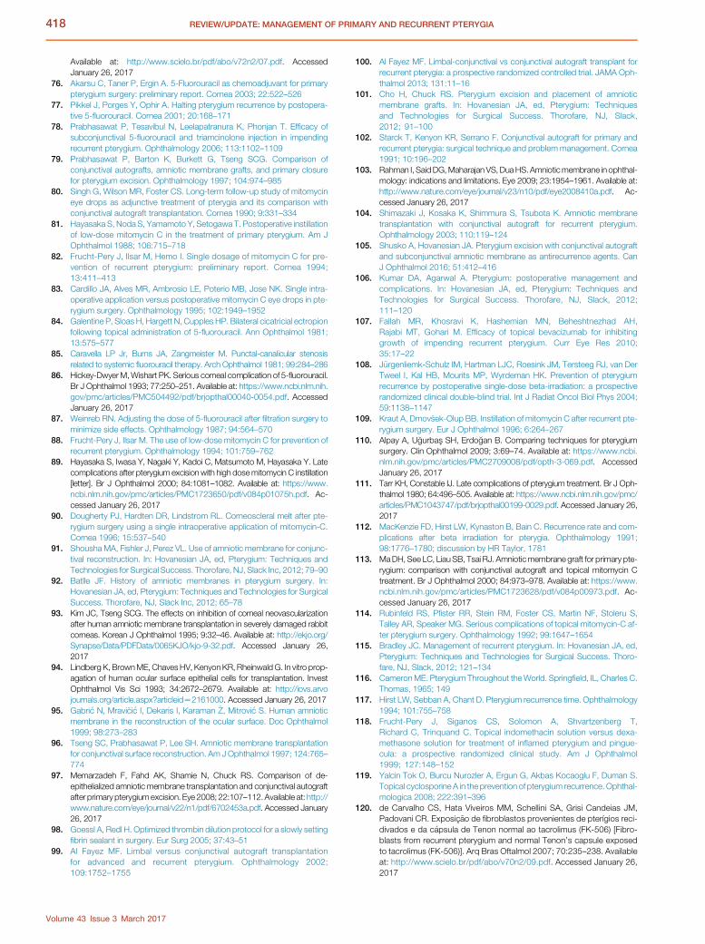
418 REVIEW/UPDATE: MANAGEMENT OF PRIMARY AND RECURRENT PTERYGIA
Available at: http://www.scielo.br/pdf/abo/v72n2/07.pdf. AccessedJanuary 26, 2017
76. Akarsu C, Taner P, Ergin A. 5-Fluorouracil as chemoadjuvant for primarypterygium surgery: preliminary report. Cornea 2003; 22:522–526
77. Pikkel J, Porges Y, Ophir A. Halting pterygium recurrence by postopera-tive 5-fluorouracil. Cornea 2001; 20:168–171
78. Prabhasawat P, Tesavibul N, Leelapatranura K, Phonjan T. Efficacy ofsubconjunctival 5-fluorouracil and triamcinolone injection in impendingrecurrent pterygium. Ophthalmology 2006; 113:1102–1109
79. Prabhasawat P, Barton K, Burkett G, Tseng SCG. Comparison ofconjunctival autografts, amniotic membrane grafts, and primary closurefor pterygium excision. Ophthalmology 1997; 104:974–985
80. Singh G, Wilson MR, Foster CS. Long-term follow-up study of mitomycineye drops as adjunctive treatment of pterygia and its comparison withconjunctival autograft transplantation. Cornea 1990; 9:331–334
81. Hayasaka S, Noda S, Yamamoto Y, Setogawa T. Postoperative instillationof low-dose mitomycin C in the treatment of primary pterygium. Am JOphthalmol 1988; 106:715–718
82. Frucht-Pery J, Ilsar M, Hemo I. Single dosage of mitomycin C for pre-vention of recurrent pterygium: preliminary report. Cornea 1994;13:411–413
83. Cardillo JA, Alves MR, Ambrosio LE, Poterio MB, Jose NK. Single intra-operative application versus postoperative mitomycin C eye drops in pte-rygium surgery. Ophthalmology 1995; 102:1949–1952
84. Galentine P, Sloas H, Hargett N, Cupples HP. Bilateral cicatricial ectropionfollowing topical administration of 5-fluorouracil. Ann Ophthalmol 1981;13:575–577
85. Caravella LP Jr, Burns JA, Zangmeister M. Punctal-canalicular stenosisrelated to systemic fluorouracil therapy. Arch Ophthalmol 1981; 99:284–286
86. Hickey-DwyerM,Wishart PK. Serious corneal complication of 5-fluorouracil.Br J Ophthalmol 1993; 77:250–251. Available at: https://www.ncbi.nlm.nih.gov/pmc/articles/PMC504492/pdf/brjopthal00040-0054.pdf. AccessedJanuary 26, 2017
87. Weinreb RN. Adjusting the dose of 5-fluorouracil after filtration surgery tominimize side effects. Ophthalmology 1987; 94:564–570
88. Frucht-Pery J, Ilsar M. The use of low-dose mitomycin C for prevention ofrecurrent pterygium. Ophthalmology 1994; 101:759–762
89. Hayasaka S, Iwasa Y, Nagaki Y, Kadoi C, Matsumoto M, Hayasaka Y. Latecomplications after pterygium excision with high dosemitomycin C instillation[letter]. Br J Ophthalmol 2000; 84:1081–1082. Available at: https://www.ncbi.nlm.nih.gov/pmc/articles/PMC1723650/pdf/v084p01075h.pdf. Ac-cessed January 26, 2017
90. Dougherty PJ, Hardten DR, Lindstrom RL. Corneoscleral melt after pte-rygium surgery using a single intraoperative application of mitomycin-C.Cornea 1996; 15:537–540
91. Shousha MA, Fishler J, Perez VL. Use of amniotic membrane for conjunc-tival reconstruction. In: Hovanesian JA, ed, Pterygium: Techniques andTechnologies for Surgical Success. Thorofare, NJ, Slack Inc, 2012; 79–90
92. Batlle JF. History of amniotic membranes in pterygium surgery. In:Hovanesian JA, ed, Pterygium: Techniques and Technologies for SurgicalSuccess. Thorofare, NJ, Slack Inc, 2012; 65–78
93. Kim JC, Tseng SCG. The effects on inhibition of corneal neovascularizationafter human amniotic membrane transplantation in severely damaged rabbitcorneas. Korean J Ophthalmol 1995; 9:32–46. Available at: http://ekjo.org/Synapse/Data/PDFData/0065KJO/kjo-9-32.pdf. Accessed January 26,2017
94. LindbergK, BrownME, ChavesHV, KenyonKR, RheinwaldG. In vitro prop-agation of human ocular surface epithelial cells for transplantation. InvestOphthalmol Vis Sci 1993; 34:2672–2679. Available at: http://iovs.arvojournals.org/article.aspx?articleidZ2161000. Accessed January 26, 2017
95. Gabri�c N, Mravi�ci�c I, Dekaris I, Karaman �Z, Mitrovi�c S. Human amnioticmembrane in the reconstruction of the ocular surface. Doc Ophthalmol1999; 98:273–283
96. Tseng SC, Prabhasawat P, Lee SH. Amniotic membrane transplantationfor conjunctival surface reconstruction. Am J Ophthalmol 1997; 124:765–774
97. Memarzadeh F, Fahd AK, Shamie N, Chuck RS. Comparison of de-epithelialized amniotic membrane transplantation and conjunctival autograftafter primary pterygiumexcision. Eye 2008; 22:107–112.Available at: http://www.nature.com/eye/journal/v22/n1/pdf/6702453a.pdf. Accessed January26, 2017
98. Goessl A, Redl H. Optimized thrombin dilution protocol for a slowly settingfibrin sealant in surgery. Eur Surg 2005; 37:43–51
99. Al Fayez MF. Limbal versus conjunctival autograft transplantationfor advanced and recurrent pterygium. Ophthalmology 2002;109:1752–1755
Volume 43 Issue 3 March 2017
100. Al Fayez MF. Limbal-conjunctival vs conjunctival autograft transplant forrecurrent pterygia: a prospective randomized controlled trial. JAMA Oph-thalmol 2013; 131:11–16
101. Cho H, Chuck RS. Pterygium excision and placement of amnioticmembrane grafts. In: Hovanesian JA, ed, Pterygium: Techniquesand Technologies for Surgical Success. Thorofare, NJ, Slack,2012; 91–100
102. Starck T, Kenyon KR, Serrano F. Conjunctival autograft for primary andrecurrent pterygia: surgical technique and problem management. Cornea1991; 10:196–202
103. Rahman I, SaidDG,Maharajan VS,DuaHS.Amnioticmembrane in ophthal-mology: indications and limitations. Eye 2009; 23:1954–1961. Available at:http://www.nature.com/eye/journal/v23/n10/pdf/eye2008410a.pdf. Ac-cessed January 26, 2017
104. Shimazaki J, Kosaka K, Shimmura S, Tsubota K. Amniotic membranetransplantation with conjunctival autograft for recurrent pterygium.Ophthalmology 2003; 110:119–124
105. Shusko A, Hovanesian JA. Pterygium excision with conjunctival autograftand subconjunctival amniotic membrane as antirecurrence agents. CanJ Ophthalmol 2016; 51:412–416
106. Kumar DA, Agarwal A. Pterygium: postoperative management andcomplications. In: Hovanesian JA, ed, Pterygium: Techniques andTechnologies for Surgical Success. Thorofare, NJ, Slack, 2012;111–120
107. Fallah MR, Khosravi K, Hashemian MN, Beheshtnezhad AH,Rajabi MT, Gohari M. Efficacy of topical bevacizumab for inhibitinggrowth of impending recurrent pterygium. Curr Eye Res 2010;35:17–22
108. J€urgenliemk-Schulz IM, Hartman LJC, Roesink JM, Tersteeg RJ, van DerTweel I, Kal HB, Mourits MP, Wyrdeman HK. Prevention of pterygiumrecurrence by postoperative single-dose beta-irradiation: a prospectiverandomized clinical double-blind trial. Int J Radiat Oncol Biol Phys 2004;59:1138–1147
109. Kraut A, Drnov�sek-Olup BB. Instillation of mitomycin C after recurrent pte-rygium surgery. Eur J Ophthalmol 1996; 6:264–267
110. Alpay A, U�gurbas SH, Erdo�gan B. Comparing techniques for pterygiumsurgery. Clin Ophthalmol 2009; 3:69–74. Available at: https://www.ncbi.nlm.nih.gov/pmc/articles/PMC2709008/pdf/opth-3-069.pdf. AccessedJanuary 26, 2017
111. Tarr KH, Constable IJ. Late complications of pterygium treatment. Br J Oph-thalmol 1980; 64:496–505. Available at: https://www.ncbi.nlm.nih.gov/pmc/articles/PMC1043747/pdf/brjopthal00199-0029.pdf. Accessed January 26,2017
112. MacKenzie FD, Hirst LW, Kynaston B, Bain C. Recurrence rate and com-plications after beta irradiation for pterygia. Ophthalmology 1991;98:1776–1780; discussion by HR Taylor, 1781
113. MaDH, See LC, LiauSB, Tsai RJ. Amnioticmembrane graft for primary pte-rygium: comparison with conjunctival autograft and topical mitomycin Ctreatment. Br J Ophthalmol 2000; 84:973–978. Available at: https://www.ncbi.nlm.nih.gov/pmc/articles/PMC1723628/pdf/v084p00973.pdf. Ac-cessed January 26, 2017
114. Rubinfeld RS, Pfister RR, Stein RM, Foster CS, Martin NF, Stoleru S,Talley AR, Speaker MG. Serious complications of topical mitomycin-C af-ter pterygium surgery. Ophthalmology 1992; 99:1647–1654
115. Bradley JC. Management of recurrent pterygium. In: Hovanesian JA, ed,Pterygium: Techniques and Technologies for Surgical Success. Thoro-fare, NJ, Slack, 2012; 121–134
116. CameronME. Pterygium Throughout theWorld. Springfield, IL, Charles C.Thomas, 1965; 149
117. Hirst LW, Sebban A, Chant D. Pterygium recurrence time. Ophthalmology1994; 101:755–758
118. Frucht-Pery J, Siganos CS, Solomon A, Shvartzenberg T,Richard C, Trinquand C. Topical indomethacin solution versus dexa-methasone solution for treatment of inflamed pterygium and pingue-cula: a prospective randomized clinical study. Am J Ophthalmol1999; 127:148–152
119. Yalcin Tok O, Burcu Nurozler A, Ergun G, Akbas Kocaoglu F, Duman S.Topical cyclosporine A in the prevention of pterygium recurrence. Ophthal-mologica 2008; 222:391–396
120. de Carvalho CS, Hata Viveiros MM, Schellini SA, Grisi Candeias JM,Padovani CR. Exposic~ao de fibroblastos provenientes de pterígios reci-divados e da c�apsula de Tenon normal ao tacrolimus (FK-506) [Fibro-blasts from recurrent pterygium and normal Tenon’s capsule exposedto tacrolimus (FK-506)]. Arq Bras Oftalmol 2007; 70:235–238. Availableat: http://www.scielo.br/pdf/abo/v70n2/09.pdf. Accessed January 26,2017

419REVIEW/UPDATE: MANAGEMENT OF PRIMARY AND RECURRENT PTERYGIA
121. Esquenazi S. Treatment of early pterygium recurrence with topicaladministration of interferon alpha-2b. Can J Ophthalmol 2005;40:185–187
122. Shimazaki J, Shinozaki N, Tsubota K. Transplantation of amniotic membraneand limbal autograft for patientswith recurrent pterygiumassociatedwith sym-blepharon. Br J Ophthalmol 1998; 82:235–240. Available at: https://www.ncbi.nlm.nih.gov/pmc/articles/PMC1722509/pdf/v082p00235.pdf. Ac-cessed January 26, 2017
OTHER CITED MATERIALA. Hovanesian JA. Pterygium surgery pearls. Available on YouTube at:
https://youtu.be/lakjdMvqBVo
B. Hovanesian JA. Three newer methods for treating pterygium: A look at auto-grafting, fibrin adhesives and amnion, a combined approach to improveoutcome. Rev Ophthalmol 2009; 16(12)46–52. Available at: https://www.reviewofophthalmology.com/article/three-newer-methods-for-treating-pterygium. Accessed January 26, 2017
C. Hovanesian JA. Pterygium: surgical strategies for success. Available onYouTube at: https://youtu.be/XGOot-us5MI
Disclosure: No author has a financial or proprietary interest in anymaterial or method mentioned.
Volume 43 Issue 3 March 2017