SURGICAL TECHNIQUE GUIDE - OsteoMed
16
NEUROSURGICAL Rethinking Possibilities, Reshaping Lives SURGICAL TECHNIQUE GUIDE
Transcript of SURGICAL TECHNIQUE GUIDE - OsteoMed
NEUROSURGICAL
Rethinking Possibilities, Reshaping Lives
SURGICAL TECHNIQUE GUIDE

TABLE OF CONTENTS
Introduction 4
Preoperative Planning 5
Manual Spring Implantation 6
Spring Removal Guide 10

www.osteomed.com4
SPRING ASSISTED SURGERY FOR THE TREATMENT OF CRANIOSYNOSTOSIS
IntroductionSmartFlex offers early minimally invasive surgical intervention to decrease the morbidity associated with an extensive decompression operation.
DO NOT USE IF PACKAGE IS DAMAGED
ATTENTION: SEE INSTRUCTIONS FOR USE
CAUTION:CONSULT ACCOMPANYING
DOCUMENTS
SINGLE USE ONLY CONSULT INSTRUCTIONSFOR USE
USED BY DATE
TEMPERATURE MANUFACTURER DATE OF MANUFACTURERFEDERAL LAW (U.S.A.)
RESTRICTS THE DEVICE TOSALE BY OR ON THE ORDER OF
A PHYSICIAN
STERILE, METHOD OF STERILIZATION USING
IRRADIATION
ONLY
STERILE R
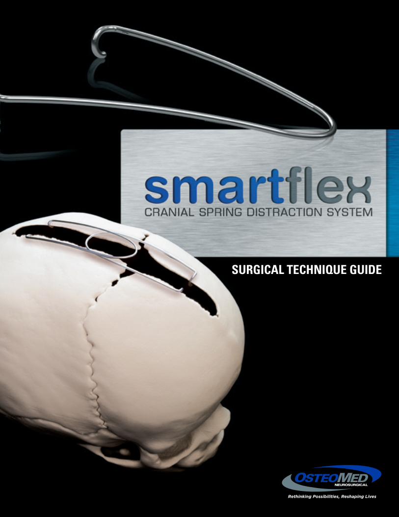
SMARTFLEX CRANIAL SPRING
Foot Plate• Beveled to reduce the possibility of dural tear• Designed to fit securely under the skull bone
Cranial Spring• Pre-formed based on an extensive analysis of infant skulls• Medical grade stainless steel• Adjustable to accommodate various skull anatomies
SPRING CUTTERRONGEUR
220-0772
220-0773
5www.osteomed.com
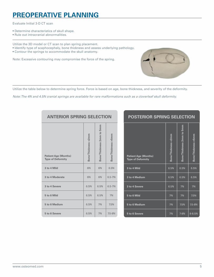
PREOPERATIVE PLANNINGEvaluate Initial 3-D CT scan
• Determine characteristics of skull shape.• Rule out intracranial abnormalities.
Utilize the 3D model or CT scan to plan spring placement.• Identify type of scaphocephaly, bone thickness and assess underlying pathology.• Contour the springs to accommodate the skull anatomy.
Note: Excessive contouring may compromise the force of the spring.
Utilize the table below to determine spring force. Force is based on age, bone thickness, and severity of the deformity.
Note: The 4N and 4.5N cranial springs are available for rare malformations such as a cloverleaf skull deformity.
ANTERIOR SPRING SELECTION
Patient Age (Months)Type of Deformity
3 to 4 Mild 6N 6N 6.5N
6N 6N 6.5-7N
6.5N 6.5N 6.5-7N
6.5N 6.5N 7N
6.5N 7N 7.5N
6.5N 7N 7.5-8N
3 to 4 Moderate
3 to 4 Severe
5 to 6 Mild
5 to 6 Medium
5 to 6 Severe
Bo
ne
Th
ickn
ess
<2m
m
Bo
ne
Th
ickn
ess
2mm
to
5m
m
Bo
ne
Th
ickn
ess
>5m
m
POSTERIOR SPRING SELECTION
Patient Age (Months)Type of Deformity
3 to 4 Mild 6.5N 6.5N 6.5N
6.5N 6.5N 6.5N
6.5N 7N 7N
7N 7N 7.5N
7N 7.5N 7.5-8N
7N 7-8N 8-8.5N
3 to 4 Medium
3 to 4 Severe
5 to 6 Mild
5 to 6 Medium
5 to 6 Severe
Bo
ne
Th
ickn
ess
<2m
m
Bo
ne
Th
ickn
ess
2mm
to
5m
m
Bo
ne
Th
ickn
ess
>5m
m
www.osteomed.com6
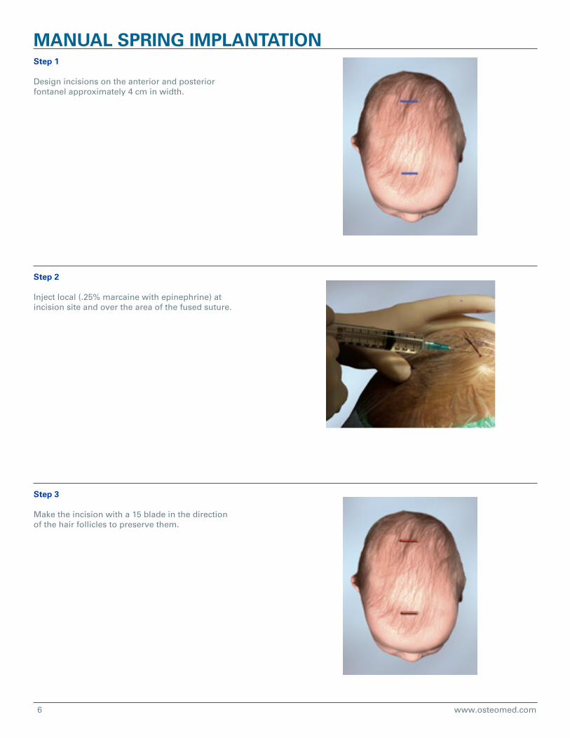
MANUAL SPRING IMPLANTATIONStep 1
Design incisions on the anterior and posterior fontanel approximately 4 cm in width.
Step 2
Inject local (.25% marcaine with epinephrine) at incision site and over the area of the fused suture.
Step 3
Make the incision with a 15 blade in the direction of the hair follicles to preserve them.
7www.osteomed.com
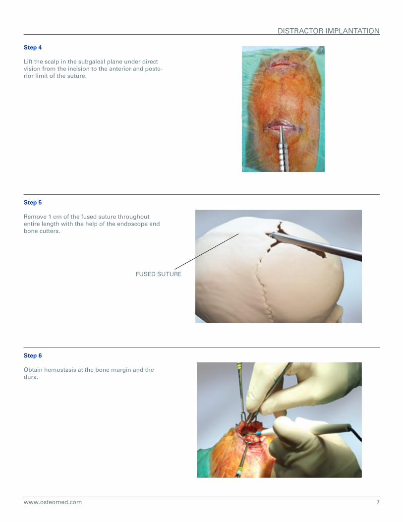
DISTRACTOR IMPLANTATION
Step 4
Lift the scalp in the subgaleal plane under direct vision from the incision to the anterior and poste-rior limit of the suture.
Step 5
Remove 1 cm of the fused suture throughout entire length with the help of the endoscope and bone cutters.
FUSED SUTURE
Step 6
Obtain hemostasis at the bone margin and the dura.

www.osteomed.com8
DISTRACTOR IMPLANTATION
Step 7
Select the spring force based on guide that considers age, bone thickness, and severity of the deformity.
If necessary, bend the spring using the supplied bending instrument to accomodate patient’s anatomy.
Note: Off plane bending and excessive bending may compromise the spring force.
Step 8
Place the springs and confirm positioning.
Note: Ensure foot plate hooks are positioned firmly on the cranial bone.
Note: To prevent spring migration, the spring shall be placed parallel to the suture line.
Step 9
Secure the springs to the bone where they overlap with a 4-0 vicryl suture by drilling a hole in the bone lateral to where the springs overlap. (Should be done on both sides)
ANTERIOR SPRING SELECTION
Patient Age (Months)Type of Deformity
3 to 4 Mild 6N 6N 6.5N
6N 6N 6.5-7N
6.5N 6.5N 6.5-7N
6.5N 6.5N 7N
6.5N 7N 7.5N
6.5N 7N 7.5-8N
3 to 4 Moderate
3 to 4 Severe
5 to 6 Mild
5 to 6 Medium
5 to 6 Severe
Bo
ne
Th
ickn
ess
<2m
m
Bo
ne
Th
ickn
ess
2mm
to
5m
m
Bo
ne
Th
ickn
ess
>5m
m
POSTERIOR SPRING SELECTION
Patient Age (Months)Type of Deformity
3 to 4 Mild 6.5N 6.5N 6.5N
6.5N 6.5N 6.5N
6.5N 7N 7N
7N 7N 7.5N
7N 7.5N 7.5-8N
7N 7-8N 8-8.5N
3 to 4 Medium
3 to 4 Severe
5 to 6 Mild
5 to 6 Medium
5 to 6 Severe
Bo
ne
Th
ickn
ess
<2m
m
Bo
ne
Th
ickn
ess
2mm
to
5m
m
Bo
ne
Th
ickn
ess
>5m
m
9www.osteomed.com
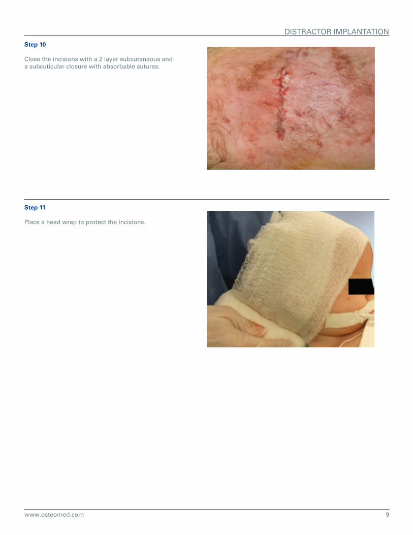
Step 10
Close the incisions with a 2 layer subcutaneous and a subcuticular closure with absorbable sutures.
Step 11
Place a head wrap to protect the incisions.
DISTRACTOR IMPLANTATION

www.osteomed.com10
SPRING REMOVAL GUIDEStep 1
Palpate the springs at their overlap and bony insertion points.
Step 2
Design a small incision over each of the 4 foot-plates and mark the portion of the previous inci-sion that will be utilized.
Step 3
Inject local into the 5 incisions.
11www.osteomed.com
SPRING REMOVAL GUIDE
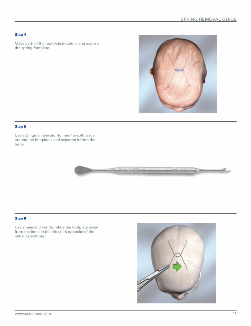
Step 4
Make each of the footplate incisions and expose the spring footplate.
Step 5
Use a Dingman elevator to free the soft tissue around the footplates and separate it from the bone.
Step 6
Use a needle driver to rotate the footplate away from the bone in the direction opposite of the initial osteotomy.
www.osteomed.com12
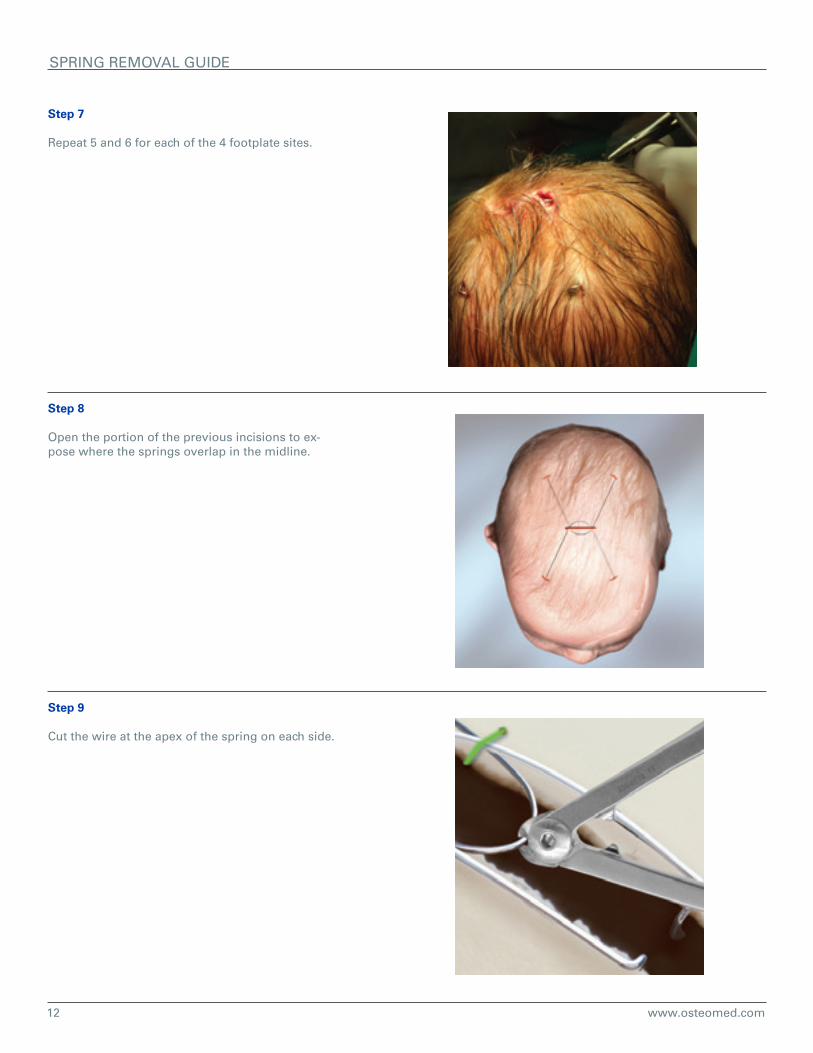
Step 7
Repeat 5 and 6 for each of the 4 footplate sites.
Step 8
Open the portion of the previous incisions to ex-pose where the springs overlap in the midline.
Step 9
Cut the wire at the apex of the spring on each side.
SPRING REMOVAL GUIDE
13www.osteomed.com
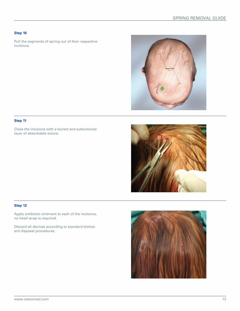
Step 10
Pull the segments of spring out of their respective incisions.
Step 11
Close the incisions with a buried and subcuticular layer of absorbable suture.
Step 12
Apply antibiotic ointment to each of the incisions, no head wrap is required.
Discard all devices according to standard biohaz-ard disposal procedures.
SPRING REMOVAL GUIDE
www.osteomed.com14
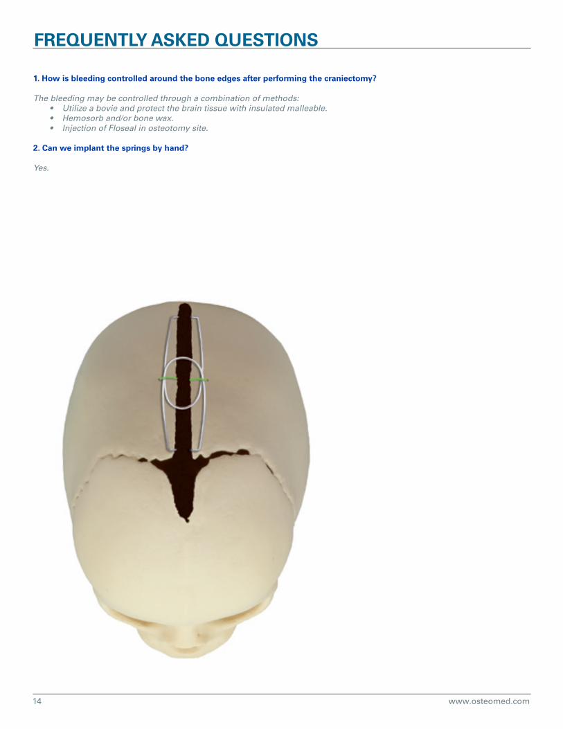
1. How is bleeding controlled around the bone edges after performing the craniectomy?
The bleeding may be controlled through a combination of methods:• Utilize a bovie and protect the brain tissue with insulated malleable.• Hemosorb and/or bone wax.• Injection of Floseal in osteotomy site.
2. Can we implant the springs by hand?
Yes.
FREQUENTLY ASKED QUESTIONS


OSTEOMED3885 Arapaho Rd.
Addison, TX 75001Customer Service: 800.456.7779
Customer Service Fax: 800.390.2620
www.osteomed.com
P/N 030-1781 Rev.B
NEUROSURGICAL
Rethinking Possibilities, Reshaping Lives