SURGICAL SITE INFECTION - gneaupp.b-cdn.net
72
PREVENTION AND MANAGEMENT ACROSS HEALTH- CARE SECTORS SURGICAL SITE INFECTION S C A L S N F E C S A JOINT DOCUMENT
Transcript of SURGICAL SITE INFECTION - gneaupp.b-cdn.net

PREVENTION AND MANAGEMENT ACROSS HEALTH-CARE SECTORS
SURGICAL SITE INFECTION
SU
RG
ICAL S I T E I NFEC
TIO
NS
A JOINT DOCUMENT
268x200_EWMA_Surgical Site Inf_200120.indd 1 20/01/2020 15.58

S 2 J O U R N A L O F WO U N D C A R E VO L 2 9 N O 2 E W M A D O C U M E N T 2 0 2 0
© EWMA 2020
All rights reserved. No reproduction, transmission or copying of this publication is allowed without written permission. No part of this publication may be reproduced, stored in a retrieval system, or transmitted in any form or by any means, mechanical, electronic, photocopying, recording, or otherwise, without the prior written permission of the European Wound Management Association (EWMA) or in accordance with the relevant copyright legislation.
Although the editor, MA Healthcare Ltd. and EWMA have taken great care to ensure accuracy, neither MA Healthcare Ltd. nor EWMA will be liable for any errors of omission or inaccuracies in this publication.
Published on behalf of EWMA by MA Healthcare Ltd.Editor: Rachel Webb Managing Director: Anthony Kerr Published by: MA Healthcare Ltd, St Jude’s Church, Dulwich Road, London, SE24 0PB, UKTel: +44 (0)20 7738 5454 Email: [email protected] Web: www.markallengroup.com
Jan Stryja (Editor) MD, Ph.D, Vascular Surgeon, Centre of vascular and miniinvasive surgery, Hospital Podlesi, Trinec, The Czech Republic. Salvatella Ltd., Centre of non-healing wounds treatment, Podiatric outpatients’ department, Trinec, The Czech Republic.
Kylie Sandy-Hodgetts (Co-editor), BSc MBA Ph.D, Senior Research Fellow – Senior Lecturer, Faculty of Medicine, School of Biomedical Sciences, University of Western Australia, Director, Skin Integrity Clinical Trials Unit, University of Western Australia.
Mark Collier, RGN, ONC, RCNT, RNT, BA (Hon’s), Nurse Consultant and Associate Lecturer – Tissue Viability, Independent – formerly at the United Lincolnshire Hospitals NHS Trust, c/o Pilgrim Hospital, Sibsey Road, Boston, Lincolnshire, PE21 9Q
Claus Moser, MD, Clinical microbiologist, Rigshospitalet, Department of Clinical Microbiology, Copenhagen, Denmark
Karen Ousey, Ph.D, MA, PGDE, BA, RN, ONC, Professor of Skin Integrity, University of Huddersfield. Institute of Skin Integrity and Infection Prevention, Huddersfield, UK
Sebastian Probst, DClinPrac, RN, professor of wound care, HES-SO University of Applied Sciences and Arts Western Switzerland, Geneva, Switzerland
Jennie Wilson, Professor of Healthcare Epidemiology, University of West London, College of Nursing, Midwifery and Healthcare, London, UK
Deborah Xuereb, RN, Senior Infection Prevention & infection Control Nurse, Mater Dei Hospital, Msida, Malta
Figs 1, 3, 4, 5, 6, 9, 10, 11, 12, 16, 17, 18, 19, 20 are published with kind permission of the Head of Hospital Podlesi, Trinec, The Czech Republic.
Fig 8 is published with the kind permission from the author.
Editorial support and coordination: Niels Fibæk Bertel, EWMA Secretariat
Corresponding author: Jan Stryja, [email protected]
The document is supported by unrestricted educational grants from: Abigo, BBraun, Essity, Ferris (Polymen), Mölnlycke Health Care, Argentum Medical and Vancive Medical Technologies.
This article should be referenced as: Stryja J, Sandy-Hodgetts K, Collier M et al. Surgical site infection: preventing and managing surgical site infection across health care sectors. J Wound Care 2020; 29: 2, Suppl 2b, S1–S69

J O U R N A L O F WO U N D C A R E VO L 2 9 N O 2 E W M A D O C U M E N T 2 0 2 0 S 3
Contents
1. Introduction 4Aims, objectives and scope of document 4Structure and content 4
2. Methodology and terminology 5
3. Epidemiology of surgical site infection 6
Surgical site infection: background 6Signs and symptoms of SSI 7Source of microorganisms related to SSI 10Type of surgery and risk of SSI 10Adjusting rates of SSI for variation in case-mix 12Patient-related factors and the risk of SSI 12Involving patients with diagnostics 13Conclusion 13
4. Principles of surgical wound management 14
Wound healing process 15Primary and secondary healing 15Principles of surgical wound closure 16Multidisciplinary team approach to surgical wound management 22Conclusion 23
5. Perioperative practice to prevent surgical site infection 25
Preoperative phase 25Intraoperative phase 27Postoperative phase 31Conclusion 37
6. Principles of postoperative care 38How to dress the wound in the outpatient setting 38How to dress the wound in the home care setting 40
7. Wound assessment and diagnostics 43
Early signs of infection 43General assessment of a patient 43Vascular assessment 43Sampling techniques 44Lab procedures 44Conclusion 45
8. Treatment of SSI 46Non-surgical treatment 46Antibiotic coverage 46Systemic antibiotic therapy 47Topical antibiotic therapy 48Impact of oxygen 49
Conclusion 50
9. Conclusion 51
10. Future perspectives 52Prevention and risk assessment 52Treatment 52
Diagnostics 52
11. References 53
Appendix 60Appendix 1: GRADE recommendation explanation 60Appendix 2: Microbiological assessment for surgical site infection, investigation of the swabs 61Appendix 3: A protocol for the prevention of surgical site infection 64Appendix 4: Patient’s Guide – frequently asked questions about SSI 67
Glossary 68Definitions�� 68Abbreviations 68

S 4 J O U R N A L O F WO U N D C A R E VO L 2 9 N O 2 E W M A D O C U M E N T 2 0 2 0
1. Introduction
Aims, objectives and scope of document
Surgical site infection (SSI) is an unfortunate
postoperative complication that affects many
surgical patients worldwide and treatment of
this type of wound is most likely to occur following
discharge from the acute care setting. While there
are several guidelines for preventing and managing
SSIs in hospitals there is an absence of guidelines
for the optimum postoperative management
in the home care setting. Furthermore, a set of
recommendations on this topic covering primary
and community health professionals remains absent
from clinical resources.
The overall aim of this document is to:
• Highlight present knowledge regarding
prevention and management of SSI in the
primary and community health-care sectors
• Present a set of recommendations to guide
clinical practice in the community setting
for maximum patient healing outcomes
following surgery.
More specifically, the main objectives of the
document are to:
• Describe the incidence and prevalence of SSI,
based on published information and data
available from SSI-registries
• Present the principles of management of
surgical wounds as well as the available modern
techniques for prevention and treatment of SSIs
across sectors
• Provide a summary of evidence-based clinical
perioperative practice recommendations to
prevent surgical site infections.
Structure and contentThis document is presented in ten chapters.
Chapter 1 is the introduction to the document
and describes the aim and objectives of the
work. Chapter 2 presents the methodology and
terminology used in the document. Chapter 3
describes the epidemiology of SSI. Chapter 4
discusses principles of the management of
surgical wounds. Chapter 5 presents a summary
of recommended practice during pre-, intra-
and postoperative phases. Chapter 6 discusses
principles of postoperative care and Chapter
7 presents contemporary methods of wound
assessment and discusses diagnosis of infection.
Chapter 8 reports on current treatments for
clinical management of SSI. Chapter 9 summarises
the main conclusions of the document, with
Chapter 10 providing a brief look at new
developments, highlighting areas that require
further research.

J O U R N A L O F WO U N D C A R E VO L 2 9 N O 2 E W M A D O C U M E N T 2 0 2 0 S 5
2. Methodology and terminology
T his document originates from a growing
interest by many EWMA stakeholders,
in the clinical management and
prevention of SSI in hospital and following
hospital discharge and their management in the
community setting.
On the basis of a literature search search conducted
by the document authors and the EWMA
secretariat, as well as input from key EWMA
stakeholders, a short description of the document
aim, objectives and scope was developed during
the first quarter of 2017. This basic document
outline was then used over the next six months
to identify the specialists, who constitute the
author group.
The expert group was established to produce the
most recent evidence-based consensus document
for health-care workers across all health-care
sectors, hospitals, primary and community
care. The group consists of representatives
from the EWMA council, Wounds Australia
and the Infection Prevention Society (IPS). The
Association for the Advancement of Wound
Care (AAWC) contributed to the development of
the document.
Each author has taken responsibility for the
development of each individual chapter, with each
author conducting relevant searches to investigate
the literature. The opinions expressed in this
document have been reached by consensus of the
author group based upon professional, clinical
and research expertise, as well as the experience
of their peers. The clinical guidance provided in
the document is based on critical analysis and
synthesis of published guidelines, literature reviews
and evidence-based recommendations as well as
consensus driven expert opinion.
Where relevant, throughout the document, the
GRADE classification for levels of evidence was
applied to assess the level of evidence relevant
to literature reviewed. The GRADE classification
system is available in Appendix 1. There is a
plethora of therapies and clinical approaches in
the prevention and management of SSI and it
was essential to grade the levels of evidence for
relevancy in the clinical setting.

S 6 J O U R N A L O F WO U N D C A R E VO L 2 9 N O 2 E W M A D O C U M E N T 2 0 2 0
3. Epidemiology of surgical site infection
Surgical site infection: background
S SIs can be defined as infections occurring up
to 30 days after surgery (or up to one year
after surgery in patients receiving implants)
and affecting either the incision or deep tissue at
the operation site.1,2 This definition is primarily
used for surveillance and reporting purposes. The
clinical definition includes signs and symptoms
of infection confirmed by the presence of typical
clinical signs of infection, such as redness, swelling
and exudate — see Chapter 7. For surveillance
purposes, the definition must be more accurate
and univocal.3 As an example of an algorithm
taking this into account, we refer to the CDC SSI
surveillance protocol.4
SSIs affect up to one-third of patients who have
undergone a surgical procedure.5 The incidence rate
Clinical definition of SSI: Infectious process
present at the site of surgery. Clinical signs and
symptoms of infection include heat, redness,
swelling, elevated body temperature and purulent
exudate from the wound or the drain.
CDC reporting definition for surgical site
infection surveillance: infections occurring
up to 30 days after surgery (or up to one year
after surgery in patients receiving implants) and
affecting either the incision or deep tissue at the
operation site. According to the range of affected
tissues SSI can be superficial, deep or organ/space.3
of SSIs vary from 2–15% depending on multiple
factors, including the type of operation.6 In the
most recent prevalence survey conducted by the
European Centre for Disease Surveillance and
Control, SSIs accounted for 18% of healthcare-
associated infections (HCAI).7,8 The incidence of SSIs
varies depending on multiple factors, including the
type of operation6 and is likely to be underestimated
given that approximately 50% of SSIs become
evident after discharge.3 Surveillance following
hospital discharge is important to accurately
determine the prevalence and incidence of this
postoperative complication. SSIs impair not only
the patient’s quality of life (QoL) but also have a
negative economic impact. They increase length
of time a patient stays in the hospital and incur
considerable extra health-care costs.9 According
to Leaper et al. additional costs attributable
to SSI of between £814 and £6626 have been
reported, depending on the type of surgery and
the severity of the infection.10 Other studies have
reported subsequent costs associated with clinical
KEY POINTS • Surgical site infection (SSI) is the third most commonly
reported healthcare-associated infection (HCAI) and results in significant patient morbidity and mortality
• The risk of SSI is influenced by a number of intrinsic and extrinsic factors, particularly the number of microorganisms present at or introduced into the incision
• The risk of SSI increases with the age of the patient; other important risk factors are severe underlying illness and obesity
• Measuring and reporting rates of SSI in surveillance systems is an important strategy for determining prevalence of SSI and measuring the impact of prevention measures

J O U R N A L O F WO U N D C A R E VO L 2 9 N O 2 E W M A D O C U M E N T 2 0 2 0 S 7
management in the acute care setting, in the US
costs exceed US$1.6 billion annually,11 Australia
AU$268 million annually12 and UK £930 million
annually.13 In addition, up to 60% of SSIs have
been estimated to be preventable and their risk
can be minimised by applying best practice in the
perioperative period.14,15
The main factors affecting the occurrence of SSI
are pre-existing health status of the patient, type
of surgery and postoperative management in the
acute care setting and following discharge.
Pathogenesis of surgical site infectionA surgical procedure exposes sterile body tissues.
Microorganisms introduced during the procedure
can multiply in the wound after it has been closed
and subsequently cause an SSI. However, the
accurate diagnosis of SSI is difficult to detect as it
may take several weeks to develop and therefore
many infections may not become apparent
until after the patient has been discharged from
hospital.16,17 Only those patients with more
severe SSI are likely to be readmitted to hospital,
accounting for one-third of patients who present
with an HCAI on admission.18 SSI is the most
common HCAI among patients who are admitted
to hospital for a surgical procedure and in this
group the prevalence of SSI is at least 5%.19
The morbidity and mortality associated with SSI is
considerable. A key indicator of the adverse effect
is the impact on length of hospital stay. Studies
suggest that an SSI, regardless of whether it was
superficial or more severe, doubles the length of
postoperative hospital stay.20–23 Other studies have
confirmed the effect of SSI on length of hospital
stay and also demonstrated the significant
increase in costs associated with additional
hospital stay and treatment.9 In economic
terms, the additional bed-days associated
with the treatment of patients with SSI reflect
‘opportunity costs’ as they could be used to treat
more patients.24 A case control study of patients
undergoing proximal femoral fracture repair
found, that when repeat admissions to hospital,
re-operations and other treatments are taken into
account, severe SSI can quadruple the costs of care
and decrease the QoL of affected patients.25
SSI also has an important impact on mortality.
Coello et al.20 found a significant increase in in-
hospital mortality associated with deep or organ-
space SSI for three major categories of surgery: hip
prosthesis, odds ratio (OR) 2.5; 95% confidence
interval (CI) 1.3 to 4.5, large bowel surgery OR
1.8; 95%CL 1.1 to 3.2 and vascular surgery OR 6.8;
95%CL 3.0 to 15.4. Astagneau et al. found a case
fatality rate of 4.5% in patients who developed
an SSI and 38% of these deaths being directly
attributable to the infection.26
Signs and symptoms of SSISSI can affect the superficial cutaneous layer
(superficial SSI), the fascial layers (deep SSI) or
nearby organs and other sites such as joints or
abdomen manipulated during the procedure
(organ/space SSI) (Fig 2).
Clinical signs and symptoms of SSI at the site
of an incision include purulent drainage, pain
or tenderness, localised swelling, redness or
heat (Table 1 and Fig 3–5). They usually become
apparent by the end of the first week after surgery.
Superficial infection come through local infection
signs given below. SSI can also manifest as a
cellulitis of soft tissue at the place of surgery or
wound abscess. In the case of deep and organ/
space SSIs the local signs of infection can be
less expressed and the first clinical signs can be
purulent drainage from the wound, unexplainable
fewer, pathological patient’s blood test results—
high C-reactive protein (CRP), BSR: blood
sedimentation rate (BSR) and pro-calcitonin level,
high leukocyte accounts—or uncommon systemic

S 8 J O U R N A L O F WO U N D C A R E VO L 2 9 N O 2 E W M A D O C U M E N T 2 0 2 0
inflammatory response of the body. Dehiscence
of the incision, if site is tender or if the patient
has a fever, may also indicate the presence of
infection if microorganisms are also isolated.27
According to Sandy-Hodgetts et al. dehiscence
may be attributable to non-microbial causes such
as obesity or pre-existing chronic disease states
in some cases.28 In diagnostics of deep-seated and
organ-affecting SSIs it is necessary to carry out full
clinical assessment of the patient and the place
of surgery, plain X-rays and further imaging (e.g.
MRI scan, CT scan, ultra-sound), blood cultures
(particularly in acute cases), organ, bone and/or
soft tissue biopsies and/or surgical sampling.
Almost half of SSIs reported in the European
Centres for Disease Prevention and
Table 1. Clinical signs and symptoms of surgical site infection (SSI)29–31
Superficial SSI symptoms Deep SSI symptoms Organ/space SSI symptoms*Increased pain and tenderness at the site of surgery
Increased pain at the site of surgery
Localised swelling and induration Spreading induration and swelling of the place of incision
Localised heat and redness of the wound
Erythema and heat of the surgical site
Purulent drainage Purulent drainage from the incision Purulent drainage from a drain placed through the skin into the organ or body space
Cellulitis limited to the wound and adjacent soft tissues
Spreading cellulitis at the site of surgery
Evident superficial wound abscess Evident deep wound abscess or fasciitis Organ or body space abscess diagnosed by radiological or histopathological examination
Separation of the edges of incision exposing the deeper tissues
Evidence of infection involving the organ or body space seen on direct examination during surgery
Unexpected postoperative fever accompanied by increasing wound pain and/or wound dehiscence
Postoperative fever
Pathological blood test findings (elevated CRP, WBC counts, erythrocyte sedimentation rates, pro-calcitonin)
Positive result of blood cultures, deep tissue biopsies, surgical sampling or pathological blood test findings (see deep SSI column)
*Involve any part of the anatomy other than the incision opened or manipulated during surgery; CRP— C-reactive protein; WBC—white blood count
Control (ECDC) surveillance system are
superficial, 30% deep, and 20% organ/space.17
However, the distribution of infection type
depends on the type of surgery and the nature
of the surveillance system. The proportion of
deep and organ/space SSI is likely to be higher
if the surveillance is primarily focused on the
postoperative hospital stay and/or patients
readmitted with infection. In procedures
associated with a very short postoperative stay
e.g. caesarean section, the majority of SSI are only
detected after the patient has been discharged
from hospital.16
The signs of infection can progress within
the time to become more evident and more
extensive (Fig 1).

J O U R N A L O F WO U N D C A R E VO L 2 9 N O 2 E W M A D O C U M E N T 2 0 2 0 S 9
Since skin is normally colonised by
microorganisms, often microbiological evidence
alone, is not a reliable indicator of SSI. However,
in the presence of clinical signs and symptoms of
infection, the results of wound cultures are helpful
in indicating the likely causative pathogen (see
Appendix 2 for microbiological investigation of
swabs). Microorganisms cultured from aseptically
aspirated fluid or tissue, or an abscess or other
Fig 1. Deep surgical site infection (SSI) following lumbar sympathectomy
Fig 3. Example of superficial surgical site infection, infected incision
Fig 2. Types of surgical site infection, adapted from Horan et al32
Subcutaneous tissue
Skin
Deep soft tissue (fascia and muscle)
Organ or body cavity Organ/space SSI
Deep incisional SSI
Superficial incisional SSI
Fig 4. Example of deep surgical site infection: infected incision after femoral artery endarterectomy
Fig 5. Organ/space surgical site infection in patient after colectomy with stercoraceous and purulent discharge from the wound. The wound was opened by the surgeon to release the pus and evaluate the range of infection

S 1 0 J O U R N A L O F WO U N D C A R E VO L 2 9 N O 2 E W M A D O C U M E N T 2 0 2 0
evidence of infection found by histopathological
or radiological examination provide an accurate
assessment of associated pathogenic activity
indicative of SSI.27 The clinical signs or symptoms
of SSI can take days or weeks to become apparent.
This can be as long as one year when foreign
material such as a prosthetic joint or sternal wire,
are left in the tissues.
Source of microorganisms related to SSIThe microorganisms associated with SSI can be
derived from sites on the patient that are colonised,
such as the skin, mucous membranes or hollow
viscera (endogenous). However, microorganisms
may also originate from an exogenous source
including operating personnel, the operating room
environment and instruments and equipment
used during the procedure (Fig 6). Occasionally, a
distant source of infection can act as a source of
microorganisms at a surgical site by attaching to
prosthesis or other implant in a biofilm formation.33
The risk of SSI may be influenced by an array
of factors that increase the risk of endogenous
contamination, for example procedures that involve
parts of the body with a dense flora such as the
bowel. It is also affected by factors that increase the
risk of exogenous contamination, such as exposure
of tissues during prolonged or major operations and
inadequate aseptic procedures. Intrinsic factors that
affect the efficacy of the general immune response,
such as diabetes, malnutrition, immunosuppressive
therapy or the local immune response, such as
foreign bodies, damaged tissue, haematoma, are also
important in determining risk of SSI occurrences.
Type of surgery and risk of SSIThe number of organisms present in the wound
following a surgical procedure are strongly
influenced by the site of the body involved.34
Procedures on normally sterile tissues, such as
bones or joints, are less likely to be contaminated
by bacteria than those involving the colon where
large numbers of bacteria are normally present.34
The rates of SSI associated with surgery on sterile
sites are therefore very low (>2%), compared
with the rates SSI associated with surgery on
contaminated sites, which may >10%.35 Minimally
invasive procedures, where the operation is
performed via an endoscope, are associated with
Fig 6. Factors that contribute to the risk of SSI
Underlying illnessObesity
Age
Number Virulence
TypeDuration Technique
Surgical procedure Microorganisms
Host patient
Fig 7. Cumulative incidence of SSI by category of procedure from European countries participating in surgical site infection surveillance. Adapted from ECDC. Annual epidemiological report for 2016, Stockholm; 2018 ECDC17
Col
on s
urge
ry
Col
on s
urge
ry
(min
imal
ly in
vasiv
e)
Cho
lecy
stec
tom
y
Cho
lecy
stec
tom
y (m
inim
ally
inva
sive)
Cor
onar
y ar
tery
byp
ass
graf
t
Cae
sare
an s
ectio
n
Hip
rep
lace
men
t
Knee
rep
lace
men
t
Lam
inec
tom
y
% S
SI
12
10
8
6
4
2
0
10.4
6.3
3.4
1.5
2.81.9
1.00.5
0.9
Notes: Open colon and cholecystectomy procedures. Rates of SSI underestimated for procedures with short postoperative hospital stay and limited post-discharge surveillance.

J O U R N A L O F WO U N D C A R E VO L 2 9 N O 2 E W M A D O C U M E N T 2 0 2 0 S 1 1
lower rates of SSI as a smaller amount of tissue is
exposed during the procedure.17 Fig 7 shows the
variation in risk of SSI attributable to different
operative procedures from data captured by the
European Centres for Disease Prevention and
Control surveillance system.17
A standard approach to classifying wounds
according to the degree of microbial
contamination likely to be present in the operative
site has been described and is widely used to both
predict the risk of SSI and enable comparisons in
risk between different types of surgical procedure
(Table 2). The classification takes account of
both the site of the procedure and events that
occur before or during the operation that may
affect the level of contamination.34 Type of
surgical procedure classification (Table 2), is an
important factor to consider when interpreting and
appraising research evidence related to SSI.34
Operations on sites where infection is already
present, referred to as ‘dirty or infected’ wounds,
for example a gangrenous appendix, are at
particular high risk of infection because of the
number of microorganisms present at the site
of incision.34
Table 2. Class of surgery
Class of surgery
Description
Clean wounds Uninfected operative wounds in which inflammation is not encountered, and the respiratory, gastrointestinal, genital, urinary tracts or the oropharynx are not entered, and there is no break in aseptic technique. In addition, clean wounds must be primarily closed and, if there is drainage, this must be closed. Includes operative wounds that follow non-penetrating trauma, e.g. fractured neck of femur, providing they meet these criteria
Clean-contaminated wounds
Operative wounds in which the respiratory, alimentary, genital, or urinary tracts are entered under controlled conditions and without unusual contamination, providing that there is no evidence of infection or a major break in aseptic technique. Note: procedures that do not enter one of these body tracts cannot be clean contaminated e.g. orthopaedic procedures
Contaminated wounds
Operations on fresh, open traumatic wounds; or operations where there is a major break in aseptic technique; or operations in which there is gross spillage from the gastrointestinal tract; or acute inflammation without pus is encountered
Dirty or infected wounds
Operations in which acute inflammation with pus is encountered, or in which perforated viscera are found; operations on traumatic wounds, which have retained devitalised tissue, foreign bodies or faecal contamination, or where the operation on the traumatic wound has been delayed. Operations included in this class are those in which the organisms causing postoperative infection are likely to have been present in the operative field before surgery
CDC/NHSN surveillance definitions for specific types of infections34
Box 1, National Healthcare Safety Network (CDC) Surgical Site Infection Risk IndexEach operation is scored by the presence or absence of three risk factors at the time of surgery:
1. American Society of Anaesthesiologists’ (ASA) preoperative assessment score of 3 or more (this indicates the patient has a severe underlying systematic disease)
2. Operation is classified as contaminated or dirty
3. Operation lasts for more than a specific period of time (‘T hours’), where T is the 75th percentile of the duration of surgery and depends on the surgical procedure being performed (e.g. hip replacement T time=2 hours)
Each of the risk factors described above contributes one point to the risk index score, which ranges from 0 (none of the risk factors present) to 3 (all of the risk factors present).Source: Public Health England (2013) Protocol for the Surveillance of Surgical Site Infection.36

S 1 2 J O U R N A L O F WO U N D C A R E VO L 2 9 N O 2 E W M A D O C U M E N T 2 0 2 0
Adjusting rates of SSI for variation in case-mixThe type of surgery classification system remains
an important, simple guide to the risk of SSI for
different procedures. However, more sophisticated
models that use a combination of intrinsic and
extrinsic factors related to both patient and the
operation are widely used to more reliably compare
the risk of SSI for the same procedures over time
and between institutions. The most widely used of
these risk adjustment frameworks was developed
for the national HCAI surveillance systems in the
US and has been widely adopted by surveillance
systems around the world (Box 1).35
More recently, a logistic regression analysis has been
applied to a large dataset of surgical procedures
captured for the National Healthcare Safety Network
(NHSN) surveillance systems in the US, in order to
develop specific risk models for different categories
of procedure.37 These specific models predict the
risk of SSI more accurately but are still only able
to account for about 60% of the variation in rates
of SSI.37 This suggests that other unknown factors
influence the risk, including the infection control
procedures applied during the procedure.
Patient-related factors and the risk of SSIFactors intrinsic to the patient undergoing surgery
may influence the risk that they subsequently
develop an SSI, either as a result of extending
the length and complexity of the surgery or
by diminishing the efficacy of the immune
response. A prolonged duration of the operation
is recognised as a strong predictor of risk of SSI
for many types of procedure.37,38 The American
Society of Anaesthesiologists’ (ASA) score is a
measure of underlying illness but increasing ASA
score is strongly associated with risk of SSI. Many
factors included in more complex risk adjustment
models are related to underlying illness in the
patients undergoing surgery e.g. diabetes, obesity
etc. Furthermore, the type and size of hospital
and type of anaesthesia play a role.37
Poor vascularisation of adipose tissue combined
with increased complexity of surgery may increase
the risk of SSI in patients with a body mass index
of 35kg/m² or more.36 Studies have identified
obesity as a risk factor for SSI in cardiac, spinal and
obstetric surgery and estimates suggest it increases
the risk by more than three-fold.39,40 In patients
with diabetes, damage to peripheral vasculature
and an impaired immune response associated with
high blood glucose levels, is shown to be associated
with the doubling of the risk of SSI compared with
patients without diabetes.41,42
Age is also an important independent predictor
of SSI risk, with many studies demonstrating
that the risk of SSI steadily increases with age
across different types of surgery.22,36,40 Adjustment
for a range of risk factors for SSI following hip
replacement demonstrated age as an independent
risk factor and found that patients over 75 years
were more than 1.5 times more likely to develop
SSI than those under 65 years.43
The literature demonstrates that there are
differences in characteristics and independent
predictors for patients developing SSI in the
hospital versus after discharge.44–46 Wiseman
et al. shows that SSIs occurring after discharge
are predominantly associated with a patient’s
comorbidity burdens such as diabetes, smoking,
hypertension, coronary artery disease (CAD), and
chronic obstructive pulmonary disease (COPD),
whereas patients who develop a SSI in the hospital
are largely associated with perioperative factors.47
Table 3 summarises the important patient- and

J O U R N A L O F WO U N D C A R E VO L 2 9 N O 2 E W M A D O C U M E N T 2 0 2 0 S 1 3
procedure-related factors that influence the risk
of SSI.
Involving patients with diagnosticsDuring the initial process of consideration/
assessment of any patient’s suitability for surgery, it
is important for all health professionals to be aware
of an appropriate validated risk assessment tool
that has been used that helps identify a patient’s
potential risk of SSI (e.g. the Surgical Site Infection
Risk Score (SSIRS).48 Especially in patient groups
that have been previously reported to be more
prone to SSIs, such as those having orthopaedic,
colorectal or gynaecological surgery; or any
patient having a surgical procedure who also has
a diagnosis of cancer. Furthermore, for all patients
undergoing surgery, it may also be helpful for
health professionals to incorporate an appropriate
validated Health Related Quality of Life (HRQoL)
assessment score/tool as part of the patients initial
assessment process, as this can help the clinician
identify in particular social factors that may
increase a patient risk of SSI.
Before surgery; health professionals and patients
should be encouraged to discuss issues such as
the patients social habits (such as smoking); their
recent travel history; their current methicillin-
resistant Staphylococcus aureus (MRSA) and
vancomycin-resistant Enterococcus (VRE) status (if
known) or the need for screening if not and their
current medical conditions (especially in relation
to diabetes and cardiopulmonary conditions);
as part of a holistic assessment process, so that
modifiable issues such as a patient's diets, smoking
and exercise habits may be addressed in advance of
planned surgery.
Why? There is a growing body of evidence that
indicates that if these issues are addressed in
a timely manner, the outcomes of surgery are
improved and the risk of the patient suffering from
an SSI are minimised.20,34,42,87,88
ConclusionSSIs are a common HCAI associated with
considerable morbidity and mortality and
additional health-care costs. The risk of SSI depends
on a combination of intrinsic and extrinsic factors,
related to the type of operation and patient.
While it may be difficult to change the intrinsic
and extrinsic risk of SSI in patients, practices and
processes aimed at reducing overall risk are essential
to ensure the lowest possible rates of SSI.49 Early and
fast diagnosing of SSI is essential in both inpatient
and outpatient setting.
Table 3. Summary of key factors that predict the risk of surgical site infection. The effect of individual factors varies by type of surgery
Intrinsic (patient–related) factors
Extrinsic (procedure–related) factors
Age Duration of procedure
Gender Minimally invasive technique
Severe underlying illness (e.g. ASA score >3)
Skill of surgeon/surgical team
Diabetes mellitus Performed as an emergency
Body mass index Type of hospital (may reflect volume of procedure and/or specialist expertise)
Procedure due to trauma
Wound classification (level of microbial contamination at site of procedure)
Based on analysis by Mu et al. 2011 and review by WHO, 2016.37

S 1 4 J O U R N A L O F WO U N D C A R E VO L 2 9 N O 2 E W M A D O C U M E N T 2 0 2 0
4. Principles of surgical wound management
An estimated 234.2 million surgeries are
conducted annually worldwide.50 While
most surgeries today are considered
relatively safe with some element of risk, from
time to time postoperative complications occur,
which have considerable impact on the patient,
their family and the wider health care system. The
resources to manage surgical wound complications
often include; surgical readmissions, extended
home nursing visits or primary care visits for the
clinical management of the wound complication.
The more serious consequences include increased
morbidity and mortality rates.51,52
Surgical procedures involve creating an opening of
the skin (wound) of which the opposed margins
are joined together for healing to take place by
using staples, stitches, glue, referred to as healing
by primary intention. Wound dressings applied
over the incision site may provide protection from
the external environment, physical support and
act to absorb exudate. Chapter 5 goes into more
detail of the different types of dressings and their
characteristics often used in wound management
for surgical wounds. While dressings are one part
of the management of surgical wounds, there are
a number of other intrinsic and extrinsic factors
that may be related to the occurrence of wound
complications after surgery (Fig 8). The clinical
preoperative and postoperative management of the
patient’s wound includes an assessment of patient-
related lifestyle factors and comorbidities, as these
may contribute to delayed healing and postoperative
complications such as SSI (Table 4).54–74
KEY POINTS • Excellent surgical technique is considered to be a
foundation of undisturbed healing process
• The choice of closure material is influenced by the type and placement of the wound, available materials, physician expertise and preferences, patient age and health
• Physiological process of wound healing comprises four phases – inflammation, proliferation, epithelialisation and remodelling. Inflammation is an integral part even of an undisturbed healing process
• Primary intention wound healing is usually achieved in uncomplicated surgical wound closure. Spontaneously and intentionally opened surgical incisions can be closed by secondary intention or delayed primary intention healing
• There are a number of intrinsic and extrinsic factors that affect wound healing after surgery
• Patients should be given clear and consistent advice about postoperatively wound management
• The information should be given written and orally not only to patients but also to community nurses and general practitioners
Fig 8. From Sandy-Hodgetts et al. Surgical wound dehiscence: a conceptual framework for patient assessment53
Lifestyle factors
Surgical factors
Pre-existing comorbities
© M
A H
ealth
care
ltd

J O U R N A L O F WO U N D C A R E VO L 2 9 N O 2 E W M A D O C U M E N T 2 0 2 0 S 1 5
While a number of identified pre-existing
comorbidities and lifestyle factors impact on
the normal wound healing trajectory, there are
a number of well-known factors in the peri and
intraoperative period that impact on wound
healing and may contribute to complications
such as surgical site infection or wound
dehiscence (Table 5).35,75–90
Wound healing process Physiological process of acute wound healing
was described by Hunt et al.91 as a cascade
of four phases: inflammation, proliferation,
epithelialisation and remodelling.
It is a general misconcepted, that inflammation
is a symptom of infection, actually inflammation
is the body's immune system response to harm
and is essential for healing. Some traditional signs
of infection can be viewed at the surgical site
during the inflammative phase of healing, it is a
temporary normal body response.
Primary and secondary healingAcute wounds are defined as disruptions in the
integrity of the skin and underlying tissues that
progress though the healing process in a timely
and uneventful manner.
Primary intention wound healingUncomplicated surgical wound healing is associated
with primary intention healing. Wound margins have
been sutured (brought close together with attention
to tissue handling, proper use of surgical instruments,
and then temporarily connected by stitches) by the
surgeon and left to spontaneously heal (Fig 9). Wound
edges are approximated and aligned immediately
to ensure appropriate and timely healing occurs.
The major activity in primary intention healing, is
connective tissue deposition and epithelialisation. No
granulation tissue is formed here. In non-complicated
conditions of healing the blood capillaries from the
wound margins can grow together. In some clinical
situations (when primary suture is too risky or
impossible) the wound can initially be left open. After
a short period of time, when the edges are covered by
granulation tissue, the edges are approximated with
sutures by a surgeon. This technique is called delayed
primary intention healing.
Secondary intention healingIn some clinical situations, the surgeon is not able
to close the wound through suturing and it has
to be left opened, for example when the wound
Table 4. Factors and conditions that may be associated with delayed healing
Lifestyle factorsSmoking
Nutrition
Pre-existing patient-related factorsDiabetes
Obesity
Depression
Chronic obstructive pulmonary disease
Peripheral arterial disease
Immunodeficiency (side effect of immunosuppressant use)
Adapted Sandy-Hodgetts et al. Surgical wound dehiscence: a conceptual framework for patient assessment53
Table 5. Intraoperative factors contributing to wound complications
Intraoperative factorsProcedural duration
Tissue oxygenation
Intraoperative body temperature
Class of surgery (clean, contaminated, dirty)
Method of closure
Surgeon’s level of experience
Adapted Sandy-Hodgetts et al. Surgical wound dehiscence: a conceptual framework for patient assessment53

S 1 6 J O U R N A L O F WO U N D C A R E VO L 2 9 N O 2 E W M A D O C U M E N T 2 0 2 0
edges cannot be approximated, non-viable wound
margins are present, large amounts of tissue
have been removed or destroyed. The wound
extends through all layers of the skin (Fig 10).
Full-thickness wound secondary intention healing
principally occurs by granulation tissue filling in
the tissue defect and subsequent contraction of the
wound. The anatomic structure of the scar does
not replicate the tissue replaced, so the scar tissue
will not be equal in elasticity or tensile strength
to the original tissue. Secondary intention wound
healing involves a process that is divided into four
orderly arranged overlapping phases of repair:
inflammation, proliferation, epithelialisation and
remodelling. This healing model is also associated
with chronic wounds.
Principles of surgical wound closure The main goal of surgical closure and healing by
primary intention is for complete healing of the
incision to occur in a timely manner and without
complication. Continual assessment of the surgical
incision site must occur to ensure the treatment
regime aligns with the management of the patient.
The principles of surgical closure for primary
intention are:92
1. Achieve haemostasis
2. Re-approximate opposed margins
3. Avoid tension on the incision site
4. Avoid local ischaemia of wound margins induced
by improper suture technique
5. Re-establish functional soft tissue
structural support
6. Elimination of dead space
7. Prevent contamination of the surgical site and
possible infection
8. Prevent scarring
Adhering to these principles can reduce both the
scarring or further postoperative complications at
the incision site.
A considerable array of surgical closure materials
are used to re-approximate margins and secure
opposing wound margins which include;
Fig 9. Uncomplicated surgical wound healing by primary intention (patient after carotic endartherectomy)`
Fig 10. Incision after hernia surgery healing by secondary intention

J O U R N A L O F WO U N D C A R E VO L 2 9 N O 2 E W M A D O C U M E N T 2 0 2 0 S 1 7
• Atraumatic monofilament sutures
• Absorbable sutures
• Antibiotic-coated sutures
• Staples
• Clips
• Adhesive tapes
• Elastic sutures
• Tissue adhesives (i.e. glue).
Many factors are involved in the choice of skin
closure material, including the type and place of
the wound, available materials, physician expertise
and preferences, and patient age and health.93
SuturesModern surgical suture materials must meet
a range of requirements to minimise trauma,
sterility, inertness (minimal tissue reactivity) and
durability. Sutures can be classified into absorbable
or non-absorbable materials. Absorbable sutures
are broken down by the body via enzymatic
reactions or hydrolysis. The time in which this
absorption takes place varies from several days to
several months. Suture materials can be further
sub-classified into monofilament or multifilament
sutures. The surgical needle allows safe placement
of the suture within the tissue and is made from
stainless steel. Absorbable sutures are commonly
used for deep tissues and tissues that heal rapidly.
Non-absorbable sutures provide long term tissue
support. Superficial wounds comprising skin are
mostly sutured by monofilament removable non-
absorbable suture fibres made from polypropylene
or nylon. Sutures are considered a foreign body
and may contribute to sustained inflammatory
response, which may lead to an acute or
chronic infection.94
Tissue adhesivesTissue adhesives offer the advantage of no needle-
stick injury and no requirement to remove
sutures later. Dumville et al. reported in a meta-
analysis sutures were significantly better than
tissue adhesives for minimising complications,
but for SSI rates no evidence of differences was
found.95,96 In some cases, tissue adhesives may be
quicker to apply than sutures. Although surgeons
may consider the use of tissue adhesives as an
alternative to other methods of surgical site closure
in the operating theatre, considerations for the use
of this type of closure include theatre time and
patient-related factors that may impact healing.
StaplesIn specific clinical situations (wound margins with
impaired microcirculation, extensive incisions,
prolonged operations), surgeons may prefer staples
to sutures for closing the wound (Fig 11).97,98
Biancari et al. published a review99 comparing
rates of SSI, between the use of staples and sutures
after saphenous vein graft harvesting for coronary
artery bypass grafting (CABG). The review yielded
limited evidence for the use of staples in this group
in a reduction. However the authors highlighted a
lack of large powered studies that are required to
answer this question.
Antimicrobial sutures and prosthetic devicesAntimicrobial sutures have been developed to
minimise the risk of wound contamination
originating during surgery causing infection in the
incision. A suture resembles a prosthetic implant
and its presence can therefore facilitate microbial
Fig 11. Wound closure by metal staples

S 1 8 J O U R N A L O F WO U N D C A R E VO L 2 9 N O 2 E W M A D O C U M E N T 2 0 2 0
multiplication, and reduce the number of organisms
required to produce a postoperative SSI.100
Antimicrobial sutures present an innovative
approach to SSI incidence reduction supported
by the evidence-based conclusions of many
randomised controlled trials.101 Triclosan
is a phenolic antiseptic that has been used
to impregnate or coat synthetic absorbable
sutures such as polydioxanone, polyglactin
and poliglecaprone. It has a broad spectrum of
antibacterial (against Gram-positive and Gram-
negative bacteria) and antifungal properties and
a low toxicity.102 The mechanisms of triclosan
antimicrobial activity are multifactorial and its
action is bacteriostatic or bactericidal, depending
on concentration.103 It has been published by
Daoud et al. that the use of triclosan antimicrobial
sutures reduces the incidence of SSI after clean,
clean-contaminated, and contaminated surgery104
(CEBM evidence level Ia). Sajid et al. meta-analysis
concluded, that the use of antibacterial sutures
for skin closure in surgical patients is effective
in reducing the risk of surgical site infection
and postoperative complications.105,106 Thimour-
Bergström et al. confirmed that leg-wound closure
with triclosan-coated sutures in CABG patients
reduces SSIs after open vein harvesting.107,108
Leaper et al. reported from a meta-analysis that
antimicrobial sutures may result in significant
savings across various surgical wound types109 due
to reductions in SSI. The NICE guideline from 2019
concluded, that when using sutures, consider using
antimicrobial triclosan-coated sutures, especially
for paediatric surgery, to reduce the risk of SSI.30
Surgical techniqueExcellent surgical technique is considered a
foundation for an effective healing process.1
Surgical closure of the incision by precisely
bringing together the wound edges promotes
the healing by primary intention. It is the fastest
and the most effective method of re-establishing
surface integrity. Application of the principles of
antisepsis (procedures to reduce contamination
by microorganisms) and aseptic operating
technique (the use of surgical practices that
prevent contamination of the surgical site) are the
necessary preconditions for uncomplicated wound
healing.110 Operation trauma minimisation is
another challenge for a surgeon. The physiological
operating approach has been published by Czech
professor of plastic surgery František Burian in
1949.95 It was based on careful manipulation with
wound edges and organs, avoiding accidental
injury and protecting healthy tissues, with the
goal to shorten healing time, patient recovery
and overall patient response. Other examples
of excellent surgical technique include gentle
handling of tissues, meticulous control of bleeding,
maintenance of blood supply, prevention of tissue
drying, removal of devitalised or contaminated
tissues, avoidance of dead space, and the use
of appropriate closure techniques.111 Although
there are no randomised controlled trials (RCT)
confirming the usefulness of physiological
operating approach, sterile, considerate and
meticulous surgical technique is one condition
of achieving good surgical results. Charoenkwan
et al.112 reported in a meta-analysis the effects of
electrosurgery compared with scalpel for major
abdominal incisions. It has shown no clear
difference in wound infections, in mean blood
loss, or difference in incision time between the
scalpel and electrosurgery (low-certainty evidence).
Conventional features of the surgeon's skills and
work at the operating room, precise haemostasis
and prevention of desiccation of exposed tissues
are supplemented by new wound closure suture
techniques by primary intention. However, there is
still little evidence to suggest greater efficacy of one
closure technique in comparison to the others.
Wound irrigationSurgical wound irrigation is an intraoperative
technique, which may reduce the rate of SSIs

J O U R N A L O F WO U N D C A R E VO L 2 9 N O 2 E W M A D O C U M E N T 2 0 2 0 S 1 9
through removal of dead or damaged tissue,
metabolic waste and wound exudate. Irrigation
can be undertaken before wound closure or
postoperatively. Results of previously published
RCT113 indicated possible association between
intraoperative application of povidone iodine and
reduction in surgical-site infections. Subsequent
meta-analysis of intraoperative povidone-iodine
application to prevent surgical-site infection
published in 2010114 reported statistically
significant reductions in SSIs observed with
povidone iodine treatment compared with no
treatment (RR 0.58, 95% CI 0.40 to 0.83; 15 RCTs;
Ι²=54%). Povidone-iodine was also associated with
significant reductions in surgical-site infections
applied by irrigation. No significant between-
group differences in SSIs were observed for spray,
abdominal surgery, before wound closure, trials
in which all patients received antibiotics or for
superficial infection (p=0.003). The effectiveness
of wound irrigation and intra-cavity lavage
on prevention of SSI has been reassessed in a
2017 Cochrane database review115 including
59 published studies with 14,738 participants.
The authors concluded, that the evidence base
for intra-cavity lavage and wound irrigation is
generally of low certainty. Antibacterial washing
solutions may reduce infection rates compared
with non-antibacterial solutions, compared with
other methods of irrigation (RR 0.57, 95% CI 0.44
to 0.75; I2=53%; 30 studies, 5141 participants).
There may be also fewer SSIs when a solution
of povidone iodine is used compared with an
alternative antiseptic.115
Usage of topical antibioticsMuch regional and geographical variation exists
in the use of topical antibiotics, and in resistance
rates of pathogens to these agents. Avoiding
antibiotics (as opposed to antiseptics) topically for
treating wound infections may be advised as there
is limited evidence of their effectiveness and they
often select for resistant colonising bacteria.116 In
addition, topical treatment may cause periwound
skin irritation, rash, eczema or impairment of
wound healing.116
The routine usage of topical antibiotics for
prevention of wound infection has been
reported to contribute to the spread of antibiotic
resistance.117 In spite of this knowledge, some
clinicians use antibiotics topically to prevent
SSI,118 which also contrasts with the conclusions
of a Review of the Clinical Effectiveness and
Guidelines: Topical Antibiotics for Infection
Prevention published in 2017.119 Here, two
systematic reviews showed that in surgical
patients, no statistically significant differences were
observed in SSI rates with mupirocin compared
with placebo, no intervention or no antibiotic;
only one systematic review showed that in surgical
patients, there was a statistically significant
reduction in SSI with bacitracin compared with
no antibiotic. A Cochrane review106 concluded,
that topical antibiotics may reduce the risk of
SSI in people with surgical wounds healing by
primary intention relative to no topical antibiotic
and relative to antiseptics. The World Health
Organization (WHO) Global Guideline on the
prevention of surgical site infection120 reports a
strong recommendation with moderate quality
evidence that mupirocin ointment with or
without chlorhexidine gluconate body wash for
the prevention of Staphylococcus aureus infection
in nasal carriers undergoing surgery. Despite this,
usage of topical antibiotics in wounds remains
controversial. Topical antiseptic agents have to be
preferred over topical antibiotic agents because
they are broader in their spectrum of activity,
practically unaffected by antimicrobial resistance
and less likely to cause allergic reactions. The joint
EWMA/BSAC position paper on antimicrobial
stewardship in wound care from 2016 concludes
that applying principles of AMS to the care of
patients with wounds ensures the safest and most
clinically effective therapy for infected wounds.121

S 2 0 J O U R N A L O F WO U N D C A R E VO L 2 9 N O 2 E W M A D O C U M E N T 2 0 2 0
Closed incision negative pressure therapyThere is emerging literature on the role of closed
incision negative pressure wound therapy (ciNPT) in
SSI, where some reduction in the rate of SSI rate has
been reported in selected surgical procedures.122
Negative pressure wound therapy (NPWT)
is considered a standard mode of open wound
treatment, but the evidence supporting the
preventive usage of negative pressure therapy in
closed surgical incisions is still sparse.123 A number
of systematic reviews have generated discourse on
the clinical efficacy of ciNPT, currently there is a
lack of evidence in support of its use due to poorly
designed and underpowered studies, as well as those
of a retrospective nature.124–126 Sandy-Hodgetts et
al.126 reported conflicting results in the use of ciNPT
for the reduction of wound dehiscence and seroma.
Furthermore the authors concluded that given
the small number of studies, mostly retrospective
comparative cohort in design, no definitive
conclusions can be reached as to the effectiveness
of the use of NPWT in the prevention of surgical
wound complications. Similarly, Webster et al.127
reported in a Cochrane review that despite the
addition of 25 trials, results are consistent with an
earlier Cochrane review,128 with the evidence judged
to be of low or very low certainty for all outcomes.
As a result, uncertainty remains regarding whether
ciNPT compared with a standard dressing reduces
or increases the incidence of important outcomes
such as morbidity, dehiscence, seroma. The WHO
Guidelines on the prevention of SSI graded the use
of ciNPT as a conditional recommendation with low
evidence to support this indication.129
When applying ciNPT, a polyurethane foam or
gauze is placed over the length of the incision,
secured with a protective occlusive tape and
attached to a commercially available NPWT device
set at between −75mmHg and −125mmHg, in a
continuous suction (Fig 12).122
In 2015, Apelqvist et al. concluded that ciNPT is used
in many different surgical disciplines. However,
the present state of knowledge is that there is no
rationale to apply ciNPT to all surgical incisions
because the costs are too high in comparison with
that of standard dressings.108,124,125
DrainsSurgical drains are placed in the site of surgery at
the end of the operation to remove fluids from a
wound. They are tailored to manage collection of
body fluids or other biological materials (blood,
pus, gas, lymph, faeces, bile, urine, etc.) and to take
them away from the skin. They are important from
diagnostic (to early characterise action of the fluid),
therapeutic and preventive reasons acting against
development of so-called dead space collections.
Dead space is defined as a space remaining in
tissues as a result of failure of proper closure of
Fig 12. Closed incision negative pressure therapy device

J O U R N A L O F WO U N D C A R E VO L 2 9 N O 2 E W M A D O C U M E N T 2 0 2 0 S 2 1
surgical wound, permitting accumulation of
blood or serum.130 Fluid collection within the
wound may disrupt the normal healing trajectory
leading to complications e.g. preventing wound
margins from close contact, slowing down tissue
reparation and creating a suitable area for micro-
organisms to multiply.
On the contrary, drains can hinder a patient’s
recovery by acting as an ‘anchor’, limiting mobility
after surgery and the drain itself may allow
infection into the wound by retrograde passage
of bacteria through drain tubes or drain exit site.
Medical drains can be classified according to their
composition (silicone, rubber, latex, antiseptic
dressing straps), shape (flat, round, half-round,
T-type, pigtail drain), purpose and function (active/
suctioning, passive; open/closed).
Management of the insertion siteThe site where the drain enters the surface of
the skin should be kept clean and sterile and be
covered an appropriate dressing. It is important
to observe the skin around the drain for signs of
possible infection. Drains should be removed when
the drainage tubing becomes obstructed or further
clinical assessment determines drains are no longer
required. To reduce the risk of wound infection,
drains should exit the skin away from the suture
line. Early removal of the drain may decrease the
risk of some complications, especially SSI.131
Evidence regarding wound drainageThere is a paucity of evidence supporting the
benefits of postoperative surgical wound drainage,
although many surgeons simply follow their 'usual
practice'.132 When deciding whether to use a drain,
surgeons consider a number of factors, including
the patient’s medical status (such as ongoing
coagulopathy, antiplatelet medications), type
of surgery (such as vascular operation, joint
replacement), location of the surgery (such as neck,
intracranial sites, other areas where complications
are potentially life-threatening), extent of surgery
(such as extensive tissue dissection, large wound
area), and potential microbiological burden of
the wound (such as traumatic wounds, obvious
intraoperative contamination). In general,
evidence regarding recommendations for routine
use of drains after uncomplicated surgery is
extremely limited or non-existent.
There is insufficient evidence from randomised
trials to support the routine use of closed suction
drainage in orthopaedic surgery133 and there is
also no evidence that drain insertion after plastic
and reconstructive surgery of the breasts reduces
complications.134 A 2005 Cochrane database
systematic review concluded that the potential
benefits and harm associated with the use of
wound drains in lower limb arterial surgery and
caesarean sections remain uncertain.132 Cochrane
concluded the potential benefits and harms
associated with the use of wound drains in lower
limb arterial surgery and caesarean sections
remain uncertain.135 It concluded that drain
insertion was associated with a reduced likelihood
of seroma formation but did not affect the risk
of infection, volume of fluid aspirated or risk
of haematoma.
Box 2. What is a wound navigator?The patient navigator is a term originating from cancer and other high risk diseases.138,139 In the 2014 EWMA Position Paper 'Managing wounds as a team' Moore et al.137 adapted the term to the field of wound management and coined the term 'wound navigator'.
It has been suggested that clinicians interested in establishing wound team services begin at the local level by assuming the role of the wound navigator. Interested clinicians could generate a list of local services, collaborate with identified services to develop referral mechanisms, aggregate assessment data collected by the services into a whole of system care plan, explore options for better use of existing remuneration schemes to fund identified patient need and collect outcomes data that supports the benefits of the wound team approach highlighted in the literature.137

S 2 2 J O U R N A L O F WO U N D C A R E VO L 2 9 N O 2 E W M A D O C U M E N T 2 0 2 0
Overall, the literature provides little evidence
that surgical drains are beneficial. Drains are
not a substitute for good surgical technique,
and although they are often used, the potential
benefits and harms should be carefully considered
because their routine use does not seem to reduce
the risk of surgical site infections. Nevertheless,
drains may be of benefit in specific high-risk
situations, such as obese patients or patients with
contaminated wounds.136
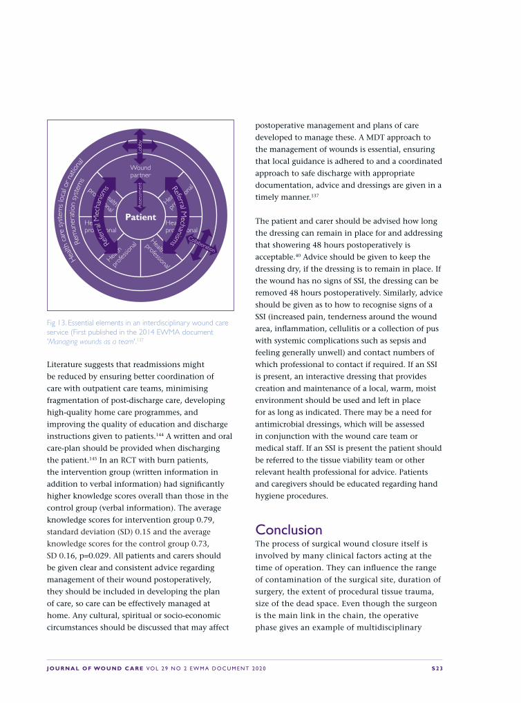
Multidisciplinary team approach to surgical wound management A multidisciplinary team (MDT) approach is
essential to successful surgical wound management
not only in acute-care but also in community
settings. These challenges include the contentious
nature of sharing professional roles and expertise,
planning and decision-making, while delivering
quality patient care within complex contexts.
Through this approach, patient-focused enhanced
clinical outcomes can be achieved.137
The essential elements for an interdisciplinary
wound care service are shown in Fig 13. This
figure shows that the patient forms the focus of
the wound care but relies on the expertise of the
different disciplines.
The patient forms the focus of the care, but
relies on the expertise of a wound navigator to
organise wound care service via established referral
mechanisms. The wound navigator and other
health professionals either collaborate to explore
beneficial remuneration and health care systems
and/or lobby to meet the needs of the patient.137
Challenges related to home care Reducing the duration of hospital stay presents
a considerable problem to both the health care
organisation and the patient. This means that
the treatment of patients is transferred to the
home care setting.140 Once a patient is discharged,
the first follow-up visit is usually scheduled 2 to
3 weeks after hospital discharge.141 During this
period, monitoring of the wound is reduced
and this lack of monitoring is a concern, as the
majority of patients do not have the experience
or expertise to recognise early-stage wound
infections.142 Thus, patients often return to the
hospital with an advanced wound infection
that often requires a re-hospitalisation.47 There
are many factors improving the SSI prevention
and treatment outcomes across in-patient and
out-patient settings (Tables 6 and 7). A US study
incorporating 346 hospitals identified SSI as the
most common reason for readmission to hospital,
accounting for 19.5% of overall readmissions.143
Table 6. Factors to improve home care management of surgical wounds
Multidisciplinary approach
Consistency in care
Patient centred wound care plan with clear and well described guidance with appropriate management goals
Access to appropriate dressing resources and clinical expertise
Patient and carer education
Written and oral information
Table 7. Factors improving the outcomes across in-patient and out-patient settings
Consistent communication and forwarding of medical reports
Clear responsibility roles
Education of patients and health care professionals
Working/functional national guidelines and standards comprising out- and inpatient sectors
J O U R N A L O F WO U N D C A R E VO L 2 9 N O 2 E W M A D O C U M E N T 2 0 2 0 S 2 3
Literature suggests that readmissions might
be reduced by ensuring better coordination of
care with outpatient care teams, minimising
fragmentation of post-discharge care, developing
high-quality home care programmes, and
improving the quality of education and discharge
instructions given to patients.144 A written and oral
care-plan should be provided when discharging
the patient.145 In an RCT with burn patients,
the intervention group (written information in
addition to verbal information) had significantly
higher knowledge scores overall than those in the
control group (verbal information). The average
knowledge scores for intervention group 0.79,
standard deviation (SD) 0.15 and the average
knowledge scores for the control group 0.73,
SD 0.16, p=0.029. All patients and carers should
be given clear and consistent advice regarding
management of their wound postoperatively,
they should be included in developing the plan
of care, so care can be effectively managed at
home. Any cultural, spiritual or socio-economic
circumstances should be discussed that may affect
postoperative management and plans of care
developed to manage these. A MDT approach to
the management of wounds is essential, ensuring
that local guidance is adhered to and a coordinated
approach to safe discharge with appropriate
documentation, advice and dressings are given in a
timely manner.137
The patient and carer should be advised how long
the dressing can remain in place for and addressing
that showering 48 hours postoperatively is
acceptable.40 Advice should be given to keep the
dressing dry, if the dressing is to remain in place. If
the wound has no signs of SSI, the dressing can be
removed 48 hours postoperatively. Similarly, advice
should be given as to how to recognise signs of a
SSI (increased pain, tenderness around the wound
area, inflammation, cellulitis or a collection of pus
with systemic complications such as sepsis and
feeling generally unwell) and contact numbers of
which professional to contact if required. If an SSI
is present, an interactive dressing that provides
creation and maintenance of a local, warm, moist
environment should be used and left in place
for as long as indicated. There may be a need for
antimicrobial dressings, which will be assessed
in conjunction with the wound care team or
medical staff. If an SSI is present the patient should
be referred to the tissue viability team or other
relevant health professional for advice. Patients
and caregivers should be educated regarding hand
hygiene procedures.
ConclusionThe process of surgical wound closure itself is
involved by many clinical factors acting at the
time of operation. They can influence the range
of contamination of the surgical site, duration of
surgery, the extent of procedural tissue trauma,
size of the dead space. Even though the surgeon
is the main link in the chain, the operative
phase gives an example of multidisciplinary
Fig 13. Essential elements in an interdisciplinary wound care service (First published in the 2014 EWMA document 'Managing wounds as a team'.137
Hea
lth c
are
syst
ems
loca
l or n
ation
alRe
mun
erat
ion
syst
ems
Health
professional
Health
professional
Health professional
Health professional
Health
pr
ofessi
onal
Health
pr
ofessi
onal
Wound partner
Patient
Refe
rral
Mec
hani
sms Referral M
echanisms Collaboration
Ass
essm
ent
Lobb
y

S 2 4 J O U R N A L O F WO U N D C A R E VO L 2 9 N O 2 E W M A D O C U M E N T 2 0 2 0
approach indispensability. Operation, carried
out excellently gives the preconditions for good
wound healing and fast patient recovery. Written
and oral patient information is key to early
identification of SSI and an appropriate pathway
for the patient to follow in seeking help.

J O U R N A L O F WO U N D C A R E VO L 2 9 N O 2 E W M A D O C U M E N T 2 0 2 0 S 2 5
5. Perioperative practice to prevent surgical site infection
P ostoperative complications arise as a
result of a combination of risk factors;
patient-related (age, obesity, underlying
illness), quality of surgical procedure (duration,
technique, type) as well as microorganisms
involved (number, virulence). Interventions can
be broadly delivered at three stages during the
patient journey: preoperatively, intraoperatively
and postoperatively. Table 8 provides a summary
of the latest published SSI guidelines:
• SSIs: prevention and treatment, National
Institute of Health and Clincal Excellence (NICE)
2008 (updated 2017)40
• Global Guidelines for the Prevention of Surgical
Site Infection, WHO, 20165
• Centers for Disease Control and Prevention
Guideline for the Prevention of Surgical Site
Infection, CDC, 20173
Preoperative phaseThe preoperative phase is the 24 hours before
the procedure and involves the admission of the
patient and the preparation leading up to the
perioperative phase.
Nasal decolonisation Recommended by WHO and NICE.5,40
Staphylococcus aureus is an important cause of
SSI. It is commonly present as part of the normal
flora of the skin and nose, with screening studies
reporting carriage rates of around 20%.146 An
association between nasal carriage of Staphylococcus
aureus and the development of SSI has been
recognised.147 Consequently, identification of
nasal carriage before surgery and treatment of
positives with a 5–day course of mupirocin and
chlorhexidine soap has been associated with
a significant reduction in rate of SSI caused by
Staphylococcus aureus.148 Decolonisation treatment
preoperatively is recommended for all patients
undergoing cardiothoracic and orthopaedic
surgeries.120 In patients undergoing other types
of surgery, it is advisable to consider other factors
including local rates of Staphylococcus aureus
and MRSA, patient-related factors such as past
KEY POINTS • Before surgery, patients should shower (full body) with
soap the night before and the day of the operation
• The incision site should be prepared with an alcohol-based antiseptic solution and hair removal should be avoided
• Antimicrobial prophylaxis should be administered only when indicated based on published clinical practice guidelines
• Normothermia should be maintained in all patients undergoing surgery
• Strategies to ensure glycaemic control should be in place
• Intraoperative factors such as tissue oxygenation, intraoperative warming and type of sutures may also influence the occurrence of SSI
• Measuring and reporting rates of SSI in surveillance systems is important practice for determining prevalence and incidence of SSI.

S 2 6 J O U R N A L O F WO U N D C A R E VO L 2 9 N O 2 E W M A D O C U M E N T 2 0 2 0
Staphylococcus aureus infection and colonisation
by Staphylococcus aureus in sites other than the
nose. While studies have yielded some reductions
in using this approach, it is recommended a
conservative approach be considered with the use
of antibiotics.
Table 8. Summary of recommendations for the prevention of surgical site infections3,5,40
Intervention Recommendation WHO5 CDC3 NICE40 NICE245
Nasal decolonisation
Consider nasal mupirocin in combination with a chlorhexidine body wash before procedures in which Staphylococcus aureus is a likely cause of a surgical site infection
✔ – ✔
Preoperative bathing
Use soap solutions to clean the skin before surgery ✔ ✔ ✔
Hair removal Avoid the removal of hair from the site of incision; if essential use hair clippers
✔ – ✔
Preoperative warming
Maintain peri-operative normothermia ✔ – ✔
Actively warm patients for 30 mins before start of anaesthesia. Temperature should be 36ºC or above before transfer to theatre
✔
Patients’ core temperature should be above 36ºC or above before and during surgery and in the recovery area
✔
Surgical hand preparation
Surgical hand preparation should be performed by scrubbing with either a suitable antimicrobial soap and water or using a suitable alcohol-based handrub before donning sterile gloves
✔ – ✔
Incision site skin disinfection
Use an alcohol-based product to disinfect skin before incision ✔ ✔ ✔
Surgical antibiotic prophylaxis
Where indicated, antibiotic prophylaxis should be given immediately before the surgical incision being made and not continued after completion of surgery
✔ ✔ ✔
Operating room ventilation
Air should be filtered to remove contaminated particles and anaesthetic gases and flow from clean to less clean areas across the operating department246
✔ – –
Operating room traffic
Reduce operating room traffic to avoid possible contamination of the site of surgery246,247
– – –
Antimicrobial sutures
Consider use of triclosan-impregnated sutures ✔ – ✔
Incise drapes The use of plastic adhesive drapes with or without antimicrobial properties is not necessary for the prevention of SSI
✔ ✔ ✔
Perioperative glycaemic control
Implement perioperative glycaemic control in patients with and without diabetes
✔ ✔ -
Perioperative oxygenation
Maintain optimal oxygenation during surgery and in the recovery period to ensure adequate haemoglobin saturation. Adult patients should receive 80% fraction of inspired oxygen
✔ - ✔
Postoperative wound management
Cover surgical incisions with an appropriate interactive dressing at the end of the operation.
✔ ✔
SSI surveillance Sustained surveillance and feedback of data on rates of SSI has been associated with reductions in rates of infection248
– – –

J O U R N A L O F WO U N D C A R E VO L 2 9 N O 2 E W M A D O C U M E N T 2 0 2 0 S 2 7
Preoperative bathing Recommended by the WHO, the CDC and
NICE,3,5,40 bacteria may gain access to the surgical
wound through several sources but the most
common source is the patient’s own skin.2
Human skin is colonised by a large number of
microorganisms known as ‘resident’ flora, which
tend to live in the skin folds, sebaceous glands and
hair follicles. The surfaces of the skin can also be
contaminated with microorganisms from body
excretions/secretions, dirt or from contact with
contaminated surfaces or items (transient flora).
While all these microorganisms are harmless on
the surface of the skin, if they access a surgical
incision they can cause a SSI. Cleansing of the
skin before surgery is therefore required to remove
as many microorganisms as possible from the
skin surface.149 Soap solutions are recommended
to physically remove dirt and transient
microorganisms from the surface of the skin.
Patients should be encouraged to have a shower
with soap. Reports by Dumville et al.,150 WHO5
and CDC151 found that bathing with chlorhexidine
soap does not significantly reduce SSI rates
compared with bathing with plain soap.
Hair removal Recommended by WHO and NICE,5,40 hair is no
more heavily colonised with microbial flora than
the skin and the criteria for hair removal at surgical
site should be based on the need to view or access
the operative site rather than to remove bacteria.
If hair removal is essential, the best approach is to
remove the minimum amount of hair, as near to the
time of operation as possible.5,40 A study has shown
that shaving the skin before operation increases
the risk of wound infection.40 According to Tanner
et al. bacteria multiply in micro abrasions caused
by the razor on the skin surrounding the operative
site and increase the risk of the incision becoming
contaminated.152 The longer the period between
hair removal and the incision, the greater the risk
of contamination. Where hair removal is essential,
this should be done using hair clippers to minimise
damage to the skin. Depilatory creams also do not
abrade the skin but are less practical as they need
to be left in place for several minutes and have the
potential to cause allergic reactions.5
Preoperative warming Recommended by NICE,149 hypothermia, defined as
a core body temperature of less than 36.0ºC149 is a
common but preventable consequence of surgery.
It can occur as a result of the suppression of the
central mechanisms of temperature regulation
due to global anaesthesia, and pharmacological
relaxation of the muscles during surgery as well
as the prolonged exposure of large surfaces of
skin to cold temperatures in operating rooms and
receiving large volumes of non-warmed fluids.
Inadvertent perioperative hypothermia has been
associated with clinical complications such as
surgical site infection, wound-healing delay,
increased bleeding or cardiovascular events.
The NICE clinical guidelines on prevention of
inadvertent hypothermia149 recommend that all
patients should be assessed within the hour before
surgery for their risk of perioperative hypothermia
and their temperature measured using a site that
produces a direct measure or direct estimate of
core temperature. All patients should be actively
warmed on the ward/emergency department at
least 30 minutes before induction of anaesthesia.
If the patient’s temperature is below 36ºC or they
are at high risk of hypothermia, they should
be warmed immediately. The patient’s core
temperature should be 36ºC or above before they
are transferred to theatre.149,153
Intraoperative phaseThe second phase, known as the intraoperative
phase, involves surgery.
Surgical scrubbing Recommended by WHO and NICE.5,40 Surgical

S 2 8 J O U R N A L O F WO U N D C A R E VO L 2 9 N O 2 E W M A D O C U M E N T 2 0 2 0
hand antisepsis by all surgical teams is routinely
carried out to remove transient microorganisms,
inhibit the growth of resident microorganisms and
maintain the lowest possible contamination of the
surgical field, especially in the event of sterile glove
puncture during the procedure. The SSI prevention
guidelines5 provide a strong recommendation that
surgical hand preparation be performed either by
scrubbing with a suitable antimicrobial soap and
water or using an alcohol-based hand-rub licensed
for surgical scrubbing, before donning sterile gloves.
A Cochrane review by Tanner et al.154 assessed the
effects of surgical hand antisepsis on preventing
SSIs and also determined the effects of surgical
hand antisepsis on the numbers of colony-forming
units (CFUs) of bacteria on the hands of the
surgical team. The review found no firm evidence
that one type of hand antisepsis is better than
another in reducing SSIs.154
Chlorhexidine gluconate scrubbing agents may
reduce the number of CFUs on hands when
compared with povidone iodine scrubs. However,
the clinical relevance of this outcome is unclear.
Alcohol hand rubs with additional antiseptic
ingredients (such as chlorhexidine) may reduce
CFUs compared with aqueous scrubs. Low-quality
evidence indicates that there is a greater CFU
reduction after a three-minute initial surgical scrub
than two-minute scrub.155 Findings on a longer
initial scrub and subsequent scrub durations were
not consistent. Tanner et al. also concluded that
it is unclear whether nail picks and brushes used
during scrubbing have an impact on the number of
CFUs remaining on the hands.154
Incision site skin disinfection Recommended by WHO, CDC and NICE,3,5,40
the aim of skin disinfection before incision is to
apply antiseptic solutions to rapidly destroy skin
microorganisms at the site of the incision and
reduce the risk of contamination of the surgical
site. Disinfection of the surgical site should occur
as close to the time of surgery as possible and
immediately before draping.40
The two main antiseptic agents used for
preoperative skin preparation are chlorhexidine
gluconate (CHG) and iodophors (povidone iodine;
PI). Both CHG and PI are effective against a broad
range of skin microorganisms and exert persistent
activity that prevents regrowth for several hours
after application.156 They are available in either an
aqueous or alcohol-based form. There is limited
evidence to suggest that one agent is better than
another. Products based on alcohol are probably
more effective than aqueous products since alcohol
is an antiseptic agent.5 Evidence for differences in
efficacy between PI and CHG is limited but tends
to favour CHG.157–159
Most studies are too small to detect differences
in rates of SSI,160–162 measure only the change in
skin colonisation or have focused on single types
of operative procedure.160,163–165 Alcohol-based
solutions should be used where they are suitable
for the particular site of incision as they include an
additional, rapid-acting antiseptic agent that dries
quickly. However, alcohol can damage mucous
membranes (eyes, ears, genitalia) and aqueous
solutions should be used for this type of surgery.
Surgical antibiotic prophylaxis Recommended by WHO, CDC and NICE,3,5,40
the purpose of surgical antibiotic prophylaxis
is to eliminate microorganisms introduced into
the surgical wound during the procedure, which
may subsequently multiply to cause SSI.166 For
many types of commonly performed surgery,
there is consistent evidence that a single dose
of antimicrobial with a long enough half-life
to achieve activity throughout the operation is
adequate.167–172 A repeat dose is only indicated
when there is excessive blood loss or if surgery is
unexpectedly prolonged.

J O U R N A L O F WO U N D C A R E VO L 2 9 N O 2 E W M A D O C U M E N T 2 0 2 0 S 2 9
Administration of surgical prophylaxis should be
timed to achieve the right levels of antibiotics in
blood and tissue before bacterial contamination
occurs during operation. The time taken for an
antibiotic to reach an effective concentration in
any particular tissue reflects its pharmacokinetic
profile and the route of administration. Antibiotic
prophylaxis administered too late or too
early (more than 120 minutes before or after
incision) reduces the efficacy of the antibiotic
and may increase the risk of SSI.173,174 A meta-
analysis on the timing of preoperative surgical
prophylaxis did not identify increased SSI risks if
administration was within 120-minute timeframe
before incision.129,174 It is broadly recommended
that the administration of the first dose of
antibiotic is made within 60 minutes before
the incision.166
Prophylaxis should be converted into a treatment
regime when an existing infection is present in
the wound at the time of surgery or significant
contamination such as from bowel contents,
occurs during the operation.149
Intraoperative warming Recommended by WHO, CDC and NICE.3,5,40 The NICE clinical guidelines on prevention of
inadvertent hypothermia149 state that induction of
anaesthesia should not begin unless the patient’s
temperature is 36.0ºC or above (unless there
is a need to expedite surgery). Patients having
anaesthesia for longer than 30 minutes are at
a higher risk of perioperative hypothermia and
are to be warmed from induction of anaesthesia
using forced air warming.149 The patient’s
temperature should be measured and documented
before induction of anaesthesia and then every
30 minutes until the end of surgery, using a
site that produces a direct measure (pulmonary
artery catheter, oesophagus and bladder) or direct
estimate of core temperature (sublingual, axilla,
rectal and zero heat flux). Devices using infrared
technology to measure temperature are not
recommended (tympanic, forehead, temporal).
Forced air warming Recommended by NICE.149 A frequently used
techniques to prevent inadvertent perioperative
hypothermia is active body surface warming
systems, which generate heat mechanically
(heating of air, water or gels) that is transferred to
the patient via skin contact. A Cochrane review
assessing the effectiveness of preoperative and/or
intraoperative active warming systems, to prevent
perioperative complications from unintended
hypothermia during surgery concluded that forced-
air warming appeared to have a beneficial effect in
reducing surgical site infection.175
Fluid warming Recommended by NICE.149 If a patient requires
a large volume of intravenous and/or irrigation
fluids (500ml or more) and the temperature of
these fluids is below core body temperature,
they can cause significant heat loss. Warming
intravenous and irrigation fluids to core body
temperature or above might prevent some of this
heat loss and subsequent hypothermia.
A recent review of clinical evidence found
that warmed intravenous fluids kept the core
temperature of study participants about half a
degree warmer than that of participants given
room temperature intravenous fluids.176 The
NICE guidelines149 recommend that all irrigation
fluids used intraoperatively should be warmed to
a temperature of 38–40ºC in a thermostatically
controlled cabinet.149
Postoperative warming Recommended by NICE.149 NICE guidelines40
recommend that the patient’s temperature should
be monitored and documented every 15 minutes in
recovery. The patient should not be transferred to
the ward, until their temperature is 36ºC or above.

S 3 0 J O U R N A L O F WO U N D C A R E VO L 2 9 N O 2 E W M A D O C U M E N T 2 0 2 0
Operating room ventilation Recommended by the CDC,5 the ventilation
systems used in operating theatres are useful to
filter out airborne microorganisms, to prevent
microorganisms from entering the theatre in the
air supply from corridors or other parts of the
hospital, and to dilute contaminated air in the
room by replenishing with fresh filtered air.177 Dust
and textile particles, skin scales, airborne bacteria
and other sources of microbial contamination
within the surgical field all have the potential to
cause SSI.
Operating room traffic In theatre, the main source of airborne bacteria is the
staff entering and leaving the theatre and adjacent
rooms, although power tools can also create aerosols
from tissues.177 The number of airborne microbial
particles in an operating room is proportional to
the number of humans present and their level of
activity.178 Each person has been estimated to emit
approximately 1000 organisms per minute at rest,
increasing to 50,000 per minute during activity as
friction of clothing against the skin releases more
squames.179,180 These particles may settle on to
instruments or gloved hands or into the wound itself
and subsequently result in wound infection.181,182
Laminar flow Recommended by WHO.5 Ultra-clean air systems
(laminar flow) have been recommended to
reduce the incidence of infection in prosthetic
orthopaedic surgery, which are susceptible to
infection even if only small numbers of bacteria are
introduced into the wound. These ‘laminar flow’
systems direct parallel streams—a laminar flow of
filtered air over the operating table—and use over
600 air changes per hour. Although early studies
suggested laminar flow systems were associated
with a reduction in wound infection rates, there is
now an emerging body of evidence that suggests
they may even increase the risk of infection,
possibly due to disruption of air flow by personal
and equipment and a reduction in wound tissue
temperature. A meta-analysis by Bischoff et al.179
showed no benefit for laminar airflow compared
with conventional turbulent ventilation of the
operating room in reducing the risk of SSIs in
total hip and knee arthroplasties, and abdominal
surgery. Indeed the WHO SSI guidelines5 do
not recommend laminar airflow as a preventive
measure to reduce the risk of SSIs, stating these
systems should not be installed in new operating
rooms. This recommendation is however based on
low to very low-level evidence.
Incise drapes Recommended by WHO, the CDC and
NICE.3,5,40 Adhesive plastic incise drapes, plain or
impregnated with an antimicrobial agent (mostly
an iodophor), are used on the patient’s skin after
surgical site preparation. The film adheres to the
skin and the surgeon cuts through the drape and
the skin. Such a drape is theoretically believed to
represent a mechanical and/or microbial barrier to
prevent the migration of microorganisms from the
skin to the operative site.
However, some reports showed an increased
recolonisation of the skin following antiseptic
preparation underneath adhesive drapes
compared to the use of no drapes. The WHO
SSI guidelines (2016),5 Cochrane review183 and
CDC (2017) SSI guidelines3 found that the use
of either non-antimicrobial-impregnated or
antimicrobial-impregnated incise drapes is not
necessary for the prevention of SSI.
Impregnated sutures Recommended by WHO and NICE,5,40 surgical
suture material is used to proximate wound edges
and is thus in direct contact with the wound
itself. To prevent microbial colonisation of the
suture material in surgical incisions, sutures with
antibacterial activity have been developed. The

J O U R N A L O F WO U N D C A R E VO L 2 9 N O 2 E W M A D O C U M E N T 2 0 2 0 S 3 1
WHO SSI prevention guidelines5 recommend the
use of triclosan-coated sutures for the purpose
of reducing the risk of SSI, independent of the
type of surgery and type of sutures (braided or
monofilament). The meta-analysis was based
on moderate-to-low quality evidence, examined
triclosan-coated, absorbable sutures only and
points out that many of the studies included
had conflicts of interest.101 There were no studies
identified that investigated other antimicrobial
agent coated sutures.
Intraoperative administration of high oxygen concentrations Recommended by WHO.5 The SSI prevention
guidelines recommends that adult patients
undergoing general anaesthesia with endotracheal
intubation for surgical procedures should receive
80% fraction of inspired oxygen intraoperatively
and, if feasible, in the immediate postoperative
period for 2–6 hours.
A meta-analysis suggested a beneficial effect of
intraoperative administration of high oxygen
concentrations during colorectal surgery.184
Glucose control Recommended by WHO and the CDC.3,5 Blood
glucose levels rise during and after surgery due to
surgical stress.185 Surgery causes a stress response
that results in a release of catabolic hormones and
the inhibition of insulin.5 Several observational
studies186–189 showed that hyperglycaemia is
associated with an increased risk of SSI and therefore
an increased risk of morbidity, mortality and higher
health care costs in both diabetic and non-diabetic
patients and in different types of surgery.
Recently published guidelines seem to disagree on
the level of glucose control required to reduce risk of
SSI. The WHO5 guidelines recommend that protocols
for intensive perioperative blood glucose control
should be used for both diabetic and non-diabetic
adult patients undergoing surgical procedures.
Centers for Disease Control (CDC) SSI prevention
guidelines3 highlight that moderate-quality evidence
suggested no benefit of strict glucose control
(80–100 mg/dl or 80–130 mg/dl) as compared with
standard blood glucose target levels. A programme
to enhance recovery after surgery is steadily in
the implementation phase across the northern
hemisphere. The enhanced recovery after surgery
(ERAS) programme is a multimodal approach to
reduce or modify the physiological and psychological
effects of surgery. Initiated by Henrik Kehlet in the
1990s implementation of the ERAS protocol has been
shown to reduce the occurrence of SSI and hospital
length of stay in certain surgical populations;
colorectal,190 urological and orthopaedic surgery.191,192
The key tenets of the ERAS protocol are underpinned
by evidence-based practices and include perioperative
counselling, preoperative nutrition (carbohydrate
loading up to 2 hours preoperatively), standardised
anaesthetic and analgesic regiments (epidural and
non-opioid analgesia) and early mobilisation.193
In some cases, implementation of the ERAS
programme have resulted in 30% to 50% reductions
in complications and reduced lengths of stay.194 The
key to success with the ERAS programme is a MDT
approach to patient management which includes
the surgical team, anaesthetist, ward staff, dieticians,
patient and carer engagement.
Postoperative phaseThe postoperative phase is the period immediately
following surgery. Application of antimicrobial
dressings should be reserved only for specific
surgical situation regarding high-risk patients
undergoing high-risk procedures and only for a
limited period of time.
Wound dressings for the prevention of SSI and management of surgical wounds Although there is no definitive evidence for the
use of any particular type of modern interactive

S 3 2 J O U R N A L O F WO U N D C A R E VO L 2 9 N O 2 E W M A D O C U M E N T 2 0 2 0
Table 9. Wound dressings for the management of surgical wounds (after NICE 201840 and WHO 2016)5
Dressings category
Dressing characteristics Wound characteristics / healing intention
Primary clinical indications
Phase of Management
Advanced (Interactive)
Vapour-permeable films
Vapour-permeable films are permeable to water vapour and oxygen, but not to water or microorganisms. They are normally transparent
• Superficial • Minimal exudate*• Primary intention
Facilitates the optimum healing environment (moist wound healing) and provides a barrier to bacteria/protects the incision site
• Intraoperative (In theatre)
• Postoperative• In community/
home care settings
Hydrocolloid dressing
Hydrocolloid dressings vary significantly in their composition and physical properties. In general, they consist of a self-adhesive gel-forming mass applied to a carrier, such as a thin polyurethane film or a foam sheet. They contain colloidal particles, such as quar, karaya, gelatic, sodium carboxymethylcellulose, gelatin and pectin, in an adhesive mass usually made of polyisobutylene. In their intact state, hydrocolloids are virtually impermeable to water vapour. By trapping wound exudates, hydrocolloids create a moist environment that softens and lifts dry eschars. They favour also granulation tissue formation and re-epithelialisation
• Superficial• Low exudate*• Primary and
secondary intention
Facilitates wound hydration and the optimum wound healing environment.
Promotes autolytic debridement and proteolytic digestion
Usually postoperative and in community/home care settings
Hydrogels or fibrous hydrocolloid dressing
Hydrogels consist of 80–90% water and insoluble cross-linked polymers, such as polyethyleneoxide, polyvinyl pyrollidone, acrylamide or carboxymethylcellulos, with hydrophilic sites that interact with aqueous solutions, absorbing and retaining significant volumes of water
• Superficial or deep
• Low-to-moderate exudate*
• Secondary intention
Rehydration of tissues and some absorption of exudate.
Facilitates the optimum healing environment and protects the incision site. Some absorbency potential
• Intraoperative (occasionally)
• Postoperative• In community/
home care settings
Polyurethane matrix hydrocolloid dressing
Polyurethane matrix hydrocolloid dressings consist of two layers: a polyurethane gel matrix and a waterproof polyurethane top film designed to act as a bacterial barrier
• Superficial or Deep
• Low to moderate exudate*
• Primary and secondary intention
Indicated for clean, granulating / sloughy or necrotic wounds.
Limited absorbency capacity - the amount of exudate that a hydrocolloid dressing can absorb will be dependent upon the MVTR (moisture vapour transfer rate) of the backing layer.
• Intraoperative• Postoperative• In community/
home care settings
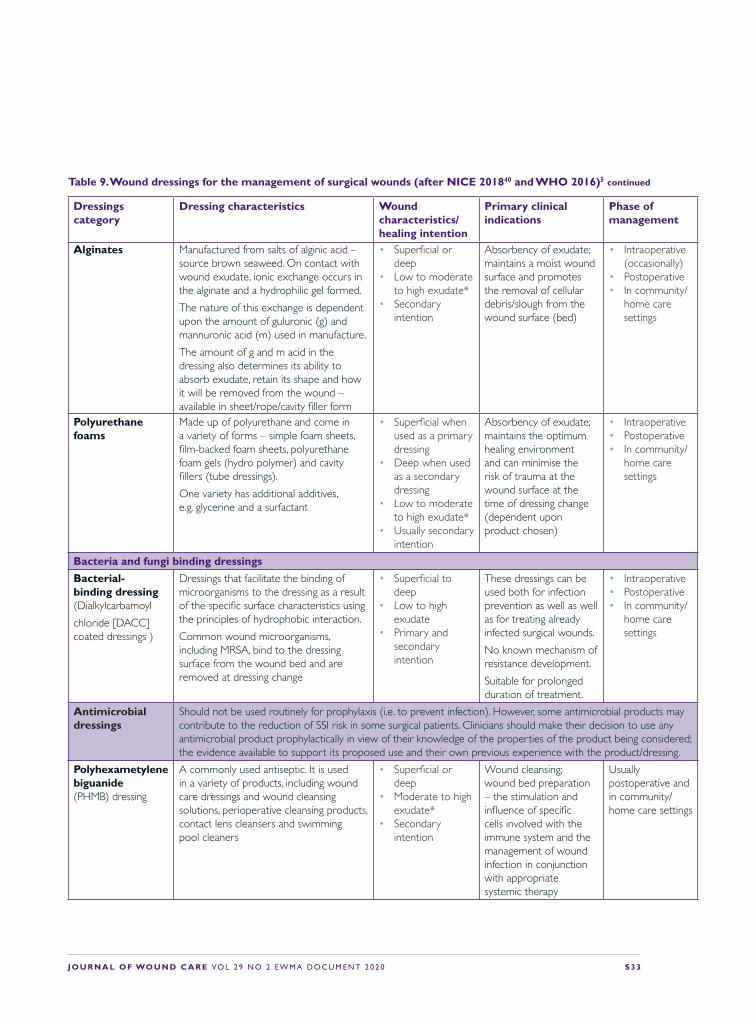
J O U R N A L O F WO U N D C A R E VO L 2 9 N O 2 E W M A D O C U M E N T 2 0 2 0 S 3 3
Table 9. Wound dressings for the management of surgical wounds (after NICE 201840 and WHO 2016)5 continued
Dressings category
Dressing characteristics Wound characteristics/healing intention
Primary clinical indications
Phase of management
Alginates Manufactured from salts of alginic acid – source brown seaweed. On contact with wound exudate, ionic exchange occurs in the alginate and a hydrophilic gel formed.
The nature of this exchange is dependent upon the amount of guluronic (g) and mannuronic acid (m) used in manufacture.
The amount of g and m acid in the dressing also determines its ability to absorb exudate, retain its shape and how it will be removed from the wound – available in sheet/rope/cavity filler form
• Superficial or deep
• Low to moderate to high exudate*
• Secondary intention
Absorbency of exudate; maintains a moist wound surface and promotes the removal of cellular debris/slough from the wound surface (bed)
• Intraoperative (occasionally)
• Postoperative• In community/
home care settings
Polyurethane foams
Made up of polyurethane and come in a variety of forms – simple foam sheets, film-backed foam sheets, polyurethane foam gels (hydro polymer) and cavity fillers (tube dressings).
One variety has additional additives, e.g. glycerine and a surfactant
• Superficial when used as a primary dressing
• Deep when used as a secondary dressing
• Low to moderate to high exudate*
• Usually secondary intention
Absorbency of exudate; maintains the optimum healing environment and can minimise the risk of trauma at the wound surface at the time of dressing change (dependent upon product chosen)
• Intraoperative• Postoperative• In community/
home care settings
Bacteria and fungi binding dressings
Bacterial-binding dressing (Dialkylcarbamoyl
chloride [DACC] coated dressings )
Dressings that facilitate the binding of microorganisms to the dressing as a result of the specific surface characteristics using the principles of hydrophobic interaction.
Common wound microorganisms, including MRSA, bind to the dressing surface from the wound bed and are removed at dressing change
• Superficial to deep
• Low to high exudate
• Primary and secondary intention
These dressings can be used both for infection prevention as well as well as for treating already infected surgical wounds.
No known mechanism of resistance development.
Suitable for prolonged duration of treatment.
• Intraoperative• Postoperative• In community/
home care settings
Antimicrobial dressings
Should not be used routinely for prophylaxis (i.e. to prevent infection). However, some antimicrobial products may contribute to the reduction of SSI risk in some surgical patients. Clinicians should make their decision to use any antimicrobial product prophylactically in view of their knowledge of the properties of the product being considered; the evidence available to support its proposed use and their own previous experience with the product/dressing.
Polyhexametylene biguanide (PHMB) dressing
A commonly used antiseptic. It is used in a variety of products, including wound care dressings and wound cleansing solutions, perioperative cleansing products, contact lens cleansers and swimming pool cleaners
• Superficial or deep
• Moderate to high exudate*
• Secondary intention
Wound cleansing; wound bed preparation – the stimulation and influence of specific cells involved with the immune system and the management of wound infection in conjunction with appropriate systemic therapy
Usually postoperative and in community/home care settings

S 3 4 J O U R N A L O F WO U N D C A R E VO L 2 9 N O 2 E W M A D O C U M E N T 2 0 2 0
Table 9. Wound dressings for the management of surgical wounds (after NICE 201840 and WHO 2016)5 Continued
Dressings Category
Dressing Characteristics Wound Characteristics / Healing intention
Primary Clinical Indications
Phase of Management
Silver-impregnated dressing
Silver provides extensive coverage against bacteria, fungi and viruses, including nosocomial pathogens and methicillin-resistant Staphylococcus aureus (MRSA) and vancomycin-resistant enterococci (VRE), make it a valuable adjunct in the prevention and treatment of infection. Silver has both bactericidal effects via oxidation of the cell membrane and bacteriostatic effects by inhibiting bacterial replication through damage to DNA
Superficial or DeepModerate to High Exudate*Secondary Intention
Wound cleansing; wound bed preparation – the stimulation and influence of specific cells involved with the immune system and the management of wound infection in conjunction with appropriate systemic therapy
Usually postoperative and in community/home care settings
Povidone Iodine impregnated dressings
Iodine is an antiseptic that targets a broad spectrum of bacteria and other pathogens. It has been used successfully without complication for the management of many hard-to-heal wounds, however there is currently little evidence to support its use for the prevention and long term management of SSI.
• Superficial wounds
• Minimal exudate• Secondary
Intention
Iodine is an oxidizing agent, and its bactericidal activity involves the inorganic and essentially no development of resistance by microorganisms has been determined
Postoperative
Advanced (Active)Negative-pressure wound therapy (NPWT) dressings
Primarily designed to prevent exudate collection while simultaneously preventing desiccation of the wound.
• Deep• Low to Moderate
to High Exudate*• Secondary
Intention
These dressings increase oxygen tension in the wound, improve blood flow to the wound bed, decrease bacterial count, increase granulation formation and minimise shear forces on the wound surface
• Intraoperative• Postoperative• In community
/home care settings
Basic wound contact layers
Absorbent dressing pads
Absorbent materials are non-occlusive permeable dressings that allow the moisture to be absorbed to evaporate into the atmosphere. Many comprise of a soft viscose, polyester bonded pad that may or may not have external polyethylene contact layer.
‘Superabsorbers’ comprise of absorbent polymers (some of which expand on absorption of fluid) however this is a comparative not absolute term.
• Superficial• Low exudate*• Primary or
Secondary Intention (when used as a secondary dressing)
• Superficial• Low to moderate
Exudate*• Usually Secondary
Intention (when used as a secondary dressing)
Additional absorbency of exudate over another primary dressing or a low adherent wound contact layer (see below)
Not generally recommended in theatre or the immediate post- operative phase. May be used as a secondary dressing – occasionally

J O U R N A L O F WO U N D C A R E VO L 2 9 N O 2 E W M A D O C U M E N T 2 0 2 0 S 3 5
wound dressing for the prevention of SSI,128 NICE
in the UK recommends covering surgical incisions
with an appropriate interactive dressing at the
end of the procedure.40 The 2013 evidence update
of the NICE guidelines agrees that no particular
dressing type emerges as the most effective in
reducing the risk of SSI, although silver nylon
dressings may be more effective than gauze.195
Furthermore in a recent Cochrane review there is
limited evidence for the use of advanced wound
dressings in the prevention of SSI. This is primarily
due to a considerable lack of level one studies such
as RCT's to determine comparative effectiveness
of wound dressings in a controlled and systematic
way.150 Furthermore in a recent Cochrane review
there is limited evidence for the use of advanced
wound dressings in the prevention of SSI. This is
primarily due to a considerable lack of level one
studies such as RCT's to determine comparative
effectiveness of wound dressings in a controlled
and systematic way.150 Nonetheless, in clinical
practice it is now generally accepted and reflected
Table 9. Wound dressings for the management of surgical wounds (after NICE 201840 and WHO 2016)5 Continued
Dressings category
Dressing characteristics Wound characteristics / healing intention
Primary clinical indications
Phase of Management
Low-adherent wound contact layers (traditional)
Low-adherent wound contact layers consist mainly of a fine mesh gauze impregnated with moisturizing, antibacterial or bactericidal compounds. They are either non-medicated (for example, paraffin gauze dressing) or medicated (for example, containing povidone iodine or chlorhexidine). As the dressing dries, fibrin from the wound bed causes temporary bonding of the dressing to the wound, thus permitting healing beneath it
• Superficial• Low exudate*• Usually primary
Intention
These dressings are widely used, primarily as interface layers between the wound surface and a secondary absorbent dressing, usually made of cotton gauze, to prevent it from adhering to the wound surface and causing trauma upon removal
• Intraoperative (occasionally)
• Postoperative• In community
/home care settings
Low adherent silicone wound contact layers
• Superficial but can be used to line a deep wound such as in combination with NPWT
• Usually low exudate* when dressing used for its prime clinical indication
• Usually primary but can be secondary when used in combination with NPWT
To minimise the risk of trauma at the wound surface and minimise the patients pain experience during dressing changes
• Intraoperative (occasionally)
• Postoperative• In community/
home care settings
*Exudate: A generic term used to identify liquid produced from wounds201 202
Bates-Jensen202 203 attempted to qualify the levels of exudate in relation to the terms often used by clinicians to describe the sameSmall (Low): wound tissues wet, moisture evenly distributed in wound, exudate affects 25% of dressingModerate: wound tissues saturated, drainage may or may not be evenly distributed in wound, exudate involves 25% to 75% of dressingLarge (High): wound tissues bathed in fluid, drainage freely expressed, may or may not be evenly distributed in wound, exudate involves 75% of dressing

S 3 6 J O U R N A L O F WO U N D C A R E VO L 2 9 N O 2 E W M A D O C U M E N T 2 0 2 0
in various postoperative care plans (predetermined
or developed further to assessment) or bundles that
surgical dressings should be kept undisturbed for
a minimum of 48 hours after surgery (longer if at
all possible, up to 4 days) unless leakage occurs/
associated symptomatology changes.196,197 Further
research regarding dressing materials for the
prevention of SSI is required, as there are still no
specific level 1 evidence based guidelines regarding
the type of surgical ‘modern interactive’ dressing
to be used. However, given the available evidence
(WHO, Nice guidelines), dressings are to be used
based upon current clinical judgement, which may
be guided by Table 9.
Most surgical wounds heal by primary intention
and are covered with a dressing that acts as a
protective barrier between the wound bed and
the outside environment. A Cochrane review150
sought to assess whether one type of dressing
is better than any other in preventing surgical
site infection. The authors reported a lack of
evidence for the use of one dressing over another
for prevention of SSIs. The NICE guidelines
recommend covering surgical incisions with an
appropriate interactive dressing at the end of
the procedure.26 A recent case control study of
834 participants who underwent either a total
knee or hip arthroplasty yielded a statistically
significant reduction between the intervention and
control arms in SSI rate with use of silver nylon
compared to standard dressing.198 A literature
review published in 2014 reported that silver-nylon
dressings are associated with decreased SSI risk in
small studies across several specialities, including
colorectal surgery, neurosurgery, spinal surgery
and some cardiac and orthopaedic procedures,
although the authors recommend larger powered
trials are required in these cohorts to determine the
comparative effectiveness of silver-nylon dressing
in the prevention of SSIs.199 Results following
a RCT of dialkylcarbamoyl chloride (DACC)-
impregnated dressings compared with a control
dressing, yielded evidence to suggest the use of
DACC-dressings may assist in the prevention of SSI
in caesarian-sections.200,201 Furthermore, the WHO
guidelines do not recommend using advanced
dressings such as hydrocolloid, silver-containing
dressings, polyhexamethylene biguanide
(PHMB), over a standard dry absorbent dressing
on primarily closed surgical wounds as an SSI
prevention measure.5 The WHO recommendation
was based upon the low-level evidence and a
limited number of level one studies available.
In Table 9 we describe the different dressing
categories, their characteristics and healing
intentions as well as their primary clinical
indications and in which phase of management
they could be considered. The following
information should help you make your
clinical decision.
SurveillanceSurveillance is deined by the CDC's Guidelines for
Evaluating Surveillance Systems as:
'The ongoing and systematic collection, analysis, and
interpretation of health data in the process of describing
and monitoring a health event. This information is
used for planning, implementing, and evaluating public
health interventions and programmes. Surveillance data
are used both to determine the need for public health
action and to assess the effectiveness of programs.'151
Surveillance of SSI is a key component of
infection prevention and control programmes,
as sustained surveillance has been shown to
reduce rates of infection.203 The risk of SSI should
be monitored using standardised surveillance
methodology to identify and investigate trends,
guide identification of improvement actions and
evaluate the effectiveness of these interventions.
SSI surveillance is performed by reviewing

J O U R N A L O F WO U N D C A R E VO L 2 9 N O 2 E W M A D O C U M E N T 2 0 2 0 S 3 7
microbiology reports and patient medical records,
carrying out surgeon and/or patient surveys
and screening for readmission and/or return to
the operating room.204 Feedback of SSI rates to
surgeons and the surgical team is important to
encourage ownership and active participation
for improvement of SSI rates.205 Indeed, the
European Council Recommendation on patient
safety, including the prevention and control
of health care-associated infections (2009/C
151/01), recommends ‘performing the surveillance
of the incidence of targeted infection types,
using surveillance methods and indicators as
recommended by ECDC and case definitions as
agreed upon at Community level in accordance
with the provisions of Decision No 2119/98/EC’.206
However, it is acknowledged that SSI surveillance
is challenging and requires expertise, time and
resource dedication.5
Surveillance is an important subject not only in
the acute care but also in the home care setting.
The transition of the surgical care from the
acute care to the home care setting poses special
challenges that may increase the morbidity of
post-discharge SSI.207 At home, patients have the
primary responsibility for problem recognition
and wound care. A supervised wound assessment
and patient-provider communication is
missing. A comparative descriptive study of
76 patients with wounds showed that patients
verbalised their concerns about going home
with a wound as they have only minimal or
ineffective discharge teaching.208 This may
result in a lack of knowledge and awareness
about SSI and therefore they may miss an early
infection.209 Evidence suggests that a missing or
an inadequate post-discharge communication
may contribute to poorer outcomes resulting in
hospital readmission.141
ConclusionPrevention of surgical site infections is complex
and requires a multidisciplinary approach
including patient and carer engagement. While
published studies are contributing to the gradually
growing evidence base for the use of dressings or
ciNPT as a prophylactic measure in the prevention
of SSI, further well designed and powered trials
are required to determine the efficacy of dressings
in the prevention of SSI. There is a considerable
evidence base in the perioperative management of
the patient, with standardised guidelines (WHO
and NICE) for prevention of SSI during this phase
of the patient’s journey.

S 3 8 J O U R N A L O F WO U N D C A R E VO L 2 9 N O 2 E W M A D O C U M E N T 2 0 2 0
Wound dressings are only one part of
healing postoperative wounds and
there are a number of other intrinsic
and extrinsic factors that are related to wound
healing after surgery (as described in Chapter 2).
The clinical postoperative management of
the patient’s surgical wound must include an
assessment of patient-related lifestyle factors and
comorbidities, as these may contribute to delayed
healing and postoperative complications such as
surgical site infection.
The primary goals for acute wound management
are to protect the approximated margins, minimise
scar formation and allow the wound to heal as
rapidly as possible without complications. Wound
dressings applied on the wound play an important
role in supporting healing. Appropriate dressing
choice must meet wound requirements, which can
vary according to the phase of the healing process.
For a detailed overview of wound dressings for
prevention of SSI and management of surgical
wounds see Table 9. Acute wound management
approaches will vary due to the location and
nature of the wound, therefore an evidence-based
multidisciplinary approach provides the basis for
wound healing (Table 3).
The primary principles for acute wound
management include:
• Promote healing by primary intention
• Assess and reduce the risk of complications
(infection, dehiscence, seroma, haematoma)
• Use aseptic technique at all times (Fig 14)
• Protect the incision site
• Promote patient recovery and wellbeing.
Regardless of procedure, all wounds must be kept
as clean as possible to prevent the occurrence of
surgical site infection. The NICE guidelines have
made recommendations for the prevention of SSI
in the postoperative phase.40
How to dress the wound in the outpatient settingIn surgical wounds healing by primary intention,
dressings act as semipermeable barrier to prevent
bacterial contamination from the external
environment. The main purpose of the use of
a surgical dressing over an incisional wound,
healing by primary intention, is to control any
postoperative bleeding, absorb exudate, ease pain
and provide protection for newly formed tissue.30
Dressing change serves for examination of the
6. Principles of postoperative care
KEY POINTS • Wounds should be covered with a protective dressing
for at least 48 hours
• Wound dressings are only a part of the postoperative management of wound
• Postoperative management includes assessment of patient related lifestyle factors, comorbidities and risk of complications
• Use aseptic technique at all times
• Surgical wounds healing by secondary intention should be managed by health professionals with tissue viability expertise and dressed with appropriate dressings

J O U R N A L O F WO U N D C A R E VO L 2 9 N O 2 E W M A D O C U M E N T 2 0 2 0 S 3 9
wound, lavage/cleansing, suture replacement,
drains removal, drug application and exchange
of dressings. If change of dressing is required in
the first 48 hours of the postoperative phase the
NICE SSI (2018) guideline update recommends
using an aseptic technique (Fig 11).40,210 This
technique helps prevent contamination of the
surgical wound by ensuring that only sterile
objects and fluids come into the contact with
the wound. Aseptic technique requires adequate
hand hygiene, the use of appropriate personal
protective equipment, preparation of the
environment and maintenance of a sterile field
at all times. Current NICE guidance (2018)40
recommends to use sterile saline for wound
cleansing up to 48 hours after surgery and to
advise patients that they may shower safely
48 hours after surgery.174 If the surgical wound
has separated or has been surgically opened to
drain exudate, tap water for wound cleansing
may be used after 48 hours of surgery.36 There
is no need to use topical antimicrobial agents
for surgical wounds that are healing by primary
intention to reduce the risk of SSI.211,212 Patients
with sutured surgical wounds should be instructed
to keep the wound clean and dry and to report
any unusual changes in the wound area to their
attending physician.
Fig 14. Aseptic technique (adapted from Wilson (2019), Infection Control in Clinical Practice, Elsevier Health Sciences)210
Aseptic technique
1 Decontaminate hands and use hygenic hand disinfection
2 Prepare equipment keeping sterile parts protected
6 Cleanse the wound from inside out, remove crusts and adhesives from surrounding skin
4 Decontaminate hands
5 Put on gloves, if indicated
3 Undress the target wound
8 Discard equipment safely
7 Prevent contamination of the dressing and cover the wound

S 4 0 J O U R N A L O F WO U N D C A R E VO L 2 9 N O 2 E W M A D O C U M E N T 2 0 2 0
How to dress the wound in the home care settingProviding health care in the home care setting may
present special challenges and health hazards such
as infection control. To prevent infections and to
offer the patient and her/his carer best practice
wound care at home, the EWMA Home Care
Document140 suggests the following procedure:
• Use safe products (with minimal
collateral effects)
• Employ simple-to-use products (to reduce risk of
mistakes and anxiety from the informal carer or
the patient)
• Use disposable products where possible (to
reduce risk of transmission of infections from
home care environment)
• Use products that reduce pain
• Use products that have a wide range of
applications (i.e. not just very specialist
products for difficult wounds but products that
can be used daily on simple wounds and have,
when necessary, the features required for the
Debridement
Moisture control
Product, devices, materials
Product categories
Device categories
Materials
Bandages and tapes
• Pressure distribution mattresses• Cushions
• Total offload devices
• Patient rights Patient information, eg. on proper nutrition including supplements
• Absorbent dressings• Antimicrobial dressings;
For further information, see EWMA Antimicrobial Document
• Foams
• Gels, eg. enzyme alginogels• Hydroactive combinations• Hydrocolloids• Polymeric memebrance dressings• Skin barriers
• See dressing category. For further information, see EWMA Debridement Document
Medical lotions and creams to care for thin and flat epidermis and loss of skin elasticity
Topical negative pressure therapy
Garments
International guidelines
Therapy shoes
National guidelines
Antimicrobial strategy• Cleansing agents• Moistened medical devices
Wound prevention
Fixation
Wound bed preparation
Dressings
Wound prevention
Patient education materials
Best practice evidence for health professions
Fig 15. Recommendations for products, devices and materials available for home care wound care.140

J O U R N A L O F WO U N D C A R E VO L 2 9 N O 2 E W M A D O C U M E N T 2 0 2 0 S 4 1
treatment of more complex situations).
The following key points for health professionals
when selecting a product in home care wound
care should be followed (Fig 15):
• Wound dressings can be used through extended
parts of the healing continuum
• Wound dressings should be non-adherent, and
should eliminate or minimise need for wound
bed cleaning
• Wound products are easy to use and access,
especially if patient or informal carer takes part
in wound management
• Wound products enable the lowest overall cost,
including the cost of home care services and
patient costs
• Wound products are eco-friendly.
In the home care setting, community
health professionals as well as visitors may
transmit germs to patients. The following
recommendations for patients are adapted from
the Australian Health and Medical Research
Council (NHMRC).213
Hand hygiene:
• Use alcohol-based hand rub on a regular base.
Personal protective equipment:
• Use disposable gloves and plastic aprons.
Cleaning:
• Use detergent and water for general cleaning.
Disinfectant is needed as well when infection is
known or suspected
• Encourage a tidy environment for a patient's
home environment.
Managing spills:
• Clean spills promptly, dispose of contaminated
materials and perform hand hygiene.
Clothing and personal hygiene:
• Change clothing daily or when soiled. Wear
short sleeves, or roll up sleeves above the elbows
• Remove clothing that is not washed daily
(such as cardigans and jackets) during personal
care activities, food preparation and cleaning
activities; and ensure lanyards and mobile
phones are secured
• Wear non-slip closed-in shoes to protect your
feet against accidental injury/spillage during
home visits
• Keep jewellery to a minimum and do not wear
gel, acrylic or false fingernails
• Keep finger nails cleaned and trimmed.
Food handling:
• Perform hand hygiene before and after
handling food
• Clean work areas with detergent and water and
allow them to dry before preparing food
• Food should be consumed by the client shortly
after preparation or covered and placed in
the fridge.
Multi-resistant organisms:
• Use standard precautions (e.g. hand hygiene,
gloves and gown if risk of blood or body fluid
splash; goggles if high risk of splash to the eye).
• Place all disposable items in the home’s general
waste bin (except sharps, which require a
specialised sharps container).
Presence of pets:
• Pets should be kept away when changing
a dressing.
Most wounds should be covered with a protective,
non-adherent dressing for at least 48 hours to
ensure sufficient epithelialisation and to protect
them from contamination (Fig 16).
Fig 16. Wound dressing applied to the incision

S 4 2 J O U R N A L O F WO U N D C A R E VO L 2 9 N O 2 E W M A D O C U M E N T 2 0 2 0
antiseptic solutions (lavage or as a poultice),
NPWT techniques etc. Health professional with
tissue viability expertise should be responsible for
selection of the dressing and treatment method.40
Surgical wounds healing by secondary intention
should be dressed appropriate with dressings
based on moist wound healing principle
(according to current wound needs), approved

J O U R N A L O F WO U N D C A R E VO L 2 9 N O 2 E W M A D O C U M E N T 2 0 2 0 S 4 3
7. Wound assessment and diagnostics
to enable an informed diagnosis, alongside
clinical assessment. While the clinical signs and
symptoms include heat, redness, swelling, elevated
body temperature and exudate from the wound
or the drain (Fig 3–5 and Table 1 presented in
Chapter 3), sampling and testing of tissue and
fluids enable early identification of pathogenic
activity and determining the appropriate
management regimes.117
Foreign bodies inserted such as implants must be
monitored for early diagnosis and treatment of
suspected biofilm activity on the implant.214,215
General assessment of a patientGeneral assessment of a patient is often designed
for detection of the most important risk factors and
comorbidities related to SSIs. This often includes
the patient’s nutritional status, medication (such as
glucocorticoids, anti-cancer drugs, anticoagulants),
presence of diabetes mellitus and other metabolic
illnesses, malignancies, peripheral arterial
occlusive disease and serious infectious diseases in
medical history.
Vascular assessment The main goal of vascular assessment is to
diagnose impairments in the patient’s macro- and
microcirculation before planned surgery and to
give priority to revascularisation. This approach
has the potential to reduce the impact of untreated
limb ischemia on tissue reparation and decrease
the number of surgical site complications.
W hen possible, an interdisciplinary
team of specialists should undertake
the assessment and diagnosis of
a possible SSI. These wound teams usually
comprise clinicians (e.g. specialised nurses,
physicians and surgeons), microbiologists,
pharmacists, prosthetists/orthotists and
others).121 While the clinician will be responsible
for the management of the patient, the
microbiologist can play an important role in
advising on whether to treat a wound with
antibiotic (systemic) or antiseptic (topically)
and, if so, on the systemic antibiotic treatment.
Engagement of microbiologists can facilitate
successful surgical wound infection management
and assist in the control of antibiotic usage,
potentially stemming the increase of antibiotic-
resistant bacteria.116
Early signs of infectionThe use of diagnostic testing can provide the
clinician with the appropriate level of information
KEY POINTS • Clinical signs of SSI include heat, redness, swelling,
elevated body temperature and purulent exudate from the wound or the drain
• Early diagnostics of comorbidities and treatment of ischaemia can decrease the number of surgical site complications
• Important to sample for microbiological diagnostics
• Pus and biopsy samples enable the use of microscopy and additional diagnostic methods, including molecular tests

S 4 4 J O U R N A L O F WO U N D C A R E VO L 2 9 N O 2 E W M A D O C U M E N T 2 0 2 0
Rest pain and claudication are the most important
complaint when taking patient case history.
During clinical examination, skin integrity and
skin appendage changes, muscle hypotrophy,
superficial skin temperature (compared with the
opposite site of the limb) and palpation of arterial
pulses are the main focuses. For assessing the
severity of peripheral arterial occlusive disease the
ankle brachial pressure index (ABPI) is measured
or duplex ultrasonography (as the non-invasive
examination) performed. Before revascularisation
CT, X-ray or MRI angiography are performed to
describe the patient’s circulation system with
present stenosis or arterial occlusions.
Sampling techniques Sampling in the case of suspected SSI is important,
but also something which may not be readily
accessible.216 Several sampling methods are
possible, providing different sample specimens.
There is much discourse on the effectiveness of
Z-swabbing or Levine (or even modified Levine)
swabbing technique is mostly representative of
bacterial load of the wound (Fig 17).217,218 217,218
Besides being the easiest method to obtain sample
species, it is also the sample species which provides
the most challenges on interpretation of clinical
significance — there can be a high degree of
colonisation by the skin microbiota, which leads
to the question to what extent the bacteria are
involved in the pathogenesis of the SSI. In addition,
the surgical wound can be colonised with normally
pathogenic bacteria, but since the sample technique
is superficial, the presence of such a species is not
necessarily involved in the aetiology of the SSI.
Sampling of wound secretion, pus, surgical biopsies
or debridement, provide the possibility to improve
the judgment of the relevance of the cultured
bacteria by microscopy (Box 3–4, Fig 18).217,218 Those
samples are representative of the pathogens most
likely to be causing the SSI.217,219 Whether several
biopsies are necessary or improve the diagnostics
of SSI is unclear. However, for hard-to-heal wounds
there is a marked difference in the distribution of
the bacteria in the wound bed.220 For deeper SSIs,
including organ-associated SSIs, surgically acquired
or aspirated material is required to provide best
microbiological diagnosis. Finally, in the case of an
implant-related SSI, removal of the foreign body
and sending this to the clinical microbiological lab
with adjacent tissue samples can provide relevant
diagnostic information – especially if a new implant
has to be inserted (Fig 19 and 20).214 For more details
regarding sampling techniques, see Appendix 2.
Lab procedures Lab procedures include the use of microscopy,
culture-based methods or molecular techniques
such as 16S polymerase chain reaction (PCR)
and sequencing.
For pus or wound secretions, culturing under
anaerobic conditions for 5–7 days, or sometimes
longer is required. In addition, all cultured isolates
Box 3. Z-swabbing217
The swab should be rotated between the fingers as the wound is swabbed from margin to margin in a 10-point, zigzag fashion
Box 4. Levine method swab culture218
1. Cleanse wound with normal saline2. Remove/debride nonviable tissue3. Wait 2–5 minutes4. If ulcer is dry, moisten swab with sterile normal saline5. Culture the healthiest looking tissue in the wound bed6. Do not culture exudate, pus, eschar or heavy
fibrous tissue7. Rotate the end of the sterile alginate-tipped
applicator over a 1cm2 area for 5 seconds8. Apply sufficient pressure to swab to cause tissue fluid
to be expressed9. Use sterile technique to break tip of swab into
collection device designed for quantitative cultures

J O U R N A L O F WO U N D C A R E VO L 2 9 N O 2 E W M A D O C U M E N T 2 0 2 0 S 4 5
should be identified and have an antibiotic
susceptibility testing performed. This should be
supplemented with microscopy of a Gram-staining
of slides from the material, to evaluate the presence
of microbes in the material. Inflammation can also
be estimated on the Gram-stained slide or can be
supplemented with a Giemsa stained slide. If more
exotic pathogens are suspected special staining can
be performed including the acid-fast staining for
mycobacteria (Appendix 2).
Fig 17. Levine method swab culture of the infected stump incision
Figure 19. Infected vascular prosthesis before explantation
Fig 18. Pus collection syringe aided Fig 20. Infected vascular prosthesis after explantation
Conclusion Concurrent wound assessment allows early
diagnosis of SSI. Untreated ischaemia is a
significant risk factor of surgical site complications
including infections. Detailed general assessment
of a patient can detect the most important
risk factors and comorbidities related to SSIs.
Sampling and testing of tissue and fluids enable
early identification of pathogenic activity and
determining the appropriate management regimes.

S 4 6 J O U R N A L O F WO U N D C A R E VO L 2 9 N O 2 E W M A D O C U M E N T 2 0 2 0
8. Treatment of SSI
Surgical treatment of SSI comprises procedures
carried out immediately after diagnosis of
SSI. Presence of fluid collection within the
wound together with clinical signs and symptoms
of infection are indications for review of the
wound (depending on the extent of infection)
and obtaining a swab or tissue samples for
microbiological assessment. The type and extent of
surgery required to manage the infection depends
upon the extent of the infection, the presence or
absence of prosthetic material and the patient’s
general health condition.
In superficial SSI wound margins separation and skin
suture removal can be sufficient. Drain the wound
using non-adherent antiseptic dressing straps, keep
KEY POINTS Non-surgical treatment
• Results from microbiological investigations should inform the choice of antibiotic therapy
• Scoring of wound and patient clinical state should inform the choice of antibiotic
• Important to choose antibiotics with acceptable penetration to the target area
• Consider combination antibiotic therapy to prevent development of antibiotic resistance
• Consider impact of biofilm
Surgical treatment
• Surgical treatment can be a part of the SSI diagnosing procedure (e.g. release and drainage of pus)
• If indicated, surgical treatment should be done as soon as possible after diagnosing the infection
• The type and extent of surgical treatment depends on the range and seriousness of infection, type of previous surgery, presence of implants
• Non-surgical techniques should be considered as a part of complex treatment
the skin opened and dress the wound with antiseptic
dressings. Cleansing the wound by antiseptic lavage
through the syringe may also be appropriate. Surgical
debridement has an important role in the open
wound management. Deep SSIs may require a more
aggressive approach with careful revision of fascia
and affected muscles, especially in cases of chronic
limb ischaemia. Treatment of organ space SSI often
requires admission into the acute care setting,
targeted antibiotic treatment, drainage of the pus
collection and complex management of the infection
with the focus according to affected organ.
Non-surgical treatmentIndications for antibiotic therapy to treat SSI include
impaired wound healing, uncontrolled spreading of
the wound infection to the adjacent skin and deeper
layers, systemic signs of infection and systemic
spreading of the infection to the blood stream.
When antibiotic therapy is necessary to manage
an SSI several things are important to consider in
making the decision — microbiological results, the
anatomical position of the SSI, previous antibiotic
therapy, knowledge of local antibiotic resistance
patterns, the clinical status of the patient and mode
of administration of the antibiotic(s).
If there are no microbiological results to be guided
by, obtaining a sample before use of antibiotic
therapy is preferable if the condition of the patient
allows for this.223,224 The more septic the patient is
the broader the spectrum has to be and the earlier
the treatment has to be initiated;223,225 also, refer to
your local sepsis clinical pathway/guideline in the
case of sepsis.
Antibiotic coverage Knowledge of local hospital or regional antibiotic
resistance patterns is very important — especially

J O U R N A L O F WO U N D C A R E VO L 2 9 N O 2 E W M A D O C U M E N T 2 0 2 0 S 4 7
as there can be substantial differences between
countries and regions in this respect. Surveys or
reports on hospital, and even department situations
are also helpful when using antibiotic therapy. Since
the situation on antibiotic resistance is dynamic,
such reports or surveys should be conducted by
the local clinical microbiological department on
a regular basis. Reports on outbreaks of specific
microorganisms, including resistant pathogens,
should also be taken into account.
Another aspect of antibiotic selection is the
pathogens expected to be causing SSI in specific
anatomical sites. There is a substantial diversity
depending on the site of infection as well as
the severity or depth of the SSI or the surgical
procedure performed.2,226 For procedures involving
implanted or graft material, e.g. orthopaedic,
cardiac, vascular or neurosurgery, as well as breast
surgery, Staphylococcus aureus (which may be MRSA,
especially in countries with high prevalence of
resistant strains) or Coagulase negative staphylococci
(CoNS) are the dominant bacteria identified.227,228
However, Enterobacteriaceae cause a quarter of
SSI in clean surgery and more than half in clean
contaminated procedures, with evidence that they
are increasing in importance as a cause of SSI.226
The picture is relatively different concerning
abdominal surgery where Gram-negative bacteria
Enterobacteriaceae and anaerobic bacteria are
most frequently identified.2 If the surgery is on
the gastroduodenal tract, streptococci are also
frequently present.2
Urogenital surgery most frequently involves
Gram-negative bacteria, whereas obstetric and
gynaecological surgery also often involves
enterococci, haemolytic streptococci Group B and
anaerobic bacteria.2
Comparable results have been reported from the
Cardinal Health Outcomes Research Database,216
which also indicates that polymicrobial SSI
where more than one pathogen is present
are frequent.2,216
The purpose is of course also to obtain a
susceptibility testing of the identified pathogens to
be able to optimise the antibiotic therapy best.
Systemic antibiotic therapySystemic antibiotic therapy of SSI, either by the
intravenous route or by oral administration is
not only a matter of antibiotic coverage (ie. the
antibiotic spectrum). A substantial challenge
is the skin focus, further complicated by the
compromised blood supply due to the surgical
procedures per se. If necrosis is induced to
microbial virulence, challenges obtaining sufficient
antibiotic concentrations at the site of infection
is even worse. It is highly important that the
antibiotic actually reaches the infection-inducing
bacteria in a sufficient concentration, and most
antibiotics have relatively reduced penetration
into the skin (Table 10).229 Drugs with good
bioavailability are preferred if the oral route is
used. Most commonly used β-lactam antibiotics
only reach the skin in a concentration of 50% or
less of the concentrations obtained in the serum.229
Meropenem and aztreonam seem to obtain
relatively high skin concentrations.229 Similarly,
flouroquinolones and azithromycin also have good
skin accessibility, azithromycin shows even supra-
serum concentrations.229 However, for macrolides
the majority of the concentrations are located
intracellularly, including in the inflammatory cells.
Surprisingly, concentrations of fucidic acid and
rifampicin did not reach high skin concentrations,
rifampicin only reached 20% of the corresponding
serum concentration.229 The reason may be that
the measurement was after a single dose only or
measurements in blisters — a hydrophilic milieu.229

S 4 8 J O U R N A L O F WO U N D C A R E VO L 2 9 N O 2 E W M A D O C U M E N T 2 0 2 0
Trimethoprim with sulfamethoxazole reached
independent concentrations of 40–50 %, the latter
lower than trimethoprim.229 Doxycyclin showed
a moderate skin penetration relative to the serum
concentration of approximately 50%.229 Although,
serum concentrations are also low and most
antibiotic may be located intracellularly.
An important issue on antibiotic penetration
to the skin is the specimen material used for
measurements. In almost all the cases referred
to the blister method was used.229 This is not
necessarily representative. A more recent study used
micro dialysis to assess the antibiotic concentrations
of the muscles and subcutis, in addition to the
blister method.230 In accordance with the statements
above, meropenem (and doripenem) reached
acceptable levels of approximately 60%. However,
for ertapenem significant differences were obtained,
depending on the used detection method. In micro
dialysis ertapenem only reached a level of 5–10%
of the corresponding plasma level, comparing
area under the curve (AUC) levels.230 Lipopeptides
(daptomycin) and glycopetides (vancomycin)
had poor penetration to the skin by means of
micro dialysis.230 Oxazolidinones (linezolid and
tedizolid) reached good skin penetration, supra-
plasma skin levels were obtained, and glycylcycline
(tigecyclin) also had good skin penetration (all
micro dialysis).230 Trimethoprim showed supra-
plasma levels whereas a sulphamethoxazole only
reached 20% of the plasma levels (skin blister, AUC)
(Table 10).230
These measurements are important to take into
account when introducing antibiotic treatment
of a SSI. If sufficient concentrations for optimal
PK/PD parameters for best outcome cannot be
obtained, induction and selection of antibiotic
resistant pathogens will occur and the SSI will
become even more difficult to treat. This has the
additional risk of the potential for dissemination of
resistant strains in the hospital environment.
NPWT has been recommended as a method
to increase the antibiotic concentrations in
wounds.231 In 32 burn or trauma patients NPWT
was found to increase concentrations to near
the serum level or even above for three β-lactam
antibiotics and tazobactam, ciprofloxacin and
most pronounced for vancomycin.231 A therapeutic
consequence could be that when NPWT is used,
antibiotics with poor skin penetration may be
used—although more data are needed to support
such a conclusion.
The issues described above illustrate the
importance of prescribing maximum doses of
antibiotics. Most antibiotics are well tolerated with
low toxicity and a high therapeutic index, with
aminoglycosides as the most obvious exception.
In addition, most antibiotic treatments of SSI are
short term. Increasing dosage to the levels used for
meningitis to cross the blood-brain barrier is an
open question to be considered.
Topical antibiotic therapy Topical antibiotic therapy can be considered
an alternative approach to compensate low
penetration of antibiotics to the skin site of
SSIs. However, in some clinical specialisations
topical antibiotics still have an important role in
topical treatment of superficial infections (such
as opthalmology). In wound management, the
topical usage of antibiotics is related with risk and
should be avoided. It is a well-known fact, that
improper use induces resistance to antibiotics.
This makes the treatment less effective and
more costly. The increase of resistant pathogens
is now a global issue across all fields of human
and animal medicine.232 Application of systemic
antibiotics should be reserved for prophylaxis
and treatment of serious bacterial infections in
high-risk patients when other treatment options
are not available or ineffective. Reduction
in the use of antibiotics via Antimicrobial

J O U R N A L O F WO U N D C A R E VO L 2 9 N O 2 E W M A D O C U M E N T 2 0 2 0 S 4 9
Table 10. Antibiotic penetration to the skin after systemic administration
Antibiotic Penetration to skin compared with serum/plasma concentrations
Comments(mode administration, method)
Benzylpenicillin + I.M. skin window
Flucloxacillin + - ++ I.V. blister
Ampicillin/amoxicillin
++ I.V. threads/blister
Cefuroxime ++ I.V. threads
Meropenem +++ I.V. blister. microdialysis
Ertapenem + I.V. microdialysis
Ciprofloxacin/Levofloxacin
+++ Oral. blister. microdialysis. *
Fusidic acid ++ Oral. blister
Rifampicin + Oral. blister. *
Trimethoprim with sulfamethoxazole
++/++ Oral. blister
Doxycyklin ++ Oral. blister
Azithromycin ++++ Oral. blister. *
Clindamycin + Oral. skin window. *
Vancomycin + I.V. microdialysis
Teicoplanin ++ I.V. blister
Daptomycin + I.V. blister. microdialysis
Linezolid/Tedizolid
+++ - ++++ I.V/oral. microdialysis
Tigecyclin +++ I.V. blister. microdialysis
Gentamicin/Tobramycin
+ - ++ I.M. skin window
+(poor) <20%;++; fair 30–70%; +++similar to serum concentration 70–100%; ++++supra serum conc >100%; *High intracellular concentration; IV—intravenous; IM—intramuscular
Stewardship Programmes (ASPs) can reduce
antibiotic resistance.233 The ASP goals for wound
management focus especially on minimising
the unnecessary use of antibiotics, overly
broad-spectrum treatment regimen avoidance
and exclusion of prescribing antibiotics for
uninfected wounds. The combination of infection
control measures and antimicrobial stewardship
can lead to a greater reduction in antibiotic
resistant bacteria.121
Impact of oxygenOxygen's impact on antibiotic efficacy has
attracted substantial attention in recent years.
It has been reported that β-lactam antibiotics
and fluoroquinolones activity is dependent on
intrabacterial accumulation of cytotoxic reactive
oxygen radicals.234 Metabolic changes are induced
in the bacteria as a stress response with superoxide
formation, displacement of iron from the iron
clusters and formation of toxic reactive oxygen
radicals.115 The generated and accumulated reactive
oxygen radicals react with the bacterial DNA, lipids
and proteins contributing to the killing effect of the
bactericidal antibiotics.234 The effect has been shown
in an experiment exposing Pseudomonas aeruginosa
to ciprofloxacin under anaerobic conditions.235
Brochman et al. reported that killing Pseudomonas
aeruginosa was effective in the presence of oxygen
but almost completely stopped in absence of
oxygen,235 this was followed by an accumulation
of oxygen radicals. In contrast, bacterial killing by
colistin was independent on the presence of oxygen
and was not accompanied by accumulation of toxic
oxygen radicals.235 Similar observations have been
demonstrated in Pseudomonas aeruginosa biofilms.236
Another consequence of a combination of relatively
low antibiotic concentration in the SSI and
intrabacterial accumulation of toxic oxygen radicals,
is an increased rate of DNA damage, and thereby
increased induction of antibiotic resistance.237
From these observations and knowledge on
the antibiotic effect, it has been obvious to test
whether exposure to excess oxygen by means
of hyperbaric oxygen therapy (HBOT) could

S 5 0 J O U R N A L O F WO U N D C A R E VO L 2 9 N O 2 E W M A D O C U M E N T 2 0 2 0
improve the bacterial killing.234 Studies in biofilm
models have investigated the HBOT effect. An
increased killing effect on Pseudomonas aeruginosa
of ciprofloxacin in conjunction with HBOT has
been observed in a biofilm model.238,239 In an
animal model of left-sided Staphylococcus aureus
endocarditis, HBOT was shown to improve
bacterial killing by tobramycin as well as reducing
levels of inflammatory markers.239,240
ConclusionIn conclusion, it is recommended to be reasonable
consistent in the use of antibiotic therapy for the
treatment of SSIs. Treatment should be guided by
site of the infection, the condition of the patient,
and microbiological diagnosis. Sufficient, if not
high antibiotic dosing should be used. Termination
of the antibiotic therapy should occur when the
infection is resolved.

J O U R N A L O F WO U N D C A R E VO L 2 9 N O 2 E W M A D O C U M E N T 2 0 2 0 S 5 1
9. Conclusion
Surgical site infections are an important
health-care associated complication with
a potential to be prevented in some cases.
They exert a considerable effect on patient
morbidity, mortality and QoL.
Adherence to evidence-based guidelines and
recommendations for SSI prevention is necessary
to reduce the risk of the patient developing SSI
following surgery. These include clinical and
surgical practices that minimise the number of
microorganisms introduced into the operative
site, prevent the multiplication of microorganisms
at the operative site, enhance the patients’
defences against infection and prevent access
of microorganisms into the
incision postoperatively.
The primary principles of acute wound
management include promotion of healing by
primary intention, protection of the incision site,
use of aseptic technique, prevention of wound-
related complications and promotion of patient
recovery and wellbeing.
Proper sampling technique can assist in the
identification of the causative microorganism(s)
and selecting appropriate antibiotic treatment, if
indicated. It is recommended to be conservative in
the use of antibiotic therapy of the prevention and
management of SSIs. Treatment should be guided
by the site of the SSI, the condition of the patient
and microbiological diagnosis. Antimicrobial
agents should be administered in a sufficient dose
to effective treat the infection. If indicted, surgical
treatment should be undertaken as soon as possible
after diagnosing the infection. The type and extent
of surgical treatment depends on the extent and
severity of infection, type of surgery and presence
of an implant.
From the patient’s perspective, an individualised
approach with the aim of lowering the impact
of identified risk factors is essential. This requires
communicating with the patient and their carers
about the planned surgery, possible complications,
safety precautions and preventive procedures.
Consistent communication between health-care
providers and patients, communicating medical
reports in a timely and expedient manner, clearly
defined roles and the education of patients and
health professionals, are some of the factors which
can improve the outcomes across inpatient and
outpatient settings.

S 5 2 J O U R N A L O F WO U N D C A R E VO L 2 9 N O 2 E W M A D O C U M E N T 2 0 2 0
10. Future perspectives
In this final chapter the authors responsible
for this EWMA position document agree on
the following perspectives of the prevention,
diagnostics and treatment of SSI.
It is crucial that development and validation of
objective definitions related to wound infection
and SSI are considered; without clear definitions
there will be a failure for accurate recording of SSI
incidence rates.
Prevention and risk assessment The research and evidence has identified that SSIs
impose a substantial burden on both the patient
and the healthcare system, yet little is known
about the true rates of infection and the cost in the
post-discharge follow-up. The subsequent reporting
and publication of findings are particularly limited.
Surgical infection prevention and control teams
need to work collaboratively to increase effective
surveillance of SSI employing standardised and
validated methods, using the results to drive
improvement and reduce the risk of SSI. Rigorously
validated and standardised risk assessment tools that
are designed to identify patients at risk of SSI, need
to be fit-for-purpose for maximum clinical impact.
Risk profiling of the patient can assist the clinician
in understanding the risk level of the patient and
provide an opportunity to implement preventative
measures based upon a sound risk assessment.
Implementation of any therapy or dressing must be
determined through the use of a risk assessment of
the patient and surgery as a whole not determined
by a single risk factor.
There is evidence that care bundles for the
prevention of SSI promote positive outcomes and
these should be reviewed and adherance with them
measured and recorded.241 There is limited level one
evidence supporting the use of antiseptic dressings128
or the use of NPWT in prevention of SSI; as such,
there is an urgent need for suitably powered, high-
quality trials to evaluate the effects and outcomes of
these types of therapeutic interventions.
TreatmentEarly and reliable identification of SSI is essential
in order to ensure timely and appropriate
treatments. A multidisciplinary approach to
treatment is required and should be underpinned
by a minimum standard of interprofessional
education supporting linking and understanding of
theory into clinical practice to identify, reduce and
manage SSIs. Use of antibiotics should be guided
by relevant laboratory results and assessment of
the patient, to include localisation of the SSI, any
signs of sepsis and local policies.
DiagnosticsHealth professionals are constantly seeking to
identify and implement evidence-based diagnostics
in the identification and management of SSI.
Digital photography has been evaluated as
increasing diagnostic confidence in identification
of SSI although this can be subjective.242 Infra-
red thermography is demonstrating positive
results in being able to detect temperature
changes associated with wound infection and
inflammation.243 Early and reliable identification
of SSI is essential in order to ensure timely and
appropriate treatments. A multidisciplinary
approach to treatment is required and should be
underpinned by a minimum standard of inter
professional education supporting linking and
understanding of theory into clinical practice to
identify, reduce and manage SSIs. Use of antibiotics
should be guided by relevant laboratory results and
assessment of the patient, to include, localisation
of the SSI, any signs of sepsis and local policies.

J O U R N A L O F WO U N D C A R E VO L 2 9 N O 2 E W M A D O C U M E N T 2 0 2 0 S 5 3
11. References 1. Mangram AJ, Horan TC, Pearson ML et al. Guideline for prevention
of surgical site infection. 1999. Infect Control Hosp Epidemiol 1999; 20(4):247–280. https://doi.org/10.1086/501620
2. Owens CD, Stoessel K. Surgical site infections: epidemiology, microbiology and prevention. J Hosp Infect 2008 Nov;70 Suppl 2:3–10. https://doi.org/10.1016/S0195-6701(08)60017-1
3. Berríos-Torres SI, Umscheid CA, Bratzler DW et al. Centers for Disease Control and Prevention Guideline for the Prevention of Surgical Site Infection, 2017. JAMA Surg 2017; 152(8):784–791. https://doi.org/10.1001/jamasurg.2017.0904
4. Centers for Disease Control and Prevention. Procedure-associated Module; Surgical Site Infection (SSI) Event. 2019.
5. World Health Organization. . Global Guidelines for the Prevention of Surgical Site Infection. World Health Organization. 2016; https://tinyurl.com/taevmpb (accessed 28 January 2020)
6. Castella A, Argentero PA, Farina EC et al. Incidence of surgical-site infections in orthopaedic surgery: a northern Italian experience. Epidemiol Infect 2011; 139(5):777–782. https://doi.org/10.1017/S0950268810001627
7. Leaper DJ. Surgical-site infection. Br J Surg 2010; 97(11):1601–1602. https://doi.org/10.1002/bjs.7275
8. Suetens C, Latour K, Kärki T et al. Prevalence of healthcare-associated infections, estimated incidence and composite antimicrobial resistance index in acute care hospitals and long-term care facilities: results from two European point prevalence surveys, 2016 to 2017. Euro Surveill 2018; 23(46):1800516. https://doi.org/10.2807/1560-7917.ES.2018.23.46.1800516
9. Jenks PJ, Laurent M, McQuarry S, Watkins R. Clinical and economic burden of surgical site infection (SSI) and predicted financial consequences of elimination of SSI from an English hospital. J Hosp Infect 2014; 86(1):24–33. https://doi.org/10.1016/j.jhin.2013.09.012
10. Leaper DJ, van Goor H, Reilly J et al. Surgical site infection — a European perspective of incidence and economic burden. Int Wound J 2004; 1(4):247–273. https://doi.org/10.1111/j.1742-4801.2004.00067.x
11. de Lissovoy G, Fraeman K, Hutchins V et al Surgical site infection: Incidence and impact on hospital utilization and treatment costs. Am J Infect Control 2009; 37(5):387–397. https://doi.org/10.1016/j.ajic.2008.12.010
12. McLaws ML, Taylor PC. The Hospital Infection Standardised Surveillance (HISS) programme: analysis of a two-year pilot. J Hosp Infect 2003; 53(4):259–267. https://doi.org/10.1053/jhin.2002.1361
13. Plowman R, Graves N, Griffin MA et al et al. The rate and cost of hospital-acquired infections occurring in patients admitted to selected specialties of a district general hospital in England and the national burden imposed. J Hosp Infect 2001; 47(3):198–209. https://doi.org/10.1053/jhin.2000.0881
14. Meeks DW, Lally KP, Carrick MM et al. Compliance with guidelines to prevent surgical site infections: As simple as 1-2-3? Am J Surg 2011; 201(1):76–83. https://doi.org/10.1016/j.amjsurg.2009.07.050
15. Umscheid CA, Mitchell MD, Doshi JA et al. Estimating the proportion of healthcare-associated infections that are reasonably preventable and the related mortality and costs. Infect Control Hosp Epidemiol 2011; 32(2):101–114. https://doi.org/10.1086/657912
16. Wilson J, Wloch C, Saei A et al. Inter-hospital comparison of rates of surgical site infection following caesarean section delivery: evaluation of a multicentre surveillance study. J Hosp Infect 2013; 84(1):44–51. https://doi.org/10.1016/j.jhin.2013.01.009
17. European Centre for Disease Prevention and Control. Surgical site infections. In: ECDC. Annual epidemiological report for 2016.
Stockholm; 2018 ECDC.18. European Centre for Disease Prevention and Control. Point
prevalence survey of healthcare-associated infections and antimicrobial use in European acute care hospitals. ECDC; 2013.
19. Smyth ET, McIlvenny G, Enstone JE et al. Four country healthcare associated infection prevalence survey 2006: overview of the results. J Hosp Infect 2008; 69(3):230–248. https://doi.org/10.1016/j.jhin.2008.04.020
20. Coello R, Charlett A, Wilson J et al Adverse impact of surgical site infections in English hospitals. J Hosp Infect 2005; 60(2):93–103. https://doi.org/10.1016/j.jhin.2004.10.019
21. Broex EC, van Asselt AD, Bruggeman CA et al. Surgical site infections: how high are the costs? J Hosp Infect 2009; 72(3):193–201. https://doi.org/10.1016/j.jhin.2009.03.020
22. Jodra VM, Soler LS, Pérez CD et al. Excess length of stay attributable to surgical site infection following hip replacement: a nested case-control study. Infect Control Hosp Epidemiol 2006; 27(12):1299–1303. https://doi.org/10.1017/S0195941700075214
23. Weber WP, Zwahlen M, Reck S et al. Economic burden of surgical site infections at a European university hospital. Infect Control Hosp Epidemiol 2008; 29(7):623–629. https://doi.org/10.1086/589331
24. Graves N, Harbarth S, Beyersmann J et al. Estimating the cost of health care-associated infections: mind your p’s and q’s. Clin Infect Dis 2010; 50(7):1017–1021. https://doi.org/10.1086/651110
25. Whitehouse JD, Friedman ND, Kirkland KB et al. The impact of surgical-site infections following orthopedic surgery at a community hospital and a university hospital: adverse quality of life, excess length of stay, and extra cost. Infect Control Hosp Epidemiol 2002; 23(4):183–189. https://doi.org/10.1086/502033
26. Astagneau P, Rioux C, Golliot F et al. Morbidity and mortality associated withsurgical site infections: results from the 1997–1999 INCISO surveillance. J Hosp Infect 2001; 48(4):267–274. https://doi.org/10.1053/jhin.2001.1003
27. European Centre for Disease Prevention and Control. Point prevalence survey of healthcare-associated infections and antimicrobial use in European acute care hospitals — protocol version 5.3. Stockholm: ECDC; 2016
28. Sandy-Hodgetts K, Leslie GD, Parsons R et al Prevention of postsurgical wound dehiscence after abdominal surgery with NPWT: a multicentre randomised controlled trial protocol. J Wound Care. 2017; 26(Sup2):S23–S26. https://doi.org/10.12968/jowc.2017.26.Sup2.S23
29. World Health Organization. Patient Safety. WHO guidelines for safe surgery 2009: safe surgery saves lives. 2009; https://tinyurl.com/rhsupup (accessed 21 November 2019)
30. National Institute for Health and Clinical Excellence (NICE). Guidance. Surgical Site Infection: Prevention and Treatment of Surgical Site Infection. London: RCOG Press; 2017; https://tinyurl.com/y9spc75p (accessed 21 November 2019)
31. Cohen J, Powderly WG, Opal SM (eds). Infectious Diseases. Volume 2. (4th edn). Elsevier Health Sciences, 2017
32. Horan TC, Gaynes RP, Martone WJ et al. CDC definitions of nosocomial surgical site infections, 1992: a modification of CDC definitions of surgical wound infections. Infect Control Hosp Epidemiol 1992; 13(10):606–608. https://doi.org/10.2307/30148464
33. David TS, Vrahas MS. Perioperative lower urinary tract infections and deep sepsis in patients undergoing total joint arthroplasty. J Am Acad Orthop Surg 2000; 8(1):66–74. https://doi.org/10.5435/00124635-200001000-00007
34. Berard F, Gandon J. Postoperative wound infections: the influence of ultraviolet irradiation of the operating room and of various other

S 5 4 J O U R N A L O F WO U N D C A R E VO L 2 9 N O 2 E W M A D O C U M E N T 2 0 2 0
factors. Ann Surg 1964; 160 Suppl 2:1–192 35. Culver DH, Horan TC, Gaynes RP et al. National Nosocomial
Infections Surveillance System. Surgical wound infection rates by wound class, operative procedure, and patient risk index. Am J Med 1991; 91(3 3B):S152–S157. https://doi.org/10.1016/0002-9343(91)90361-Z
36. Public Health England. Surveillance of surgical site infections in NHS hospitals in England 2013/14. 2014; https://tinyurl.com/y8shco3f (accessed 21 November 2019)
37. Mu Y, Edwards JR, Horan TC et al. Improving risk-adjusted measures of surgical site infection for the national healthcare safety network. Infect Control Hosp Epidemiol 2011; 32(10):970–986. https://doi.org/10.1086/662016
38. Leong G, Wilson J, Charlett A. Duration of operation as a risk factor for surgical site infection: comparison of English and US data. J Hosp Infect 2006; 63(3):255–262. https://doi.org/10.1016/j.jhin.2006.02.007
39. Wloch C, Wilson J, Lamagni T et al. Risk factors for surgical site infection following caesarean section in England: results from a multicentre cohort study. BJOG 2012; 119(11):1324–1333. https://doi.org/10.1111/j.1471-0528.2012.03452.x
40. NICE. Guidance. Surgical Site Infection: Prevention and Treatment of Surgical Site Infection. London: RCOG Press; 2017. https://tinyurl.com/y9spc75p (accessed 21 November 2019)
41. Zhang Y, Zheng QJ, Wang S et al. Diabetes mellitus is associated with increased risk of surgical site infections: a meta-analysis of prospective cohort studies. Am J Infect Control 2015; 43(8):810–815. https://doi.org/10.1016/j.ajic.2015.04.003
42. Martin ET, Kaye KS, Knott C et al. Diabetes and risk of surgical site infection: a systematic review and meta-analysis. infect control hosp epidemiol 2016; 37(1):88–99. https://doi.org/10.1017/ice.2015.249
43. Ridgeway S, Wilson J, Charlet A et al Infection of the surgical site after arthroplasty of the hip. J Bone Joint Surg Br 2005; 87-B(6):844–850. https://doi.org/10.1302/0301-620X.87B6.15121
44. Gibson A, Tevis S, Kennedy G. Readmission after delayed diagnosis of surgical site infection: a focus on prevention using the American College of Surgeons National Surgical Quality Improvement Program. Am J Surg 2014; 207(6):832–839. https://doi.org/10.1016/j.amjsurg.2013.05.017
45. Daneman N, Lu H, Redelmeier DA. Discharge after discharge: predicting surgical site infections after patients leave hospital. J Hosp Infect 2010; 75(3):188–194. https://doi.org/10.1016/j.jhin.2010.01.029
46. Delgado-Rodríguez M, Gómez-Ortega A, Sillero-Arenas M, Llorca J. Epidemiology of surgical-site infections diagnosed after hospital discharge: a prospective cohort study. Infect Control Hosp Epidemiol 2001; 22(1):24–30. https://doi.org/10.1086/501820
47. Wiseman JT, Fernandes-Taylor S, Barnes ML et al. Predictors of surgical site infection after hospital discharge in patients undergoing major vascular surgery. J Vasc Surg 2015; 62(4):1023–1031.e5. https://doi.org/10.1016/j.jvs.2015.04.453
48. van Walraven C, Musselman R. The surgical site infection risk score (SSIRS): a model to predict the risk of surgical site infections. PLoS One 2013; 8(6):e67167. https://doi.org/10.1371/journal.pone.0067167
49. Campbell DA Jr, Henderson WG, Englesbe MJ et al. Surgical site infection prevention: the importance of operative duration and blood transfusion—results of the first American College of Surgeons-National Surgical Quality Improvement Program Best Practices Initiative. J Am Coll Surg 2008; 207(6):810–820. https://doi.org/10.1016/j.jamcollsurg.2008.08.018
50. Weiser TG, Regenbogen SE, Thompson KD et al. An estimation of the global volume of surgery: a modelling strategy based on available data. Lancet 2008; 372(9633):139–144. https://doi.org/10.1016/S0140-6736(08)60878-8
51. Waqar SH, Malik ZI, Razzaq A et al. Frequency and risk factors for
wound dehiscence/burst abdomen in midline laparotomies. J Ayub Med Coll Abbottabad 2005; 17(4):70–73
52. Spiliotis J, Tsiveriotis K, Datsis AD et al. Wound dehiscence: is still a problem in the 21th century: a retrospective study. World J Emerg Surg 2009; 4(1):12. https://doi.org/10.1186/1749-7922-4-12
53. Sandy-Hodgetts K, Carville K, Leslie GD. Surgical wound dehiscence: a conceptual framework for patient assessment. J Wound Care 2018; 27(3):119–126. https://doi.org/10.12968/jowc.2018.27.3.119
54. Siana JE, Rex S, Gottrup F. The effect of cigarette smoking on wound healing. Scand J Plast Reconstr Surg Hand Surg 1989; 23(3):207–209
55. Kean J. The effects of smoking on the wound healing process. J Wound Care 2010; 19(1):5–8. https://doi.org/10.12968/jowc.2010.19.1.46092
56. Møller AM, Villebro N, Pedersen T, Tønnesen H. Effect of preoperative smoking intervention on postoperative complications: a randomised clinical trial. Lancet 2002; 359(9301):114–117. https://doi.org/10.1016/S0140-6736(02)07369-5
57. Sharma M, Fakih MG, Berriel-Cass D et al. Harvest surgical site infection following coronary artery bypass grafting: Risk factors, microbiology, and outcomes. Am J Infect Control 2009; 37(8):653–657. https://doi.org/10.1016/j.ajic.2008.12.012
58. Shepherd AA. Nutrition for optimum wound healing. Nurs Stand 2003; 18(6):55–58
59. Stechmiller JK. Understanding the role of nutrition and wound healing. Nutr Clin Pract 2010; 25(1):61–68. https://doi.org/10.1177/0884533609358997
60. Todorovic V. Food and wounds: nutritional factors in wound formation and healing. Br J Community Nurs 2002;7 Sup2:43–54. https://doi.org/10.12968/bjcn.2002.7.Sup2.12981
61. Borger MA, Rao V, Weisel RD et al. Deep sternal wound infection: risk factors and outcomes. Ann Thorac Surg 1998; 65(4):1050–1056. https://doi.org/10.1016/S0003-4975(98)00063-0
62. Streeter NB. Considerations in prevention of surgical site infections following cardiac surgery: when your patient is diabetic. J Cardiovasc Nurs 2006; 21(3):E14–E20. https://doi.org/10.1097/00005082-200605000-00014
63. Chen SY, Stem M, Schweitzer MA et al. Assessment of postdischarge complications after bariatric surgery: a national surgical quality improvement program analysis. Surgery 2015; 158(3):777–786. https://doi.org/10.1016/j.surg.2015.04.028
64. Sood A, Abdollah F, Sammon JD et al. The effect of body mass index on perioperative outcomes after major surgery: results from the national surgical quality improvement program (ACS-NSQIP) 2005–2011. World J Surg 2015; 39(10):2376–2385. https://doi.org/10.1007/s00268-015-3112-7
65. Williams TK, Rosato EL, Kennedy EP et al. Impact of obesity on perioperative morbidity and mortality after pancreaticoduodenectomy. J Am Coll Surg 2009; 208(2):210–217. https://doi.org/10.1016/j.jamcollsurg.2008.10.019
66. Ghoneim MM, O’Hara MW. Depression and postoperative complications: an overview. BMC Surg 2016; 16(1):5. https://tinyurl.com/ttoa94d (accessed 21 November)
67. Baskett RJ, MacDougall CE, Ross DB. Is mediastinitis a preventable complication? A 10-year review. Ann Thorac Surg 1999; 67(2):462–465. https://doi.org/10.1016/S0003-4975(98)01195-3
68. Celik S, Kirbas A, Gurer O, Yildiz Y, Isik O. Sternal dehiscence in patients with moderate and severe chronic obstructive pulmonary disease undergoing cardiac surgery: The value of supportive thorax vests. J Thorac Cardiovasc Surg 2011; 141(6):1398–1402. https://doi.org/10.1016/j.jtcvs.2011.01.042
69. Gao D, Grunwald GK, Rumsfeld JS et al. Variation in mortality risk factors with time after coronary artery bypass graft operation. Ann Thorac Surg 2003; 75(1):74–81. https://doi.org/10.1016/S0003-4975(02)04163-2
70. Ridderstolpe L, Gill H, Granfeldt H et al. Superficial and deep sternal

J O U R N A L O F WO U N D C A R E VO L 2 9 N O 2 E W M A D O C U M E N T 2 0 2 0 S 5 5
wound complications: incidence, risk factors and mortality. Eur J Cardiothorac Surg 2001; 20(6):1168–1175. https://doi.org/10.1016/S1010-7940(01)00991-5
71. van Ramshorst GH, Nieuwenhuizen J, Hop WC et al. Abdominal wound dehiscence in adults: development and validation of a risk model. World J Surg 2010; 34(1):20–27. https://doi.org/10.1007/s00268-009-0277-y
72. Paletta CE, Huang DB, Fiore AC et al. Major leg wound complications after saphenous vein harvest for coronary revascularization. Ann Thorac Surg 2000; 70(2):492–497. https://doi.org/10.1016/S0003-4975(00)01414-4
73. Torpy JM, Burke A, Glass RM. Wound Infections. JAMA 2005; 294(16):2122–2122. https://doi.org/10.1001/jama.294.16.2122
74. Sorensen LT, Karlsmark T, Gottrup F. Abstinence from smoking reduces incisional wound infection: a randomized controlled trial. Ann Surg 2003; 238(1):1–5. https://doi.org/10.1097/01.SLA.0000074980.39700.31
75. Salehi Omran A, Karimi A, Ahmadi SH et al. Superficial and deep sternal wound infection after more than 9000 coronary artery bypass graft (CABG): incidence, risk factors and mortality. BMC Infect Dis 2007;7(1):112. https://tinyurl.com/rlh96t9
76. Webster C, Neumayer L, Smout R et. Prognostic models of abdominal wound dehiscence after laparotomy. J Surg Res 2003; 109(2):130–137. https://doi.org/10.1016/S0022-4804(02)00097-5
77. Hunt TK, Pai MP. The effect of varying ambient oxygen tensions on wound metabolism and collagen synthesis. Surg Gynecol Obstet 1972; 135(4):561–567
78. Gottrup F. Oxygen in wound healing and infection. World J Surg 2004; 28(3):312–315. https://doi.org/10.1007/s00268-003-7398-5
79. Leaper D. Perfusion, oxygenation and warming. Int Wound J 2007; 4(s3 Suppl 3):4–8. https://doi.org/10.1111/j.1742-481X.2007.00382.x
80. Munoz-Price LS, Sands L, Lubarsky DA. Effect of high perioperative oxygen supplementation on surgical site infections. Clin Infect Dis 2013; 57(10):1465–1472. https://doi.org/10.1093/cid/cit493
81. Thibon P, Borgey F, Boutreux S et al. Effect of perioperative oxygen supplementation on 30-day surgical site infection rate in abdominal, gynecologic, and breast surgery: the ISO2 randomized controlled trial. Anesthesiology 2012; 117(3):504–511. https://doi.org/10.1097/ALN.0b013e3182632341
82. Melling AC, Ali B, Scott EM, Leaper DJ. Effects of preoperative warming on the incidence of wound infection after clean surgery: a randomised controlled trial. Lancet 2001; 358(9285):876–880. https://doi.org/10.1016/S0140-6736(01)06071-8
83. Kumar S, Wong PF, Melling AC, Leaper DJ. Effects of perioperative hypothermia and warming in surgical practice. Int Wound J 2005; 2(3):193–204. https://doi.org/10.1111/j.1742-4801.2005.00102.x
84. Leaper D. Effects of local and systemic warming on postoperative infections. Surg Infect (Larchmt) 2006; 7 supplement 2:s-101–s-103. https://doi.org/10.1089/sur.2006.7.s2-101
85. Wong PF, Kumar S, Bohra A et al. Randomized clinical trial of perioperative systemic warming in major elective abdominal surgery. Br J Surg 2007; 94(4):421–426. https://doi.org/10.1002/bjs.5631
86. Lilani SP, Jangale N, Chowdhary A, Daver GB. Surgical site infection in clean and clean-contaminated cases. Indian J Med Microbiol 2005; 23(4):249–252
87. Ortega G, Rhee DS, Papandria DJ et al. An evaluation of surgical site infections by wound classification system using the ACS-NSQIP. J Surg Res 2012; 174(1):33–38. https://doi.org/10.1016/j.jss.2011.05.056
88. Hadar E, Melamed N, Tzadikevitch-Geffen K, Yogev Y. Timing and risk factors of maternal complications of cesarean section. Arch Gynecol Obstet 2011; 283(4):735–741. https://doi.org/10.1007/s00404-010-1450-0
89. Basha SL, Rochon ML, Quiñones JN et al. Randomized controlled
trial of wound complication rates of subcuticular suture vs staples for skin closure at cesarean delivery. Am J Obstet Gynecol 2010 ;203(3):285.e1–285.e8. https://doi.org/10.1016/j.ajog.2010.07.011
90. Sandy-Hodgetts K, Carville K, Leslie GD. Determining risk factors for surgical wound dehiscence: a literature review. Int Wound J 2015; 12(3):265–275. https://doi.org/10.1111/iwj.12088
91. Hunt T, Heppenstall R, Pines E, Roove D. Soft and Hard Tissue Repair : Biological and Clinical Aspects. N Y Praegar Publ,1984
92. Boffard KD. Manual of Definitive Surgical Trauma Care 3E. 201193. Al-Mubarak L, Al-Haddab M. Cutaneous wound closure materials:
An overview and update. J Cutan Aesthet Surg 2013; 6(4):178–188. https://doi.org/10.4103/0974-2077.123395
94. Hansen KB, Gottrup F. Chronic ulceration and sinus formation due to foreign body. Int J Low Extrem Wounds 2015; 14(4):393–395. https://doi.org/10.1177/1534734614550687
95. Mešták J. Univerzita Karlova. Úvod do plastické chirurgie. ; [Introduction to the plastic surgery]. [Artilce in Cech] 2005. Praha: Karolinum, Charl's University, Karolinum; 2005
96. Dumville JC, Coulthard P, Worthington HV et al. Tissue adhesives for closure of surgical incisions. Cochrane Database Syst Rev. 2014; (5):CD004287. https://doi.org/10.1002/14651858.CD004287.pub3
97. Singh BI, Mcgarvey C. Staples for skin closure in surgery. BMJ 2010; 340 mar16 1:c403. https://doi.org/10.1136/bmj.c403
98. Gatt D, Quick CR, Owen-Smith MS. Staples for wound closure: a controlled trial. Ann R Coll Surg Engl 1985; 67(5):318–320
99. Biancari F, Tiozzo V. Staples versus sutures for closing leg wounds after vein graft harvesting for coronary artery bypass surgery. Cochrane Database Syst Rev. 2010; (5):CD008057. https://doi.org/10.1002/14651858.CD008057.pub2
100. Charnley J, Eftekhar N. Postoperative infection in total prosthetic replacement arthroplasty of the hip-joint with special reference to the bacterial content of the air of the operating room. Br J Surg 1969 Sep;56(9):641–649. https://doi.org/10.1002/bjs.1800560902
101. Wu X, Kubilay NZ, Ren J et al. Antimicrobial-coated sutures to decrease surgical site infections: a systematic review and meta-analysis. Eur J Clin Microbiol Infect Dis 2017; 36(1):19–32. https://doi.org/10.1007/s10096-016-2765-y
102. Leaper D, Assadian O, Hubner NO et al. Antimicrobial sutures and prevention of surgical site infection: assessment of the safety of the antiseptic triclosan. Int Wound J 2011; 8(6):556–566. https://doi.org/10.1111/j.1742-481X.2011.00841.x
103. Leaper D, Wilson P, Assadian O et al. The role of antimicrobial sutures in preventing surgical site infection. Ann R Coll Surg Engl 2017; 99(6):439–443. https://doi.org/10.1308/rcsann.2017.0071
104. Daoud FC, Edmiston CE Jr, Leaper D. Meta-analysis of prevention of surgical site infections following incision closure with triclosan-coated sutures: robustness to new evidence. Surg Infect (Larchmt) 2014; 15(3):165–181. https://doi.org/10.1089/sur.2013.177
105. Sajid MS, Craciunas L, Sains P et al. Use of antibacterial sutures for skin closure in controlling surgical site infections: a systematic review of published randomized, controlled trials. Gastroenterol Rep (Oxf) 2013; 1(1):42–50. https://doi.org/10.1093/gastro/got003
106. Heal CF, Banks JL, Lepper PD et al. Topical antibiotics for preventing surgical site infection in wounds healing by primary intention. Cochrane Database Syst Rev 2016; 11:CD011426. https://doi.org/10.1002/14651858.CD011426.pub2
107. Thimour-Bergström L, Roman-Emanuel C, Scherstén H et al. Triclosan-coated sutures reduce surgical site infection after open vein harvesting in coronary artery bypass grafting patients: a randomized controlled trial. Eur J Cardiothorac Surg 2013; 44(5):931–938. https://doi.org/10.1093/ejcts/ezt063
108. Apelqvist J, Willy C, Fagerdahl A-M, Fraccalvieri M et al. EWMA document: negative pressure wound therapy. J Wound Care 2017; 26(Sup3):S1–154. https://doi.org/10.12968/jowc.2017.26.Sup3.S1
109. Leaper DJ, Edmiston CE Jr, Holy CE. Meta-analysis of the potential

S 5 6 J O U R N A L O F WO U N D C A R E VO L 2 9 N O 2 E W M A D O C U M E N T 2 0 2 0
economic impact following introduction of absorbable antimicrobial sutures. Br J Surg 2017; 104(2):e134–e144. https://doi.org/10.1002/bjs.10443
110. Guideline For Prevention of Surgical Wound Infections. 1985 https://tinyurl.com/tycfx4p (accessed 20 November 2019)
111. World Union of Wound Healing Societies (WUWHS) Consensus Document. Surgical wound dehiscence: improving prevention and outcomes. Wounds International, 2018
112. Charoenkwan K, Iheozor-Ejiofor Z, Rerkasem K, Matovinovic E. Scalpel versus electrosurgery for major abdominal incisions. Cochrane Database Syst Rev 2017; 6:CD005987. https://doi.org/10.1002/14651858.CD005987.pub3
113. Barnes S, Spencer M, Graham D, Johnson HB. Surgical wound irrigation: A call for evidence-based standardization of practice. Am J Infect Control 2014; 42(5):525–529. https://doi.org/10.1016/j.ajic.2014.01.012
114. Fournel I, Tiv M, Soulias M et al. Meta-analysis of intraoperative povidone-iodine application to prevent surgical-site infection. Br J Surg 2010; 97(11):1603–1613. https://doi.org/10.1002/bjs.7212
115. Norman G, Atkinson RA, Smith TA et al. Intracavity lavage and wound irrigation for prevention of surgical site infection. Cochrane Database Syst Rev 2017; 10:CD012234. https://doi.org/10.1002/14651858.CD012234.pub2
116. Gottrup F, Apelqvist J, Bjarnsholt T et al. Antimicrobials and non-healing wounds. evidence, controversies and suggestions—key messages. J Wound Care 2014; 23(10):477–482. https://doi.org/10.12968/jowc.2014.23.10.477
117. Lipsky BA, Dryden M, Gottrup F et al. Antimicrobial stewardship in wound care: a Position Paper from the British Society for Antimicrobial Chemotherapy and European Wound Management Association. J Antimicrob Chemother 2016; 71(11):3026–3035. https://doi.org/10.1093/jac/dkw287
118. Cooper C, Horner C, Barlow G et al. A survey of practice and opinions on the use of topical antibiotics to prevent surgical site infection: more confusion than consensus. J Antimicrob Chemother 2018; 73(7):1978–1983. https://doi.org/10.1093/jac/dky097
119. Banerjee S, Argáez C. Topical Antibiotics for Infection Prevention: A Review of the Clinical Effectiveness and Guidelines. Canadian Agency for Drugs and Technologies in Health; 2017. https://tinyurl.com/ux69hbd (accessed 20 November 2019)
120. World Health Organisation (WHO). WHO Surgical Site Infection Prevention Guidelines - Web Appendix 26 WHO; https://tinyurl.com/s4lpqbr (accessed 20 January 2020)
121. Lipsky BA, Dryden M, Gottrup F et al. Antimicrobial stewardship in wound care: a Position Paper from the British Society for Antimicrobial Chemotherapy and European Wound Management Association. J Antimicrob Chemother 2016; 71(11):3026–3035. https://doi.org/10.1093/jac/dkw287
122. Apelqvist J, Willy C, Fagerdahl A-M, Fraccalvieri M et al. EWMA Document: Negative Pressure Wound Therapy. J Wound Care. 2017; 26(Sup3):S1–154. https://doi.org/10.12968/jowc.2017.26.sup3.s1
123. Webster J, Scuffham P, Stankiewicz M, Chaboyer WP. Negative pressure wound therapy for skin grafts and surgical wounds healing by primary intention. Cochrane Database Syst Rev 2012; (4):CD009261. https://doi.org/10.1002/14651858.CD009261.pub2
124. Karlakki S, Brem M, Giannini S et al. Negative pressure wound therapy for managementof the surgical incision in orthopaedic surgery: a review of evidence and mechanisms for an emerging indication. Bone Joint Res 2013; 2(12):276–284. https://doi.org/10.1302/2046-3758.212.2000190
125. Gillespie BM, Rickard CM, Thalib L et al. Use of Negative-Pressure Wound Dressings to Prevent Surgical Site Complications After Primary Hip Arthroplasty. Surg Innov 2015; 22(5):488–495. https://doi.org/10.1177/1553350615573583
126. Sandy-Hodgetts K, Watts R. Effectiveness of negative pressure wound therapy/closed incision management in the prevention of post-surgical wound complications: a systematic review and meta-analysis. JBI Database Syst Rev Implement Reports 2015; 13(1):253–303. https://doi.org/10.11124/jbisrir-2015-1687
127. Webster J, Scuffham P, Stankiewicz M, Chaboyer WP. Negative pressure wound therapy for skin grafts and surgical wounds healing by primary intention. Cochrane Database Syst Rev 2012; (4):CD009261. https://doi.org/10.1002/14651858.CD009261.pub2
128. Dumville JC, Gray TA, Walter CJ,et al. Dressings for the prevention of surgical site infection. Cochrane Database Syst Rev. 2011; (7):CD003091. https://doi.org/10.1002/14651858.CD003091.pub2
129. WHO. Global guidelines on the prevention of surgical site infection. WHO. https://tinyurl.com/mklpquv (accessed 21 November 2019)
130. Aho JM, Nickerson TP, Thiels CA et al. Prevention of postoperative seromas with dead space obliteration: A case-control study. Int J Surg 2016; 29:70–73. https://doi.org/10.1016/j.ijsu.2016.03.004
131. Guyot A, Layer G. MRSA - ‘bug-bear’ of a surgical practice: reducing the incidence of MRSA surgical site infections. Ann R Coll Surg Engl 2006; 88(2):222–223. https://doi.org/10.1308/003588406X94841
132. Gates S, Anderson ER. Wound drainage for caesarean section. Cochrane Database Syst Rev 2005; (1):CD004549. https://doi.org/10.1002/14651858.CD004549.pub3
133. Parker MJ, Livingstone V, Clifton R, McKee A. Closed suction surgical wound drainage after orthopaedic surgery. Cochrane Database Syst Rev 2007; (3):CD001825. https://doi.org/10.1002/14651858.CD001825.pub2
134. Khan SM, Smeulders MJ, Van der Horst CM. Wound drainage after plastic and reconstructive surgery of the breast. Cochrane Database Syst Rev 2015; (10):CD007258. https://doi.org/10.1002/14651858.CD007258.pub3
135. Thomson DR, Sadideen H, Furniss D. Wound drainage after axillary dissection for carcinoma of the breast. Cochrane Database Syst Rev 2013; (10):CD006823. https://doi.org/10.1002/14651858.CD006823.pub2
136. Manzoor B, Heywood N, Sharma A. Review of Subcutaneous Wound Drainage in Reducing Surgical Site Infections after Laparotomy. Surg Res Pract 2015; 2015:715803. https://dx.doi.org/10.1155%2F2015%2F715803
137. Moore Z, Butcher G, Corbett LQ et al. Exploring the concept of a team approach to wound care: managing wounds as a team. J Wound Care 2014; 23(Sup5b Suppl 5b):S1–S38. https://doi.org/10.12968/jowc.2014.23.Sup5b.S1
138. McMullen L. Oncology nurse navigators and the continuum of cancer care. Semin Oncol Nurs 2013; 29(2):105–117. https://doi.org/10.1016/j.soncn.2013.02.005
139. Naylor MD, Sochalski JA. Scaling up: bringing the transitional care model into the mainstream. Issue Brief (Commonw Fund) 2010; 103:1–12
140. Probst S, Seppänen S, Gerber V et al. EWMA document: home care-wound care: overview, challenges and perspectives. J Wound Care 2014; 23(Sup5a Suppl 5a):S1–S41. https://doi.org/10.12968/jowc.2014.23.Sup5a.S1
141. Saunders RS, Fernandes-Taylor S, Rathouz PJ et al. Outpatient follow-up versus 30-day readmission among general and vascular surgery patients: A case for redesigning transitional care. Surgery 2014; 156(4):949–958. https://doi.org/10.1016/j.surg.2014.06.041
142. Whitby M, McLaws ML, Collopy B et al. Post-discharge surveillance: can patients reliably diagnose surgical wound infections? J Hosp Infect 2002; 52(3):155–160. https://doi.org/10.1053/jhin.2002.1275
143. Merkow RP, Ju MH, Chung JW et al. Underlying reasons associated with hospital readmission following surgery in the United States. JAMA 2015; 313(5):483–495. https://doi.org/10.1001/jama.2014.18614
144. World Union of Wound Healing Societies (WUWHS) Consensus

J O U R N A L O F WO U N D C A R E VO L 2 9 N O 2 E W M A D O C U M E N T 2 0 2 0 S 5 7
Document. Closed surgical incision management: understanding the role of NPWT. Wounds International, 2016
145. Johnson A, Sandford J. Written and verbal information versus verbal information only for patients being discharged from acute hospital settings to home: systematic review. Health Educ Res 2005; 20(4):423–429. https://doi.org/10.1093/her/cyg141
146. Langenberg JC, Kluytmans JA, Mulder PG et al. Peri-operative nasal eradication therapy prevents staphylococcus aureus surgical site infections in aortoiliac surgery. Surg Infect (Larchmt) 2018; 19(5):510–515. https://doi.org/10.1089/sur.2018.029
147. Kallen AJ, Wilson CT, Larson RJ. Perioperative intranasal mupirocin for the prevention of surgical-site infections: systematic review of the literature and meta-analysis. Infect Control Hosp Epidemiol 2005; 26(12):916–922. https://doi.org/10.1086/505453
148. Bode LG, Kluytmans JA, Wertheim HF et al. Preventing surgical-site infections in nasal carriers of Staphylococcus aureus. N Engl J Med 2010; 362(1):9–17. https://doi.org/10.1056/NEJMoa0808939
149. NICE. Hypothermia: prevention and management in adults having surgery. 2016; https://www.nice.org.uk/guidance/cg65 (accessed 21 November 2019)
150. Dumville JC, Gray TA, Walter CJ et al. Dressings for the prevention of surgical site infection. Cochrane Database Syst Rev 2014; (9):CD003091. https://doi.org/10.1002/14651858.CD003091.pub3
151. Centers for Disease Control (CDC). Guidelines for evaluating surveillance systems. MMWR Suppl; 37(5):1–18
152. Tanner J, Norrie P, Melen K. Preoperative hair removal to reduce surgical site infection. Cochrane Database Syst Rev 2011; (11):CD004122. https://doi.org/10.1002/14651858.CD004122.pub4
153. Ousey K, Edward KL, Lui S et al. Perioperative, local and systemic warming in surgical site infection: a systematic review and meta-analysis. J Wound Care 2017; 26(11):614–624. https://doi.org/10.12968/jowc.2017.26.11.614
154. Tanner J, Dumville JC, Norman G, Fortnam M. Surgical hand antisepsis to reduce surgical site infection. Cochrane Database Syst Rev 2016; (1):CD004288. https://doi.org/10.1002/14651858.CD004288.pub3
155. Wheelock SM, Lookinland S. Effect of surgical hand scrub time on subsequent bacterial growth. AORN J 1997; 65(6):1087–1098. https://doi.org/10.1016/S0001-2092(06)62949–62949
156. Ison S, Beattie M. Disinfection, sterilization and preservation (5th ed). Aust Infect Control. 2002; 7(2):74.
157. Berry AR, Watt B, Goldacre MJ et al. A comparison of the use of povidone-iodine and chlorhexidine in the prophylaxis of postoperative wound infection. J Hosp Infect 1982; 3(1):55–63. https://doi.org/10.1016/0195-6701(82)90031–90037
158. Tuuli MG, Liu J, Stout MJ et al. A Randomized Trial Comparing Skin Antiseptic Agents at Cesarean Delivery. N Engl J Med 2016; 374(7):647–655. https://doi.org/10.1056/NEJMoa1511048
159. Ayoub F, Quirke M, Conroy R, Hill A. Chlorhexidine-alcohol versus povidone-iodine for pre-operative skin preparation: A systematic review and meta-analysis. Surg Artic [Internet]. 2015; 1:41–46. https://doi.org/10.1016/j.ijso.2016.02.002
160. Savage JW, Weatherford BM, Sugrue PA et al. Efficacy of surgical preparation solutions in lumbar spine surgery. J Bone Joint Surg Am 2012; 94(6):490–494. https://doi.org/10.2106/JBJS.K.00471
161. Cheng K, Robertson H, St Mart JP et al. Quantitative analysis of bacteria in forefoot surgery: a comparison of skin preparation techniques. Foot Ankle Int 2009; 30(10):992–997. https://doi.org/10.3113/FAI.2009.0992
162. Hort KR, DeOrio JK. Residual bacterial contamination after surgical preparation of the foot or ankle with or without alcohol. Foot Ankle Int 2002; 23(10):946–948. https://doi.org/10.1177/107110070202301010
163. Saltzman MD, Nuber GW, Gryzlo SM et al. Efficacy of surgical
preparation solutions in shoulder surgery. J Bone Joint Surg Am 2009; 91(8):1949–1953. https://doi.org/10.2106/JBJS.H.00768
164. Darouiche RO, Wall MJ Jr, Itani KM et al. Chlorhexidine–alcohol versus povidone–iodine for surgical-site antisepsis. N Engl J Med 2010; 362(1):18–26. https://doi.org/10.1056/NEJMoa0810988
165. Bibbo C, Patel DV, Gehrmann RM, Lin SS. Chlorhexidine provides superior skin decontamination in foot and ankle surgery: a prospective randomized study. Clin Orthop Relat Res 2005; (438):204–208. https://doi.org/10.1097/01.blo.0000167832.47464.22
166. Scottish Intercollegiate Guidelines Network (SIGN). Antibiotic prophylaxis in surgery: a national clinical guideline. Edinburgh: Scottish Intercollegiate Guidelines Network; 2008
167. Andrews PJ, East CA, Jayaraj SM et al. Prophylactic vs postoperative antibiotic use in complex septorhinoplasty surgery: a prospective, randomized, single-blind trial comparing efficacy. Arch Facial Plast Surg 2006; 8(2):84–87. https://doi.org/10.1001/archfaci.8.2.84
168. Andreasen JO, Jensen SS, Schwartz O, Hillerup Y. A systematic review of prophylactic antibiotics in the surgical treatment of maxillofacial fractures. J Oral Maxillofac Surg 2006; 64(11):1664–1668. https://doi.org/10.1016/j.joms.2006.02.032
169. Mui LM, Ng CS, Wong SK, Lam YH, Fung TM, Fok KL et al. Optimum duration of prophylactic antibiotics in acute non-perforated appendicitis. ANZ J Surg 2005; 75(6):425–428. https://doi.org/10.1111/j.1445-2197.2005.03397.x
170. Velmahos GC, Toutouzas KG, Sarkisyan G et al. Severe trauma is not an excuse for prolonged antibiotic prophylaxis. Arch Surg Chic Ill 1960. 2002; 137(5):537–41; discussion 541–542
171. Smaill F, Hofmeyr GJ. Antibiotic prophylaxis for cesarean section. Cochrane Database Syst Rev 2002; (3):CD000933–CD000933
172. Song F, Glenny AM. Antimicrobial prophylaxis in colorectal surgery: a systematic review of randomized controlled trials. Br J Surg 1998; 85(9):1232–1241. https://doi.org/10.1046/j.1365-2168.1998.00883.x
173. Zelenitsky SA, Ariano RE, Harding GK, Silverman RE. Antibiotic pharmacodynamics in surgical prophylaxis: an association between intraoperative antibiotic concentrations and efficacy. Antimicrob Agents Chemother 2002; 46(9):3026–3030. https://doi.org/10.1128/AAC.46.9.3026-3030.2002
174. de Jonge SW, Gans SL, Atema JJ et al. Timing of preoperative antibiotic prophylaxis in 54,552 patients and the risk of surgical site infection. Medicine (Baltimore) 2017; 96(29):e6903. https://doi.org/10.1097/MD.0000000000006903
175. Madrid E, Urrútia G, Roqué i Figuls M et al. Active body surface warming systems for preventing complications caused by inadvertent perioperative hypothermia in adults. Cochrane Database Syst Rev 2016; 4:CD009016. https://doi.org/10.1002/14651858.CD009016.pub2
176. Campbell G, Alderson P, Smith AF, Warttig S. Warming of intravenous and irrigation fluids for preventing inadvertent perioperative hypothermia. Cochrane Database Syst Rev 2015; (4):CD009891. https://doi.org/10.1002/14651858.CD009891.pub2
177. Hoffman PN, Williams J, Stacey A et al. Microbiological commissioning and monitoring of operating theatre suites. J Hosp Infect 2002; 52(1):1–28. https://doi.org/10.1053/jhin.2002.1237
178. Taaffe K, Lee B, Ferrand Y, Fredendall L et al. The Influence of Traffic, Area Location, and Other Factors on Operating Room Microbial Load. Infect Control Hosp Epidemiol 2018; 39(4):391–397. https://doi.org/10.1017/ice.2017.323
179. Bischoff P, Kubilay NZ, Allegranzi B et al Effect of laminar airflow ventilation on surgical site infections: a systematic review and meta-analysis. Lancet Infect Dis 2017; 17(5):553–561. https://doi.org/10.1016/S1473-3099(17)30059-2
180. Howorth FH. Prevention of airborne infection during surgery. Lancet 1985; 325(8425):386–388. https://doi.org/10.1016/S0140-

S 5 8 J O U R N A L O F WO U N D C A R E VO L 2 9 N O 2 E W M A D O C U M E N T 2 0 2 0
6736(85)91399-6181. Hambraeus A. Aerobiology in the operating room--a review. J Hosp
Infect. 1988 Feb;11 Suppl A:68–76 182. Whyte W, Bailey PV, Tinkler J et al. An evaluation of the routes of
bacterial contamination occurring during aseptic pharmaceutical manufacturing. J Parenter Sci Technol 1982; 36(3):102–107
183. Webster J, Alghamdi AA. Use of plastic adhesive drapes during surgery for preventing surgical site infection. Cochrane Database Syst Rev 2007; (4), CD006353 https://doi.org/10.1002/14651858.cd006353.pub2
184. Yang W, Liu Y, Zhang Y, Zhao QH, He SF. Effect of intra-operative high inspired oxygen fraction on surgical site infection: a meta-analysis of randomized controlled trials. J Hosp Infect 2016; 93(4):329–338. https://doi.org/10.1016/j.jhin.2016.03.015
185. Qadan M, Weller EB, Gardner SA et al. Glucose and surgical sepsis: a study of underlying immunologic mechanisms. J Am Coll Surg 2010; 210(6):966–974. https://doi.org/10.1016/j.jamcollsurg.2010.02.001
186. Kao LS, Phatak UR. Glycemic control and prevention of surgical site infection. Surg Infect (Larchmt) 2013; 14(5):437–444. https://doi.org/10.1089/sur.2013.008
187. Kotagal M, Symons RG, Hirsch IB et al. Perioperative hyperglycemia and risk of adverse events among patients with and without diabetes. Ann Surg 2015; 261(1):97–103. https://doi.org/10.1097/SLA.0000000000000688
188. Kiran RP, Turina M, Hammel J, Fazio V. The clinical significance of an elevated postoperative glucose value in nondiabetic patients after colorectal surgery: evidence for the need for tight glucose control? Ann Surg 2013; 258(4):599–605. https://doi.org/10.1097/SLA.0b013e3182a501e3
189. Kwon S, Thompson R, Dellinger P et al. Importance of perioperative glycemic control in general surgery: a report from the Surgical Care and Outcomes Assessment Program. Ann Surg 2013; 257(1):8–14. https://doi.org/10.1097/SLA.0b013e31827b6bbc
190. Teeuwen PH, Bleichrodt RP, Strik C et al. Enhanced recovery after surgery (ERAS) versus conventional postoperative care in colorectal surgery. J Gastrointest Surg 2010; 14(1):88–95. https://doi.org/10.1007/s11605-009-1037-x
191. Scott NB, McDonald D, Campbell et al. The use of enhanced recovery after surgery (ERAS) principles in Scottish orthopaedic units—an implementation and follow-up at 1 year, 2010–2011: a report from the Musculoskeletal Audit, Scotland. Arch Orthop Trauma Surg 2013; 133(1):117–124. https://doi.org/10.1007/s00402-012-1619-z
192. Auyong DB, Allen CJ, Pahang JA et al. Reduced length of hospitalization in primary total knee arthroplasty patients using an updated enhanced recovery after orthopedic surgery (ERAS) Pathway. J Arthroplasty 2015; 30(10):1705–1709. https://doi.org/10.1016/j.arth.2015.05.007
193. Azhar RA, Bochner B, Catto J et al. Enhanced recovery after urological surgery: a contemporary systematic review of outcomes, key elements, and research needs. Eur Urol 2016; 70(1):176–187. https://doi.org/10.1016/j.eururo.2016.02.051
194. Ljungqvist O, Scott M, Fearon KC. Enhanced Recovery After Surgery. JAMA Surg 2017; 152(3):292–298. https://doi.org/10.1001/jamasurg.2016.4952
195. Surgical site infection: evidence update 43. Manchester, UK: National Institute for Health and Care Excellence; 2013
196. Targeted literature review: What are the key infection prevention and control recommendations to inform a surgical site infection (SSI) prevention quality improvement tool? Edinburgh: Health Protection Scotland; 2012 Dec
197. High impact intervention. Care bundle to prevent surgical site infection. London: Department of Health; 2011
198. Tisosky AJ, Iyoha-Bello O, Demosthenes N et al. Use of a silver nylon dressing following total hip and knee arthroplasty decreases the
postoperative infection rate. J Am Acad Orthop Surg Glob Res Rev. 2017; 1(7):e034. https://doi.org/10.5435/JAAOSGlobal-D-17-00034
199. Abboud EC, Settle JC, Legare TB et al. Silver-based dressings for the reduction of surgical site infection: Review of current experience and recommendation for future studies. Burns 2014; 40 Suppl 1:S30–S39. https://doi.org/10.1016/j.burns.2014.09.011
200. Stanirowski PJ, Bizoń M, Cendrowski K, Sawicki W. Randomized controlled trial evaluating dialkylcarbamoyl chloride impregnated dressings for the prevention of surgical site infections in adult women undergoing cesarean section. Surg Infect (Larchmt) 2016; 17(4):427–435. https://doi.org/10.1089/sur.2015.223
201. Ban KA, Minei JP, Laronga C et al. Surgical site infection guidelines, 2016 Update. J Am Coll Surg 2017; 224(1):59–74. https://doi.org/10.1016/j.jamcollsurg.2016.10.029
202. Thomas S. Assessment and management of wound exudate. J Wound Care 1997; 6(7):327–330
203. Bates-Jensen BM. The Pressure Sore Status Tool a few thousand assessments later. Adv Wound Care 1997; 10(5):65–73s
204. Geubbels E, Bakker H, Houtman P et al. Promoting quality through surveillance of surgical site infections: Five prevention success stories. Am J Infect Control 2004; 32(7):424–430. https://doi.org/10.1016/j.ajic.2004.07.001
205. Anderson DJ, Podgorny K, Berríos-Torres SI et al. Strategies to prevent surgical site infections in acute care hospitals: 2014 update. Infect Control Hosp Epidemiol 2014; 35(6):605–627. https://doi.org/10.1086/676022
206. Surveillance of surgical site infections and prevention indicators in European hospitals HAI-Net SSI protocol, version 2.2. Stockholm: European Centre for Disease Prevention and Control (ECDC); 2017.
207. Rhodes KV. Completing the play or dropping the ball?: the case for comprehensive patient-centered discharge planning. JAMA Intern Med 2013; 173(18):1723–1724. https://doi.org/10.1001/jamainternmed.2013.7854
208. Pieper B, Sieggreen M, Nordstrom CK et al. Discharge knowledge and concerns of patients going home with a wound. J Wound Ostomy Continence Nurs 2007; 34(3):245–253. https://doi.org/10.1097/01.WON.0000270817.06942.00
209. Tanner J, Padley W, Davey S et al. Patient narratives of surgical site infection: implications for practice. J Hosp Infect 2013; 83(1):41–45. https://doi.org/10.1016/j.jhin.2012.07.025
210. Wilson J. Infection control in clinical practices. Elsevier, 2019211. Gottrup F, Apelqvist J, Bjarnsholt T et al. Antimicrobials and Non-
Healing Wounds. Evidence, controversies and suggestions—key messages. J Wound Care 2014; 23(10):477–482. https://doi.org/10.12968/jowc.2014.23.10.477
212. Guideline NI. Surgical site infections: prevention and treatment. Update 2019. https://tinyurl.com/tcpn7z4
213. National Health and Medical Research Council. Prevention and control of infection in residential and community aged care. https://tinyurl.com/s6cgnv2 (accessed 21 November 2019)
214. Høiby N, Bjarnsholt T, Moser C et al. ESCMID guideline for the diagnosis and treatment of biofilm infections 2014. Clin Microbiol Infect 2015; 21 Suppl 1:S1–S25. https://doi.org/10.1016/j.cmi.2014.10.024
215. Wu H, Moser C, Wang HZ et al. Strategies for combating bacterial biofilm infections. Int J Oral Sci 2015; 7(1):1–7. https://doi.org/10.1038/ijos.2014.65
216. Weigelt JA, Lipsky BA, Tabak YP et al. Surgical site infections: Causative pathogens and associated outcomes. Am J Infect Control 2010; 38(2):112–120. https://doi.org/10.1016/j.ajic.2009.06.010
217. Tedeschi S, Negosanti L, Sgarzani R et al. Superficial swab versus deep-tissue biopsy for the microbiological diagnosis of local infection in advanced-stage pressure ulcers of spinal-cord-injured patients: a prospective study. Clin Microbiol Infect 2017; 23(12):943–

J O U R N A L O F WO U N D C A R E VO L 2 9 N O 2 E W M A D O C U M E N T 2 0 2 0 S 5 9
947. https://doi.org/10.1016/j.cmi.2017.04.015218. Copeland-Halperin LR, Kaminsky AJ, Bluefeld N, Miraliakbari R.
Sample procurement for cultures of infected wounds: a systematic review. J Wound Care. 2016; 25(4):S4–S10. https://doi.org/10.12968/jowc.2016.25.Sup4.S4
219. Kirketerp-Møller K, Jensen PO, Fazli M et al. Distribution, organization, and ecology of bacteria in chronic wounds. J Clin Microbiol 2008; 46(8):2717–2722. https://doi.org/10.1128/JCM.00501-08
220. Thomsen TR, Aasholm MS, Rudkjà et al. The bacteriology of chronic venous leg ulcer examined by culture-independent molecular methods. Wound Repair Regen 2010; 18(1):38–49. https://doi.org/10.1111/j.1524-475X.2009.00561.x
221. Dow G, Browne A, Sibbald RG. Infection in chronic wounds: controversies in diagnosis and treatment. Ostomy Wound Manage 1999; 45(8):23–27
222. National Pressure Ulcer Advisory Panel, European Pressure Ulcer Advisory Panel, Pan Pacific Pressure Injury Alliance. Prevention and treatment ofpressure ulcers: quick reference guide. Emily Haesler (ed). Cambridge Media, 2014
223. Kumar A, Roberts D, Wood KE et al. Duration of hypotension before initiation of effective antimicrobial therapy is the critical determinant of survival in human septic shock. Crit Care Med 2006; 34(6):1589–1596. https://doi.org/10.1097/01.CCM.0000217961.75225.E9
224. Singer M, Deutschman CS, Seymour CW et al. The third international consensus definitions for sepsis and septic shock (Sepsis-3). JAMA 2016; 315(8):801–810. https://doi.org/10.1001/jama.2016.0287
225. Ferrer R, Martin-Loeches I, Phillips G et al. Empiric antibiotic treatment reduces mortality in severe sepsis and septic shock from the first hour: results from a guideline-based performance improvement program. Crit Care Med 2014; 42(8):1749–1755. https://doi.org/10.1097/CCM.0000000000000330
226. Elgohari S, Wilson J, Saei A et al. Impact of national policies on the microbial aetiology of surgical site infections in acute NHS hospitals in England: analysis of trends between 2000 and 2013 using multi-centre prospective cohort data. Epidemiol Infect 2017; 145(5):957–969. https://doi.org/10.1017/S0950268816003058
227. European Centre for Disease Prevention and Control. Annual Epidemiological Report 2016 – Surgical site infections. 2016; https://tinyurl.com/w2b22kh (accessed 11 November 2019)
228. Public Health England. Surveillance of surgical site infections in NHS hospitals in England. 2017; https://tinyurl.com/yaun6jh2 (acessed 11 Novemeber 2019)
229. Lorian V. Antibiotics in laboratory medicine (5th edn). Lippincott, Williams & Wilkins, 2005
230. Pea F. Practical concept of pharmacokinetics/pharmacodynamics in the management of skin and soft tissue infections. Curr Opin Infect Dis 2016; 29(2):153–159. https://doi.org/10.1097/QCO.0000000000000256
231. Rowan MP, Niece KL, Rizzo JA, Akers KS. Wound Penetration of Cefazolin, Ciprofloxacin, Piperacillin, Tazobactam, and Vancomycin During Negative Pressure Wound Therapy. Adv Wound Care 2017; 6(2):55–62. https://doi.org/10.1089/wound.2016.0698
232. Ventola CL. The antibiotic resistance crisis: part 1: causes and threats. P T Peer-Rev J Formul Manag. 2015; 40(4):277–83.
233. Lonks JR. Infection Control and Antimicrobial Stewardship. R I Med J 2013. 2018 01;101(5):35–37.
234. Kohanski MA, Dwyer DJ, Hayete B et al. A common mechanism
of cellular death induced by bactericidal antibiotics. Cell 2007; 130(5):797–810. https://doi.org/10.1016/j.cell.2007.06.049
235. Brochmann RP, Toft A, Ciofu O et al. Bactericidal effect of colistin on planktonic Pseudomonas aeruginosa is independent of hydroxyl radical formation. Int J Antimicrob Agents 2014; 43(2):140–147. https://doi.org/10.1016/j.ijantimicag.2013.10.015
236. Jensen PØ, Briales A, Brochmann RP et al. Formation of hydroxyl radicals contributes to the bactericidal activity of ciprofloxacin against Pseudomonas aeruginosa biofilms. Pathog Dis 2014; 70(3):440–443. https://doi.org/10.1111/2049-632X.12120
237. Ahmed MN, Porse A, Sommer MO et al. Evolution of antibiotic resistance in biofilm and planktonic pseudomonas aeruginosa populations exposed to subinhibitory levels of ciprofloxacin. Antimicrob Agents Chemother 2018; 62(8):e00320-18. https://doi.org/10.1128/AAC.00320-18
238. Kolpen M, Mousavi N, Sams T et al. Reinforcement of the bactericidal effect of ciprofloxacin on Pseudomonas aeruginosa biofilm by hyperbaric oxygen treatment. Int J Antimicrob Agents 2016; 47(2):163–167. https://doi.org/10.1016/j.ijantimicag.2015.12.005
239. Lerche CJ, Christophersen LJ, Kolpen M et al. Hyperbaric oxygen therapy augments tobramycin efficacy in experimental Staphylococcus aureus endocarditis. Int J Antimicrob Agents 2017; 50(3):406–412. https://doi.org/10.1016/j.ijantimicag.2017.04.025
240. Gottrup F, Dissemond J, Baines C et al. Use of oxygen therapies in wound healing. J Wound Care. 2017; 26(Sup5):S1–43. https://doi.org/10.12968/jowc.2017.26.sup5.s1
241. Yamada K, Abe H, Higashikawa A et al. Evidence-based Care Bundles for Preventing Surgical Site Infections in Spinal Instrumentation Surgery. Spine 2018; 43(24):1765–1773. https://doi.org/10.1097/BRS.0000000000002709
242. Sanger PC, Simianu VV, Gaskill CE et al. Diagnosing Surgical Site Infection Using Wound Photography: A Scenario-Based Study. J Am Coll Surg 2017; 224(1):8–15.e1. https://doi.org/10.1016/j.jamcollsurg.2016.10.027
243. Childs C, Wright N, Willmott J et al. The surgical wound in infrared: thermographic profiles and early stage test-accuracy to predict surgical site infection in obese women during the first 30 days after caesarean section. Antimicrob Resist Infect Control 2019; 7,8 https://doi.org/10.1186/s13756-018-0461-7
244. Guyatt G Schünemann HJ, Cook D et al. Applying the grades of recommendation for antithrombotic and thrombolytic therapy. Chest 2004; 126; 179S–187S. https://doi.org/10.1378/chest.126.3_suppl.179S
245. National Institute for Health and Clinical Excellence (NICE) Addendum to Clinical Guideline CG65, Inadvertent Perioperative Hypothermia. 2016; https://tinyurl.com/yx7fqgb2 (accessed 29 January 2020)
246. Department of Health. Heating and Ventilation Systems Health Technical Memorandum 03 01: Specialised ventilation for healthcare premises Part A: Design and validation. 2007; https://tinyurl.com/t94m9x5 (accessed 29 January 2020)
247. Hoffman PN, Williams J, Stacey A et al. Microbiological commissioning and monitoring of operating theatre suites. J Hosp Infect 2002; 52(1):1–28. https://doi.org/10.1053/jhin.2002.1237
248. Geubbels E, Bakker H, Houtman P, Van Noort-Klaassen M et al. Promoting quality through surveillance of surgical site infections: five prevention success stories. Am J Infect Control 2004; 32(7):424–30. https://doi.org/10.1177%2F1757177412471147

S 6 0 J O U R N A L O F WO U N D C A R E VO L 2 9 N O 2 E W M A D O C U M E N T 2 0 2 0
Appendix Appendix 1: GRADE recommendation explanationThe committee used the GRADE approach (Grades
of Recommendation Assessment, Development and
Evaluation) system153 to rate the quality of evidence
(confidence in the estimates) and grade the strength
of recommendations. This system, adopted by more
than 70 organisations, categorises recommendations
as strong GRADE 1 or weak GRADE 2, based
on the quality of evidence, the balance between
desirable effects and undesirable ones, the values
and preferences and the resources and costs.
GRADE 1 recommendations are meant to
identify practices where benefit clearly outweighs
risk. These recommendations can be made by
clinicians and accepted by patients with a high
degree of confidence. GRADE 2 recommendations
are made when the benefits and risks are more
closely matched and are more dependent on
specific clinical scenarios. In general, physician
and patient preferences play a more important
role in the decision-making process in these latter
circumstances.
In GRADE, the level of evidence to support the
recommendation is divided into 3 categories:
A (high quality), B (moderate quality), and
C (low quality).
GRADE approach to treatment recommendations
Recommendation Benefit versus risk Quality of evidence Comment 1A Clear High: consistent results from RCTs or
observational studies with large effects Strong recommendation, generaliseable
1B Clear Moderate: RCTs with limitations and very strong observational studies
Strong recommendation; may change with further research
1C Clear Low: observational studies Very low: case series, descriptive reports, expert opinion
Intermediate recommendation; likely to change with further research
2A Balanced or unclear High: consistent results from RCTs or observational studies with large effects
Intermediate recommendation: may vary with patient values
2B Balanced or unclear Moderate: RCTs with limitations and very strong observational studies
Weak recommendation; may vary with patient values
2C Balanced or unclear Low: observational studies Very low: case series, descriptive re-ports, expert opinion
Weak recommendation; alternative treatments may be equally valid
Adapted from Guyatt G et al. Applying the grades of recommendation for antithrombotic and thrombolytic therapy.243

J O U R N A L O F WO U N D C A R E VO L 2 9 N O 2 E W M A D O C U M E N T 2 0 2 0 S 6 1
Appendix 2: Microbiological assessment for surgical site infection (SSI), investigation of the swabsClinical form of SSI
Superficial SSI: skin and soft tissue infection, presence of abscess or purulent discharge within an incision, infected wound dehiscences and skin lesions at the place of surgery
Deep-seated and organ affecting surgical site infections
SSI associated with osteomyelitis
Implant-associated SSI
How to diagnose Clinical presentation of infection, local signs of infection, microbiological assessment of the wound
Full clinical assessment of the patient and the place of surgery, plain X-rays and further imaging (e.g. MRI scan, CT scan, ultrasound), blood cultures (particularly in acute cases), organ, bone and/or soft tissue biopsies and/or surgical sampling
Clinical presentation of infection: Acute infection: hot, swollen painful joint, febrile or septic patient, inflammatory markers raised. Chronic infection: painful and stiff joint, inflammatory markers slightly raised, presence of a discharging sinus. Joint aspirate for cell count, culture and histology, periprosthetic joint biopsy (with sonication to disrupt the bacterial biofilm on the prosthetic material), blood cultures, explanted prostheses microbiological investigation, plain X-rays, ultrasound (fluid in the joint itself), nuclear radiology, magnetic resonance imaging, computerised tomography
Recommended type of specimen
No or low wound exudation: skin swab, swab from superficial surgical wounds, swab of pus, irrigation-aspiration method. Copious discharge: microbiological assessment of a pus or exudate
Pus, tissue biopsy (percutaneous thin needle biopsy samples, an open biopsy procedure at operation), artificial materials sent to the laboratory (prosthetic cardiac valves, pacemakers, grafts, artificial joints and tissue implants)
Intraoperative samples of bone, percutaneous bone biopsies, soft tissue specimens, aspirates, samples from around devices
Pus, exudate, prosthetic joint aspirate, periprosthetic biopsy, intraoperative specimens (debridement and retention or revision arthroplasty), prostheses, fixation devices
Specimen collection, transport and storage
Use aseptic technique. Collect swabs into appropriate transport medium and transport in sealed plastic bags. Compliance with postal, transport and storage regulations is essential. Recommended incubation time for specimens: 48 hours to 7 days, implant associated SSIs samples can be cultured for up to 14 days. All results should be issued to the requesting clinician as soon as they become available. Urgent results should be telephoned or transmitted electronically in accordance with local policies

S 6 2 J O U R N A L O F WO U N D C A R E VO L 2 9 N O 2 E W M A D O C U M E N T 2 0 2 0
Optimal method of collection
No wound exudation: Superficial swabs, skin and tissue biopsies, blood or fluid from bullae culturing. Wound exudation present: Samples of pus/exudate are preferred to swabs. Ideally, a minimum volume of 1ml of pus should be submitted. If only a minute amount of pus or exudate is available it is preferable to send a pus/exudate swab in transport medium to minimise the risk of desiccation during transport. When using a swab disinfect the superficial area of the wound first, the deepest part of the wound should be sampled, avoiding the superficial microflora. If specimens are taken from ulcers, the debris on the ulcer should be removed and the ulcer should be cleaned with saline. A biopsy or preferably, a needle aspiration of the edge of the wound should be the taken
If specimen is small, place it in sterile water to prevent desiccation. Grind or homogenise specimen
If specimen is small, place it in sterile water to prevent desiccation. Grind or homogenise specimen. Acute infections in patients who have not recently received antimicrobials are often monomicrobial (almost always with aerobic Gram-positive cocci such as S. aureus and β-haemolytic streptococci), whereas chronic infections are often polymicrobial
If possible stop all antibiotics at two weeks before sampling and consider not giving routine surgical prophylaxis until after sampling. For aspirates and radiologically guided biopsies the specimen size should approximate 1ml
General key recommendation
The specimen type and clinical details must be taken into consideration when processing samples. Collect specimens before starting antimicrobial therapy where possible. Specimens should be transported and processed as soon as possible. A mechanism for urgent reporting should be in place to communicate significant results. The volume of specimen influences the transport time that is acceptable. Large volumes of purulent material maintain the viability of anaerobes for longer. Non-healing wounds are invariably colonised by a polymicrobial flora and microbiology samples should be taken only if a clinical diagnosis of infection has been made
Osteomyelitis is a progressive infection which results in inflammation of the bone and causes bone destruction, necrosis and deformation. Hospitalisation, surgical procedures, and, especially, prolonged or broad spectrum antibiotic therapy may predispose patients to colonisation and/or infection with antibiotic resistant organisms
Collect specimens before starting antimicrobial therapy where possible. Specimens should be transported and processed as soon as possible. In cases of suspected prosthetic joint infection, with low virulence organisms culture may be extended to 14 days. Once infection is established around a prosthetic joint, organisms can form a biofilm. The 'persisters' within the biofilm are very difficult to kill so that infection may not be eradicated without removal of the prosthesis.

J O U R N A L O F WO U N D C A R E VO L 2 9 N O 2 E W M A D O C U M E N T 2 0 2 0 S 6 3
Common cause Staphylococcus aureus, β haemolytic streptococci groups A, C and G , Streptococcus anginosus, Enterobacteriaceae
Group A streptococci, anaerobes, Staphylococcus aureus, Enterobacteriaceae, Mycobacterium species and fungi
Staphylococcus aureus, coagulase negative staphylococci, Enterococcus species. Always consider other organisms such as Mycobacterium species, fungi and actinomycetes. The bacterial species in haematogenous osteomyelitis are usually dependent on the age of the patient.
Staphylococcus aureus, coagulase negative staphylococci, streptococci, coliforms, enterococci
References Public Health England. (2018). Investigation of swabs from skin and superficial soft tissue infections. UK Standards for Microbiology Investigations. B 11 Issue 6.3. Public Health England. (2016). Investigation of pus and exudates. UK Standards for Microbiology Investigations. B 14 Issue 6.2.
Public Health England. (2018). Investigation of tissues and biopsies from deep-seated sites and organs. UK Standards for Microbiology Investigations. B 17 Issue 6.3.
Public Health England. (2015). Investigation of bone and soft tissue associated with osteomyelitis. UK Standards for Microbiology Investigations. B 42 Issue 2.
Public Health England. (2016). Investigation of orthopaedic implant associated infections. UK Standards for Microbiology Investigations. B 44 Issue 2.

S 6 4 J O U R N A L O F WO U N D C A R E VO L 2 9 N O 2 E W M A D O C U M E N T 2 0 2 0
Appendix 3: A protocol for the prevention of surgical site infection (SSI)A protocol for the prevention of surgical site infection [after NICE (2008)(2013), WHO (2016) & CDC (2017)]3,4,120,211
‘Offer patients and carers appropriate information and advice throughout all stages of their care’
Recommendations for your consideration/inclusion into your local protocol:Note: During the initial process of consideration/assessment of any patient’s suitability for surgery, it is important for all health care workers (HCW’s) to use/be aware of an appropriate validated ‘Risk Assessment tool’ that has been used that helps identify a patient’s potential risk of surgical site infection (SSI). Especially in patient groups where SSIs are particularly problematic or common, such as those having Orthopaedic, Colorectal or Gynaecological surgery; or any patient having a surgical procedure who has also has a diagnosis of Cancer. Furthermore, for all patients undergoing surgery, it may also be helpful for HCW’s to incorporate an appropriate validated health-related quality of life (HRQoL) Assessment Score/tool as part of the patients initial assessment process, as this can help the clinician’s identify in particular social factors that may increase a patient risk of SSI
Before surgery; HCW’s and patients should be encouraged to discuss the following as part of a holistic assessment process, so that modifiable issues such as a patient diets, smoking and exercise habits may be addressed in advance of planned surgery
Preoperative phase: patients and health care workers• Multidrug-resistant organism risk (MDRO): Patients should inform their HCW’s about any travel history or
previous recent hospitalisation
• Staphylococcus aureus screening and decolonisation: If undergoing high-risk surgery (including cardiothoracic and orthopaedic surgery) patients should have screening swabs taken. Any patient with nasal carriage of Staphylococcus aureus should receive perioperative intranasal applications of mupirocin 2% ointment with or without a combination of chlorhexidine gluconate (CHG) body wash
• Smoking: Patients should inform their doctor about their smoking history in advance and or be encouraged to quit at least four weeks before the surgery or earlier
• Body temperature: Patients when in hospital should ask for blankets to keep themselves warm when transferring between hospital departments and not be afraid of speaking up if they feel cold, want an extra blanket
• Diabetes mellitus: If patient have diabetes mellitus, they should have an appointment with their general practitioner at least one month before surgery; maintain stable blood glucose levels before (and after) surgery and inform all HCW’s about their normal management regime
• Modification of long term existing medical conditions: HCW’s and patients should work together to minimise any risks associated with surgery and the medical condition that the patient suffers with such as cardiovascular disease and chronic obstructive pulmonary disease
Preoperative Phase: Health Care Workers• Advise patients to shower or bathe (full body) with soap (antimicrobial or non-antimicrobial) either the day before, or
on the day of surgery
• Hair removal should only be undertaken if required to visualise the site as per local hospital protocol. If hair has to be removed, use electric clippers with a single-use head on the day of surgery. Do not use razors for hair removal because they increase the risk of SSI

J O U R N A L O F WO U N D C A R E VO L 2 9 N O 2 E W M A D O C U M E N T 2 0 2 0 S 6 5
• Provide patients with specific theatre wear that is appropriate for the procedure and clinical setting and that provides easy access to the operative site and areas for placing devices, such as intravenous cannulas. Consider also the patient’s comfort and dignity
• All staff should wear specific non-sterile theatre wear in all areas where operations are undertaken. All staff should keep their movements in and out of the operating area to a minimum
• Do not use nasal decontamination with topical antimicrobial agents aimed at eliminating Staphylococcus aureus routinely to reduce the risk of SSI
• Do not use mechanical bowel preparation (MBP) routinely to reduce the risk of SSI
• Preoperative oral antibiotics combined with MBP (mechanical bowel preparation) are suggested for use in adult patients undergoing elective colorectal surgery
• MBP alone (without the administration of oral antibiotics) should not be used in adult patients undergoing elective colorectal surgery
• Surgical antibiotic prophylaxis (SAP) should be administered before the surgical incision when indicated. SAP should be administered within two hours before incision. Give antibiotic prophylaxis to patients before:• Clean surgery involving the placement of a prosthesis or implant • Clean-contaminated surgery • Contaminated surgery
Do not use antibiotic prophylaxis routinely for clean non-prosthetic uncomplicated surgery
Use the local antibiotic formulary and always consider potential adverse effects when choosing specific antibiotics for prophylaxis
Only give a repeat dose of antibiotic prophylaxis when the operation is longer than the half-life of the antibiotic given
Give antibiotic treatment (in addition to prophylaxis) to patients having surgery on a dirty or infected wound
Inform patients before the operation, whenever possible, if they will need antibiotic prophylaxis, and afterwards if they have been given antibiotics during their operation
Immunosuppressive medication should not be discontinued before surgery.
Intraoperative phase:• Surgical hand preparation should be performed either by scrubbing with a suitable antimicrobial soap and water
or using a suitable alcohol-based hand rub before donning sterile gloves. Before subsequent operations, hands should be washed using either an alcoholic hand rub or an antiseptic surgical solution. If hands are ‘contaminated’ during a surgical procedure, then they should be washed again with an antiseptic surgical solution and another pair of gloves donned
• Do not use non-iodophor-impregnated incise drapes routinely for surgery as they may increase the risk of SSI If an incise drape is required, use an iodophor-impregnated drape unless the patient has an iodine allergy
• The surgical team should wear sterile gowns in the operating theatre during the operation
• Surgeons and the immediate ‘scrub team’ should consider wearing two pairs of sterile gloves when there is a high risk of glove perforation during a procedure, as the consequences of contamination may be serious
• Prepare the skin at the surgical site immediately before incision using an antiseptic (aqueous or alcohol-based) preparation: povidone-iodine or chlorhexidine gluconate (CHG) are most suitable. If diathermy is to be used, ensure that antiseptic skin preparations are dried by evaporation and pooling of alcohol-based preparations is avoided
• Antimicrobial sealants should not be used after surgical site skin preparation for the purpose of reducing SSI
• Do not use diathermy for surgical incision to reduce the risk of SSI
• Maintain perioperative normothermia
• Maintain a patient’s temperature in line with ‘Inadvertent perioperative hypothermia’ guidance (NICE clinical guideline 65)211
• Maintain optimal oxygenation during surgery. In particular, give patients sufficient oxygen during major surgery and in the recovery period to ensure that a haemoglobin saturation of more than 95% is maintained

S 6 6 J O U R N A L O F WO U N D C A R E VO L 2 9 N O 2 E W M A D O C U M E N T 2 0 2 0
• Implement intraoperative glycaemic control and use blood glucose target levels less than 200 mg/dl in patients with and without diabetes
• Do not give insulin routinely to patients who do not have diabetes to optimise blood glucose postoperatively as a means of reducing the risk of SSI
• Do not use wound irrigation to reduce the risk of surgical site infection. Do not use intracavity lavage to reduce the risk of SSI
• Do not apply antimicrobial agents (i.e., ointments, solutions, or powders) to the surgical incision for the prevention of SSI
• Cover surgical incisions with an appropriate interactive dressing at the end of the operation (see Table 9)
Postoperative phase• Consider the administration of oral or enteral multiple nutrient-enhanced nutritional formulas in underweight
patients who undergo major surgical operations
• Use an aseptic technique for changing or removing surgical wound dressings
• Use a sterile solution for wound cleansing for at least 48 hours after surgery. Advise patients that they may shower safely 48 hours after surgery
• Do not use topical antimicrobial agents for surgical wounds that are healing by primary intention to reduce the risk of SSI
• Do not use Eusol and gauze, or moist cotton gauze or mercuric antiseptic solutions to manage surgical wounds that are healing by secondary intentionUse an appropriate interactive dressing to manage surgical wounds that are healing by secondary intention
Refer to an appropriately qualified health professional with tissue viability expertise (such as a tissue viability nurse) for advice on appropriate dressings for the management of surgical wounds that are healing by secondary intention
• When SSI is suspected (i.e. cellulitis), either de novo or because of treatment failure, give the patient an antibiotic that covers the likely causative organisms. Consider local resistance patterns and the results of microbiological tests in choosing an antibiotic
• Wound care after discharge from hospital: community or home care nurses should be informed before the patients discharge from hospital or patients should be informed how to care for their wound at home and to recognise early signs of an infection (redness/fever/swelling/pain) — as per local policy
References:
NICE (2008) Surgical Site Infection: Prevention and treatment CG74 National Collaborating Centre for Women’s and Children’s Health London – last updated 2017
NICE (2008) Hypothermia: prevention and management in adults having surgery CG65 National Collaborating Centre for Nursing and Supportive Care UK – last updated 2016
Health and Care Excellence (NICE) (2013) Surgical site infection: evidence update 43 (June). Manchester, UK – last updated 2017
Allegranzi B, Bischoff P, de Jonge S, et al (2016) New WHO recommendations on preoperative measures for surgical site infection prevention: an evidence-based global perspective. Lancet Infect Dis.16:e276–87
Centers for Disease Control and Prevention (2017) Guideline for the Prevention of Surgical Site Infection JAMA Surg. 2017;152(8):784-791. https://doi.org/10.1001/jamasurg.2017.0904
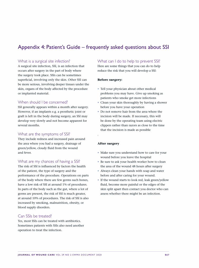
J O U R N A L O F WO U N D C A R E VO L 2 9 N O 2 E W M A D O C U M E N T 2 0 2 0 S 6 7
What is a surgical site infection?A surgical site infection, SSI, is an infection that
occurs after surgery in the part of body where
the surgery took place. SSIs can be sometimes
superficial, involving only the skin. Other SSI can
be more serious, involving deeper tissues under the
skin, organs of the body affected by the procedure
or implanted material.
When should I be concerned?SSI generally appears within a month after surgery.
However, if an implants e.g. a prosthetic joint or
graft is left in the body during surgery, an SSI may
develop very slowly and not become apparent for
several months.
What are the symptoms of SSI?They include redness and increased pain around
the area where you had a surgery, drainage of
green/yellow, cloudy fluid from the wound
and fever.
What are my chances of having a SSI?The risk of SSI is influenced by factors the health
of the patient, the type of surgery and the
performance of the procedure. Operations on parts
of the body where there are few germs such bones,
have a low risk of SSI at around 1% of procedures.
In parts of the body such as the gut, where a lot of
germs are present, the risk of SSI is much greater,
at around 10% of procedures. The risk of SSI is also
increased by smoking, malnutrition, obesity, or
blood supply disorders.
Can SSIs be treated?Yes, most SSIs can be treated with antibiotics.
Sometimes patients with SSIs also need another
operation to treat the infection.
What can I do to help to prevent SSI?Here are some things that you can do to help
reduce the risk that you will develop a SSI:
Before surgery:
• Tell your physician about other medical
problems you may have. Give up smoking as
patients who smoke get more infections
• Clean your skin thoroughly by having a shower
before you have your operation
• Do not remove hair from the area where the
incision will be made. If necessary, this will
be done by the operating team using electric
clippers rather than razors as close to the time
that the incision is made as possible
.
After surgery
• Make sure you understand how to care for your
wound before you leave the hospital
• Be sure to ask your health worker how to clean
the area of the wound 48 hours after surgery
• Always clean your hands with soap and water
before and after caring for your wound.
• If the wound starts to look red, leak green/yellow
fluid, become more painful or the edges of the
skin split apart then contact you doctor who can
assess whether there might be an infection.
Appendix 4: Patient’s Guide – frequently asked questions about SSI

S 6 8 J O U R N A L O F WO U N D C A R E VO L 2 9 N O 2 E W M A D O C U M E N T 2 0 2 0
Glossary
DefinitionsBiologic treatment: a type of treatment that
uses substances made from living organisms to
treat disease. Types of biological therapy include
immunotherapy (such as vaccines, cytokines,
and some antibodies), gene therapy and some
targeted therapies
Cribriform: scars perforated like a sieve
Immunomodulatory therapy: treatment
that modulates the activity of the body’s
immune system
Immunosuppressive therapy: treatment that
lowers the activity of the body’s immune system
Multidisciplinary team: a group of health
professionals who are members of different
disciplines each providing specific services to the
patient. The team members independently treat
various issues a patient may have, focusing on the
issues in which they specialise1
Interdisciplinary team: An interdisciplinary
clinical team is a consistent grouping of people
from relevant clinical disciplines, ideally inclusive
of the patient, whose interactions are guided by
specific team functions and processes to achieve
team-defined favourable patient outcomes2
Pathergy: A skin condition in which a minor
trauma leads to the development of specific
skin lesions
Purpura: Red to dark purple spots in the skin that
do not disappear on compression. Associated with
vessel damage in the dermis
Systemic treatment: Treatment using substances
that travel through the bloodstream, reaching and
affecting cells all over the body
AbbreviationsAAWC: Association for the Advancement of
Wound Care
ABPI: ankle brachial pressure index
ASA: American Society of Anaesthesiologists’
AUC: Area under curve
ARC: Augmented renal clearance
BSR: blood sedimentation rate
ciNPT: Closed incision negative pressure therapy
CABG: coronary artery bypass graft
CAD: coronary artery disease
CDC: Centres for Disease Control and Prevention
CFUs: colony forming units
CHG: chlorhexidine gluconate

J O U R N A L O F WO U N D C A R E VO L 2 9 N O 2 E W M A D O C U M E N T 2 0 2 0 S 6 9
CoNS: coagulase negative staphylococci
CT: computerised tomography
COPD: chronic obstructive pulmonary disease
CRP: C-reactive protein
DNA: Deoxyribonucleic acid
EWMA: European Wound
Management Association
ECDC: European Centre for Disease Prevention
and Control
HBOT: hyperbaric oxygen therapy
HCAI: health care acquired infections
HCW: health care worker
HRQoL: health related quality of life
IPS: Infection Prevention Society
MDT: multidisciplinary team
NICE: National Institute for Health and Care
Excellence
NPWT: negative pressure wound therapy
NHSN: National Healthcare Safety Network
MRI: magnetic resonance imaging
MRSA: Methicillin-resistant Staphylococcus aureus
OR: Odds Ratio
PI: povidone iodine
PK/PD: pharmacokinetic/pharmacodynamic -
this interplay is important for the correct dosing
regimens of antibiotics, to obtain the most optimal
effect (dynamic)
QoL: Quality of life
RCT: randomised controlled trial
SD: standard deviation
SSI: surgical site infection
VRE: vancomycin-resistant enterococci
Wound navigator: usually, a clinician who acts
as an advocate for the patient
WHO: World Health Organization

S 7 0 J O U R N A L O F WO U N D C A R E VO L 2 9 N O 2 E W M A D O C U M E N T 2 0 2 0
Notes

J O U R N A L O F WO U N D C A R E VO L 2 9 N O 2 E W M A D O C U M E N T 2 0 2 0 S 7 1

PREVENTION AND MANAGEMENT ACROSS HEALTH-CARE SECTORS
SURGICAL SITE INFECTION
SU
RG
ICAL S I T E I NFEC
TIO
NS
A JOINT DOCUMENT
268x200_EWMA_Surgical Site Inf_200120.indd 1 20/01/2020 15.58