SURGICAL MANAGEMENT OF BLUNT HEAD...
19
4/27/2015 1 SURGICAL MANAGEMENT OF BLUNT HEAD TRAUMA Mark Duncan, D.O. MMG Neurosurgery CCMH Lawton, OK Learning Objectives: After the lecture, the trauma physician should be better able to: 1. Recognize patients with head trauma. 2. Indentify which patient might need surgical intervention. 3. Determine when surgical intervention is warranted for increasing intracranial pressure TBI EPIDEMIOLOGY:UNITED STATES TBI in the United States and developed countries has declined in recent years Mortality rates from severe TBI declining from 80% in the1950s to about 20% in the past 5 years. Attributed most likely to the combined effects of prevention, evidence-based guidelines that promote early intervention, and improved neurocritical care.
Transcript of SURGICAL MANAGEMENT OF BLUNT HEAD...

4/27/2015
1
SURGICAL MANAGEMENT
OF BLUNT HEAD TRAUMA
Mark Duncan, D.O.
MMG Neurosurgery
CCMH Lawton, OK
Learning Objectives: After the lecture, the
trauma physician should be better able to:
1. Recognize patients with head trauma.
2. Indentify which patient might need surgical
intervention.
3. Determine when surgical intervention is warranted
for increasing intracranial pressure
TBI EPIDEMIOLOGY:UNITED STATES
TBI in the United States and
developed countries has declined in recent years
Mortality rates from severe TBI declining from 80%
in the1950s to about 20% in the past 5 years.
Attributed most likely to the combined effects of
prevention, evidence-based guidelines that
promote early intervention, and improved
neurocritical care.

4/27/2015
2
Head injury causes about
75,000 deaths in the United
States each year. Thus, it has
a great impact on public health,
and continuous improvements
in prevention and care are
needed.
TRAUMA COMA DATA BANK
Death can be expected in approximately 30% to 36% of patients with severe TBI (GCS ≤8)
Good outcome in only 25%.
Gunshot wounds to the head tend to have a worse prognosis
-prehospital mortality rates approaching 90% -mortality rate of at least 60% for those who are alive upon
arrival at a hospital.
TRAUMA COMA DATA BANK
The incidence of TBI is:
• Increasing within the elderly populationElderly population- increase in falls complicated
by anticoagulant use.
• Decreasing for those younger than 45
years.< 45 year - increase motor vehicle safety
(texting, mobile phones???)

4/27/2015
3
DIAGNOSIS
A CT scan ideally should be obtained in all head
injury patients except those who are
completely asymptomatic.
Patients with Glasgow Coma Score (GCS) 14 or
GCS 15 (mild head injury) with documented:
1.loss of consciousness,
2.amnesia,
3.focal neurological deficit,
4.seizures
5.signs of skull fractures
GLASGOW COMA SCALE
Score Best motor response Best verbal response Best eye-opening response6 Obeys — —5 Localizes Oriented —4 Withdraws Confused Spontaneously open3 Flexes (Decorticate) Words/phrases Open to voice2 Extends (Decerebrate) Incomprehensible sounds Open to pain1 No response None Remain closed
Severity of injury• Minor (GCS = 15)
• Mild head injury (GCS = 13-14)
• Moderate head injury (GCS = 9–12)
• Severe head injury (GCS ≤8)
• 25% to 45% of severe TBI cases (GCS ≤8)
• 3% to 12% of moderate TBI cases (GCS = 9–12)
• Approximately 1 in 500 mild TBI cases (GCS 13-
14)
The general principle is prompt evacuation of masslesions that are causing mass effect, neurologic deficits, and poor or worsening Glasgow Coma Scale (GCS) score, in an attempt to LIMIT OR PREVENT THE PROGRESSION OF SECONDARY INJURY.

4/27/2015
4
PREHOSPITAL MANAGEMENT
Main focus is identification and management of
hypoxia and hypotension by reversing conditions
that may jeopardize brain oxygenation.
The basic field principles of airway, breathing, and
circulation remain critical to the management of
patients with cerebral trauma.
PREHOSPITAL MANAGEMENT
Hypoxemia is a strong predictor of poor outcome
and should be addressed immediately with
oxygen supplementation.
*Because of conflicting results from recent studies
demonstrating worse outcomes for patients intubated in the
field, endotracheal intubation should be reserved for patients
who are unable to adequately oxygenate, ventilate, or protect
their airway or in those whose clinical course suggests
that the benefits of intubation outweigh the risks. Contemporary
Neurosurgery: Volume 34; Number 25:Dec 15, 2012
TBI BASIC MEDICAL GUIDELINES
Oxygen saturation kept greater than 90% with normal
breathing rates and end-tidal CO2 levels between 35 and
40 mm Hg.
Hyperventilation with end-tidal CO2 levels below
35 mm Hg should be avoided unless there is evidence of
cerebral herniation.
Routine use of paralytic agents to assist endotracheal intubation
in spontaneously breathing patients should be avoided
Use of induction agents has not been demonstrated to improve outcomes,
although evidence is inconclusive.
The effects of hypoxemia seem to be amplified
when combined with hypotension, resulting in worse
outcomes than those associated with hypoxemia alone.

4/27/2015
5
TBI BASIC MEDICAL GUIDELINES
Hypotension, which is systolic blood pressure less than
90 mm Hg, has been determined to be one of the 5
factors to have at least 70% positive predictive value for
mortality.
Intravascular volume repletion is essential for restoring
blood pressure and thereby cerebral perfusion and
oxygen delivery.
Hypotensive patients should be resuscitated with isotonic
fluids
INITIAL MEDICAL MANAGEMENT OF
SEVERE TBI (GCS ≤8)
1. Head of bed elevation- 30 degrees or higher
2. Keep neck straight/ avoid tight trach tape
3. Avoid hypotension < 90 mm Hg/ control
hypertension
4. Avoid hypoxemia PaO2 <60 mm Hg or O2 sat <
90%
5. Ventilate to normocarbia (PaCO2 35-40 mm Hg)
6. Sedation- Morphine 2-4 mg/1hr /Propofol drip or
Paralysis (Vecuronium 8-10 mg IV)
MEDICAL MANAGEMENT
7. Routine Steroids is not recommended for treatment of
head injury
8. Head CT without contrast
9. Mannitol 0.25-1 gm/kg q6h (serum osmol <320), Hold If
hypotensive (SBP<90 mmHg) or hypovolemia
10. Seizure prophylaxis Yes or No??? Grey Zone.
11. Cerebrospinal fluid (CSF) drainage- Ventriculostomy
(ICP)
12. Do not hyperventilate unless ongoing brain herniation-if
indicated PaCO2 = 25-30 mmHg, <25 mmHg mm Hg
Avoid at all times

4/27/2015
6
BP Guidelines
Hypertension should be treated only if mean arterial pressure is > 130 mm Hg or systolic BP is > 185 mm Hg.
Nicardipine (Cardene)
2.5 mg/h IV is given initially; dose is increased by 2.5 mg/h q 5 min to a maximum of 15 mg/h as needed to decrease systolic BP by 10 to 15%.
HIGH-RISK CRITERIA FOR POST-TRAUMATIC SEIZURES*
• Acute SDH, EDH, or ICH
• Open–depressed skull fracture with brain
injury
• Seizure within 24 hours of injury
• Glasgow Coma Scale score 10
• Penetrating brain injury
• Significant ethanol abuse history
• + /- Cortical contusion on head CT
EARLY POST-TRAUMATIC SEIZURES
In patients at high risk for seizure activity especially
comatose and neurologic examinations are unreliable:
• EEG monitoring is recommended
• administration of phenytoin within 24 hours of injury and continuing for 7
days to prevent the occurrence of early seizures.
Phenytoin should be loaded at 20 mg/kg followed by a maintenance dose to
ensure high therapeutic levels between 10 and 20 mcg/mL.
***Continuation beyond 7 days is not recommended unless:
• penetrating injury
• undergone craniotomy
• history of seizures
• experiences repeated seizures

4/27/2015
7
SURGICAL AND NEUROCRITICALCARE
The main objective of surgical and neurocritical care is to
anticipate the evolution of secondary brain injury and
prevent its deleterious
effects.
SURGICAL CARE: PREVENTING SECONDARY BRAIN INJURY
Need for Ventriculostomy or placing an intraparenchymal
monitor in patients with GCS score of 8 or less to measure
ICP.
The goal has been to maintain ICP below and CPP above
specific thresholds to ensure adequate cerebral perfusion and
oxygenation. Although there is consensus for treating persistently
elevated ICP higher than 20 mm Hg, CPP management
has been less straightforward.
Some studies have demonstrated improved outcomes with CPP higher than
70 mm Hg, whereas others have not and have revealed high
rates of pulmonary complications such as acute respiratory
distress syndrome.
ICP
MONITOR/VENTRICULOSTOMY
4/27/2015
8
NEUROCRITICAL CARE
According to Current Brain Trauma Foundation
guidelines,the critical threshold below which CPP
should not fall below is 50 mm Hg, but artificially
driving CPP (MAP-ICP) higher than 70 mm Hg
has fallen out of favor.
*Avoid aggressive use of fluids and pressors to
maintain CPP > 70 mmHG ( risk of adult
respiratory distress syndrome (ARDS) )
CURRENT TRENDS
Prophylactic hypothermia: If used hold at target temp 32-35 o C (91.4-95o F) > 48 hrs ( reduce CMRO2 –efficacy not proven: Brain Trauma Foundation: Prophylactic hypothermia. J Neurotrauma 24: S21-5,2007
Hypertonic Saline: May reduce ICP in patients refractory to mannitol, although no improvement in outcome over mannitol has been demonstrated. J Trauma 44 (1): 50-58, 1998
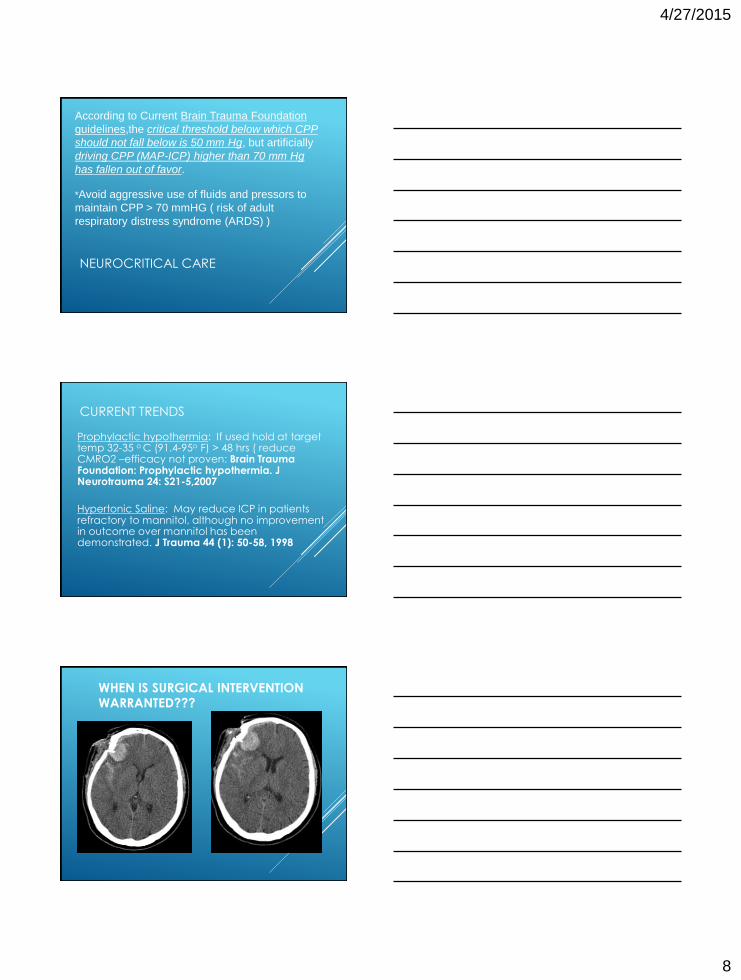
WHEN IS SURGICAL INTERVENTION WARRANTED???

4/27/2015
9
TRAUMATIC INTRACRANIAL
HEMORRHAGE
Guidelines can be useful, but they may be a simplification
of a complicated and, sometimes, unpredictable clinical
situation. The most critical factors in deciding whether
to operate on a traumatic intracranial hematoma are :
1.Patient’s neurological status (GCS)
2.Imaging findings (Head CT- mass effect, midline shift)
3.Presence and severity of extracranial lesions.
NON-OPERATIVE
MANAGEMENT
In a patient with a small hematoma
causing less than 5 mm shift who is
neurologically intact, a conservative approach is justified. However, the patient
may deteriorate rapidly, so very close
observation is vital.
4/27/2015
4/27/2015
There is general agreement that an urgent operative course is indicated in a rapidly deteriorating patient with an expanding intracranial hematoma that is causing significant mass effect.
Significant mass effect may be arbitrarily defined as
displacement of midline structures more than 5 mm or
effacement of basal cisterns on CT scan (or both).

4/27/2015
10
In general, surgical intervention is decided more readily
for intracranial hemorrhages located:
• Temporal lobe
• Posterior fossa lesions
In these locations small lesion may lead to compression
and irreversible brain stem damage within a shorter
period of time.
TRAUMATIC INTRACRANIAL
HEMORRHAGE CLASSIFICATION
•Epidural Hematoma (EDH)
•Subdural Hematoma (SDH)
•Traumatic Brain Injury
•Cerebral Contusion/Hemorrhagic Contusion
•Traumatic SAH
•Skull fracture
•Penetrating injury- GSW
•Diffuse Axonal Injury- rotational deceleration/acceleration injury-nonsurgical
ANATOMY OF SDH VS EDH
4/27/2015

4/27/2015
11
EDH
EPIDURAL HEMATOMA (EDH)
84% cases have Classic appearance
High density biconvex shape
Textbook presentation < 10-27%
Brief posttraumatic LOC
Followed by “lucid Interval” for several hours
Then, obtundation, contralateral hemiparesis, ipsilateral pupillary dilatation
Untreated: Decerebrateposturing, HTN, respiratory failure and death
ACUTE EDH
Acute Epidural Hematoma
• Surgical evacuation should occur as soon as possible if GCS score is GCS ≤8, midline shift is > 5 mm and the patient has anisocoria.
• If the thickness is < 15 mm with < 5 mm midline shift in a patient with GCS score higher than 8 without focal deficit, the acute epidural hematoma may also be managed
non-operatively.
4/27/2015

4/27/2015
12
EDH MANAGEMENT
50% of cases there will be a slight
transient increase in size between
days 5-16 and some patients
required emergency craniotomy
Close Neuro observation if
nonsurgical
Serial Head CT’S
EDH
Nonsurgical
Small < 1cm max thickness
Subacute or Chronic
Minimal neurologic signs/symptoms
HA, lethargy
No mass effect, no herniation
SDH

4/27/2015
13
SUBDURAL HEMATOMA (SDH)
Acute/Chronic
Midline shift > 5 mm midline shift
Effacement of 3rd and posterior horn of right lateral ventricle
NONSURGICAL SDH
Subdural often do not need surgical intervention **
If asymptomatic or mild headache Maintain BP < 165/90
Stop all NSAIDS, Anticoagulants
Medical Management

4/27/2015
14
ACUTE SDH SURGICAL INDICATION
Surgical evacuation should occur when
the hematoma thickness is greater than
10 mm or there is a midline shift more
than 5 mm regardless of GCS score.
Symptomatic-lethargy, hemiparesis etc
Patients with a GCS score higher than 9
or above does not meet the earlier criteria
should undergo surgical evacuation if:• GCS score declines by 2 or more points
• patient develops anisocoria (unequal pupils)
• and/or ICP exceeds 20 mm Hg
CEREBRAL (HEMORRHAGIC) CONTUSION
High density area on CT
Most commonly occur in areas where sudden deceleration of the head causes the brain to impact a bony prominences
Coup or Contrecoup (Counter Blow) injury
Temporal
Frontal
Occipital
Majority
nonsurgical

4/27/2015
15
CONTUSION OR ICH
The decision to remove an intracerebral
hematoma or cerebral contusion should
be based on several key factors, including
(but not limited to):
• Blood Volume of the lesion > 20 cm3
• Depth and location of the lesion
in relation to eloquent brain areas
• Patient’s neurological status
• ICP > 20 mmHg with medical
mangement
•Most are nonsurgical
4/27/2015
TRAUMATIC ICH
Elderly with history of fall on anticoagulants
Delayed ICH Incidence is 10%
Most occur within 72 hrs of trauma
“Talk and deteriorate” only 12%
Poor outcome: mortality ranging form 50-75%
Common with coagulopathy
Plavix- give platelets
Coumadin-FFP and Vit. K
4/27/2015
16
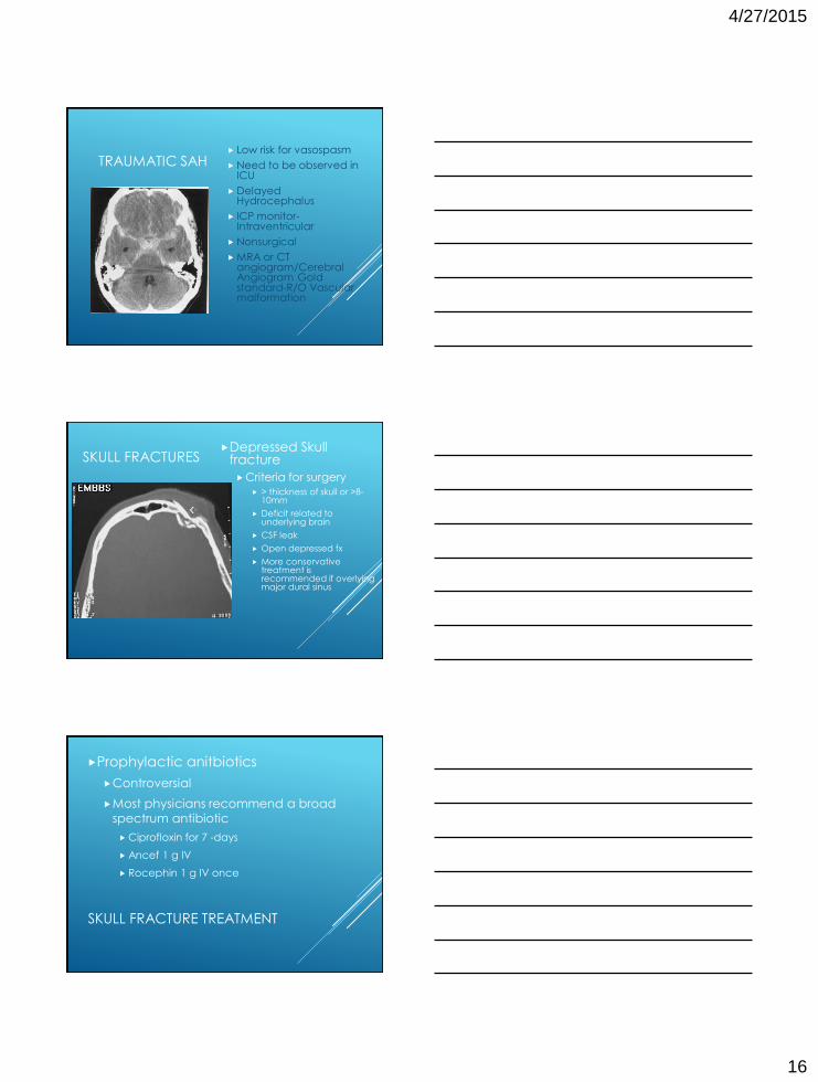
TRAUMATIC SAH Low risk for vasospasm
Need to be observed in ICU
Delayed Hydrocephalus
ICP monitor-Intraventricular
Nonsurgical
MRA or CT angiogram/Cerebral Angiogram Gold standard-R/O Vascular malformation
SKULL FRACTURESDepressed Skull
fracture
Criteria for surgery
> thickness of skull or >8-10mm
Deficit related to underlying brain
CSF leak
Open depressed fx
More conservative treatment is recommended if overlying major dural sinus
SKULL FRACTURE TREATMENT
Prophylactic anitbiotics
Controversial
Most physicians recommend a broad
spectrum antibiotic
Ciprofloxin for 7 -days
Ancef 1 g IV
Rocephin 1 g IV once

4/27/2015
17
SKULL FRACTURE WITH UNDERLYING HEMATOMA (VENOUS VS ARTERIAL)
CEREBELLAR HEMATOMA
Surgical emergency
If > 3 cm needs emergent surgery
Neuro status: in question intubate
Maintain SBP <185

4/27/2015
18
State of the Art in the Treatment of Cerebral Trauma: Part I
Keen, Joseph R.; Colohan, Austin R.T.
Contemporary Neurosurgery. 34(25):1-7, December 15, 2012.
doi: 10.1097/01.CNE.0000423005.25206.2c
Figure 1. Stepwise therapy for increased ICP.
State of the Art in the Treatment of Cerebral Trauma: Part II
Keen, Joseph R.; Colohan, Austin R.T.
Contemporary Neurosurgery. 34(26):1-6, December 31, 2012.
doi: 10.1097/01.CNE.0000423866.00314.4e
Figure 1. Decompressive craniectomy techniques. The 2 main decompressive techniques include unilateral hemicraniectomy and bifrontal craniectomy, depending on the presence, location, and extent of mass lesions, penetrating injuries, and midline shift.
Decompressive Craniectomy trial
(DECRA), published in the New England
Journal of Medicine 2011, demonstrated
higher rates of unfavorable
outcome compared with standard
medical management.
Cooper DJ, Rosenfeld JV, Murray L, et al. Decompressive
craniectomy in diffuse traumatic brain injury. N Engl J Med.
2011;364(16):1493-1502.

4/27/2015
19
The goal in managing a patient with
cerebral trauma is to anticipate the
evolution of secondary brain injury
and intervene early, before its
progression.