Surgical management of an impacted sharp metallic foreign body in ...
4
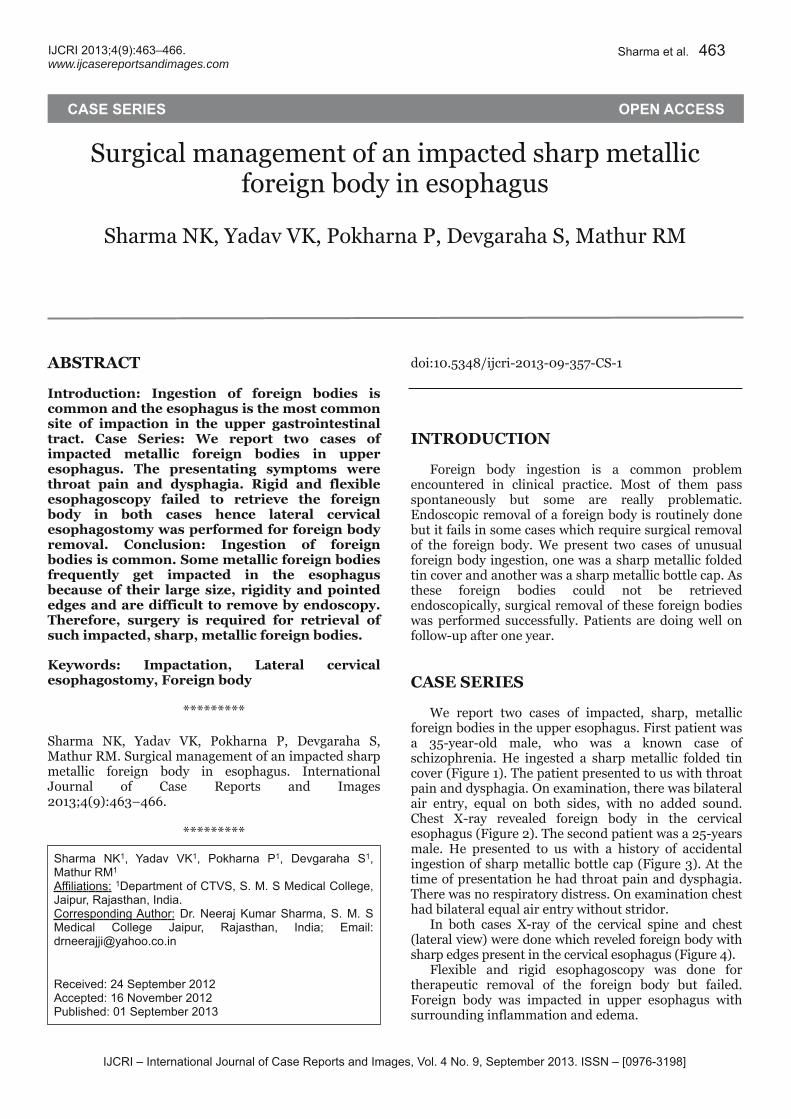
www.ijcasereportsandimages.com Surgical management of an impacted sharp metallic foreign body in esophagus Sharma NK, Yadav VK, Pokharna P, Devgaraha S, Mathur RM ABSTRACT Introduction: Ingestion of foreign bodies is common and the esophagus is the most common site of impaction in the upper gastrointestinal tract. Case Series: We report two cases of impacted metallic foreign bodies in upper esophagus. The presentating symptoms were throat pain and dysphagia. Rigid and flexible esophagoscopy failed to retrieve the foreign body in both cases hence lateral cervical esophagostomy was performed for foreign body removal. Conclusion: Ingestion of foreign bodies is common. Some metallic foreign bodies frequently get impacted in the esophagus because of their large size, rigidity and pointed edges and are difficult to remove by endoscopy. Therefore, surgery is required for retrieval of such impacted, sharp, metallic foreign bodies. Keywords: Impactation, Lateral cervical esophagostomy, Foreign body ********* Sharma NK, Yadav VK, Pokharna P, Devgaraha S, Mathur RM. Surgical management of an impacted sharp metallic foreign body in esophagus. International Journal of Case Reports and Images 2013;4(9):463–466. ********* doi:10.5348/ijcri201309357CS1 INTRODUCTION Foreign body ingestion is a common problem encountered in clinical practice. Most of them pass spontaneously but some are really problematic. Endoscopic removal of a foreign body is routinely done but it fails in some cases which require surgical removal of the foreign body. We present two cases of unusual foreign body ingestion, one was a sharp metallic folded tin cover and another was a sharp metallic bottle cap. As these foreign bodies could not be retrieved endoscopically, surgical removal of these foreign bodies was performed successfully. Patients are doing well on followup after one year. CASE SERIES We report two cases of impacted, sharp, metallic foreign bodies in the upper esophagus. First patient was a 35yearold male, who was a known case of schizophrenia. He ingested a sharp metallic folded tin cover (Figure 1). The patient presented to us with throat pain and dysphagia. On examination, there was bilateral air entry, equal on both sides, with no added sound. Chest Xray revealed foreign body in the cervical esophagus (Figure 2). The second patient was a 25years male. He presented to us with a history of accidental ingestion of sharp metallic bottle cap (Figure 3). At the time of presentation he had throat pain and dysphagia. There was no respiratory distress. On examination chest had bilateral equal air entry without stridor. In both cases Xray of the cervical spine and chest (lateral view) were done which reveled foreign body with sharp edges present in the cervical esophagus (Figure 4). Flexible and rigid esophagoscopy was done for therapeutic removal of the foreign body but failed. Foreign body was impacted in upper esophagus with surrounding inflammation and edema.
Transcript of Surgical management of an impacted sharp metallic foreign body in ...
IJCRI – International Journal of Case Reports and Images, Vol. 4 No. 9, September 201 3. ISSN – [0976-31 98]
IJCRI 201 3;4(9):463–466.www.ijcasereportsandimages.com
Surgical management of an impacted sharp metallicforeign body in esophagusSharma NK, Yadav VK, Pokharna P, Devgaraha S, Mathur RM
ABSTRACTIntroduction: Ingestion of foreign bodies iscommon and the esophagus is the most commonsite of impaction in the upper gastrointestinaltract. Case Series: We report two cases ofimpacted metallic foreign bodies in upperesophagus. The presentating symptoms werethroat pain and dysphagia. Rigid and flexibleesophagoscopy failed to retrieve the foreignbody in both cases hence lateral cervicalesophagostomy was performed for foreign bodyremoval. Conclusion: Ingestion of foreignbodies is common. Some metallic foreign bodiesfrequently get impacted in the esophagusbecause of their large size, rigidity and pointededges and are difficult to remove by endoscopy.Therefore, surgery is required for retrieval ofsuch impacted, sharp, metallic foreign bodies.Keywords: Impactation, Lateral cervicalesophagostomy, Foreign body
*********Sharma NK, Yadav VK, Pokharna P, Devgaraha S,Mathur RM. Surgical management of an impacted sharpmetallic foreign body in esophagus. InternationalJournal of Case Reports and Images2013;4(9):463–466.
*********
doi:10.5348/ijcri201309357CS1
INTRODUCTIONForeign body ingestion is a common problemencountered in clinical practice. Most of them passspontaneously but some are really problematic.Endoscopic removal of a foreign body is routinely donebut it fails in some cases which require surgical removalof the foreign body. We present two cases of unusualforeign body ingestion, one was a sharp metallic foldedtin cover and another was a sharp metallic bottle cap. Asthese foreign bodies could not be retrievedendoscopically, surgical removal of these foreign bodieswas performed successfully. Patients are doing well onfollowup after one year.
CASE SERIESWe report two cases of impacted, sharp, metallicforeign bodies in the upper esophagus. First patient wasa 35yearold male, who was a known case ofschizophrenia. He ingested a sharp metallic folded tincover (Figure 1). The patient presented to us with throatpain and dysphagia. On examination, there was bilateralair entry, equal on both sides, with no added sound.Chest Xray revealed foreign body in the cervicalesophagus (Figure 2). The second patient was a 25yearsmale. He presented to us with a history of accidentalingestion of sharp metallic bottle cap (Figure 3). At thetime of presentation he had throat pain and dysphagia.There was no respiratory distress. On examination chesthad bilateral equal air entry without stridor.In both cases Xray of the cervical spine and chest(lateral view) were done which reveled foreign body withsharp edges present in the cervical esophagus (Figure 4).Flexible and rigid esophagoscopy was done fortherapeutic removal of the foreign body but failed.Foreign body was impacted in upper esophagus withsurrounding inflammation and edema.
CASE SERIES OPEN ACCESS
Sharma NK1 , Yadav VK1 , Pokharna P1 , Devgaraha S1 ,Mathur RM1
Affi l iations: 1Department of CTVS, S. M. S Medical College,Jaipur, Rajasthan, India.Corresponding Author: Dr. Neeraj Kumar Sharma, S. M. SMedical College Jaipur, Rajasthan, India; Email :drneerajj [email protected]. in
Received: 24 September 201 2Accepted: 1 6 November 201 2Published: 01 September 201 3
Sharma et al. 463

IJCRI – International Journal of Case Reports and Images, Vol. 4 No. 9, September 201 3. ISSN – [0976-31 98]
IJCRI 201 3;4(9):463–466.www.ijcasereportsandimages.com Sharma et al. 464
Emergency surgery was planned. Longitudinalincision was given on the left side of neck in front ofsternocleidomastoid. Carotid artery and thyroid glandwere retracted. Esophagus was found to be inflammedand edematous. Longitudinal incision was given onesophagus over impacted foreign body and foreign bodywas gently disimpacted and removed to prevent furtherinjury to esophagus. Esophagus was repaired using 3.0vicryl interrupted sutures in two layers, with Ryle’s tubeleft in situ for three weeks. Postoperative period was
Figure 1: Folded metallic tin cover removed from esophagus(case 1).
Figure 2: Bottle cap removed from esophagus (case 2).
Figure 3: Chest Xray showing foreign body in the cervicalesohagus.
Figure 4: Lateral chest Xray showing foreign body in theupper esophagus.
IJCRI – International Journal of Case Reports and Images, Vol. 4 No. 9, September 201 3. ISSN – [0976-31 98]
IJCRI 201 3;4(9):463–466.www.ijcasereportsandimages.com Sharma et al. 465
uneventful and both the patients are doing well onfollowup.
DISCUSSIONIt is estimated that up to 90% of all foreign bodiespass spontaneously from the esophagus. Endoscopicmanagement is needed in less than 10% cases, whereassurgery is required for foreign body retrieval ormanagement of complications in approximately 1%patients [1–3]. In both our cases, the patients requiredlateral cervical esophagostomy, after failed endoscopicretrieval.Although most of the foreign bodies in thegastrointestinal tract are swallowed accidentally, otherconditions such as imprisonment, mental illness,mental retardation, bulimia, alcohol consumption ordrug abuse may also be involved, particularly in theWestern countries [4]. The majority of foreign bodyingestions occur in the pediatric population with a peakincidence between six months and six years of age[2–8]. Edentulous adults are also at greater risk forforeign body ingestion, including that of dentalprosthesis [8, 9]. One of our cases had an accidentalingestion of bottle cap while another had a history ofmental illness.Esophageal objects can cause a foreign bodysensation, drooling, respiratory distress due to trachealcompression, gagging, dysphonia, vomiting, anddysphagia, depending on the location and nature of theforeign body [10]. Although rare, perforating objects arepotentially life threatening because they may lead to theformation of a fistula between the esophagus and theinnominate artery thus causing catastrophic bleeding[11, 12]. Young children and those with mental illnessmay present with choking, refusal to eat, vomiting,drooling, wheezing, bloodstained saliva, or respiratorydistress [5, 7, 13, 14]. Our patients had symptoms ofthroat pain and dysphagia.The foreign bodies usually lie close to one of thethree esophageal anatomical constrictions: thecricopharyngeal ring, the aortic arch narrowing or theesophagogastric junction [15, 16]. The most frequentlodgement site of described in literature is thecricopharyngeus muscle [11, 12].Plain films (cervical and chest Xrays) are a veryimportant diagnostic tool, especially in defining thelocation of the foreign body [10]. A bariumswallow Xray study could be useful in cases of nonradioopaqueforeign body, but due to possible barium aspiration andor irritation of the damaged esophageal mucosa, thisprocedure is no longer used [17–19]. Computedtomography scans can be used to confirm the presenceand location of the foreign body (especially in the casesof fish bones), and to evaluate any eventual damage tothe neighboring structures [18, 19].In our cases, it was the clinical history, Xrays andendoscopy that guided us in diagnosing and definingthe location of the foreign body.The treatment of choice for esophageal forein bodies
depends on various parameters such as patient’s age,clinical condition; the type, size, shape, site andnumber of foreign bodies. [10, 20] Endoscopy is thepreferred method with a reported success rate of 83%[20]. Today, both rigid or flexible endoscopy, performedunder general anesthesia or conscious sedation,respectively, are considered to be safe and representeffective methods in experienced hands. For themanagement of sharp and penetrating foreign bodies,rigid endoscopy is often the treatment of choice. Majorrisk during esophagoscopy maneuvers include directinstrumental wounds and perforations [16]. Surgicaltreatment is unavoidable in cases of irretrievableforeign body or esophageal perforation [10].In our cases, flexible and rigid endoscopies wereunsuccessful in the removal maneuvers mainly due tothe size and shape of the foreign bodies. Further, theforeign bodies were impacted in esophagus thereforesurgical treatment was necessary.
CONCLUSIONIngestion of foreign bodies end their lodgment in theesophagus are common. Some metallic foreign bodiesget frequently impacted in the esophagus and dosedifficulty in endoscopic retrieval due to their large size,rigidity and pointed edges. Therefore in such casessurgery is required for retrieval of the impacted sharpmetallic foreign bodies.
*********Author ContributionsSharma NK – Substantial contributions to conceptionand design, Analysis and interpretation of data,Drafting the article, Revising it critically for importantintellectual content, Final approval of the version to bepublishedYadav VK – Substantial contributions to conception anddesign, Acquisition of data, Drafting the article,Revising it critically for important intellectual content,Final approval of the version to be publishedPokharna P – Substantial contributions to conceptionand design, Acquisition of data, Revising it critically forimportant intellectual content, Final approval of theversion to be publishedDevgaraha S – Analysis and interpretation of data,Revising it critically for important intellectual content,Final approval of the version to be publishedMathur RM – Analysis and interpretation of data,Revising it critically for important intellectual content,Final approval of the version to be publishedGuarantorThe corresponding author is the guarantor ofsubmission.Conflict of InterestAuthors declare no conflict of interest.
IJCRI – International Journal of Case Reports and Images, Vol. 4 No. 9, September 201 3. ISSN – [0976-31 98]
IJCRI 201 3;4(9):463–466.www.ijcasereportsandimages.com Sharma et al. 466
Copyright© Sharma NK et al. 2013; This article is distributedunder the terms of Creative Commons Attribution 3.0License which permits unrestricted use, distribution andreproduction in any means provided the original authorsand original publisher are properly credited. (Please seewww.ijcasereportsandimages.com/copyrightpolicy.phpfor more information.)
REFERENCES1. Eisen GM, Baron TH, Dominitz JA, et al. Guidelinefor the management of ingested foreign bodies.Gastrointest Endosc 2002;55(7):802–6.2. Webb WA. Management of foreign bodies of theupper gastrointestinal tract: update. GastrointestEndosc 1995;41(1):39–51.3. Chaves DM, Ishioka S, Félix VN, Sakai P, GamaRodrigues JJ. Removal of a foreign body from theupper gastrointestinal tract with a flexibleendoscope: a prospective study. Endoscopy2004;36(10):887–92.4. Lai AT, Chow TL, Lee DT, Kwok SP. Risk factorspredicting the development of complications afterforeign body ingestion. Br J Surg 2003Dec;90(12):1531–5.5. Cheng W, Tam PK. Foreignbody ingestion inchildren in children: experience with 1,265 cases. JPediatr Surg 1999;34(10):1472–6.6. Panieri E, Bass DH. The management of ingestedforeign bodies in children—a review of 663 cases.Eur J Emerg Med 1995;2(2):83–7.7. HachimiIdrissi S, Corne L, Vandenplas Y.Management of ingested foreign bodies inchildhood: our experience and review of theliterature. Eur J Emerg Med 1998;5(3):319–23.8. Blaho KE, Merigian KS, Winbery SL, Park LJ,Cockrell M. Foreign body ingestions in theemergency department: case reports and review oftreatment. J Emerg Med 1998;16(1):21–6.9. Abdullah BJ, Teong LK, Mahadevan J, Jalaludin A.Dental prosthesis ingested and impacted in the
esophagus and orolaryngopharynx. J Otolaryngol1998;27(4):190–4.10. Athanassiadi K, Gerazounis M, Metaxas E, KalantziN. Management of esophageal foreign bodies: aretrospective review of 400 cases. Eur J CardiothoracSurg 2002;21(4):653–6.11. Byard RW. Esophageal causes of sudden andunexpected death. J Forensic Sci 2006;51(2):390–5.12. Tokar B, Cevik AA, Ilhan H. Ingested gastrointestinalforeign bodies: predisposing factors forcomplications in children having surgical orendoscopic removal. Pediatric Surgery International2007;23(2)135–9.13. Kamal I, Thompson J, Paquette DM. The hazards ofvinyl glove ingestion in the mentally retarded patientwith pica: new implications for surgicalmanagement. Can J Surg 1999;42(3):201–4.14. Chowdhury CR, Bricknell MC, MacIver D.Oesophageal foreign body: an unusual cause ofrespiratory symptoms in a threeweekold baby. JLaryngol Otol 1992;106(6):556–7.15. von Rahden BH, Feith M, Dittler HJ, Stein HJ.Cervical esophageal perforation with severemediastinitis due to an impacted dental prosthesis.Dis Esophagus 2002;15(4):340–4.16. Karaman A, Cavusoglu YH , Karaman I, Erdogan D,Aslan MK, Cakmak O. Magill forceps technique forremoval of safety pins in upper esophagus: apreliminary report. Int J Pediatr Otorhinolaryngol2004;68(9):1189–1.17. AlQudah A, Daradkeh S, AbuKhalaf M. Esophagealforeign bodies. Eur J Cardiothorac Surg1998;13(5):494–8.18. Akazawa Y, Watanabe S, Nobukiyo S, et al. Themanagement of possible fishbone ingestion. AurisNasus Larynx 2004;31(4):413–6.19. de Lucas EM, RuizDelgado ML, GarcíaBarón PL,Sádaba P, Pagola MA. Foreign esophageal bodyimpaction: multimodality imaging diagnosis. EmergRadiol 2004;10(4):216–7.20. Furihata M, Tagaya N, Furihata T, Kubota K.Laparoscopic removal of an intragastric foreign bodywith endoscopic assistance. Surg Laparosc EndoscPercutan Tech 2004;14(4):234–7.
Access full text article onother devices Access PDF of article onother devices