surgical manag of colorectal liver mets
107
Surgical management of Colorectal liver metastasis Dr Dharma Poonia
-
Upload
dharma-punia -
Category
Healthcare
-
view
84 -
download
0
Transcript of surgical manag of colorectal liver mets
Surgical management of Colorectal liver metastasis
Dr Dharma Poonia
• Background, epidemiology.
• Patient selection and assessment.
• Prognostic variables.
• Define resectability.
• Methods to increase resectability.
• Adjunct methods.
• Recent development.
Background, epidemiology.
• It is 3rd MC cancer in men and 2nd MC cancer in women in western world.
• 80% of patients with colorectal cancer (CRC) present with local or regional disease.
• For these patients, the general plan of treatment is clear: surgery with the intent of cure.
• Remaining 20% of newly diagnosed patients continue to present with synchronously diagnosed stage 4 disease.
• 25% patients with primary colorectal carcinoma present with synchronous hepatic metastasis, and 50% of the patients will eventually develop metachronous liver metastasis
» (Bozzetti et al, 1987; Ekberg et al, 1987).
• For these patients, the treatment plan is less obvious.
• Despite their advanced stage of disease, a subset of stage 4 patients are potentially curable.
• The liver is the most common site for hematogenous metastasis from colorectal cancers (CRCs).
• In patients with isolated hepatic metastases, the extent of liver disease is the principal determinant of survival, and when left untreated, survival is measured in months
» (Norstein & Silen, 1997).
• Following resection for CRLM, 5 years OS is 40%, with a 10-year survival approaching 20%.
• No PRTCs comparing liver resection to systemic, regional, or other local therapies have been performed, the outcome for patients after liver resection for metastatic CRC is sufficiently favorable that surgery is now considered the primary therapy in selected patients
» Pawlik & Choti, 2007; Ito et al, 2010).
• With newer multimodal treatments and careful patient selection, it is anticipated that 5-year survival approaching 70% can be achieved after resection and comparable outcomes are likely to be reported in larger studies in the near future.
» (Nikfarjam et al, 2009)
What are the options for patients with colorectal liver metastasis?
• Do nothing– median survival of 6 to 9 months.
• Chemotherapy – 14.5-month median survival,
• RFA – 40% 3-year survival rate,
• Resection– 5-year survival rate of 45% to 60%

Patient selection & assessment
• Standard staging by AJCC did not provide prognostic informations.
• Therefore, a classification system that can discriminate between these patients and provide additional prognostic information is paramount.
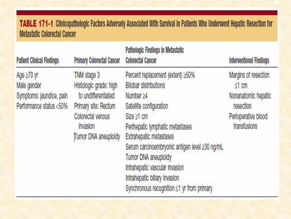
• Prognostic variables
1) clinical and pathologic variables associated with the primary tumor;
2) clinical variables associated with the liver metastases, such as response to neoadjuvantchemotherapy; and
3) pathologic characteristics of the liver metastases.
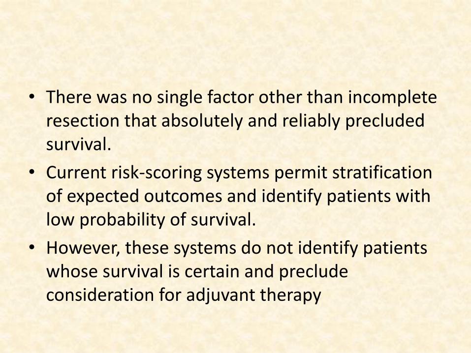
• There was no single factor other than incomplete resection that absolutely and reliably precluded survival.
• Current risk-scoring systems permit stratification of expected outcomes and identify patients with low probability of survival.
• However, these systems do not identify patients whose survival is certain and preclude consideration for adjuvant therapy
Predictive Models and Clinical Risk Scores
• Four large studies on multivariate analyses of prognostic factors designed a of useful predictive models for favorable survival after metastasectomy.
• Nordlinger et al, 1996;
• Fong et al, 1999;
• Kattan et al, 2008;
• Rees et al, 2008
• In the series by Fong and colleagues (1999), independent predictors of unfavorable prognosis:
1) the presence of extrahepatic disease,
2) a positive resection margin,
3) Node positive primary CRC,
4) a short disease-free interval(<1year)
5) largest liver metastasis greater than 5 cm,
6) more than one liver metastasis, and
7) serum CEA greater than 200 ng/mL.
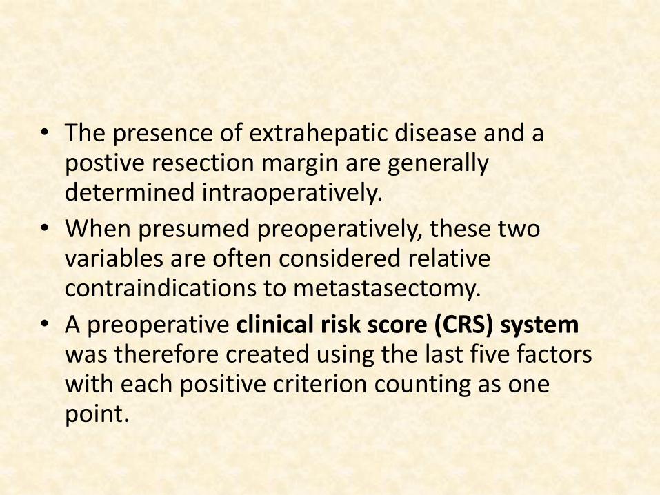
• The presence of extrahepatic disease and a postive resection margin are generally determined intraoperatively.
• When presumed preoperatively, these two variables are often considered relative contraindications to metastasectomy.
• A preoperative clinical risk score (CRS) system was therefore created using the last five factors with each positive criterion counting as one point.
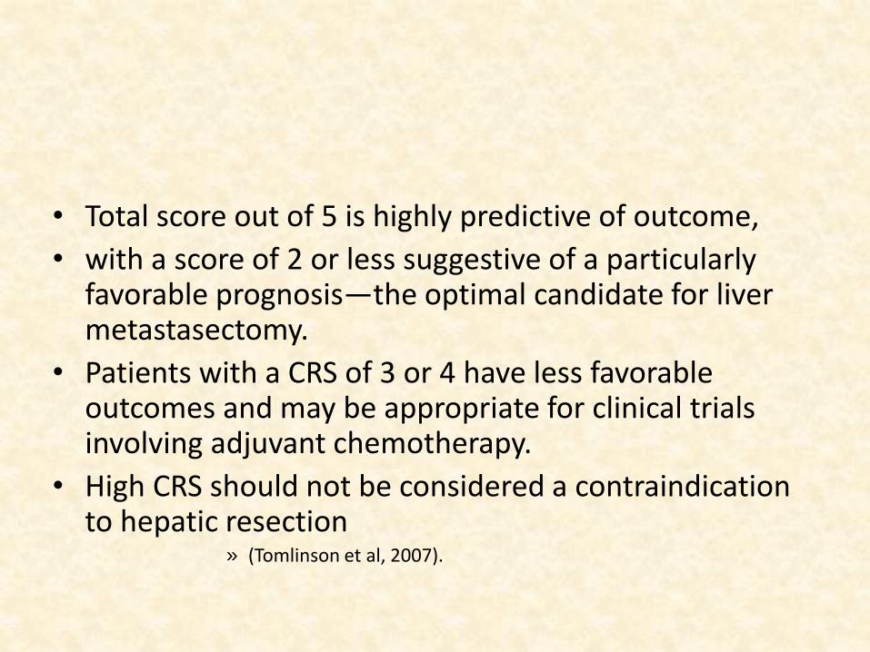
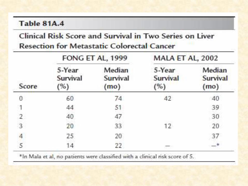
• Total score out of 5 is highly predictive of outcome,
• with a score of 2 or less suggestive of a particularly favorable prognosis—the optimal candidate for liver metastasectomy.
• Patients with a CRS of 3 or 4 have less favorable outcomes and may be appropriate for clinical trials involving adjuvant chemotherapy.
• High CRS should not be considered a contraindication to hepatic resection
» (Tomlinson et al, 2007).
Use of clinical risk score system
• It is validated by an independent group from Norway (Mala et al, 2002),
• The CRS is generalizable to populations outside of the index cohort from a single, large, tertiary
• In addition to appropriate patient selection for surgery, the CRS has proven useful in selecting patients for – neoadjuvant treatment
– preoperative evaluation, ablation, and
– stratification in clinical trials.
• DL should be indicated in CRS>2, as risk of occult extrahepatic disease is around 40%.
» Jarnagin et al 2001
• PET CT should be advised for CRS1 or more, as chances of occult mets >14% in these patients
» Schussler fiorenza et al 2004
• Predictive models with added sophistication have been developed recently, such as a
– nomogram (Kattan et al, 2008) and
– a multifactorial predictive index (Rees et al, 2008).
• These models are based on the same basic prognostic factors as the earlier models described above,
• They are more complex and difficult to use;
Pre op assesment
• 1) establishing the diagnosis and characterization of primary.
• 2) anatomic definition of the liver lesion for surgical planning,
• 3) staging to rule out extrahepatic disease.
• 4) general fitness for surgical resection. – A confirmatory biopsy of hepatic lesions is only
indicated to confirm the diagnosis when the clinical picture is unclear
Imaging
Should address the following five critical issues:
1. Evaluation of liver metastases.
2. Possible hilar lymph node involvement.
3. Vascular invasion.
4. Liver volumetry.
5. Presence of extrahepatic disease.
(Valls et al. 2009).
• CT scan
• MRI abdomen
• USG CEUS,
• PET CT
• IOUS
USG
• US is a rapid and non-invasive method
• operator dependent.
• Its sensitivity (50-70%)
• surpassed by other imaging studies.» (Oldenburg & Albrecht 2008).
CEUS
• CEUS sensitivity and specificity in staging liver metastases (80–95% and 84–98%, respectively) approach those of CT and MRI.
• It is useful to improve the detection rate of metastases smaller than 1 cm or of those lesions that are isoechoic with respect to adjacent liver parenchyma,
• (Oldenburg & Albrecht 2008).
CEUS
• Limited ability to observe certain parts of the liver,
• Obese patients and/or in cases of steatosis and it is not possible to simultaneously examine multiple lesions in the arterial and early portal phases.
• Hypervascular metastases and haemangiomas on one hand and metastases and small cysts on the other can be difficult to differentiate
» (Larsen 2010).
CT
• MDCT has a sensitivity of 70–85% and a specificity of 90%, especially for lesions bigger than 1.5–2 cm.
• Sensitivity is lower for small subglissonianmetastases, even though multi-slice CT allows identification of hepatic lesions of 0.5 cm in size
» (Guglielmi et al. 2005).
CT
• Hepatic volumetry, necessary to evaluate the feasibility of major hepatectomies.
• In the case of atypical resections, CT software able to highlight different liver segments and to create vascular maps for arterial and portal afferences, and for hepatic vein drainage
» Laghi et al 2005
MRI
• MDCT is usually preferred because it is more widely available and because it is a well established technique for surveying the extrahepatic abdominal organs and tissues.
• characterization of focal lesions and • is also preferred for patients who cannot receive
intravenous iodinated contrast material.• when concerns about the risk of radiation from
repeated exposure to CT, as in children or young adults, exists.
• In general, MRI sensitivity varies from 85-90% and its specificity is up to 95%,
FDG PET
• FDG PET is a highly sensitive and specific imaging study detecting hepatic metastases from CRC (92–100% and 85–100% respectively).
• With regard to the initial staging of patients with metastatic CRC, FDG PET imaging leads to a change in management in 2% to 36% of patients.
» (Lucey et al. 2006).
• CT scanning remains a dominant imaging modality not only for lesion detection and preoperative planning, but also for treatment monitoring and post-treatment surveillance.
• FDG PET/CT may obviate the need for additional studies and may improve patient management.
» (Bipat et al. 2007; Doan et al. 2010; Vauthey 2006).
• MRI has the highest sensitivity for lesion detection, but because of its low sensitivity in detecting extrahepaticdisease in the peritoneum and chest, it is not a desirable primary imaging modality
» (Vauthey 2006)
• Ultimately, the modality used must be tailored not only to the patient and the clinical situation, but also to the imaging expertise within the institution.
NCCN 2015
• (NCCN) guidelines do recommend that an FDG PET scan be considered in the – follow-up of a patient with CRC in the setting of CEA
elevation and suspected recurrence.
– also in the setting of a resectable synchronous or metachronous liver metastasis.
• The utility of preoperative staging with FDG PET in metastatic CRC is the focus of a currently accruing randomized Phase III trial (PET START).
• Enrollment for this 400-patient study is nearing completion, with results expected in the near future.
Diagnostic Laparoscopy
• As negative laparoscopy lengthens anesthetic time and increases operating costs it should be reserved
– Suspicious extrahepatic disease on imaging.
– CRS more than 2.» Jarnagin and colleagues (2001),
IOUS
• IOUS has higher sensitivity of 98% and a specificity of 95%, than transabdominal US, MDCT and MRI.
• Allows identification of metastases 0.5 cm in size and defining the relationship between lesion, vessels and biliary structures.
• Modifying the planned surgical intervention in 18-30% of the patients.
• Hence it has become necessary tool, in addition to palpation.
Starren ED Am Surg 1997
Defining resectability
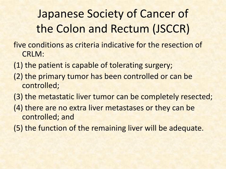
Japanese Society of Cancer ofthe Colon and Rectum (JSCCR)
five conditions as criteria indicative for the resection of CRLM:
(1) the patient is capable of tolerating surgery;
(2) the primary tumor has been controlled or can be controlled;
(3) the metastatic liver tumor can be completely resected;
(4) there are no extra liver metastases or they can be controlled; and
(5) the function of the remaining liver will be adequate.
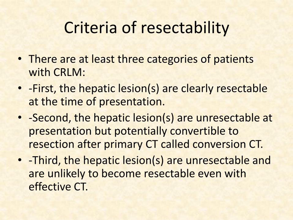
Criteria of resectability
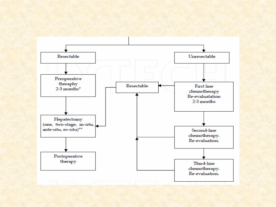
• There are at least three categories of patients with CRLM:
• -First, the hepatic lesion(s) are clearly resectableat the time of presentation.
• -Second, the hepatic lesion(s) are unresectable at presentation but potentially convertible to resection after primary CT called conversion CT.
• -Third, the hepatic lesion(s) are unresectable and are unlikely to become resectable even with effective CT.
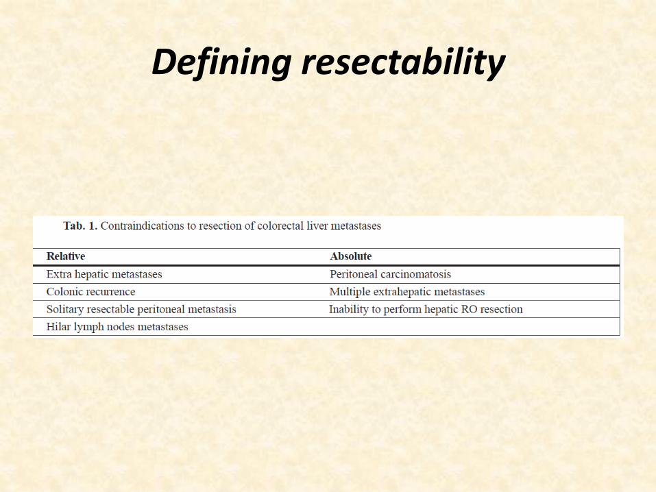
Defining resectability
Resectability
• No. of mets have not found to affect long term survival of R0 resection achieved.
• Degree of response to chemotherapy is a stronger predictor factor for long term survival than the number of metastasis.
• Evidence shows that size is not a resectabilityfactor, but a factor related to tumouraggressiveness
» (Altendorf-Hofmann et al. 2003)
Resectability
• Actual width of the surgical margin has no effect on survival as long as the margin is microscopically negative.
» (Figueras et al. 2007; Lordan 2007; Pawlik et al. 2005).
• A margin greater than 10mm is considered to be optimum.
» (Casanova et al. 2004).
• Surgeons should to plan achive 10mm margin but, a predicted margin of less than 1 cm should no longer be considered an exclusion criterion for resection
Resectability
• Historically, extrahepatic disease has been almost universally accepted as a contraindication to liver resection.
• Recently, however, some series have shown a 5-year survival rate of 12% to 37% after liver resection in selected patients with extrahepaticdisease, independent of the location of that disease (lung, primary colorectal recurrence, retroperitoneal or hepatic pedicle lymph nodes, peritoneal carcinomatosis, miscellaneous)
» (Elias et al. 2003, 2005).
Resectabilty
• Incidental peritoneal disease found at laparotomy would contraindicate hepatic resection.
• In general, resection in such patients should only be considered after documentation of stable/responsive disease with systemic chemotherapy and when an R0 resection of both intrahepatic and extrahepatic disease is feasible.
» (Vauthey 2007).
• Positive hilar lymph nodes are associated with a poor outcome and have been traditionally considered as a contraindication to hepatic resection of CRC liver disease.
• Recent papers shown long-term survival in some patients with hilar nodal metastases, (hepatoduodenal-retropancreatic area and not in the common hepatic artery/celiac-axis region)
» (Adam et al. 2008; Jaeck 2003).
• At present, the criteria for resectability include any patient in whom all disease can be
– removed with a negative margin and
– who has adequate hepatic reserve.
• That is to say, instead of resectability being defined by what is removed, now it is sustained by what will remain after resection, including patients with extrahepatic disease
» (Pawlik et al. 2008).
• There has been a shift in the concept of ‘resectability’ over the past two decades.
• Traditionally unresectable disease if any of the following criteria(EKEBERG’s ).(i) more than four metastatic deposits; (ii) Resection margin ,1 cm; (iii) bilobar disease; (iv) extrahepatic disease.
• Although these factors continue to convey a worse prognosis, they are no longer regarded as an absolute contraindication to liver resection, since a proportion of these patients can undergo successful tumor clearance and have long-term survival.
• American Hepato- Pancreato-Biliary Association (AHPBA) consensus conference 2006
• Resectable
– when the disease can be completely resected,
– two adjacent liver segments can be spared with an adequate vascular inflow and outflow and biliarydrainage,
– and the volume of the liver remaining after resection (future liver remnant [FLR]) will be adequate
» (Vauthey 2006).
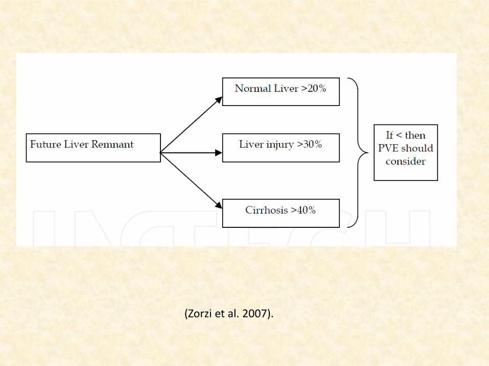
(Zorzi et al. 2007).
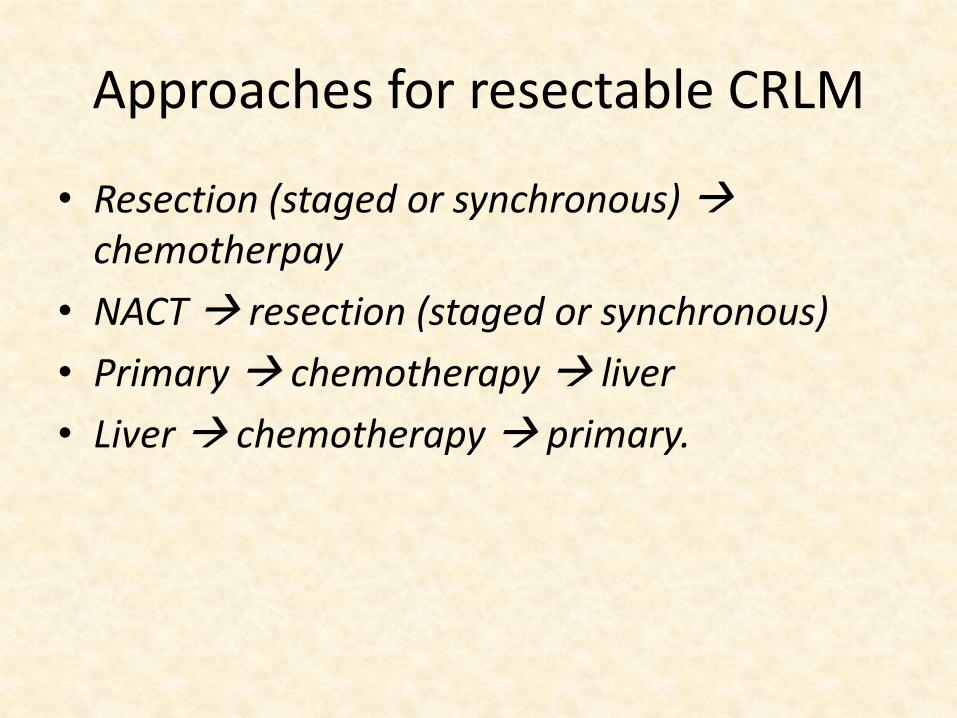
Approaches for resectable CRLM
• Resection (staged or synchronous) chemotherpay
• NACT resection (staged or synchronous)
• Primary chemotherapy liver
• Liver chemotherapy primary.
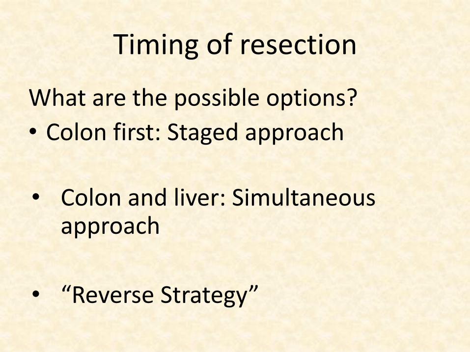
Timing of resection
What are the possible options?
• Colon first: Staged approach
• Colon and liver: Simultaneous approach
• “Reverse Strategy”
Factors determine the decision:
1. The presence of symptoms.
2. Location of primary tumor and liver metastases.
3. Extent of tumor (both primary and metastatic).
4. Patient performance status, and underlying comorbidities.
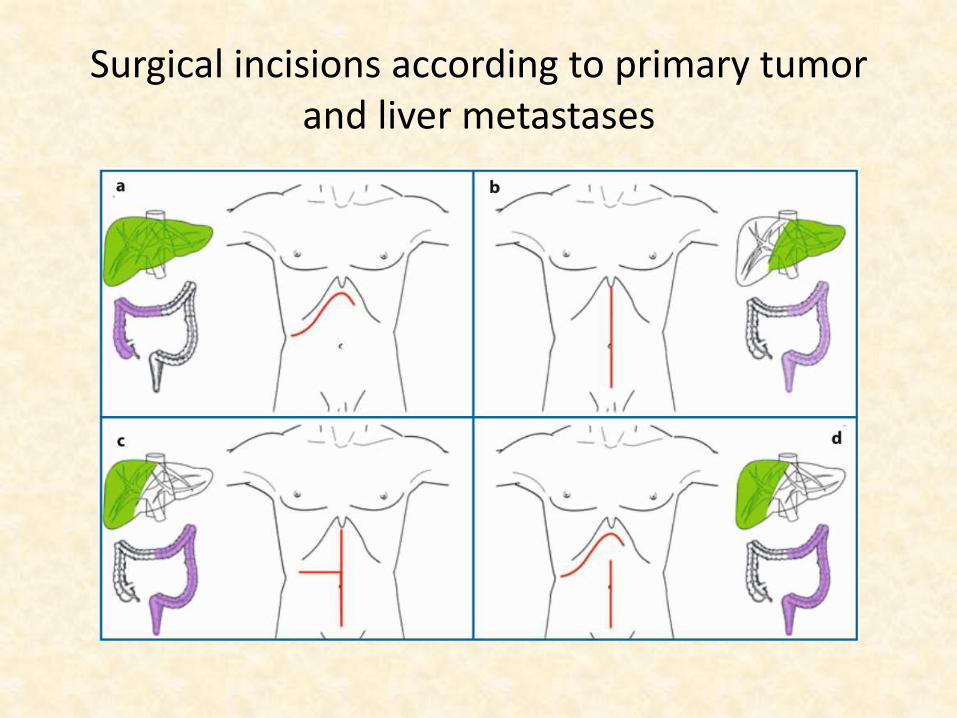
Surgical incisions according to primary tumor and liver metastases
Advantages of simultaneous resection
• a single surgical procedure.
• Reduced length of hospital stay
• The removal of all neoplastic foci and interruption of the “metastatic cascade”.
• No delay in initiating systemic treatment
Disadvantages of simultaneous resections
• The combination of a “clean” and a “contaminated” surgical procedure and thus the higher risk of septic complications, which could cause or worsen a liver dysfunction
• The increased risk of anastomotic leak due to splanchnic congestion if prolonged pedicle clamping is needed.
Disadvantages of simultaneous resections, cont.,
• The need for a double surgical team for liver and colorectal surgery/inadequate treatment if a single team performs the entire procedure.
• The inadequate surgical exposure through a single incision
Criteria for synchronous approach
– Age<70 years
– good surgical fitness.
– an adequate tumor-free margin,
– lesions that are not advanced(T4),
– less than 4 colorectal lymph node metastases
– histology that is not poorly differentiated or mucinous adenocarcinoma.
– 3 or fewer liver metastases.
– a minor liver resection (less than 3 segments) is plannedAnn Acad Med Singapore 2010;39:719-33
“Reverse Strategy”
• Mentha et al. and the group from M.D. Anderson Cancer Center reported it in 2006.
• Preoperative chemotherapy is followed by resection of the hepatic metastases and then by resection of the colorectal primary at a second operation.
• The conclude treatment completion rate are better in reverse approach but equall OS.
• J Am Coll Surg 2010;210:934-41.
The rationale for this approach
• It can be considered as an alternative option in patients with advanced hepatic metastases and an asymptomatic primary.
• Its can be better indicated to patient with rectal tumor where pelvic surgical morbidity may delay addressing liver mets.
• Only a minority of patients with liver metastases is amenable directly to surgery (10-20%).
• Therefore, efforts have been made to increase the resectability of patients with initially unresectable colorectal liver metastases.
• Reasons of unresectability in liver limited CRLM.
– Inadequate FLR
– Bad anatomical location.
– Poor PS of patients.
Measures to increase resectability
• increase/preserve hepatic reserve,
- Portal Vein Embolisation: inadequate FLR
- Two- stage resection: bilobar disease
• combined local therapy: Resection Plus RFA
- Resect- larger lesion
- Ablate- smaller lesions
• decrease tumor size: Chemotherapy (near major vessels.)
Ante-situ, in situ, and ex situ procedures : extreme liver surgery.ALLPS
Down staging chemotherapy
• Optimal regimen and sequencing remain matter of debate.
• Some suggest low CRS should put on immediate surgery if resectable and other should receive preopchemotherpy.
» Fong et al 1999
• Using Chemotherapy for borderline resectable tumor is standard method, but its role in resectable CRLM is a matter of debate.
• Although there are no reports of outcomes of liver resection after HAI, its complication rates are so high (57%) that it is dismissed as a first option.
Chemotherapy
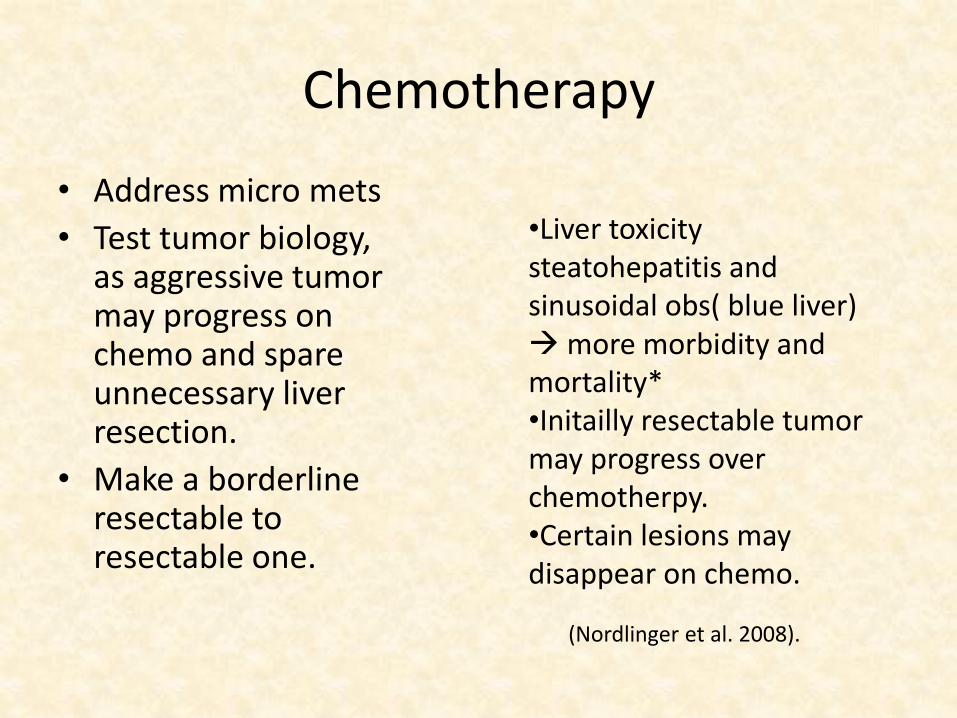
• Address micro mets
• Test tumor biology, as aggressive tumor may progress on chemo and spare unnecessary liver resection.
• Make a borderline resectable to resectable one.
•Liver toxicity steatohepatitis and sinusoidal obs( blue liver) more morbidity and mortality*•Initailly resectable tumor may progress over chemotherpy.•Certain lesions may disappear on chemo.
(Nordlinger et al. 2008).
• NCCN recommends complete 6 months systemic chemotherapy to address residual microscopic disease.
• systemic therapy can be given before, between, or after resections, the total duration of perioperativechemotherapy should not exceed 6 months.
• A 2012 meta-analysis of 3RCTS comparing S vs S & CT in 642 eligible patients (resectable CRLM)
• PFS and DFS better in CT arm (pooled HR, 0.75; CI, 0.62–0.91; P = .003).
• But equal OS (pooled HR, 0.74; CI, 0.53–1.05; P = .088.)» Ciliberto D, Prati U, Roveda L, et al. Role of systemicchemotherapy in the
management of resected or resectable colorectal liver metastases: a systematic review and meta-analysis of randomized controlled trials. Oncol Rep 2012;27:1849-1856. Available
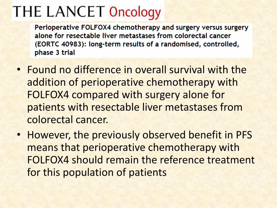
• Found no difference in overall survival with the addition of perioperative chemotherapy with FOLFOX4 compared with surgery alone for patients with resectable liver metastases from colorectal cancer.
• However, the previously observed benefit in PFS means that perioperative chemotherapy with FOLFOX4 should remain the reference treatment for this population of patients
• Peri-operative chemotherapy with FOLFOX4 improves PFS but does not statistically significantly improve OS over surgery alone.
• The optimal sequencing of chemotherapy remains unclear for resectable tumor
• Patients with resectable disease may undergo liver resection first, followed by postoperative adjuvant chemotherapy.
• Alternatively, perioperative (neoadjuvant plus postoperative) chemotherapy can be used.
Araujo et al 2013. Bilchik AJ et al 2008, Adams et al 2009
Chemotherapy
• Novel chemotherapeutic regimens have been associated with response rates (approximately 50%), allowing 10-30% of the patients with initially unresectable disease to be successfully treated with liver surgery
» (Adam et al. 2004).
• In addition, combination with biologic agents that target angiogenesis and the epidermal growth factor receptor (EGFR), bevacizumab and cetuximab, achieves response rates of up to 70%, increasing these figures
» (Vauthey 2006).
NCCN 1.2015
• FOLFOXIRI (infusional 5-FU, LV, oxaliplatin, irinotecan) has been compared with FOLFIRI in 2 randomized clinical trials in patients with unresectable disease, showed better resectabilityrates, but more toxicity.
• Gruppo Oncologico Nord Ovest (GONO) 15 vs 6% • Gastrointestinal Committee of the Hellenic Oncology
Research Group (HORG) trial 10 vs 4%
– FOLFIRI, FOLFOX, or CapeOx chemotherapy alone or with bevacizumab;
– FOLFIRI or FOLFOX with panitumumab or cetuximab; – FOLFOXIRI alone or with bevacizumab).
• Re-evaluation for resection should be done after 2 or 3 months of pre-operative chemotherapy and every 2 months thereafter.
• Tumor progression before surgery is associated with a poor outcome, even after potentially curative hepatectomy.
• Tumor control before surgery is crucial to offer a chance of prolonged remission in patients with multiple metastases
» (Adam et al. 2004.)
• Patients should be referred early for evaluation for resection.
• The peri-operative complication rate, including hepatobiliary complications, is higher with lengthy pre-operative chemotherapy and is likely related to the prolonged and sequential use of multiple regimens
» (de Haas et al. 2011).
• Although resection has proven to be safe after preoperative chemotherapy, the mortality rate is increased in certain types of liver damage associated with chemotherapy, specifically, steatohepatitis associated with irinotecantherapy.
» Vauthey et al 2006
Chemotherapy
• If bevacizumab is included as a component of the conversion therapy, an interval of at least 6 weeks between the last dose of bevacizumab and surgery should be applied, with a 6- to 8- week postoperative period before re-initiation of bevacizumab.
• Patients with disease converted to a resectablestate should undergo synchronized or staged resection of colon and metastatic cancer as soon as it become resectable.
• And patient should receive complete 6 months treatment.
• In the case of liver metastases only, HAI therapy with or without systemic 5-FU/LV (category 2B) remains an option at centers with experience.
Disappearing mets
• 6-9 % develop radiological CR following NACT.• Not all radiologically CR lesion shown PCR,
correlation ranging from 17-65% in various series.• So concern will to find out these ghost lesions. • Many variables are described correlating PCR.
– Lesion disappeared on HAI based Ct.– Not visible on MRI– CEA normalize.
» Benoist et al. 2006» Elias et al 2008» Auer et al 2010

• Patient with PCR showed better OS » (thomay et al 2010)
• In general, all the original sites of disease noted on the pre-therapy imaging need to be resected or ablated.
Portal Vein Embolization
• To minimize risk of POLF, inadequate FLR and IL hypertrophy of Liver parenchyma selective embolization of Portal vein brach or ligation done.
• It coz ipsilateral atrophy with FLR hypertrophy.
• Chemotherpy need not to modify except bavacizumab.
• Chemotherapy does not seem to affect the hypertrophy induced by PVE.
• A few studies using bevazucimab recommend a 6 week waiting period between the last dose and the hepatectomy, although its influence on the hypertrophy is unclear.
• Azoulay et al. reported that PVE increased the feasibility of liver resection by 19% and that the actuarial survival rate was 40% at 5 years, similar to that of patients resected without PVE
» (Azoulay et al. 2000).
• There is a variety of substances used for embolization, e.g. absolute alcohol, ethiodizedoil, cyanoacrylate with no clear difference between them.
• PVE is well tolerated with minimum side effects such as fever, nausea, and transient abnormality of liver function test.
• Contraindications for PVE
– tumor invasion of the portal vein,
– portal vein thrombosis,
– Uncorrectable coagulopathy
– severe portal hypertension and
– renal failure
• Thus, the optimal time to evaluate the hypertrophy after PvE is 3 to 4 weeks.
• Volumetric CT can be repeated at this time because it provides two key pieces of information:
– (1) if adequate liver volume has been reached;
– (2) growth rate, which is informative for the liver capacity for regeneration
Two stage hepatectomy
• When planed resection not feasible in one stage particularly in bilobar liver mets TSH should be offered.
• It consists of combining two sequential and planned liver resections, usually 4-6 week apart (?)
• Frequently, it is associated with peri-operative systemic chemotherapy and PVE, although it is not a rule
» (Jaeck et al. 2004)
• In 2000, Adam et al.[63] proposed a new two-stage approach for initially resectable liver tumors.
• The maximum number of tumors removed in the first operation, and a second surgery is performed to remove the rest after a period of liver regeneration.
• The goal of the double stage hepatectomy is to minimize the risk of liver failure after massive hepatectomy in patients with bilateral metastases
• Usually, on the first hepatectomy the FLR is cleared out of tumors with non-anatomic resections and/or radiofrequency ablation or at most a single segment resection.
• It can be associated to the removal of the primary colorectal tumor, preferably through a laparoscopic approach or using a “J” incision if it is located on the right colon.
• PVO can be done by PVE , 2-4 week after first stage• Alternatively, PVE can be done during the first hepatectomy
through the ligation and alcoholization of the right portal vein• The second hepatectomy can be done on the fourth of fifth week
after PVE, when an adequate hypertrophy of the non-embolizedhemi-liver is achieved.
Jaeck et al 2004 reverse approach
• Some authors recommend pre-operative chemotherapy during the entire process. This should be determined by the criteria of the multidisciplinary team according to each individual case
» (Adam et al. 2000).
• If during the second stage hepatectomy new liver metastases or extrahepatic lesions are discovered, such as localized peritoneum implants, the procedure can still be performed if a R0 resection can be achieved.(?)
• This procedure may be the only therapy able to provide long-term survival and a possible cure for patients with initially unresectablemultiple and bilobar CRC liver metastases.
• A recent series reports a 5 year overall survival rate of 32% for patients on whom the procedure had been completed
» (Narita et al. 2011).
Radiofrequency ablation
• Not all patients are suitable for liver resection for CRLM and alternative therapies have been proposed.
• The most common alternative treatment for CRLM is radiofrequency ablation (RFA).
• RFA involves placing an electrode into the liver tumor under radiological guidance (ultrasound [U.S.], CT or MRI),
• It generates thermal (radio frequency) energy which destroys the tumor and a margin of normal parenchyma.
• RFA can be performed percutaneously, laparoscopicallyor during laparotomy.
• Larger follow-up data confirm the safety process, but suggest that RFA may not be equivalent to a local resection as a modality.
– Abdalla et al 2004
• Therefore, the long-term survival after resection is better then after local ablation (65% vs 22%) [8]
• solitary CLM and showed that liver resection is associated with greater survival rates 37% vs 5%
• The disadvantages of RFA include:
(1) High recurrence rate for large tumors (>5 cm);
(2) Necrosis of adjacent structures—major bile ducts, stomach, duodenum, colon, and diaphragm;
(3) Delayed tumor recurrence on late (>3 years) follow up higher than resection.
(4) Metastases must be clearly visible by imaging.(post NACT)
(5) Not effective for surface tumor, near major vessels
• Currently, RFA is used primarily as an adjunct to resection or as primary therapy when resection is precluded regardless of cause.
• Neodymium-doped yttrium-aluminum-garnet (Nd:YAG) laser and microwave are other hyperthermic abalative methods.
Cryoablation
• Chemico physiologic sequelae of rapid freeze-thaw cycles on cellular membranes to achieve a total cell kill, tissue temperatures of˜ −50° C or below are required.
• Depending on the size of the metastases, an appropriate-sized cryoprobe is placed through the metastases, and cryoablation is initiated under ultrasonographic guidance
• Potential intraoperative complications of hepatic cryosurgery include – Accidental freezing of adjacent tissues,
– cracking of the liver parenchyma,
– bleeding due to the introduction of trotter probes,
– hypothermia and related cardiac arrhythmias,
– nitrogen embolism,
– bile duct or major vascular injury, and
– renal failure from myoglobinuria.
• Large vessels tolerate cryotherapy extremely well without rupture or occlusion because of the continued dissipation of thermal energy by the flow of blood.
• In contrast, large bile ducts are extremely vulnerable to cryoinjury, and caution should be exercised in treating tumors located near the hilum.
• Whether survival after cryoablation will be equivalent to resection is yet undetermined.
• No randomized, controlled trials have been performed to compare these treatments.
• Adjuvant cryoablation has been used concurrently with resection for the treatment of small, deep-seated hepatic metastases during major hepatectomy, and consequently, has extended the role of resection in some patients who were otherwise unresectable.
Extreme liver surgery
• Total vascular occlusion. (IVC clamping and pringle manuever).
• Useful when major vessels involved by tumor.
• Still investigational.
• Lesson learn from liver transplantation.
• In situ, ante situ, ex situ depending on level of vascular detachment of liver
• The common basis for in-situ, ante-situ and ex-situ resection is the total vascular exclusion (TVE) of the liver, and the perfusion of the organ by preservation hypothermic solution.
• Generally, a veno-venous bypass is used to avoid venous congestion during prolonged caval and portal crossclamping and a hypothermic preservation solution is instilled through the portal vein.
• Main indications of the three techniques are tumors that involve vascular structures of the hilum, venous confluence or inferior vena cava (IVC), or are in close proximity to them.
• Ex-situ technique is losing support due to its high morbidity and mortality.
• The location of the lesion or lesions in or near the suprahepatic IVC represents a true challenge due to the impossibility of using conventional resection techniques.
• Furthermore, optimal perioperative anaestheticmanagement is crucial in this setting, and the anaesthesiateam should be familiar with the hepatic transplant procedure.
• The involvement of the inferior vena cava does not necessarily preclude resection.
• Liver resection with reconstruction of the IVC can be performed in selected cases.
• The resected IVC may then be replaced with an autogenousvein graft or a prosthetic material. The mortality rate of resection IVC is 4.5-25% and morbidity up to 40%
» (Azoulay et al. 2005).
• The increased risk associated with the procedure appears to be balanced by the possible benefits, particularly when the lack of alternative approaches is considered
» (Hemming et al. 2004).
Re resection
• Most patients who undergo liver resection for CLM have recurrence, and one third of these recurrences develop only in the liver(MC).
• Selected patients with isolated hepatic recurrence (30%) may undergo repeat hepatectomy and achieve long-term survival.
Adam R et al 2003Petrowky et al 2002 MSKCC
• After the third hepatectomy, survival rates at 5 years are estimated as 32% and postoperative morbidity and mortality are not higher than after the first hepatectomy.
• As bilobar multiple CLM recurrence are very likely for recurrence, early diagnosis of the relapse is important to maximize the number of patietsappropriate for resection, and long-term survival can be achieved with this approach.
(ALPPS)
• Associated liver partition and portalvein ligation for staged hepatectomy.
• Recently, a new two-stage technique has been developed with the acronym (ALPPS) associating liver partitioning and portal vein ligation for staged hepatectomy with the aim of obtaining a more rapid and effective increase in FLR, even though indications are not clear yet.
Advantage
• Rapid and superior amount of FLR hypertrophy compared with PVO alone.
• A short interval period, meaning early definitive liver resection, unlikely tumor progression, and faster recovery for the patient with early restart of chemotherapy;
• In cases of synchronic disease, for which combined surgical procedures may require a greater functional hepatic reserve, this new strategy enables the simultaneous resection of the primary tumor and aggressive tumor cleaning of the FLR;
• ALPPS might make curative resection possible even for patients with a history of failed PVE or PVL.
• Despite the potential benefits of this novel approach, there are some concerns.
• Probably the most important is the potential drawback of manipulating a liver with a high tumor load and leaving it for a week or more in a environment of immunosuppression, inflammation, and stress, which could cause spillage of tumor cells into the pulmonary and systemic circulation.
• Whether or not this spillage does occur, and if so whether it adversely affects the survival of patients with CRLM, remains unknown

Thanking you.


