A TWELFTH CENTURY HOSPITAL AT POLONNARUVA AND MEDICAL AND SURGICAL EQUIPMENT
Upload
scu-hospitalCategory
view
417download
70
Differentiating Surgical Equipment
and Supplies
1572_FM_i-xii.qxd 8/6/09 12:04 PM Page i

1572_FM_i-xii.qxd 8/6/09 12:04 PM Page ii
DifferentiatingSurgical Equipment
and SuppliesColleen J. Rutherford, RN, MSN, CNOREducatorConcord HospitalConcord, New Hampshire
1572_FM_i-xii.qxd 8/6/09 12:04 PM Page iii
F.A. Davis Company
1915 Arch Street
Philadelphia, PA 19103
www.fadavis.com
Copyright © 2010 by F.A. Davis Company
Copyright © 2010 by F.A. Davis Company.All rights reserved.This product is protected by copyright. No part of it may be reproduced, stored in a retrieval
system, or transmitted in any form or by any means, electronic, mechanical, photocopying, recording, or otherwise, without written permission from the
publisher.
Printed in the United States of America
Last digit indicates print number: 10 9 8 7 6 5 4 3 2 1
Acquisitions Editor: Christa Fratantoro
Developmental Editor: Yvonne Gillam
Publisher: Margaret Biblis
Manager of Content Development: George W. Lang
Art and Design Manager: Carolyn O’Brien
As new scientific information becomes available through basic and clinical research, recommended treatments and drug therapies undergo changes.The author(s)
and publisher have done everything possible to make this book accurate, up to date, and in accord with accepted standards at the time of publication.The author(s),
editors, and publisher are not responsible for errors or omissions or for consequences from application of the book, and make no warranty, expressed or implied, in
regard to the contents of the book.Any practice described in this book should be applied by the reader in accordance with professional standards of care used in
regard to the unique circumstances that may apply in each situation.The reader is advised always to check product information (package inserts) for changes and
new information regarding dose and contraindications before administering any drug. Caution is especially urged when using new or infrequently ordered drugs.
Library of Congress Cataloging-in-Publication Data
Rutherford, Colleen.
Differentiating surgical equipment and supplies / Colleen J. Rutherford.
p. ; cm.
Includes index.
ISBN 978-0-8036-1572-4
1. Surgery—Equipment and supplies—Handbooks, manuals, etc. I.Title.
[DNLM: 1. Surgical Equipment—Atlases. 2. Equipment and Supplies—Atlases.
3. Operating Rooms—organization & administration—Atlases.WO 517 R975d 2009]
RD32.R695 2009
617.9—dc22 2009023553
Authorization to photocopy items for internal or personal use, or the internal or personal use of specific clients, is granted by F.A. Davis Company for users regis-
tered with the Copyright Clearance Center (CCC) Transactional Reporting Service, provided that the fee of $.10 per copy is paid directly to CCC, 222 Rosewood
Drive, Danvers, MA 01923. For those organizations that have been granted a photocopy license by CCC, a separate system of payment has been arranged.The fee
code for users of the Transactional Reporting Service is: 8036-1572/10 0 + $.10.
1572_FM_i-xii.qxd 8/6/09 12:04 PM Page iv

Acknowledgements
v
No one writes a book alone. I would like to acknowledge the following people and resources
for their contributions to this project:
First, to Christa Fratantoro,Acquisitions Editor of F.A.Davis Publishing Company—you had
enough faith in me to let me write a second book.Yvonne Gillam, Developmental Editor of
F. A. Davis Publishing Company—your energy and enthusiasm toward this project helped to
make it happen. Both of you make writing a pleasure and keep me motivated. I cherish you as
business associates and friends.
To Jamie—Thanks for your patience and perseverance in taking the photos for this book.
You are a true artist.
To my mother—Thank you for the love and support you have given me throughout my
life. I would not be the person I am today without your love and caring.
To all of my friends—you continue to enrich my life on a daily basis. I am truly blessed
to have each and every one of you in my life. To Marie DeRosia—you are a wonderful
friend and terrific colleague.Thank you for your support and your willingness to “model”
for some of the photos in this book—what a true friend!
A special “thank you” to Sharon Guldin, CRNA, Sean O’Brien, CRNA, and Robert
Sanborn, CRNA, for their careful review and input regarding the anesthesia content.
I have used numerous websites and surgical equipment catalogs to double check facts and
information on the equipment and supplies in this book.
1572_FM_i-xii.qxd 8/6/09 12:04 PM Page v

1572_FM_i-xii.qxd 8/6/09 12:04 PM Page vi

vii
Reviewers
David A. Alfaro, CSTProgram Director
American Career College
Surgical Technology Department
Los Angeles, California
Christina Lynn Baumer, RN, PhD, MEd, CNOR, CHESChair
Division of Continuing Education
Program Director
Surgical Technology
Lancaster General College of Nursing and Health Sciences
Continuing Education
Surgical Technology
Lancaster, Pennsylvania
Karen L. Chambers, CST-ACLSDirector
Long Island University
Surgical Technology Institute
School of Continuing Studies
Brooklyn, New York
Kevin R. Craycraft, CSTProgram Director
Bluegrass Community and Technical College
Health and Human Services Department
Lexington, Kentucky
Javier E. Espinales, MEd, CSTDirector, Surgical Technology Program
Virginia College at Austin
Surgical Technology Department
Austin,Texas
Robin Hueske, CSTConcorde Career Institute
Surgical Technology Department
Aurora, Colorado
Tammy Mangold, MEd, CST, CFASurgical Technologist
Columbia Regional Hospital
Surgery Department
Colombia, Missouri
Diane May, CSTAssociate Professor
Santa Fe Community College
Health Sciences Division
Gainesville, Florida
Rosemary A. Nagler, RN, BS, CNORClinical Education Coordinator
Western Suffolk BOCES
School of Surgical Technology
Northport, New York
Judith Schatte, RN, CNOR, CRNFAProgram Director and Instructor
Surgical Technology
Brevard Community College
Cocoa, Florida
Deborah S. Smith, RN, CNORDirector and Instructor
Southeastern Technical College
Surgical Technology Department
Glenwood, Georgia
Roy G. Zacharias, Jr, BS, CST Associate Academic Dean
Concorde Career Institute
Surgical Technology Department
Arlington,Texas
1572_FM_i-xii.qxd 8/6/09 12:04 PM Page vii

1572_FM_i-xii.qxd 8/6/09 12:04 PM Page viii

ix
Table of Contents
Chapter One: Common Equipment and Furniture 1Aseptic (Sterile) Technique 1Types of Sterilization 8Operating Room Attire 10Operating Room Table 16Operating Room Furniture 23Electrosurgical Units 30Medical Gases 34Pneumatic Tourniquet 37Autotransfusion 39Patient Positioning Equipment 41Miscellaneous Equipment 45Surgical Session Review 49
Chapter Two: General Surgical Supplies 51Surgical Sponges 51Hemostatics 54Syringes 56Surgical Wound Dressings 58Drapes 66Skin Prepping Solutions 76Basins 78Other Supplies 79Surgical Session Review 91
Chapter Three: Tubes, Drains, Catheters and Postop Splints 93Drains 93Gastrointestinal Tubes 98Urethral Catheters 100Splints 104Surgical Session Review 106
Chapter Four: Sutures and Suture Needles 107Comparison of Suture Materials 107Components of Suture Packaging 107Suture Sizing 109Suture 110Surgical Needle Points 118
1572_FM_i-xii.qxd 8/6/09 12:04 PM Page ix

x Contents
Individual Suture Needles 121Surgical Session Review 130
Chapter Five: Endoscopic Equipment 133Endoscopic Surgery 134Video Towers 135Endoscopic Cameras 136Rigid Scope Components 137Cystoscopy/Ureteroscopy Equipment 138Rigid ENT/Thoracic Scopes 140Arthroscopy Equipment 142Flexible Endoscopes 143Needles, Trocars and Instruments 143Surgical Session Review 147
Chapter Six: Lasers 149Types of Lasers 149Other Types of Lasers 151Laser Safety 151Surgical Session Review 154
Chapter Seven: Specialty Equipment 157Neurosurgery Equipment 157Cardiopulmonary Bypass Machine 158Bone Screws, Plates, Nails, Rods, Pins and Staples 159Vitrectomy/Cataract Removal Machines 161Ear Nose Throat Machines 163Gynecological Surgery Machines 164Liposuction 164Microscope 164Robotics 165Surgical Session Review 168
Chapter Eight: Anesthesia Supplies and Equipment 171Types of Anesthesia 171Airways 176Supplemental Oxygen 179Suction 182Anesthesia Machinery 182Other Equipment 189Block Needles and Supplies 190IV Supplies and Solutions 194Medications 200Surgical Session Review 229
Appendix: Answers to Surgical Session Reviews 231
Index 233
1572_FM_i-xii.qxd 8/6/09 12:04 PM Page x
xi
Introduction
Entering the operating room for the first time can be intimidating; it is like entering a for-
eign land.The clothes are different (scrubs, masks and hats), the temperature cold and there
are lots of equipment and supplies that are not used anywhere else except in surgery.What
is a “lap pad”? What is the difference between “Chromic” and “Vicryl” suture? What safety
precautions are necessary when the laser is in use? This book answers those questions and a
whole lot more.
The purpose of this book is to lessen the intimidation by describing common equipment
and supplies unique to the surgical setting.Throughout the chapters there are special pages
that describe some equipment or supplies in detail, help differentiate between similar supplies
(e.g., types of suture), aid in troubleshooting equipment or, in the case of anesthesia, give the
reader key points to look for when aiding in patient care. In some cases, the equipment and
supplies require less narration; on those pages you will find pictures of the supply and a short
description, including its use.
Throughout the chapters you will find boxes titled “Watch Out!”The purpose of these
boxes is to draw your attention to important information.
Students and new personnel can use the book as a reference, look up supplies and equip-
ment that they have come across in surgical cases or read more information about a subject.
Instructors and preceptors can use the book as a learning aid, assigning personnel or students
certain chapters to read or items to look up. Instructors who adopt this book for use in their
classrooms also receive a CD-ROM containing test questions and pictures of the supplies and
equipment that can be inserted into their own tests or presentations.
The operating room is an exciting and wonderful foreign land, full of joys and some sor-
rows but most of all wonderful opportunities to learn and make a difference for others. It is
my hope that you find this book a valuable aid in navigating the exciting world of surgery!
1572_FM_i-xii.qxd 8/6/09 12:04 PM Page xi

1572_FM_i-xii.qxd 8/6/09 12:04 PM Page xii
ASEPTIC (STERILE) TECHNIQUE
Aseptic technique is the foundation of all that goes on in the
operating room (OR). No matter what else happens, failure to
practice proper aseptic technique increases the patient’s risk of
infection, injury or even death.
Asepsis literally means “without infection,” but the generally
accepted definition is “absence of all microbes.” In surgery, an
inanimate object (e.g., instrument or supply) is considered ster-
ile (aseptic) only after it has been exposed to a process that kills
all microbes, including spores. Sterilization processes can use
steam, gas, chemicals, hydrogen peroxide or radiation to render
an item free of microbes.The skin can never be rendered sterile
(i.e., free of all microbes); therefore, aseptic technique dealing
with skin (e.g., scrubbing or prepping) is aimed at reducing mi-
crobes to an absolute minimum.
This section of the book covers principles of aseptic tech-
nique and provides an overview of sterilization methods. It is
imperative that ALL personnel working in the OR have a
thorough understanding of aseptic technique in order to pro-
tect their patients.
C H A P T E R 1
Common Equipment and Furniture
Surgical Conscience
� Surgical conscience is the ethical motivation to practice proper aseptic technique.ALL personnel must be willing to police themselves and each other, be willing toadmit to a break in technique and be ready to take corrective action. The willing-ness to police oneself, whether or not there is anyone else around to notice thebreak in technique, is imperative. Anyone who is not willing to admit to a breakin technique should NOT be working in the OR.
� ANY break in sterile technique, no matter how minor it may seem, puts the pa-tient at increased risk of infection.
� There is no such thing as “slightly contaminated” or “almost sterile.” Sterility ofan item is an absolute—either an item is sterile or it is not. If there is ANYdoubt about an item’s sterility, DO NOT use it—dispose of it and get a new one.
WATCH OUT!
Principles and Guidelines of Aseptic TechniqueThe principles listed here are adapted from various OR textbooks as well as the Association of Operating Room Nurses (AORN) guidelines and standards of practice. Although some ofthese principles and guidelines may be practiced and enforced to varying degrees, depending on institutional policies, they have a solid foundation in patient care and safety.
� Only sterile (scrubbed) personnel handle sterile items. Only nonsterile personnel handle nonsterile items.
� Sterility of an item is an absolute—either an item is sterile or it is not. If there is ANY doubt about an item’s sterility, DO NOT use it—dispose of it and get a new one.
� Proper attire must be worn by all personnel. This includes scrub suits, masks covering the mouth and nose, hats or caps that cover all hair (including facial hair) and, in some cases,eye protection and shoe covers. Waist ties and scrub tops should be tucked inside the pants to prevent inadvertent contamination of the field. Long sleeved jackets may be worn bynonsterile personnel but need to be buttoned.
� The wearing of cloth hats or caps depends on institutional policy. Cloth caps should be thrown into the laundry at the end of the day and when they become visibly soiled. Clothhats that are worn for more than 1 day without being laundered become a source of increased microbial contamination.
� Jewelry is a potential source of microbes and should NOT be worn in the OR.
� Nonsterile persons must stand a minimum of 12 to 18 inches away from any sterile area (e.g., the sterile field, sterile tables or ring basins). Nonsterile items should be kept aminimum of 12 inches away from sterile items.
� The draped patient is the center of the sterile field, and all other components of the sterile field (e.g., Mayo stand, back tables and ring stand) should be placed close to andaround the patient. Nonsterile items or personnel should not be within this area.
� The sterile field should be created as close to the time of surgery as possible. The longer sterile items are left out, the greater the chance of contamination. Rooms with sterile itemsleft open for more than 1 to 2 hours (this time varies depending on institutional policy but 2 hours is the maximum allowable time) should be broken down and the items removed.
� If sterile items are opened in a room and the surgery starting time is delayed, the room must be continuously monitored. It is NOT acceptable to leave the room without someonethere to monitor it at all times. If the room is left unattended there is no way to tell if an item or items might have been inadvertently contaminated.
� Talking at the sterile field should be kept to a minimum. Excessive, non-essential talking increases the amount of airborne microbes.
� Movement (including traffic in and out of the room) should be kept to a minimum during the surgical procedure. Doors should be kept closed. Opening of doors and movement ofpersonnel stir up air currents that can lead to increased microbial contamination.
Continued
1572_Ch01_001-050.qxd 8/6/09 10:48 AM Page 1
2 Chapter 1 Common Equipment and Furniture
Principles and Guidelines of Aseptic Technique—cont’d� Handling and moving drapes and linen should be kept to a minimum to prevent the spread of lint and dust.
� The only part of the surgical table that is considered sterile is the TABLETOP. Sterile items should NOT hang over the table edge.
� Once placed, table drapes and patient drapes should be left in place. Repositioning drapes could cause a part of the drape that previously had been below tabletop or patient level(therefore unsterile) to be brought back into the sterile field.
� Tubing that falls below the level of the patient is considered contaminated. If the electrosurgical hand unit or suction tip falls below the patient level it must be discarded and re-placed with a new one.
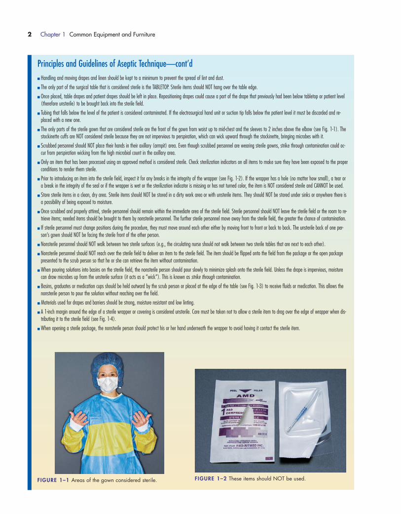
� The only parts of the sterile gown that are considered sterile are the front of the gown from waist up to mid-chest and the sleeves to 2 inches above the elbow (see Fig. 1-1). Thestockinette cuffs are NOT considered sterile because they are not impervious to perspiration, which can wick upward through the stockinette, bringing microbes with it.
� Scrubbed personnel should NOT place their hands in their axillary (armpit) area. Even though scrubbed personnel are wearing sterile gowns, strike through contamination could oc-cur from perspiration wicking from the high microbial count in the axillary area.
� Only an item that has been processed using an approved method is considered sterile. Check sterilization indicators on all items to make sure they have been exposed to the properconditions to render them sterile.
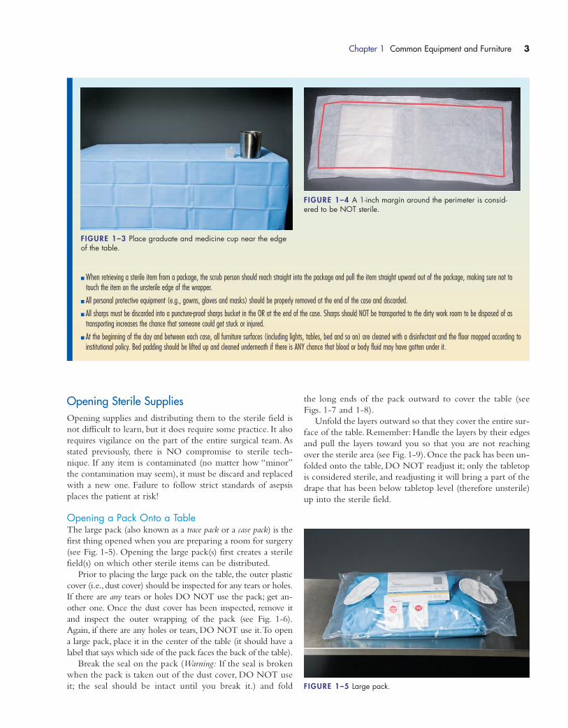
� Prior to introducing an item into the sterile field, inspect it for any breaks in the integrity of the wrapper (see Fig. 1-2). If the wrapper has a hole (no matter how small), a tear ora break in the integrity of the seal or if the wrapper is wet or the sterilization indicator is missing or has not turned color, the item is NOT considered sterile and CANNOT be used.
� Store sterile items in a clean, dry area. Sterile items should NOT be stored in a dirty work area or with unsterile items. They should NOT be stored under sinks or anywhere there isa possibility of being exposed to moisture.
� Once scrubbed and properly attired, sterile personnel should remain within the immediate area of the sterile field. Sterile personnel should NOT leave the sterile field or the room to re-trieve items; needed items should be brought to them by nonsterile personnel. The further sterile personnel move away from the sterile field, the greater the chance of contamination.
� If sterile personnel must change positions during the procedure, they must move around each other either by moving front to front or back to back. The unsterile back of one per-son’s gown should NOT be facing the sterile front of the other person.
� Nonsterile personnel should NOT walk between two sterile surfaces (e.g., the circulating nurse should not walk between two sterile tables that are next to each other).
� Nonsterile personnel should NOT reach over the sterile field to deliver an item to the sterile field. The item should be flipped onto the field from the package or the open packagepresented to the scrub person so that he or she can retrieve the item without contamination.
� When pouring solutions into basins on the sterile field, the nonsterile person should pour slowly to minimize splash onto the sterile field. Unless the drape is impervious, moisturecan draw microbes up from the unsterile surface (it acts as a “wick”). This is known as strike through contamination.
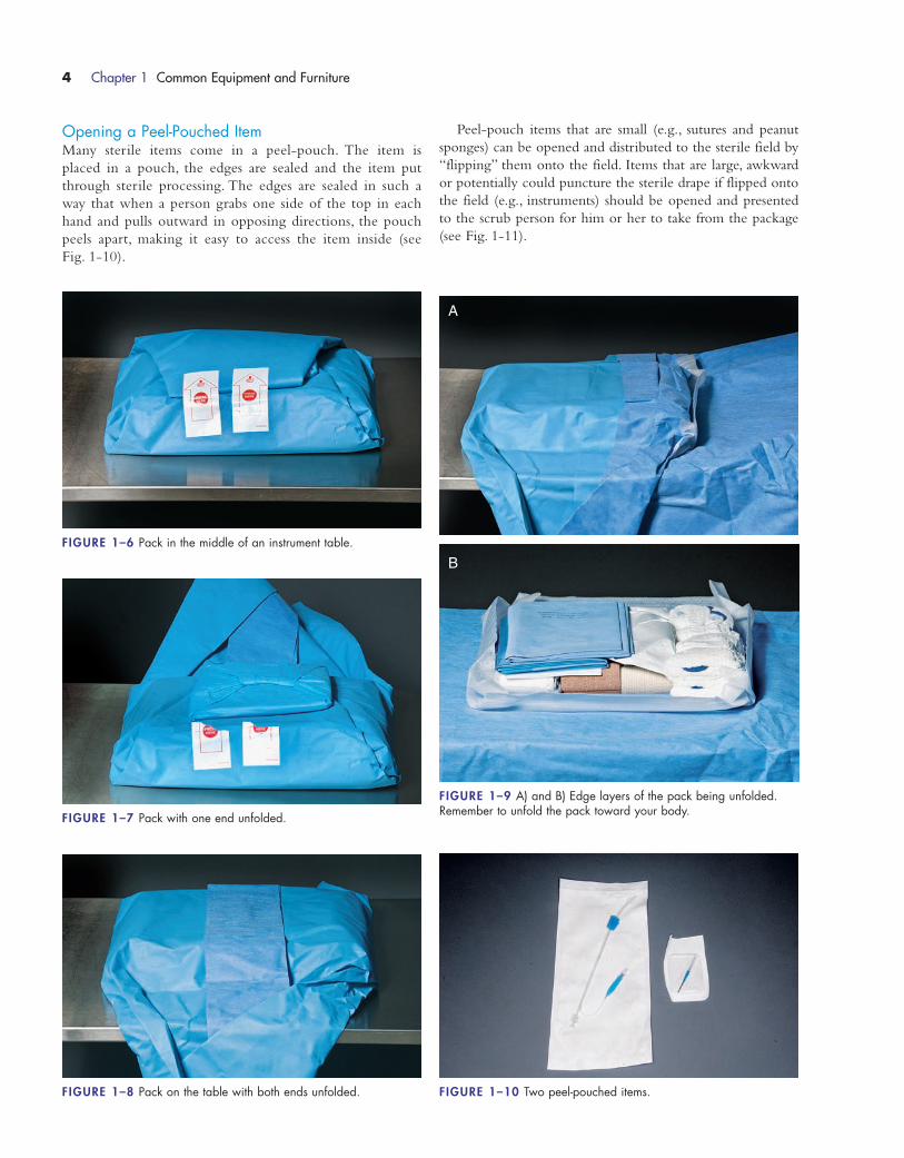
� Basins, graduates or medication cups should be held outward by the scrub person or placed at the edge of the table (see Fig. 1-3) to receive fluids or medication. This allows thenonsterile person to pour the solution without reaching over the field.
� Materials used for drapes and barriers should be strong, moisture resistant and low linting.
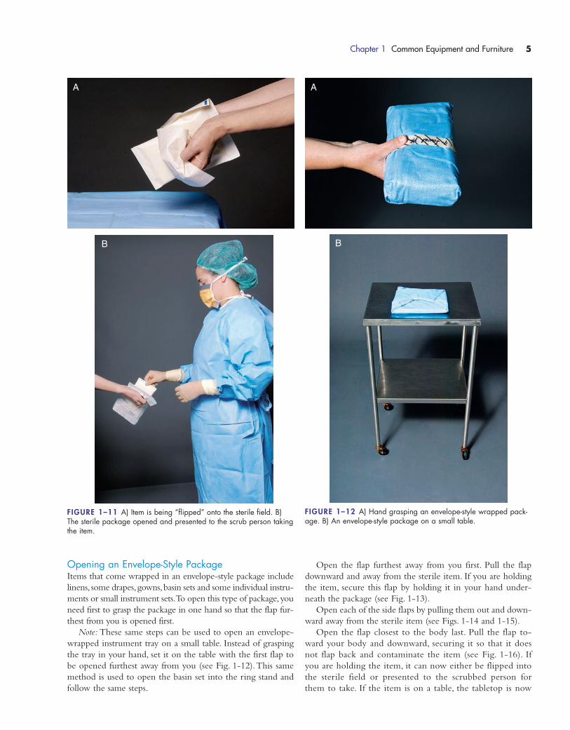
� A 1-inch margin around the edge of a sterile wrapper or covering is considered unsterile. Care must be taken not to allow a sterile item to drag over the edge of wrapper when dis-tributing it to the sterile field (see Fig. 1-4).
� When opening a sterile package, the nonsterile person should protect his or her hand underneath the wrapper to avoid having it contact the sterile item.
FIGURE 1–1 Areas of the gown considered sterile. FIGURE 1–2 These items should NOT be used.
1572_Ch01_001-050.qxd 8/6/09 10:48 AM Page 2
Chapter 1 Common Equipment and Furniture 3
� When retrieving a sterile item from a package, the scrub person should reach straight into the package and pull the item straight upward out of the package, making sure not totouch the item on the unsterile edge of the wrapper.
� All personal protective equipment (e.g., gowns, gloves and masks) should be properly removed at the end of the case and discarded.
� All sharps must be discarded into a puncture-proof sharps bucket in the OR at the end of the case. Sharps should NOT be transported to the dirty work room to be disposed of astransporting increases the chance that someone could get stuck or injured.
� At the beginning of the day and between each case, all furniture surfaces (including lights, tables, bed and so on) are cleaned with a disinfectant and the floor mopped according toinstitutional policy. Bed padding should be lifted up and cleaned underneath if there is ANY chance that blood or body fluid may have gotten under it.
FIGURE 1–3 Place graduate and medicine cup near the edgeof the table.
FIGURE 1–4 A 1-inch margin around the perimeter is consid-ered to be NOT sterile.
Opening Sterile SuppliesOpening supplies and distributing them to the sterile field is
not difficult to learn, but it does require some practice. It also
requires vigilance on the part of the entire surgical team. As
stated previously, there is NO compromise to sterile tech-
nique. If any item is contaminated (no matter how “minor”
the contamination may seem), it must be discard and replaced
with a new one. Failure to follow strict standards of asepsis
places the patient at risk!
Opening a Pack Onto a TableThe large pack (also known as a trace pack or a case pack) is the
first thing opened when you are preparing a room for surgery
(see Fig. 1-5). Opening the large pack(s) first creates a sterile
field(s) on which other sterile items can be distributed.
Prior to placing the large pack on the table, the outer plastic
cover (i.e., dust cover) should be inspected for any tears or holes.
If there are any tears or holes DO NOT use the pack; get an-
other one. Once the dust cover has been inspected, remove it
and inspect the outer wrapping of the pack (see Fig. 1-6).
Again, if there are any holes or tears, DO NOT use it.To open
a large pack, place it in the center of the table (it should have a
label that says which side of the pack faces the back of the table).
Break the seal on the pack (Warning: If the seal is broken
when the pack is taken out of the dust cover, DO NOT use
it; the seal should be intact until you break it.) and fold
the long ends of the pack outward to cover the table (see
Figs. 1-7 and 1-8).
Unfold the layers outward so that they cover the entire sur-
face of the table. Remember: Handle the layers by their edges
and pull the layers toward you so that you are not reaching
over the sterile area (see Fig. 1-9). Once the pack has been un-
folded onto the table, DO NOT readjust it; only the tabletop
is considered sterile, and readjusting it will bring a part of the
drape that has been below tabletop level (therefore unsterile)
up into the sterile field.
FIGURE 1–5 Large pack.
1572_Ch01_001-050.qxd 8/6/09 10:48 AM Page 3
4 Chapter 1 Common Equipment and Furniture
Opening a Peel-Pouched ItemMany sterile items come in a peel-pouch. The item is
placed in a pouch, the edges are sealed and the item put
through sterile processing. The edges are sealed in such a
way that when a person grabs one side of the top in each
hand and pulls outward in opposing directions, the pouch
peels apart, making it easy to access the item inside (see
Fig. 1-10).
Peel-pouch items that are small (e.g., sutures and peanut
sponges) can be opened and distributed to the sterile field by
“flipping” them onto the field. Items that are large, awkward
or potentially could puncture the sterile drape if flipped onto
the field (e.g., instruments) should be opened and presented
to the scrub person for him or her to take from the package
(see Fig. 1-11).
FIGURE 1–7 Pack with one end unfolded.
FIGURE 1–8 Pack on the table with both ends unfolded. FIGURE 1–10 Two peel-pouched items.
FIGURE 1–9 A) and B) Edge layers of the pack being unfolded. Remember to unfold the pack toward your body.
A
B
FIGURE 1–6 Pack in the middle of an instrument table.
1572_Ch01_001-050.qxd 8/6/09 10:48 AM Page 4
Chapter 1 Common Equipment and Furniture 5
A
B
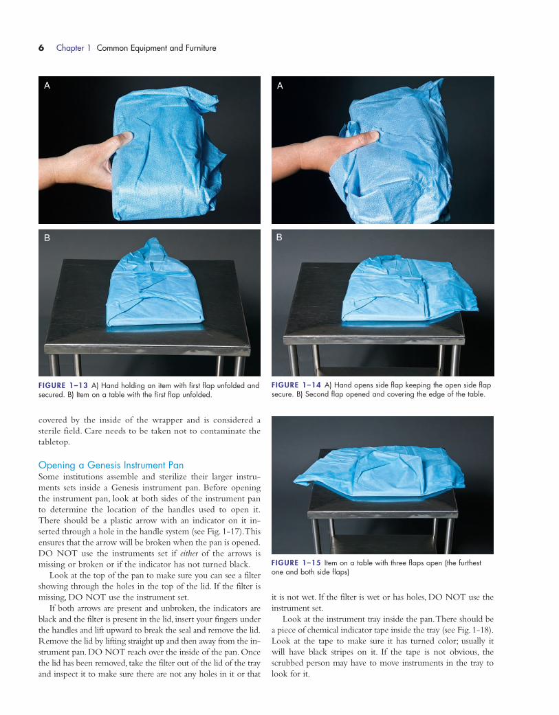
Opening an Envelope-Style Package Items that come wrapped in an envelope-style package include
linens, some drapes,gowns,basin sets and some individual instru-
ments or small instrument sets.To open this type of package, you
need first to grasp the package in one hand so that the flap fur-
thest from you is opened first.
Note: These same steps can be used to open an envelope-
wrapped instrument tray on a small table. Instead of grasping
the tray in your hand, set it on the table with the first flap to
be opened furthest away from you (see Fig. 1-12).This same
method is used to open the basin set into the ring stand and
follow the same steps.
Open the flap furthest away from you first. Pull the flap
downward and away from the sterile item. If you are holding
the item, secure this flap by holding it in your hand under-
neath the package (see Fig. 1-13).
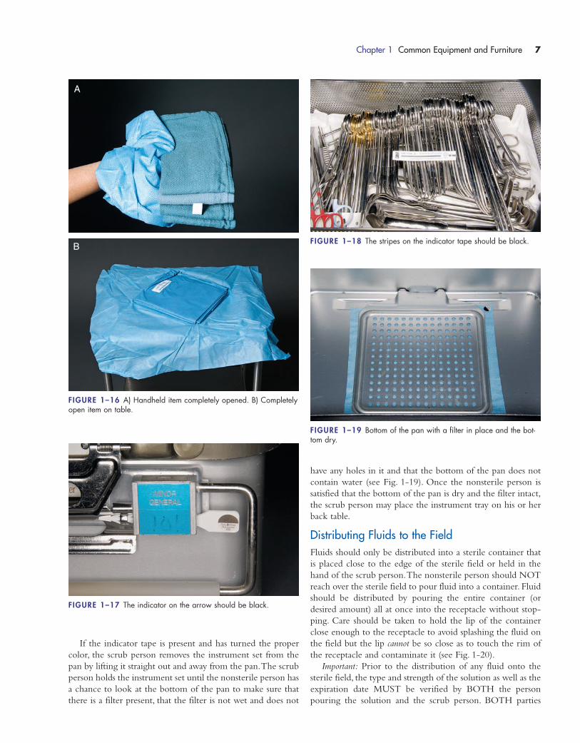
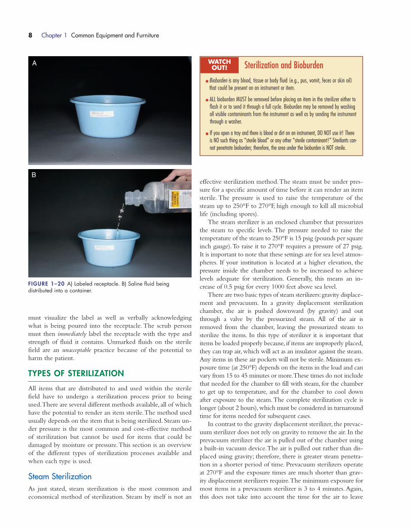
Open each of the side flaps by pulling them out and down-
ward away from the sterile item (see Figs. 1-14 and 1-15).
Open the flap closest to the body last. Pull the flap to-
ward your body and downward, securing it so that it does
not flap back and contaminate the item (see Fig. 1-16). If
you are holding the item, it can now either be flipped into
the sterile field or presented to the scrubbed person for
them to take. If the item is on a table, the tabletop is now
FIGURE 1–11 A) Item is being “flipped” onto the sterile field. B)The sterile package opened and presented to the scrub person takingthe item.
A
B
FIGURE 1–12 A) Hand grasping an envelope-style wrapped pack-age. B) An envelope-style package on a small table.
1572_Ch01_001-050.qxd 8/6/09 10:48 AM Page 5
covered by the inside of the wrapper and is considered a
sterile field. Care needs to be taken not to contaminate the
tabletop.
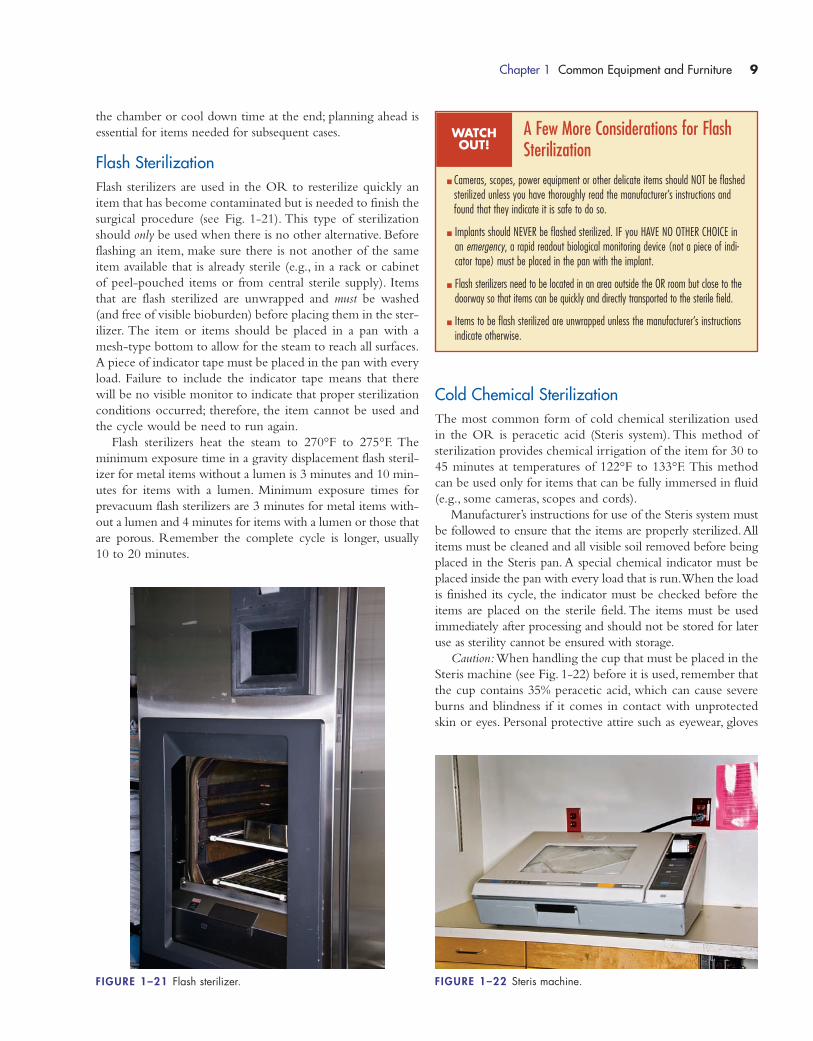
Opening a Genesis Instrument PanSome institutions assemble and sterilize their larger instru-
ments sets inside a Genesis instrument pan. Before opening
the instrument pan, look at both sides of the instrument pan
to determine the location of the handles used to open it.
There should be a plastic arrow with an indicator on it in-
serted through a hole in the handle system (see Fig. 1-17).This
ensures that the arrow will be broken when the pan is opened.
DO NOT use the instruments set if either of the arrows is
missing or broken or if the indicator has not turned black.
Look at the top of the pan to make sure you can see a filter
showing through the holes in the top of the lid. If the filter is
missing, DO NOT use the instrument set.
If both arrows are present and unbroken, the indicators are
black and the filter is present in the lid, insert your fingers under
the handles and lift upward to break the seal and remove the lid.
Remove the lid by lifting straight up and then away from the in-
strument pan. DO NOT reach over the inside of the pan. Once
the lid has been removed, take the filter out of the lid of the tray
and inspect it to make sure there are not any holes in it or that
it is not wet. If the filter is wet or has holes, DO NOT use the
instrument set.
Look at the instrument tray inside the pan.There should be
a piece of chemical indicator tape inside the tray (see Fig. 1-18).
Look at the tape to make sure it has turned color; usually it
will have black stripes on it. If the tape is not obvious, the
scrubbed person may have to move instruments in the tray to
look for it.
6 Chapter 1 Common Equipment and Furniture
FIGURE 1–13 A) Hand holding an item with first flap unfolded andsecured. B) Item on a table with the first flap unfolded.
A
B
FIGURE 1–14 A) Hand opens side flap keeping the open side flapsecure. B) Second flap opened and covering the edge of the table.
A
B
FIGURE 1–15 Item on a table with three flaps open (the furthestone and both side flaps)
1572_Ch01_001-050.qxd 8/6/09 10:48 AM Page 6
Chapter 1 Common Equipment and Furniture 7
FIGURE 1–16 A) Handheld item completely opened. B) Completelyopen item on table.
A
B
If the indicator tape is present and has turned the proper
color, the scrub person removes the instrument set from the
pan by lifting it straight out and away from the pan.The scrub
person holds the instrument set until the nonsterile person has
a chance to look at the bottom of the pan to make sure that
there is a filter present, that the filter is not wet and does not
have any holes in it and that the bottom of the pan does not
contain water (see Fig. 1-19). Once the nonsterile person is
satisfied that the bottom of the pan is dry and the filter intact,
the scrub person may place the instrument tray on his or her
back table.
Distributing Fluids to the FieldFluids should only be distributed into a sterile container that
is placed close to the edge of the sterile field or held in the
hand of the scrub person.The nonsterile person should NOT
reach over the sterile field to pour fluid into a container. Fluid
should be distributed by pouring the entire container (or
desired amount) all at once into the receptacle without stop-
ping. Care should be taken to hold the lip of the container
close enough to the receptacle to avoid splashing the fluid on
the field but the lip cannot be so close as to touch the rim of
the receptacle and contaminate it (see Fig. 1-20).
Important: Prior to the distribution of any fluid onto the
sterile field, the type and strength of the solution as well as the
expiration date MUST be verified by BOTH the person
pouring the solution and the scrub person. BOTH parties
FIGURE 1–17 The indicator on the arrow should be black.
FIGURE 1–18 The stripes on the indicator tape should be black.
FIGURE 1–19 Bottom of the pan with a filter in place and the bot-tom dry.
1572_Ch01_001-050.qxd 8/6/09 10:48 AM Page 7
must visualize the label as well as verbally acknowledging
what is being poured into the receptacle. The scrub person
must then immediately label the receptacle with the type and
strength of fluid it contains. Unmarked fluids on the sterile
field are an unacceptable practice because of the potential to
harm the patient.
TYPES OF STERILIZATION
All items that are distributed to and used within the sterile
field have to undergo a sterilization process prior to being
used.There are several different methods available, all of which
have the potential to render an item sterile.The method used
usually depends on the item that is being sterilized. Steam un-
der pressure is the most common and cost-effective method
of sterilization but cannot be used for items that could be
damaged by moisture or pressure.This section is an overview
of the different types of sterilization processes available and
when each type is used.
Steam SterilizationAs just stated, steam sterilization is the most common and
economical method of sterilization. Steam by itself is not an
effective sterilization method.The steam must be under pres-
sure for a specific amount of time before it can render an item
sterile. The pressure is used to raise the temperature of the
steam up to 250°F to 270°F, high enough to kill all microbial
life (including spores).
The steam sterilizer is an enclosed chamber that pressurizes
the steam to specific levels. The pressure needed to raise the
temperature of the steam to 250°F is 15 psig (pounds per square
inch gauge).To raise it to 270°F requires a pressure of 27 psig.
It is important to note that these settings are for sea level atmos-
pheres. If your institution is located at a higher elevation, the
pressure inside the chamber needs to be increased to achieve
levels adequate for sterilization. Generally, this means an in-
crease of 0.5 psig for every 1000 feet above sea level.
There are two basic types of steam sterilizers:gravity displace-
ment and prevacuum. In a gravity displacement sterilization
chamber, the air is pushed downward (by gravity) and out
through a valve by the pressurized steam. All of the air is
removed from the chamber, leaving the pressurized steam to
sterilize the items. In this type of sterilizer it is important that
items be loaded properly because, if items are improperly placed,
they can trap air,which will act as an insulator against the steam.
Any items in these air pockets will not be sterile. Minimum ex-
posure time (at 250°F) depends on the items in the load and can
vary from 15 to 45 minutes or more.These times do not include
that needed for the chamber to fill with steam, for the chamber
to get up to temperature, and for the chamber to cool down
after exposure to the steam.The complete sterilization cycle is
longer (about 2 hours),which must be considered in turnaround
time for items needed for subsequent cases.
In contrast to the gravity displacement sterilizer, the prevac-
uum sterilizer does not rely on gravity to remove the air. In the
prevacuum sterilizer the air is pulled out of the chamber using
a built-in vacuum device.The air is pulled out rather than dis-
placed using gravity; therefore, there is greater steam penetra-
tion in a shorter period of time. Prevacuum sterilizers operate
at 270°F and the exposure times are much shorter than grav-
ity displacement sterilizers require.The minimum exposure for
most items in a prevacuum sterilizer is 3 to 4 minutes. Again,
this does not take into account the time for the air to leave
8 Chapter 1 Common Equipment and Furniture
FIGURE 1–20 A) Labeled receptacle. B) Saline fluid being distributed into a container.
A
B
Sterilization and Bioburden
� Bioburden is any blood, tissue or body fluid (e.g., pus, vomit, feces or skin oil)that could be present on an instrument or item.
� ALL bioburden MUST be removed before placing an item in the sterilizer either toflash it or to send it through a full cycle. Bioburden may be removed by washingall visible contaminants from the instrument as well as by sending the instrumentthrough a washer.
� If you open a tray and there is blood or dirt on an instrument, DO NOT use it! Thereis NO such thing as “sterile blood” or any other “sterile contaminant!” Sterilants can-not penetrate bioburden; therefore, the area under the bioburden is NOT sterile.
WATCH OUT!
1572_Ch01_001-050.qxd 8/6/09 10:48 AM Page 8
the chamber or cool down time at the end; planning ahead is
essential for items needed for subsequent cases.
Flash SterilizationFlash sterilizers are used in the OR to resterilize quickly an
item that has become contaminated but is needed to finish the
surgical procedure (see Fig. 1-21). This type of sterilization
should only be used when there is no other alternative. Before
flashing an item, make sure there is not another of the same
item available that is already sterile (e.g., in a rack or cabinet
of peel-pouched items or from central sterile supply). Items
that are flash sterilized are unwrapped and must be washed
(and free of visible bioburden) before placing them in the ster-
ilizer. The item or items should be placed in a pan with a
mesh-type bottom to allow for the steam to reach all surfaces.
A piece of indicator tape must be placed in the pan with every
load. Failure to include the indicator tape means that there
will be no visible monitor to indicate that proper sterilization
conditions occurred; therefore, the item cannot be used and
the cycle would be need to run again.
Flash sterilizers heat the steam to 270°F to 275°F. The
minimum exposure time in a gravity displacement flash steril-
izer for metal items without a lumen is 3 minutes and 10 min-
utes for items with a lumen. Minimum exposure times for
prevacuum flash sterilizers are 3 minutes for metal items with-
out a lumen and 4 minutes for items with a lumen or those that
are porous. Remember the complete cycle is longer, usually
10 to 20 minutes.
Cold Chemical SterilizationThe most common form of cold chemical sterilization used
in the OR is peracetic acid (Steris system). This method of
sterilization provides chemical irrigation of the item for 30 to
45 minutes at temperatures of 122°F to 133°F. This method
can be used only for items that can be fully immersed in fluid
(e.g., some cameras, scopes and cords).
Manufacturer’s instructions for use of the Steris system must
be followed to ensure that the items are properly sterilized.All
items must be cleaned and all visible soil removed before being
placed in the Steris pan. A special chemical indicator must be
placed inside the pan with every load that is run.When the load
is finished its cycle, the indicator must be checked before the
items are placed on the sterile field. The items must be used
immediately after processing and should not be stored for later
use as sterility cannot be ensured with storage.
Caution:When handling the cup that must be placed in the
Steris machine (see Fig. 1-22) before it is used, remember that
the cup contains 35% peracetic acid, which can cause severe
burns and blindness if it comes in contact with unprotected
skin or eyes. Personal protective attire such as eyewear, gloves
Chapter 1 Common Equipment and Furniture 9
FIGURE 1–21 Flash sterilizer.
A Few More Considerations for FlashSterilization
� Cameras, scopes, power equipment or other delicate items should NOT be flashedsterilized unless you have thoroughly read the manufacturer’s instructions andfound that they indicate it is safe to do so.
� Implants should NEVER be flashed sterilized. IF you HAVE NO OTHER CHOICE inan emergency, a rapid readout biological monitoring device (not a piece of indi-cator tape) must be placed in the pan with the implant.
� Flash sterilizers need to be located in an area outside the OR room but close to thedoorway so that items can be quickly and directly transported to the sterile field.
� Items to be flash sterilized are unwrapped unless the manufacturer’s instructionsindicate otherwise.
WATCH OUT!
FIGURE 1–22 Steris machine.
1572_Ch01_001-050.qxd 8/6/09 10:48 AM Page 9
and long sleeves (gown or jacket) should be worn when re-
moving the cup from the package and placing it into the
machine. The same attire should be worn when removing the
used cup from the machine at the end of the cycle.
Another cold chemical that can be used to sterilize immers-
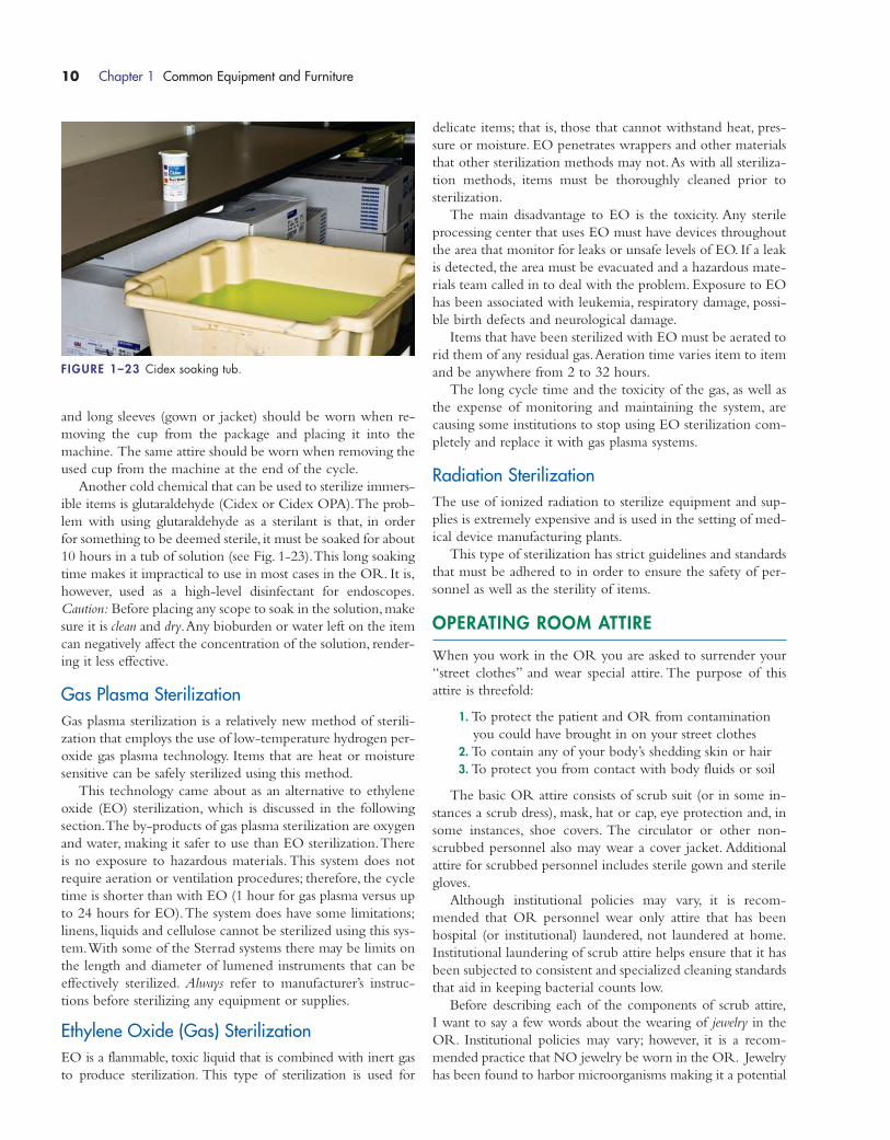
ible items is glutaraldehyde (Cidex or Cidex OPA).The prob-
lem with using glutaraldehyde as a sterilant is that, in order
for something to be deemed sterile, it must be soaked for about
10 hours in a tub of solution (see Fig. 1-23).This long soaking
time makes it impractical to use in most cases in the OR. It is,
however, used as a high-level disinfectant for endoscopes.
Caution: Before placing any scope to soak in the solution,make
sure it is clean and dry.Any bioburden or water left on the item
can negatively affect the concentration of the solution, render-
ing it less effective.
Gas Plasma SterilizationGas plasma sterilization is a relatively new method of sterili-
zation that employs the use of low-temperature hydrogen per-
oxide gas plasma technology. Items that are heat or moisture
sensitive can be safely sterilized using this method.
This technology came about as an alternative to ethylene
oxide (EO) sterilization, which is discussed in the following
section.The by-products of gas plasma sterilization are oxygen
and water, making it safer to use than EO sterilization.There
is no exposure to hazardous materials. This system does not
require aeration or ventilation procedures; therefore, the cycle
time is shorter than with EO (1 hour for gas plasma versus up
to 24 hours for EO).The system does have some limitations;
linens, liquids and cellulose cannot be sterilized using this sys-
tem.With some of the Sterrad systems there may be limits on
the length and diameter of lumened instruments that can be
effectively sterilized. Always refer to manufacturer’s instruc-
tions before sterilizing any equipment or supplies.
Ethylene Oxide (Gas) SterilizationEO is a flammable, toxic liquid that is combined with inert gas
to produce sterilization. This type of sterilization is used for
delicate items; that is, those that cannot withstand heat, pres-
sure or moisture. EO penetrates wrappers and other materials
that other sterilization methods may not.As with all steriliza-
tion methods, items must be thoroughly cleaned prior to
sterilization.
The main disadvantage to EO is the toxicity. Any sterile
processing center that uses EO must have devices throughout
the area that monitor for leaks or unsafe levels of EO. If a leak
is detected, the area must be evacuated and a hazardous mate-
rials team called in to deal with the problem. Exposure to EO
has been associated with leukemia, respiratory damage, possi-
ble birth defects and neurological damage.
Items that have been sterilized with EO must be aerated to
rid them of any residual gas.Aeration time varies item to item
and be anywhere from 2 to 32 hours.
The long cycle time and the toxicity of the gas, as well as
the expense of monitoring and maintaining the system, are
causing some institutions to stop using EO sterilization com-
pletely and replace it with gas plasma systems.
Radiation SterilizationThe use of ionized radiation to sterilize equipment and sup-
plies is extremely expensive and is used in the setting of med-
ical device manufacturing plants.
This type of sterilization has strict guidelines and standards
that must be adhered to in order to ensure the safety of per-
sonnel as well as the sterility of items.
OPERATING ROOM ATTIRE
When you work in the OR you are asked to surrender your
“street clothes” and wear special attire. The purpose of this
attire is threefold:
1. To protect the patient and OR from contamination
you could have brought in on your street clothes
2. To contain any of your body’s shedding skin or hair
3. To protect you from contact with body fluids or soil
The basic OR attire consists of scrub suit (or in some in-
stances a scrub dress), mask, hat or cap, eye protection and, in
some instances, shoe covers. The circulator or other non-
scrubbed personnel also may wear a cover jacket. Additional
attire for scrubbed personnel includes sterile gown and sterile
gloves.
Although institutional policies may vary, it is recom-
mended that OR personnel wear only attire that has been
hospital (or institutional) laundered, not laundered at home.
Institutional laundering of scrub attire helps ensure that it has
been subjected to consistent and specialized cleaning standards
that aid in keeping bacterial counts low.
Before describing each of the components of scrub attire,
I want to say a few words about the wearing of jewelry in the
OR. Institutional policies may vary; however, it is a recom-
mended practice that NO jewelry be worn in the OR. Jewelry
has been found to harbor microorganisms making it a potential
10 Chapter 1 Common Equipment and Furniture
FIGURE 1–23 Cidex soaking tub.
1572_Ch01_001-050.qxd 8/6/09 10:48 AM Page 10
source of contamination.Earrings or necklaces that are not fully
contained under the scrub attire could come loose and fall into
the surgical field or the wound. Jewelry could become contam-
inated with blood or other particles and pose a risk to person-
nel wearing it or others who contact it.
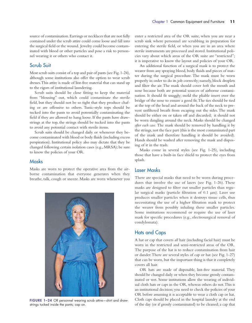
Scrub SuitMost scrub suits consist of a top and pair of pants (see Fig. 1-24),
although some institutions also offer the option to wear scrub
dresses.This attire is made of lint-free material that can stand up
to the rigors of institutional laundering.
Scrub suits should be close fitting to keep the material
from “blousing” out, which could contaminate the sterile
field, but they should not be so tight that they produce chaf-
ing or are offensive to others. Tunic-style tops should be
tucked into the pants to avoid potentially contaminating the
field if they are allowed to hang loose. If the pants have draw-
strings at the top, the strings should be tucked into the pants
to avoid any potential contact with sterile items.
Scrub suits should be changed daily or whenever they be-
come contaminated with blood or body fluids (including excess
perspiration). Institutional policy also may dictate that they be
changed following certain isolation cases (e.g., MRSA); be sure
to know the policies of your OR.
MasksMasks are worn to protect the operative area from the air-
borne contamination that everyone generates when they
breathe, talk, cough or sneeze. Masks are worn whenever you
enter a restricted area of the OR suite, when you are near a
scrub sink where personnel are scrubbing in preparation for
entering the sterile field, or when you are in an area where
sterile instruments are processed and stored. Institutional poli-
cies vary about which areas of the OR suite are “restricted”;
it is imperative to know the layout and policies of your OR.
An additional function of a surgical mask is to protect the
wearer from any spraying blood, body fluids and pieces of mat-
ter during the surgical procedure. The mask must be worn
properly in order to do its job correctly; namely, block droplets
and filter the air.The mask should cover both the mouth and
nose because both are potential sources of airborne contami-
nation. It should fit snuggly; mold the pliable insert over the
bridge of the nose to ensure a good fit.The ties should be tied
at the top of the head and around the back of the neck to pre-
vent unfiltered breath from escaping out the sides. The mask
should be either on or taken off and discarded; it should not
be worn dangling around the neck. Masks should be changed
after each case. The mask should be removed by handling it by
the strings, not the face part (this is the most contaminated part
of the mask and therefore handling it should be avoided).
Hands should be washed after removing the mask and dispos-
ing of it in the trash.
Masks come in several styles (see Fig. 1-25), including
those that have a built-in face shield to protect the eyes from
splash.
Laser MasksThere are special masks that need to be worn during proce-
dures that involve the use of lasers (see Fig. 1-26). These
masks are designed to filter out smaller particles than regu-
lar surgical masks (particle filtration of 0.1 μm). Laser use
produces smaller particles when it destroys tissue cells, thus
necessitating the use of a higher filtration mask to protect
the wearer from possibly inhaling these smaller particles.
Some institutions recommend or require the use of laser
mask for specific procedures (e.g., electrosurgical removal of
condylomata).
Hats and CapsA hat or cap that covers all hair (including facial hair) must be
worn in the restricted and semi-restricted areas of the OR.
The purpose of the hat is to reduce contamination from hair
or dander.There are several styles of cap or hat (see Fig. 1-27)
that can be worn, but the important thing is that it completely
covers all hair.
OR hats are made of disposable, lint-free material. They
should be changed daily or when they become grossly contam-
inated or wet. Some institutions allow the wearing of individ-
ual cloth hats or caps in the OR, whereas others do not.This is
an institutional decision; you need to check the policies of your
OR before assuming it is acceptable to wear a cloth cap or hat.
Cloth caps should be placed in the hospital laundry at the end
of the day (or if grossly contaminated) to be cleaned; a cap that
Chapter 1 Common Equipment and Furniture 11
FIGURE 1–24 OR personnel wearing scrub attire—shirt and draw-strings tucked inside the pants; cap on.
1572_Ch01_001-050.qxd 8/6/09 10:48 AM Page 11
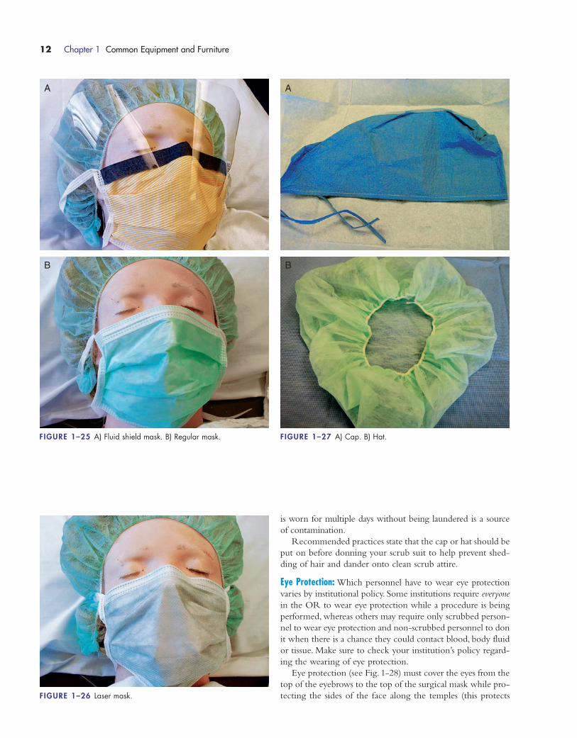
FIGURE 1–27 A) Cap. B) Hat.
is worn for multiple days without being laundered is a source
of contamination.
Recommended practices state that the cap or hat should be
put on before donning your scrub suit to help prevent shed-
ding of hair and dander onto clean scrub attire.
Eye Protection: Which personnel have to wear eye protection
varies by institutional policy. Some institutions require everyone
in the OR to wear eye protection while a procedure is being
performed, whereas others may require only scrubbed person-
nel to wear eye protection and non-scrubbed personnel to don
it when there is a chance they could contact blood, body fluid
or tissue. Make sure to check your institution’s policy regard-
ing the wearing of eye protection.
Eye protection (see Fig. 1-28) must cover the eyes from the
top of the eyebrows to the top of the surgical mask while pro-
tecting the sides of the face along the temples (this protects
12 Chapter 1 Common Equipment and Furniture
FIGURE 1–26 Laser mask.
A
B
FIGURE 1–25 A) Fluid shield mask. B) Regular mask.
A
B
1572_Ch01_001-050.qxd 8/6/09 10:48 AM Page 12
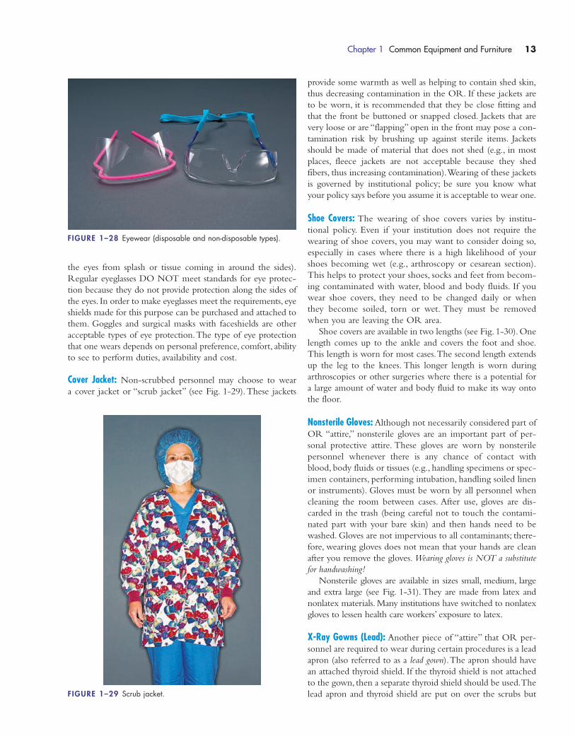
the eyes from splash or tissue coming in around the sides).
Regular eyeglasses DO NOT meet standards for eye protec-
tion because they do not provide protection along the sides of
the eyes. In order to make eyeglasses meet the requirements, eye
shields made for this purpose can be purchased and attached to
them. Goggles and surgical masks with faceshields are other
acceptable types of eye protection.The type of eye protection
that one wears depends on personal preference, comfort, ability
to see to perform duties, availability and cost.
Cover Jacket: Non-scrubbed personnel may choose to wear
a cover jacket or “scrub jacket” (see Fig. 1-29). These jackets
provide some warmth as well as helping to contain shed skin,
thus decreasing contamination in the OR. If these jackets are
to be worn, it is recommended that they be close fitting and
that the front be buttoned or snapped closed. Jackets that are
very loose or are “flapping” open in the front may pose a con-
tamination risk by brushing up against sterile items. Jackets
should be made of material that does not shed (e.g., in most
places, fleece jackets are not acceptable because they shed
fibers, thus increasing contamination).Wearing of these jackets
is governed by institutional policy; be sure you know what
your policy says before you assume it is acceptable to wear one.
Shoe Covers: The wearing of shoe covers varies by institu-
tional policy. Even if your institution does not require the
wearing of shoe covers, you may want to consider doing so,
especially in cases where there is a high likelihood of your
shoes becoming wet (e.g., arthroscopy or cesarean section).
This helps to protect your shoes, socks and feet from becom-
ing contaminated with water, blood and body fluids. If you
wear shoe covers, they need to be changed daily or when
they become soiled, torn or wet. They must be removed
when you are leaving the OR area.
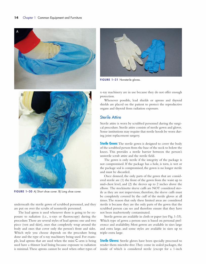
Shoe covers are available in two lengths (see Fig. 1-30).One
length comes up to the ankle and covers the foot and shoe.
This length is worn for most cases.The second length extends
up the leg to the knees. This longer length is worn during
arthroscopies or other surgeries where there is a potential for
a large amount of water and body fluid to make its way onto
the floor.
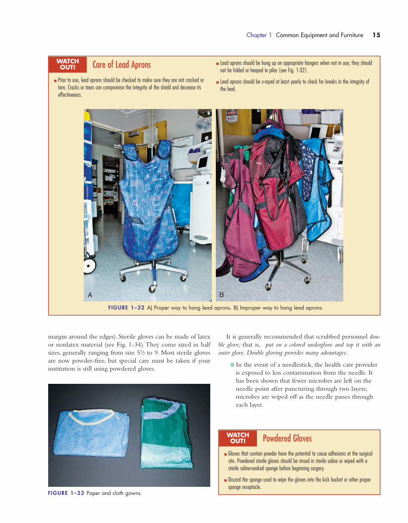
Nonsterile Gloves: Although not necessarily considered part of
OR “attire,” nonsterile gloves are an important part of per-
sonal protective attire. These gloves are worn by nonsterile
personnel whenever there is any chance of contact with
blood, body fluids or tissues (e.g., handling specimens or spec-
imen containers, performing intubation, handling soiled linen
or instruments). Gloves must be worn by all personnel when
cleaning the room between cases. After use, gloves are dis-
carded in the trash (being careful not to touch the contami-
nated part with your bare skin) and then hands need to be
washed. Gloves are not impervious to all contaminants; there-
fore, wearing gloves does not mean that your hands are clean
after you remove the gloves. Wearing gloves is NOT a substitute
for handwashing!
Nonsterile gloves are available in sizes small, medium, large
and extra large (see Fig. 1-31).They are made from latex and
nonlatex materials. Many institutions have switched to nonlatex
gloves to lessen health care workers’ exposure to latex.
X-Ray Gowns (Lead): Another piece of “attire” that OR per-
sonnel are required to wear during certain procedures is a lead
apron (also referred to as a lead gown).The apron should have
an attached thyroid shield. If the thyroid shield is not attached
to the gown, then a separate thyroid shield should be used.The
lead apron and thyroid shield are put on over the scrubs but
Chapter 1 Common Equipment and Furniture 13
FIGURE 1–28 Eyewear (disposable and non-disposable types).
FIGURE 1–29 Scrub jacket.
1572_Ch01_001-050.qxd 8/6/09 10:48 AM Page 13
underneath the sterile gown of scrubbed personnel, and they
are put on over the scrubs of nonsterile personnel.
The lead apron is used whenever there is going to be ex-
posure to radiation (i.e., x-rays or fluoroscopy) during the
procedure.There are several styles of lead aprons: one and two
piece (vest and skirt), ones that completely wrap around the
body and ones that cover only the person’s front and sides.
Which style you choose depends on the procedure being
done and the type of x-ray machinery being used. For exam-
ple, lead aprons that are used when the mini C-arm is being
used have a thinner lead lining because exposure to radiation
is minimal.These aprons cannot be used when other types of
x-ray machinery are in use because they do not offer enough
protection.
Whenever possible, lead shields or aprons and thyroid
shields are placed on the patient to protect the reproductive
organs and thyroid from radiation exposure.
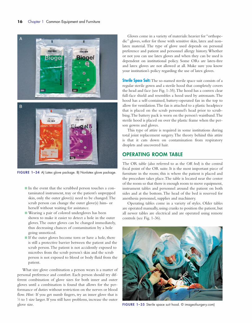
Sterile AttireSterile attire is worn by scrubbed personnel during the surgi-
cal procedure. Sterile attire consists of sterile gown and gloves.
Some institutions may require that sterile hoods be worn dur-
ing joint replacement surgery.
Sterile Gown: The sterile gown is designed to cover the body
of the scrubbed person from the base of the neck to below the
knees. This provides a sterile barrier between the person’s
unsterile scrub attire and the sterile field.
The gown is only sterile if the integrity of the package is
not compromised. If the package has a hole, is torn, is wet or
the package seal is compromised, the gown is no longer sterile
and must be discarded.
Once donned, the only parts of the gown that are consid-
ered sterile are (1) the front of the gown from the waist up to
mid-chest level, and (2) the sleeves up to 2 inches above the
elbow. The stockinette sleeve cuffs are NOT considered ster-
ile as they are not impervious; therefore, the sleeve cuffs must
be completely covered by the cuff of the sterile gloves at all
times.The reason that only these limited areas are considered
sterile is because they are the only parts of the gown that the
scrubbed person can see and therefore ensure that they have
not been inadvertently contaminated.
Sterile gowns are available in cloth or paper (see Fig. 1-33).
Which type of gown a person uses is based on personal pref-
erence and availability. Most gowns are available in sizes large
and extra large, and some styles are available in sizes up to
triple-extra large.
Sterile Gloves: Sterile gloves have been specially processed to
render them microbe-free.They come in sealed packages, the
inside of which is considered sterile (except for a 1-inch
14 Chapter 1 Common Equipment and Furniture
FIGURE 1–31 Nonsterile gloves.
FIGURE 1–30 A) Short shoe cover. B) Long shoe cover.
A
B
1572_Ch01_001-050.qxd 8/6/09 10:48 AM Page 14
margin around the edges). Sterile gloves can be made of latex
or nonlatex material (see Fig. 1-34).They come sized in half
sizes, generally ranging from size 51⁄2 to 9. Most sterile gloves
are now powder-free, but special care must be taken if your
institution is still using powdered gloves.
Chapter 1 Common Equipment and Furniture 15
Care of Lead Aprons
� Prior to use, lead aprons should be checked to make sure they are not cracked ortorn. Cracks or tears can compromise the integrity of the shield and decrease itseffectiveness.
WATCH OUT!
� Lead aprons should be hung up on appropriate hangers when not in use; they shouldnot be folded or heaped in piles (see Fig. 1-32).
� Lead aprons should be x-rayed at least yearly to check for breaks in the integrity ofthe lead.
FIGURE 1–32 A) Proper way to hang lead aprons. B) Improper way to hang lead aprons.
A B
It is generally recommended that scrubbed personnel dou-
ble glove; that is, put on a colored underglove and top it with an
outer glove. Double gloving provides many advantages:
� In the event of a needlestick, the health care provider
is exposed to less contamination from the needle. It
has been shown that fewer microbes are left on the
needle point after puncturing through two layers;
microbes are wiped off as the needle passes through
each layer.
FIGURE 1–33 Paper and cloth gowns.
Powdered Gloves
� Gloves that contain powder have the potential to cause adhesions at the surgicalsite. Powdered sterile gloves should be rinsed in sterile saline or wiped with asterile saline-soaked sponge before beginning surgery.
� Discard the sponge used to wipe the gloves into the kick bucket or other propersponge receptacle.
WATCH OUT!
1572_Ch01_001-050.qxd 8/6/09 10:48 AM Page 15
� In the event that the scrubbed person touches a con-
taminated instrument, tray or the patient’s unprepped
skin, only the outer glove(s) need to be changed.The
scrub person can change the outer glove(s) him- or
herself without waiting for assistance.
� Wearing a pair of colored undergloves has been
shown to make it easier to detect a hole in the outer
gloves.The outer gloves can be changed immediately,
thus decreasing chances of contamination by a hole
going unnoticed.
� If the outer gloves become torn or have a hole, there
is still a protective barrier between the patient and the
scrub person.The patient is not accidently exposed to
microbes from the scrub person’s skin and the scrub
person is not exposed to blood or body fluid from the
patient.
What size glove combination a person wears is a matter of
personal preference and comfort. Each person should try dif-
ferent combination of glove sizes for both inner and outer
gloves until a combination is found that allows for the per-
formance of duties without restriction on the nerves or blood
flow. Hint: If you get numb fingers, try an inner glove that is1⁄2 to 1 size larger. If you still have problems, increase the outer
glove size.
Gloves come in a variety of materials: heavier for “orthope-
dic” gloves, softer for those with sensitive skin, latex and non-
latex material. The type of glove used depends on personal
preference and patient and personnel allergy history.Whether
or not you can use latex gloves and when they can be used is
dependent on institutional policy. Some ORs are latex-free
and latex gloves are not allowed at all. Make sure you know
your institution’s policy regarding the use of latex gloves.
Sterile Space Suit: The so-named sterile space suit consists of a
regular sterile gown and a sterile hood that completely covers
the head and face (see Fig. 1-35).The hood has a convex clear
full-face shield and resembles a hood used by astronauts.The
hood has a self-contained, battery-operated fan in the top to
allow for ventilation.The fan is attached to a plastic headpiece
that is placed on the scrub personnel’s head prior to scrub-
bing.The battery pack is worn on the person’s waistband.The
sterile hood is placed on over the plastic frame when the per-
son gowns and gloves.
This type of attire is required in some institutions during
total joint replacement surgery.The theory behind this attire
is that it cuts down on contamination from respiratory
droplets and uncovered hair.
OPERATING ROOM TABLE
The OR table (also referred to as the OR bed) is the central
focal point of the OR suite. It is the most important piece of
furniture in the room; this is where the patient is placed and
the procedure takes place.The table is located near the center
of the room so that there is enough room to move equipment,
instrument tables and personnel around the patient on both
sides and at the bottom.The head of the bed is reserved for
anesthesia personnel, supplies and machinery.
Operating tables come in a variety of styles. Older tables
are operated manually, using cranks to position the patient, but
all newer tables are electrical and are operated using remote
controls (see Fig. 1-36).
16 Chapter 1 Common Equipment and Furniture
FIGURE 1–35 Sterile space suit hood. © imageofsurgery.com)
FIGURE 1–34 A) Latex glove package. B) Nonlatex glove package.
A
B
1572_Ch01_001-050.qxd 8/6/09 10:48 AM Page 16
As with any piece of medical equipment, it is important to
know how to use the OR table in the room you are assigned
(see Fig. 1-37). Make sure you know the manufacturer’s in-
structions for table use. All personnel should receive instruction
on the tables being used in their institution as part of their ori-
entation as well as when any new table is purchased. Failure to
be familiar with proper table use could result in surgery delays,
increased anesthesia time for the patient (while personnel try to
figure out how to work the bed during a procedure) or
injury to the patient or staff.
Parts of an Operating Room TableHeadpiece: The headpiece is a removable padded section at-
tached at the head of the bed (usually with metal rods or bars).
This section can be adjusted up or down to aid in airway main-
tenance.The headpiece can be removed to allow for attachment
of other headrests such as a Mayfield headrest (neurosurgery) or
a horseshoe headrest.The headpiece also can be removed and
placed at the bottom of the table for some gynecological
procedures.
Body: The body of the table usually consists of two padded
pieces with a “break” in between them.The break allows for
each piece to be flexed or extended independently for patient
positioning (e.g., Fowler’s or sitting position).There is a metal
bar in the break area that can be moved up and down.This bar
is known as a kidney rest and is used to elevate the flank area
for better exposure during renal procedures.
Footpiece: The footpiece or bottom of the bed is a padded re-
movable section where the patient’s lower legs and feet rest.
The footpiece can be flexed or extended independently of the
rest of the bed for patient positioning, or it can be moved out
of the way during vaginal procedures.
Operating Table AttachmentsMayfield Headrest: This attachment (see Fig. 1-38) is used for
some craniotomy procedures.The Mayfield headrest generally
is used to hold the head still when the patient is prone or in
Fowler’s (sitting) position. The headrest is attached to the
patient’s skull using sterile pins. This headrest is attached to
the patient by the surgeon and positioning is done with the
surgeon present.
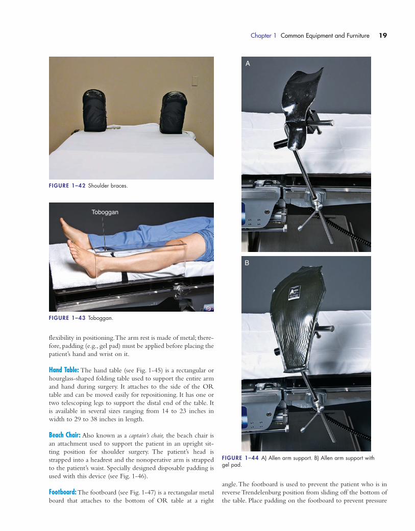
Gardner Tongs: Gardner tongs (see Fig. 1-39) are used to apply
cervical traction to patients with cervical spine fractures.They
are attached into each side of the patient’s skull using sterile
pins.Weight is attached to a rope loop that goes over a pulley
to create the traction.
Chapter 1 Common Equipment and Furniture 17
Body Head
RemoteControl
Foot
Kidney Rest BreakFIGURE 1–37 OR table parts.
FIGURE 1–36 A) Manual OR table. B) Electric OR table.
A
B
Operating Room Table Weight Limits
� Some older manual models of OR tables have a 350-lb weight limit.
� Some older electric models of OR tables have a 500- to 550-lb weight limit.
� Some newer models of OR tables have capacities of 1000 lbs plus, but only incertain positions (e.g., supine); the weight limit is lower if the patient is in lithotomy or other position.
It is imperative that you know the weight limit of the OR table you are usingPRIOR to placing the patient on it. Failure to use the proper table for large patientscould result in severe injury to the patient or staff.
WATCH OUT!
1572_Ch01_001-050.qxd 8/6/09 10:48 AM Page 17
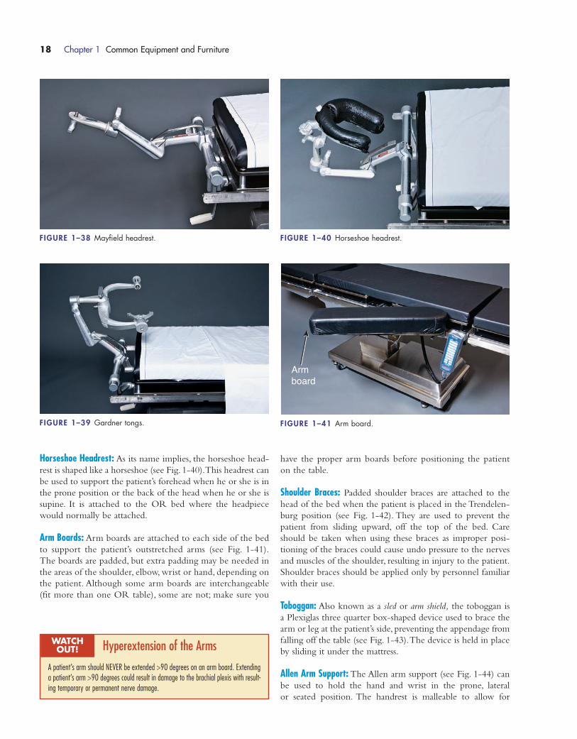
Horseshoe Headrest: As its name implies, the horseshoe head-
rest is shaped like a horseshoe (see Fig. 1-40).This headrest can
be used to support the patient’s forehead when he or she is in
the prone position or the back of the head when he or she is
supine. It is attached to the OR bed where the headpiece
would normally be attached.
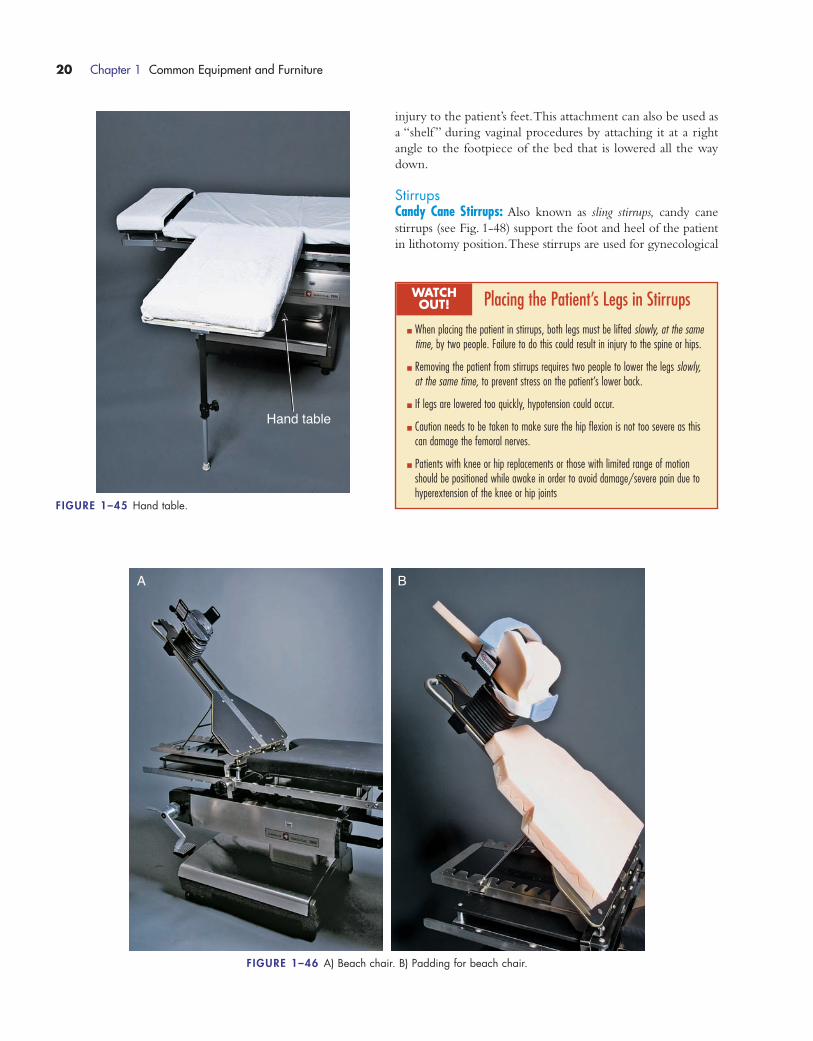
Arm Boards: Arm boards are attached to each side of the bed
to support the patient’s outstretched arms (see Fig. 1-41).
The boards are padded, but extra padding may be needed in
the areas of the shoulder, elbow, wrist or hand, depending on
the patient. Although some arm boards are interchangeable
(fit more than one OR table), some are not; make sure you
have the proper arm boards before positioning the patient
on the table.
Shoulder Braces: Padded shoulder braces are attached to the
head of the bed when the patient is placed in the Trendelen-
burg position (see Fig. 1-42). They are used to prevent the
patient from sliding upward, off the top of the bed. Care
should be taken when using these braces as improper posi-
tioning of the braces could cause undo pressure to the nerves
and muscles of the shoulder, resulting in injury to the patient.
Shoulder braces should be applied only by personnel familiar
with their use.
Toboggan: Also known as a sled or arm shield, the toboggan is
a Plexiglas three quarter box-shaped device used to brace the
arm or leg at the patient’s side, preventing the appendage from
falling off the table (see Fig. 1-43).The device is held in place
by sliding it under the mattress.
Allen Arm Support: The Allen arm support (see Fig. 1-44) can
be used to hold the hand and wrist in the prone, lateral
or seated position. The handrest is malleable to allow for
18 Chapter 1 Common Equipment and Furniture
FIGURE 1–39 Gardner tongs.
FIGURE 1–38 Mayfield headrest. FIGURE 1–40 Horseshoe headrest.
Arm board
FIGURE 1–41 Arm board.
Hyperextension of the Arms
A patient’s arm should NEVER be extended >90 degrees on an arm board. Extendinga patient’s arm >90 degrees could result in damage to the brachial plexis with result-ing temporary or permanent nerve damage.
WATCH OUT!
1572_Ch01_001-050.qxd 8/6/09 10:48 AM Page 18
flexibility in positioning.The arm rest is made of metal; there-
fore, padding (e.g., gel pad) must be applied before placing the
patient’s hand and wrist on it.
Hand Table: The hand table (see Fig. 1-45) is a rectangular or
hourglass-shaped folding table used to support the entire arm
and hand during surgery. It attaches to the side of the OR
table and can be moved easily for repositioning. It has one or
two telescoping legs to support the distal end of the table. It
is available in several sizes ranging from 14 to 23 inches in
width to 29 to 38 inches in length.
Beach Chair: Also known as a captain’s chair, the beach chair is
an attachment used to support the patient in an upright sit-
ting position for shoulder surgery. The patient’s head is
strapped into a headrest and the nonoperative arm is strapped
to the patient’s waist. Specially designed disposable padding is
used with this device (see Fig. 1-46).
Footboard: The footboard (see Fig. 1-47) is a rectangular metal
board that attaches to the bottom of OR table at a right
angle.The footboard is used to prevent the patient who is in
reverse Trendelenburg position from sliding off the bottom of
the table. Place padding on the footboard to prevent pressure
Chapter 1 Common Equipment and Furniture 19
FIGURE 1–42 Shoulder braces.
Toboggan
FIGURE 1–43 Toboggan.
A
B
FIGURE 1–44 A) Allen arm support. B) Allen arm support with gel pad.
1572_Ch01_001-050.qxd 8/6/09 10:48 AM Page 19
injury to the patient’s feet.This attachment can also be used as
a “shelf ” during vaginal procedures by attaching it at a right
angle to the footpiece of the bed that is lowered all the way
down.
StirrupsCandy Cane Stirrups: Also known as sling stirrups, candy cane
stirrups (see Fig. 1-48) support the foot and heel of the patient
in lithotomy position.These stirrups are used for gynecological
20 Chapter 1 Common Equipment and Furniture
A B
FIGURE 1–46 A) Beach chair. B) Padding for beach chair.
Hand table
FIGURE 1–45 Hand table.
Placing the Patient’s Legs in Stirrups
� When placing the patient in stirrups, both legs must be lifted slowly, at the sametime, by two people. Failure to do this could result in injury to the spine or hips.
� Removing the patient from stirrups requires two people to lower the legs slowly,at the same time, to prevent stress on the patient’s lower back.
� If legs are lowered too quickly, hypotension could occur.
� Caution needs to be taken to make sure the hip flexion is not too severe as thiscan damage the femoral nerves.
� Patients with knee or hip replacements or those with limited range of motionshould be positioned while awake in order to avoid damage/severe pain due tohyperextension of the knee or hip joints
WATCH OUT!
1572_Ch01_001-050.qxd 8/6/09 10:48 AM Page 20
procedures. Care must be taken to make sure the side of the
patient’s lower leg is NOT resting on the metal post of the
candy cane stirrup. Do not place the sling directly over the
Achilles area as pressure could cause injury to the peroneal
nerve.
Knee-Crutch Stirrups: These stirrups provide support under
the knee and areas just above and below the knee (see
Fig. 1-49). They are generally used for short-duration knee
arthroscopy or cystoscopy procedures. Care must be taken to
Chapter 1 Common Equipment and Furniture 21
FIGURE 1–47 Footboard.
FIGURE 1–48 Candy cane stirrups.
FIGURE 1–49 Knee-crutch stirrups.
properly pad and position the stirrups to prevent popliteal
nerve damage.
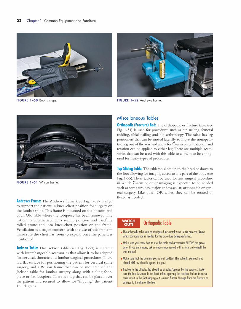
Boot Stirrups: Rapidly becoming the stirrup of choice for
many gynecological and rectal procedures, the boot stirrup
provides support to the patient’s foot and entire lower leg (see
Fig. 1-50).This type of stirrup is completely adjustable both
vertically and for abduction.The boot is padded, but care must
be taken to properly place the patient’s leg in the boot (make
sure the heel is all the way back into the boot) to prevent
nerve injury. There are several manufacturers of boot-type
stirrups so make sure you are familiar with the operation of
the stirrups (how to raise, lower and abduct them) used at
your facility.
Frames for Lumbar SurgeryWilson Frame:The Wilson frame (see Fig. 1-51) is used to sup-
port a patient’s torso in a flexed, prone position.This frame is
used for surgeries of the lumbar spine including laminectomy,
diskectomy and microdiskectomy as well as insertion of blad-
der control stimulators. It consists of two padded half circles
mounted on a frame that is placed under the prone patient’s
torso. Using a crank system, the supports can be raised or
lowered to achieve the desired amount of flexion. The sup-
ports also can be adjusted laterally to fit multiple patient body
types.
1572_Ch01_001-050.qxd 8/6/09 10:48 AM Page 21
Miscellaneous TablesOrthopedic (Fracture) Bed: The orthopedic or fracture table (see
Fig. 1-54) is used for procedures such as hip nailing, femoral
rodding, tibial nailing and hip arthroscopy. The table has leg
positioners that can be moved laterally to move the nonopera-
tive leg out of the way and allow for C-arm access.Traction and
rotation can be applied to either leg.There are multiple acces-
sories that can be used with this table to allow it to be config-
ured for many types of procedures.
Top Sliding Table: The tabletop slides up to the head or down to
the foot allowing for imaging access to any part of the body (see
Fig. 1-55).These tables can be used for any surgical procedure
in which C-arm or other imaging is expected to be needed
such as some urology, major endovascular, orthopedic or gen-
eral surgery. Like other OR tables, they can be rotated or
flexed as needed.
22 Chapter 1 Common Equipment and Furniture
FIGURE 1–50 Boot stirrups.
FIGURE 1–51 Wilson frame.
FIGURE 1–52 Andrews frame.
Andrews Frame: The Andrews frame (see Fig. 1-52) is used
to support the patient in knee-chest position for surgery on
the lumbar spine.This frame is mounted on the bottom end
of an OR table where the footpiece has been removed.The
patient is anesthetized in a supine position and carefully
rolled prone and into knee-chest position on the frame.
Ventilation is a major concern with the use of this frame—
make sure the chest has room to expand once the patient is
positioned.
Jackson Table: The Jackson table (see Fig. 1-53) is a frame
with interchangeable accessories that allow it to be adapted
for cervical, thoracic and lumbar surgical procedures.There
is a flat surface for positioning the patient for cervical spine
surgery, and a Wilson frame that can be mounted on the
Jackson table for lumbar surgery along with a sling foot-
piece or flat footpiece.There is a top that can be placed over
the patient and secured to allow for “flipping” the patient
180 degrees.
Orthopedic Table
� The orthopedic table can be configured in several ways. Make sure you knowwhich configuration is needed for the procedure being performed.
� Make sure you know how to use the table and accessories BEFORE the proce-dure. If you are unsure, ask someone experienced with its use and consult theuser manual.
� Make sure that the perineal post is well padded. The patient’s perineal areashould NOT rest directly against the post.
� Traction to the affected leg should be directed/applied by the surgeon. Makesure the foot is secure in the boot before applying the traction. Failure to do socould result in the foot slipping out, causing further damage from the fracture ordamage to the skin of the foot.
WATCH OUT!
1572_Ch01_001-050.qxd 8/6/09 10:48 AM Page 22
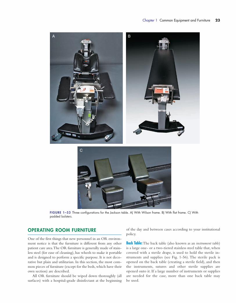
OPERATING ROOM FURNITURE
One of the first things that new personnel in an OR environ-
ment notice is that the furniture is different from any other
patient care area.The OR furniture is generally made of stain-
less steel (for ease of cleaning), has wheels to make it portable
and is designed to perform a specific purpose. It is not deco-
rative but plain and utilitarian. In this section, the most com-
mon pieces of furniture (except for the beds, which have their
own section) are described.
All OR furniture should be wiped down thoroughly (all
surfaces) with a hospital-grade disinfectant at the beginning
of the day and between cases according to your institutional
policy.
Back Table: The back table (also known as an instrument table)
is a large one- or a two-tiered stainless steel table that, when
covered with a sterile drape, is used to hold the sterile in-
struments and supplies (see Fig. 1-56). The sterile pack is
opened on the back table (creating a sterile field), and then
the instruments, sutures and other sterile supplies are
opened onto it. If a large number of instruments or supplies
are needed for the case, more than one back table may
be used.
Chapter 1 Common Equipment and Furniture 23
A
C
B
FIGURE 1–53 Three configurations for the Jackson table. A) With Wilson frame. B) With flat frame. C) Withpadded bolsters.
1572_Ch01_001-050.qxd 8/6/09 10:48 AM Page 23

Back tables come in a variety of sizes and heights.The table
may have one or two top shelves. The model with two top
shelves is generally used for vascular or orthopedic surgery, and
both top shelves are covered with a sterile drape. Some back
tables have a bottom shelf about 12 to 18 inches under the top
shelf. On these models, the bottom shelf is not covered with a
drape and NOT considered sterile.
Mayo Stand: The Mayo stand is covered with a special ster-
ile drape made specifically for that purpose and is used to
set up instruments that need to be readily available for the
case (see Fig. 1-57). The instruments placed on the Mayo
stand vary depending on the case but should be kept to a
minimum to avoid disorganization and wasted time hunting
for needed items. The space on top of the Mayo stand is
limited; use it only for the items needed to start the case
(e.g., scalpels and superficial retractors) and those that will
be used repeatedly (e.g., common rings such as kochers,
allis and hemostats; Mayo and Metzenbaum scissors; smooth
and tissue forceps) on it.The rest of the instruments should
be organized on the back table. Instruments can be removed
and replaced with others (e.g., deeper retractors) as the
surgery progresses.
Mayo stands come in a variety of styles. The height on
most Mayo stands can be adjusted from 36 to 62 inches to
allow them to be moved up to the field, over the patient.
Height adjustment is made either by stepping on a foot but-
ton or pulling a hand lever, depending on the model. Stan-
dard Mayo stands have a tray surface area of 12 × 19 inches
or 16 × 21 inches. Extra large Mayo stands (used mostly in
orthopedic or neurosurgical procedures) have a surface area
of 20 × 25 inches.The extra large stand requires an extra large
sterile Mayo cover.
Utility Table: The utility table (also called a prep table) is a
small stainless steel table (see Fig. 1-58) that can be used for
a variety of things.When it is covered with a sterile drape,
it can be used to hold the basins and other supplies used
24 Chapter 1 Common Equipment and Furniture
FIGURE 1–54 A) Fracture table closed. B) Fracture table opened.
A
B
FIGURE 1–55 Top-sliding table.
FIGURE 1–56 Back table.
1572_Ch01_001-050.qxd 8/6/09 10:48 AM Page 24
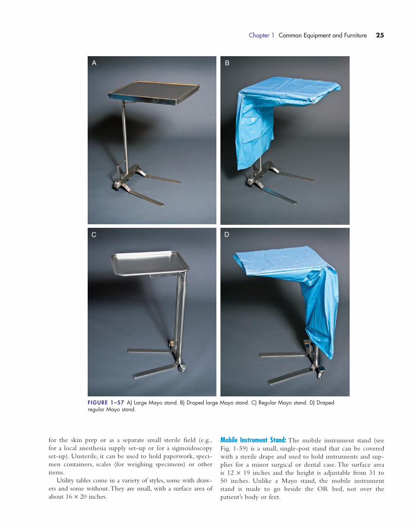
for the skin prep or as a separate small sterile field (e.g.,
for a local anesthesia supply set-up or for a sigmoidoscopy
set-up). Unsterile, it can be used to hold paperwork, speci-
men containers, scales (for weighing specimens) or other
items.
Utility tables come in a variety of styles, some with draw-
ers and some without.They are small, with a surface area of
about 16 × 20 inches.
Mobile Instrument Stand: The mobile instrument stand (see
Fig. 1-59) is a small, single-post stand that can be covered
with a sterile drape and used to hold instruments and sup-
plies for a minor surgical or dental case. The surface area
is 12 × 19 inches and the height is adjustable from 31 to
50 inches. Unlike a Mayo stand, the mobile instrument
stand is made to go beside the OR bed, not over the
patient’s body or feet.
Chapter 1 Common Equipment and Furniture 25
FIGURE 1–57 A) Large Mayo stand. B) Draped large Mayo stand. C) Regular Mayo stand. D) Draped regular Mayo stand.
A B
C D
1572_Ch01_001-050.qxd 8/6/09 10:48 AM Page 25
Kick Bucket: The kick bucket (see Fig. 1-60) is a stainless steel
13-quart bucket set into a round, rolling stainless steel frame.
The bucket is commonly used to contain dirty sponges dis-
carded from the field. The sponges are discarded into the
bucket; the circulator (using proper standard precautions) may
drape the sponges over the side of the bucket for ease of
counting. Some institutions require that discarded sponges be
placed in plastic impervious sponge count bags for counting,
not draped on the side of the bucket. Check your institution’s
policies and procedures.
Ring Stand: The ring stand (also known as a basin stand) is
a round stainless steel ring mounted on a rolling stand (see
Fig. 1-61).The large (8.5 quart) sterile basin is opened in this
stand with the wrapper opened outward to cover the top and
side of the stand, creating a sterile field.The sterile basin can
then be used as a place into which to organize drapes or
gowns or to open other sterile items.
Ring stands are available as single or double (holds two
basins side by side) models. They have open frames to allow
easy access from any side and may or may not have a shelf
underneath.
Standing Stool: The standing stool (also known as a step stool)
comes in a variety of styles (see Fig. 1-62). Like the name
implies, this stool is made for members of the surgical team
to stand on during a procedure for better visualization of the
surgical field. The important thing to check on a standing
26 Chapter 1 Common Equipment and Furniture
FIGURE 1–58 Utility table.
FIGURE 1–59 Mobile instrument stand. FIGURE 1–60 Kick bucket.
1572_Ch01_001-050.qxd 8/6/09 10:48 AM Page 26
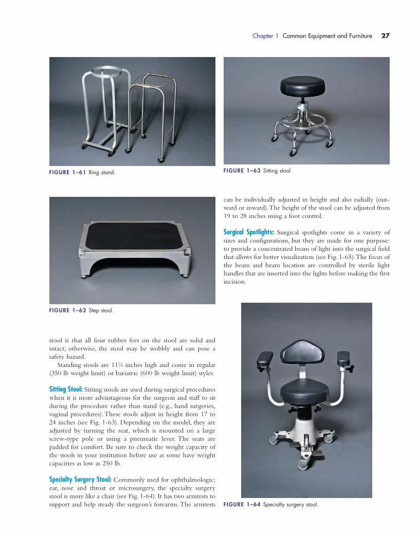
stool is that all four rubber feet on the stool are solid and
intact; otherwise, the stool may be wobbly and can pose a
safety hazard.
Standing stools are 111⁄4 inches high and come in regular
(350 lb weight limit) or bariatric (600 lb weight limit) styles.
Sitting Stool: Sitting stools are used during surgical procedures
when it is more advantageous for the surgeon and staff to sit
during the procedure rather than stand (e.g., hand surgeries,
vaginal procedures).These stools adjust in height from 17 to
24 inches (see Fig. 1-63). Depending on the model, they are
adjusted by turning the seat, which is mounted on a large
screw-type pole or using a pneumatic lever. The seats are
padded for comfort. Be sure to check the weight capacity of
the stools in your institution before use as some have weight
capacities as low as 250 lb.
Specialty Surgery Stool: Commonly used for ophthalmologic;
ear, nose and throat or microsurgery, the specialty surgery
stool is more like a chair (see Fig. 1-64). It has two armrests to
support and help steady the surgeon’s forearms.The armrests
can be individually adjusted in height and also radially (out-
ward or inward).The height of the stool can be adjusted from
19 to 28 inches using a foot control.
Surgical Spotlights: Surgical spotlights come in a variety of
sizes and configurations, but they are made for one purpose:
to provide a concentrated beam of light into the surgical field
that allows for better visualization (see Fig. 1-65).The focus of
the beam and beam location are controlled by sterile light
handles that are inserted into the lights before making the first
incision.
Chapter 1 Common Equipment and Furniture 27
FIGURE 1–61 Ring stand.
FIGURE 1–62 Step stool.
FIGURE 1–63 Sitting stool.
FIGURE 1–64 Specialty surgery stool.
1572_Ch01_001-050.qxd 8/6/09 10:48 AM Page 27
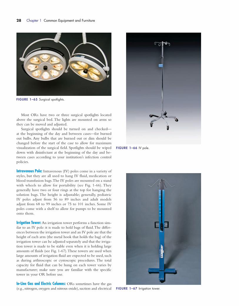
Most ORs have two or three surgical spotlights located
above the surgical bed. The lights are mounted on arms so
they can be moved and adjusted.
Surgical spotlights should be turned on and checked—
at the beginning of the day and between cases—for burned
out bulbs. Any bulbs that are burned out or dim should be
changed before the start of the case to allow for maximum
visualization of the surgical field. Spotlights should be wiped
down with disinfectant at the beginning of the day and be-
tween cases according to your institution’s infection control
policies.
Intravenous Pole: Intravenous (IV) poles come in a variety of
styles, but they are all used to hang IV fluid, medication or
blood transfusion bags.The IV poles are mounted on a stand
with wheels to allow for portability (see Fig. 1-66). They
generally have two or four rings at the top for hanging the
solution bags. The height is adjustable; generally, pediatric
IV poles adjust from 56 to 89 inches and adult models
adjust from 68 to 99 inches or 75 to 101 inches. Some IV
poles come with a shelf to allow for pumps to be mounted
onto them.
Irrigation Tower: An irrigation tower performs a function sim-
ilar to an IV pole: it is made to hold bags of fluid.The differ-
ences between the irrigation tower and an IV pole are that the
height of each arm (the metal hook that holds the bag) of the
irrigation tower can be adjusted separately and that the irriga-
tion tower is made to be stable even when it is holding large
amounts of fluids (see Fig. 1-67).These towers are used when
large amounts of irrigation fluid are expected to be used, such
as during arthroscopic or cystoscopic procedures. The total
capacity for fluid that can be hung on each tower varies by
manufacturer; make sure you are familiar with the specific
tower in your OR before use.
In-Line Gas and Electric Columns: ORs sometimes have the gas
(e.g., nitrogen, oxygen and nitrous oxide), suction and electrical
28 Chapter 1 Common Equipment and Furniture
FIGURE 1–66 IV pole.
FIGURE 1–67 Irrigation tower.
FIGURE 1–65 Surgical spotlights.
1572_Ch01_001-050.qxd 8/6/09 10:48 AM Page 28
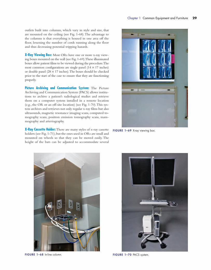
outlets built into columns, which vary in style and size, that
are mounted on the ceiling (see Fig. 1-68).The advantage to
the columns is that everything is housed in one area off the
floor, lessening the number of cords running along the floor
and thus decreasing potential tripping hazards.
X-Ray Viewing Box: Most ORs have one or more x-ray view-
ing boxes mounted on the wall (see Fig.1-69).These illuminated
boxes allow patient films to be viewed during the procedure.The
most common configurations are single panel (14 × 17 inches)
or double panel (28 × 17 inches).The boxes should be checked
prior to the start of the case to ensure that they are functioning
properly.
Picture Archiving and Communication System: The Picture
Archiving and Communication System (PACS) allows institu-
tions to archive a patient’s radiological studies and retrieve
them on a computer system installed in a remote location
(e.g., the OR or an off-site location) (see Fig. 1-70).This sys-
tem archives and retrieves not only regular x-ray films but also
ultrasounds, magnetic resonance imaging scans, computed to-
mography scans, positron emission tomography scans, mam-
mography and arteriography.
X-Ray Cassette Holder: There are many styles of x-ray cassette
holders (see Fig. 1-71), but the ones used in ORs are small and
mounted on wheels so that they can be moved easily. The
height of the bars can be adjusted to accommodate several
Chapter 1 Common Equipment and Furniture 29
FIGURE 1–68 In-line column.
FIGURE 1–69 X-ray viewing box.
FIGURE 1–70 PACS system.
1572_Ch01_001-050.qxd 8/6/09 10:48 AM Page 29
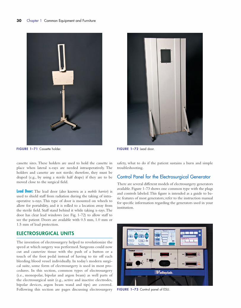
cassette sizes. These holders are used to hold the cassette in
place when lateral x-rays are needed intraoperatively. The
holders and cassette are not sterile; therefore, they must be
draped (e.g., by using a sterile half drape) if they are to be
moved close to the surgical field.
Lead Door: The lead door (also known as a mobile barrier) is
used to shield staff from radiation during the taking of intra-
operative x-rays.This type of door is mounted on wheels to
allow for portability, and it is rolled to a location away from
the sterile field. Staff stand behind it while taking x-rays.The
door has clear lead windows (see Fig. 1-72) to allow staff to
see the patient. Doors are available with 0.5 mm, 1.0 mm or
1.5 mm of lead protection.
ELECTROSURGICAL UNITS
The invention of electrosurgery helped to revolutionize the
speed at which surgery was performed. Surgeons could now
cut and cauterize tissue with the push of a button or a
touch of the foot pedal instead of having to tie off each
bleeding blood vessel individually. In today’s modern surgi-
cal suite, some form of electrosurgery is used in most pro-
cedures. In this section, common types of electrosurgery
(i.e., monopolar, bipolar and argon beam) as well parts of
the electrosurgical unit (e.g., active and inactive electrodes,
bipolar devices, argon beam wand and tips) are covered.
Following this section are pages discussing electrosurgery
30 Chapter 1 Common Equipment and Furniture
FIGURE 1–71 Cassette holder. FIGURE 1–72 Lead door.
FIGURE 1–73 Control panel of ESU.
safety, what to do if the patient sustains a burn and simple
troubleshooting.
Control Panel for the Electrosurgical GeneratorThere are several different models of electrosurgery generators
available. Figure 1-73 shows one common type with the plugs
and controls labeled.This figure is intended as a guide to ba-
sic features of most generators; refer to the instruction manual
for specific information regarding the generators used in your
institution.
1572_Ch01_001-050.qxd 8/6/09 10:48 AM Page 30
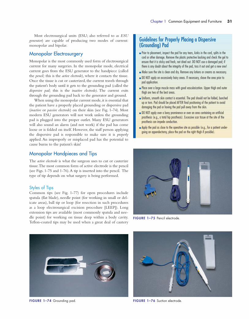
Chapter 1 Common Equipment and Furniture 31
Most electrosurgical units (ESU; also referred to as ESU
generator) are capable of producing two modes of current:
monopolar and bipolar.
Monopolar ElectrosurgeryMonopolar is the most commonly used form of electrosurgical
current for many surgeries. In the monopolar mode, electrical
current goes from the ESU generator to the handpiece (called
the pencil; this is the active electrode), where it contacts the tissue.
Once the tissue is cut or cauterized, the current travels through
the patient’s body until it gets to the grounding pad (called the
dispersive pad; this is the inactive electrode). The current exits
through the grounding pad back to the generator and ground.
When using the monopolar current mode, it is essential that
the patient have a properly placed grounding or dispersive pad
(inactive or passive electrode) on their skin (see Fig. 1-74). Most
modern ESU generators will not work unless the grounding
pad is plugged into the proper outlet. Many ESU generators
will also sound an alarm (and not work) if the pad has come
loose or is folded on itself. However, the staff person applying
the dispersive pad is responsible to make sure it is properly
applied. An improperly or misplaced pad has the potential to
cause burns to the patient’s skin!
Monopolar Handpieces and TipsThe active electrode is what the surgeon uses to cut or cauterize
tissue.The most common form of active electrode is the pencil
(see Figs. 1-75 and 1-76).A tip is inserted into the pencil. The
type of tip depends on what surgery is being performed.
Styles of TipsCommon tips (see Fig. 1-77) for open procedures include
spatula (flat blade), needle point (for working in small or del-
icate areas), ball tip or loop (for resection in such procedures
as a loop electrosurgical excision procedure [LEEP]). Long
extension tips are available (most commonly spatula and nee-
dle point) for working on tissue deep within a body cavity.
Teflon-coated tips may be used when a great deal of cautery
FIGURE 1–74 Grounding pad.
Guidelines for Properly Placing a Dispersive(Grounding) Pad� Prior to placement, inspect the pad for any tears, kinks in the cord, splits in the
cord or other damage. Remove the plastic protective backing and check the gel toensure that it is sticky and fresh, not dried out. DO NOT use a damaged pad; ifthere is any doubt about the integrity of the pad, toss it out and get a new one!
� Make sure the site is clean and dry. Remove any lotions or creams as necessary.
� DO NOT apply on excessively hairy areas. If necessary, shave the area prior topad application.
� Place over a large muscle mass with good vascularization. Upper thigh and outerthigh are two of the best areas.
� Uniform, smooth skin contact is essential. The pad should not be folded, bunchedup or torn. Pad should be placed AFTER final positioning of the patient to avoiddamaging the pad or having the pad pull away from the skin.
� DO NOT apply over a bony prominence or over an area containing an artificialprosthesis (e.g., a total hip prosthesis). Excessive scar tissue at the site of theprosthesis can impede conduction.
� Apply the pad as close to the operative site as possible (e.g., for a patient under-going an appendectomy, place the pad on the right thigh if possible).
FIGURE 1–75 Pencil electrode.
FIGURE 1–76 Suction electrode.
1572_Ch01_001-050.qxd 8/6/09 10:48 AM Page 31
32 Chapter 1 Common Equipment and Furniture
coagulation produces less tissue damage, less smoke plume
and less charring than traditional electrosurgery methods.
A special instrument or wand (see Fig. 1-81) is needed
when using the argon beam coagulation mode of the machine.
On some argon beam machines, you can turn the gas tank off
and use it with a traditional pencil as a monopolar device or
Loop
Teflon extension
ExtensionSpatulaPoint
Large flat tip
FIGURE 1–77 Loop, Teflon extension, extension tip, spatula, point,large flat tip.
is anticipated (these tips are easier to keep free of tissue
debris). Endoscopic procedures require special electrodes that
are long and thin.There are a variety of endoscopic electrodes
including probes and graspers.
The pencil can be activated by pressing a button or step-
ping on the foot pedal. While the electrode is activated, the
ESU makes an audible signal that alerts personnel that the tip
is hot and therefore a potential hazard. These signals are in-
tended for patient safety device and should never be disabled or
turned down so low as to make them inaudible to personnel in
the room.
Bipolar ElectrosurgeryIn bipolar electrosurgery, the current passes from one tine of the
forceps to the other; the current never passes through the pa-
tient. The power is lower than what can be achieved with
monopolar cautery and is used for cauterization during neuro-
surgery or on delicate tissue.The current does not pass through
the patient, thus a dispersive (or grounding) pad is not needed.
Note: If the surgeon is going to be using both monopolar
and bipolar current during the procedure, a dispersive elec-
trode pad must be placed on the patient.
Special forceps are used to deliver bipolar current.The for-
ceps are available in a wide variety of styles and plug into the
ESU using a bipolar cord (see Fig. 1-78). Many of the forceps
resemble regular grasping forceps (see Fig. 1-79) but with one
important difference; these forceps have a plug on the end of
the handle to plug them into the bipolar cord.
Argon Beam CoagulationArgon beam coagulation (also referred to as ABC or argon plasma
coagulation) uses a combination of argon gas and monopolar
electrosurgery to coagulate tissue.Within the machine (see
Fig. 1-80) the gas is ionized, making it electrically conduc-
tive.This provides a more efficient pathway for the current
to travel along to the tissue.The tip of the instrument does
not need to touch the tissue; the beam of ionized gas coag-
ulates bleeders within 2 to 10 mm of the tip. Argon beam
FIGURE 1–79 Bipolar forceps.
FIGURE 1–78 Bipolar cord.
FIGURE 1–80 Argon beam machine.
1572_Ch01_001-050.qxd 8/6/09 10:49 AM Page 32
Chapter 1 Common Equipment and Furniture 33
plug it in for use with bipolar coagulation. Be sure to refer to
the manufacturer’s instruction book to see if the unit in your
institution can be used this way.
Remember: Argon beam coagulation allows the beam to
travel through the patient’s body; therefore, a dispersive elec-
trode (grounding pad) must be used.
Electrosurgical SafetyAs with any electrical device, there are certain safety measures
that should be followed when an ESU is in use:
� Know your machine. Only personnel knowledgeable
about electrosurgical use and safety precautions should
use the device. If you have questions, refer to the
manufacturer’s instructions.
� Check the unit for frayed cords or damaged plugs
prior to use. If the electric cord is split, frayed or oth-
erwise compromised or the prongs on the plug are
bent, damaged or missing DO NOT use the machine.
A different machine should be obtained and the dam-
aged one checked by biomedical engineering.
� Do not store liquids on top of the machine. Spills
could occur and cause electrical shock or shortage.
� Make sure the electrosurgical grounding pad (inactive
electrode) is properly placed on the patient prior to
the start of surgery. Plug the patient ground pad into
the machine (most newer machines will not allow the
machine to activate in monopolar mode unless the
grounding pad is plugged in).
� Fire safety precautions must be followed.These include:
� Allow all prep solutions to dry completely before
using the ESU.
� Use a special endotracheal tube if electrosurgery
will be performed near the airway.
� Use flame-retardant drapes.
� When not using the handpiece, place it back in the
holder; DO NOT place it on dry drapes or towels.
� Oxygen should be delivered at the lowest possible
concentration.
� Water or saline must be available on the surgical
field (usually on the back table).
� Portable fire extinguishers must be readily available
(halon extinguishers are recommended).
� Nonflammable anesthetics must be used.
� During anorectal procedures, the rectum must be
packed with a sterile water-soaked sponge to pre-
vent a methane gas fire.
� NEVER turn off or disable alarms. Do not turn the
volume too low; personnel should be able to hear the
“beeping sound” when the electrode is activated.This
sounds alerts all personnel that the tip is activated (hot).
� Use an instrument (not your hands!) to change the
electrode tip.
� Do not loop the cords around metal devices or
instruments.
� Make sure all jewelry is removed from the patient
prior to surgery. Metal jewelry can act as a conductor
and cause alternate-site burns.
� If the patient has a pacemaker or an implanted defib-
rillator, consult the cardiologist before surgery to find
out whether or not the device needs to be shut off.
� DO NOT allow patient to come in contact with
metal surfaces on the OR bed.
� Suction smoke and plume during electrosurgery use
(a smoke evacuation system is recommended rather
than just regular suctioning).
What to Do If the Patient Sustains a BurnIf a patient is burned and the electrosurgical device might
have been the cause (e.g., you remove the grounding pad
and the patient’s skin underneath the pad is burned), this falls
under federal mandatory reporting laws. What does staff
need to do?
� Notify the surgeon immediately that a burn has
occurred and have him or her look at the area.The
surgeon will need to talk to the patient about what
happened and may need to order treatment (e.g.,
ointments or dressings) for the burn.
� Document the facts of how the skin looks in the pa-
tient’s perioperative record. Document that the surgeon
was notified and what was done to treat the burn area.
� Save ALL disposable parts of the electrosurgical unit
used on that patient, including the grounding pad,
electrosurgical pencil and tips. Put these supplies in a
bag and send them with the machine to biomedical
engineering to be checked.
� The electrosurgical machine should NOT be used
on any other patient until it has been checked and
cleared by biomedical engineering.
� Notify your supervisor (who in turn needs to notify
risk management). Fill out your institution’s quality
assurance paperwork (usually known as an incidence or
variance report).
FIGURE 1–81 Argon beam wand.
1572_Ch01_001-050.qxd 8/6/09 10:49 AM Page 33
� The supervisor and/or risk management department
will report the incident to the proper federal report-
ing agency following your institution’s policy.
MEDICAL GASES
There are six common medical gases used in the OR: oxy-
gen, nitrogen, nitrous oxide, carbon dioxide, helium and
argon. Each gas has a specific purpose and a mix-up could
be disastrous.
The Food and Drug Administration (FDA) recommends
that each medical gas be stored in a cylinder of a specific color
to make identification easier and a mix-up less likely. DO
NOT rely solely on the cylinder color; read the label on the
cylinder carefully! The regulator for the cylinder is another
safety feature. Regulators are gas-specific and are not inter-
changeable; never modify a gas regulator from one type of gas
to try to make it fit another.
These pages contain pictures and descriptions of each gas
as well as specific safety tips.At the end of this section, general
safety tips for handling medical gases are presented.
General Medical Gas Cylinder SafetyMedical gases are the only ones that have a regulated color sys-
tem for the cylinders. Although it is never recommended that
you rely on cylinder color alone (read the label!),here are the FDA
cylinder color recommendations to help with identification:
� Air: yellow
� Carbon dioxide: gray
� Cyclopropane: orange
� Helium: brown
� Nitrogen: black
� Nitrous oxide: blue
� Oxygen: green
34 Chapter 1 Common Equipment and Furniture
Troubleshooting TipsAs with any device, troubleshooting starts with the seemingly obvious and simple:
� Make sure the machine is plugged in.� Make sure the machine is turned on.� Check to see that the machine is in “ready” mode, not “standby.”
After checking that the three troubleshooting tasks are done, start to look for the problem.
PROBLEM WHAT TO LOOK ATWhen the surgeon presses the button, the handpiece doesn’t work. � Is the handpiece plugged into the proper place?
� Is the handpiece plugged in all the way (snuggly)?� Is the grounding pad plugged into the machine?� Is there a problem with the grounding pad (has it lifted off the skin, folded on itself or torn)?
The grounding pad light is lit up. � Is the grounding pad plugged into the machine?� Has the grounding pad begun to lift off the patient’s skin?� Is there a kink or tear in the grounding pad cord?� Has the grounding pad torn or folded (especially if the grounding pad was applied prior to
positioning)?
I turn the machine “on” but the power doesn’t come on. � Is the machine plugged in?� Is the cord kinked or frayed? If the cord is frayed or split, do not use this machine—get
another one.� Try another electrical outlet (sometimes there is a problem with power to one outlet).� If the machine is plugged in and you’ve tried other outlets without any luck, get another machine
and have biomedical engineering check out the problem machine.
General Guidelines for the Safe Handling ofMedical Gas Cylinders1. Read the label! Know what you are using before you use it. Never use an
unmarked cylinder or one bearing a label that is unreadable.
2. Store cylinders away from flammable material. Do not use oily or greasy substances to clean the cylinders.
3. Store cylinders in an upright position and make sure they are secured at all times. Usehandcarts to transport cylinders. Do not allow cylinders to hit against one another.
4. Store cylinders away from sources of heat or electricity.
5. Do not try to modify regulators from one type of gas to fit another type of gascylinder. Remember: Regulators are gas-specific and NOT interchangeable!
6. Store all cylinders in a well-ventilated area.
7. Let the experts do any repairs that are necessary. Do not try to repair damagedcylinders, regulators or gauges.
8. Never use a cylinder that is damaged or has a damaged pressure gauge or regulator.
1572_Ch01_001-050.qxd 8/6/09 10:49 AM Page 34
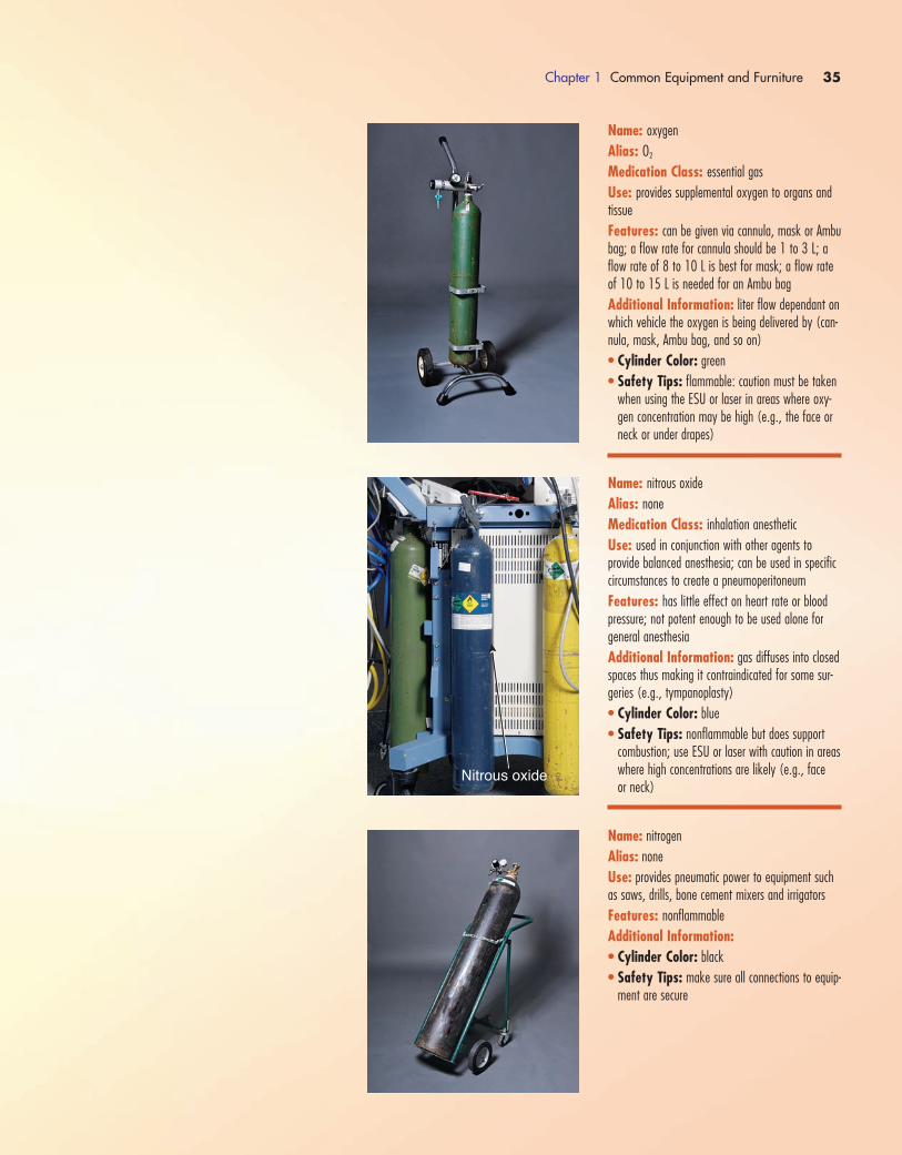
Chapter 1 Common Equipment and Furniture 35
Name: oxygen
Alias: O2
Medication Class: essential gas
Use: provides supplemental oxygen to organs andtissue
Features: can be given via cannula, mask or Ambubag; a flow rate for cannula should be 1 to 3 L; aflow rate of 8 to 10 L is best for mask; a flow rateof 10 to 15 L is needed for an Ambu bag
Additional Information: liter flow dependant onwhich vehicle the oxygen is being delivered by (can-nula, mask, Ambu bag, and so on)
• Cylinder Color: green
• Safety Tips: flammable: caution must be takenwhen using the ESU or laser in areas where oxy-gen concentration may be high (e.g., the face orneck or under drapes)
Name: nitrous oxide
Alias: none
Medication Class: inhalation anesthetic
Use: used in conjunction with other agents to provide balanced anesthesia; can be used in specific circumstances to create a pneumoperitoneum
Features: has little effect on heart rate or bloodpressure; not potent enough to be used alone forgeneral anesthesia
Additional Information: gas diffuses into closedspaces thus making it contraindicated for some sur-geries (e.g., tympanoplasty)
• Cylinder Color: blue
• Safety Tips: nonflammable but does supportcombustion; use ESU or laser with caution in areaswhere high concentrations are likely (e.g., face or neck)
Name: nitrogen
Alias: none
Use: provides pneumatic power to equipment suchas saws, drills, bone cement mixers and irrigators
Features: nonflammable
Additional Information:• Cylinder Color: black
• Safety Tips: make sure all connections to equip-ment are secure
Nitrous oxide
1572_Ch01_001-050.qxd 8/6/09 10:49 AM Page 35
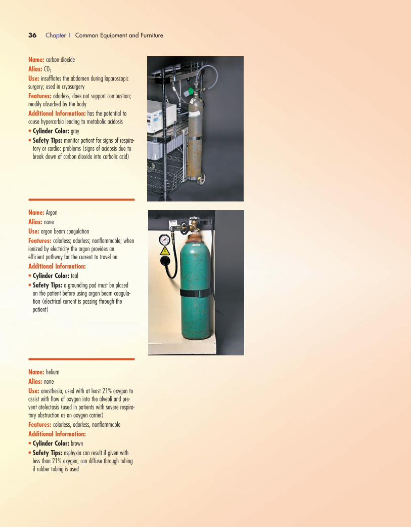
36 Chapter 1 Common Equipment and Furniture
Name: carbon dioxide
Alias: CO2
Use: insufflates the abdomen during laparoscopicsurgery; used in cryosurgery
Features: odorless; does not support combustion;readily absorbed by the body
Additional Information: has the potential tocause hypercarbia leading to metabolic acidosis
• Cylinder Color: gray
• Safety Tips: monitor patient for signs of respira-tory or cardiac problems (signs of acidosis due tobreak down of carbon dioxide into carbolic acid)
Name: Argon
Alias: none
Use: argon beam coagulation
Features: colorless; odorless; nonflammable; whenionized by electricity the argon provides an efficient pathway for the current to travel on
Additional Information:• Cylinder Color: teal
• Safety Tips: a grounding pad must be placedon the patient before using argon beam coagula-tion (electrical current is passing through the patient)
Name: helium
Alias: none
Use: anesthesia; used with at least 21% oxygen toassist with flow of oxygen into the alveoli and pre-vent atelectasis (used in patients with severe respira-tory obstruction as an oxygen carrier)
Features: colorless, odorless, nonflammable
Additional Information:• Cylinder Color: brown
• Safety Tips: asphyxia can result if given withless than 21% oxygen; can diffuse through tubingif rubber tubing is used
1572_Ch01_001-050.qxd 8/6/09 10:49 AM Page 36
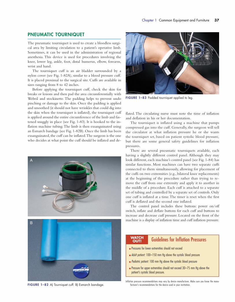
PNEUMATIC TOURNIQUET
The pneumatic tourniquet is used to create a bloodless surgi-
cal area by limiting circulation to a patient’s operative limb.
Sometimes, it can be used in the administration of regional
anesthesia. This device is used for procedures involving the
knee, lower leg, ankle, foot, distal humerus, elbow, forearm,
wrist and hand.
The tourniquet cuff is an air bladder surrounded by a
nylon cover (see Fig. 1-82A), similar to a blood pressure cuff.
It is placed proximal to the surgical site. Cuffs are available in
sizes ranging from 8 to 42 inches.
Before applying the tourniquet cuff, check the skin for
breaks or lesions and then pad the area circumferentially with
Webril and stockinette. The padding helps to prevent undo
pinching or damage to the skin. Once the padding is applied
and smoothed (it should not have wrinkles that could dig into
the skin when the tourniquet is inflated), the tourniquet cuff
is applied around the entire circumference of the limb and fas-
tened snuggly in place (see Fig. 1-83). It is hooked to the in-
flation machine tubing.The limb is then exsanguinated using
an Esmarch bandage (see Fig. 1-82B). Once the limb has been
exsanguinated, the cuff can be inflated.The surgeon is the one
who decides at what point the cuff should be inflated and de-
flated. The circulating nurse must note the time of inflation
and deflation in his or her documentation.
The tourniquet is inflated using a machine that pumps
compressed gas into the cuff. Generally, the surgeon will tell
the circulator at what inflation pressure he or she wants
the tourniquet set, based on patient systolic blood pressure,
but there are some general safety guidelines for inflation
pressures.
There are several pneumatic tourniquets available, each
having a slightly different control panel. Although they may
look different, each machine’s control panel (see Fig. 1-84) has
similar functions. Most machines can have two separate cuffs
connected to them simultaneously, allowing for placement of
the cuffs on two extremities (e.g., bilateral knee replacements)
at the beginning of the procedure rather than trying to re-
move the cuff from one extremity and apply it to another in
the middle of a procedure. Each cuff is attached to a separate
set of tubing and controlled by a separate set of controls. Only
one cuff is inflated at a time.The timer is reset when the first
cuff is deflated and the second one inflated.
The control panel includes these buttons: power on/off
switch, inflate and deflate buttons for each cuff and buttons to
increase and decrease cuff pressure. Located on the front of the
machine is a display of inflation time and cuff inflation pressure.
Chapter 1 Common Equipment and Furniture 37
FIGURE 1–82 A) Tourniquet cuff. B) Esmarch bandage.
Guidelines for Inflation Pressures
� Pressures for lower extremities should not exceed
� Adult patient: 100–150 mm Hg above the systolic blood pressure
� Pediatric patient: 100 mm Hg above the systolic blood pressure
� Pressure for upper extremities should not exceed 30–75 mm Hg above the patient’s systolic blood pressure.
Inflation pressure recommendations may vary by device manufacturer. Make sure you know the manu-facturer’s recommendations for the device used in your institution.
WATCH OUT!
FIGURE 1–83 Padded tourniquet applied to leg.
A
B
1572_Ch01_001-050.qxd 8/6/09 10:49 AM Page 37
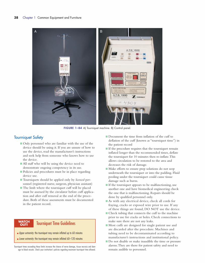
Tourniquet Safety� Only personnel who are familiar with the use of the
device should be using it. If you are unsure of how to
use the device, read the manufacturer’s instructions
and seek help from someone who knows how to use
the device.
� All staff who will be using the device need to
demonstrate ongoing competency in its use.
� Policies and procedures must be in place regarding
device use.
� Tourniquets should be applied only by licensed per-
sonnel (registered nurse, surgeon, physician assistant)
� The limb where the tourniquet cuff will be placed
must be assessed by the circulator before cuff applica-
tion and after cuff removal at the end of the proce-
dure. Both of these assessments must be documented
in the patient record.
� Document the time from inflation of the cuff to
deflation of the cuff (known as “tourniquet time”) in
the patient record
� If the procedure requires that the tourniquet remain
inflated longer than the recommended times, deflate
the tourniquet for 10 minutes then re-inflate.This
allows circulation to be restored to the area and
decreases the risk of damage.
� Make efforts to ensure prep solutions do not seep
underneath the tourniquet or into the padding. Fluid
pooling under the tourniquet could cause tissue
damage such as burns.
� If the tourniquet appears to be malfunctioning, use
another one and have biomedical engineering check
the one that is malfunctioning. Repairs should be
done by qualified personnel only.
� As with any electrical device, check all cords for
fraying, cracks or exposed wire prior to use. If any
of these things are found, DO NOT use the device.
� Check tubing that connects the cuff to the machine
prior to use for cracks or holes. Check connections to
make sure there are not any leaks.
� Most cuffs are designed for single patient use and
are discarded after the procedure. Machines and
tubing need to be decontaminated according to
manufacturer’s instructions and institutional policy.
� Do not disable or make inaudible the time or pressure
alarms.They are there for patient safety and need to
remain audible to personnel.
38 Chapter 1 Common Equipment and Furniture
A B
FIGURE 1–84 A) Tourniquet machine. B) Control panel.
Tourniquet Time Guidelines
� Upper extremity: the tourniquet may remain inflated up to 60 minutes
� Lower extremity: the tourniquet may remain inflated 60–120 minutes
Tourniquet times exceeding these limits increase the chance of nerve damage, tissue necrosis and dam-age to blood vessels. Check your institution’s policies regarding maximum tourniquet time allowed.
WATCH OUT!
1572_Ch01_001-050.qxd 8/6/09 10:49 AM Page 38
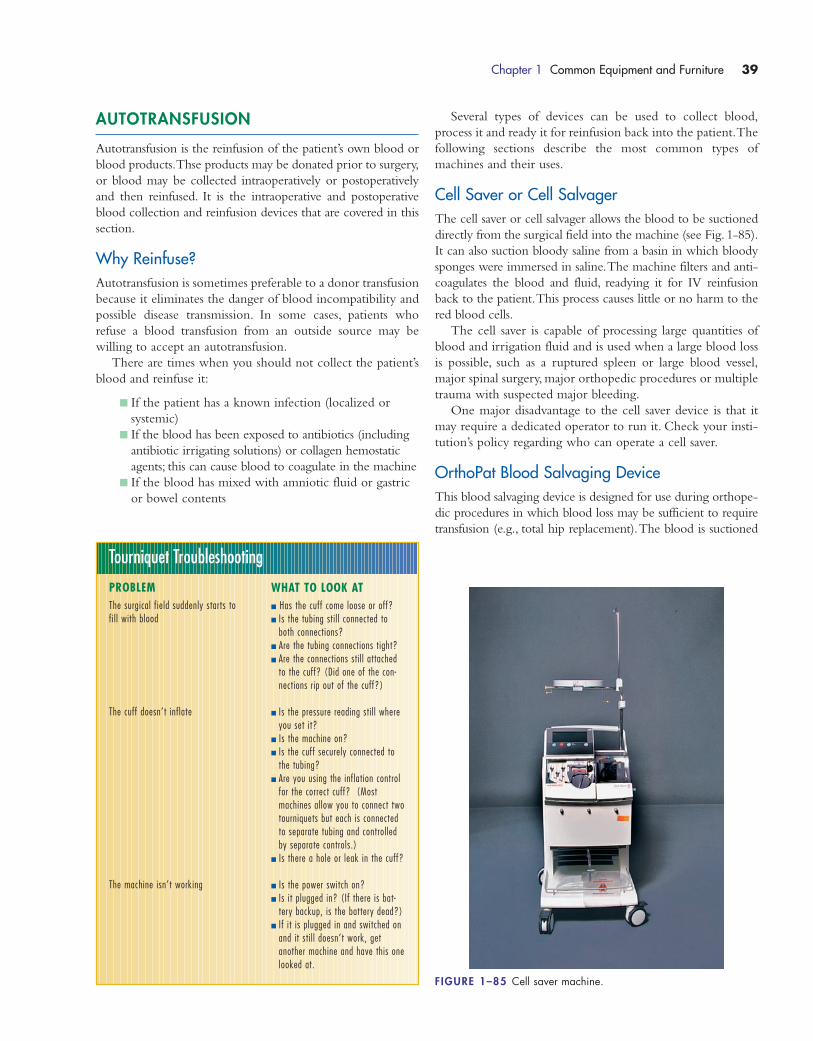
AUTOTRANSFUSION
Autotransfusion is the reinfusion of the patient’s own blood or
blood products.Thse products may be donated prior to surgery,
or blood may be collected intraoperatively or postoperatively
and then reinfused. It is the intraoperative and postoperative
blood collection and reinfusion devices that are covered in this
section.
Why Reinfuse?Autotransfusion is sometimes preferable to a donor transfusion
because it eliminates the danger of blood incompatibility and
possible disease transmission. In some cases, patients who
refuse a blood transfusion from an outside source may be
willing to accept an autotransfusion.
There are times when you should not collect the patient’s
blood and reinfuse it:
� If the patient has a known infection (localized or
systemic)
� If the blood has been exposed to antibiotics (including
antibiotic irrigating solutions) or collagen hemostatic
agents; this can cause blood to coagulate in the machine
� If the blood has mixed with amniotic fluid or gastric
or bowel contents
Several types of devices can be used to collect blood,
process it and ready it for reinfusion back into the patient.The
following sections describe the most common types of
machines and their uses.
Cell Saver or Cell SalvagerThe cell saver or cell salvager allows the blood to be suctioned
directly from the surgical field into the machine (see Fig. 1-85).
It can also suction bloody saline from a basin in which bloody
sponges were immersed in saline.The machine filters and anti-
coagulates the blood and fluid, readying it for IV reinfusion
back to the patient.This process causes little or no harm to the
red blood cells.
The cell saver is capable of processing large quantities of
blood and irrigation fluid and is used when a large blood loss
is possible, such as a ruptured spleen or large blood vessel,
major spinal surgery, major orthopedic procedures or multiple
trauma with suspected major bleeding.
One major disadvantage to the cell saver device is that it
may require a dedicated operator to run it. Check your insti-
tution’s policy regarding who can operate a cell saver.
OrthoPat Blood Salvaging DeviceThis blood salvaging device is designed for use during orthope-
dic procedures in which blood loss may be sufficient to require
transfusion (e.g., total hip replacement).The blood is suctioned
Chapter 1 Common Equipment and Furniture 39
Tourniquet Troubleshooting
PROBLEMThe surgical field suddenly starts tofill with blood
The cuff doesn’t inflate
The machine isn’t working
WHAT TO LOOK AT� Has the cuff come loose or off?� Is the tubing stil l connected to
both connections? � Are the tubing connections tight?� Are the connections stil l attached
to the cuff? (Did one of the con-nections rip out of the cuff?)
� Is the pressure reading stil l whereyou set it?
� Is the machine on?� Is the cuff securely connected to
the tubing?� Are you using the inflation control
for the correct cuff? (Most machines allow you to connect twotourniquets but each is connectedto separate tubing and controlledby separate controls.)
� Is there a hole or leak in the cuff?
� Is the power switch on?� Is it plugged in? (If there is bat-
tery backup, is the battery dead?)� If it is plugged in and switched on
and it stil l doesn’t work, get another machine and have this onelooked at.
FIGURE 1–85 Cell saver machine.
1572_Ch01_001-050.qxd 8/6/09 10:49 AM Page 39
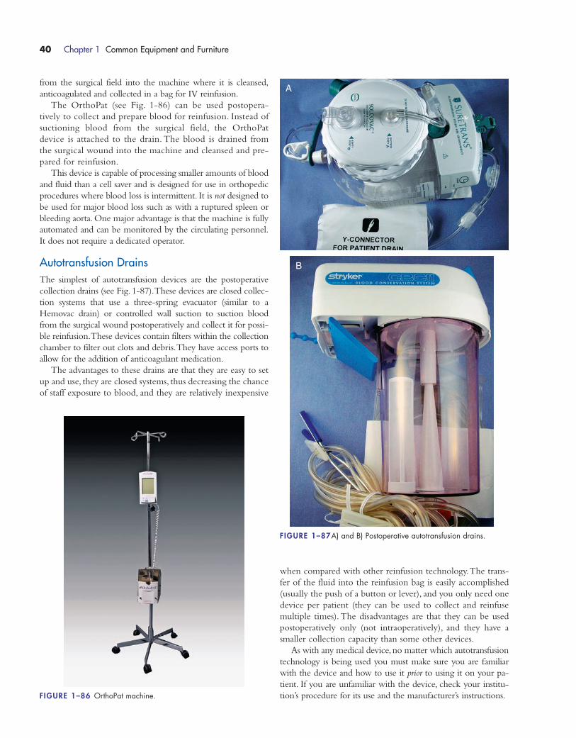
from the surgical field into the machine where it is cleansed,
anticoagulated and collected in a bag for IV reinfusion.
The OrthoPat (see Fig. 1-86) can be used postopera-
tively to collect and prepare blood for reinfusion. Instead of
suctioning blood from the surgical field, the OrthoPat
device is attached to the drain. The blood is drained from
the surgical wound into the machine and cleansed and pre-
pared for reinfusion.
This device is capable of processing smaller amounts of blood
and fluid than a cell saver and is designed for use in orthopedic
procedures where blood loss is intermittent. It is not designed to
be used for major blood loss such as with a ruptured spleen or
bleeding aorta. One major advantage is that the machine is fully
automated and can be monitored by the circulating personnel.
It does not require a dedicated operator.
Autotransfusion DrainsThe simplest of autotransfusion devices are the postoperative
collection drains (see Fig. 1-87).These devices are closed collec-
tion systems that use a three-spring evacuator (similar to a
Hemovac drain) or controlled wall suction to suction blood
from the surgical wound postoperatively and collect it for possi-
ble reinfusion.These devices contain filters within the collection
chamber to filter out clots and debris.They have access ports to
allow for the addition of anticoagulant medication.
The advantages to these drains are that they are easy to set
up and use, they are closed systems, thus decreasing the chance
of staff exposure to blood, and they are relatively inexpensive
40 Chapter 1 Common Equipment and Furniture
FIGURE 1–86 OrthoPat machine.
FIGURE 1–87A) and B) Postoperative autotransfusion drains.
A
B
when compared with other reinfusion technology.The trans-
fer of the fluid into the reinfusion bag is easily accomplished
(usually the push of a button or lever), and you only need one
device per patient (they can be used to collect and reinfuse
multiple times). The disadvantages are that they can be used
postoperatively only (not intraoperatively), and they have a
smaller collection capacity than some other devices.
As with any medical device, no matter which autotransfusion
technology is being used you must make sure you are familiar
with the device and how to use it prior to using it on your pa-
tient. If you are unfamiliar with the device, check your institu-
tion’s procedure for its use and the manufacturer’s instructions.
1572_Ch01_001-050.qxd 8/6/09 10:49 AM Page 40
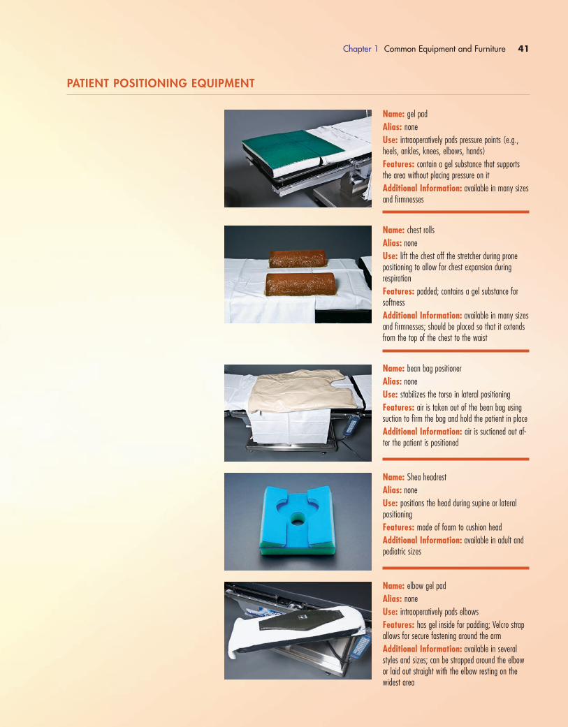
Chapter 1 Common Equipment and Furniture 41
PATIENT POSITIONING EQUIPMENT
Name: gel pad
Alias: none
Use: intraoperatively pads pressure points (e.g.,heels, ankles, knees, elbows, hands)
Features: contain a gel substance that supportsthe area without placing pressure on it
Additional Information: available in many sizesand firmnesses
Name: chest rolls
Alias: none
Use: lift the chest off the stretcher during prone positioning to allow for chest expansion during respiration
Features: padded; contains a gel substance forsoftness
Additional Information: available in many sizesand firmnesses; should be placed so that it extendsfrom the top of the chest to the waist
Name: bean bag positioner
Alias: none
Use: stabilizes the torso in lateral positioning
Features: air is taken out of the bean bag usingsuction to firm the bag and hold the patient in place
Additional Information: air is suctioned out af-ter the patient is positioned
Name: Shea headrest
Alias: none
Use: positions the head during supine or lateral positioning
Features: made of foam to cushion head
Additional Information: available in adult andpediatric sizes
Name: elbow gel pad
Alias: none
Use: intraoperatively pads elbows
Features: has gel inside for padding; Velcro strapallows for secure fastening around the arm
Additional Information: available in severalstyles and sizes; can be strapped around the elbowor laid out straight with the elbow resting on thewidest area
1572_Ch01_001-050.qxd 8/6/09 10:49 AM Page 41
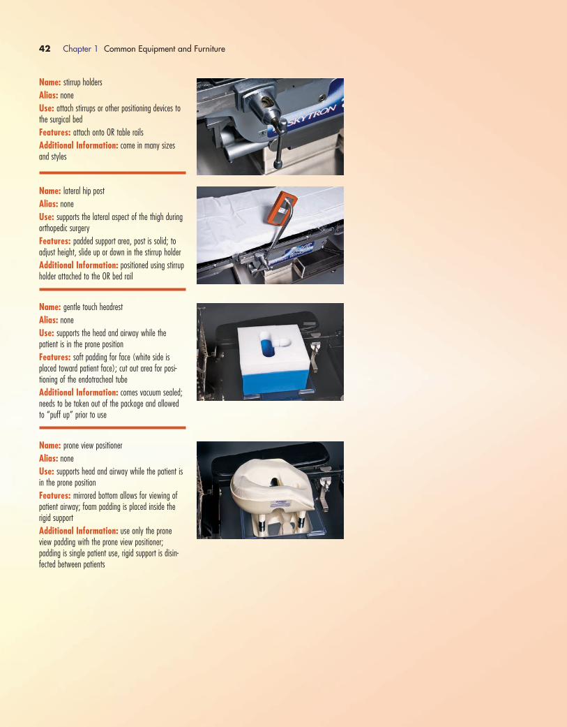
42 Chapter 1 Common Equipment and Furniture
Name: stirrup holders
Alias: none
Use: attach stirrups or other positioning devices tothe surgical bed
Features: attach onto OR table rails
Additional Information: come in many sizesand styles
Name: lateral hip post
Alias: none
Use: supports the lateral aspect of the thigh duringorthopedic surgery
Features: padded support area, post is solid; toadjust height, slide up or down in the stirrup holder
Additional Information: positioned using stirrupholder attached to the OR bed rail
Name: gentle touch headrest
Alias: none
Use: supports the head and airway while the patient is in the prone position
Features: soft padding for face (white side isplaced toward patient face); cut out area for posi-tioning of the endotracheal tube
Additional Information: comes vacuum sealed;needs to be taken out of the package and allowedto “puff up” prior to use
Name: prone view positioner
Alias: none
Use: supports head and airway while the patient isin the prone position
Features: mirrored bottom allows for viewing ofpatient airway; foam padding is placed inside therigid support
Additional Information: use only the proneview padding with the prone view positioner;padding is single patient use, rigid support is disin-fected between patients
1572_Ch01_001-050.qxd 8/6/09 10:49 AM Page 42
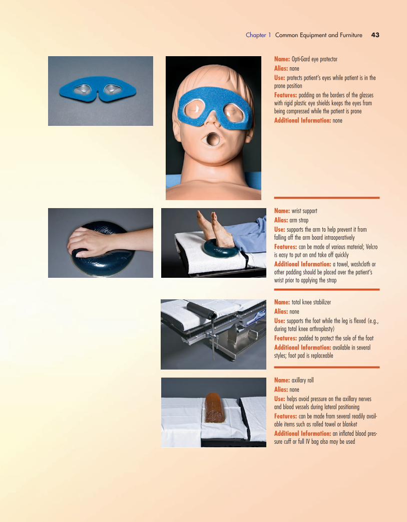
Chapter 1 Common Equipment and Furniture 43
Name: Opti-Gard eye protector
Alias: none
Use: protects patient’s eyes while patient is in theprone position
Features: padding on the borders of the glasseswith rigid plastic eye shields keeps the eyes from being compressed while the patient is prone
Additional Information: none
Name: wrist support
Alias: arm strap
Use: supports the arm to help prevent it fromfalling off the arm board intraoperatively
Features: can be made of various material; Velcrois easy to put on and take off quickly
Additional Information: a towel, washcloth orother padding should be placed over the patient’swrist prior to applying the strap
Name: total knee stabilizer
Alias: none
Use: supports the foot while the leg is flexed (e.g.,during total knee arthroplasty)
Features: padded to protect the sole of the foot
Additional Information: available in severalstyles; foot pad is replaceable
Name: axillary roll
Alias: none
Use: helps avoid pressure on the axillary nervesand blood vessels during lateral positioning
Features: can be made from several readily avail-able items such as rolled towel or blanket
Additional Information: an inflated blood pres-sure cuff or full IV bag also may be used
1572_Ch01_001-050.qxd 8/6/09 10:49 AM Page 43
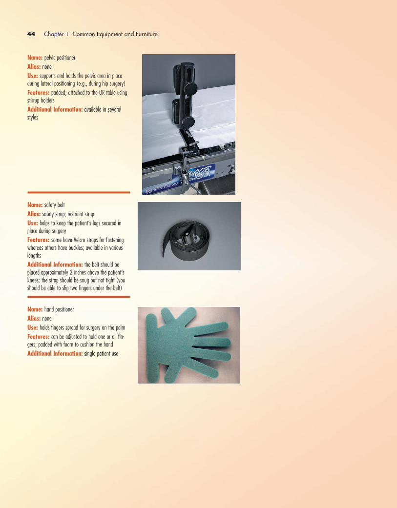
44 Chapter 1 Common Equipment and Furniture
Name: pelvic positioner
Alias: none
Use: supports and holds the pelvic area in placeduring lateral positioning (e.g., during hip surgery)
Features: padded; attached to the OR table usingstirrup holders
Additional Information: available in severalstyles
Name: safety belt
Alias: safety strap; restraint strap
Use: helps to keep the patient’s legs secured inplace during surgery
Features: some have Velcro straps for fasteningwhereas others have buckles; available in variouslengths
Additional Information: the belt should beplaced approximately 2 inches above the patient’sknees; the strap should be snug but not tight (youshould be able to slip two fingers under the belt)
Name: hand positioner
Alias: none
Use: holds fingers spread for surgery on the palm
Features: can be adjusted to hold one or all fin-gers; padded with foam to cushion the hand
Additional Information: single patient use
1572_Ch01_001-050.qxd 8/6/09 10:49 AM Page 44
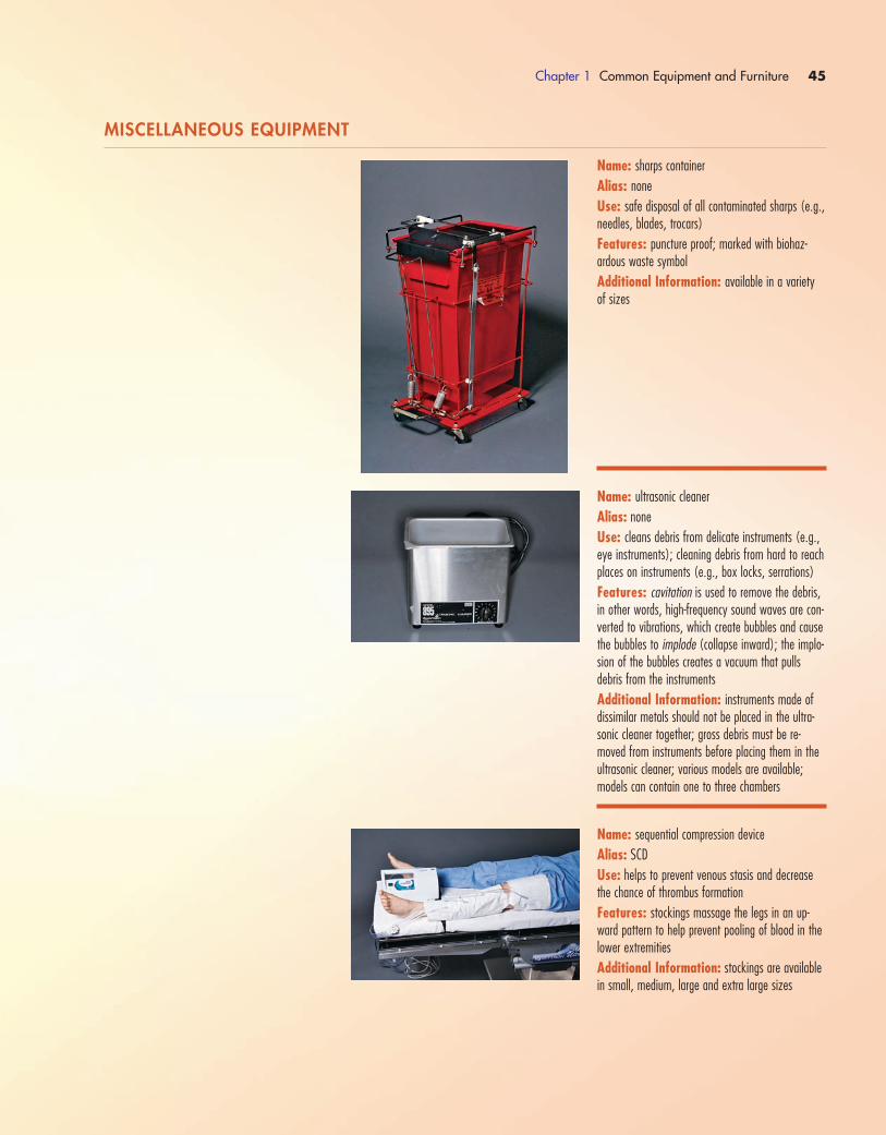
Chapter 1 Common Equipment and Furniture 45
Name: sharps container
Alias: none
Use: safe disposal of all contaminated sharps (e.g.,needles, blades, trocars)
Features: puncture proof; marked with biohaz-ardous waste symbol
Additional Information: available in a varietyof sizes
Name: ultrasonic cleaner
Alias: none
Use: cleans debris from delicate instruments (e.g.,eye instruments); cleaning debris from hard to reachplaces on instruments (e.g., box locks, serrations)
Features: cavitation is used to remove the debris,in other words, high-frequency sound waves are con-verted to vibrations, which create bubbles and causethe bubbles to implode (collapse inward); the implo-sion of the bubbles creates a vacuum that pulls debris from the instruments
Additional Information: instruments made ofdissimilar metals should not be placed in the ultra-sonic cleaner together; gross debris must be re-moved from instruments before placing them in theultrasonic cleaner; various models are available;models can contain one to three chambers
Name: sequential compression device
Alias: SCD
Use: helps to prevent venous stasis and decreasethe chance of thrombus formation
Features: stockings massage the legs in an up-ward pattern to help prevent pooling of blood in thelower extremities
Additional Information: stockings are availablein small, medium, large and extra large sizes
MISCELLANEOUS EQUIPMENT
1572_Ch01_001-050.qxd 8/6/09 10:49 AM Page 45
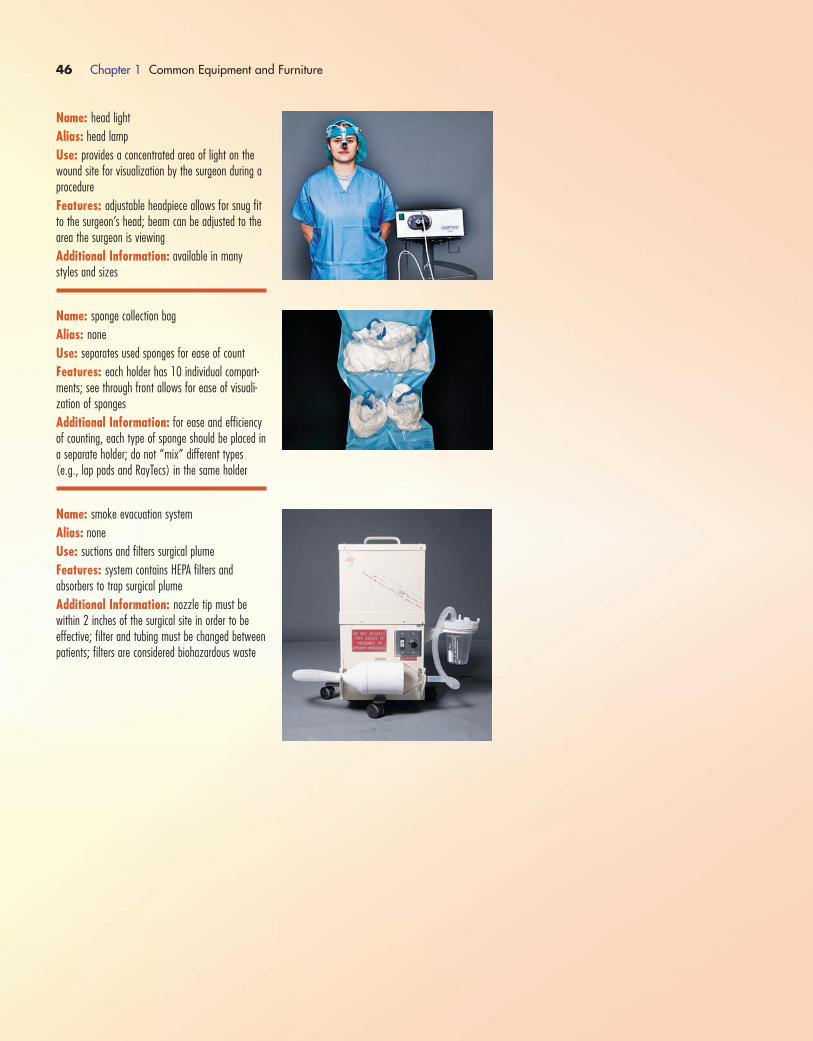
46 Chapter 1 Common Equipment and Furniture
Name: head light
Alias: head lamp
Use: provides a concentrated area of light on thewound site for visualization by the surgeon during aprocedure
Features: adjustable headpiece allows for snug fitto the surgeon’s head; beam can be adjusted to thearea the surgeon is viewing
Additional Information: available in manystyles and sizes
Name: sponge collection bag
Alias: none
Use: separates used sponges for ease of count
Features: each holder has 10 individual compart-ments; see through front allows for ease of visuali-zation of sponges
Additional Information: for ease and efficiencyof counting, each type of sponge should be placed ina separate holder; do not “mix” different types(e.g., lap pads and RayTecs) in the same holder
Name: smoke evacuation system
Alias: none
Use: suctions and filters surgical plume
Features: system contains HEPA filters and absorbers to trap surgical plume
Additional Information: nozzle tip must bewithin 2 inches of the surgical site in order to be effective; filter and tubing must be changed betweenpatients; filters are considered biohazardous waste
1572_Ch01_001-050.qxd 8/6/09 10:49 AM Page 46
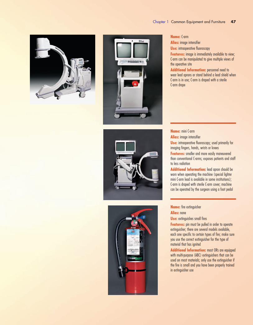
Chapter 1 Common Equipment and Furniture 47
Name: C-arm
Alias: image intensifier
Use: intraoperative fluoroscopy
Features: image is immediately available to view;C-arm can be manipulated to give multiple views ofthe operative site
Additional Information: personnel need towear lead aprons or stand behind a lead shield whenC-arm is in use; C-arm is draped with a sterile C-arm drape
Name: mini C-arm
Alias: image intensifier
Use: intraoperative fluoroscopy; used primarily forimaging fingers, hands, wrists or knees
Features: smaller and more easily maneuveredthan conventional C-arms; exposes patients and staffto less radiation
Additional Information: lead apron should beworn when operating the machine (special lightermini C-arm lead is available in some institutions); C-arm is draped with sterile C-arm cover; machinecan be operated by the surgeon using a foot pedal
Name: fire extinguisher
Alias: none
Use: extinguishes small fires
Features: pin must be pulled in order to operateextinguisher; there are several models available,each one specific to certain types of fire; make sureyou use the correct extinguisher for the type of material that has ignited
Additional Information: most ORs are equippedwith multi-purpose (ABC) extinguishers that can beused on most materials; only use the extinguisher ifthe fire is small and you have been properly trainedin extinguisher use
1572_Ch01_001-050.qxd 8/6/09 10:49 AM Page 47
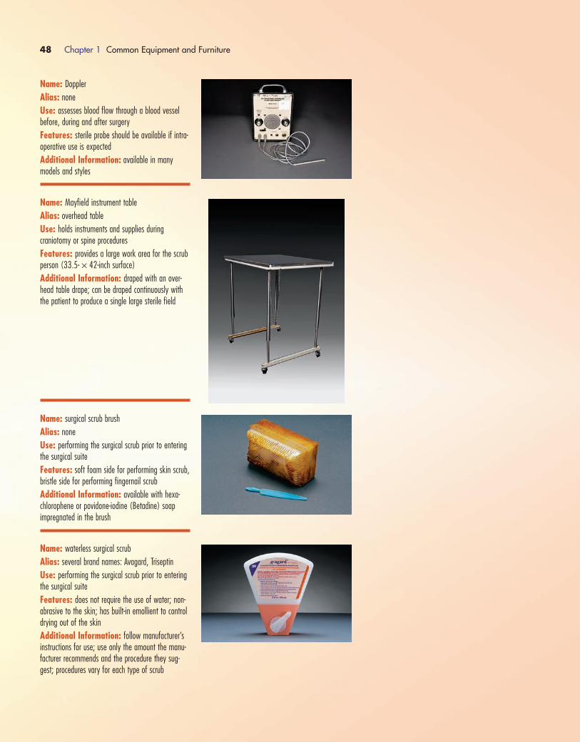
48 Chapter 1 Common Equipment and Furniture
Name: Doppler
Alias: none
Use: assesses blood flow through a blood vesselbefore, during and after surgery
Features: sterile probe should be available if intra-operative use is expected
Additional Information: available in many models and styles
Name: Mayfield instrument table
Alias: overhead table
Use: holds instruments and supplies during craniotomy or spine procedures
Features: provides a large work area for the scrubperson (33.5- × 42-inch surface)
Additional Information: draped with an over-head table drape; can be draped continuously withthe patient to produce a single large sterile field
Name: surgical scrub brush
Alias: none
Use: performing the surgical scrub prior to enteringthe surgical suite
Features: soft foam side for performing skin scrub,bristle side for performing fingernail scrub
Additional Information: available with hexa-chlorophene or povidone-iodine (Betadine) soap impregnated in the brush
Name: waterless surgical scrub
Alias: several brand names: Avagard, Triseptin
Use: performing the surgical scrub prior to enteringthe surgical suite
Features: does not require the use of water; non-abrasive to the skin; has built-in emollient to controldrying out of the skin
Additional Information: follow manufacturer’sinstructions for use; use only the amount the manu-facturer recommends and the procedure they sug-gest; procedures vary for each type of scrub
1572_Ch01_001-050.qxd 8/6/09 10:49 AM Page 48

Chapter 1 Common Equipment and Furniture 49
Surgical Session Review
1) All of the following are true about masks except:
a. they should be changed after each case
b. they protect the wearer from spraying of blood/body
fluids
c. they have to be worn in unrestricted areas of the OR
d. they must be worn so that they cover the mouth and
the nose
2) Which of the following statements is false regarding OR
attire:
a. caps or hats must cover all hair (including facial hair)
b. cloth caps or hats can be worn for 3 consecutive days
without laundering
c. tunic style scrub tops should be tucked into your scrub
pants
d. Regular eyeglasses alone do not meet the standards for
protective eyewear
3) Unsterile personnel should stand a minimum of ________
inches from the sterile field.
a. 6-8
b. 12-18
c. 20-24
d. 24-28
4) Principles of aseptic technique include:
a. keep talking at the field to a minimum
b. only sterile (scrubbed) personnel handle sterile items
c. create the sterile field as close to the time of surgery as
possible
d. all of the above
5) All of the following are true about sterile drapes except:
a. they should be made of a low-linting material
b. once in place, they should not be moved
c. handling should be kept to a minimum
d. the part of the drape that falls below the level of the
table top is sterile
6) The maximum continuous time that a tourniquet should
be left inflated on a leg is _______ minutes.
a. 30
b. 60
c. 90
d. 120
7) Which of the following might be used to position a patient
who is undergoing surgery on the lower back?
a. Jackson table
b. Wilson frame
c. Andrews frame
d. all of the above
8) A horseshoe, Mayfield or Shea may be used to position
the patient’s _______.
a. head
b. armc. foot
d. shoulders
9) Arms should NEVER be extended more than _______
degrees on an arm board.
a. 45
b. 65
c. 90
d. 120
10) Hyperextension of the arm can cause damage to the:
a. peroneal nerve
b. popliteal nerve
c. brachial plexus
d. chordae tendineae
11) All of the following are types of stirrups except:
a. candy cane
b. boot
c. knee release
d. knee crutch
12) A __________ is one of the most common types of suc-
tion tip used to clear the airway.
a. Yankauer
b. Neuro
c. Frasier
d. Poole
13) Which of the following types of electrosurgery unit does
not require the use of a grounding pad?
a. monopolar
b. argon beam
c. bipolar
d. all of the above require a grounding pad
14) Fire safety rules during the use of electrosurgery include:
a. allow all prep solutions to dry completely
b. water or saline must be available on the surgical field
c. when not in use, the handpiece should be placed back
in a holder
d. all of the above
15) In reference to electrosurgery units,ABC stands for:
a. aluminum beam coagulation
b. argon beam coagulation
c. argon beam cautery
d. aluminum beam cautery
1572_Ch01_001-050.qxd 8/6/09 10:49 AM Page 49
16) Which of the following staff members should not apply
the pneumatic tourniquet?
a. surgeon
b. physician assistant
c. registered nurse
d. surgical technologist
17) Ultrasonic cleaners use ___________ to remove debris
from instruments.
a. cavitation
b. steam
c. ethylene oxide
d. Cidex
18) ____________ is an anesthetic gas.
a. oxygen
b. nitrogen
c. nitrous oxide
d. argon
19) An Optigard protector is used when the patient is prone
to protect his or her _______.
a. fingers
b. hands
c. toes
d. eyes
20) A Doppler is used to assess ________ flow.
a. urine
b. coronary artery
c. blood
d. brain wave
50 Chapter 1 Common Equipment and Furniture
1572_Ch01_001-050.qxd 8/6/09 10:49 AM Page 50
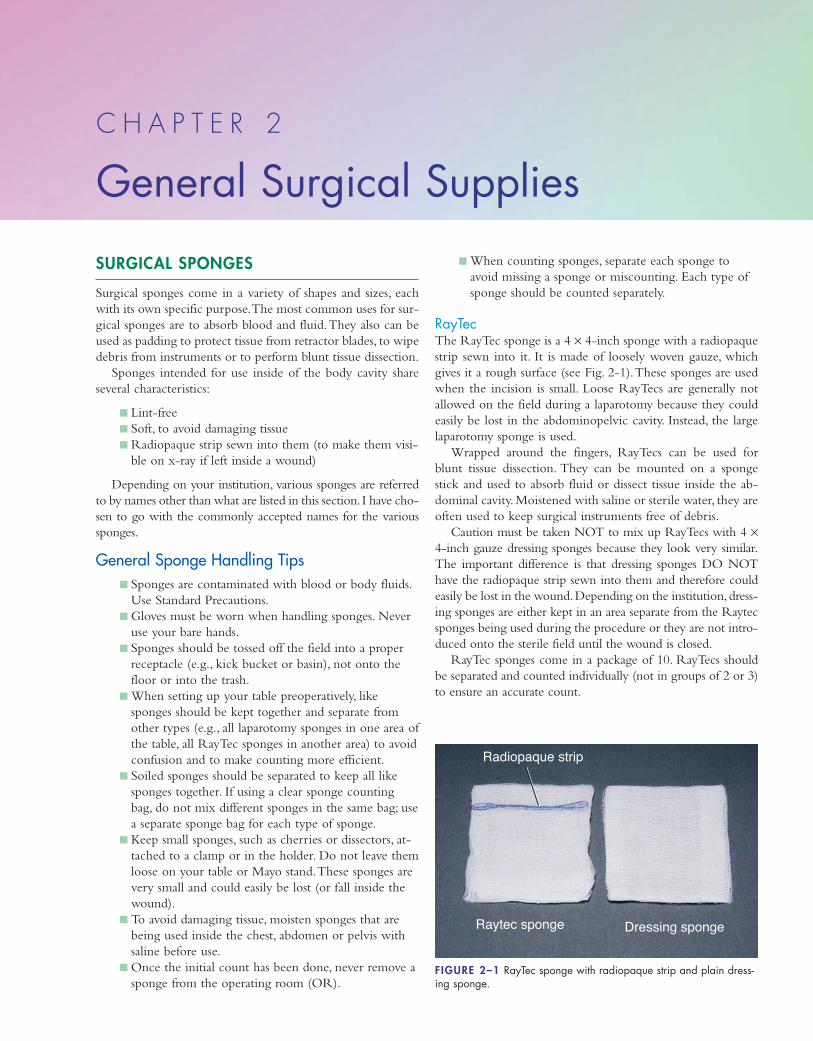
SURGICAL SPONGES
Surgical sponges come in a variety of shapes and sizes, each
with its own specific purpose.The most common uses for sur-
gical sponges are to absorb blood and fluid.They also can be
used as padding to protect tissue from retractor blades, to wipe
debris from instruments or to perform blunt tissue dissection.
Sponges intended for use inside of the body cavity share
several characteristics:
� Lint-free
� Soft, to avoid damaging tissue
� Radiopaque strip sewn into them (to make them visi-
ble on x-ray if left inside a wound)
Depending on your institution, various sponges are referred
to by names other than what are listed in this section. I have cho-
sen to go with the commonly accepted names for the various
sponges.
General Sponge Handling Tips� Sponges are contaminated with blood or body fluids.
Use Standard Precautions.
� Gloves must be worn when handling sponges. Never
use your bare hands.
� Sponges should be tossed off the field into a proper
receptacle (e.g., kick bucket or basin), not onto the
floor or into the trash.
� When setting up your table preoperatively, like
sponges should be kept together and separate from
other types (e.g., all laparotomy sponges in one area of
the table, all RayTec sponges in another area) to avoid
confusion and to make counting more efficient.
� Soiled sponges should be separated to keep all like
sponges together. If using a clear sponge counting
bag, do not mix different sponges in the same bag; use
a separate sponge bag for each type of sponge.
� Keep small sponges, such as cherries or dissectors, at-
tached to a clamp or in the holder. Do not leave them
loose on your table or Mayo stand.These sponges are
very small and could easily be lost (or fall inside the
wound).
� To avoid damaging tissue, moisten sponges that are
being used inside the chest, abdomen or pelvis with
saline before use.
� Once the initial count has been done, never remove a
sponge from the operating room (OR).
� When counting sponges, separate each sponge to
avoid missing a sponge or miscounting. Each type of
sponge should be counted separately.
RayTecThe RayTec sponge is a 4 × 4-inch sponge with a radiopaque
strip sewn into it. It is made of loosely woven gauze, which
gives it a rough surface (see Fig. 2-1).These sponges are used
when the incision is small. Loose RayTecs are generally not
allowed on the field during a laparotomy because they could
easily be lost in the abdominopelvic cavity. Instead, the large
laparotomy sponge is used.
Wrapped around the fingers, RayTecs can be used for
blunt tissue dissection. They can be mounted on a sponge
stick and used to absorb fluid or dissect tissue inside the ab-
dominal cavity. Moistened with saline or sterile water, they are
often used to keep surgical instruments free of debris.
Caution must be taken NOT to mix up RayTecs with 4 ×4-inch gauze dressing sponges because they look very similar.
The important difference is that dressing sponges DO NOT
have the radiopaque strip sewn into them and therefore could
easily be lost in the wound.Depending on the institution,dress-
ing sponges are either kept in an area separate from the Raytec
sponges being used during the procedure or they are not intro-
duced onto the sterile field until the wound is closed.
RayTec sponges come in a package of 10. RayTecs should
be separated and counted individually (not in groups of 2 or 3)
to ensure an accurate count.
C H A P T E R 2
General Surgical Supplies
Radiopaque strip
Raytec sponge Dressing sponge
FIGURE 2–1 RayTec sponge with radiopaque strip and plain dress-ing sponge.
1572_Ch02_051-092.qxd 8/6/09 11:06 AM Page 51
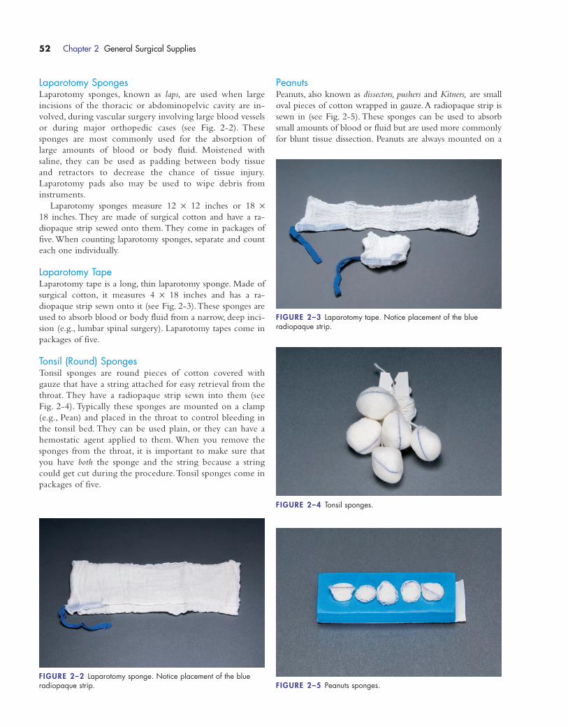
Laparotomy SpongesLaparotomy sponges, known as laps, are used when large
incisions of the thoracic or abdominopelvic cavity are in-
volved, during vascular surgery involving large blood vessels
or during major orthopedic cases (see Fig. 2-2). These
sponges are most commonly used for the absorption of
large amounts of blood or body fluid. Moistened with
saline, they can be used as padding between body tissue
and retractors to decrease the chance of tissue injury.
Laparotomy pads also may be used to wipe debris from
instruments.
Laparotomy sponges measure 12 × 12 inches or 18 ×18 inches. They are made of surgical cotton and have a ra-
diopaque strip sewed onto them. They come in packages of
five.When counting laparotomy sponges, separate and count
each one individually.
Laparotomy TapeLaparotomy tape is a long, thin laparotomy sponge. Made of
surgical cotton, it measures 4 × 18 inches and has a ra-
diopaque strip sewn onto it (see Fig. 2-3).These sponges are
used to absorb blood or body fluid from a narrow, deep inci-
sion (e.g., lumbar spinal surgery). Laparotomy tapes come in
packages of five.
Tonsil (Round) SpongesTonsil sponges are round pieces of cotton covered with
gauze that have a string attached for easy retrieval from the
throat. They have a radiopaque strip sewn into them (see
Fig. 2-4). Typically these sponges are mounted on a clamp
(e.g., Pean) and placed in the throat to control bleeding in
the tonsil bed. They can be used plain, or they can have a
hemostatic agent applied to them. When you remove the
sponges from the throat, it is important to make sure that
you have both the sponge and the string because a string
could get cut during the procedure.Tonsil sponges come in
packages of five.
PeanutsPeanuts, also known as dissectors, pushers and Kitners, are small
oval pieces of cotton wrapped in gauze. A radiopaque strip is
sewn in (see Fig. 2-5). These sponges can be used to absorb
small amounts of blood or fluid but are used more commonly
for blunt tissue dissection. Peanuts are always mounted on a
52 Chapter 2 General Surgical Supplies
FIGURE 2–2 Laparotomy sponge. Notice placement of the blue radiopaque strip.
FIGURE 2–3 Laparotomy tape. Notice placement of the blue radiopaque strip.
FIGURE 2–4 Tonsil sponges.
FIGURE 2–5 Peanuts sponges.
1572_Ch02_051-092.qxd 8/6/09 11:06 AM Page 52
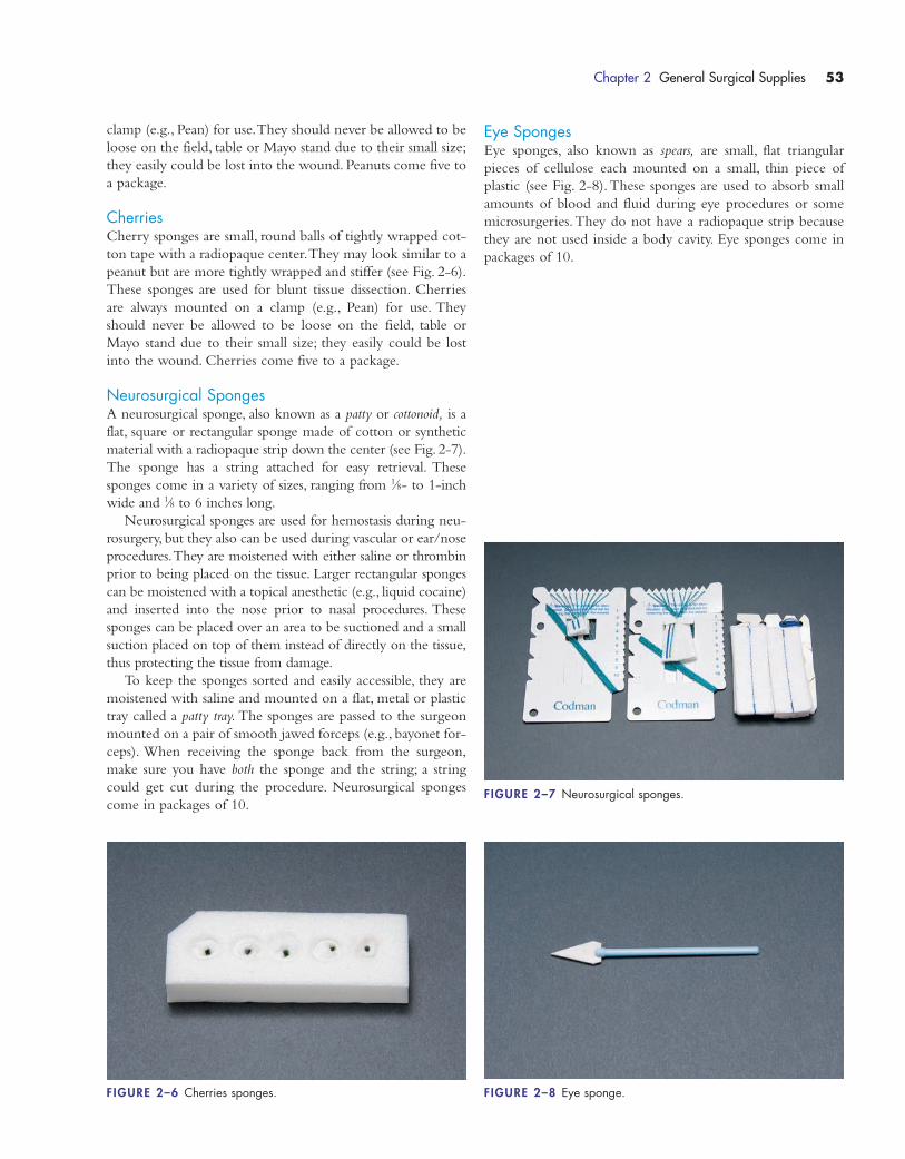
clamp (e.g., Pean) for use.They should never be allowed to be
loose on the field, table or Mayo stand due to their small size;
they easily could be lost into the wound. Peanuts come five to
a package.
CherriesCherry sponges are small, round balls of tightly wrapped cot-
ton tape with a radiopaque center.They may look similar to a
peanut but are more tightly wrapped and stiffer (see Fig. 2-6).
These sponges are used for blunt tissue dissection. Cherries
are always mounted on a clamp (e.g., Pean) for use. They
should never be allowed to be loose on the field, table or
Mayo stand due to their small size; they easily could be lost
into the wound. Cherries come five to a package.
Neurosurgical SpongesA neurosurgical sponge, also known as a patty or cottonoid, is a
flat, square or rectangular sponge made of cotton or synthetic
material with a radiopaque strip down the center (see Fig. 2-7).
The sponge has a string attached for easy retrieval. These
sponges come in a variety of sizes, ranging from 1⁄8- to 1-inch
wide and 1⁄8 to 6 inches long.
Neurosurgical sponges are used for hemostasis during neu-
rosurgery, but they also can be used during vascular or ear/nose
procedures.They are moistened with either saline or thrombin
prior to being placed on the tissue. Larger rectangular sponges
can be moistened with a topical anesthetic (e.g., liquid cocaine)
and inserted into the nose prior to nasal procedures. These
sponges can be placed over an area to be suctioned and a small
suction placed on top of them instead of directly on the tissue,
thus protecting the tissue from damage.
To keep the sponges sorted and easily accessible, they are
moistened with saline and mounted on a flat, metal or plastic
tray called a patty tray. The sponges are passed to the surgeon
mounted on a pair of smooth jawed forceps (e.g., bayonet for-
ceps). When receiving the sponge back from the surgeon,
make sure you have both the sponge and the string; a string
could get cut during the procedure. Neurosurgical sponges
come in packages of 10.
Eye SpongesEye sponges, also known as spears, are small, flat triangular
pieces of cellulose each mounted on a small, thin piece of
plastic (see Fig. 2-8).These sponges are used to absorb small
amounts of blood and fluid during eye procedures or some
microsurgeries.They do not have a radiopaque strip because
they are not used inside a body cavity. Eye sponges come in
packages of 10.
Chapter 2 General Surgical Supplies 53
FIGURE 2–6 Cherries sponges. FIGURE 2–8 Eye sponge.
FIGURE 2–7 Neurosurgical sponges.
1572_Ch02_051-092.qxd 8/6/09 11:06 AM Page 53
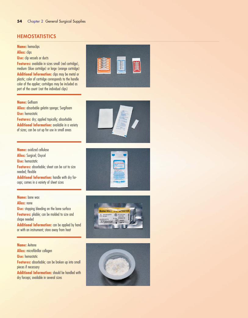
54 Chapter 2 General Surgical Supplies
HEMOSTATISTICS
Name: hemoclips
Alias: clips
Use: clip vessels or ducts
Features: available in sizes small (red cartridge),medium (blue cartridge) or large (orange cartridge)
Additional Information: clips may be metal orplastic; color of cartridge corresponds to the handlecolor of the applier; cartridges may be included aspart of the count (not the individual clips)
Name: Gelfoam
Alias: absorbable gelatin sponge; Surgifoam
Use: hemostatic
Features: dry; applied topically; absorbable
Additional Information: available in a varietyof sizes; can be cut up for use in small areas
Name: oxidized cellulose
Alias: Surgicel; Oxycel
Use: hemostatic
Features: absorbable; sheet can be cut to sizeneeded; flexible
Additional Information: handle with dry for-ceps; comes in a variety of sheet sizes
Name: bone wax
Alias: none
Use: stopping bleeding on the bone surface
Features: pliable; can be molded to size andshape needed
Additional Information: can be applied by handor with an instrument; store away from heat
Name: Avitene
Alias: microfibrillar collagen
Use: hemostatic
Features: absorbable; can be broken up into smallpieces if necessary
Additional Information: should be handled withdry forceps; available in several sizes
1572_Ch02_051-092.qxd 8/6/09 11:06 AM Page 54
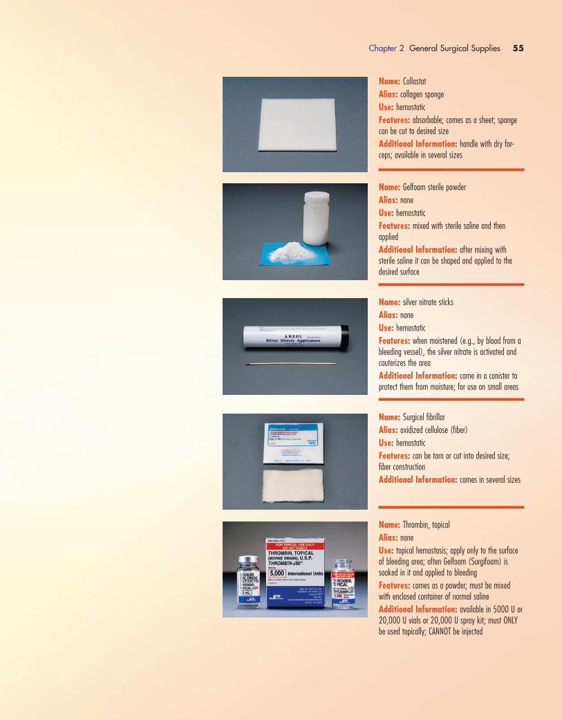
Chapter 2 General Surgical Supplies 55
Name: Collastat
Alias: collagen sponge
Use: hemostatic
Features: absorbable; comes as a sheet; spongecan be cut to desired size
Additional Information: handle with dry for-ceps; available in several sizes
Name: Gelfoam sterile powder
Alias: none
Use: hemostatic
Features: mixed with sterile saline and then applied
Additional Information: after mixing with sterile saline it can be shaped and applied to the desired surface
Name: silver nitrate sticks
Alias: none
Use: hemostatic
Features: when moistened (e.g., by blood from ableeding vessel), the silver nitrate is activated andcauterizes the area
Additional Information: come in a canister toprotect them from moisture; for use on small areas
Name: Surgicel fibrillar
Alias: oxidized cellulose (fiber)
Use: hemostatic
Features: can be torn or cut into desired size;fiber construction
Additional Information: comes in several sizes
Name: Thrombin, topical
Alias: none
Use: topical hemostasis; apply only to the surfaceof bleeding area; often Gelfoam (Surgifoam) issoaked in it and applied to bleeding
Features: comes as a powder; must be mixedwith enclosed container of normal saline
Additional Information: available in 5000 U or20,000 U vials or 20,000 U spray kit; must ONLYbe used topically; CANNOT be injected
1572_Ch02_051-092.qxd 8/6/09 11:06 AM Page 55
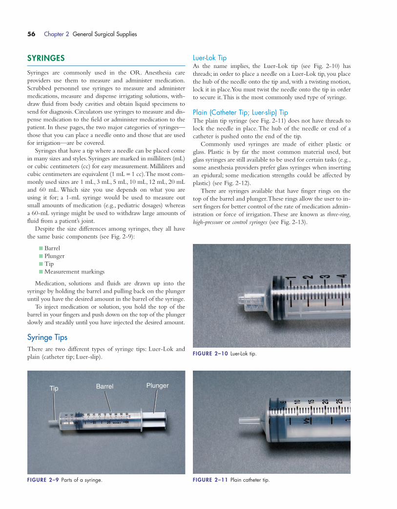
SYRINGES
Syringes are commonly used in the OR. Anesthesia care
providers use them to measure and administer medication.
Scrubbed personnel use syringes to measure and administer
medications, measure and dispense irrigating solutions, with-
draw fluid from body cavities and obtain liquid specimens to
send for diagnosis. Circulators use syringes to measure and dis-
pense medication to the field or administer medication to the
patient. In these pages, the two major categories of syringes—
those that you can place a needle onto and those that are used
for irrigation—are be covered.
Syringes that have a tip where a needle can be placed come
in many sizes and styles. Syringes are marked in milliliters (mL)
or cubic centimeters (cc) for easy measurement. Milliliters and
cubic centimeters are equivalent (1 mL = 1 cc).The most com-
monly used sizes are 1 mL, 3 mL, 5 mL, 10 mL, 12 mL, 20 mL
and 60 mL. Which size you use depends on what you are
using it for; a 1-mL syringe would be used to measure out
small amounts of medication (e.g., pediatric dosages) whereas
a 60-mL syringe might be used to withdraw large amounts of
fluid from a patient’s joint.
Despite the size differences among syringes, they all have
the same basic components (see Fig. 2-9):
� Barrel
� Plunger
� Tip
� Measurement markings
Medication, solutions and fluids are drawn up into the
syringe by holding the barrel and pulling back on the plunger
until you have the desired amount in the barrel of the syringe.
To inject medication or solution, you hold the top of the
barrel in your fingers and push down on the top of the plunger
slowly and steadily until you have injected the desired amount.
Syringe TipsThere are two different types of syringe tips: Luer-Lok and
plain (catheter tip; Luer-slip).
Luer-Lok TipAs the name implies, the Luer-Lok tip (see Fig. 2-10) has
threads; in order to place a needle on a Luer-Lok tip, you place
the hub of the needle onto the tip and, with a twisting motion,
lock it in place.You must twist the needle onto the tip in order
to secure it.This is the most commonly used type of syringe.
Plain (Catheter Tip; Luer-slip) TipThe plain tip syringe (see Fig. 2-11) does not have threads to
lock the needle in place.The hub of the needle or end of a
catheter is pushed onto the end of the tip.
Commonly used syringes are made of either plastic or
glass. Plastic is by far the most common material used, but
glass syringes are still available to be used for certain tasks (e.g.,
some anesthesia providers prefer glass syringes when inserting
an epidural; some medication strengths could be affected by
plastic) (see Fig. 2-12).
There are syringes available that have finger rings on the
top of the barrel and plunger.These rings allow the user to in-
sert fingers for better control of the rate of medication admin-
istration or force of irrigation.These are known as three-ring,
high-pressure or control syringes (see Fig. 2-13).
56 Chapter 2 General Surgical Supplies
Tip Barrel Plunger
FIGURE 2–9 Parts of a syringe.
FIGURE 2–10 Luer-Lok tip.
FIGURE 2–11 Plain catheter tip.
1572_Ch02_051-092.qxd 8/6/09 11:06 AM Page 56
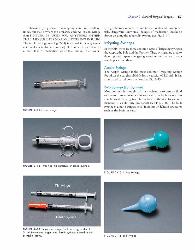
syringe, the measurement would be inaccurate and thus poten-
tially dangerous. Only small dosages of medication should be
drawn up using the tuberculin syringe (see Fig. 2-14).
Irrigating SyringesIn the OR, there are three common types of irrigating syringes:
the Asepto, the bulb and the Toomey. These syringes are used to
draw up and dispense irrigating solutions and do not have a
needle placed on them.
Asepto SyringeThe Asepto syringe is the most common irrigating syringe
found on the surgical field. It has a capacity of 120 mL. It has
a bulb and barrel construction (see Fig. 2-15).
Bulb Syringe (Ear Syringe)Most commonly thought of as a mechanism to remove fluid
or mucus from an infant’s nose or mouth, the bulb syringe can
also be used for irrigation. In contrast to the Asepto, its con-
struction is a bulb only (no barrel) (see Fig. 2-16).The bulb
syringe is used to irrigate small incisions or delicate structures
such as the brain or ears.
Chapter 2 General Surgical Supplies 57
FIGURE 2–12 Glass syringe.
FIGURE 2–13 Three-ring, high-pressure or control syringe.
TB syringe
Insulin syringe
FIGURE 2–14 Tuberculin syringe: 1-mL capacity; marked in 0.1-mL increments (larger lines). Insulin syringe: marked in units of insulin (not mL).
FIGURE 2–15 Asepto syringe.
FIGURE 2–16 Bulb syringe.
Tuberculin syringes and insulin syringes are both small sy-
ringes, but that is where the similarity ends. An insulin syringe
should NEVER BE USED FOR ANYTHING OTHER
THAN MEASURING AND ADMINISTERING INSULIN!
The insulin syringe (see Fig. 2-14) is marked in units of insulin
not milliliters (cubic centimeters) of volume. If you were to
measure fluid or medication (other than insulin) in an insulin
1572_Ch02_051-092.qxd 8/6/09 11:06 AM Page 57
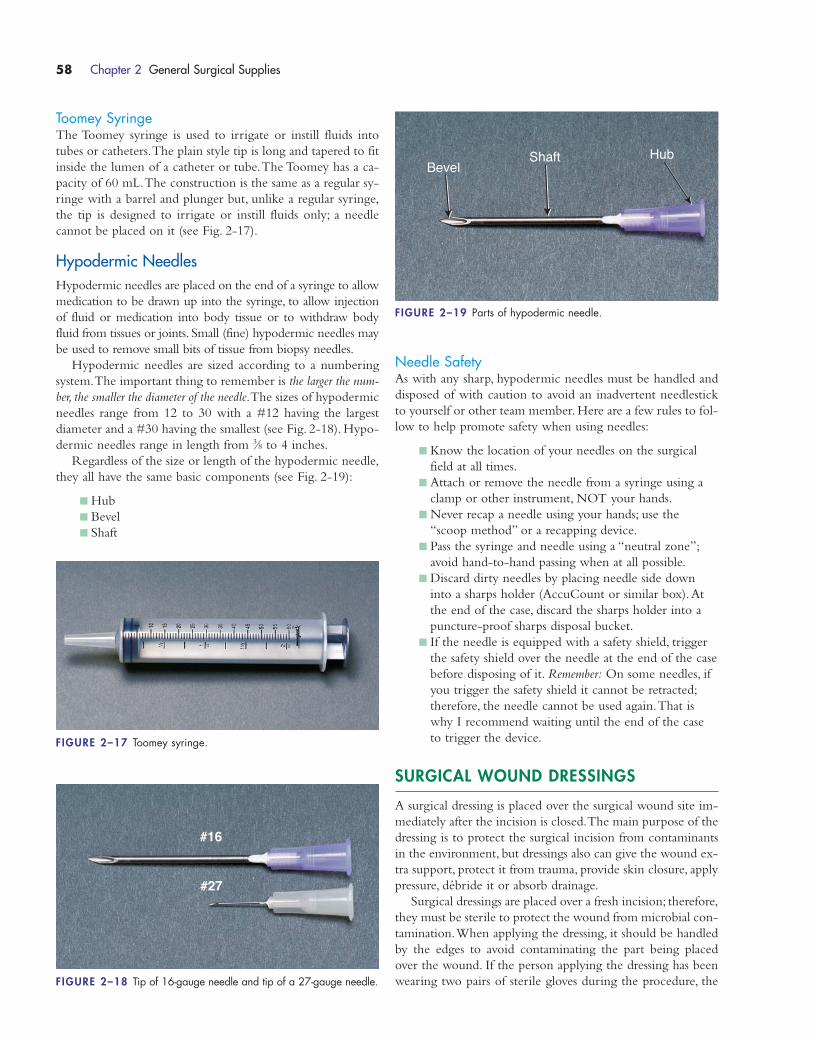
Toomey SyringeThe Toomey syringe is used to irrigate or instill fluids into
tubes or catheters.The plain style tip is long and tapered to fit
inside the lumen of a catheter or tube.The Toomey has a ca-
pacity of 60 mL.The construction is the same as a regular sy-
ringe with a barrel and plunger but, unlike a regular syringe,
the tip is designed to irrigate or instill fluids only; a needle
cannot be placed on it (see Fig. 2-17).
Hypodermic NeedlesHypodermic needles are placed on the end of a syringe to allow
medication to be drawn up into the syringe, to allow injection
of fluid or medication into body tissue or to withdraw body
fluid from tissues or joints. Small (fine) hypodermic needles may
be used to remove small bits of tissue from biopsy needles.
Hypodermic needles are sized according to a numbering
system.The important thing to remember is the larger the num-
ber, the smaller the diameter of the needle.The sizes of hypodermic
needles range from 12 to 30 with a #12 having the largest
diameter and a #30 having the smallest (see Fig. 2-18). Hypo-
dermic needles range in length from 3⁄8 to 4 inches.
Regardless of the size or length of the hypodermic needle,
they all have the same basic components (see Fig. 2-19):
� Hub
� Bevel
� Shaft
Needle SafetyAs with any sharp, hypodermic needles must be handled and
disposed of with caution to avoid an inadvertent needlestick
to yourself or other team member. Here are a few rules to fol-
low to help promote safety when using needles:
� Know the location of your needles on the surgical
field at all times.
� Attach or remove the needle from a syringe using a
clamp or other instrument, NOT your hands.
� Never recap a needle using your hands; use the
“scoop method” or a recapping device.
� Pass the syringe and needle using a “neutral zone”;
avoid hand-to-hand passing when at all possible.
� Discard dirty needles by placing needle side down
into a sharps holder (AccuCount or similar box).At
the end of the case, discard the sharps holder into a
puncture-proof sharps disposal bucket.
� If the needle is equipped with a safety shield, trigger
the safety shield over the needle at the end of the case
before disposing of it. Remember: On some needles, if
you trigger the safety shield it cannot be retracted;
therefore, the needle cannot be used again.That is
why I recommend waiting until the end of the case
to trigger the device.
SURGICAL WOUND DRESSINGS
A surgical dressing is placed over the surgical wound site im-
mediately after the incision is closed.The main purpose of the
dressing is to protect the surgical incision from contaminants
in the environment, but dressings also can give the wound ex-
tra support, protect it from trauma, provide skin closure, apply
pressure, débride it or absorb drainage.
Surgical dressings are placed over a fresh incision; therefore,
they must be sterile to protect the wound from microbial con-
tamination.When applying the dressing, it should be handled
by the edges to avoid contaminating the part being placed
over the wound. If the person applying the dressing has been
wearing two pairs of sterile gloves during the procedure, the
58 Chapter 2 General Surgical Supplies
FIGURE 2–17 Toomey syringe.
BevelShaft Hub
FIGURE 2–19 Parts of hypodermic needle.
#16
#27
FIGURE 2–18 Tip of 16-gauge needle and tip of a 27-gauge needle.
1572_Ch02_051-092.qxd 8/6/09 11:06 AM Page 58
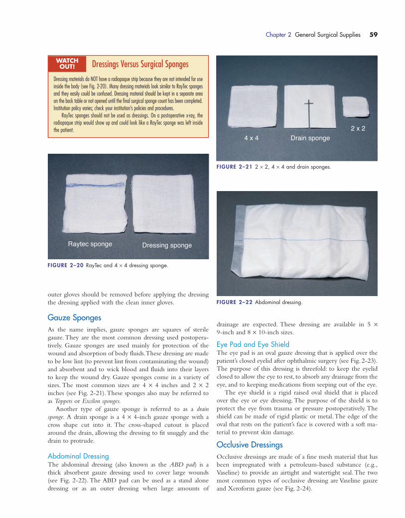
outer gloves should be removed before applying the dressing
the dressing applied with the clean inner gloves.
Gauze SpongesAs the name implies, gauze sponges are squares of sterile
gauze. They are the most common dressing used postopera-
tively. Gauze sponges are used mainly for protection of the
wound and absorption of body fluids.These dressing are made
to be low lint (to prevent lint from contaminating the wound)
and absorbent and to wick blood and fluids into their layers
to keep the wound dry. Gauze sponges come in a variety of
sizes. The most common sizes are 4 × 4 inches and 2 × 2
inches (see Fig. 2-21).These sponges also may be referred to
as Toppers or Excilon sponges.
Another type of gauze sponge is referred to as a drain
sponge. A drain sponge is a 4 × 4-inch gauze sponge with a
cross shape cut into it. The cross-shaped cutout is placed
around the drain, allowing the dressing to fit snuggly and the
drain to protrude.
Abdominal DressingThe abdominal dressing (also known as the ABD pad) is a
thick absorbent gauze dressing used to cover large wounds
(see Fig. 2-22). The ABD pad can be used as a stand alone
dressing or as an outer dressing when large amounts of
drainage are expected. These dressing are available in 5 ×9-inch and 8 × 10-inch sizes.
Eye Pad and Eye ShieldThe eye pad is an oval gauze dressing that is applied over the
patient’s closed eyelid after ophthalmic surgery (see Fig. 2-23).
The purpose of this dressing is threefold: to keep the eyelid
closed to allow the eye to rest, to absorb any drainage from the
eye, and to keeping medications from seeping out of the eye.
The eye shield is a rigid raised oval shield that is placed
over the eye or eye dressing.The purpose of the shield is to
protect the eye from trauma or pressure postoperatively. The
shield can be made of rigid plastic or metal.The edge of the
oval that rests on the patient’s face is covered with a soft ma-
terial to prevent skin damage.
Occlusive DressingsOcclusive dressings are made of a fine mesh material that has
been impregnated with a petroleum-based substance (e.g.,
Vaseline) to provide an airtight and watertight seal. The two
most common types of occlusive dressing are Vaseline gauze
and Xeroform gauze (see Fig. 2-24).
Chapter 2 General Surgical Supplies 59
Raytec sponge Dressing sponge
FIGURE 2–20 RayTec and 4 × 4 dressing sponge.
4 x 4 Drain sponge2 x 2
FIGURE 2–21 2 × 2, 4 × 4 and drain sponges.
FIGURE 2–22 Abdominal dressing.
Dressings Versus Surgical Sponges
Dressing materials do NOT have a radiopaque strip because they are not intended for useinside the body (see Fig. 2-20). Many dressing materials look similar to RayTec spongesand they easily could be confused. Dressing material should be kept in a separate areaon the back table or not opened until the final surgical sponge count has been completed.Institution policy varies; check your institution’s policies and procedures.
RayTec sponges should not be used as dressings. On a postoperative x-ray, theradiopaque strip would show up and could look like a RayTec sponge was left insidethe patient.
WATCH OUT!
1572_Ch02_051-092.qxd 8/6/09 11:06 AM Page 59
The petroleum-based substance on the dressing prevents it
from sticking to the wound while the mesh allows drainage
to seep into the absorbent gauze dressing placed over it.This
type of dressing is often used over a suture line to protect it
and to keep the absorbent gauze dressing layers from sticking
to it. It also can be used around a chest tube insertion site to
prevent outside air from entering the chest around the stab
wound site.
Vaseline dressings are available in sheets or rolls. Sizes range
from 1 × 8 inches to 6 × 36 inches.The most common sizes
of Xeroform dressings are 1 × 8 inches and 5 × 9 inches.
AdapticAdaptic is gauze that has been impregnated with a petroleum-
based substance. Unlike the Vaseline or Xeroform dressing,
Adaptic is nonocclusive. It does not provide a watertight or air-
tight seal. Adaptic does, however, provide a nonadhering layer
that wicks drainage away from the wound and allows the dress-
ing to be removed without sticking (see Fig. 2-25). Adaptic is
available in three sizes: 3 × 3 inches, 3 × 8 inches and 3 ×16 inches.
TelfaTelfa is a nonadhering dressing material. It is made of absorbent
gauze that is bonded on both sides with a nonadherent mate-
rial to prevent it from sticking to the wound. It allows air to
reach the wound and wicks the drainage away from the wound
(see Fig. 2-26).Telfa is available in three sizes: 2 × 3 inches, 3 ×4 inches and 6 × 10 inches.
Telfa also can be used as a mat for tissue specimens. The
nonadherent covering keeps the specimen from sticking, thus
preserving the tissue integrity. Scrubbed personnel often place
small tissue specimens on a piece of Telfa to keep them from
becoming lost.
TegadermTegaderm is a thin, transparent, waterproof film dressing. It is
used as an outer dressing for wounds from which large amounts
of drainage are not expected or as a stand alone dressing over
intravenous (IV) sites. One advantage of Tegaderm is that it is
clear, allowing visualization of the dressing or IV site under-
neath it (see Fig. 2-27). Tegaderm is available in sizes ranging
from 23⁄4 × 23⁄4 inches to 8 × 12 inches.
60 Chapter 2 General Surgical Supplies
FIGURE 2–23 Eye pad and eye shield. FIGURE 2–25 Adaptic.
FIGURE 2–26 Telfa.FIGURE 2–24 Vaseline and Xeroform gauzes.
1572_Ch02_051-092.qxd 8/6/09 11:06 AM Page 60
Island dressingsIsland dressings combine a nonadherent gauze dressing with a
nonirritating adhesive border. The built-in adhesive border al-
lows these dressings to adhere directly to the skin, negating the
need to use tape or a wrap to secure them (see Fig. 2-28). These
dressings are used on wounds from which large amounts of
drainage are not expected. Two of the most common brand
names are Covaderm and Primapore.These dressings come in a
variety of shapes (e.g., square, round and rectangular) and sizes.
Dressing Wraps
KerlixKerlix (see Fig. 2-29) is a six-ply roll of loosely woven gauze
material. Generally used to secure a dressing or splint in place,
it conforms to the contours of the patient’s body, allowing
freedom of movement. It also can be used to help apply pres-
sure to a wound or to pack a large wound.
Kerlix is available as a 41⁄2-inch × 4-yard or 21⁄4-inch ×3-yard roll. Kerlix is also available as a dressing sponge impreg-
nated with an antimicrobial substance.
KlingKling (see Fig. 2-29) is a roll of nonwoven gauze material used
to secure bandages or splints or to provide pressure to a wound.
It conforms to body contours, allowing freedom of movement.
It is available in 1-, 2-, 3-, 4- or 6-inch widths.
Ace BandageAn Ace bandage (also known as an Ace wrap) is a roll of elas-
tic bandage commonly used to provide support to a sprain or
strain of a joint (see Fig. 2-30). It also can be used to secure a
dressing or to hold a splint in place.
Chapter 2 General Surgical Supplies 61
FIGURE 2–27 Tegaderm.
Kerlix
Kling
FIGURE 2–29 Kerlix and Kling.
FIGURE 2–30 Ace wrap.FIGURE 2–28 Island dressings.
Wrapping Bandages
Use caution when wrapping any bandage (regardless of material) in a circular fash-ion that encompasses the entire diameter of an extremity or other body part. Thebandage should fit snuggly so that it does not slide off but it should NOT be wrappedso tight that it causes circulatory problems.
After wrapping the bandage, check the areas distal to the bandage for signs ofimpaired circulation: cyanosis, skin coolness, skin mottling or patient complaints ofnumbness or tingling.
WATCH OUT!
1572_Ch02_051-092.qxd 8/6/09 11:06 AM Page 61
The elastic in the bandage allows it to stretch to conform
to the area and provide limited freedom of movement. Ace
bandages can be secured with tape, although most come with
metal fasteners or Velcro closures. Available sizes are 2-, 3-,
4- and 6-inch widths.
Self-Adherent WrapsSelf-adherent wraps are made of a blend of nonwoven fibers
and elastic to allow for stretch.They contain a cohesive mate-
rial designed to allow the bandage to stick to itself but not to
skin or other materials (see Fig. 2-31). These bandages are
used to secure dressings or splints in place.The two most com-
mon brand names for this type of dressing are Coban and
Flex-wrap. Self-adherent wraps are available in widths ranging
from 1 to 6 inches.
PackingsAs the name implies, packing materials are packed inside the
wound to provide support, to provide hemostasis or to allow
wound drainage and healing (see Fig. 2-32). Uses include boils,
abscesses, fistulas and other draining or tunneling wounds as
well as packing nasal passages, the rectum and the vagina.
Packings are usually made of a long strip of gauze material
that is plain or impregnated with antimicrobial substances
such as iodoform or silver. Creams (e.g., estrogen cream or
hemorrhoid cream) may be used to coat plain packing for use
after specific types of surgery.
Packing material is available in widths ranging from 1⁄4 inch
to 4 inches.
Liquid Chemical DressingsCollodionCollodion is a thick, yellow liquid consisting of pyroxylin
mixed with alcohol and ether (see Fig. 2-33). As the solvent
evaporates, collodion dries to form a waterproof film over the
wound. This type of dressing is used on small wounds from
which drainage is not expected. It is highly flammable and has
a strong odor.
Collodion is applied over the dry suture or staple line us-
ing a cotton-tipped applicator.A thin layer is applied and left
open to the air to dry. Once dry, the film provides protection
from environmental contaminants and water.
Liquid Skin AdhesivesThis category of dressings consists of topically applied liquid
adhesives that are designed to close small, superficial lacera-
tions and surgical incisions. In addition to wound closure they
provide a chemical film barrier for protection. Brand names
include Dermabond, New-Skin and LiquiBand.These adhe-
sives come in small ampules with an applicator on one end
(see Fig. 2-34).The ampule is squeezed, allowing the adhesive
to saturate the applicator.A thin layer of adhesive is applied to
the wound and allowed to dry and then another layer is
applied (application techniques may vary with brand; always
follow the manufacturer’s instructions).
Prior to use, it is important to check patient allergies to make
sure it is appropriate to use these adhesives (e.g., Dermabond
contains formaldehyde and cannot be used on patients with
formaldehyde allergies).
62 Chapter 2 General Surgical Supplies
FIGURE 2–31 Coban (Self-adherent wraps).
FIGURE 2–32 Plain and iodoform packing strip. FIGURE 2–33 Collodion.
1572_Ch02_051-092.qxd 8/6/09 11:06 AM Page 62
Adhesive TapesMedical adhesive tape (also known as surgical tape) is the
most common method of securing dressings. Available in
many types and widths, it is an easy and inexpensive way to
keep dressings in place. Listed are the most common types of
adhesive tape:
� Paper tape: Hypoallergenic. Leaves very little adhesive
residue when removed and is gentle on the skin. Used
on patients with adhesive sensitivities or patients who
have friable (fragile) skin. Good for use when repeated
dressing changes are anticipated on sensitive skin.
Used by some anesthesia personnel to tape eyelids
shut during general anesthesia.Tends not to stick well
when wet.Available in 1⁄2-, 1-, 2- and 3-inch widths.
Common brand names include Micropore, Hypo-Pore
and Tenderskin.
� Silk tape: “Silk-like tape.” Most commonly used
general purpose adhesive tape (see Fig. 2-35).
Hypoallergenic adhesive leaves tape residue after
removal. Available in 1⁄2-, 1-, 2- and 3-inch widths.
Common brand names include Durapore and
Hypo Silk.
� Elastic tape: Cotton elastic tape (see Fig. 2-36) de-
signed for use for pressure dressings or support strap-
ping of orthopedic injuries. Conforms to knees,
shoulders and elbows.Allows for wicking of moisture
away from the skin. Has a rubber-based adhesive.
Available in 1-, 2- , 3- and 4-inch widths. Common
brand names include Elastoplast and Elastikon.
� Plastic tape: Flexible, contouring, clear plastic tape that
allows viewing of the wound site. Used for securing
dressings, IV catheters or tubes.Available in 1-, 2- or
3-inch widths. Brand names include Blenderm,Trans-
pore, DermiView and HypoClear.
� Fabric tape: Porous and nonabsorbing fabric used to
secure dressings, tubes and cannulas. Nonirritating ad-
hesive allows skin underneath it to breath.Available in
1-, 2-, 4-, 6- and 8-inch widths; brand names include
Hypafix and Mefix.
� Foam elastic tape: Elastic foam material (see
Fig. 2-36) used for compression dressing or dressing
in hard-to-secure areas. Stretches in all directions.
Waterproof and hypoallergenic. Available in 1-,
2-, 3- and 4-inch widths. Common brand name is
Microfoam.
� Cloth tape: Nonwoven polyester tape (see
Fig. 2-35). Easy to tear, hypoallergenic, general
purpose tape. Has moderate stretch to allow for
movement and swelling. Available in 1-, 2-, 3-, 4-,
6- and 8-inch widths. Brand names include
Dermicel and Curasilk.
� Waterproof tape: Zinc oxide–based tape (see
Fig. 2-35).Waterproof and oil resistant and gentle to
all skin types, it will remain intact for long periods
of time, even when wet. Conforms easily to body
contours. Used to secure dressings or tubes. Used by
some anesthesia personnel to secure endotracheal
tubes. Available in 1⁄2-, 1-, and 2-inch widths. Brand
names include Hy-Tape, Scanpor,Wet Pruf and
Megazinc.
Chapter 2 General Surgical Supplies 63
FIGURE 2–34 Dermabond.
Paper
Waterproof
Silk
Cloth
FIGURE 2–35 Paper, silk, cloth and waterproof tapes.
Cloth
Elastic
FIGURE 2–36 Elastic and cloth tapes.
1572_Ch02_051-092.qxd 8/6/09 11:06 AM Page 63
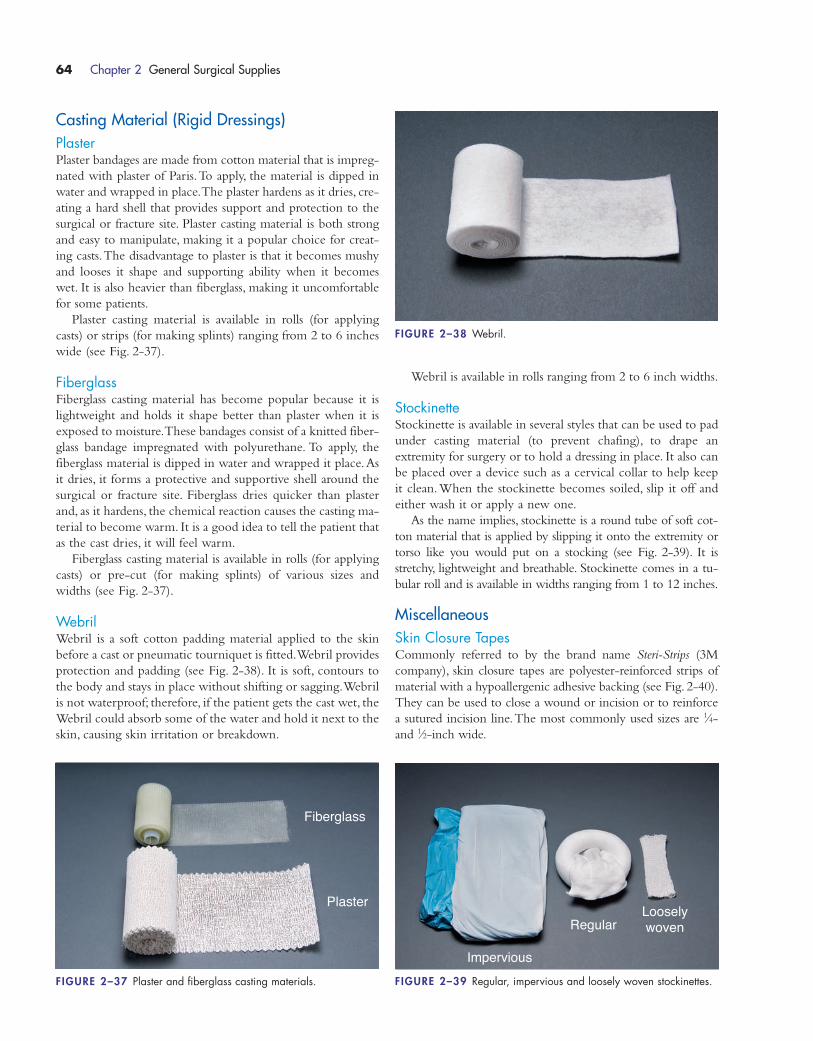
Casting Material (Rigid Dressings)PlasterPlaster bandages are made from cotton material that is impreg-
nated with plaster of Paris.To apply, the material is dipped in
water and wrapped in place.The plaster hardens as it dries, cre-
ating a hard shell that provides support and protection to the
surgical or fracture site. Plaster casting material is both strong
and easy to manipulate, making it a popular choice for creat-
ing casts.The disadvantage to plaster is that it becomes mushy
and looses it shape and supporting ability when it becomes
wet. It is also heavier than fiberglass, making it uncomfortable
for some patients.
Plaster casting material is available in rolls (for applying
casts) or strips (for making splints) ranging from 2 to 6 inches
wide (see Fig. 2-37).
FiberglassFiberglass casting material has become popular because it is
lightweight and holds it shape better than plaster when it is
exposed to moisture.These bandages consist of a knitted fiber-
glass bandage impregnated with polyurethane. To apply, the
fiberglass material is dipped in water and wrapped it place.As
it dries, it forms a protective and supportive shell around the
surgical or fracture site. Fiberglass dries quicker than plaster
and, as it hardens, the chemical reaction causes the casting ma-
terial to become warm. It is a good idea to tell the patient that
as the cast dries, it will feel warm.
Fiberglass casting material is available in rolls (for applying
casts) or pre-cut (for making splints) of various sizes and
widths (see Fig. 2-37).
WebrilWebril is a soft cotton padding material applied to the skin
before a cast or pneumatic tourniquet is fitted.Webril provides
protection and padding (see Fig. 2-38). It is soft, contours to
the body and stays in place without shifting or sagging.Webril
is not waterproof; therefore, if the patient gets the cast wet, the
Webril could absorb some of the water and hold it next to the
skin, causing skin irritation or breakdown.
Webril is available in rolls ranging from 2 to 6 inch widths.
StockinetteStockinette is available in several styles that can be used to pad
under casting material (to prevent chafing), to drape an
extremity for surgery or to hold a dressing in place. It also can
be placed over a device such as a cervical collar to help keep
it clean.When the stockinette becomes soiled, slip it off and
either wash it or apply a new one.
As the name implies, stockinette is a round tube of soft cot-
ton material that is applied by slipping it onto the extremity or
torso like you would put on a stocking (see Fig. 2-39). It is
stretchy, lightweight and breathable. Stockinette comes in a tu-
bular roll and is available in widths ranging from 1 to 12 inches.
MiscellaneousSkin Closure TapesCommonly referred to by the brand name Steri-Strips (3M
company), skin closure tapes are polyester-reinforced strips of
material with a hypoallergenic adhesive backing (see Fig. 2-40).
They can be used to close a wound or incision or to reinforce
a sutured incision line.The most commonly used sizes are 1⁄4-
and 1⁄2-inch wide.
64 Chapter 2 General Surgical Supplies
Fiberglass
Plaster
FIGURE 2–37 Plaster and fiberglass casting materials.
Impervious
RegularLoosely woven
FIGURE 2–39 Regular, impervious and loosely woven stockinettes.
FIGURE 2–38 Webril.
1572_Ch02_051-092.qxd 8/6/09 11:06 AM Page 64
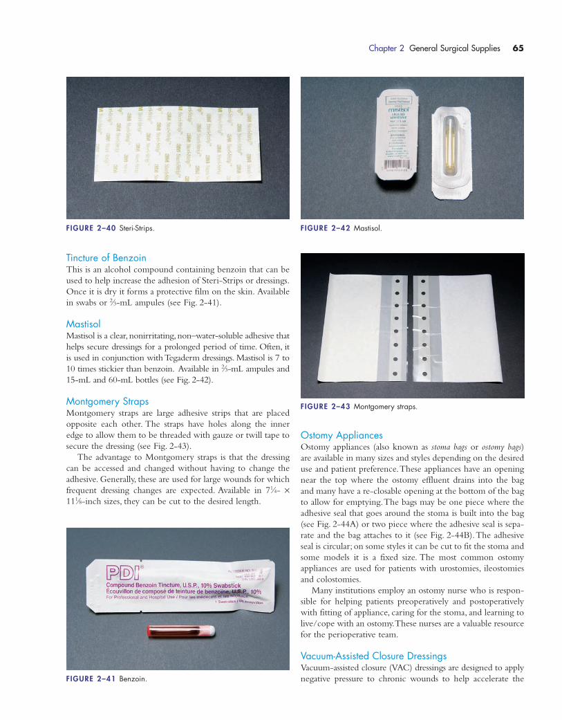
Tincture of BenzoinThis is an alcohol compound containing benzoin that can be
used to help increase the adhesion of Steri-Strips or dressings.
Once it is dry it forms a protective film on the skin. Available
in swabs or 2⁄3-mL ampules (see Fig. 2-41).
MastisolMastisol is a clear,nonirritating,non–water-soluble adhesive that
helps secure dressings for a prolonged period of time. Often, it
is used in conjunction with Tegaderm dressings. Mastisol is 7 to
10 times stickier than benzoin. Available in 2⁄3-mL ampules and
15-mL and 60-mL bottles (see Fig. 2-42).
Montgomery StrapsMontgomery straps are large adhesive strips that are placed
opposite each other. The straps have holes along the inner
edge to allow them to be threaded with gauze or twill tape to
secure the dressing (see Fig. 2-43).
The advantage to Montgomery straps is that the dressing
can be accessed and changed without having to change the
adhesive. Generally, these are used for large wounds for which
frequent dressing changes are expected. Available in 71⁄4- ×111⁄8-inch sizes, they can be cut to the desired length.
Ostomy AppliancesOstomy appliances (also known as stoma bags or ostomy bags)
are available in many sizes and styles depending on the desired
use and patient preference.These appliances have an opening
near the top where the ostomy effluent drains into the bag
and many have a re-closable opening at the bottom of the bag
to allow for emptying.The bags may be one piece where the
adhesive seal that goes around the stoma is built into the bag
(see Fig. 2-44A) or two piece where the adhesive seal is sepa-
rate and the bag attaches to it (see Fig. 2-44B).The adhesive
seal is circular; on some styles it can be cut to fit the stoma and
some models it is a fixed size. The most common ostomy
appliances are used for patients with urostomies, ileostomies
and colostomies.
Many institutions employ an ostomy nurse who is respon-
sible for helping patients preoperatively and postoperatively
with fitting of appliance, caring for the stoma, and learning to
live/cope with an ostomy.These nurses are a valuable resource
for the perioperative team.
Vacuum-Assisted Closure DressingsVacuum-assisted closure (VAC) dressings are designed to apply
negative pressure to chronic wounds to help accelerate the
Chapter 2 General Surgical Supplies 65
FIGURE 2–40 Steri-Strips.
FIGURE 2–41 Benzoin.
FIGURE 2–42 Mastisol.
FIGURE 2–43 Montgomery straps.
1572_Ch02_051-092.qxd 8/6/09 11:06 AM Page 65
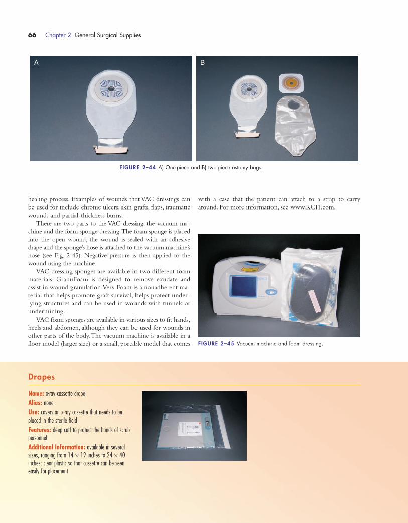
healing process. Examples of wounds that VAC dressings can
be used for include chronic ulcers, skin grafts, flaps, traumatic
wounds and partial-thickness burns.
There are two parts to the VAC dressing: the vacuum ma-
chine and the foam sponge dressing.The foam sponge is placed
into the open wound, the wound is sealed with an adhesive
drape and the sponge’s hose is attached to the vacuum machine’s
hose (see Fig. 2-45). Negative pressure is then applied to the
wound using the machine.
VAC dressing sponges are available in two different foam
materials. GranuFoam is designed to remove exudate and
assist in wound granulation.Vers-Foam is a nonadherent ma-
terial that helps promote graft survival, helps protect under-
lying structures and can be used in wounds with tunnels or
undermining.
VAC foam sponges are available in various sizes to fit hands,
heels and abdomen, although they can be used for wounds in
other parts of the body.The vacuum machine is available in a
floor model (larger size) or a small, portable model that comes
with a case that the patient can attach to a strap to carry
around. For more information, see www.KCI1.com.
66 Chapter 2 General Surgical Supplies
FIGURE 2–44 A) One-piece and B) two-piece ostomy bags.
A B
FIGURE 2–45 Vacuum machine and foam dressing.
Drapes
Name: x-ray cassette drape
Alias: none
Use: covers an x-ray cassette that needs to beplaced in the sterile field
Features: deep cuff to protect the hands of scrubpersonnel
Additional Information: available in severalsizes, ranging from 14 × 19 inches to 24 × 40inches; clear plastic so that cassette can be seeneasily for placement
1572_Ch02_051-092.qxd 8/6/09 11:07 AM Page 66
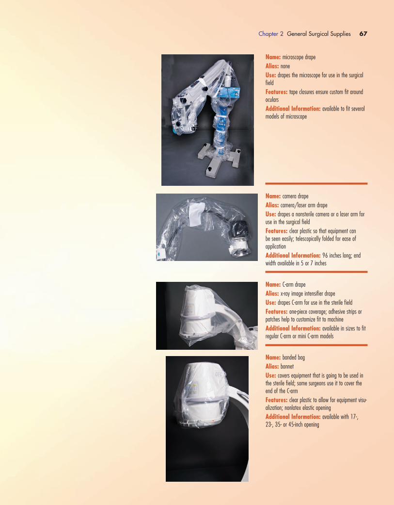
Chapter 2 General Surgical Supplies 67
Name: microscope drape
Alias: none
Use: drapes the microscope for use in the surgicalfield
Features: tape closures ensure custom fit aroundoculars
Additional Information: available to fit severalmodels of microscope
Name: camera drape
Alias: camera/laser arm drape
Use: drapes a nonsterile camera or a laser arm foruse in the surgical field
Features: clear plastic so that equipment can be seen easily; telescopically folded for ease of application
Additional Information: 96 inches long; endwidth available in 5 or 7 inches
Name: C-arm drape
Alias: x-ray image intensifier drape
Use: drapes C-arm for use in the sterile field
Features: one-piece coverage; adhesive strips orpatches help to customize fit to machine
Additional Information: available in sizes to fitregular C-arm or mini C-arm models
Name: banded bag
Alias: bonnet
Use: covers equipment that is going to be used inthe sterile field; some surgeons use it to cover theend of the C-arm
Features: clear plastic to allow for equipment visu-alization; nonlatex elastic opening
Additional Information: available with 17-, 23-, 35- or 45-inch opening
1572_Ch02_051-092.qxd 8/6/09 11:07 AM Page 67
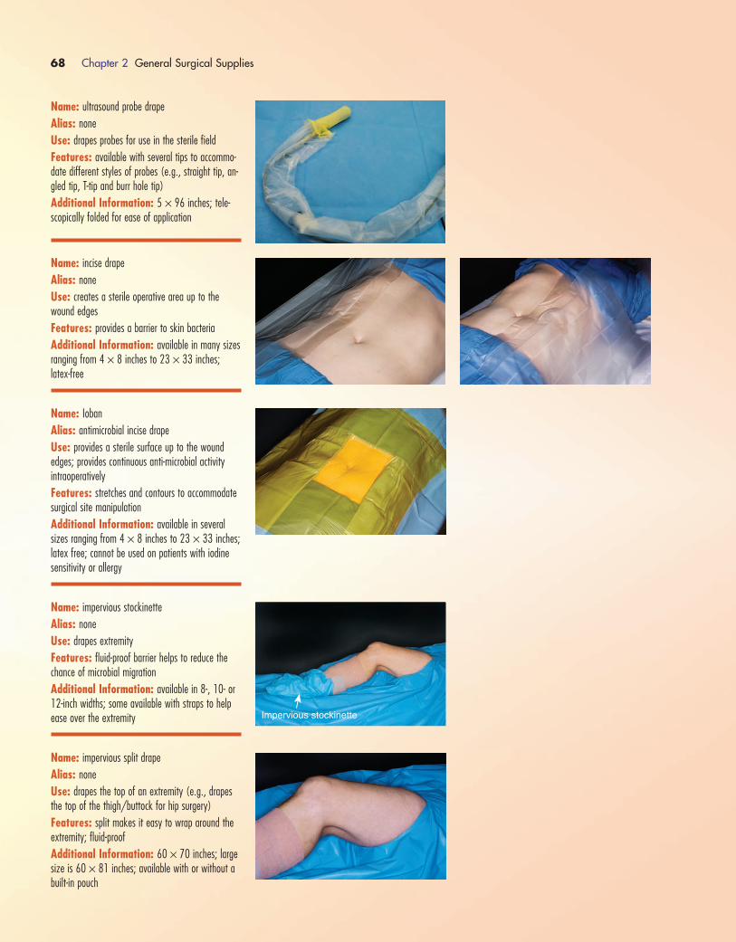
68 Chapter 2 General Surgical Supplies
Name: ultrasound probe drape
Alias: none
Use: drapes probes for use in the sterile field
Features: available with several tips to accommo-date different styles of probes (e.g., straight tip, an-gled tip, T-tip and burr hole tip)
Additional Information: 5 × 96 inches; tele-scopically folded for ease of application
Name: incise drape
Alias: none
Use: creates a sterile operative area up to thewound edges
Features: provides a barrier to skin bacteria
Additional Information: available in many sizesranging from 4 × 8 inches to 23 × 33 inches; latex-free
Name: Ioban
Alias: antimicrobial incise drape
Use: provides a sterile surface up to the woundedges; provides continuous anti-microbial activity intraoperatively
Features: stretches and contours to accommodatesurgical site manipulation
Additional Information: available in severalsizes ranging from 4 × 8 inches to 23 × 33 inches;latex free; cannot be used on patients with iodinesensitivity or allergy
Name: impervious stockinette
Alias: none
Use: drapes extremity
Features: fluid-proof barrier helps to reduce thechance of microbial migration
Additional Information: available in 8-, 10- or12-inch widths; some available with straps to helpease over the extremity
Name: impervious split drape
Alias: none
Use: drapes the top of an extremity (e.g., drapesthe top of the thigh/buttock for hip surgery)
Features: split makes it easy to wrap around theextremity; fluid-proof
Additional Information: 60 × 70 inches; largesize is 60 × 81 inches; available with or without abuilt-in pouch
Impervious stockinette
1572_Ch02_051-092.qxd 8/6/09 11:07 AM Page 68
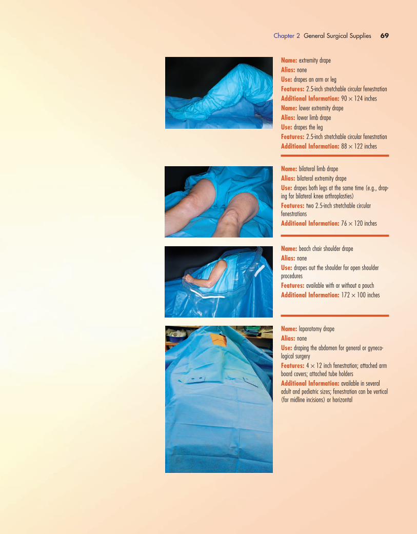
Chapter 2 General Surgical Supplies 69
Name: extremity drape
Alias: none
Use: drapes an arm or leg
Features: 2.5-inch stretchable circular fenestration
Additional Information: 90 × 124 inches
Name: lower extremity drape
Alias: lower limb drape
Use: drapes the leg
Features: 2.5-inch stretchable circular fenestration
Additional Information: 88 × 122 inches
Name: bilateral limb drape
Alias: bilateral extremity drape
Use: drapes both legs at the same time (e.g., drap-ing for bilateral knee arthroplasties)
Features: two 2.5-inch stretchable circular fenestrations
Additional Information: 76 × 120 inches
Name: beach chair shoulder drape
Alias: none
Use: drapes out the shoulder for open shoulder procedures
Features: available with or without a pouch
Additional Information: 172 × 100 inches
Name: laparotomy drape
Alias: none
Use: draping the abdomen for general or gyneco-logical surgery
Features: 4 × 12 inch fenestration; attached armboard covers; attached tube holders
Additional Information: available in severaladult and pediatric sizes; fenestration can be vertical(for midline incisions) or horizontal
1572_Ch02_051-092.qxd 8/6/09 11:07 AM Page 69
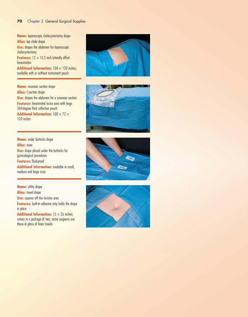
70 Chapter 2 General Surgical Supplies
Name: laparoscopic cholecystectomy drape
Alias: lap chole drape
Use: drapes the abdomen for laparoscopic cholecystectomy
Features: 12 × 13.5 inch laterally offset fenestration
Additional Information: 104 × 120 inches;available with or without instrument pouch
Name: cesarean section drape
Alias: C-section drape
Use: drapes the abdomen for a cesarean section
Features: fenestrated incise area with large 360-degree fluid collection pouch
Additional Information: 100 × 72 ×120 inches
Name: under buttocks drape
Alias: none
Use: drape placed under the buttocks for gynecological procedures
Features: fluid-proof
Additional Information: available in small,medium and large sizes
Name: utility drape
Alias: towel drape
Use: squares off the incision area
Features: built-in adhesive strip holds the drape in place
Additional Information: 15 × 26 inches;comes in a package of two; some surgeons usethese in place of linen towels
1572_Ch02_051-092.qxd 8/6/09 11:07 AM Page 70
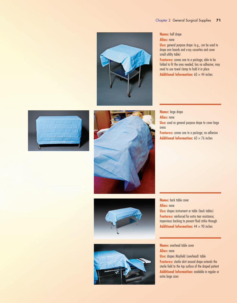
Chapter 2 General Surgical Supplies 71
Name: half drape
Alias: none
Use: general purpose drape (e.g., can be used todrape arm boards and x-ray cassettes and coversmall utility table)
Features: comes one to a package; able to befolded to fit the area needed; has no adhesive; mayneed to use towel clamp to hold it in place
Additional Information: 60 × 44 inches
Name: large drape
Alias: none
Use: used as general purpose drape to cover largeareas
Features: comes one to a package; no adhesive
Additional Information: 60 × 76 inches
Name: back table cover
Alias: none
Use: drapes instrument or table (back tables)
Features: reinforced for extra tear resistance; impervious backing to prevent fluid strike through
Additional Information: 44 × 90 inches
Name: overhead table cover
Alias: none
Use: drapes Mayfield (overhead) table
Features: sterile skirt around drape extends thesterile field to the top surface of the draped patient
Additional Information: available in regular orextra large sizes
1572_Ch02_051-092.qxd 8/6/09 11:07 AM Page 71
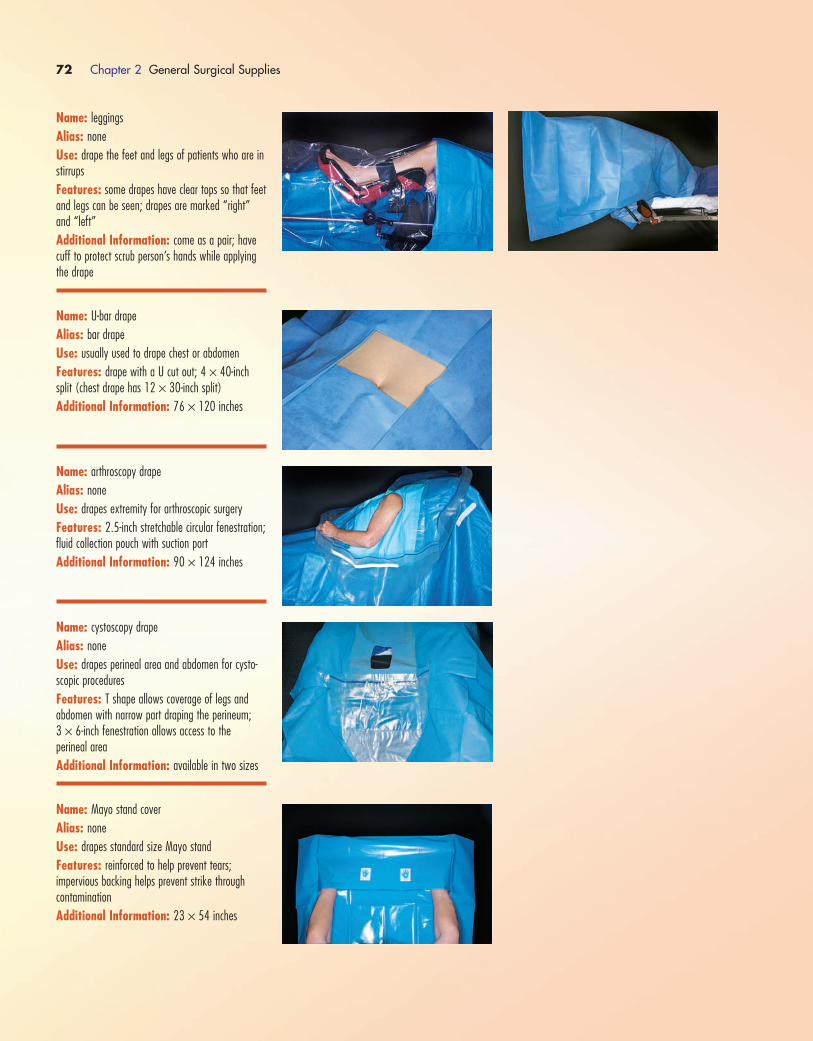
72 Chapter 2 General Surgical Supplies
Name: leggings
Alias: none
Use: drape the feet and legs of patients who are instirrups
Features: some drapes have clear tops so that feetand legs can be seen; drapes are marked “right”and “left”
Additional Information: come as a pair; havecuff to protect scrub person’s hands while applyingthe drape
Name: U-bar drape
Alias: bar drape
Use: usually used to drape chest or abdomen
Features: drape with a U cut out; 4 × 40-inchsplit (chest drape has 12 × 30-inch split)
Additional Information: 76 × 120 inches
Name: arthroscopy drape
Alias: none
Use: drapes extremity for arthroscopic surgery
Features: 2.5-inch stretchable circular fenestration;fluid collection pouch with suction port
Additional Information: 90 × 124 inches
Name: cystoscopy drape
Alias: none
Use: drapes perineal area and abdomen for cysto-scopic procedures
Features: T shape allows coverage of legs and abdomen with narrow part draping the perineum; 3 × 6-inch fenestration allows access to the perineal area
Additional Information: available in two sizes
Name: Mayo stand cover
Alias: none
Use: drapes standard size Mayo stand
Features: reinforced to help prevent tears; impervious backing helps prevent strike through contamination
Additional Information: 23 × 54 inches
1572_Ch02_051-092.qxd 8/6/09 11:07 AM Page 72
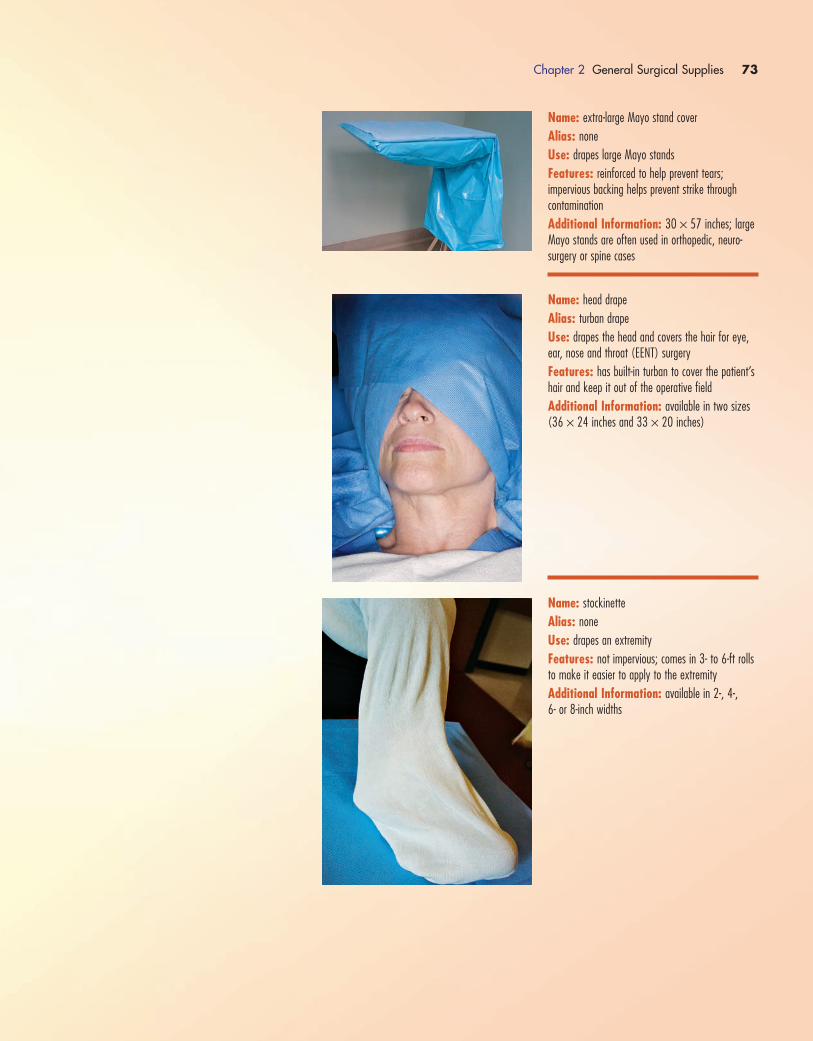
Chapter 2 General Surgical Supplies 73
Name: extra-large Mayo stand cover
Alias: none
Use: drapes large Mayo stands
Features: reinforced to help prevent tears; impervious backing helps prevent strike through contamination
Additional Information: 30 × 57 inches; largeMayo stands are often used in orthopedic, neuro-surgery or spine cases
Name: head drape
Alias: turban drape
Use: drapes the head and covers the hair for eye,ear, nose and throat (EENT) surgery
Features: has built-in turban to cover the patient’shair and keep it out of the operative field
Additional Information: available in two sizes(36 × 24 inches and 33 × 20 inches)
Name: stockinette
Alias: none
Use: drapes an extremity
Features: not impervious; comes in 3- to 6-ft rollsto make it easier to apply to the extremity
Additional Information: available in 2-, 4-, 6- or 8-inch widths
1572_Ch02_051-092.qxd 8/6/09 11:07 AM Page 73
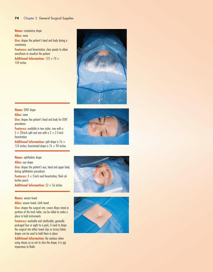
74 Chapter 2 General Surgical Supplies
Name: craniotomy drape
Alias: none
Use: drapes the patient’s head and body during acraniotomy
Features: oval fenestration; clear panels to allowanesthesia to visualize the patient
Additional Information: 122 × 74 ×134 inches
Name: EENT drape
Alias: none
Use: drapes the patient’s head and body for EENTprocedures
Features: available in two styles: one with a 3 × 28-inch split and one with a 2 × 2.5-inch fenestration
Additional Information: split drape is 76 ×124 inches; fenestrated drape is 76 × 90 inches
Name: ophthalmic drape
Alias: eye drape
Use: drapes the patient’s eye, head and upper bodyduring ophthalmic procedures
Features: 3 × 2-inch oval fenestration; fluid col-lection pouch
Additional Information: 52 × 56 inches
Name: woven towel
Alias: woven towel; cloth towel
Use: drapes the surgical site; covers Mayo stand orportions of the back table; can be rolled to make aplace to hold instruments
Features: washable and sterilizable; generallypackaged four or eight to a pack; if used to drapethe surgical site either towel clips or incise/Iobandrapes can be used to hold them in place
Additional Information: Be cautious when using sharps so as not to slice the drape; it is notimpervious to fluids
1572_Ch02_051-092.qxd 8/6/09 11:07 AM Page 74
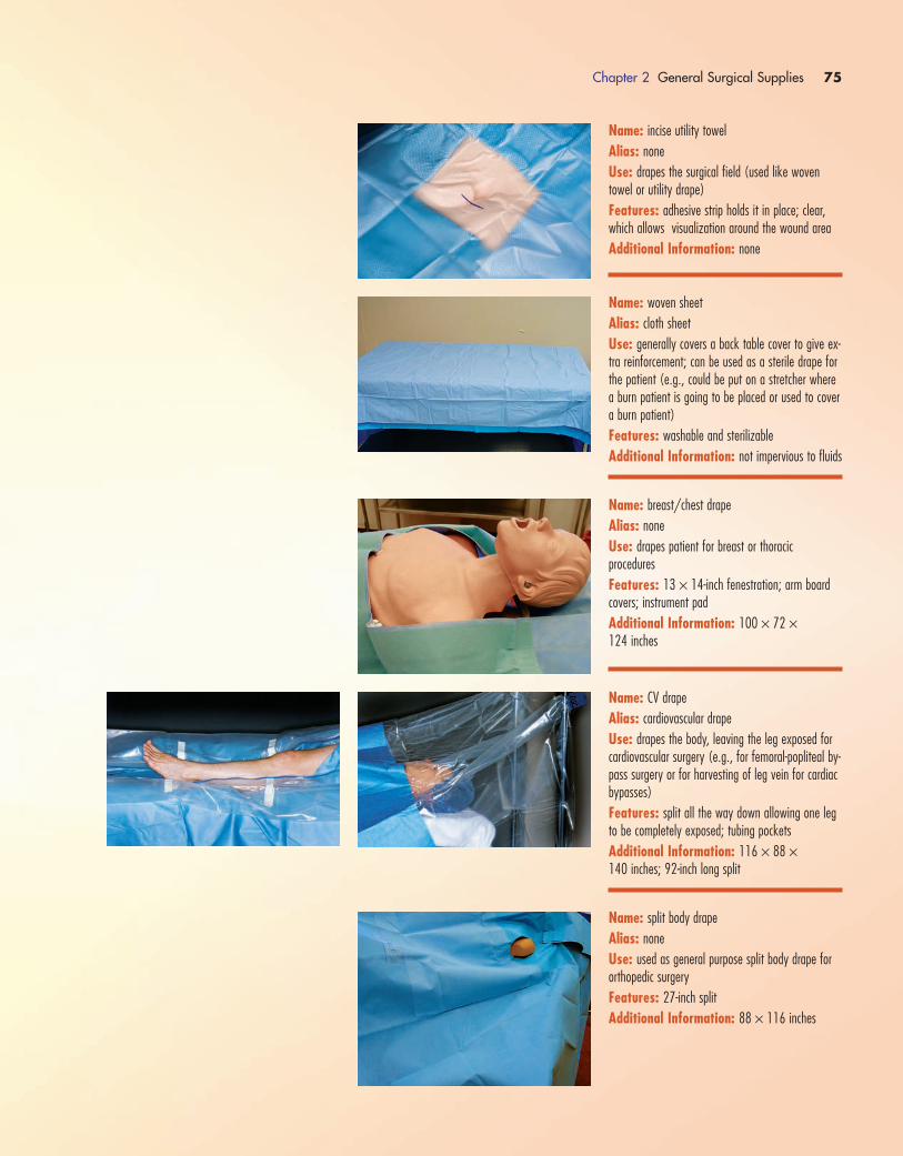
Chapter 2 General Surgical Supplies 75
Name: incise utility towel
Alias: none
Use: drapes the surgical field (used like woventowel or utility drape)
Features: adhesive strip holds it in place; clear,which allows visualization around the wound area
Additional Information: none
Name: woven sheet
Alias: cloth sheet
Use: generally covers a back table cover to give ex-tra reinforcement; can be used as a sterile drape forthe patient (e.g., could be put on a stretcher wherea burn patient is going to be placed or used to covera burn patient)
Features: washable and sterilizable
Additional Information: not impervious to fluids
Name: breast/chest drape
Alias: none
Use: drapes patient for breast or thoracic procedures
Features: 13 × 14-inch fenestration; arm boardcovers; instrument pad
Additional Information: 100 × 72 ×124 inches
Name: CV drape
Alias: cardiovascular drape
Use: drapes the body, leaving the leg exposed forcardiovascular surgery (e.g., for femoral-popliteal by-pass surgery or for harvesting of leg vein for cardiacbypasses)
Features: split all the way down allowing one legto be completely exposed; tubing pockets
Additional Information: 116 × 88 ×140 inches; 92-inch long split
Name: split body drape
Alias: none
Use: used as general purpose split body drape fororthopedic surgery
Features: 27-inch split
Additional Information: 88 × 116 inches
1572_Ch02_051-092.qxd 8/6/09 11:07 AM Page 75
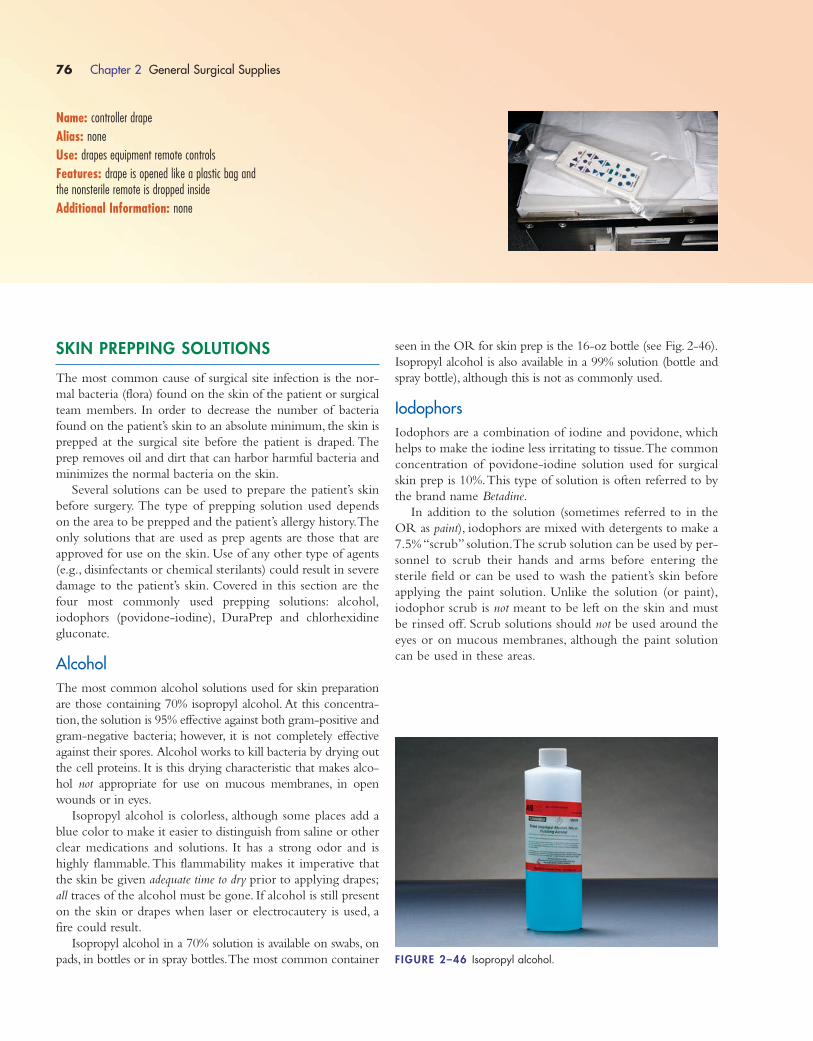
76 Chapter 2 General Surgical Supplies
Name: controller drape
Alias: none
Use: drapes equipment remote controls
Features: drape is opened like a plastic bag andthe nonsterile remote is dropped inside
Additional Information: none
SKIN PREPPING SOLUTIONS
The most common cause of surgical site infection is the nor-
mal bacteria (flora) found on the skin of the patient or surgical
team members. In order to decrease the number of bacteria
found on the patient’s skin to an absolute minimum, the skin is
prepped at the surgical site before the patient is draped. The
prep removes oil and dirt that can harbor harmful bacteria and
minimizes the normal bacteria on the skin.
Several solutions can be used to prepare the patient’s skin
before surgery. The type of prepping solution used depends
on the area to be prepped and the patient’s allergy history.The
only solutions that are used as prep agents are those that are
approved for use on the skin. Use of any other type of agents
(e.g., disinfectants or chemical sterilants) could result in severe
damage to the patient’s skin. Covered in this section are the
four most commonly used prepping solutions: alcohol,
iodophors (povidone-iodine), DuraPrep and chlorhexidine
gluconate.
AlcoholThe most common alcohol solutions used for skin preparation
are those containing 70% isopropyl alcohol. At this concentra-
tion, the solution is 95% effective against both gram-positive and
gram-negative bacteria; however, it is not completely effective
against their spores. Alcohol works to kill bacteria by drying out
the cell proteins. It is this drying characteristic that makes alco-
hol not appropriate for use on mucous membranes, in open
wounds or in eyes.
Isopropyl alcohol is colorless, although some places add a
blue color to make it easier to distinguish from saline or other
clear medications and solutions. It has a strong odor and is
highly flammable.This flammability makes it imperative that
the skin be given adequate time to dry prior to applying drapes;
all traces of the alcohol must be gone. If alcohol is still present
on the skin or drapes when laser or electrocautery is used, a
fire could result.
Isopropyl alcohol in a 70% solution is available on swabs, on
pads, in bottles or in spray bottles.The most common container FIGURE 2–46 Isopropyl alcohol.
seen in the OR for skin prep is the 16-oz bottle (see Fig. 2-46).
Isopropyl alcohol is also available in a 99% solution (bottle and
spray bottle), although this is not as commonly used.
IodophorsIodophors are a combination of iodine and povidone, which
helps to make the iodine less irritating to tissue.The common
concentration of povidone-iodine solution used for surgical
skin prep is 10%.This type of solution is often referred to by
the brand name Betadine.
In addition to the solution (sometimes referred to in the
OR as paint), iodophors are mixed with detergents to make a
7.5% “scrub” solution.The scrub solution can be used by per-
sonnel to scrub their hands and arms before entering the
sterile field or can be used to wash the patient’s skin before
applying the paint solution. Unlike the solution (or paint),
iodophor scrub is not meant to be left on the skin and must
be rinsed off. Scrub solutions should not be used around the
eyes or on mucous membranes, although the paint solution
can be used in these areas.
1572_Ch02_051-092.qxd 8/6/09 11:07 AM Page 76
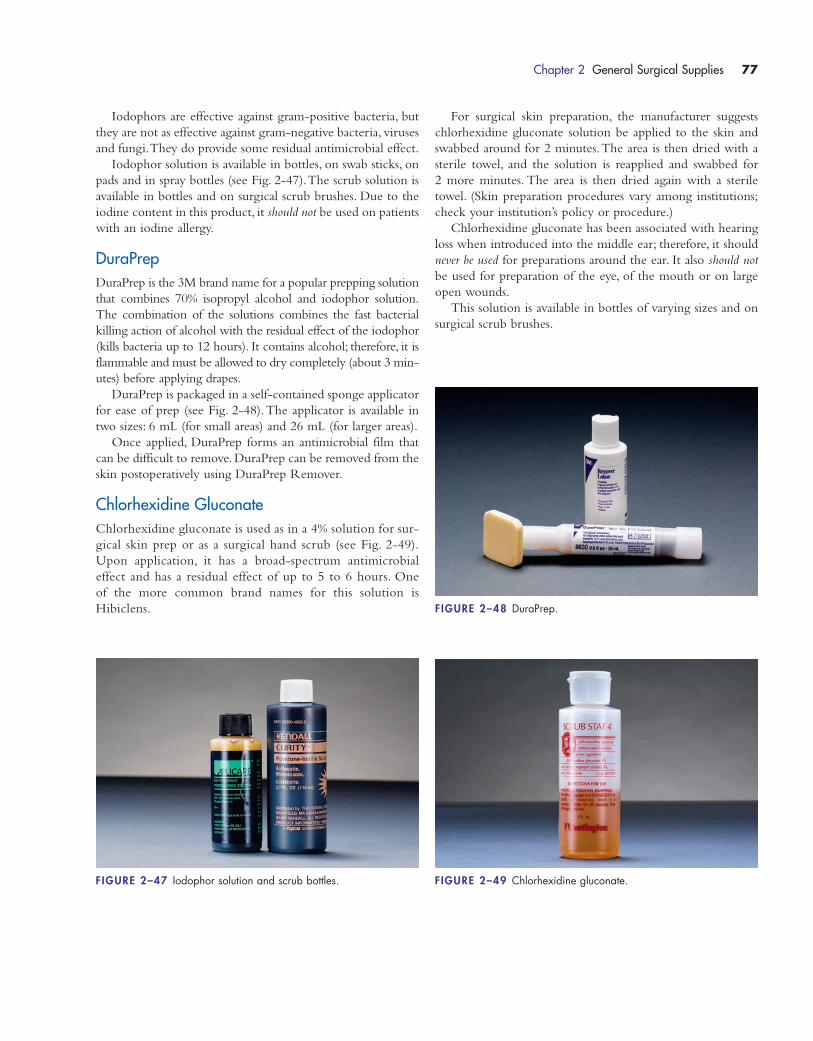
Iodophors are effective against gram-positive bacteria, but
they are not as effective against gram-negative bacteria, viruses
and fungi.They do provide some residual antimicrobial effect.
Iodophor solution is available in bottles, on swab sticks, on
pads and in spray bottles (see Fig. 2-47).The scrub solution is
available in bottles and on surgical scrub brushes. Due to the
iodine content in this product, it should not be used on patients
with an iodine allergy.
DuraPrepDuraPrep is the 3M brand name for a popular prepping solution
that combines 70% isopropyl alcohol and iodophor solution.
The combination of the solutions combines the fast bacterial
killing action of alcohol with the residual effect of the iodophor
(kills bacteria up to 12 hours). It contains alcohol; therefore, it is
flammable and must be allowed to dry completely (about 3 min-
utes) before applying drapes.
DuraPrep is packaged in a self-contained sponge applicator
for ease of prep (see Fig. 2-48).The applicator is available in
two sizes: 6 mL (for small areas) and 26 mL (for larger areas).
Once applied, DuraPrep forms an antimicrobial film that
can be difficult to remove. DuraPrep can be removed from the
skin postoperatively using DuraPrep Remover.
Chlorhexidine GluconateChlorhexidine gluconate is used as in a 4% solution for sur-
gical skin prep or as a surgical hand scrub (see Fig. 2-49).
Upon application, it has a broad-spectrum antimicrobial
effect and has a residual effect of up to 5 to 6 hours. One
of the more common brand names for this solution is
Hibiclens.
Chapter 2 General Surgical Supplies 77
FIGURE 2–47 Iodophor solution and scrub bottles.
FIGURE 2–48 DuraPrep.
FIGURE 2–49 Chlorhexidine gluconate.
For surgical skin preparation, the manufacturer suggests
chlorhexidine gluconate solution be applied to the skin and
swabbed around for 2 minutes.The area is then dried with a
sterile towel, and the solution is reapplied and swabbed for
2 more minutes. The area is then dried again with a sterile
towel. (Skin preparation procedures vary among institutions;
check your institution’s policy or procedure.)
Chlorhexidine gluconate has been associated with hearing
loss when introduced into the middle ear; therefore, it should
never be used for preparations around the ear. It also should not
be used for preparation of the eye, of the mouth or on large
open wounds.
This solution is available in bottles of varying sizes and on
surgical scrub brushes.
1572_Ch02_051-092.qxd 8/6/09 11:07 AM Page 77
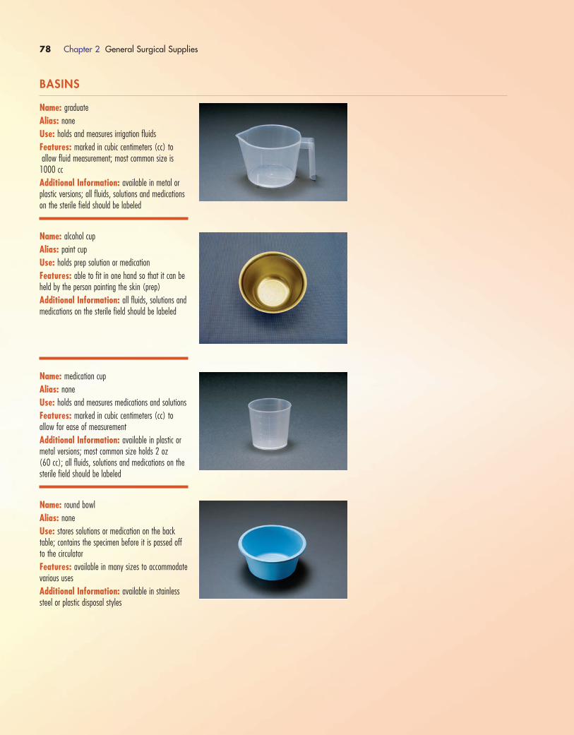
78 Chapter 2 General Surgical Supplies
BASINS
Name: graduate
Alias: none
Use: holds and measures irrigation fluids
Features: marked in cubic centimeters (cc) toallow fluid measurement; most common size is
1000 cc
Additional Information: available in metal orplastic versions; all fluids, solutions and medicationson the sterile field should be labeled
Name: alcohol cup
Alias: paint cup
Use: holds prep solution or medication
Features: able to fit in one hand so that it can beheld by the person painting the skin (prep)
Additional Information: all fluids, solutions andmedications on the sterile field should be labeled
Name: medication cup
Alias: none
Use: holds and measures medications and solutions
Features: marked in cubic centimeters (cc) to allow for ease of measurement
Additional Information: available in plastic ormetal versions; most common size holds 2 oz (60 cc); all fluids, solutions and medications on thesterile field should be labeled
Name: round bowl
Alias: none
Use: stores solutions or medication on the backtable; contains the specimen before it is passed offto the circulator
Features: available in many sizes to accommodatevarious uses
Additional Information: available in stainlesssteel or plastic disposal styles
1572_Ch02_051-092.qxd 8/6/09 11:07 AM Page 78
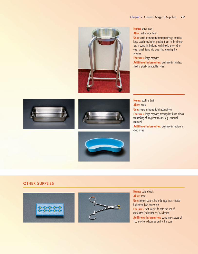
Chapter 2 General Surgical Supplies 79
OTHER SUPPLIES
Name: wash bowl
Alias: extra large basin
Use: soaks instruments intraoperatively; containslarge specimens before passing them to the circula-tor; in some institutions, wash bowls are used toopen small items into when first opening the supplies
Features: large capacity
Additional Information: available in stainlesssteel or plastic disposable styles
Name: soaking basin
Alias: none
Use: soaks instruments intraoperatively
Features: large capacity; rectangular shape allowsfor soaking of long instruments (e.g., femoral reamers)
Additional Information: available in shallow ordeep styles
Name: suture boots
Alias: shods
Use: protect sutures from damage that serrated instrument jaws can cause
Features: soft plastic; fit onto the tips of mosquitos (Halstead) or Crile clamps
Additional Information: come in packages of10; may be included as part of the count
1572_Ch02_051-092.qxd 8/6/09 11:07 AM Page 79
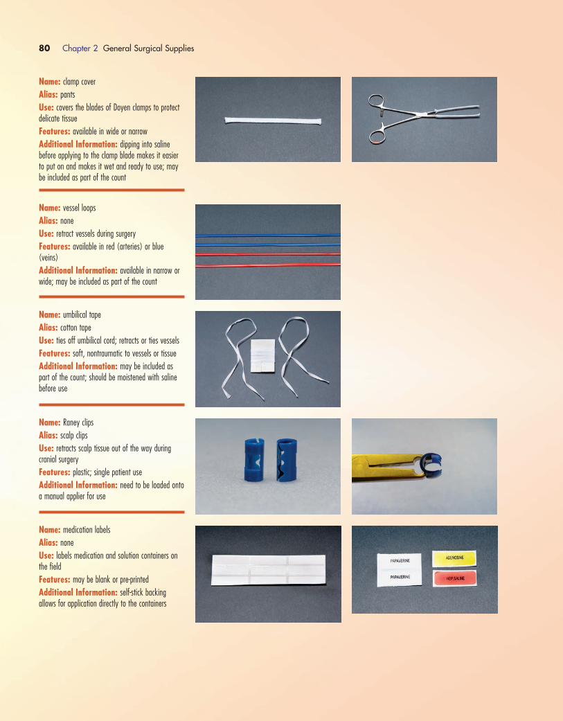
80 Chapter 2 General Surgical Supplies
Name: clamp cover
Alias: pants
Use: covers the blades of Doyen clamps to protectdelicate tissue
Features: available in wide or narrow
Additional Information: dipping into saline before applying to the clamp blade makes it easierto put on and makes it wet and ready to use; maybe included as part of the count
Name: vessel loops
Alias: none
Use: retract vessels during surgery
Features: available in red (arteries) or blue(veins)
Additional Information: available in narrow orwide; may be included as part of the count
Name: umbilical tape
Alias: cotton tape
Use: ties off umbilical cord; retracts or ties vessels
Features: soft, nontraumatic to vessels or tissue
Additional Information: may be included aspart of the count; should be moistened with salinebefore use
Name: Raney clips
Alias: scalp clips
Use: retracts scalp tissue out of the way during cranial surgery
Features: plastic; single patient use
Additional Information: need to be loaded ontoa manual applier for use
Name: medication labels
Alias: none
Use: labels medication and solution containers onthe field
Features: may be blank or pre-printed
Additional Information: self-stick backing allows for application directly to the containers
1572_Ch02_051-092.qxd 8/6/09 11:07 AM Page 80
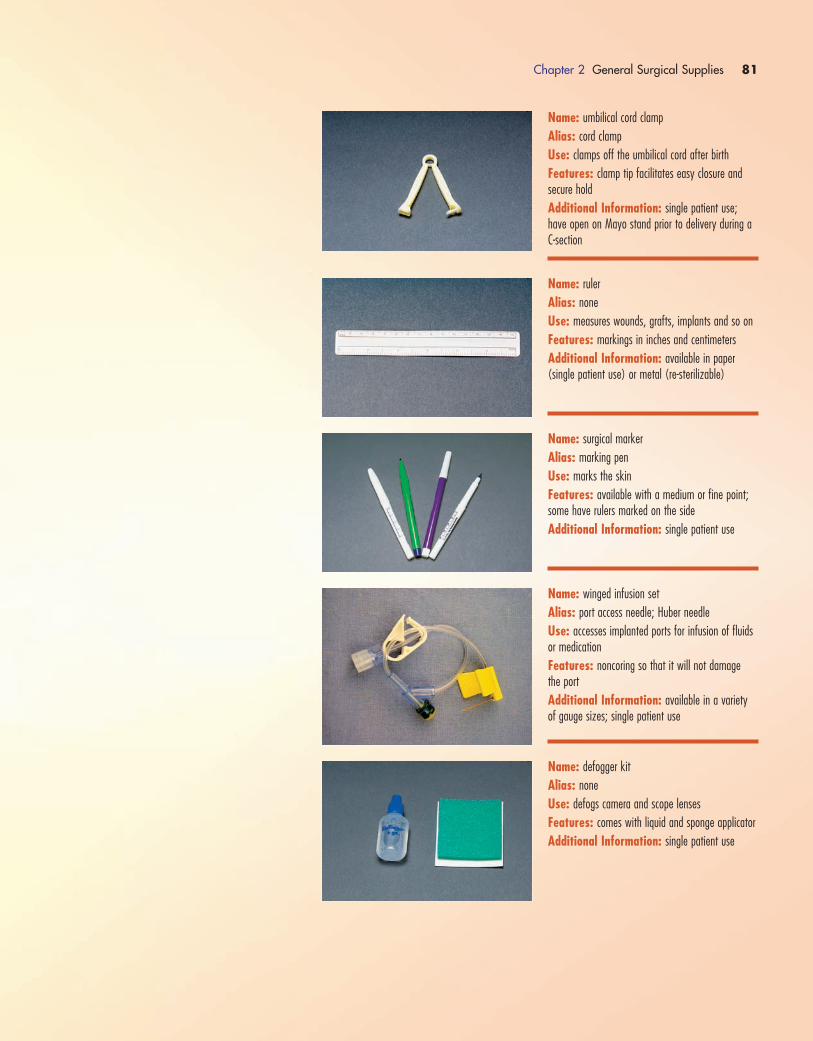
Chapter 2 General Surgical Supplies 81
Name: umbilical cord clamp
Alias: cord clamp
Use: clamps off the umbilical cord after birth
Features: clamp tip facilitates easy closure and secure hold
Additional Information: single patient use;have open on Mayo stand prior to delivery during aC-section
Name: ruler
Alias: none
Use: measures wounds, grafts, implants and so on
Features: markings in inches and centimeters
Additional Information: available in paper (single patient use) or metal (re-sterilizable)
Name: surgical marker
Alias: marking pen
Use: marks the skin
Features: available with a medium or fine point;some have rulers marked on the side
Additional Information: single patient use
Name: winged infusion set
Alias: port access needle; Huber needle
Use: accesses implanted ports for infusion of fluidsor medication
Features: noncoring so that it will not damage the port
Additional Information: available in a varietyof gauge sizes; single patient use
Name: defogger kit
Alias: none
Use: defogs camera and scope lenses
Features: comes with liquid and sponge applicator
Additional Information: single patient use
1572_Ch02_051-092.qxd 8/6/09 11:07 AM Page 81
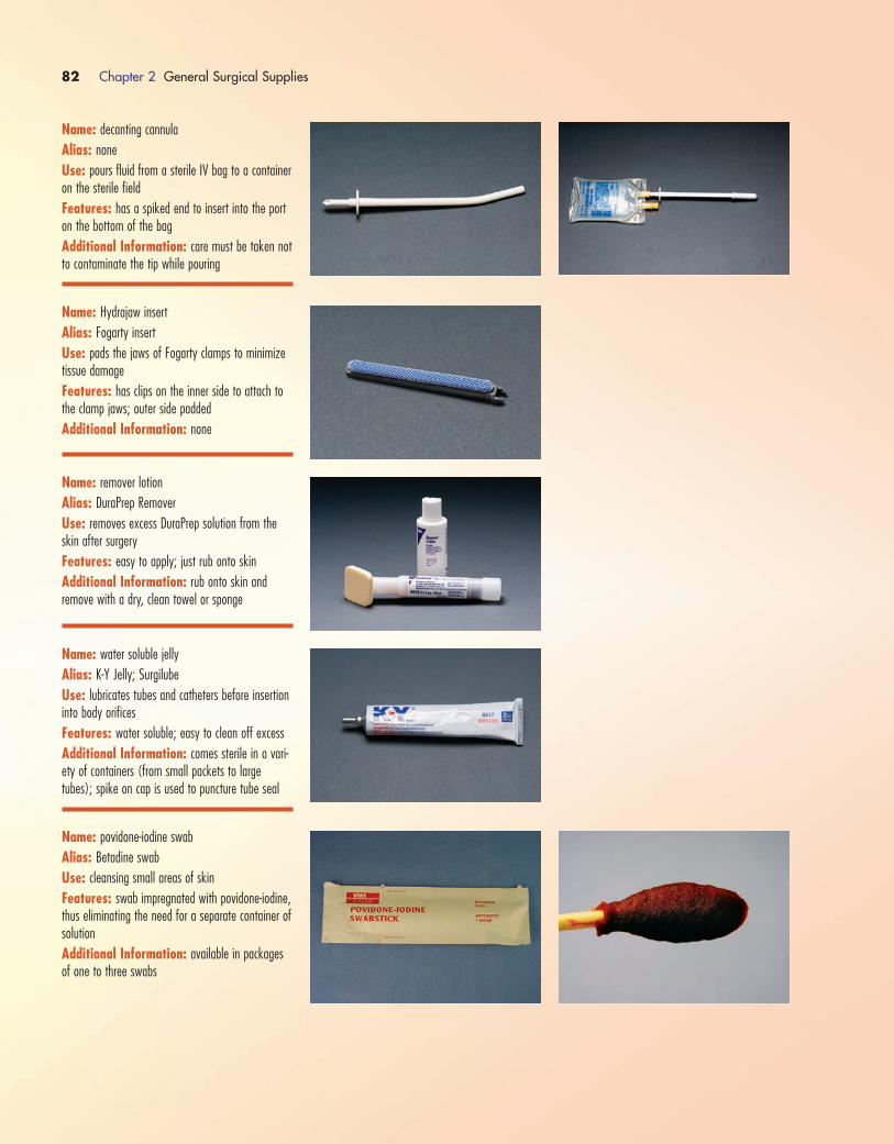
82 Chapter 2 General Surgical Supplies
Name: decanting cannula
Alias: none
Use: pours fluid from a sterile IV bag to a containeron the sterile field
Features: has a spiked end to insert into the porton the bottom of the bag
Additional Information: care must be taken notto contaminate the tip while pouring
Name: Hydrajaw insert
Alias: Fogarty insert
Use: pads the jaws of Fogarty clamps to minimizetissue damage
Features: has clips on the inner side to attach tothe clamp jaws; outer side padded
Additional Information: none
Name: remover lotion
Alias: DuraPrep Remover
Use: removes excess DuraPrep solution from theskin after surgery
Features: easy to apply; just rub onto skin
Additional Information: rub onto skin and remove with a dry, clean towel or sponge
Name: water soluble jelly
Alias: K-Y Jelly; Surgilube
Use: lubricates tubes and catheters before insertioninto body orifices
Features: water soluble; easy to clean off excess
Additional Information: comes sterile in a vari-ety of containers (from small packets to largetubes); spike on cap is used to puncture tube seal
Name: povidone-iodine swab
Alias: Betadine swab
Use: cleansing small areas of skin
Features: swab impregnated with povidone-iodine,thus eliminating the need for a separate container ofsolution
Additional Information: available in packagesof one to three swabs
1572_Ch02_051-092.qxd 8/6/09 11:07 AM Page 82
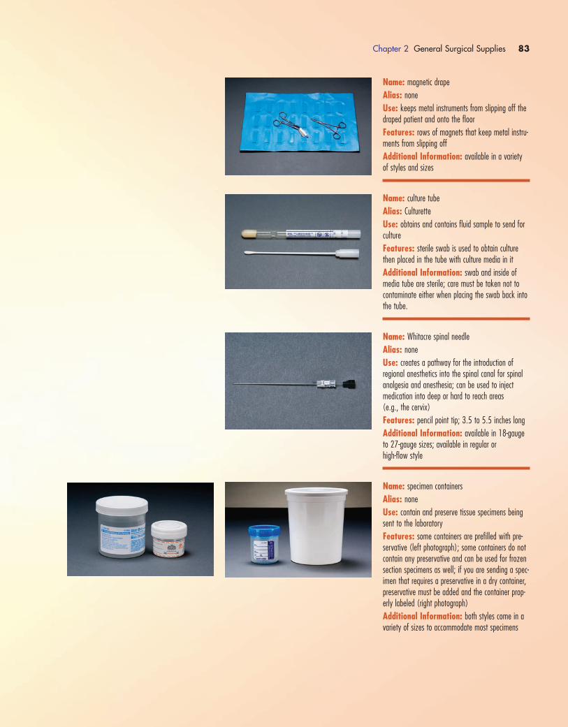
Chapter 2 General Surgical Supplies 83
Name: magnetic drape
Alias: none
Use: keeps metal instruments from slipping off thedraped patient and onto the floor
Features: rows of magnets that keep metal instru-ments from slipping off
Additional Information: available in a varietyof styles and sizes
Name: culture tube
Alias: Culturette
Use: obtains and contains fluid sample to send forculture
Features: sterile swab is used to obtain culturethen placed in the tube with culture media in it
Additional Information: swab and inside of media tube are sterile; care must be taken not tocontaminate either when placing the swab back intothe tube.
Name: Whitacre spinal needle
Alias: none
Use: creates a pathway for the introduction of regional anesthetics into the spinal canal for spinalanalgesia and anesthesia; can be used to inject medication into deep or hard to reach areas (e.g., the cervix)
Features: pencil point tip; 3.5 to 5.5 inches long
Additional Information: available in 18-gaugeto 27-gauge sizes; available in regular or high-flow style
Name: specimen containers
Alias: none
Use: contain and preserve tissue specimens beingsent to the laboratory
Features: some containers are prefilled with pre-servative (left photograph); some containers do notcontain any preservative and can be used for frozensection specimens as well; if you are sending a spec-imen that requires a preservative in a dry container,preservative must be added and the container prop-erly labeled (right photograph)
Additional Information: both styles come in avariety of sizes to accommodate most specimens
1572_Ch02_051-092.qxd 8/6/09 11:07 AM Page 83
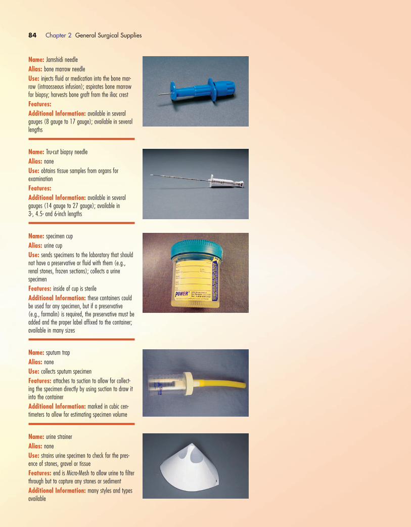
84 Chapter 2 General Surgical Supplies
Name: Jamshidi needle
Alias: bone marrow needle
Use: injects fluid or medication into the bone mar-row (intraosseous infusion); aspirates bone marrowfor biopsy; harvests bone graft from the iliac crest
Features: Additional Information: available in severalgauges (8 gauge to 17 gauge); available in severallengths
Name: Tru-cut biopsy needle
Alias: none
Use: obtains tissue samples from organs for examination
Features: Additional Information: available in severalgauges (14 gauge to 27 gauge); available in 3-, 4.5- and 6-inch lengths
Name: specimen cup
Alias: urine cup
Use: sends specimens to the laboratory that shouldnot have a preservative or fluid with them (e.g., renal stones, frozen sections); collects a urine specimen
Features: inside of cup is sterile
Additional Information: these containers couldbe used for any specimen, but if a preservative(e.g., formalin) is required, the preservative must beadded and the proper label affixed to the container;available in many sizes
Name: sputum trap
Alias: none
Use: collects sputum specimen
Features: attaches to suction to allow for collect-ing the specimen directly by using suction to draw itinto the container
Additional Information: marked in cubic cen-timeters to allow for estimating specimen volume
Name: urine strainer
Alias: none
Use: strains urine specimen to check for the pres-ence of stones, gravel or tissue
Features: end is Micro-Mesh to allow urine to filterthrough but to capture any stones or sediment
Additional Information: many styles and typesavailable
1572_Ch02_051-092.qxd 8/6/09 11:07 AM Page 84
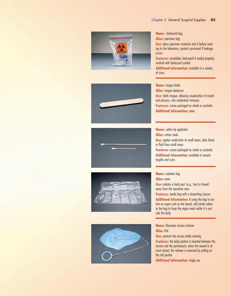
Chapter 2 General Surgical Supplies 85
Name: biohazard bag
Alias: specimen bag
Use: place specimen container into it before send-ing to the laboratory; protects personnel if leakageoccurs
Features: resealable; leak-proof if sealed properly;marked with biohazard symbol
Additional Information: available in a varietyof sizes
Name: tongue blade
Alias: tongue depressor
Use: holds tongue, allowing visualization of mouthand pharynx; stirs medication mixtures
Features: comes packaged as sterile or unsterile
Additional Information: none
Name: cotton tip applicator
Alias: cotton swab
Use: applies medication to small areas; dabs bloodor fluid from small areas
Features: comes packaged as sterile or unsterile
Additional Information: available in severallengths and sizes
Name: isolation bag
Alias: none
Use: isolates a body part (e.g., foot or bowel)away from the operative area
Features: sterile bag with a drawstring closure
Additional Information: if using the bag to iso-late an organ such as the bowel, add sterile salineto the bag to keep the organ moist while it is out-side the body
Name: Glassman viscera retainer
Alias: fishUse: protects the viscera while suturing
Features: the body portion is inserted between theviscera and the peritoneum; when the wound is al-most closed, the retainer is removed by pulling onthe tail portion
Additional Information: single use
1572_Ch02_051-092.qxd 8/6/09 11:07 AM Page 85
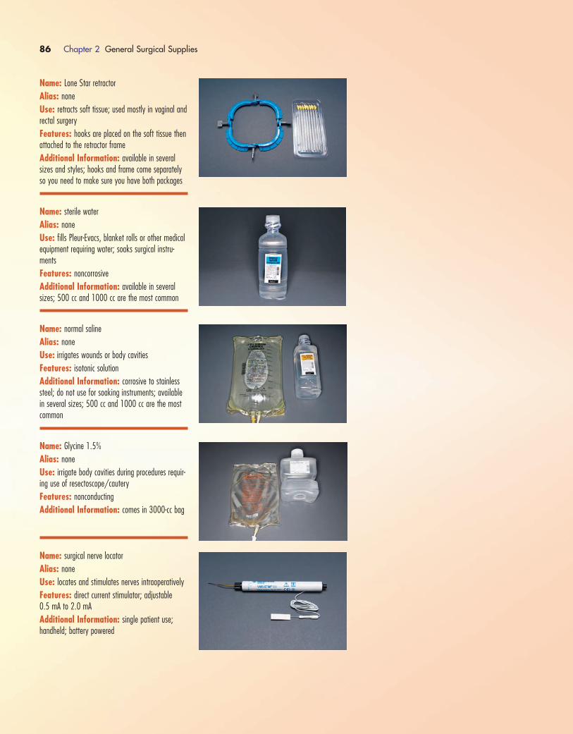
86 Chapter 2 General Surgical Supplies
Name: Lone Star retractor
Alias: none
Use: retracts soft tissue; used mostly in vaginal andrectal surgery
Features: hooks are placed on the soft tissue thenattached to the retractor frame
Additional Information: available in severalsizes and styles; hooks and frame come separatelyso you need to make sure you have both packages
Name: sterile water
Alias: none
Use: fills Pleur-Evacs, blanket rolls or other medicalequipment requiring water; soaks surgical instru-ments
Features: noncorrosive
Additional Information: available in severalsizes; 500 cc and 1000 cc are the most common
Name: normal saline
Alias: none
Use: irrigates wounds or body cavities
Features: isotonic solution
Additional Information: corrosive to stainlesssteel; do not use for soaking instruments; availablein several sizes; 500 cc and 1000 cc are the mostcommon
Name: Glycine 1.5%
Alias: none
Use: irrigate body cavities during procedures requir-ing use of resectoscope/cautery
Features: nonconducting
Additional Information: comes in 3000-cc bag
Name: surgical nerve locator
Alias: none
Use: locates and stimulates nerves intraoperatively
Features: direct current stimulator; adjustable 0.5 mA to 2.0 mA
Additional Information: single patient use;handheld; battery powered
1572_Ch02_051-092.qxd 8/6/09 11:07 AM Page 86
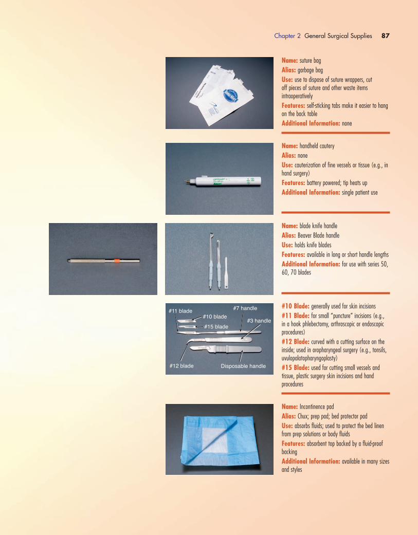
Chapter 2 General Surgical Supplies 87
#10 blade#7 handle
#15 blade#3 handle
#11 blade
#12 blade Disposable handle
Name: suture bag
Alias: garbage bag
Use: use to dispose of suture wrappers, cut off pieces of suture and other waste items intraoperatively
Features: self-sticking tabs make it easier to hangon the back table
Additional Information: none
Name: handheld cautery
Alias: none
Use: cauterization of fine vessels or tissue (e.g., inhand surgery)
Features: battery powered; tip heats up
Additional Information: single patient use
Name: blade knife handle
Alias: Beaver Blade handle
Use: holds knife blades
Features: available in long or short handle lengths
Additional Information: for use with series 50,60, 70 blades
#10 Blade: generally used for skin incisions
#11 Blade: for small “puncture” incisions (e.g., in a hook phlebectomy, arthroscopic or endoscopicprocedures)
#12 Blade: curved with a cutting surface on theinside; used in oropharyngeal surgery (e.g., tonsils,uvulopalatopharyngoplasty)
#15 Blade: used for cutting small vessels and tissue, plastic surgery skin incisions and hand procedures
Name: Incontinence pad
Alias: Chux; prep pad; bed protector pad
Use: absorbs fluids; used to protect the bed linenfrom prep solutions or body fluids
Features: absorbent top backed by a fluid-proofbacking
Additional Information: available in many sizesand styles
1572_Ch02_051-092.qxd 8/6/09 11:07 AM Page 87
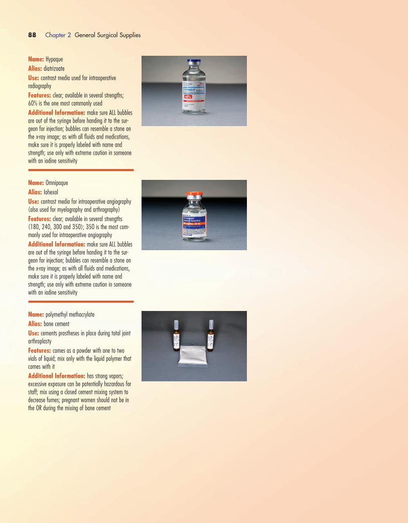
88 Chapter 2 General Surgical Supplies
Name: Hypaque
Alias: diatrizoate
Use: contrast media used for intraoperative radiography
Features: clear; available in several strengths;60% is the one most commonly used
Additional Information: make sure ALL bubblesare out of the syringe before handing it to the sur-geon for injection; bubbles can resemble a stone onthe x-ray image; as with all fluids and medications,make sure it is properly labeled with name andstrength; use only with extreme caution in someonewith an iodine sensitivity
Name: Omnipaque
Alias: Iohexol
Use: contrast media for intraoperative angiography(also used for myelography and arthrography)
Features: clear; available in several strengths(180, 240, 300 and 350); 350 is the most com-monly used for intraoperative angiography
Additional Information: make sure ALL bubblesare out of the syringe before handing it to the sur-geon for injection; bubbles can resemble a stone onthe x-ray image; as with all fluids and medications,make sure it is properly labeled with name andstrength; use only with extreme caution in someonewith an iodine sensitivity
Name: polymethyl methacrylate
Alias: bone cement
Use: cements prostheses in place during total jointarthroplasty
Features: comes as a powder with one to twovials of liquid; mix only with the liquid polymer thatcomes with it
Additional Information: has strong vapors; excessive exposure can be potentially hazardous forstaff; mix using a closed cement mixing system todecrease fumes; pregnant women should not be inthe OR during the mixing of bone cement
1572_Ch02_051-092.qxd 8/6/09 11:07 AM Page 88
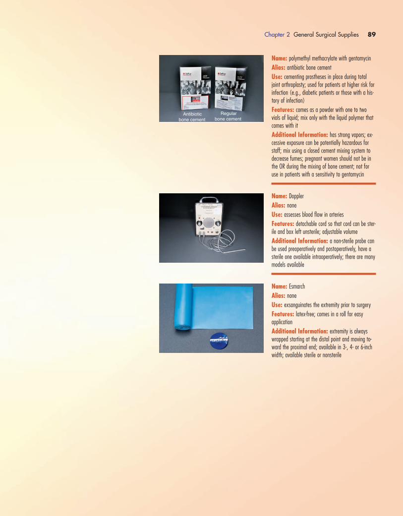
Chapter 2 General Surgical Supplies 89
Antibiotic bone cement
Regular bone cement
Name: polymethyl methacrylate with gentamycin
Alias: antibiotic bone cement
Use: cementing prostheses in place during totaljoint arthroplasty; used for patients at higher risk forinfection (e.g., diabetic patients or those with a his-tory of infection)
Features: comes as a powder with one to twovials of liquid; mix only with the liquid polymer thatcomes with it
Additional Information: has strong vapors; ex-cessive exposure can be potentially hazardous forstaff; mix using a closed cement mixing system todecrease fumes; pregnant women should not be inthe OR during the mixing of bone cement; not foruse in patients with a sensitivity to gentamycin
Name: Doppler
Alias: none
Use: assesses blood flow in arteries
Features: detachable cord so that cord can be ster-ile and box left unsterile; adjustable volume
Additional Information: a non-sterile probe canbe used preoperatively and postoperatively, have asterile one available intraoperatively; there are manymodels available
Name: Esmarch
Alias: none
Use: exsanguinates the extremity prior to surgery
Features: latex-free; comes in a roll for easy application
Additional Information: extremity is alwayswrapped starting at the distal point and moving to-ward the proximal end; available in 3-, 4- or 6-inchwidth; available sterile or nonsterile
1572_Ch02_051-092.qxd 8/6/09 11:08 AM Page 89
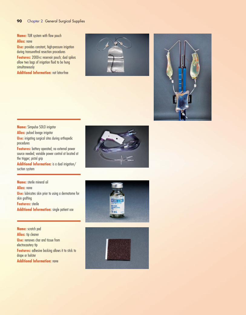
90 Chapter 2 General Surgical Supplies
Name: TUR system with flow pouch
Alias: none
Use: provides constant, high-pressure irrigation during transurethral resection procedures
Features: 2000-cc reservoir pouch; dual spikes allow two bags of irrigation fluid to be hung simultaneously
Additional Information: not latex-free
Name: Simpulse SOLO irrigator
Alias: pulsed lavage irrigator
Use: irrigating surgical sites during orthopedic procedures
Features: battery operated; no external powersource needed; variable power control at located atthe trigger; pistol grip
Additional Information: is a dual irrigation/suction system
Name: sterile mineral oil
Alias: none
Use: lubricates skin prior to using a dermatome forskin grafting
Features: sterile
Additional Information: single patient use
Name: scratch pad
Alias: tip cleaner
Use: removes char and tissue from electrocautery tip
Features: adhesive backing allows it to stick todrape or holster
Additional Information: none
1572_Ch02_051-092.qxd 8/6/09 11:08 AM Page 90
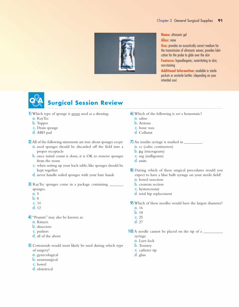
Chapter 2 General Surgical Supplies 91
Name: ultrasonic gel
Alias: none
Use: provides an acoustically correct medium forthe transmission of ultrasonic waves; provides lubri-cation for the probe to glide over the skin
Features: hypoallergenic; nonirritating to skin;non-staining
Additional Information: available in sterilepackets or unsterile bottles (depending on your intended use)
Surgical Session Review
1) Which type of sponge is never used as a dressing:
a. RayTec
b. Topper
c. Drain sponge
d. ABD pad
2) All of the following statements are true about sponges except:
a. used sponges should be discarded off the field into a
proper receptacle
b. once initial count is done, it is OK to remove sponges
from the room
c. when setting up your back table, like sponges should be
kept together
d. never handle soiled sponges with your bare hands
3) RayTec sponges come in a package containing _______
sponges.
a. 5b. 8c. 10
d. 12
4) “Peanuts” may also be known as:
a. Kitners
b. dissectors
c. pushers
d. all of the above
5) Cottonoids would most likely be used during which type
of surgery?
a. gynecological
b. neurosurgical
c. bowel
d. obstetrical
6) Which of the following is not a hemostatic?
a. saline
b. Avitene
c. bone wax
d. Collastat
7) An insulin syringe is marked in _________.
a. cc (cubic centimeters)
b. μg (micrograms)
c. mg (milligrams)
d. units
8) During which of these surgical procedures would you
expect to have a blue bulb syringe on your sterile field?
a. bowel resection
b. cesarean section
c. hysterectomy
d. total hip replacement
9) Which of these needles would have the largest diameter?
a. 16
b. 18
c. 25
d. 27
10) A needle cannot be placed on the tip of a __________
syringe.
a. Luer-lock
b. Toomey
c. catheter tip
d. glass
1572_Ch02_051-092.qxd 8/6/09 11:08 AM Page 91
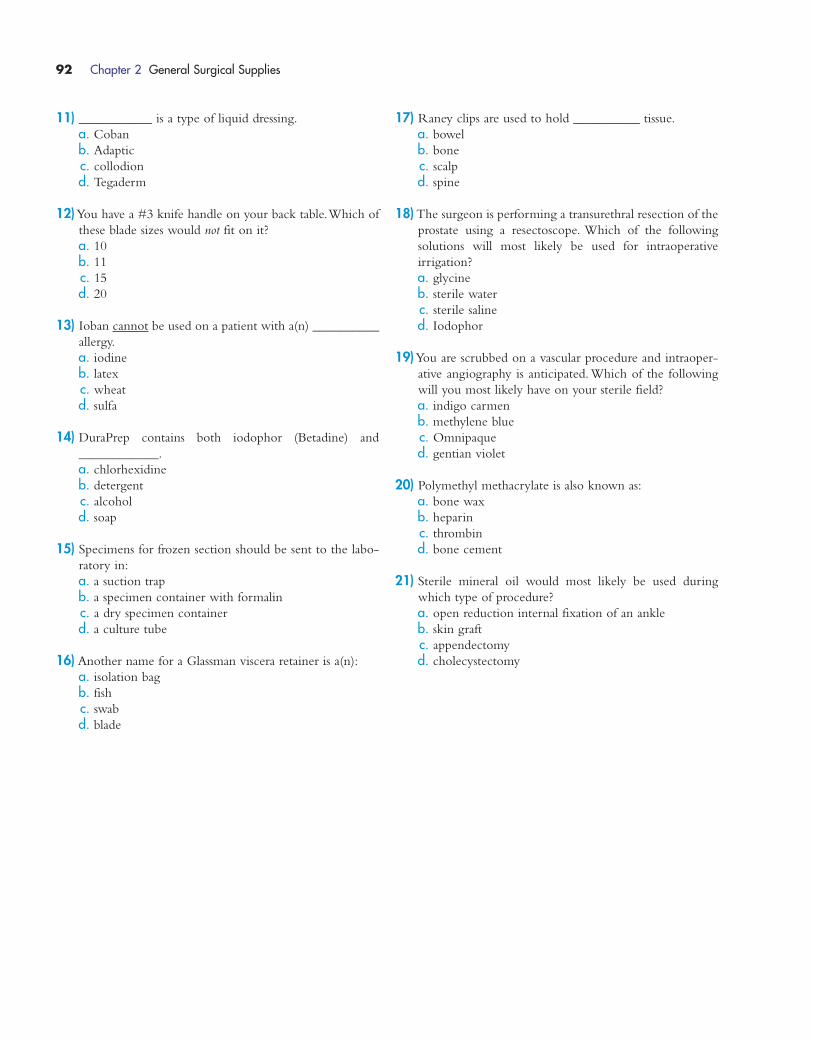
11) ___________ is a type of liquid dressing.
a. Coban
b. Adaptic
c. collodion
d. Tegaderm
12)You have a #3 knife handle on your back table.Which of
these blade sizes would not fit on it?
a. 10
b. 11
c. 15
d. 20
13) Ioban cannot be used on a patient with a(n) __________
allergy.
a. iodine
b. latex
c. wheat
d. sulfa
14) DuraPrep contains both iodophor (Betadine) and
____________.
a. chlorhexidine
b. detergent
c. alcohol
d. soap
15) Specimens for frozen section should be sent to the labo-
ratory in:
a. a suction trap
b. a specimen container with formalin
c. a dry specimen container
d. a culture tube
16) Another name for a Glassman viscera retainer is a(n):
a. isolation bag
b. fishc. swab
d. blade
17) Raney clips are used to hold __________ tissue.
a. bowel
b. bone
c. scalp
d. spine
18) The surgeon is performing a transurethral resection of the
prostate using a resectoscope. Which of the following
solutions will most likely be used for intraoperative
irrigation?
a. glycine
b. sterile water
c. sterile saline
d. Iodophor
19)You are scrubbed on a vascular procedure and intraoper-
ative angiography is anticipated.Which of the following
will you most likely have on your sterile field?
a. indigo carmen
b. methylene blue
c. Omnipaque
d. gentian violet
20) Polymethyl methacrylate is also known as:
a. bone wax
b. heparin
c. thrombin
d. bone cement
21) Sterile mineral oil would most likely be used during
which type of procedure?
a. open reduction internal fixation of an ankle
b. skin graft
c. appendectomy
d. cholecystectomy
92 Chapter 2 General Surgical Supplies
1572_Ch02_051-092.qxd 8/6/09 11:08 AM Page 92
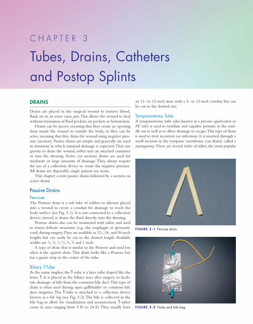
DRAINS
Drains are placed in the surgical wound to remove blood,
fluid, air or, in some cases, pus.This allows the wound to heal
without formation of fluid pockets, air pockets or hematomas.
Drains can be passive, meaning that they create an opening
from inside the wound to outside the body, or they can be
active, meaning that they drain the wound using negative pres-
sure (suction). Passive drains are simple and generally are used
in situations in which minimal drainage is expected.They use
gravity to drain the wound, either into an attached container
or into the dressing. Active (or suction) drains are used for
moderate or large amounts of drainage. They always require
the use of a collection device to create the negative pressure.
All drains are disposable, single patient use items.
This chapter covers passive drains followed by a section on
active drains.
Passive DrainsPenroseThe Penrose drain is a soft tube of rubber or silicone placed
into a wound to create a conduit for drainage to reach the
body surface (see Fig. 3-1). It is not connected to a collection
device; instead, it drains the fluid directly into the dressing.
Penrose drains also can be moistened with saline and used
to retract delicate structures (e.g., the esophagus or spermatic
cord) during surgery.They are available in 12-, 18- and 36-inch
lengths but can easily be cut to the desired length. Available
widths are 1⁄4, 3⁄8, 1⁄2, 5⁄9, 3⁄4, 7⁄8 and 1 inch.
A type of drain that is similar to the Penrose and used less
often is the cigarette drain. This drain looks like a Penrose but
has a gauze strip in the center of the tube.
Biliary T-TubeAs the name implies, the T-tube is a latex tube shaped like the
letter T. It is placed in the biliary tract after surgery to facili-
tate drainage of bile from the common bile duct.This type of
drain is often used during open gallbladder or common bile
duct surgeries. The T-tube is attached to a collection device
known as a bile bag (see Fig. 3-2).The bile is collected in the
bile bag to allow for visualization and measurement. T-tubes
come in sizes ranging from 5 Fr to 24 Fr.They usually have
an 11- to 12-inch stem with a 5- or 12-inch crossbar but can
be cut to the desired size.
Tympanostomy TubeA tympanostomy tube (also known as a pressure equalization or
PE tube) is used to ventilate and equalize pressure in the mid-
dle ear as well as to allow drainage to escape.This type of drain
is used to treat recurrent ear infections. It is inserted through a
small incision in the tympanic membrane (ear drum) called a
myringotomy.There are several styles of tubes; the most popular
C H A P T E R 3
Tubes, Drains, Catheters and Postop Splints
FIGURE 3–1 Penrose drain.
FIGURE 3–2 T-tube and bile bag.
1572_Ch03_093-106.qxd 8/6/09 11:27 AM Page 93
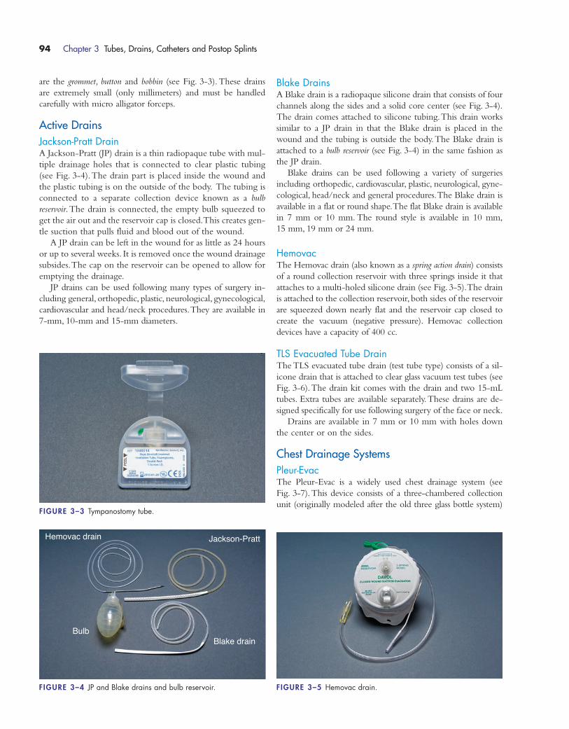
are the grommet, button and bobbin (see Fig. 3-3).These drains
are extremely small (only millimeters) and must be handled
carefully with micro alligator forceps.
Active DrainsJackson-Pratt DrainA Jackson-Pratt (JP) drain is a thin radiopaque tube with mul-
tiple drainage holes that is connected to clear plastic tubing
(see Fig. 3-4).The drain part is placed inside the wound and
the plastic tubing is on the outside of the body. The tubing is
connected to a separate collection device known as a bulb
reservoir.The drain is connected, the empty bulb squeezed to
get the air out and the reservoir cap is closed.This creates gen-
tle suction that pulls fluid and blood out of the wound.
A JP drain can be left in the wound for as little as 24 hours
or up to several weeks. It is removed once the wound drainage
subsides.The cap on the reservoir can be opened to allow for
emptying the drainage.
JP drains can be used following many types of surgery in-
cluding general, orthopedic, plastic, neurological, gynecological,
cardiovascular and head/neck procedures.They are available in
7-mm, 10-mm and 15-mm diameters.
Blake DrainsA Blake drain is a radiopaque silicone drain that consists of four
channels along the sides and a solid core center (see Fig. 3-4).
The drain comes attached to silicone tubing.This drain works
similar to a JP drain in that the Blake drain is placed in the
wound and the tubing is outside the body.The Blake drain is
attached to a bulb reservoir (see Fig. 3-4) in the same fashion as
the JP drain.
Blake drains can be used following a variety of surgeries
including orthopedic, cardiovascular, plastic, neurological, gyne-
cological, head/neck and general procedures.The Blake drain is
available in a flat or round shape.The flat Blake drain is available
in 7 mm or 10 mm. The round style is available in 10 mm,
15 mm, 19 mm or 24 mm.
HemovacThe Hemovac drain (also known as a spring action drain) consists
of a round collection reservoir with three springs inside it that
attaches to a multi-holed silicone drain (see Fig. 3-5).The drain
is attached to the collection reservoir, both sides of the reservoir
are squeezed down nearly flat and the reservoir cap closed to
create the vacuum (negative pressure). Hemovac collection
devices have a capacity of 400 cc.
TLS Evacuated Tube DrainThe TLS evacuated tube drain (test tube type) consists of a sil-
icone drain that is attached to clear glass vacuum test tubes (see
Fig. 3-6).The drain kit comes with the drain and two 15-mL
tubes. Extra tubes are available separately.These drains are de-
signed specifically for use following surgery of the face or neck.
Drains are available in 7 mm or 10 mm with holes down
the center or on the sides.
Chest Drainage SystemsPleur-EvacThe Pleur-Evac is a widely used chest drainage system (see
Fig. 3-7).This device consists of a three-chambered collection
unit (originally modeled after the old three glass bottle system)
94 Chapter 3 Tubes, Drains, Catheters and Postop Splints
FIGURE 3–3 Tympanostomy tube.
Jackson-Pratt
BulbBlake drain
Hemovac drain
FIGURE 3–4 JP and Blake drains and bulb reservoir. FIGURE 3–5 Hemovac drain.
1572_Ch03_093-106.qxd 8/6/09 11:27 AM Page 94
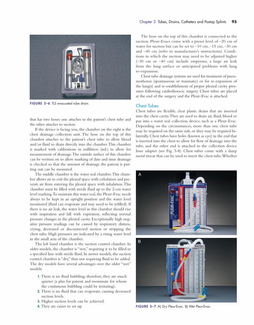
The hose on the top of this chamber is connected to the
suction. Pleur-Evacs come with a preset level of –20 cm of
water for suction but can be set to –10 cm, –15 cm, –30 cm
and –40 cm (refer to manufacturer’s instructions). Condi-
tions in which the suction may need to be adjusted higher
(–30 cm or –40 cm) include empyema, a large air leak
from the lung surface or anticipated problems with lung
re-expansion.
Chest tube drainage systems are used for treatment of pneu-
mothorax (spontaneous or traumatic) or for re-expansion of
the lung(s) and re-establishment of proper pleural cavity pres-
sures following cardiothoracic surgery. Chest tubes are placed
at the end of the surgery and the Pleur-Evac is attached.
Chest TubesChest tubes are flexible, clear plastic drains that are inserted
into the chest cavity.They are used to drain air, fluid, blood or
pus into a water seal collection device, such as a Pleur-Evac.
Depending on the circumstances, more than one chest tube
may be required on the same side, or they may be required bi-
laterally.Chest tubes have holes (known as eyes) in the end that
is inserted into the chest to allow for flow of drainage into the
tube, and the other end is attached to the collection device
hose adapter (see Fig. 3-8). Chest tubes come with a sharp
metal trocar that can be used to insert the chest tube.Whether
Chapter 3 Tubes, Drains, Catheters and Postop Splints 95
FIGURE 3–6 TLS evacuated tube drain.
FIGURE 3–7 A) Dry Pleur-Evac. B) Wet Pleur-Evac.
A
B
that has two hoses: one attaches to the patient’s chest tube and
the other attaches to suction.
If the device is facing you, the chamber on the right is the
chest drainage collection unit. The hose on the top of this
chamber attaches to the patient’s chest tube to allow blood
and/or fluid to drain directly into the chamber.This chamber
is marked with calibrations in milliliters (mL) to allow for
measurement of drainage.The outside surface of this chamber
can be written on to allow marking of date and time drainage
is checked so that the amount of drainage the patient is put-
ting out can be measured.
The middle chamber is the water seal chamber.This cham-
ber allows air to exit the pleural space with exhalation and pre-
vents air from entering the pleural space with inhalation.This
chamber must be filled with sterile fluid up to the 2-cm water
level marking.To maintain this water seal, the Pleur-Evac needs
always to be kept in an upright position and the water level
monitored (fluid can evaporate and may need to be refilled). If
there is no air leak, the water level in this chamber should rise
with inspiration and fall with expiration, reflecting normal
pressure changes in the pleural cavity. Exceptionally high neg-
ative pressure readings can be caused by respiratory distress,
crying, decreased or disconnected suction or stripping the
chest tube. High pressures are indicated by a rising water level
in the small arm of the chamber.
The left hand chamber is the suction control chamber. In
older models, the chamber is “wet,” requiring it to be filled to
a specified line with sterile fluid. In newer models, the suction
control chamber is “dry,” thus not requiring fluid to be added.
The dry models have several advantages over the older “wet”
models:
1. There is no fluid bubbling; therefore, they are much
quieter (a plus for patient and roommate for whom
the continuous bubbling could be irritating).
2. There is no fluid that can evaporate, causing decreased
suction levels.
3. Higher suction levels can be achieved.
4. They are easier to set up.
1572_Ch03_093-106.qxd 8/6/09 11:27 AM Page 95
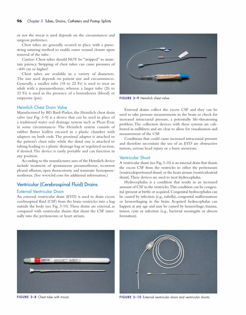
or not the trocar is used depends on the circumstances and
surgeon preference.
Chest tubes are generally secured in place with a purse-
string suturing method to enable easier wound closure upon
removal of the tube.
Caution: Chest tubes should NOT be “stripped” to main-
tain patency. Stripping of chest tubes can cause pressures of
–400 cm or higher!
Chest tubes are available in a variety of diameters.
The size used depends on patient size and circumstances.
Generally, a smaller tube (18 to 22 Fr) is used to treat an
adult with a pneumothorax, whereas a larger tube (26 to
32 Fr) is used in the presence of a hemothorax (blood) or
empyema (pus).
Heimlich Chest Drain ValveManufactured by BD Bard-Parker, the Heimlich chest drain
valve (see Fig. 3-9) is a device that can be used in place of
a traditional water seal drainage system such as Pleur-Evac
in some circumstances. The Heimlich system consists of
rubber flutter leaflets encased in a plastic chamber with
adapters on both ends.The proximal adapter is attached to
the patient’s chest tube while the distal one is attached to
tubing leading to a plastic drainage bag or regulated suction,
if desired.The device is easily portable and can function in
any position.
According to the manufacturer, uses of the Heimlich device
include treatment of spontaneous pneumothorax, recurrent
pleural effusion, open thoracotomy and traumatic hemopneu-
mothorax. (See www.bd.com for additional information.)
Ventricular (Cerebrospinal Fluid) DrainsExternal Ventricular DrainAn external ventricular drain (EVD) is used to drain excess
cerebrospinal fluid (CSF) from the brain ventricles into a bag
outside the body (see Fig. 3-10).These drains are external, as
compared with ventricular shunts that shunt the CSF inter-
nally into the peritoneum or heart atrium.
External drains collect the excess CSF and they can be
used to take pressure measurements in the brain to check for
increased intracranial pressure, a potentially life-threatening
problem. The collection devices with these systems are cali-
brated in milliliters and are clear to allow for visualization and
measurement of the CSF.
Conditions that could cause increased intracranial pressure
and therefore necessitate the use of an EVD are obstructive
tumors, serious head injury or a burst aneurysm.
Ventricular ShuntA ventricular shunt (see Fig.3-10) is an internal drain that shunts
the excess CSF from the ventricles to either the peritoneum
(ventriculoperitoneal shunt) or the heart atrium (ventriculoatrial
shunt).These devices are used to treat hydrocephalus.
Hydrocephalus is a condition that results in an increased
amount of CSF in the ventricles.This condition can be congen-
ital (present at birth) or acquired.Congenital hydrocephalus can
be caused by infection (e.g., rubella), congenital malformations
or hemorrhaging in the brain. Acquired hydrocephalus can
happen at any age and may be caused by hemorrhage, trauma,
tumor, cysts or infection (e.g., bacterial meningitis or abscess
formation).
96 Chapter 3 Tubes, Drains, Catheters and Postop Splints
FIGURE 3–8 Chest tube with trocar. FIGURE 3–10 External ventricular drain and ventricular shunts.
FIGURE 3–9 Heimlich chest valve.
1572_Ch03_093-106.qxd 8/6/09 11:27 AM Page 96
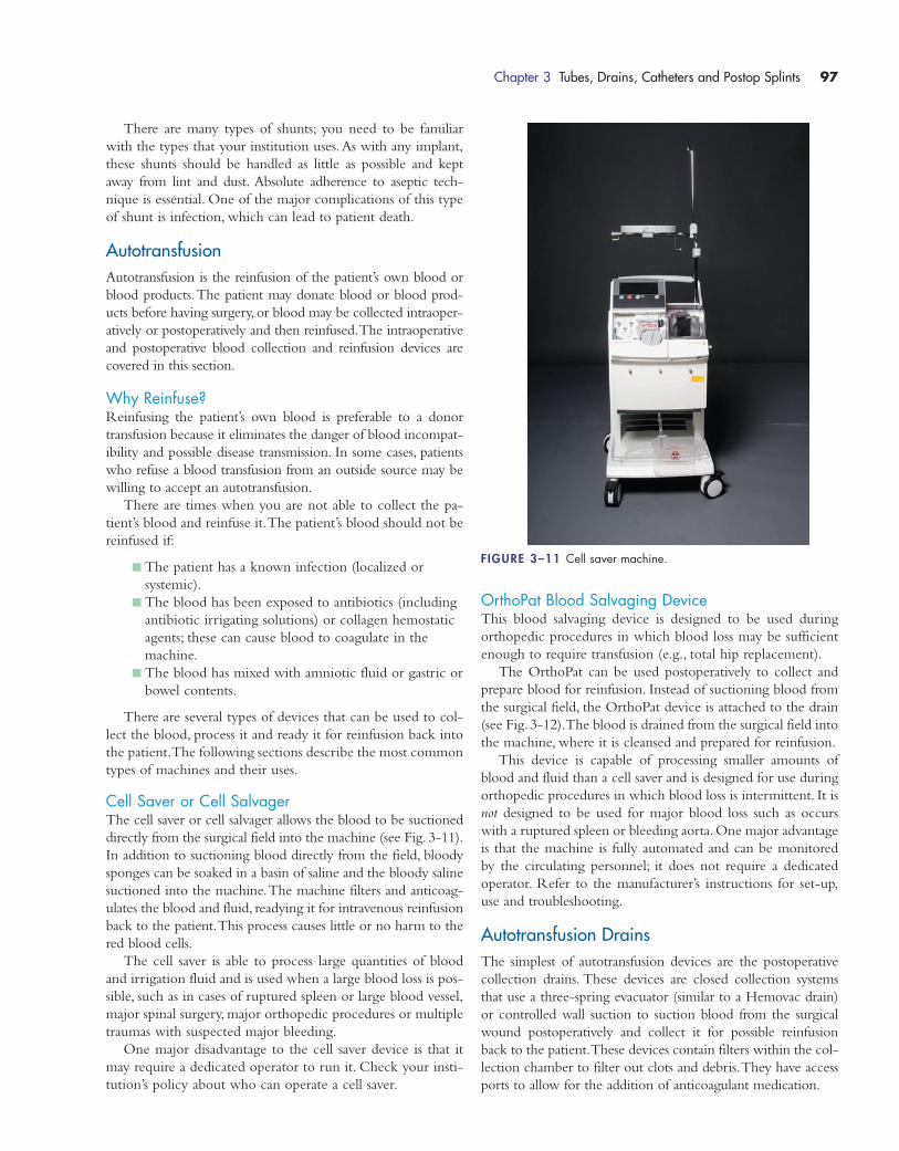
There are many types of shunts; you need to be familiar
with the types that your institution uses.As with any implant,
these shunts should be handled as little as possible and kept
away from lint and dust. Absolute adherence to aseptic tech-
nique is essential. One of the major complications of this type
of shunt is infection, which can lead to patient death.
AutotransfusionAutotransfusion is the reinfusion of the patient’s own blood or
blood products.The patient may donate blood or blood prod-
ucts before having surgery, or blood may be collected intraoper-
atively or postoperatively and then reinfused.The intraoperative
and postoperative blood collection and reinfusion devices are
covered in this section.
Why Reinfuse?Reinfusing the patient’s own blood is preferable to a donor
transfusion because it eliminates the danger of blood incompat-
ibility and possible disease transmission. In some cases, patients
who refuse a blood transfusion from an outside source may be
willing to accept an autotransfusion.
There are times when you are not able to collect the pa-
tient’s blood and reinfuse it.The patient’s blood should not be
reinfused if:
� The patient has a known infection (localized or
systemic).
� The blood has been exposed to antibiotics (including
antibiotic irrigating solutions) or collagen hemostatic
agents; these can cause blood to coagulate in the
machine.
� The blood has mixed with amniotic fluid or gastric or
bowel contents.
There are several types of devices that can be used to col-
lect the blood, process it and ready it for reinfusion back into
the patient.The following sections describe the most common
types of machines and their uses.
Cell Saver or Cell SalvagerThe cell saver or cell salvager allows the blood to be suctioned
directly from the surgical field into the machine (see Fig. 3-11).
In addition to suctioning blood directly from the field, bloody
sponges can be soaked in a basin of saline and the bloody saline
suctioned into the machine.The machine filters and anticoag-
ulates the blood and fluid, readying it for intravenous reinfusion
back to the patient.This process causes little or no harm to the
red blood cells.
The cell saver is able to process large quantities of blood
and irrigation fluid and is used when a large blood loss is pos-
sible, such as in cases of ruptured spleen or large blood vessel,
major spinal surgery, major orthopedic procedures or multiple
traumas with suspected major bleeding.
One major disadvantage to the cell saver device is that it
may require a dedicated operator to run it. Check your insti-
tution’s policy about who can operate a cell saver.
OrthoPat Blood Salvaging DeviceThis blood salvaging device is designed to be used during
orthopedic procedures in which blood loss may be sufficient
enough to require transfusion (e.g., total hip replacement).
The OrthoPat can be used postoperatively to collect and
prepare blood for reinfusion. Instead of suctioning blood from
the surgical field, the OrthoPat device is attached to the drain
(see Fig. 3-12).The blood is drained from the surgical field into
the machine, where it is cleansed and prepared for reinfusion.
This device is capable of processing smaller amounts of
blood and fluid than a cell saver and is designed for use during
orthopedic procedures in which blood loss is intermittent. It is
not designed to be used for major blood loss such as occurs
with a ruptured spleen or bleeding aorta. One major advantage
is that the machine is fully automated and can be monitored
by the circulating personnel; it does not require a dedicated
operator. Refer to the manufacturer’s instructions for set-up,
use and troubleshooting.
Autotransfusion DrainsThe simplest of autotransfusion devices are the postoperative
collection drains. These devices are closed collection systems
that use a three-spring evacuator (similar to a Hemovac drain)
or controlled wall suction to suction blood from the surgical
wound postoperatively and collect it for possible reinfusion
back to the patient.These devices contain filters within the col-
lection chamber to filter out clots and debris.They have access
ports to allow for the addition of anticoagulant medication.
Chapter 3 Tubes, Drains, Catheters and Postop Splints 97
FIGURE 3–11 Cell saver machine.
1572_Ch03_093-106.qxd 8/6/09 11:27 AM Page 97
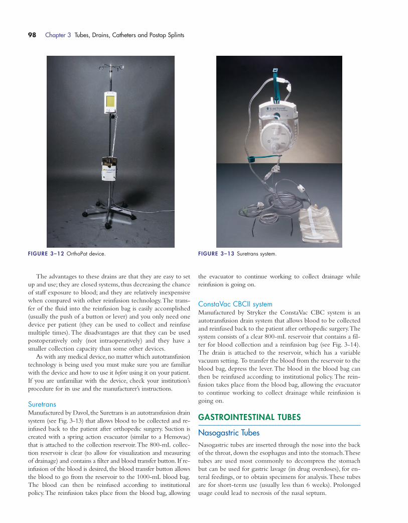
The advantages to these drains are that they are easy to set
up and use; they are closed systems, thus decreasing the chance
of staff exposure to blood; and they are relatively inexpensive
when compared with other reinfusion technology.The trans-
fer of the fluid into the reinfusion bag is easily accomplished
(usually the push of a button or lever) and you only need one
device per patient (they can be used to collect and reinfuse
multiple times). The disadvantages are that they can be used
postoperatively only (not intraoperatively) and they have a
smaller collection capacity than some other devices.
As with any medical device, no matter which autotransfusion
technology is being used you must make sure you are familiar
with the device and how to use it before using it on your patient.
If you are unfamiliar with the device, check your institution’s
procedure for its use and the manufacturer’s instructions.
SuretransManufactured by Davol, the Suretrans is an autotransfusion drain
system (see Fig. 3-13) that allows blood to be collected and re-
infused back to the patient after orthopedic surgery. Suction is
created with a spring action evacuator (similar to a Hemovac)
that is attached to the collection reservoir.The 800-mL collec-
tion reservoir is clear (to allow for visualization and measuring
of drainage) and contains a filter and blood transfer button. If re-
infusion of the blood is desired, the blood transfer button allows
the blood to go from the reservoir to the 1000-mL blood bag.
The blood can then be reinfused according to institutional
policy.The reinfusion takes place from the blood bag, allowing
the evacuator to continue working to collect drainage while
reinfusion is going on.
ConstaVac CBCII systemManufactured by Stryker the ConstaVac CBC system is an
autotransfusion drain system that allows blood to be collected
and reinfused back to the patient after orthopedic surgery.The
system consists of a clear 800-mL reservoir that contains a fil-
ter for blood collection and a reinfusion bag (see Fig. 3-14).
The drain is attached to the reservoir, which has a variable
vacuum setting. To transfer the blood from the reservoir to the
blood bag, depress the lever.The blood in the blood bag can
then be reinfused according to institutional policy.The rein-
fusion takes place from the blood bag, allowing the evacuator
to continue working to collect drainage while reinfusion is
going on.
GASTROINTESTINAL TUBES
Nasogastric TubesNasogastric tubes are inserted through the nose into the back
of the throat, down the esophagus and into the stomach.These
tubes are used most commonly to decompress the stomach
but can be used for gastric lavage (in drug overdoses), for en-
teral feedings, or to obtain specimens for analysis.These tubes
are for short-term use (usually less than 6 weeks). Prolonged
usage could lead to necrosis of the nasal septum.
98 Chapter 3 Tubes, Drains, Catheters and Postop Splints
FIGURE 3–12 OrthoPat device. FIGURE 3–13 Suretrans system.
1572_Ch03_093-106.qxd 8/6/09 11:27 AM Page 98
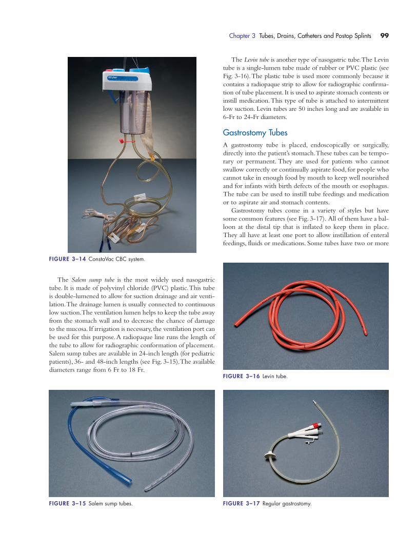
The Salem sump tube is the most widely used nasogastric
tube. It is made of polyvinyl chloride (PVC) plastic.This tube
is double-lumened to allow for suction drainage and air venti-
lation.The drainage lumen is usually connected to continuous
low suction.The ventilation lumen helps to keep the tube away
from the stomach wall and to decrease the chance of damage
to the mucosa. If irrigation is necessary, the ventilation port can
be used for this purpose.A radiopaque line runs the length of
the tube to allow for radiographic conformation of placement.
Salem sump tubes are available in 24-inch length (for pediatric
patients), 36- and 48-inch lengths (see Fig. 3-15).The available
diameters range from 6 Fr to 18 Fr.
The Levin tube is another type of nasogastric tube.The Levin
tube is a single-lumen tube made of rubber or PVC plastic (see
Fig. 3-16).The plastic tube is used more commonly because it
contains a radiopaque strip to allow for radiographic confirma-
tion of tube placement. It is used to aspirate stomach contents or
instill medication.This type of tube is attached to intermittent
low suction. Levin tubes are 50 inches long and are available in
6-Fr to 24-Fr diameters.
Gastrostomy TubesA gastrostomy tube is placed, endoscopically or surgically,
directly into the patient’s stomach.These tubes can be tempo-
rary or permanent. They are used for patients who cannot
swallow correctly or continually aspirate food, for people who
cannot take in enough food by mouth to keep well nourished
and for infants with birth defects of the mouth or esophagus.
The tube can be used to instill tube feedings and medication
or to aspirate air and stomach contents.
Gastrostomy tubes come in a variety of styles but have
some common features (see Fig. 3-17). All of them have a bal-
loon at the distal tip that is inflated to keep them in place.
They all have at least one port to allow instillation of enteral
feedings, fluids or medications. Some tubes have two or more
Chapter 3 Tubes, Drains, Catheters and Postop Splints 99
FIGURE 3–14 ConstaVac CBC system.
FIGURE 3–15 Salem sump tubes.
FIGURE 3–16 Levin tube.
FIGURE 3–17 Regular gastrostomy.
1572_Ch03_093-106.qxd 8/6/09 11:27 AM Page 99
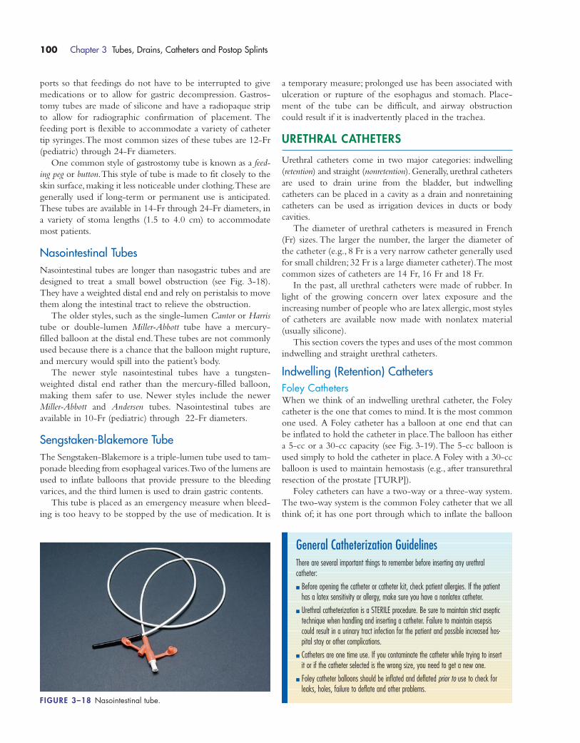
ports so that feedings do not have to be interrupted to give
medications or to allow for gastric decompression. Gastros-
tomy tubes are made of silicone and have a radiopaque strip
to allow for radiographic confirmation of placement. The
feeding port is flexible to accommodate a variety of catheter
tip syringes.The most common sizes of these tubes are 12-Fr
(pediatric) through 24-Fr diameters.
One common style of gastrostomy tube is known as a feed-
ing peg or button.This style of tube is made to fit closely to the
skin surface, making it less noticeable under clothing.These are
generally used if long-term or permanent use is anticipated.
These tubes are available in 14-Fr through 24-Fr diameters, in
a variety of stoma lengths (1.5 to 4.0 cm) to accommodate
most patients.
Nasointestinal TubesNasointestinal tubes are longer than nasogastric tubes and are
designed to treat a small bowel obstruction (see Fig. 3-18).
They have a weighted distal end and rely on peristalsis to move
them along the intestinal tract to relieve the obstruction.
The older styles, such as the single-lumen Cantor or Harris
tube or double-lumen Miller-Abbott tube have a mercury-
filled balloon at the distal end.These tubes are not commonly
used because there is a chance that the balloon might rupture,
and mercury would spill into the patient’s body.
The newer style nasointestinal tubes have a tungsten-
weighted distal end rather than the mercury-filled balloon,
making them safer to use. Newer styles include the newer
Miller-Abbott and Andersen tubes. Nasointestinal tubes are
available in 10-Fr (pediatric) through 22-Fr diameters.
Sengstaken-Blakemore TubeThe Sengstaken-Blakemore is a triple-lumen tube used to tam-
ponade bleeding from esophageal varices.Two of the lumens are
used to inflate balloons that provide pressure to the bleeding
varices, and the third lumen is used to drain gastric contents.
This tube is placed as an emergency measure when bleed-
ing is too heavy to be stopped by the use of medication. It is
a temporary measure; prolonged use has been associated with
ulceration or rupture of the esophagus and stomach. Place-
ment of the tube can be difficult, and airway obstruction
could result if it is inadvertently placed in the trachea.
URETHRAL CATHETERS
Urethral catheters come in two major categories: indwelling
(retention) and straight (nonretention).Generally, urethral catheters
are used to drain urine from the bladder, but indwelling
catheters can be placed in a cavity as a drain and nonretaining
catheters can be used as irrigation devices in ducts or body
cavities.
The diameter of urethral catheters is measured in French
(Fr) sizes. The larger the number, the larger the diameter of
the catheter (e.g., 8 Fr is a very narrow catheter generally used
for small children; 32 Fr is a large diameter catheter).The most
common sizes of catheters are 14 Fr, 16 Fr and 18 Fr.
In the past, all urethral catheters were made of rubber. In
light of the growing concern over latex exposure and the
increasing number of people who are latex allergic, most styles
of catheters are available now made with nonlatex material
(usually silicone).
This section covers the types and uses of the most common
indwelling and straight urethral catheters.
Indwelling (Retention) CathetersFoley CathetersWhen we think of an indwelling urethral catheter, the Foley
catheter is the one that comes to mind. It is the most common
one used. A Foley catheter has a balloon at one end that can
be inflated to hold the catheter in place.The balloon has either
a 5-cc or a 30-cc capacity (see Fig. 3-19).The 5-cc balloon is
used simply to hold the catheter in place.A Foley with a 30-cc
balloon is used to maintain hemostasis (e.g., after transurethral
resection of the prostate [TURP]).
Foley catheters can have a two-way or a three-way system.
The two-way system is the common Foley catheter that we all
think of; it has one port through which to inflate the balloon
100 Chapter 3 Tubes, Drains, Catheters and Postop Splints
FIGURE 3–18 Nasointestinal tube.
General Catheterization GuidelinesThere are several important things to remember before inserting any urethralcatheter:
� Before opening the catheter or catheter kit, check patient allergies. If the patienthas a latex sensitivity or allergy, make sure you have a nonlatex catheter.
� Urethral catheterization is a STERILE procedure. Be sure to maintain strict aseptictechnique when handling and inserting a catheter. Failure to maintain asepsiscould result in a urinary tract infection for the patient and possible increased hos-pital stay or other complications.
� Catheters are one time use. If you contaminate the catheter while trying to insertit or if the catheter selected is the wrong size, you need to get a new one.
� Foley catheter balloons should be inflated and deflated prior to use to check forleaks, holes, failure to deflate and other problems.
1572_Ch03_093-106.qxd 8/6/09 11:27 AM Page 100
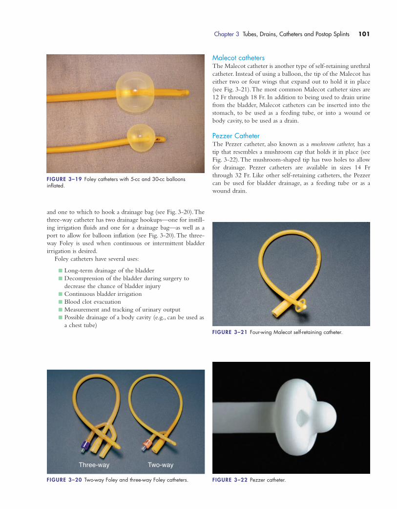
and one to which to hook a drainage bag (see Fig. 3-20).The
three-way catheter has two drainage hookups—one for instill-
ing irrigation fluids and one for a drainage bag—as well as a
port to allow for balloon inflation (see Fig. 3-20).The three-
way Foley is used when continuous or intermittent bladder
irrigation is desired.
Foley catheters have several uses:
� Long-term drainage of the bladder
� Decompression of the bladder during surgery to
decrease the chance of bladder injury
� Continuous bladder irrigation
� Blood clot evacuation
� Measurement and tracking of urinary output
� Possible drainage of a body cavity (e.g., can be used as
a chest tube)
Malecot cathetersThe Malecot catheter is another type of self-retaining urethral
catheter. Instead of using a balloon, the tip of the Malecot has
either two or four wings that expand out to hold it in place
(see Fig. 3-21).The most common Malecot catheter sizes are
12 Fr through 18 Fr. In addition to being used to drain urine
from the bladder, Malecot catheters can be inserted into the
stomach, to be used as a feeding tube, or into a wound or
body cavity, to be used as a drain.
Pezzer CatheterThe Pezzer catheter, also known as a mushroom catheter, has a
tip that resembles a mushroom cap that holds it in place (see
Fig. 3-22).The mushroom-shaped tip has two holes to allow
for drainage. Pezzer catheters are available in sizes 14 Fr
through 32 Fr. Like other self-retaining catheters, the Pezzer
can be used for bladder drainage, as a feeding tube or as a
wound drain.
Chapter 3 Tubes, Drains, Catheters and Postop Splints 101
FIGURE 3–19 Foley catheters with 5-cc and 30-cc balloons inflated.
Three-way Two-way
FIGURE 3–20 Two-way Foley and three-way Foley catheters.
FIGURE 3–21 Four-wing Malecot self-retaining catheter.
FIGURE 3–22 Pezzer catheter.
1572_Ch03_093-106.qxd 8/6/09 11:27 AM Page 101
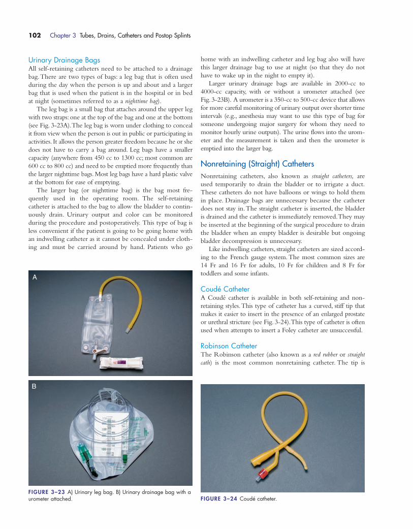
Urinary Drainage BagsAll self-retaining catheters need to be attached to a drainage
bag.There are two types of bags: a leg bag that is often used
during the day when the person is up and about and a larger
bag that is used when the patient is in the hospital or in bed
at night (sometimes referred to as a nighttime bag).
The leg bag is a small bag that attaches around the upper leg
with two straps: one at the top of the bag and one at the bottom
(see Fig. 3-23A).The leg bag is worn under clothing to conceal
it from view when the person is out in public or participating in
activities. It allows the person greater freedom because he or she
does not have to carry a bag around. Leg bags have a smaller
capacity (anywhere from 450 cc to 1300 cc; most common are
600 cc to 800 cc) and need to be emptied more frequently than
the larger nighttime bags. Most leg bags have a hard plastic valve
at the bottom for ease of emptying.
The larger bag (or nighttime bag) is the bag most fre-
quently used in the operating room. The self-retaining
catheter is attached to the bag to allow the bladder to contin-
uously drain. Urinary output and color can be monitored
during the procedure and postoperatively. This type of bag is
less convenient if the patient is going to be going home with
an indwelling catheter as it cannot be concealed under cloth-
ing and must be carried around by hand. Patients who go
home with an indwelling catheter and leg bag also will have
this larger drainage bag to use at night (so that they do not
have to wake up in the night to empty it).
Larger urinary drainage bags are available in 2000-cc to
4000-cc capacity, with or without a urometer attached (see
Fig.3-23B). A urometer is a 350-cc to 500-cc device that allows
for more careful monitoring of urinary output over shorter time
intervals (e.g., anesthesia may want to use this type of bag for
someone undergoing major surgery for whom they need to
monitor hourly urine outputs). The urine flows into the urom-
eter and the measurement is taken and then the urometer is
emptied into the larger bag.
Nonretaining (Straight) CathetersNonretaining catheters, also known as straight catheters, are
used temporarily to drain the bladder or to irrigate a duct.
These catheters do not have balloons or wings to hold them
in place. Drainage bags are unnecessary because the catheter
does not stay in.The straight catheter is inserted, the bladder
is drained and the catheter is immediately removed.They may
be inserted at the beginning of the surgical procedure to drain
the bladder when an empty bladder is desirable but ongoing
bladder decompression is unnecessary.
Like indwelling catheters, straight catheters are sized accord-
ing to the French gauge system. The most common sizes are
14 Fr and 16 Fr for adults, 10 Fr for children and 8 Fr for
toddlers and some infants.
Coudé CatheterA Coudé catheter is available in both self-retaining and non-
retaining styles.This type of catheter has a curved, stiff tip that
makes it easier to insert in the presence of an enlarged prostate
or urethral stricture (see Fig. 3-24).This type of catheter is often
used when attempts to insert a Foley catheter are unsuccessful.
Robinson CatheterThe Robinson catheter (also known as a red rubber or straight
cath) is the most common nonretaining catheter. The tip is
102 Chapter 3 Tubes, Drains, Catheters and Postop Splints
FIGURE 3–23 A) Urinary leg bag. B) Urinary drainage bag with aurometer attached.
A
B
FIGURE 3–24 Coudé catheter.
1572_Ch03_093-106.qxd 8/6/09 11:27 AM Page 102
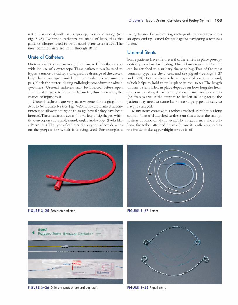
soft and rounded, with two opposing eyes for drainage (see
Fig. 3-25). Robinson catheters are made of latex, thus the
patient’s allergies need to be checked prior to insertion.The
most common sizes are 12 Fr through 18 Fr.
Ureteral CathetersUreteral catheters are narrow tubes inserted into the ureters
with the use of a cystoscope.These catheters can be used to
bypass a tumor or kidney stone, provide drainage of the ureter,
keep the ureter open, instill contrast media, allow stones to
pass, block the ureters during radiologic procedures or obtain
specimens. Ureteral catheters may be inserted before open
abdominal surgery to identify the ureter, thus decreasing the
chance of injury to it.
Ureteral catheters are very narrow, generally ranging from
3-Fr to 6-Fr diameter (see Fig. 3-26).They are marked in cen-
timeters to allow the surgeon to gauge how far they have been
inserted.These catheters come in a variety of tip shapes: whis-
tle, cone, open end, spiral, round, angled and wedge (looks like
a Pezzer tip).The type of catheter the surgeon selects depends
on the purpose for which it is being used. For example, a
wedge tip may be used during a retrograde pyelogram,whereas
an open-end tip is used for drainage or navigating a tortuous
ureter.
Ureteral StentsSome patients have the ureteral catheter left in place postop-
eratively to allow for healing.This is known as a stent and it
can be attached to a urinary drainage bag.Two of the most
common types are the J stent and the pigtail (see Figs. 3-27
and 3-28). Both catheters have a spiral shape to the end,
which helps to hold them in place in the ureter.The length
of time a stent is left in place depends on how long the heal-
ing process takes; it can be anywhere from days to months
(or even years). If the stent is to be left in long-term, the
patient may need to come back into surgery periodically to
have it changed.
Many stents come with a tether attached. A tether is a long
strand of material attached to the stent that aids in the manip-
ulation or removal of the stent. The surgeon may choose to
leave the tether attached (in which case it is often secured to
the inside of the upper thigh) or cut it off.
Chapter 3 Tubes, Drains, Catheters and Postop Splints 103
FIGURE 3–25 Robinson catheter.
FIGURE 3–26 Different types of ureteral catheters.
FIGURE 3–27 J stent.
FIGURE 3–28 Pigtail stent.
1572_Ch03_093-106.qxd 8/6/09 11:27 AM Page 103
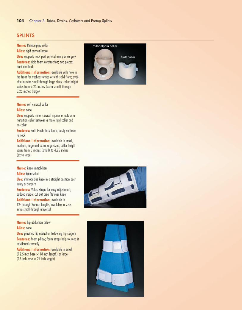
104 Chapter 3 Tubes, Drains, Catheters and Postop Splints
Philadelphia collar
Soft collar
SPLINTS
Name: Philadelphia collar
Alias: rigid cervical brace
Use: supports neck post cervical injury or surgery
Features: rigid foam construction; two pieces:front and back
Additional Information: available with hole inthe front for tracheostomies or with solid front; avail-able in extra small through large sizes; collar heightvaries from 2.25 inches (extra small) through 5.25 inches (large)
Name: soft cervical collar
Alias: none
Use: supports minor cervical injuries or acts as atransition collar between a more rigid collar and no collar
Features: soft 1-inch thick foam; easily contoursto neck
Additional Information: available in small,medium, large and extra large sizes; collar heightvaries from 3 inches (small) to 4.25 inches (extra large)
Name: knee immobilizer
Alias: knee splint
Use: immobilizes knee in a straight position post injury or surgery
Features: Velcro straps for easy adjustment;padded inside; cut out area fits over knee
Additional Information: available in 12- through 26-inch lengths; available in sizes extra small through universal
Name: hip abduction pillow
Alias: none
Use: provides hip abduction following hip surgery
Features: foam pillow; foam straps help to keep itpositioned correctly
Additional Information: available in small(12.5-inch base × 18-inch length) or large (17-inch base × 24-inch length)
1572_Ch03_093-106.qxd 8/6/09 11:27 AM Page 104
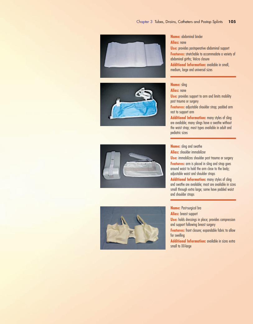
Chapter 3 Tubes, Drains, Catheters and Postop Splints 105
Name: abdominal binder
Alias: none
Use: provides postoperative abdominal support
Features: stretchable to accommodate a variety ofabdominal girths; Velcro closure
Additional Information: available in small,medium, large and universal sizes
Name: sling
Alias: none
Use: provides support to arm and limits mobilitypost trauma or surgery
Features: adjustable shoulder strap; padded armrest to support arm
Additional Information: many styles of slingare available; many slings have a swathe withoutthe waist strap; most types available in adult andpediatric sizes
Name: sling and swathe
Alias: shoulder immobilizer
Use: immobilizes shoulder post trauma or surgery
Features: arm is placed in sling and strap goesaround waist to hold the arm close to the body; adjustable waist and shoulder straps
Additional Information: many styles of slingand swathe are available; most are available in sizessmall through extra large; some have padded waistand shoulder straps
Name: Post-surgical bra
Alias: breast support
Use: holds dressings in place; provides compressionand support following breast surgery
Features: front closure; expandable fabric to allowfor swelling
Additional Information: available in sizes extrasmall to XX-large
1572_Ch03_093-106.qxd 8/6/09 11:27 AM Page 105
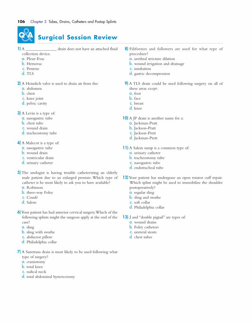
106 Chapter 3 Tubes, Drains, Catheters and Postop Splints
Surgical Session Review
1) A _______________ drain does not have an attached fluid
collection device.
a. Pleur-Evac
b. Hemovac
c. Penrose
d. TLS
2) A Heimlich valve is used to drain air from the:
a. abdomen
b. chest
c. knee joint
d. pelvic cavity
3) A Levin is a type of:
a. nasogastric tube
b. chest tube
c. wound drain
d. tracheostomy tube
4) A Malecot is a type of:
a. nasogastric tube
b. wound drain
c. ventricular drain
d. urinary catheter
5) The urologist is having trouble catheterizing an elderly
male patient due to an enlarged prostate. Which type of
catheter is he most likely to ask you to have available?
a. Robinson
b. three-way Foley
c. Coudé
d. Salem
6)Your patient has had anterior cervical surgery.Which of the
following splints might the surgeon apply at the end of the
case?
a. sling
b. sling with swathe
c. abductor pillow
d. Philadelphia collar
7) A Suretrans drain is most likely to be used following what
type of surgery?
a. craniotomy
b. total knee
c. radical neck
d. total abdominal hysterectomy
8) Filiformes and followers are used for what type of
procedure?
a. urethral stricture dilation
b. wound irrigation and drainage
c. intubation
d. gastric decompression
9) A TLS drain could be used following surgery on all of
these areas except:
a. foot
b. face
c. breast
d. knee
10) A JP drain is another name for a:
a. Jackman-Pratt
b. Jackson-Pratt
c. Jackson-Prett
d. Jackman-Prett
11) A Salem sump is a common type of:
a. urinary catheter
b. tracheostomy tube
c. nasogastric tube
d. endotracheal tube
12)Your patient has undergone an open rotator cuff repair.
Which splint might be used to immobilize the shoulder
postoperatively?
a. regular sling
b. sling and swathe
c. soft collar
d. Philadelphia collar
13) J and “double pigtail” are types of:
a. wound drains
b. Foley catheters
c. ureteral stents
d. chest tubes
1572_Ch03_093-106.qxd 8/6/09 11:27 AM Page 106
COMPARISON OF SUTURE MATERIALS
Sutures are composed of various materials, which make them
suitable for certain procedures or tissues.Tables 4-1, 4-2 and
4-3 allow you to compare suture materials used by various
manufacturers. Approximate wound support times are listed
with the absorbable sutures to allow for further comparisons.
COMPONENTS OF SUTURE PACKAGING
You can judge some things by their covers. Shown here are the
components that make up the information found on the outer
packaging of the suture material produced by four major suture
manufacturers.
Ethicon (see Fig. 4-1)Manufacturer’s Major Suture Brands
� Ethilon
� Surgical Stainless Steel
� Nurolon
� Mersilene
� Prolene
� Ethibond
� Silk (Mersilk)
� Surgical Gut (Plain)
� Surgical Gut (Chromic)
� Vicryl
� Vicryl Plus
� Vicryl Rapide
� Monocryl
� Panacryl
Syneture (see Fig. 4-2)Manufacturer’s Major Suture Brands
� Dermalon
� TiCron
� Novafil
� SurgiDac
� Vascufil
� Novafil
� Surgipro
� Flexon
C H A P T E R 4
Sutures and Suture Needles
Table 4-1 Non-absorbable Sutures
SYNETURE PRODUCT SUTURE MATERIAL (US SUTURE/DAVIS AND GECK) ETHICON PRODUCTMonofilament nylon Dermalon; Monosof Ethilon
Braided nylon Surgilon Nurolon
Braided polyester (uncoated) Surgidac Mersilene
Braided polyester (coated) Ti Cron Ethibond Excel
Monofilament polybutester Novafil —
Monofilament polybutester (coated) Vascufil —
Monofilament polypropylene Surgipro; Surgipro II Prolene
Hexafluoropropylene VDF — Pronova Poly
Monofilament steel Steel Surgical Stainless Steel
Multistrand steel Flexon Surgical Stainless Steel
Silk Sofsilk Silk; Perma-Hand
1572_Ch04_107-132.qxd 8/6/09 11:36 AM Page 107
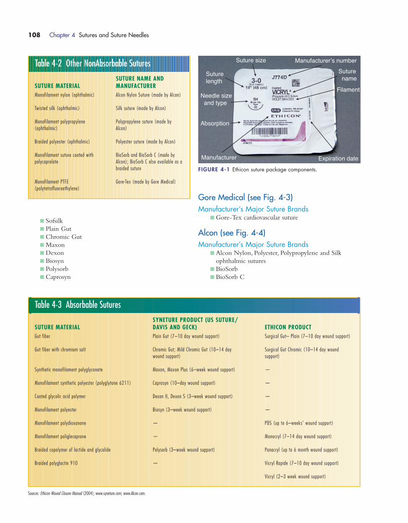
� Sofsilk
� Plain Gut
� Chromic Gut
� Maxon
� Dexon
� Biosyn
� Polysorb
� Caprosyn
Gore Medical (see Fig. 4-3)Manufacturer’s Major Suture Brands
� Gore-Tex cardiovascular suture
Alcon (see Fig. 4-4)Manufacturer’s Major Suture Brands
� Alcon Nylon, Polyester, Polypropylene and Silk
ophthalmic sutures
� BioSorb
� BioSorb C
108 Chapter 4 Sutures and Suture Needles
Table 4-2 Other NonAbsorbable Sutures
SUTURE NAME AND SUTURE MATERIAL MANUFACTURERMonofilament nylon (ophthalmic) Alcon Nylon Suture (made by Alcon)
Twisted silk (ophthalmic) Silk suture (made by Alcon)
Monofilament polypropylene Polypropylene suture (made by (ophthalmic) Alcon)
Braided polyester (ophthalmic) Polyester suture (made by Alcon)
Monofilament suture coated with BioSorb and BioSorb C (made by polycaprolate Alcon); BioSorb C also available as a
braided suture
Monofilament PTFE Gore-Tex (made by Gore Medical)(polytetrafluoroethylene)
Table 4-3 Absorbable Sutures
SYNETURE PRODUCT (US SUTURE/ SUTURE MATERIAL DAVIS AND GECK) ETHICON PRODUCTGut fiber Plain Gut (7–10 day wound support) Surgical Gut– Plain (7–10 day wound support)
Gut fiber with chromium salt Chromic Gut; Mild Chromic Gut (10–14 day Surgical Gut Chromic (10–14 day wound wound support) support)
Synthetic monofilament polyglyconate Maxon, Maxon Plus (6–week wound support) —
Monofilament synthetic polyester (polyglytone 6211) Caprosyn (10–day wound support) —
Coated glycolic acid polymer Dexon II, Dexon S (3–week wound support) —
Monofilament polyester Biosyn (3–week wound support) —
Monofilament polydioxanone — PDS (up to 6–weeks’ wound support)
Monofilament poliglecaprone — Monocryl (7–14 day wound support)
Braided copolymer of lactide and glycolide Polysorb (3–week wound support) Panacryl (up to 6 month wound support)
Braided polyglactin 910 — Vicryl Rapide (7–10 day wound support)
Vicryl (2–3 week wound support)
Sources: Ethicon Wound Closure Manual (2004); www.syneture.com; www.Alcon.com.
Suture size
Suture name
Manufacturer’s number
Suture length
Needle size and type
Absorption
Manufacturer Expiration date
Filament
FIGURE 4-1 Ethicon suture package components.
1572_Ch04_107-132.qxd 8/6/09 11:36 AM Page 108
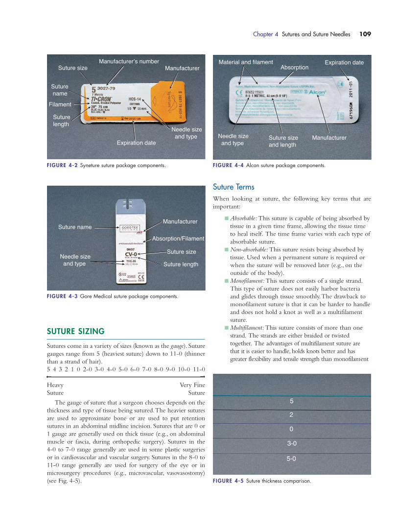
SUTURE SIZING
Sutures come in a variety of sizes (known as the gauge). Suture
gauges range from 5 (heaviest suture) down to 11-0 (thinner
than a strand of hair).
5 4 3 2 1 0 2-0 3-0 4-0 5-0 6-0 7-0 8-0 9-0 10-0 11-0
Heavy Very Fine
Suture Suture
The gauge of suture that a surgeon chooses depends on the
thickness and type of tissue being sutured.The heavier sutures
are used to approximate bone or are used to put retention
sutures in an abdominal midline incision. Sutures that are 0 or
1 gauge are generally used on thick tissue (e.g., on abdominal
muscle or fascia, during orthopedic surgery). Sutures in the
4-0 to 7-0 range generally are used in some plastic surgeries
or in cardiovascular and vascular surgery. Sutures in the 8-0 to
11-0 range generally are used for surgery of the eye or in
microsurgery procedures (e.g., microvascular, vasovasostomy)
(see Fig. 4-5).
Suture TermsWhen looking at suture, the following key terms that are
important:
� Absorbable: This suture is capable of being absorbed by
tissue in a given time frame, allowing the tissue time
to heal itself. The time frame varies with each type of
absorbable suture.
� Non-absorbable: This suture resists being absorbed by
tissue. Used when a permanent suture is required or
when the suture will be removed later (e.g., on the
outside of the body).
� Monofilament: This suture consists of a single strand.
This type of suture does not easily harbor bacteria
and glides through tissue smoothly.The drawback to
monofilament suture is that it can be harder to handle
and does not hold a knot as well as a multifilament
suture.
� Multifilament: This suture consists of more than one
strand. The strands are either braided or twisted
together. The advantages of multifilament suture are
that it is easier to handle, holds knots better and has
greater flexibility and tensile strength than monofilament
Chapter 4 Sutures and Suture Needles 109
Suture sizeManufacturer’s number
Manufacturer
Needle size and type
Expiration date
Filament
Suture name
Suture length
FIGURE 4-2 Syneture suture package components.
Suture name
Needle size and type
Manufacturer
Absorption/Filament
Suture size
Suture length
FIGURE 4-3 Gore Medical suture package components.
Material and filament
Manufacturer
Expiration date
Needle size and type
Suture size and length
Absorption
FIGURE 4-4 Alcon suture package components.
5
2
0
3-0
5-0
FIGURE 4-5 Suture thickness comparison.
��
1572_Ch04_107-132.qxd 8/6/09 11:36 AM Page 109
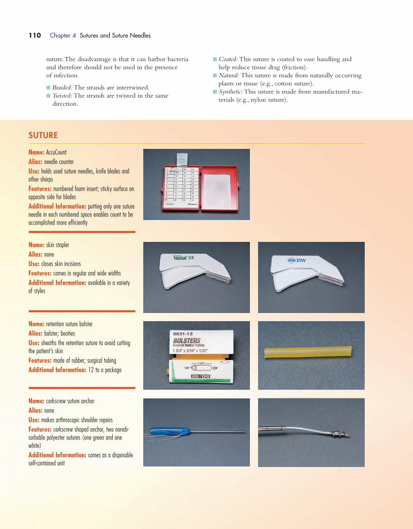
SUTURE
Name: AccuCount
Alias: needle counter
Use: holds used suture needles, knife blades andother sharps
Features: numbered foam insert; sticky surface onopposite side for blades
Additional Information: putting only one sutureneedle in each numbered space enables count to beaccomplished more efficiently
Name: skin stapler
Alias: none
Use: closes skin incisions
Features: comes in regular and wide widths
Additional Information: available in a varietyof styles
Name: retention suture bolster
Alias: bolster; booties
Use: sheaths the retention suture to avoid cuttingthe patient’s skin
Features: made of rubber; surgical tubing
Additional Information: 12 to a package
Name: corkscrew suture anchor
Alias: none
Use: makes arthroscopic shoulder repairs
Features: corkscrew shaped anchor, two nonab-sorbable polyester sutures (one green and onewhite)
Additional Information: comes as a disposableself-contained unit
suture.The disadvantage is that it can harbor bacteria
and therefore should not be used in the presence
of infection.
� Braided: The strands are intertwined.
� Twisted: The strands are twisted in the same
direction.
� Coated: This suture is coated to ease handling and
help reduce tissue drag (friction).
� Natural: This suture is made from naturally occurring
plants or tissue (e.g., cotton suture).
� Synthetic: This suture is made from manufactured ma-
terials (e.g., nylon suture).
110 Chapter 4 Sutures and Suture Needles
1572_Ch04_107-132.qxd 8/6/09 11:36 AM Page 110
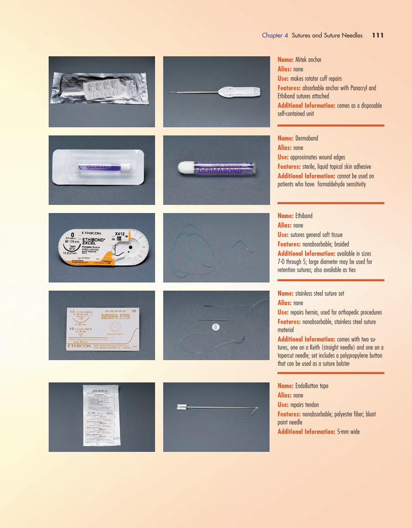
Chapter 4 Sutures and Suture Needles 111
Name: Mitek anchor
Alias: none
Use: makes rotator cuff repairs
Features: absorbable anchor with Panacryl andEthibond sutures attached
Additional Information: comes as a disposableself-contained unit
Name: Dermabond
Alias: none
Use: approximates wound edges
Features: sterile, liquid topical skin adhesive
Additional Information: cannot be used on patients who have formaldehyde sensitivity
Name: Ethibond
Alias: none
Use: sutures general soft tissue
Features: nonabsorbable; braided
Additional Information: available in sizes 7-0 through 5; large diameter may be used for retention sutures; also available as ties
Name: stainless steel suture set
Alias: none
Use: repairs hernia, used for orthopedic procedures
Features: nonabsorbable, stainless steel suturematerial
Additional Information: comes with two su-tures, one on a Keith (straight needle) and one on atapercut needle; set includes a polypropylene buttonthat can be used as a suture bolster
Name: EndoButton tape
Alias: none
Use: repairs tendon
Features: nonabsorbable; polyester fiber; bluntpoint needle
Additional Information: 5-mm wide
1572_Ch04_107-132.qxd 8/6/09 11:36 AM Page 111
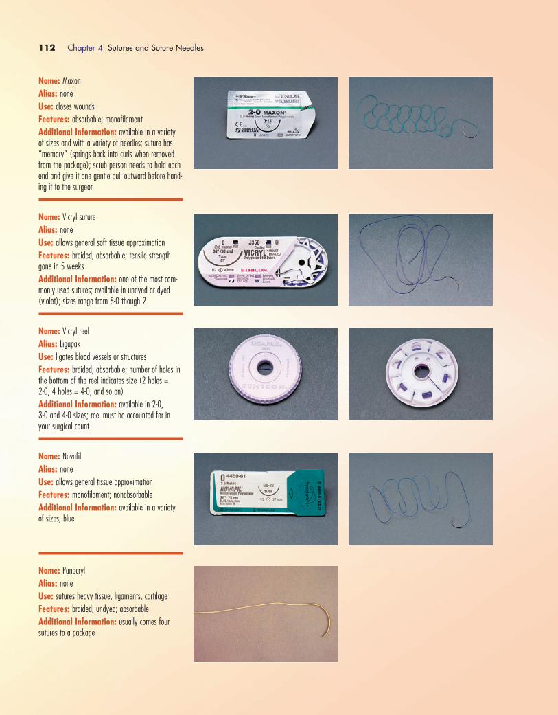
112 Chapter 4 Sutures and Suture Needles
Name: Maxon
Alias: none
Use: closes wounds
Features: absorbable; monofilament
Additional Information: available in a varietyof sizes and with a variety of needles; suture has“memory” (springs back into curls when removedfrom the package); scrub person needs to hold eachend and give it one gentle pull outward before hand-ing it to the surgeon
Name: Vicryl suture
Alias: none
Use: allows general soft tissue approximation
Features: braided; absorbable; tensile strengthgone in 5 weeks
Additional Information: one of the most com-monly used sutures; available in undyed or dyed (violet); sizes range from 8-0 though 2
Name: Vicryl reel
Alias: Ligapak
Use: ligates blood vessels or structures
Features: braided; absorbable; number of holes inthe bottom of the reel indicates size (2 holes = 2-0, 4 holes = 4-0, and so on)
Additional Information: available in 2-0, 3-0 and 4-0 sizes; reel must be accounted for inyour surgical count
Name: Novafil
Alias: none
Use: allows general tissue approximation
Features: monofilament; nonabsorbable
Additional Information: available in a varietyof sizes; blue
Name: Panacryl
Alias: none
Use: sutures heavy tissue, ligaments, cartilage
Features: braided; undyed; absorbable
Additional Information: usually comes four sutures to a package
1572_Ch04_107-132.qxd 8/6/09 11:36 AM Page 112
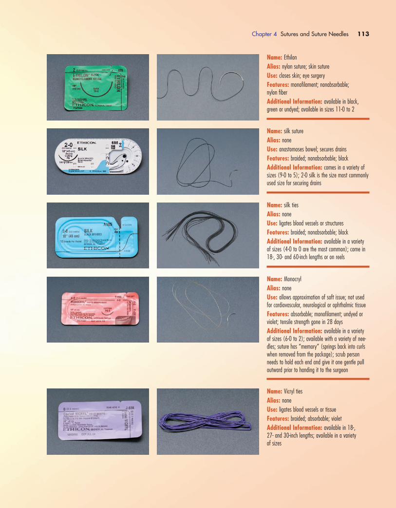
Chapter 4 Sutures and Suture Needles 113
Name: Ethilon
Alias: nylon suture; skin suture
Use: closes skin; eye surgery
Features: monofilament; nonabsorbable; nylon fiber
Additional Information: available in black,green or undyed; available in sizes 11-0 to 2
Name: silk suture
Alias: none
Use: anastomoses bowel; secures drains
Features: braided; nonabsorbable; black
Additional Information: comes in a variety ofsizes (9-0 to 5); 2-0 silk is the size most commonlyused size for securing drains
Name: silk ties
Alias: none
Use: ligates blood vessels or structures
Features: braided; nonabsorbable; black
Additional Information: available in a varietyof sizes (4-0 to 0 are the most common); come in18-, 30- and 60-inch lengths or on reels
Name: Monocryl
Alias: none
Use: allows approximation of soft issue; not usedfor cardiovascular, neurological or ophthalmic tissue
Features: absorbable; monofilament; undyed or violet; tensile strength gone in 28 days
Additional Information: available in a varietyof sizes (6-0 to 2); available with a variety of nee-dles; suture has “memory” (springs back into curlswhen removed from the package); scrub personneeds to hold each end and give it one gentle pulloutward prior to handing it to the surgeon
Name: Vicryl ties
Alias: none
Use: ligates blood vessels or tissue
Features: braided; absorbable; violet
Additional Information: available in 18-, 27- and 30-inch lengths; available in a variety of sizes
1572_Ch04_107-132.qxd 8/6/09 11:36 AM Page 113
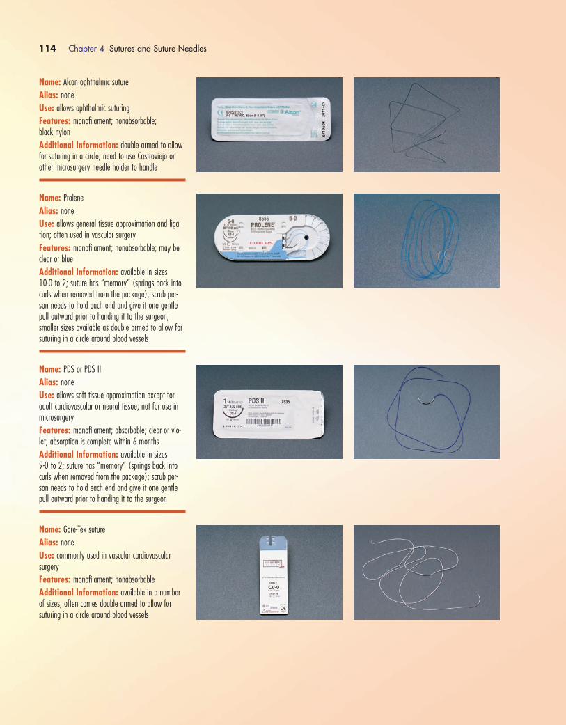
114 Chapter 4 Sutures and Suture Needles
Name: Alcon ophthalmic suture
Alias: none
Use: allows ophthalmic suturing
Features: monofilament; nonabsorbable; black nylon
Additional Information: double armed to allowfor suturing in a circle; need to use Castroviejo orother microsurgery needle holder to handle
Name: Prolene
Alias: none
Use: allows general tissue approximation and liga-tion; often used in vascular surgery
Features: monofilament; nonabsorbable; may beclear or blue
Additional Information: available in sizes 10-0 to 2; suture has “memory” (springs back intocurls when removed from the package); scrub per-son needs to hold each end and give it one gentlepull outward prior to handing it to the surgeon;smaller sizes available as double armed to allow forsuturing in a circle around blood vessels
Name: PDS or PDS II
Alias: none
Use: allows soft tissue approximation except foradult cardiovascular or neural tissue; not for use inmicrosurgery
Features: monofilament; absorbable; clear or vio-let; absorption is complete within 6 months
Additional Information: available in sizes 9-0 to 2; suture has “memory” (springs back intocurls when removed from the package); scrub per-son needs to hold each end and give it one gentlepull outward prior to handing it to the surgeon
Name: Gore-Tex suture
Alias: none
Use: commonly used in vascular cardiovascular surgery
Features: monofilament; nonabsorbable
Additional Information: available in a numberof sizes; often comes double armed to allow for suturing in a circle around blood vessels
1572_Ch04_107-132.qxd 8/6/09 11:36 AM Page 114
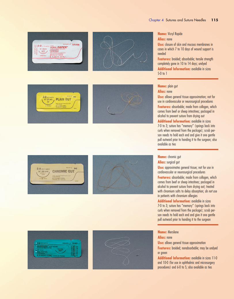
Chapter 4 Sutures and Suture Needles 115
Name: Vicryl Rapide
Alias: none
Use: closure of skin and mucous membranes incases in which 7 to 10 days of wound support isneeded
Features: braided; absorbable; tensile strengthcompletely gone in 10 to 14 days; undyed
Additional Information: available in sizes 5-0 to 1
Name: plain gut
Alias: none
Use: allows general tissue approximation; not foruse in cardiovascular or neurosurgical procedures
Features: absorbable; made from collagen, whichcomes from beef or sheep intestines; packaged in alcohol to prevent suture from drying out
Additional Information: available in sizes 7-0 to 3; suture has “memory” (springs back intocurls when removed from the package); scrub per-son needs to hold each end and give it one gentlepull outward prior to handing it to the surgeon; alsoavailable as ties
Name: chromic gut
Alias: surgical gut
Use: approximates general tissue; not for use incardiovascular or neurosurgical procedures
Features: absorbable; made from collagen, whichcomes from beef or sheep intestines; packaged in alcohol to prevent suture from drying out; treatedwith chromium salts to delay absorption; do not usein patients with chromium allergies
Additional Information: available in sizes 7-0 to 3; suture has “memory” (springs back intocurls when removed from the package); scrub per-son needs to hold each end and give it one gentlepull outward prior to handing it to the surgeon
Name: Mersilene
Alias: none
Use: allows general tissue approximation
Features: braided; nonabsorbable; may be undyedor green
Additional Information: available in sizes 11-0and 10-0 (for use in ophthalmic and microsurgeryprocedures) and 6-0 to 5; also available as ties
1572_Ch04_107-132.qxd 8/6/09 11:36 AM Page 115
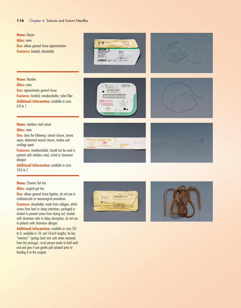
116 Chapter 4 Sutures and Suture Needles
Name: Dexon
Alias: none
Use: allows general tissue approximation
Features: braided; absorbable
Name: Nurolon
Alias: none
Use: approximates general tissue
Features: braided; nonabsorbable; nylon fiber
Additional Information: available in sizes 6-0 to 1
Name: stainless steel suture
Alias: none
Use: does the following: sternal closure, hernia repair, abdominal wound closure, tendon and cartilage repair
Features: nonabsorbable; should not be used inpatients with stainless steel, nickel or chromium allergies
Additional Information: available in sizes 10-0 to 7
Name: Chromic Gut ties
Alias: surgical gut ties
Use: allows general tissue ligation; do not use incardiovascular or neurosurgical procedures
Features: absorbable; made from collagen, whichcomes from beef or sheep intestines; packaged in alcohol to prevent suture from drying out; treatedwith chromium salts to delay absorption; do not usein patients with chromium allergies
Additional Information: available in sizes 3-0to 0; available in 18- and 54-inch lengths; tie has“memory” (springs back into curls when removedfrom the package); scrub person needs to hold eachend and give it one gentle pull outward prior tohanding it to the surgeon
1572_Ch04_107-132.qxd 8/6/09 11:37 AM Page 116
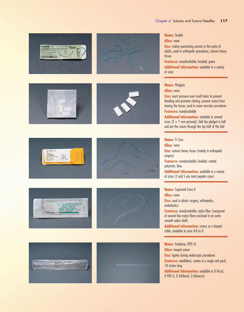
Chapter 4 Sutures and Suture Needles 117
Name: Tevdek
Alias: none
Use: makes pursestring sutures in the aorta ofadults; used in orthopedic procedures; sutures heavytissue
Features: nonabsorbable; braided; green
Additional Information: available in a varietyof sizes
Name: Pledgets
Alias: none
Use: exert pressure over small holes to preventbleeding and promote clotting; prevent suture fromtearing the tissue; used in some vascular procedures
Features: nonabsorbable
Additional Information: available in severalsizes (3 × 7 mm pictured); fold the pledget in halfand put the suture through the top half of the fold
Name: Ti Cron
Alias: none
Use: sutures heavy tissue (mainly in orthopedic surgery)
Features: nonabsorbable; braided; coated polyester; blue
Additional Information: available in a varietyof sizes (2 and 5 are most popular sizes)
Name: Supramid Extra II
Alias: none
Use: used in plastic surgery, orthopedics, endodontics
Features: nonabsorbable; nylon fiber (composedof several fine nylon fibers enclosed in an outersmooth nylon shell)
Additional Information: comes as a looped cable; available in sizes 4-0 to 8
Name: Endoloop (PDS II)
Alias: looped suture
Use: ligates during endoscopic procedures
Features: needleless; comes in a single unit pack;18 inches long
Additional Information: available in 0 Vicryl, 0 PDS II, 0 Ethibond, 0 Monocryl
1572_Ch04_107-132.qxd 8/6/09 11:37 AM Page 117
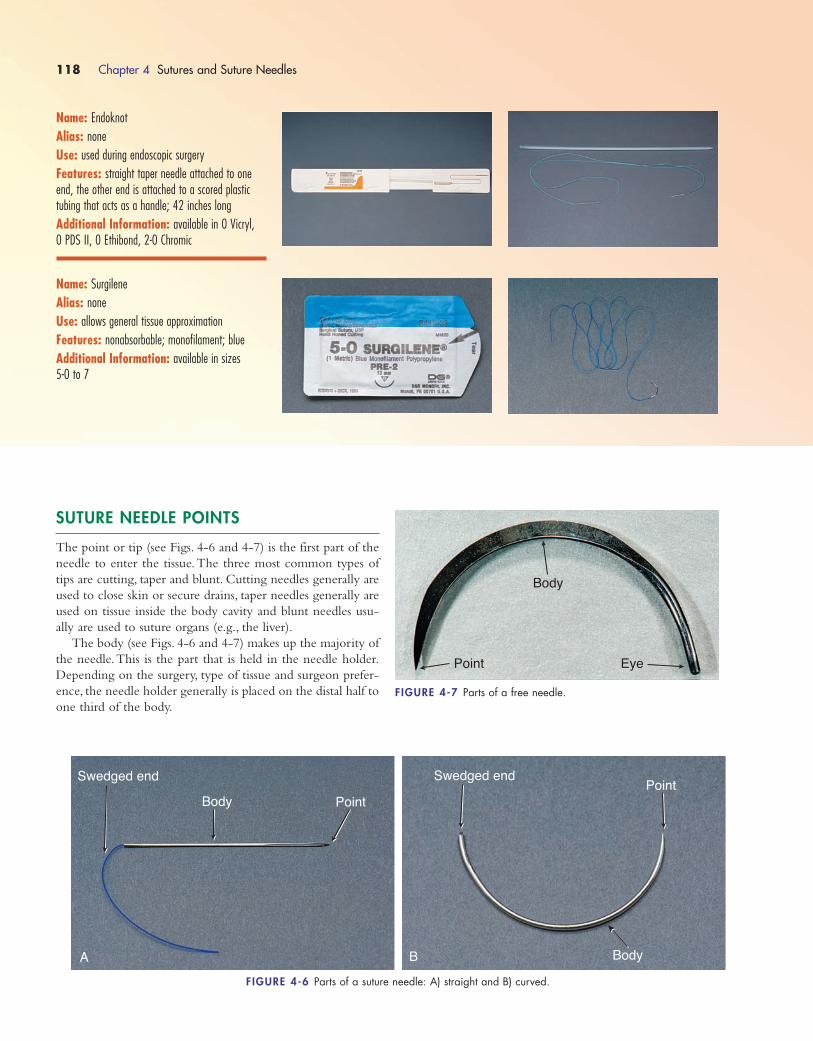
118 Chapter 4 Sutures and Suture Needles
Name: Endoknot
Alias: none
Use: used during endoscopic surgery
Features: straight taper needle attached to oneend, the other end is attached to a scored plastictubing that acts as a handle; 42 inches long
Additional Information: available in 0 Vicryl, 0 PDS II, 0 Ethibond, 2-0 Chromic
Name: Surgilene
Alias: none
Use: allows general tissue approximation
Features: nonabsorbable; monofilament; blue
Additional Information: available in sizes 5-0 to 7
SUTURE NEEDLE POINTS
The point or tip (see Figs. 4-6 and 4-7) is the first part of the
needle to enter the tissue.The three most common types of
tips are cutting, taper and blunt. Cutting needles generally are
used to close skin or secure drains, taper needles generally are
used on tissue inside the body cavity and blunt needles usu-
ally are used to suture organs (e.g., the liver).
The body (see Figs. 4-6 and 4-7) makes up the majority of
the needle.This is the part that is held in the needle holder.
Depending on the surgery, type of tissue and surgeon prefer-
ence, the needle holder generally is placed on the distal half to
one third of the body.
Swedged end
Body
PointBody
Swedged end
Point
FIGURE 4-6 Parts of a suture needle: A) straight and B) curved.
A B
Point
Body
Eye
FIGURE 4-7 Parts of a free needle.
1572_Ch04_107-132.qxd 8/6/09 11:37 AM Page 118
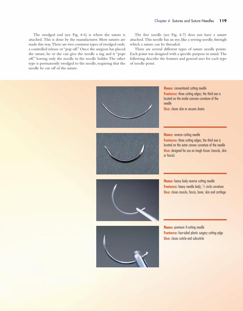
Chapter 4 Sutures and Suture Needles 119
Name: conventional cutting needle
Features: three cutting edges; the third one is located on the inside concave curvature of the needle
Use: closes skin or secures drains
Name: reverse cutting needle
Features: three cutting edges; the third one is located on the outer convex curvature of the needle
Use: designed for use on tough tissue (muscle, skinor fascia)
Name: heavy body reverse cutting needle
Features: heavy needle body; 1⁄2 circle curvature
Use: closes muscle, fascia, bone, skin and cartilage
Name: premium X-cutting needle
Features: four-sided plastic surgery cutting edge
Use: closes cuticle and subcuticle
The swedged end (see Fig. 4-6) is where the suture is
attached. This is done by the manufacturer. Most sutures are
made this way. There are two common types of swedged ends:
a controlled release or “pop off.” Once the surgeon has placed
the suture, he or she can give the needle a tug and it “pops
off,” leaving only the needle in the needle holder.The other
type is permanently swedged to the needle, requiring that the
needle be cut off of the suture.
The free needle (see Fig. 4-7) does not have a suture
attached. This needle has an eye, like a sewing needle, through
which a suture can be threaded.
There are several different types of suture needle points.
Each point was designed with a specific purpose in mind. The
following describe the features and general uses for each type
of needle point.
1572_Ch04_107-132.qxd 8/6/09 11:37 AM Page 119
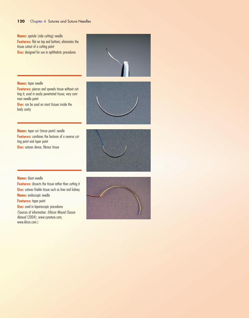
120 Chapter 4 Sutures and Suture Needles
Name: spatula (side cutting) needle
Features: flat on top and bottom; eliminates thetissue cutout of a cutting point
Use: designed for use in ophthalmic procedures
Name: taper needle
Features: pierces and spreads tissue without cut-ting it; used in easily penetrated tissue; very com-mon needle point
Use: can be used on most tissues inside the body cavity
Name: taper cut (trocar point) needle
Features: combines the features of a reverse cut-ting point and taper point
Use: sutures dense, fibrous tissue
Name: blunt needle
Features: dissects the tissue rather than cutting it
Use: sutures friable tissue such as liver and kidney
Name: endoscopic needle
Features: taper point
Use: used in laparoscopic procedures
(Sources of information: Ethicon Wound ClosureManual (2004); www.syneture.com;www.Alcon.com.)
1572_Ch04_107-132.qxd 8/6/09 11:37 AM Page 120
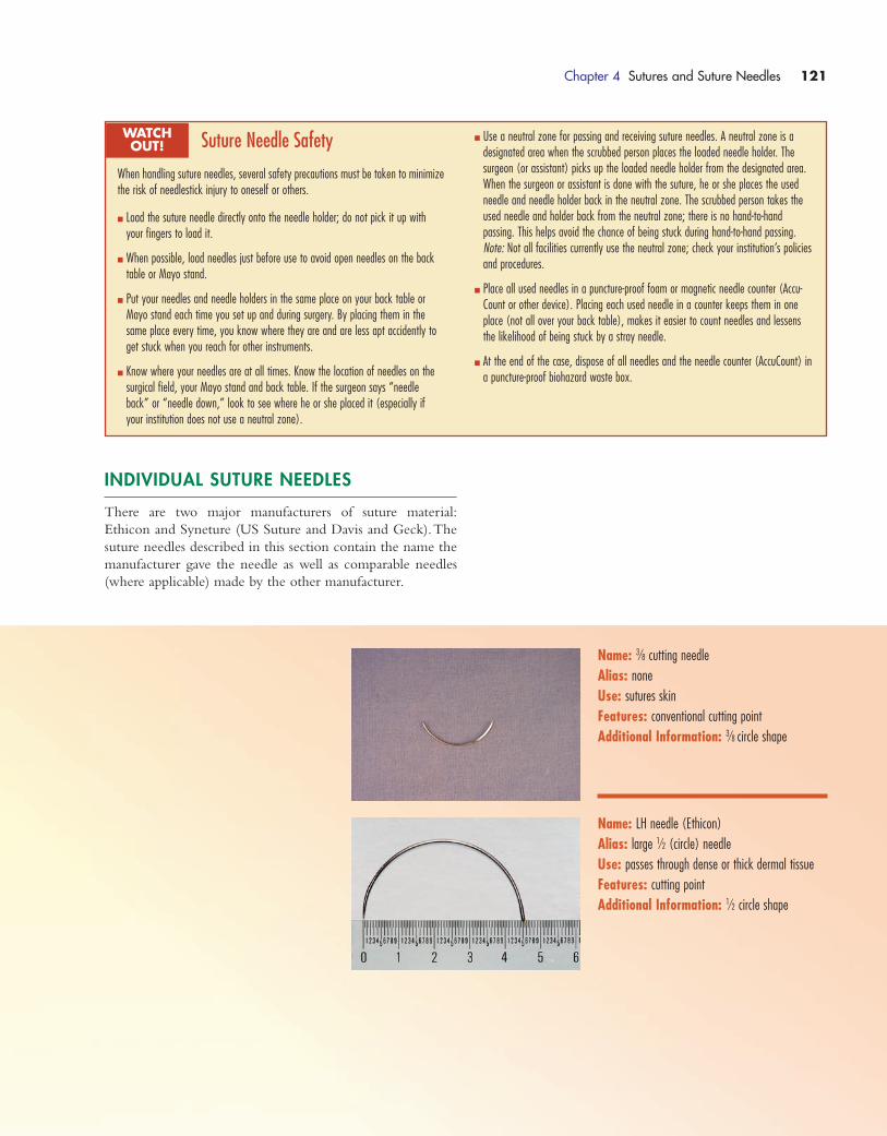
Name: 3⁄8 cutting needle
Alias: none
Use: sutures skin
Features: conventional cutting point
Additional Information: 3⁄8 circle shape
Name: LH needle (Ethicon)
Alias: large 1⁄2 (circle) needle
Use: passes through dense or thick dermal tissue
Features: cutting point
Additional Information: 1⁄2 circle shape
INDIVIDUAL SUTURE NEEDLES
There are two major manufacturers of suture material:
Ethicon and Syneture (US Suture and Davis and Geck).The
suture needles described in this section contain the name the
manufacturer gave the needle as well as comparable needles
(where applicable) made by the other manufacturer.
Chapter 4 Sutures and Suture Needles 121
Suture Needle Safety
When handling suture needles, several safety precautions must be taken to minimizethe risk of needlestick injury to oneself or others.
� Load the suture needle directly onto the needle holder; do not pick it up withyour fingers to load it.
� When possible, load needles just before use to avoid open needles on the backtable or Mayo stand.
� Put your needles and needle holders in the same place on your back table orMayo stand each time you set up and during surgery. By placing them in thesame place every time, you know where they are and are less apt accidently toget stuck when you reach for other instruments.
� Know where your needles are at all times. Know the location of needles on thesurgical field, your Mayo stand and back table. If the surgeon says “needleback” or “needle down,” look to see where he or she placed it (especially ifyour institution does not use a neutral zone).
WATCH OUT!
� Use a neutral zone for passing and receiving suture needles. A neutral zone is a designated area when the scrubbed person places the loaded needle holder. The surgeon (or assistant) picks up the loaded needle holder from the designated area.When the surgeon or assistant is done with the suture, he or she places the usedneedle and needle holder back in the neutral zone. The scrubbed person takes theused needle and holder back from the neutral zone; there is no hand-to-hand passing. This helps avoid the chance of being stuck during hand-to-hand passing.Note: Not all facilities currently use the neutral zone; check your institution’s policiesand procedures.
� Place all used needles in a puncture-proof foam or magnetic needle counter (Accu-Count or other device). Placing each used needle in a counter keeps them in oneplace (not all over your back table), makes it easier to count needles and lessensthe likelihood of being stuck by a stray needle.
� At the end of the case, dispose of all needles and the needle counter (AccuCount) ina puncture-proof biohazard waste box.
1572_Ch04_107-132.qxd 8/6/09 11:37 AM Page 121
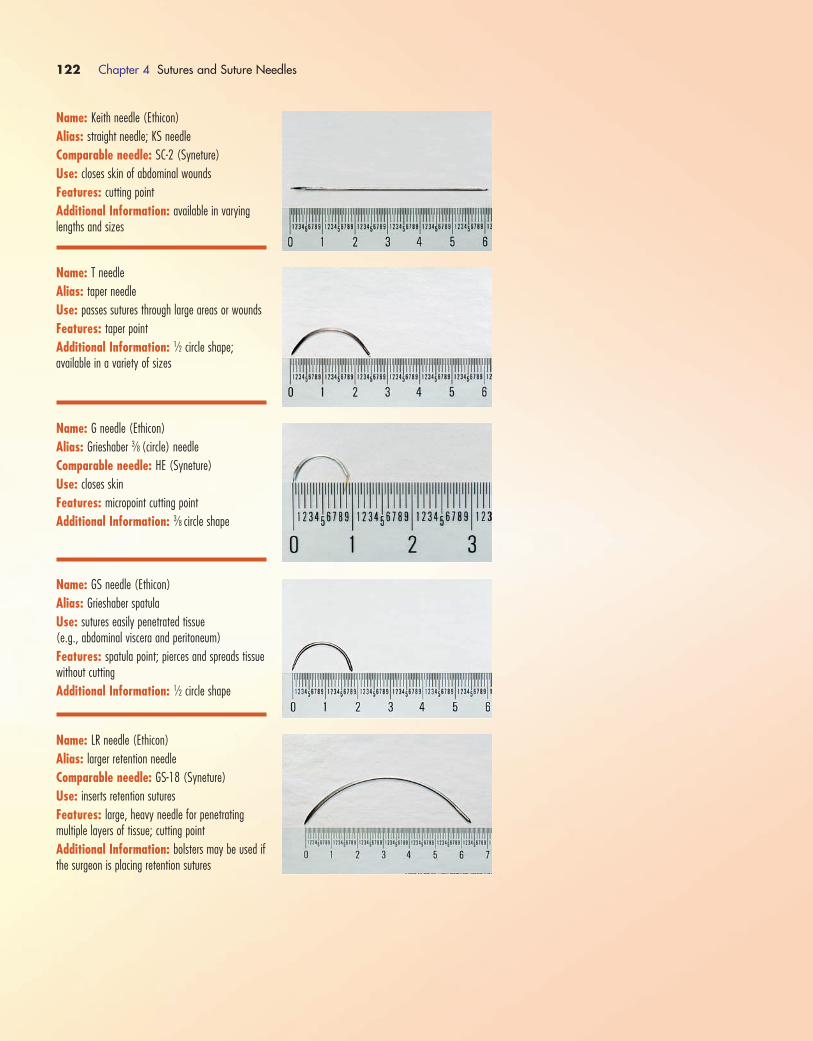
122 Chapter 4 Sutures and Suture Needles
Name: Keith needle (Ethicon)
Alias: straight needle; KS needle
Comparable needle: SC-2 (Syneture)
Use: closes skin of abdominal wounds
Features: cutting point
Additional Information: available in varyinglengths and sizes
Name: T needle
Alias: taper needle
Use: passes sutures through large areas or wounds
Features: taper point
Additional Information: 1⁄2 circle shape; available in a variety of sizes
Name: G needle (Ethicon)
Alias: Grieshaber 3⁄8 (circle) needle
Comparable needle: HE (Syneture)
Use: closes skin
Features: micropoint cutting point
Additional Information: 3⁄8 circle shape
Name: GS needle (Ethicon)
Alias: Grieshaber spatula
Use: sutures easily penetrated tissue (e.g., abdominal viscera and peritoneum)
Features: spatula point; pierces and spreads tissuewithout cutting
Additional Information: 1⁄2 circle shape
Name: LR needle (Ethicon)
Alias: larger retention needle
Comparable needle: GS-18 (Syneture)
Use: inserts retention sutures
Features: large, heavy needle for penetrating multiple layers of tissue; cutting point
Additional Information: bolsters may be used ifthe surgeon is placing retention sutures
1572_Ch04_107-132.qxd 8/6/09 11:37 AM Page 122
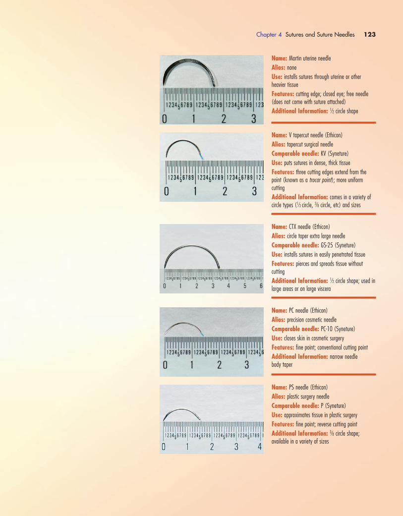
Chapter 4 Sutures and Suture Needles 123
Name: Martin uterine needle
Alias: none
Use: installs sutures through uterine or other heavier tissue
Features: cutting edge; closed eye; free needle(does not come with suture attached)
Additional Information: 1⁄2 circle shape
Name: V tapercut needle (Ethicon)
Alias: tapercut surgical needle
Comparable needle: KV (Syneture)
Use: puts sutures in dense, thick tissue
Features: three cutting edges extend from thepoint (known as a trocar point); more uniform cutting
Additional Information: comes in a variety ofcircle types (1⁄2 circle, 3⁄8 circle, etc) and sizes
Name: CTX needle (Ethicon)
Alias: circle taper extra large needle
Comparable needle: GS-25 (Syneture)
Use: installs sutures in easily penetrated tissue
Features: pierces and spreads tissue without cutting
Additional Information: 1⁄2 circle shape; used inlarge areas or on large viscera
Name: PC needle (Ethicon)
Alias: precision cosmetic needle
Comparable needle: PC-10 (Syneture)
Use: closes skin in cosmetic surgery
Features: fine point; conventional cutting point
Additional Information: narrow needle body taper
Name: PS needle (Ethicon)
Alias: plastic surgery needle
Comparable needle: P (Syneture)
Use: approximates tissue in plastic surgery
Features: fine point; reverse cutting point
Additional Information: 3⁄8 circle shape; available in a variety of sizes
1572_Ch04_107-132.qxd 8/6/09 11:37 AM Page 123
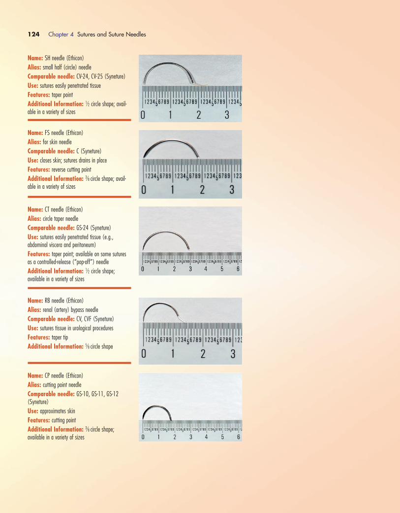
124 Chapter 4 Sutures and Suture Needles
Name: SH needle (Ethicon)
Alias: small half (circle) needle
Comparable needle: CV-24, CV-25 (Syneture)
Use: sutures easily penetrated tissue
Features: taper point
Additional Information: 1⁄2 circle shape; avail-able in a variety of sizes
Name: FS needle (Ethicon)
Alias: for skin needle
Comparable needle: C (Syneture)
Use: closes skin; sutures drains in place
Features: reverse cutting point
Additional Information: 3⁄8 circle shape; avail-able in a variety of sizes
Name: CT needle (Ethicon)
Alias: circle taper needle
Comparable needle: GS-24 (Syneture)
Use: sutures easily penetrated tissue (e.g., abdominal viscera and peritoneum)
Features: taper point; available on some suturesas a controlled-release (“pop-off”) needle
Additional Information: 1⁄2 circle shape; available in a variety of sizes
Name: RB needle (Ethicon)
Alias: renal (artery) bypass needle
Comparable needle: CV, CVF (Syneture)
Use: sutures tissue in urological procedures
Features: taper tip
Additional Information: 5⁄8 circle shape
Name: CP needle (Ethicon)
Alias: cutting point needle
Comparable needle: GS-10, GS-11, GS-12(Syneture)
Use: approximates skin
Features: cutting point
Additional Information: 3⁄8 circle shape; available in a variety of sizes
1572_Ch04_107-132.qxd 8/6/09 11:37 AM Page 124
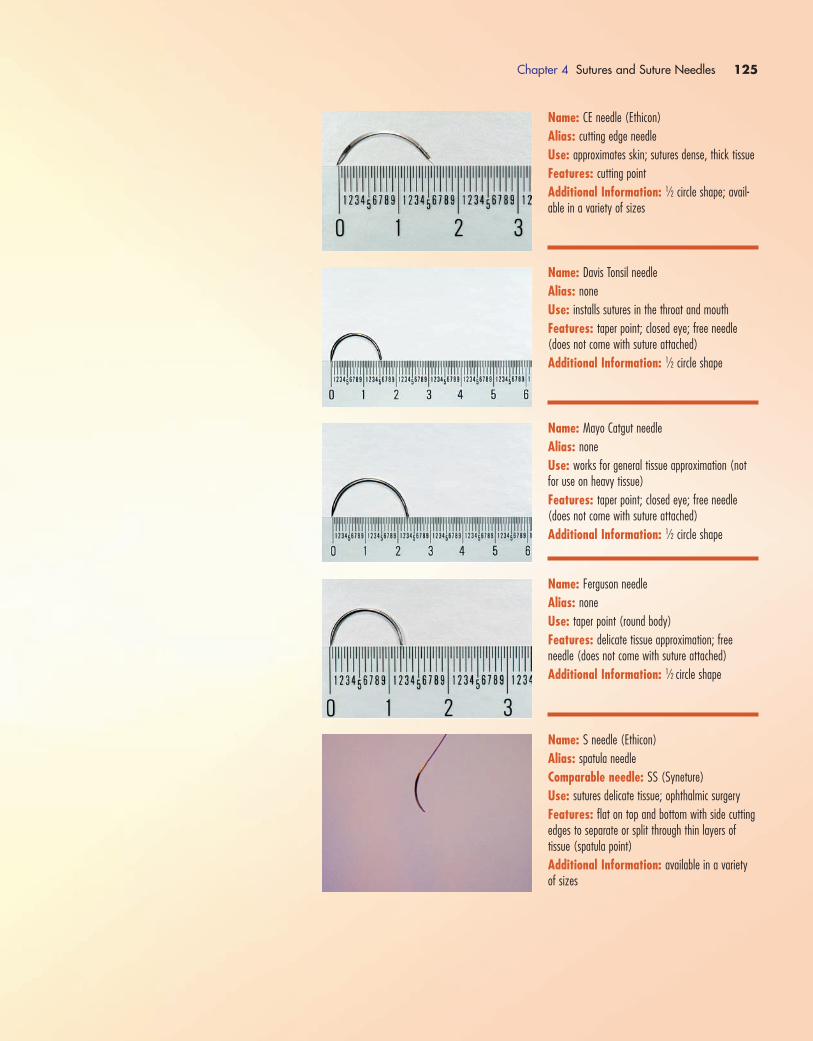
Chapter 4 Sutures and Suture Needles 125
Name: CE needle (Ethicon)
Alias: cutting edge needle
Use: approximates skin; sutures dense, thick tissue
Features: cutting point
Additional Information: 1⁄2 circle shape; avail-able in a variety of sizes
Name: Davis Tonsil needle
Alias: none
Use: installs sutures in the throat and mouth
Features: taper point; closed eye; free needle(does not come with suture attached)
Additional Information: 1⁄2 circle shape
Name: Mayo Catgut needle
Alias: none
Use: works for general tissue approximation (notfor use on heavy tissue)
Features: taper point; closed eye; free needle(does not come with suture attached)
Additional Information: 1⁄2 circle shape
Name: Ferguson needle
Alias: none
Use: taper point (round body)
Features: delicate tissue approximation; free needle (does not come with suture attached)
Additional Information: 1⁄2 circle shape
Name: S needle (Ethicon)
Alias: spatula needle
Comparable needle: SS (Syneture)
Use: sutures delicate tissue; ophthalmic surgery
Features: flat on top and bottom with side cuttingedges to separate or split through thin layers of tissue (spatula point)
Additional Information: available in a varietyof sizes
1572_Ch04_107-132.qxd 8/6/09 11:37 AM Page 125
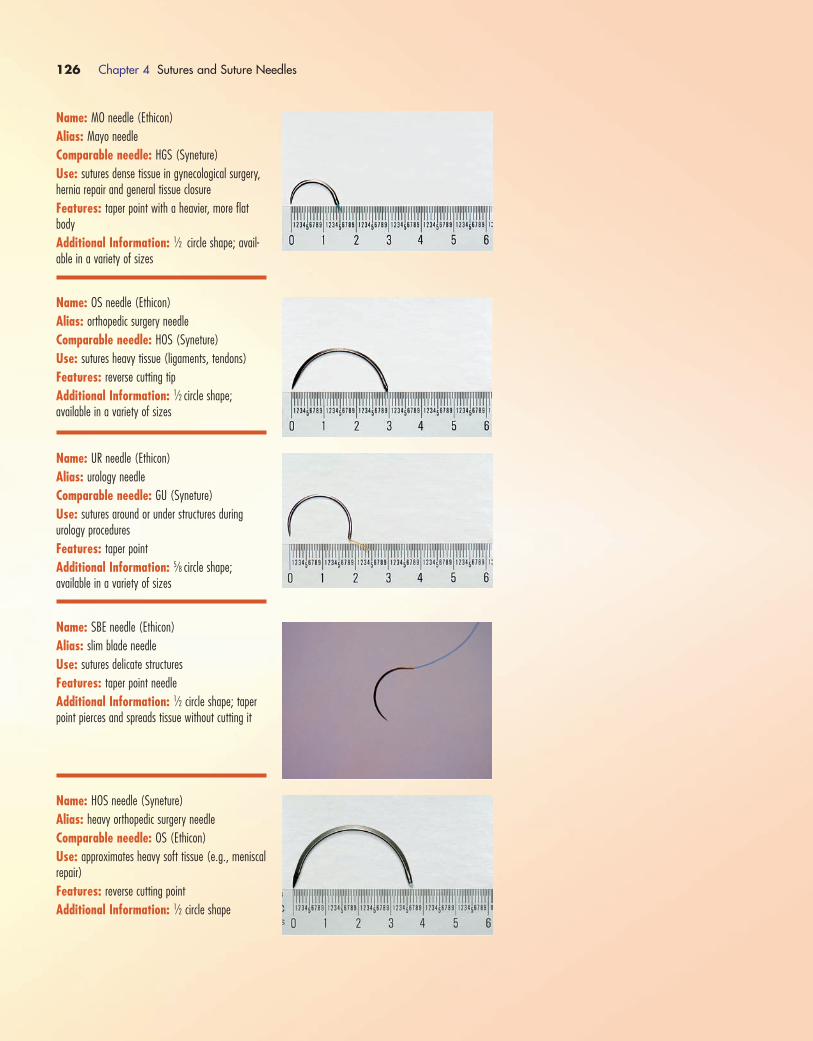
126 Chapter 4 Sutures and Suture Needles
Name: MO needle (Ethicon)
Alias: Mayo needle
Comparable needle: HGS (Syneture)
Use: sutures dense tissue in gynecological surgery,hernia repair and general tissue closure
Features: taper point with a heavier, more flatbody
Additional Information: 1⁄2 circle shape; avail-able in a variety of sizes
Name: OS needle (Ethicon)
Alias: orthopedic surgery needle
Comparable needle: HOS (Syneture)
Use: sutures heavy tissue (ligaments, tendons)
Features: reverse cutting tip
Additional Information: 1⁄2 circle shape; available in a variety of sizes
Name: UR needle (Ethicon)
Alias: urology needle
Comparable needle: GU (Syneture)
Use: sutures around or under structures during urology procedures
Features: taper point
Additional Information: 5⁄8 circle shape; available in a variety of sizes
Name: SBE needle (Ethicon)
Alias: slim blade needle
Use: sutures delicate structures
Features: taper point needle
Additional Information: 1⁄2 circle shape; taperpoint pierces and spreads tissue without cutting it
Name: HOS needle (Syneture)
Alias: heavy orthopedic surgery needle
Comparable needle: OS (Ethicon)
Use: approximates heavy soft tissue (e.g., meniscalrepair)
Features: reverse cutting point
Additional Information: 1⁄2 circle shape
1572_Ch04_107-132.qxd 8/6/09 11:37 AM Page 126
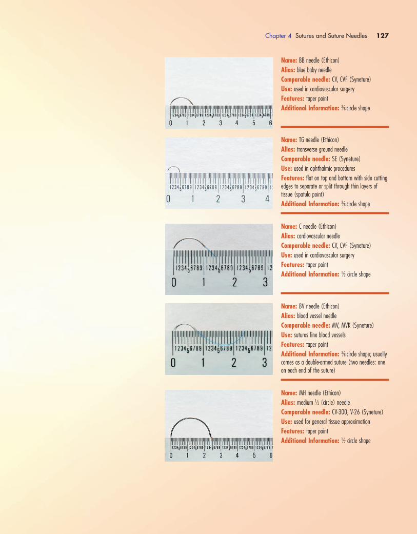
Chapter 4 Sutures and Suture Needles 127
Name: BB needle (Ethicon)
Alias: blue baby needle
Comparable needle: CV, CVF (Syneture)
Use: used in cardiovascular surgery
Features: taper point
Additional Information: 3⁄8 circle shape
Name: TG needle (Ethicon)
Alias: transverse ground needle
Comparable needle: SE (Syneture)
Use: used in ophthalmic procedures
Features: flat on top and bottom with side cuttingedges to separate or split through thin layers of tissue (spatula point)
Additional Information: 3⁄8 circle shape
Name: C needle (Ethicon)
Alias: cardiovascular needle
Comparable needle: CV, CVF (Syneture)
Use: used in cardiovascular surgery
Features: taper point
Additional Information: 1⁄2 circle shape
Name: BV needle (Ethicon)
Alias: blood vessel needle
Comparable needle: MV, MVK (Syneture)
Use: sutures fine blood vessels
Features: taper point
Additional Information: 3⁄8 circle shape; usuallycomes as a double-armed suture (two needles: oneon each end of the suture)
Name: MH needle (Ethicon)
Alias: medium 1⁄2 (circle) needle
Comparable needle: CV-300, V-26 (Syneture)
Use: used for general tissue approximation
Features: taper point
Additional Information: 1⁄2 circle shape
1572_Ch04_107-132.qxd 8/6/09 11:37 AM Page 127
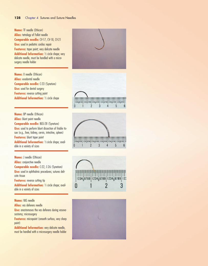
128 Chapter 4 Sutures and Suture Needles
Name: TF needle (Ethicon)
Alias: tetralogy of Fallot needle
Comparable needle: CV-17, CV-18, CV-21
Use: used in pediatric cardiac repair
Features: taper point; very delicate needle
Additional Information: 1⁄2 circle shape; verydelicate needle, must be handled with a micro-surgery needle holder
Name: X needle (Ethicon)
Alias: exodontal needle
Comparable needle: C-23 (Syneture)
Use: used for dental surgery
Features: reverse cutting point
Additional Information: 1⁄2 circle shape
Name: BP needle (Ethicon)
Alias: blunt point needle
Comparable needle: BGS-28 (Syneture)
Use: used to perform blunt dissection of friable tis-sue (e.g., liver, kidney, cervix, intestine, spleen)
Features: blunt taper point
Additional Information: 1⁄2 circle shape; avail-able in a variety of sizes
Name: J needle (Ethicon)
Alias: conjunctive needle
Comparable needle: C-22, C-26 (Syneture)
Use: used in ophthalmic procedures; sutures deli-cate tissue
Features: reverse cutting tip
Additional Information: 1⁄2 circle shape; avail-able in a variety of sizes
Name: VAS needle
Alias: vas deferens needle
Use: anastomoses the vas deferens during vasova-sostomy; microsurgery
Features: micropoint (smooth surface, very sharppoint)
Additional Information: very delicate needle,must be handled with a microsurgery needle holder
1572_Ch04_107-132.qxd 8/6/09 11:37 AM Page 128
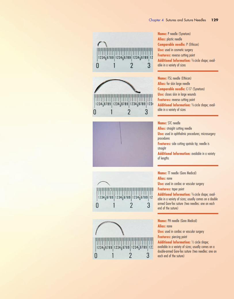
Chapter 4 Sutures and Suture Needles 129
Name: P needle (Syneture)
Alias: plastic needle
Comparable needle: P (Ethicon)
Use: used in cosmetic surgery
Features: reverse cutting point
Additional Information: 3⁄8 circle shape; avail-able in a variety of sizes
Name: FSL needle (Ethicon)
Alias: for skin large needle
Comparable needle: C-17 (Syneture)
Use: closes skin in large wounds
Features: reverse cutting point
Additional Information: 3⁄8 circle shape; avail-able in a variety of sizes
Name: STC needle
Alias: straight cutting needle
Use: used in ophthalmic procedures; microsurgeryprocedures
Features: side cutting spatula tip; needle isstraight
Additional Information: available in a varietyof lengths
Name: TT needle (Gore Medical)
Alias: none
Use: used in cardiac or vascular surgery
Features: taper point
Additional Information: 3⁄8 circle shape; avail-able in a variety of sizes; usually comes on a doublearmed Gore-Tex suture (two needles: one on eachend of the suture)
Name: PH needle (Gore Medical)
Alias: none
Use: used in cardiac or vascular surgery
Features: piercing point
Additional Information: 1⁄2 circle shape; available in a variety of sizes; usually comes on adouble-armed Gore-Tex suture (two needles: one oneach end of the suture)
1572_Ch04_107-132.qxd 8/6/09 11:37 AM Page 129
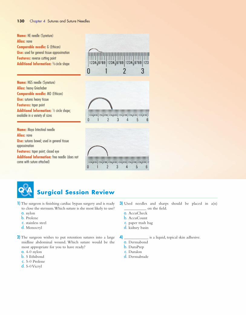
130 Chapter 4 Sutures and Suture Needles
Name: HE needle (Syneture)
Alias: none
Comparable needle: G (Ethicon)
Use: used for general tissue approximation
Features: reverse cutting point
Additional Information: 3⁄8 circle shape
Name: HGS needle (Syneture)
Alias: heavy Grieshaber
Comparable needle: MO (Ethicon)
Use: sutures heavy tissue
Features: taper point
Additional Information: 1⁄2 circle shape; available in a variety of sizes
Name: Mayo Intestinal needle
Alias: none
Use: sutures bowel; used in general tissue approximation
Features: taper point; closed eye
Additional Information: free needle (does notcome with suture attached)
Surgical Session Review
1) The surgeon is finishing cardiac bypass surgery and is ready
to close the sternum.Which suture is she most likely to use?
a. nylon
b. Prolene
c. stainless steel
d. Monocryl
2) The surgeon wishes to put retention sutures into a large
midline abdominal wound. Which suture would be the
most appropriate for you to have ready?
a. 4-0 nylon
b. 5 Ethibond
c. 5-0 Prolene
d. 5-0 Vicryl
3) Used needles and sharps should be placed in a(n)
___________ on the field.
a. AccuCheck
b. AccuCount
c. paper trash bag
d. kidney basin
4) ____________ is a liquid, topical skin adhesive.
a. Dermabond
b. DuraPrep
c. Duralon
d. Dermabrade
1572_Ch04_107-132.qxd 8/6/09 11:37 AM Page 130

5) Which of the following is made of nylon fiber?
a. Monocryl
b. Panacryl
c. Vicryl
d. Ethilon
6) One of the following sutures does NOT have memory,
thus eliminating the need to give it a gentle tug prior to
handing it to the surgeon.Which suture does NOT have
memory?
a. Maxon
b. Prolene
c. Chromic
d. Vicryl
7) During eye surgery, which of the following sutures would
most likely be used?
a. 4 Ethilon
b. 2 Ethilon
c. 2-0 Ethilon
d. 9-0 Ethilon
8) All but one of the following are non-absorbable sutures.
Which one is the absorbable suture?
a. stainless steel
b. Ethilon
c. Vicryl
d. Prolene
9) Another name for a straight needle is a(n) ___________
needle.
a. Kevin
b. Keith
c. LH
d. CT
10) The “PS” in PS needle stands for:
a. pointed straight
b. plain suture
c. plastic surgery
d. pointed suture
11) The surgeon requests a taper point needle.Which of the
following would be appropriate to hand him?
a. CP needle
b. CT needle
c. CE needle
d. OS needle
12) An RB needle is usually used during what type of
procedures?
a. cardiovascular
b. gynecological
c. urological
d. obstetrical
13) An MH needle has a __________ circle shape.
a. 1⁄4
b. 3⁄8
c. 1⁄2
d. 5⁄8
14) The needle designed specifically for suturing in the throat
(tonsils) is a:
a. Davis
b. Ferguson
c. Martin
d. Mayo Catgut
15) The S needle has a ___________ point.
a. cutting
b. taper
c. taper point
d. spatula
16) What type of needle would most likely be used to place
retention sutures into a large abdominal wound?
a. PC
b. LR
c. Martin
d. PS
17) Which of these suture sizes is the heaviest (thickest)?
1. 3
2. 0
3. 2-0
4. 3-0
18) Gore-Tex suture is:
a. monofilament
b. multifilament
c. braided
d. polyfilament
19) All of the following statements about sharps safety are
true except:
a. use a neutral zone when passing sharps
b. know where your needles and sharps are located at all
times
c. load needles as far in advance of use as possible
d. at the end of the case, dispose of all needles in a
puncture-proof container
20) A needle without a suture attached is called a(n)
_________ needle.
a. swedged
b. free
c. orphaned
d. solo
Chapter 4 Sutures and Suture Needles 131
1572_Ch04_107-132.qxd 8/6/09 11:37 AM Page 131

1572_Ch04_107-132.qxd 8/6/09 11:37 AM Page 132

ENDOSCOPIC SURGERY
The root word endo means “within.”The suffix scopy means “to
view with an instrument.” The term endoscopy means “using an
instrument to view within (a body cavity).” Endoscopy is a
general term referring to any procedure that uses a scope to
view inside a body cavity, but there are several terms that are
used to be more specific about which area is being viewed:
� Anoscopy: Viewing inside the anus
� Arthroscopy:Viewing inside a joint
� Bronchoscopy:Viewing inside the lungs (bronchus)
� Colonoscopy:Viewing inside the colon
� Colposcopy:Viewing inside the vagina and cervix
� Cystoscopy:Viewing inside the bladder
� Duodenoscopy:Viewing inside the duodenum
� Esophagoscopy:Viewing inside the esophagus
� Esophagogastroduodenoscopy (EGD):Viewing inside the
esophagus, stomach and duodenum
� Fluoroscopy:Viewing inside organs or tissues using x-ray
� Gastroscopy:Viewing inside the stomach
� Hepatoscopy:Viewing the liver
� Hysteroscopy:Viewing inside the uterus
� Laparoscopy: Viewing inside the abdominal and pelvic
cavities
� Laryngoscopy:Viewing inside the larynx
� Meatoscopy:Viewing inside the urinary meatus
� Mediastinoscopy:Viewing inside the mediastinum
(mediastinal space)
� Pelvioscopy (pelvoscopy):Viewing inside the pelvis
� Pharyngoscopy:Viewing inside the throat
� Rhinoscopy: Viewing inside the nose
� Sigmoidoscopy:Viewing inside the sigmoid colon
� Thoracoscopy (pleuroscopy):Viewing inside the chest
(thorax)
� Tracheoscopy: Viewing inside the trachea
� Ureteroscopy:Viewing inside the ureter
� Urethroscopy:Viewing inside the urethra
When a surgical procedure is performed using a scope and
very small incisions (known as stab wound incisions), it is referred
to as minimally invasive surgery. Minimally invasive surgery came
onto the surgical scene in the 1980s and revolutionized the way
surgery is performed. Today, more and more procedures (in-
cluding gastric bypass and coronary bypass surgeries) are being
performed using minimally invasive techniques.
There are several advantages to minimally invasive surgery,
including smaller incisions (less scarring and less pain), reduced
length of hospital stay, reduced time recovering at home and
less time out of work. The actual time it takes to perform a
minimally invasive procedure in the operating room (OR) is
not shorter than an open procedure, but the advantages of less
pain and disability for the patient as well as less tissue trauma
are reasons to perform the procedure endoscopically.
It must be stated that not every procedure can or should be
performed endoscopically. Some procedures still must be per-
formed as open procedures (e.g., in the presence of a large,
infected gallbladder). Often, it is not known until the surgery
has begun and the surgeon has had a chance to look inside
whether or not the surgery can be safely performed endoscop-
ically. For this reason, every endoscopic case should be set up
with the ability to convert to an open case quickly and easily.
Most of the time, this means setting up two sets of instruments
on the back table: endoscopic instruments and a traditional
stringer of instruments to be used for an open case.
Every minimally invasive procedure requires certain basic
equipment: a video viewing system (TV system), a recording
system (still pictures, videotape or DVD) and a light source for
the camera. Procedures involving the abdomen and pelvis
require a gas (carbon dioxide) insufflating system. Procedures
involving the joints or urinary system require a constant fluid
irrigation system. This is usually normal saline, but for some
urinary procedures, glycine is used.
The following sections of this chapter cover video towers
(including basic troubleshooting), cameras, scopes and equip-
ment (including care and cleaning) and some specifics for
arthroscopic, flexible and rigid scopes.
VIDEO TOWERS
Minimally invasive procedures require the surgeon to be able
to view inside the body cavity where he or she is working.
This is done using a camera with the image projected up onto
a video system (usually a TV system of some sort).
Although the types of video systems vary from institution
to institution, the requirement is the same; scrubbed person-
nel must be able to view clearly and easily what is going on
inside the operative field.The simplest way to accomplish this
is to have two TV towers, one on each side of the patient, to-
ward the head of the bed so that personnel on both sides of
the operative field are looking directly at a monitor (Fig. 5-1).
C H A P T E R 5
Endoscopic Equipment
1572_Ch05_133-148.qxd 8/6/09 11:40 AM Page 133
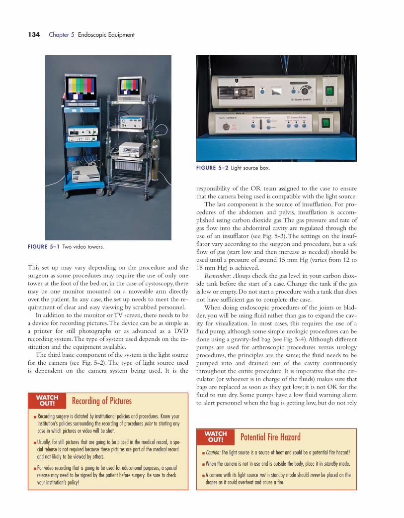
This set up may vary depending on the procedure and the
surgeon as some procedures may require the use of only one
tower at the foot of the bed or, in the case of cystoscopy, there
may be one monitor mounted on a moveable arm directly
over the patient. In any case, the set up needs to meet the re-
quirement of clear and easy viewing by scrubbed personnel.
In addition to the monitor or TV screen, there needs to be
a device for recording pictures.The device can be as simple as
a printer for still photographs or as advanced as a DVD
recording system.The type of system used depends on the in-
stitution and the equipment available.
The third basic component of the system is the light source
for the camera (see Fig. 5-2). The type of light source used
is dependent on the camera system being used. It is the
responsibility of the OR team assigned to the case to ensure
that the camera being used is compatible with the light source.
The last component is the source of insufflation. For pro-
cedures of the abdomen and pelvis, insufflation is accom-
plished using carbon dioxide gas.The gas pressure and rate of
gas flow into the abdominal cavity are regulated through the
use of an insufflator (see Fig. 5-3).The settings on the insuf-
flator vary according to the surgeon and procedure, but a safe
flow of gas (start low and then increase as needed) should be
used until a pressure of around 15 mm Hg (varies from 12 to
18 mm Hg) is achieved.
Remember: Always check the gas level in your carbon diox-
ide tank before the start of a case. Change the tank if the gas
is low or empty.Do not start a procedure with a tank that does
not have sufficient gas to complete the case.
When doing endoscopic procedures of the joints or blad-
der, you will be using fluid rather than gas to expand the cav-
ity for visualization. In most cases, this requires the use of a
fluid pump, although some simple urologic procedures can be
done using a gravity-fed bag (see Fig. 5-4).Although different
pumps are used for arthroscopic procedures versus urology
procedures, the principles are the same; the fluid needs to be
pumped into and drained out of the cavity continuously
throughout the entire procedure. It is imperative that the cir-
culator (or whoever is in charge of the fluids) makes sure that
bags are replaced as soon as they get low; it is not OK for the
fluid to run dry. Some pumps have a low fluid warning alarm
to alert personnel when the bag is getting low, but do not rely
134 Chapter 5 Endoscopic Equipment
FIGURE 5–1 Two video towers.
FIGURE 5–2 Light source box.
Recording of Pictures
� Recording surgery is dictated by institutional policies and procedures. Know yourinstitution’s policies surrounding the recording of procedures prior to starting anycase in which pictures or video will be shot.
� Usually, for still pictures that are going to be placed in the medical record, a spe-cial release is not required because these pictures are part of the medical recordand not likely to be viewed by others.
� For video recording that is going to be used for educational purposes, a specialrelease may need to be signed by the patient before surgery. Be sure to checkyour institution’s policy!
WATCH OUT!
Potential Fire Hazard
� Caution: The light source is a source of heat and could be a potential fire hazard!
� When the camera is not in use and is outside the body, place it in standby mode.
� A camera with its light source not in standby mode should never be placed on thedrapes as it could overheat and cause a fire.
WATCH OUT!
1572_Ch05_133-148.qxd 8/6/09 11:40 AM Page 134
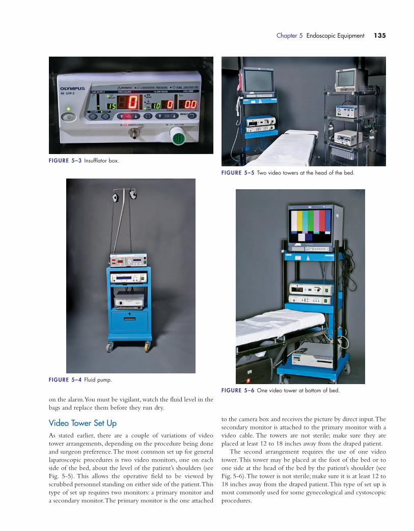
on the alarm.You must be vigilant, watch the fluid level in the
bags and replace them before they run dry.
Video Tower Set UpAs stated earlier, there are a couple of variations of video
tower arrangements, depending on the procedure being done
and surgeon preference.The most common set up for general
laparoscopic procedures is two video monitors, one on each
side of the bed, about the level of the patient’s shoulders (see
Fig. 5-5). This allows the operative field to be viewed by
scrubbed personnel standing on either side of the patient.This
type of set up requires two monitors: a primary monitor and
a secondary monitor.The primary monitor is the one attached
to the camera box and receives the picture by direct input.The
secondary monitor is attached to the primary monitor with a
video cable. The towers are not sterile; make sure they are
placed at least 12 to 18 inches away from the draped patient.
The second arrangement requires the use of one video
tower.This tower may be placed at the foot of the bed or to
one side at the head of the bed by the patient’s shoulder (see
Fig. 5-6).The tower is not sterile; make sure it is at least 12 to
18 inches away from the draped patient.This type of set up is
most commonly used for some gynecological and cystoscopic
procedures.
Chapter 5 Endoscopic Equipment 135
FIGURE 5–3 Insufflator box.
FIGURE 5–4 Fluid pump.
FIGURE 5–5 Two video towers at the head of the bed.
FIGURE 5–6 One video tower at bottom of bed.
1572_Ch05_133-148.qxd 8/6/09 11:40 AM Page 135
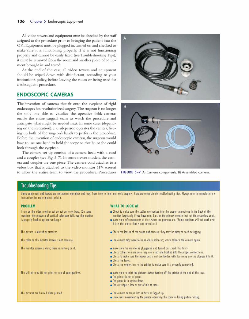
All video towers and equipment must be checked by the staff
assigned to the procedure prior to bringing the patient into the
OR. Equipment must be plugged in, turned on and checked to
make sure it is functioning properly. If it is not functioning
properly and cannot be easily fixed (see Troubleshooting Tips),
it must be removed from the room and another piece of equip-
ment brought in and tested.
At the end of the case, all video towers and equipment
should be wiped down with disinfectant, according to your
institution’s policy, before leaving the room or being used for
a subsequent procedure.
ENDOSCOPIC CAMERAS
The invention of cameras that fit onto the eyepiece of rigid
endoscopes has revolutionized surgery. The surgeon is no longer
the only one able to visualize the operative field; cameras
enable the entire surgical team to watch the procedure and
anticipate what might be needed next. In some cases (depend-
ing on the institution), a scrub person operates the camera, free-
ing up both of the surgeon’s hands to perform the procedure.
Before the invention of endoscopic cameras, the surgeon would
have to use one hand to hold the scope so that he or she could
look through the eyepiece.
The camera set up consists of a camera head with a cord
and a coupler (see Fig. 5-7). In some newer models, the cam-
era and coupler are one piece.The camera cord attaches to a
video box that is attached to the video monitor (TV screen)
to allow the entire team to view the procedure. Procedures
136 Chapter 5 Endoscopic Equipment
Troubleshooting TipsVideo equipment and towers are mechanical machines and may, from time to time, not work properly. Here are some simple troubleshooting tips. Always refer to manufacturer’sinstructions for more in-depth advice.
PROBLEM WHAT TO LOOK ATI turn on the video monitor but do not get color bars. (On some � Check to make sure the cables are hooked into the proper connections in the back of the monitors, the presence of vertical color bars tells you the monitor monitor (especially if you have color bars on the primary monitor but not the secondary one).is properly hooked up and working.) � Make sure all components of the system are powered on. (Some monitors will not work even
if it is the printer that is not turned on.)
The picture is blurred or streaked. � Check the lenses of the scope and camera; they may be dirty or need defogging.
The color on the monitor screen is not accurate. � The camera may need to be re-white balanced; white balance the camera again.
The monitor screen is dark; there is nothing on it. � Make sure the monitor is plugged in and turned on (check this first).� Check cables to make sure they are intact and hooked into the proper connections.� Check to make sure the power box is not overloaded with too many devices plugged into it.� Check the fuses.� Check the connection to the printer to make sure it is properly connected.
The stil l pictures did not print (or are of poor quality). � Make sure to print the pictures before turning off the printer at the end of the case.� The printer is out of paper.� The paper is in upside down.� The cartridge is low or out of ink or toner.
The pictures are blurred when printed. � The camera or scope lens is dirty or fogged up.� There was movement by the person operating the camera during picture taking.
FIGURE 5–7 A) Camera components. B) Assembled camera.
A
B
1572_Ch05_133-148.qxd 8/6/09 11:40 AM Page 136
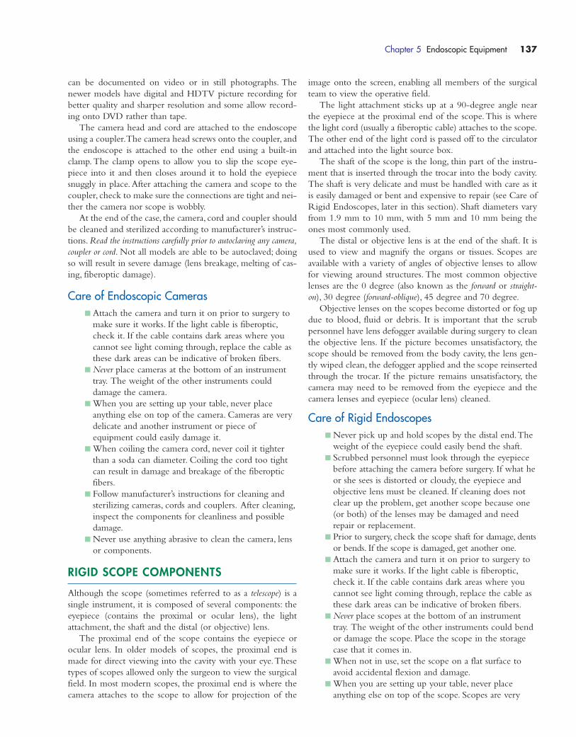
can be documented on video or in still photographs. The
newer models have digital and HDTV picture recording for
better quality and sharper resolution and some allow record-
ing onto DVD rather than tape.
The camera head and cord are attached to the endoscope
using a coupler.The camera head screws onto the coupler, and
the endoscope is attached to the other end using a built-in
clamp. The clamp opens to allow you to slip the scope eye-
piece into it and then closes around it to hold the eyepiece
snuggly in place. After attaching the camera and scope to the
coupler, check to make sure the connections are tight and nei-
ther the camera nor scope is wobbly.
At the end of the case, the camera, cord and coupler should
be cleaned and sterilized according to manufacturer’s instruc-
tions. Read the instructions carefully prior to autoclaving any camera,
coupler or cord. Not all models are able to be autoclaved; doing
so will result in severe damage (lens breakage, melting of cas-
ing, fiberoptic damage).
Care of Endoscopic Cameras� Attach the camera and turn it on prior to surgery to
make sure it works. If the light cable is fiberoptic,
check it. If the cable contains dark areas where you
cannot see light coming through, replace the cable as
these dark areas can be indicative of broken fibers.
� Never place cameras at the bottom of an instrument
tray. The weight of the other instruments could
damage the camera.
� When you are setting up your table, never place
anything else on top of the camera. Cameras are very
delicate and another instrument or piece of
equipment could easily damage it.
� When coiling the camera cord, never coil it tighter
than a soda can diameter. Coiling the cord too tight
can result in damage and breakage of the fiberoptic
fibers.
� Follow manufacturer’s instructions for cleaning and
sterilizing cameras, cords and couplers. After cleaning,
inspect the components for cleanliness and possible
damage.
� Never use anything abrasive to clean the camera, lens
or components.
RIGID SCOPE COMPONENTS
Although the scope (sometimes referred to as a telescope) is a
single instrument, it is composed of several components: the
eyepiece (contains the proximal or ocular lens), the light
attachment, the shaft and the distal (or objective) lens.
The proximal end of the scope contains the eyepiece or
ocular lens. In older models of scopes, the proximal end is
made for direct viewing into the cavity with your eye.These
types of scopes allowed only the surgeon to view the surgical
field. In most modern scopes, the proximal end is where the
camera attaches to the scope to allow for projection of the
image onto the screen, enabling all members of the surgical
team to view the operative field.
The light attachment sticks up at a 90-degree angle near
the eyepiece at the proximal end of the scope.This is where
the light cord (usually a fiberoptic cable) attaches to the scope.
The other end of the light cord is passed off to the circulator
and attached into the light source box.
The shaft of the scope is the long, thin part of the instru-
ment that is inserted through the trocar into the body cavity.
The shaft is very delicate and must be handled with care as it
is easily damaged or bent and expensive to repair (see Care of
Rigid Endoscopes, later in this section). Shaft diameters vary
from 1.9 mm to 10 mm, with 5 mm and 10 mm being the
ones most commonly used.
The distal or objective lens is at the end of the shaft. It is
used to view and magnify the organs or tissues. Scopes are
available with a variety of angles of objective lenses to allow
for viewing around structures. The most common objective
lenses are the 0 degree (also known as the forward or straight-
on), 30 degree (forward-oblique), 45 degree and 70 degree.
Objective lenses on the scopes become distorted or fog up
due to blood, fluid or debris. It is important that the scrub
personnel have lens defogger available during surgery to clean
the objective lens. If the picture becomes unsatisfactory, the
scope should be removed from the body cavity, the lens gen-
tly wiped clean, the defogger applied and the scope reinserted
through the trocar. If the picture remains unsatisfactory, the
camera may need to be removed from the eyepiece and the
camera lenses and eyepiece (ocular lens) cleaned.
Care of Rigid Endoscopes� Never pick up and hold scopes by the distal end.The
weight of the eyepiece could easily bend the shaft.
� Scrubbed personnel must look through the eyepiece
before attaching the camera before surgery. If what he
or she sees is distorted or cloudy, the eyepiece and
objective lens must be cleaned. If cleaning does not
clear up the problem, get another scope because one
(or both) of the lenses may be damaged and need
repair or replacement.
� Prior to surgery, check the scope shaft for damage, dents
or bends. If the scope is damaged, get another one.
� Attach the camera and turn it on prior to surgery to
make sure it works. If the light cable is fiberoptic,
check it. If the cable contains dark areas where you
cannot see light coming through, replace the cable as
these dark areas can be indicative of broken fibers.
� Never place scopes at the bottom of an instrument
tray. The weight of the other instruments could bend
or damage the scope. Place the scope in the storage
case that it comes in.
� When not in use, set the scope on a flat surface to
avoid accidental flexion and damage.
� When you are setting up your table, never place
anything else on top of the scope. Scopes are very
Chapter 5 Endoscopic Equipment 137
1572_Ch05_133-148.qxd 8/6/09 11:40 AM Page 137
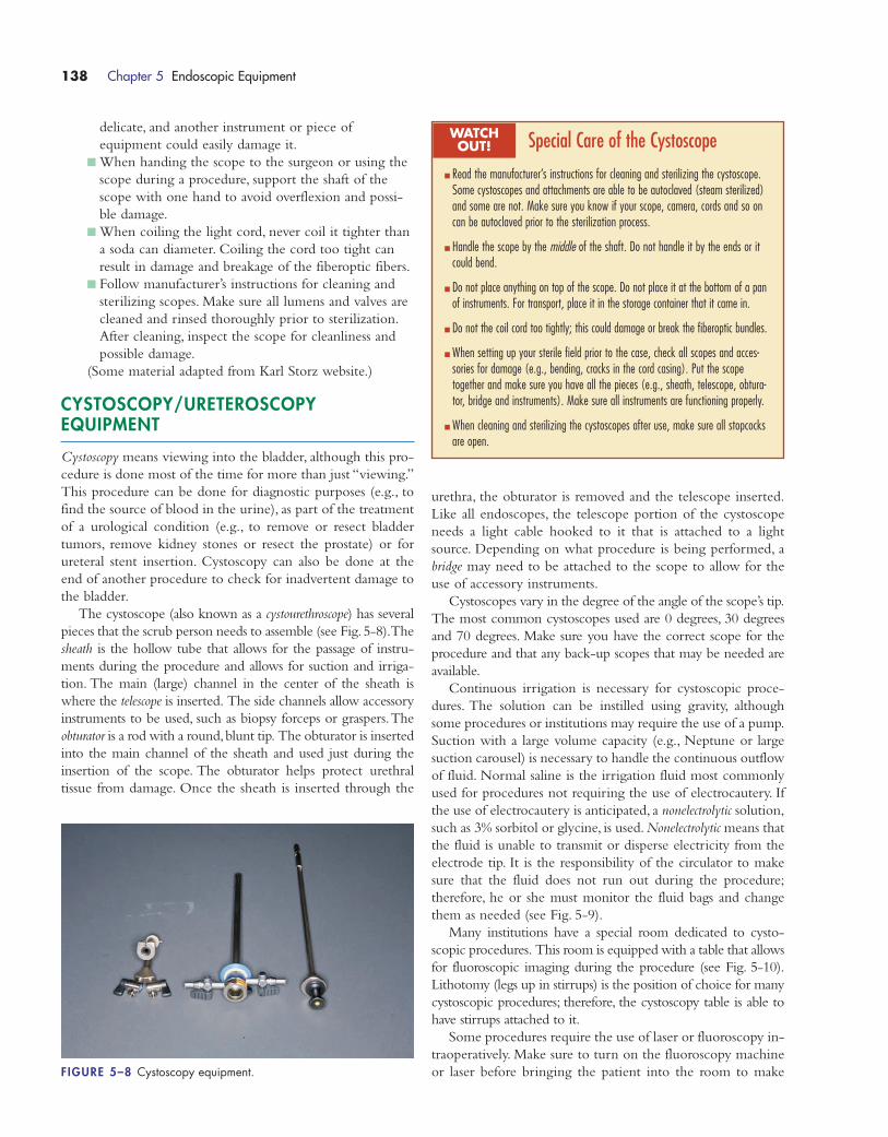
delicate, and another instrument or piece of
equipment could easily damage it.
� When handing the scope to the surgeon or using the
scope during a procedure, support the shaft of the
scope with one hand to avoid overflexion and possi-
ble damage.
� When coiling the light cord, never coil it tighter than
a soda can diameter. Coiling the cord too tight can
result in damage and breakage of the fiberoptic fibers.
� Follow manufacturer’s instructions for cleaning and
sterilizing scopes. Make sure all lumens and valves are
cleaned and rinsed thoroughly prior to sterilization.
After cleaning, inspect the scope for cleanliness and
possible damage.
(Some material adapted from Karl Storz website.)
CYSTOSCOPY/URETEROSCOPYEQUIPMENT
Cystoscopy means viewing into the bladder, although this pro-
cedure is done most of the time for more than just “viewing.”
This procedure can be done for diagnostic purposes (e.g., to
find the source of blood in the urine), as part of the treatment
of a urological condition (e.g., to remove or resect bladder
tumors, remove kidney stones or resect the prostate) or for
ureteral stent insertion. Cystoscopy can also be done at the
end of another procedure to check for inadvertent damage to
the bladder.
The cystoscope (also known as a cystourethroscope) has several
pieces that the scrub person needs to assemble (see Fig.5-8).The
sheath is the hollow tube that allows for the passage of instru-
ments during the procedure and allows for suction and irriga-
tion. The main (large) channel in the center of the sheath is
where the telescope is inserted. The side channels allow accessory
instruments to be used, such as biopsy forceps or graspers.The
obturator is a rod with a round,blunt tip. The obturator is inserted
into the main channel of the sheath and used just during the
insertion of the scope. The obturator helps protect urethral
tissue from damage. Once the sheath is inserted through the
urethra, the obturator is removed and the telescope inserted.
Like all endoscopes, the telescope portion of the cystoscope
needs a light cable hooked to it that is attached to a light
source. Depending on what procedure is being performed, a
bridge may need to be attached to the scope to allow for the
use of accessory instruments.
Cystoscopes vary in the degree of the angle of the scope’s tip.
The most common cystoscopes used are 0 degrees, 30 degrees
and 70 degrees. Make sure you have the correct scope for the
procedure and that any back-up scopes that may be needed are
available.
Continuous irrigation is necessary for cystoscopic proce-
dures. The solution can be instilled using gravity, although
some procedures or institutions may require the use of a pump.
Suction with a large volume capacity (e.g., Neptune or large
suction carousel) is necessary to handle the continuous outflow
of fluid. Normal saline is the irrigation fluid most commonly
used for procedures not requiring the use of electrocautery. If
the use of electrocautery is anticipated, a nonelectrolytic solution,
such as 3% sorbitol or glycine, is used. Nonelectrolytic means that
the fluid is unable to transmit or disperse electricity from the
electrode tip. It is the responsibility of the circulator to make
sure that the fluid does not run out during the procedure;
therefore, he or she must monitor the fluid bags and change
them as needed (see Fig. 5-9).
Many institutions have a special room dedicated to cysto-
scopic procedures. This room is equipped with a table that allows
for fluoroscopic imaging during the procedure (see Fig. 5-10).
Lithotomy (legs up in stirrups) is the position of choice for many
cystoscopic procedures; therefore, the cystoscopy table is able to
have stirrups attached to it.
Some procedures require the use of laser or fluoroscopy in-
traoperatively. Make sure to turn on the fluoroscopy machine
or laser before bringing the patient into the room to make
138 Chapter 5 Endoscopic Equipment
FIGURE 5–8 Cystoscopy equipment.
Special Care of the Cystoscope
� Read the manufacturer’s instructions for cleaning and sterilizing the cystoscope.Some cystoscopes and attachments are able to be autoclaved (steam sterilized)and some are not. Make sure you know if your scope, camera, cords and so oncan be autoclaved prior to the sterilization process.
� Handle the scope by the middle of the shaft. Do not handle it by the ends or itcould bend.
� Do not place anything on top of the scope. Do not place it at the bottom of a panof instruments. For transport, place it in the storage container that it came in.
� Do not the coil cord too tightly; this could damage or break the fiberoptic bundles.
� When setting up your sterile field prior to the case, check all scopes and acces-sories for damage (e.g., bending, cracks in the cord casing). Put the scope together and make sure you have all the pieces (e.g., sheath, telescope, obtura-tor, bridge and instruments). Make sure all instruments are functioning properly.
� When cleaning and sterilizing the cystoscopes after use, make sure all stopcocksare open.
WATCH OUT!
1572_Ch05_133-148.qxd 8/6/09 11:40 AM Page 138
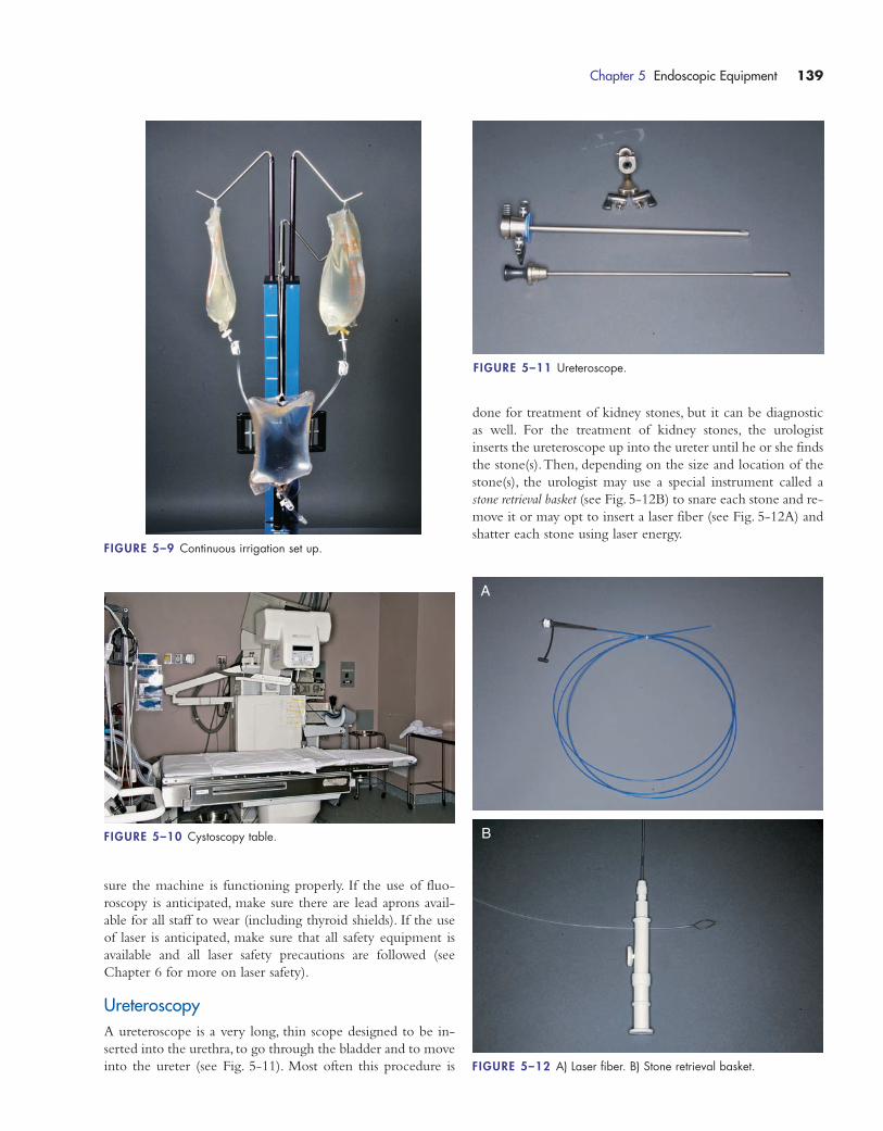
sure the machine is functioning properly. If the use of fluo-
roscopy is anticipated, make sure there are lead aprons avail-
able for all staff to wear (including thyroid shields). If the use
of laser is anticipated, make sure that all safety equipment is
available and all laser safety precautions are followed (see
Chapter 6 for more on laser safety).
UreteroscopyA ureteroscope is a very long, thin scope designed to be in-
serted into the urethra, to go through the bladder and to move
into the ureter (see Fig. 5-11). Most often this procedure is
done for treatment of kidney stones, but it can be diagnostic
as well. For the treatment of kidney stones, the urologist
inserts the ureteroscope up into the ureter until he or she finds
the stone(s).Then, depending on the size and location of the
stone(s), the urologist may use a special instrument called a
stone retrieval basket (see Fig. 5-12B) to snare each stone and re-
move it or may opt to insert a laser fiber (see Fig. 5-12A) and
shatter each stone using laser energy.
Chapter 5 Endoscopic Equipment 139
FIGURE 5–9 Continuous irrigation set up.
FIGURE 5–11 Ureteroscope.
FIGURE 5–10 Cystoscopy table.
FIGURE 5–12 A) Laser fiber. B) Stone retrieval basket.
A
B
1572_Ch05_133-148.qxd 8/6/09 11:40 AM Page 139
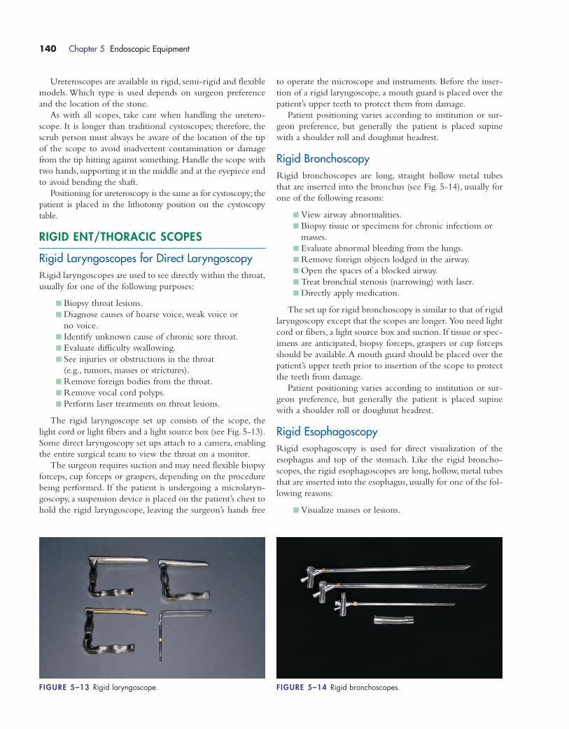
Ureteroscopes are available in rigid, semi-rigid and flexible
models. Which type is used depends on surgeon preference
and the location of the stone.
As with all scopes, take care when handling the uretero-
scope. It is longer than traditional cystoscopes; therefore, the
scrub person must always be aware of the location of the tip
of the scope to avoid inadvertent contamination or damage
from the tip hitting against something. Handle the scope with
two hands, supporting it in the middle and at the eyepiece end
to avoid bending the shaft.
Positioning for ureteroscopy is the same as for cystoscopy; the
patient is placed in the lithotomy position on the cystoscopy
table.
RIGID ENT/THORACIC SCOPES
Rigid Laryngoscopes for Direct LaryngoscopyRigid laryngoscopes are used to see directly within the throat,
usually for one of the following purposes:
� Biopsy throat lesions.
� Diagnose causes of hoarse voice, weak voice or
no voice.
� Identify unknown cause of chronic sore throat.
� Evaluate difficulty swallowing.
� See injuries or obstructions in the throat
(e.g., tumors, masses or strictures).
� Remove foreign bodies from the throat.
� Remove vocal cord polyps.
� Perform laser treatments on throat lesions.
The rigid laryngoscope set up consists of the scope, the
light cord or light fibers and a light source box (see Fig. 5-13).
Some direct laryngoscopy set ups attach to a camera, enabling
the entire surgical team to view the throat on a monitor.
The surgeon requires suction and may need flexible biopsy
forceps, cup forceps or graspers, depending on the procedure
being performed. If the patient is undergoing a microlaryn-
goscopy, a suspension device is placed on the patient’s chest to
hold the rigid laryngoscope, leaving the surgeon’s hands free
to operate the microscope and instruments. Before the inser-
tion of a rigid laryngoscope, a mouth guard is placed over the
patient’s upper teeth to protect them from damage.
Patient positioning varies according to institution or sur-
geon preference, but generally the patient is placed supine
with a shoulder roll and doughnut headrest.
Rigid BronchoscopyRigid bronchoscopes are long, straight hollow metal tubes
that are inserted into the bronchus (see Fig. 5-14), usually for
one of the following reasons:
� View airway abnormalities.
� Biopsy tissue or specimens for chronic infections or
masses.
� Evaluate abnormal bleeding from the lungs.
� Remove foreign objects lodged in the airway.
� Open the spaces of a blocked airway.
� Treat bronchial stenosis (narrowing) with laser.
� Directly apply medication.
The set up for rigid bronchoscopy is similar to that of rigid
laryngoscopy except that the scopes are longer. You need light
cord or fibers, a light source box and suction. If tissue or spec-
imens are anticipated, biopsy forceps, graspers or cup forceps
should be available.A mouth guard should be placed over the
patient’s upper teeth prior to insertion of the scope to protect
the teeth from damage.
Patient positioning varies according to institution or sur-
geon preference, but generally the patient is placed supine
with a shoulder roll or doughnut headrest.
Rigid EsophagoscopyRigid esophagoscopy is used for direct visualization of the
esophagus and top of the stomach. Like the rigid broncho-
scopes, the rigid esophagoscopes are long, hollow, metal tubes
that are inserted into the esophagus, usually for one of the fol-
lowing reasons:
� Visualize masses or lesions.
140 Chapter 5 Endoscopic Equipment
FIGURE 5–13 Rigid laryngoscope. FIGURE 5–14 Rigid bronchoscopes.
1572_Ch05_133-148.qxd 8/6/09 11:40 AM Page 140
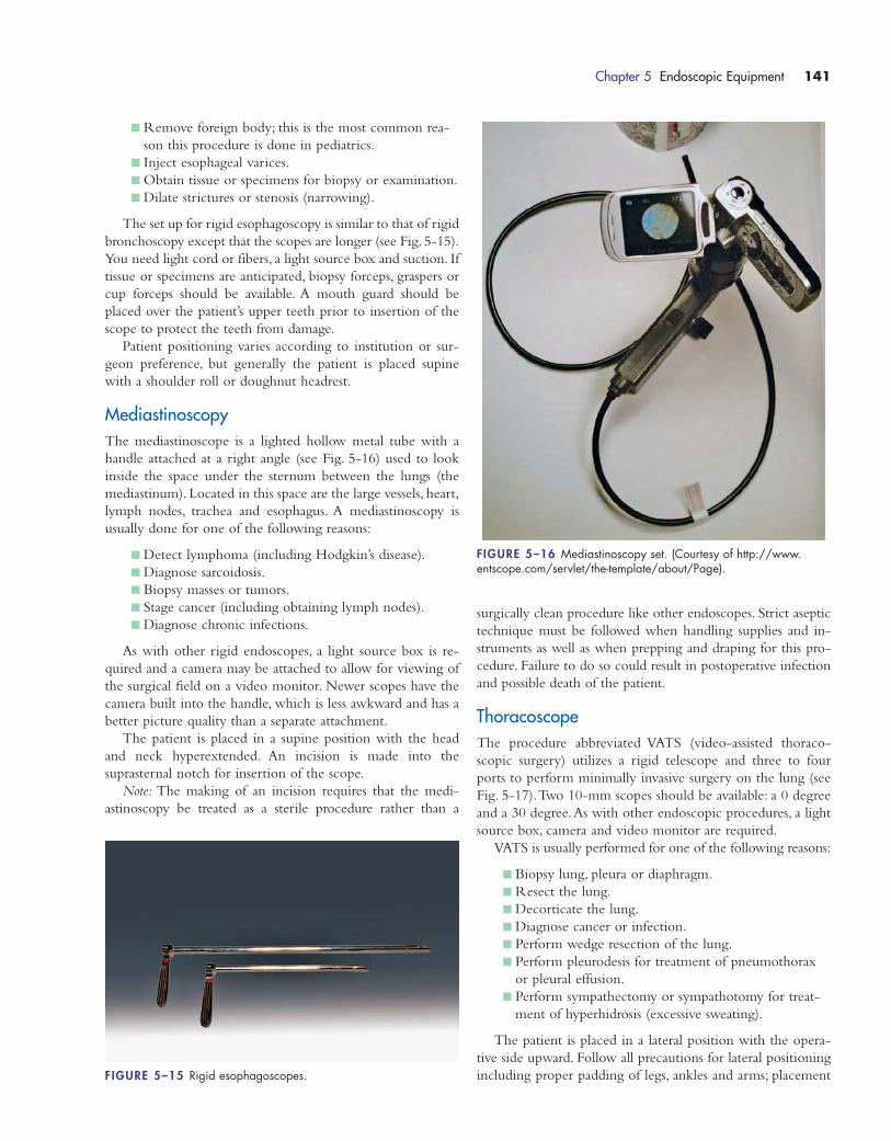
� Remove foreign body; this is the most common rea-
son this procedure is done in pediatrics.
� Inject esophageal varices.
� Obtain tissue or specimens for biopsy or examination.
� Dilate strictures or stenosis (narrowing).
The set up for rigid esophagoscopy is similar to that of rigid
bronchoscopy except that the scopes are longer (see Fig. 5-15).
You need light cord or fibers, a light source box and suction. If
tissue or specimens are anticipated, biopsy forceps, graspers or
cup forceps should be available. A mouth guard should be
placed over the patient’s upper teeth prior to insertion of the
scope to protect the teeth from damage.
Patient positioning varies according to institution or sur-
geon preference, but generally the patient is placed supine
with a shoulder roll or doughnut headrest.
MediastinoscopyThe mediastinoscope is a lighted hollow metal tube with a
handle attached at a right angle (see Fig. 5-16) used to look
inside the space under the sternum between the lungs (the
mediastinum). Located in this space are the large vessels, heart,
lymph nodes, trachea and esophagus. A mediastinoscopy is
usually done for one of the following reasons:
� Detect lymphoma (including Hodgkin’s disease).
� Diagnose sarcoidosis.
� Biopsy masses or tumors.
� Stage cancer (including obtaining lymph nodes).
� Diagnose chronic infections.
As with other rigid endoscopes, a light source box is re-
quired and a camera may be attached to allow for viewing of
the surgical field on a video monitor. Newer scopes have the
camera built into the handle, which is less awkward and has a
better picture quality than a separate attachment.
The patient is placed in a supine position with the head
and neck hyperextended. An incision is made into the
suprasternal notch for insertion of the scope.
Note: The making of an incision requires that the medi-
astinoscopy be treated as a sterile procedure rather than a
surgically clean procedure like other endoscopes. Strict aseptic
technique must be followed when handling supplies and in-
struments as well as when prepping and draping for this pro-
cedure. Failure to do so could result in postoperative infection
and possible death of the patient.
ThoracoscopeThe procedure abbreviated VATS (video-assisted thoraco-
scopic surgery) utilizes a rigid telescope and three to four
ports to perform minimally invasive surgery on the lung (see
Fig. 5-17).Two 10-mm scopes should be available: a 0 degree
and a 30 degree.As with other endoscopic procedures, a light
source box, camera and video monitor are required.
VATS is usually performed for one of the following reasons:
� Biopsy lung, pleura or diaphragm.
� Resect the lung.
� Decorticate the lung.
� Diagnose cancer or infection.
� Perform wedge resection of the lung.
� Perform pleurodesis for treatment of pneumothorax
or pleural effusion.
� Perform sympathectomy or sympathotomy for treat-
ment of hyperhidrosis (excessive sweating).
The patient is placed in a lateral position with the opera-
tive side upward. Follow all precautions for lateral positioning
including proper padding of legs, ankles and arms; placement
Chapter 5 Endoscopic Equipment 141
FIGURE 5–15 Rigid esophagoscopes.
FIGURE 5–16 Mediastinoscopy set. (Courtesy of http://www.entscope.com/servlet/the-template/about/Page).
1572_Ch05_133-148.qxd 8/6/09 11:40 AM Page 141
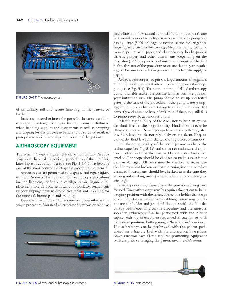
of an axillary roll and secure fastening of the patient to
the bed.
Incisions are used to insert the ports for the camera and in-
struments; therefore, strict aseptic technique must be followed
when handling supplies and instruments as well as prepping
and draping for this procedure. Failure to do so could result in
postoperative infection and possible death of the patient.
ARTHROSCOPY EQUIPMENT
The term arthroscopy means to look within a joint. Arthro-
scopes can be used to perform procedures of the shoulder,
knee, hip, elbow, wrist and ankle (see Fig. 5-18). It has become
one of the most common orthopedic procedures performed.
Arthroscopies are performed to diagnose and repair injury
to a joint. Some of the most common arthroscopic procedures
include ligament, tendon and cartilage repair; ligament re-
placement; foreign body removal; chondroplasty; rotator cuff
surgery; impingement syndrome treatment and searching for
the cause of chronic pain and swelling.
Equipment set up is much the same as for any other endo-
scopic procedure. You need an arthroscope, trocars or cannulas
(including an inflow cannula to instill fluid into the joint), one
or two video monitors, a light source, arthroscopy pump and
tubing, large (3000 cc) bags of normal saline for irrigation,
large capacity suction device (e.g., Neptune or jug suction),
camera, printer with paper, and electrocautery, hooks, probes,
shavers, graspers and other instruments (depending on the
procedure). All equipment and instruments must be checked
before the start of the procedure to ensure that they are work-
ing. Make sure to check the printer for an adequate supply of
paper.
Arthroscopic surgery requires a large amount of irrigation
fluid.The fluid is pumped into the joint using an arthroscopy
pump (see Fig. 5-4). There are many models of arthroscopy
pumps available; make sure you are familiar with the pump(s)
your institution uses.The pump should be set up and tested
prior to the start of the procedure. If the pump is not pump-
ing fluid properly, check the tubing to make sure it is inserted
correctly and does not have a kink in it. If the pump still fails
to pump properly, get another pump.
It is the responsibility of the circulator to keep an eye on
the fluid level in the irrigation bag. Fluid should never be
allowed to run out. Newer pumps have an alarm that signals a
low fluid level, but do not rely solely on the alarm. Keep an
eye on the fluid level and change the bag before it runs out.
It is the responsibility of the scrub person to check the
arthroscope (see Fig. 5-19) and camera to make sure the pic-
ture is clear and that the lens or fibers are not broken or
cracked.The scope should be checked to make sure it is not
bent or damaged. All cords must be checked to make sure
the fibers are not broken or that the casing is not cracked or
damaged. Instruments should be checked to make sure they
are in good working order (not difficult to open or close, not
sticking).
Patient positioning depends on the procedure being per-
formed. Knee arthroscopy usually requires the patient to be in
a supine position with the affected knee in a holder that keeps
it bent (e.g., knee-crutch stirrup), although some surgeons do
not use the holder and just bend the knee with the foot flat
on the bed. Depending on the procedure and the surgeon,
shoulder arthroscopy can be performed with the patient
supine with the affected arm suspended in traction or with
the patient positioned sitting using a “beach chair” positioner.
Hip arthroscopy can be performed with the patient posi-
tioned on a fracture bed, with the affected leg in traction.
Make sure you have all the required positioning equipment
available prior to bringing the patient into the OR room.
142 Chapter 5 Endoscopic Equipment
FIGURE 5–17 Thoracoscopy set.
FIGURE 5–18 Shaver and arthroscopic instruments. FIGURE 5–19 Arthroscope.
1572_Ch05_133-148.qxd 8/6/09 11:40 AM Page 142
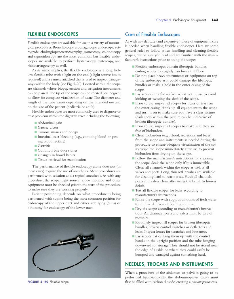
FLEXIBLE ENDOSCOPES
Flexible endoscopes are available for use in a variety of nonsur-
gical procedures.Bronchoscopy, esophagoscopy, endoscopic ret-
rograde cholangiopancreaticography, gastroscopy, colonoscopy
and sigmoidoscopy are the most common, but flexible endo-
scopes are available to perform hysteroscopy, cystoscopy, and
rhinolaryngoscopy as well.
As its name implies, the flexible endoscope is a long, hol-
low, flexible tube with a light on the end (a light source box is
required) and a camera attached that is used to inspect passage-
ways within the body (see Fig. 5-20). Located within the scope
are channels where biopsy, suction and irrigation instruments
can be passed.The tip of the scope can be rotated 360 degrees
to allow for complete visualization of tissue.The diameter and
length of the tube varies depending on the intended use and
on the size of the patient (pediatric or adult).
Flexible endoscopies are most commonly used to diagnose or
treat problems within the digestive tract including the following:
� Abdominal pain
� Gastric ulcers
� Tumors, masses and polyps
� Intestinal tract bleeding (e.g., vomiting blood or pass-
ing blood rectally)
� Gastritis
� Common bile duct stones
� Changes in bowel habits
� Tissue retrieval for examination
The performance of flexible endoscopy alone does not (in
most cases) require the use of anesthesia. Most procedures are
performed with sedation and a topical anesthetic.As with any
procedure, the scope, light source, video monitor and other
equipment must be checked prior to the start of the procedure
to make sure they are working properly.
Patient positioning depends on what procedure is being
performed, with supine being the most common position for
endoscopy of the upper tract and either side lying (Sims) or
lithotomy for endoscopy of the lower tract.
Care of Flexible EndoscopesAs with any delicate (and expensive!) piece of equipment, care
is needed when handling flexible endoscopes. Here are some
general rules to follow when handling and cleaning flexible
scopes, but be sure you read and are familiar with the manu-
facturer’s instructions prior to using the scope:
� Flexible endoscopes contain fiberoptic bundles;
coiling scopes too tightly can break the fibers.
� Do not place heavy instruments or equipment on top
of the endoscope as it could damage the fiberoptic
bundles or make a hole in the outer casing of the
scope.
� Lay scopes on a flat surface when not in use to avoid
kinking or twisting the shaft of the scope.
� Prior to use, inspect all scopes for holes or tears on
the outer casing. Hook up all equipment to the scope
and turn it on to make sure you have a clear picture
(dark spots within the picture can be indicative of
broken fiberoptic bundles).
� Prior to use, inspect all scopes to make sure they are
free of bioburden.
� Clean bioburden (e.g., blood, secretions and feces)
from the scope and instruments as needed during the
procedure to ensure adequate visualization of the cav-
ity.Wipe the scope immediately after use to prevent
bioburden from drying on the scope.
� Follow the manufacturer’s instructions for cleaning
the scope. Soak the scope only if it is immersible.
Clean all channels within the scope as well as all
valves and ports. Long, thin soft brushes are available
for cleaning hard to reach areas. Flush all channels,
ports and valves clean after using the brush to loosen
debris.
� Test all flexible scopes for leaks according to
manufacturer’s instructions.
� Rinse the scope with copious amounts of fresh water
to remove debris and cleaning solution.
� Dry the scope according to manufacturer’s instruc-
tions.All channels, ports and valves must be free of
moisture.
� Routinely inspect all scopes for broken fiberoptic
bundles, broken control switches or deflectors and
leaks. Inspect lenses for scratches and looseness.
� Lay scopes flat or hang them up with the control
handle in the upright position and the tube hanging
downward for storage.They should not be stored near
the edge of a table or where they could easily be
bumped and damaged against something hard.
NEEDLES, TROCARS AND INSTRUMENTS
When a procedure of the abdomen or pelvis is going to be
performed laparoscopically, the abdominopelvic cavity must
first be filled with carbon dioxide, creating a pneumoperitoneum.
Chapter 5 Endoscopic Equipment 143
FIGURE 5–20 Flexible scope.
1572_Ch05_133-148.qxd 8/6/09 11:41 AM Page 143
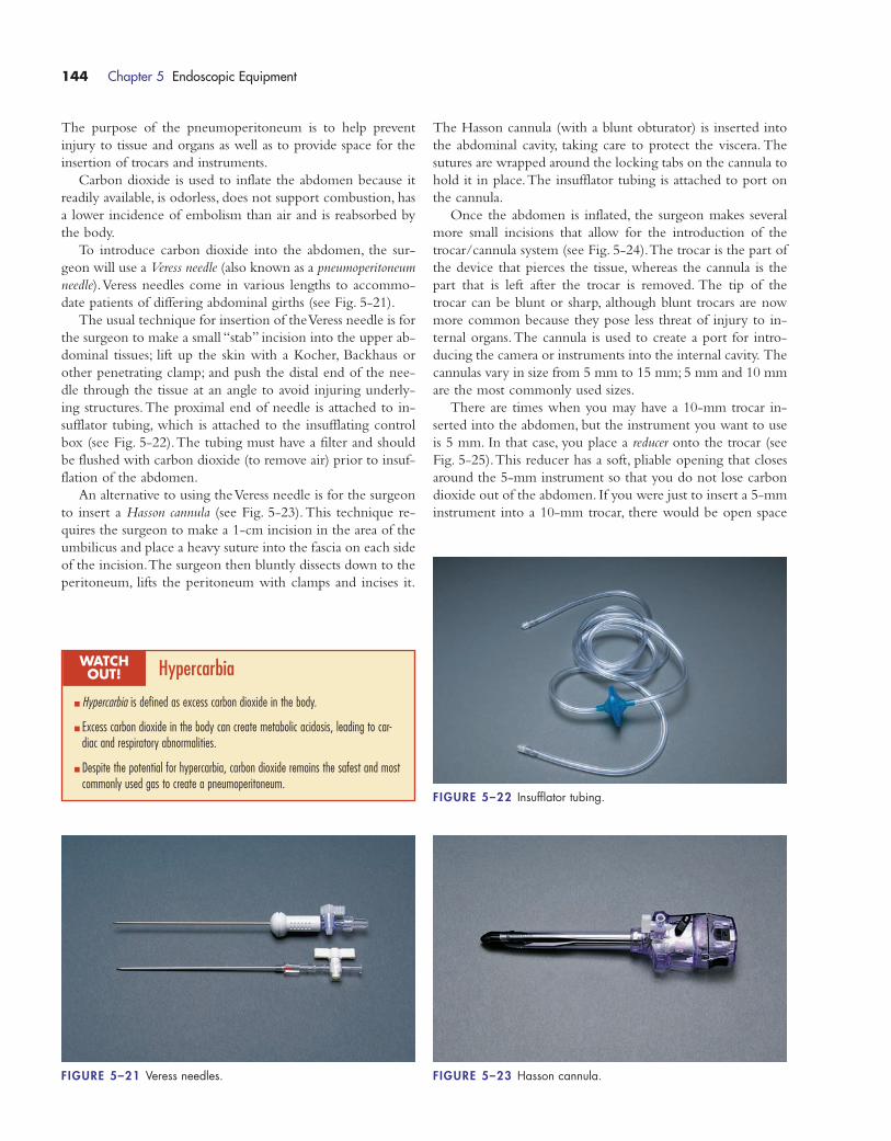
The purpose of the pneumoperitoneum is to help prevent
injury to tissue and organs as well as to provide space for the
insertion of trocars and instruments.
Carbon dioxide is used to inflate the abdomen because it
readily available, is odorless, does not support combustion, has
a lower incidence of embolism than air and is reabsorbed by
the body.
To introduce carbon dioxide into the abdomen, the sur-
geon will use a Veress needle (also known as a pneumoperitoneum
needle).Veress needles come in various lengths to accommo-
date patients of differing abdominal girths (see Fig. 5-21).
The usual technique for insertion of the Veress needle is for
the surgeon to make a small “stab” incision into the upper ab-
dominal tissues; lift up the skin with a Kocher, Backhaus or
other penetrating clamp; and push the distal end of the nee-
dle through the tissue at an angle to avoid injuring underly-
ing structures.The proximal end of needle is attached to in-
sufflator tubing, which is attached to the insufflating control
box (see Fig. 5-22).The tubing must have a filter and should
be flushed with carbon dioxide (to remove air) prior to insuf-
flation of the abdomen.
An alternative to using the Veress needle is for the surgeon
to insert a Hasson cannula (see Fig. 5-23).This technique re-
quires the surgeon to make a 1-cm incision in the area of the
umbilicus and place a heavy suture into the fascia on each side
of the incision.The surgeon then bluntly dissects down to the
peritoneum, lifts the peritoneum with clamps and incises it.
The Hasson cannula (with a blunt obturator) is inserted into
the abdominal cavity, taking care to protect the viscera. The
sutures are wrapped around the locking tabs on the cannula to
hold it in place.The insufflator tubing is attached to port on
the cannula.
Once the abdomen is inflated, the surgeon makes several
more small incisions that allow for the introduction of the
trocar/cannula system (see Fig. 5-24).The trocar is the part of
the device that pierces the tissue, whereas the cannula is the
part that is left after the trocar is removed. The tip of the
trocar can be blunt or sharp, although blunt trocars are now
more common because they pose less threat of injury to in-
ternal organs.The cannula is used to create a port for intro-
ducing the camera or instruments into the internal cavity. The
cannulas vary in size from 5 mm to 15 mm; 5 mm and 10 mm
are the most commonly used sizes.
There are times when you may have a 10-mm trocar in-
serted into the abdomen, but the instrument you want to use
is 5 mm. In that case, you place a reducer onto the trocar (see
Fig. 5-25).This reducer has a soft, pliable opening that closes
around the 5-mm instrument so that you do not lose carbon
dioxide out of the abdomen. If you were just to insert a 5-mm
instrument into a 10-mm trocar, there would be open space
144 Chapter 5 Endoscopic Equipment
Hypercarbia
� Hypercarbia is defined as excess carbon dioxide in the body.
� Excess carbon dioxide in the body can create metabolic acidosis, leading to car-diac and respiratory abnormalities.
� Despite the potential for hypercarbia, carbon dioxide remains the safest and mostcommonly used gas to create a pneumoperitoneum.
WATCH OUT!
FIGURE 5–21 Veress needles.
FIGURE 5–22 Insufflator tubing.
FIGURE 5–23 Hasson cannula.
1572_Ch05_133-148.qxd 8/6/09 11:41 AM Page 144
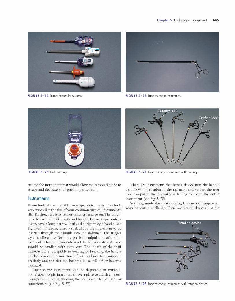
around the instrument that would allow the carbon dioxide to
escape and decrease your pneumoperitoneum.
InstrumentsIf you look at the tips of laparoscopic instruments, they look
very much like the tips of your common surgical instruments:
allis, Kocher, hemostat, scissors, mixters, and so on.The differ-
ence lies in the shaft length and handle. Laparoscopic instru-
ments have a long, narrow shaft and a trigger style handle (see
Fig. 5-26).The long narrow shaft allows the instrument to be
inserted through the cannula into the abdomen. The trigger
style handle allows for more precise manipulation of the in-
strument. These instruments tend to be very delicate and
should be handled with extra care. The length of the shaft
makes it more susceptible to bending or breaking, the handle
mechanisms can become too stiff or too loose to manipulate
precisely and the tips can become loose, fall off or become
damaged.
Laparoscopic instruments can be disposable or reusable.
Some laparoscopic instruments have a place to attach an elec-
trosurgery unit cord, allowing the instrument to be used for
cauterization (see Fig. 5-27).
There are instruments that have a device near the handle
that allows for rotation of the tip, making it so that the user
can manipulate the tip without having to rotate the entire
instrument (see Fig. 5-28).
Suturing inside the cavity during laparoscopic surgery al-
ways presents a challenge. There are several devices that are
Chapter 5 Endoscopic Equipment 145
FIGURE 5–24 Trocar/cannula systems.
FIGURE 5–25 Reducer cap.
FIGURE 5–26 Laparoscopic instrument.
Cautery post
Cautery post
FIGURE 5–27 Laparoscopic instrument with cautery.
Rotation device
FIGURE 5–28 Laparoscopic instrument with rotation device.
1572_Ch05_133-148.qxd 8/6/09 11:41 AM Page 145
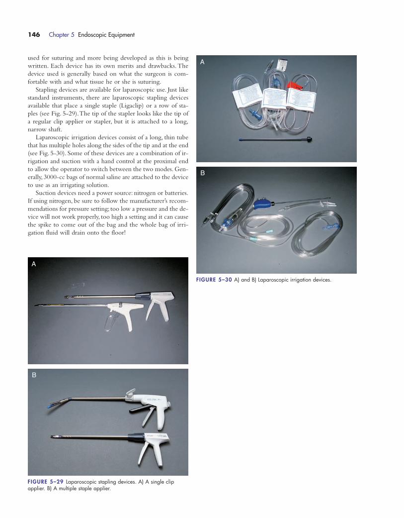
used for suturing and more being developed as this is being
written. Each device has its own merits and drawbacks. The
device used is generally based on what the surgeon is com-
fortable with and what tissue he or she is suturing.
Stapling devices are available for laparoscopic use. Just like
standard instruments, there are laparoscopic stapling devices
available that place a single staple (Ligaclip) or a row of sta-
ples (see Fig. 5-29).The tip of the stapler looks like the tip of
a regular clip applier or stapler, but it is attached to a long,
narrow shaft.
Laparoscopic irrigation devices consist of a long, thin tube
that has multiple holes along the sides of the tip and at the end
(see Fig. 5-30). Some of these devices are a combination of ir-
rigation and suction with a hand control at the proximal end
to allow the operator to switch between the two modes. Gen-
erally, 3000-cc bags of normal saline are attached to the device
to use as an irrigating solution.
Suction devices need a power source: nitrogen or batteries.
If using nitrogen, be sure to follow the manufacturer’s recom-
mendations for pressure setting; too low a pressure and the de-
vice will not work properly, too high a setting and it can cause
the spike to come out of the bag and the whole bag of irri-
gation fluid will drain onto the floor!
146 Chapter 5 Endoscopic Equipment
FIGURE 5–29 Laparoscopic stapling devices. A) A single clip applier. B) A multiple staple applier.
A
B
FIGURE 5–30 A) and B) Laparoscopic irrigation devices.
A
B
1572_Ch05_133-148.qxd 8/6/09 11:41 AM Page 146

Chapter 5 Endoscopic Equipment 147
Surgical Session Review
1) Rhinoscopy refers to viewing inside the:
a. mouth
b. nose
c. ear
d. throat
2) Fluoroscopy uses __________ to view inside tissues or organs.
a. ultrasound waves
b. laser
c. x-ray
d. electrical current
3) Gastroscopy refers to viewing inside the:
a. small intestine
b. large intestine
c. esophagus
d. stomach
4) Which of the following is false concerning a ureteroscope?
a. The shaft is longer and thinner than a cystoscope.
b. It can be rigid, semi-rigid or flexible.
c. The shaft is shorter and thinner than a cystoscope.
d. It is used to view inside the ureter.
5) The most common gas used to create a pneumoperi-
toneum is:
a. carbon dioxide
b. carbon monoxide
c. oxygen
d. helium
6) The gas pressure in the abdomen during laparoscopy
should generally be around:
a. 5 mm Hg
b. 10 mm Hg
c. 15 mm Hg
d. 25 mm Hg
7)Video towers are not sterile; therefore, you need to make
sure they are positioned at least ___________ inches from
the sterile drapes.
a. 6-10
b. 12-18
c. 28-26
d. 32-40
8) If the monitor video screen is dark, what should you check
first?
a. the fuses
b. the power box to make sure it is not overloaded
c. make sure it is plugged in and turned on
d. make sure it is connected to the printer
9)Your patient is scheduled for a laryngoscopy.The surgeon
will be looking inside the patient’s ___________.
a. nose
b. throat
c. ear
d. stomach
10) A mediastinoscopy can be used to aid in diagnosing:
a. cervical cancer
b. Hodgkin’s disease
c. AIDS
d. Paget’s disease
11) All of the following structures are located in the medi-
astinum except the:
a. trachea
b. heart
c. esophagus
d. lungs
12) Thoracoscopy can be performed for all of the following
reasons except:
a. lung biopsy
b. wedge resection of the long
c. decortication
d. pneumonectomy
13) The acronym VATS means:
a. Video- activated thoracoscopic surgery
b. Video- assisted thoracoscopic surgery
c. Video- aided thoracic surgery
d. Video- aligned thoracic surgery
14) To avoid possible damage, cystoscopes should be handled
by the:
a. middle of the shaft
b. tip end of the shaft
c. light cable connector
d. bridge
15) The patient is undergoing a TURBT that will involve
electrocautery of bladder tumors. Which irrigation fluid
would you anticipate using?
a. normal saline
b. dextrose in water
c. lactated Ringer’s
d. glycine
1572_Ch05_133-148.qxd 8/6/09 11:41 AM Page 147

16) ___________ is the most common position for a patient
undergoing cystoscopy.
a. supine
b. prone
c. Kraske
d. lithotomy
17) Arthroscopy can be used for all of the following except:
a. ligament damage repair
b. tendon damage repair
c. bone fractures
d. chondroplasty
18) The needle used to insert gas into the abdomen is known
as a __________ needle.
a. Veress
b. Vegan
c. Vargan
d. Verrin
19) Excess carbon dioxide in the body is known as:
a. hypoxia
b. hypocarbia
c. hypercarbia
d. hyperglycemia
20) The three most common types of cystoscopes used have
_______________ degree tips.
a. 0, 30, 70
b. 0, 30, 65
c. 0. 20, 70
d. 10, 30, 70
148 Chapter 5 Endoscopic Equipment
1572_Ch05_133-148.qxd 8/6/09 11:41 AM Page 148
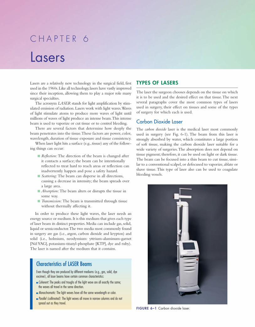
Lasers are a relatively new technology in the surgical field, first
used in the 1960s.Like all technology, lasers have vastly improved
since their inception, allowing them to play a major role many
surgical specialties.
The acronym LASER stands for light amplification by stim-
ulated emission of radiation.Lasers work with light waves.Waves
of light stimulate atoms to produce more waves of light until
millions of waves of light produce an intense beam.This intense
beam is used to vaporize or cut tissue or to control bleeding.
There are several factors that determine how deeply the
beam penetrates into the tissue.These factors are power, color,
wavelength, duration of tissue exposure and tissue consistency.
When laser light hits a surface (e.g., tissue) any of the follow-
ing things can occur:
� Reflection: The direction of the beam is changed after
it contacts a surface; the beam can be intentionally
reflected to treat hard to reach areas or reflection can
inadvertently happen and pose a safety hazard.
� Scattering: The beam can disperse in all directions,
causing a decrease in intensity; the beam spreads over
a large area.
� Absorption: The beam alters or disrupts the tissue in
some way.
� Transmission: The beam is transmitted through tissue
without thermally affecting it.
In order to produce these light waves, the laser needs an
energy source or medium. It is this medium that gives each type
of laser beam its distinct properties. Media can include gas, solid,
liquid or semiconductor.The two media most commonly found
in surgery are gas (i.e., argon, carbon dioxide and krypton) and
solid (i.e., holmium, neodymium: yttrium-aluminum-garnet
[Nd:YAG], potassium-titanyl-phosphate [KTP], dye and ruby).
The laser is named after the medium that it contains.
TYPES OF LASERS
The laser the surgeon chooses depends on the tissue on which
it is to be used and the desired effect on that tissue.The next
several paragraphs cover the most common types of lasers
used in surgery, their effect on tissues and some of the types
of surgery for which each is used.
Carbon Dioxide LaserThe carbon dioxide laser is the medical laser most commonly
used in surgery (see Fig. 6-1). The beam from this laser is
strongly absorbed by water, which constitutes a large portion
of soft tissue, making the carbon dioxide laser suitable for a
wide variety of surgeries.The absorption does not depend on
tissue pigment; therefore, it can be used on light or dark tissue.
The beam can be focused into a thin beam to cut tissue, simi-
lar to a conventional scalpel, or defocused to vaporize, ablate or
shave tissue. This type of laser also can be used to coagulate
bleeding vessels.
C H A P T E R 6
Lasers
FIGURE 6–1 Carbon dioxide laser.
Characteristics of LASER BeamsEven though they are produced by different mediums (e.g., gas, solid, dye excimer), all laser beams have certain common characteristics:
� Coherent: The peaks and troughs of the light wave are all exactly the same; the waves all travel in the same direction.
� Monochromatic: The light waves have all the same wavelength or color.
� Parallel (collimated): The light waves all move in narrow columns and do notspread out as they travel.
1572_Ch06_149-156.qxd 8/6/09 11:43 AM Page 149
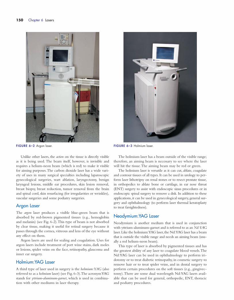
Unlike other lasers, the action on the tissue is directly visible
as it is being used. The beam itself, however, is invisible and
requires a helium-neon beam (which is red) to make it visible
for aiming purposes.The carbon dioxide laser has a wide vari-
ety of uses in many surgical specialties including laparoscopic
gynecological surgeries, wart ablation, laryngectomy, benign
laryngeal lesions, middle ear procedures, skin lesion removal,
breast biopsy, breast reduction, tumor removal from the brain
and spinal cord, skin resurfacing (for irregularities or wrinkles),
vascular surgeries and some podiatry surgeries.
Argon LaserThe argon laser produces a visible blue-green beam that is
absorbed by red-brown pigmented tissues (e.g., hemoglobin
and melanin) (see Fig. 6-2).This type of beam is not absorbed
by clear tissue, making it useful for retinal surgery because it
passes through the cornea, vitreous and lens of the eye without
any effect on them.
Argon lasers are used for sealing and coagulation. Uses for
argon lasers include treatment of port wine stains, dark moles
or lesions, spider veins on the face, retinopathy, glaucoma and
inner ear surgery.
Holmium:YAG LaserA third type of laser used in surgery is the holmium:YAG (also
referred to as a holmium laser) (see Fig. 6-3).The acronym YAG
stands for yttrium-aluminum-garnet, which is used in combina-
tion with other mediums in laser therapy.
The holmium laser has a beam outside of the visible range;
therefore, an aiming beam is necessary to see where the laser
will hit the tissue.The aiming beam may be red or green.
The holmium laser is versatile as it can cut, ablate, coagulate
and contour tissues of all types. It can be used in urology to per-
form laser lithotripsy on renal stones or to resect prostate tissue,
in orthopedics to ablate bone or cartilage, in ear nose throat
(ENT) surgery to assist with endoscopic sinus procedures or in
endoscopic spinal surgery to remove a disk. In addition to these
applications, it can be used in gynecological surgery, general sur-
gery and ophthalmology (to perform laser thermal keratoplasty
to treat farsightedness).
Neodymium:YAG LaserNeodymium is another medium that is used in conjunction
with yttrium-aluminum-garnet and is referred to as an Nd:YAG
laser.Like the holmium:YAG laser, the Nd:YAG laser has a beam
that is outside the visible range and needs an aiming beam (usu-
ally a red helium-neon beam).
This type of laser is absorbed by pigmented tissues and has
the greatest ability of any laser to coagulate blood vessels.The
Nd:YAG laser can be used in ophthalmology to perform iri-
dotomy or to treat diabetic retinopathy, in cosmetic surgery to
remove hair or to treat spider veins, and in dental surgery to
perform certain procedures on the soft tissues (e.g., gingivec-
tomy).There are some dual wavelength Nd:YAG lasers avail-
able that can be used for general, orthopedic, ENT, thoracic
and podiatry procedures.
150 Chapter 6 Lasers
FIGURE 6–2 Argon laser. FIGURE 6–3 Holmium laser.
1572_Ch06_149-156.qxd 8/6/09 11:43 AM Page 150
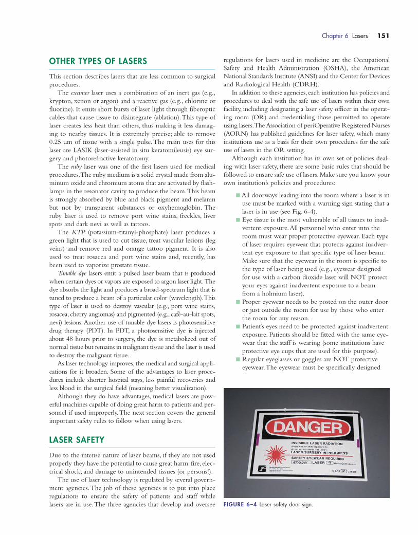
OTHER TYPES OF LASERS
This section describes lasers that are less common to surgical
procedures.
The excimer laser uses a combination of an inert gas (e.g.,
krypton, xenon or argon) and a reactive gas (e.g., chlorine or
fluorine). It emits short bursts of laser light through fiberoptic
cables that cause tissue to disintegrate (ablation).This type of
laser creates less heat than others, thus making it less damag-
ing to nearby tissues. It is extremely precise; able to remove
0.25 μm of tissue with a single pulse.The main uses for this
laser are LASIK (laser-assisted in situ keratomileusis) eye sur-
gery and photorefractive keratotomy.
The ruby laser was one of the first lasers used for medical
procedures.The ruby medium is a solid crystal made from alu-
minum oxide and chromium atoms that are activated by flash-
lamps in the resonator cavity to produce the beam.This beam
is strongly absorbed by blue and black pigment and melanin
but not by transparent substances or oxyhemoglobin. The
ruby laser is used to remove port wine stains, freckles, liver
spots and dark nevi as well as tattoos.
The KTP (potassium-titanyl-phosphate) laser produces a
green light that is used to cut tissue, treat vascular lesions (leg
veins) and remove red and orange tattoo pigment. It is also
used to treat rosacea and port wine stains and, recently, has
been used to vaporize prostate tissue.
Tunable dye lasers emit a pulsed laser beam that is produced
when certain dyes or vapors are exposed to argon laser light.The
dye absorbs the light and produces a broad-spectrum light that is
tuned to produce a beam of a particular color (wavelength).This
type of laser is used to destroy vascular (e.g., port wine stains,
rosacea, cherry angiomas) and pigmented (e.g., café-au-lait spots,
nevi) lesions.Another use of tunable dye lasers is photosensitive
drug therapy (PDT). In PDT, a photosensitive dye is injected
about 48 hours prior to surgery, the dye is metabolized out of
normal tissue but remains in malignant tissue and the laser is used
to destroy the malignant tissue.
As laser technology improves, the medical and surgical appli-
cations for it broaden. Some of the advantages to laser proce-
dures include shorter hospital stays, less painful recoveries and
less blood in the surgical field (meaning better visualization).
Although they do have advantages, medical lasers are pow-
erful machines capable of doing great harm to patients and per-
sonnel if used improperly.The next section covers the general
important safety rules to follow when using lasers.
LASER SAFETY
Due to the intense nature of laser beams, if they are not used
properly they have the potential to cause great harm: fire, elec-
trical shock, and damage to unintended tissues (or persons!).
The use of laser technology is regulated by several govern-
ment agencies.The job of these agencies is to put into place
regulations to ensure the safety of patients and staff while
lasers are in use.The three agencies that develop and oversee
regulations for lasers used in medicine are the Occupational
Safety and Health Administration (OSHA), the American
National Standards Institute (ANSI) and the Center for Devices
and Radiological Health (CDRH).
In addition to these agencies, each institution has policies and
procedures to deal with the safe use of lasers within their own
facility, including designating a laser safety officer in the operat-
ing room (OR) and credentialing those permitted to operate
using lasers.The Association of periOperative Registered Nurses
(AORN) has published guidelines for laser safety, which many
institutions use as a basis for their own procedures for the safe
use of lasers in the OR setting.
Although each institution has its own set of policies deal-
ing with laser safety, there are some basic rules that should be
followed to ensure safe use of lasers.Make sure you know your
own institution’s policies and procedures:
� All doorways leading into the room where a laser is in
use must be marked with a warning sign stating that a
laser is in use (see Fig. 6-4).
� Eye tissue is the most vulnerable of all tissues to inad-
vertent exposure.All personnel who enter into the
room must wear proper protective eyewear. Each type
of laser requires eyewear that protects against inadver-
tent eye exposure to that specific type of laser beam.
Make sure that the eyewear in the room is specific to
the type of laser being used (e.g., eyewear designed
for use with a carbon dioxide laser will NOT protect
your eyes against inadvertent exposure to a beam
from a holmium laser).
� Proper eyewear needs to be posted on the outer door
or just outside the room for use by those who enter
the room for any reason.
� Patient’s eyes need to be protected against inadvertent
exposure. Patients should be fitted with the same eye-
wear that the staff is wearing (some institutions have
protective eye cups that are used for this purpose).
� Regular eyeglasses or goggles are NOT protective
eyewear.The eyewear must be specifically designed
Chapter 6 Lasers 151
FIGURE 6–4 Laser safety door sign.
1572_Ch06_149-156.qxd 8/6/09 11:43 AM Page 151
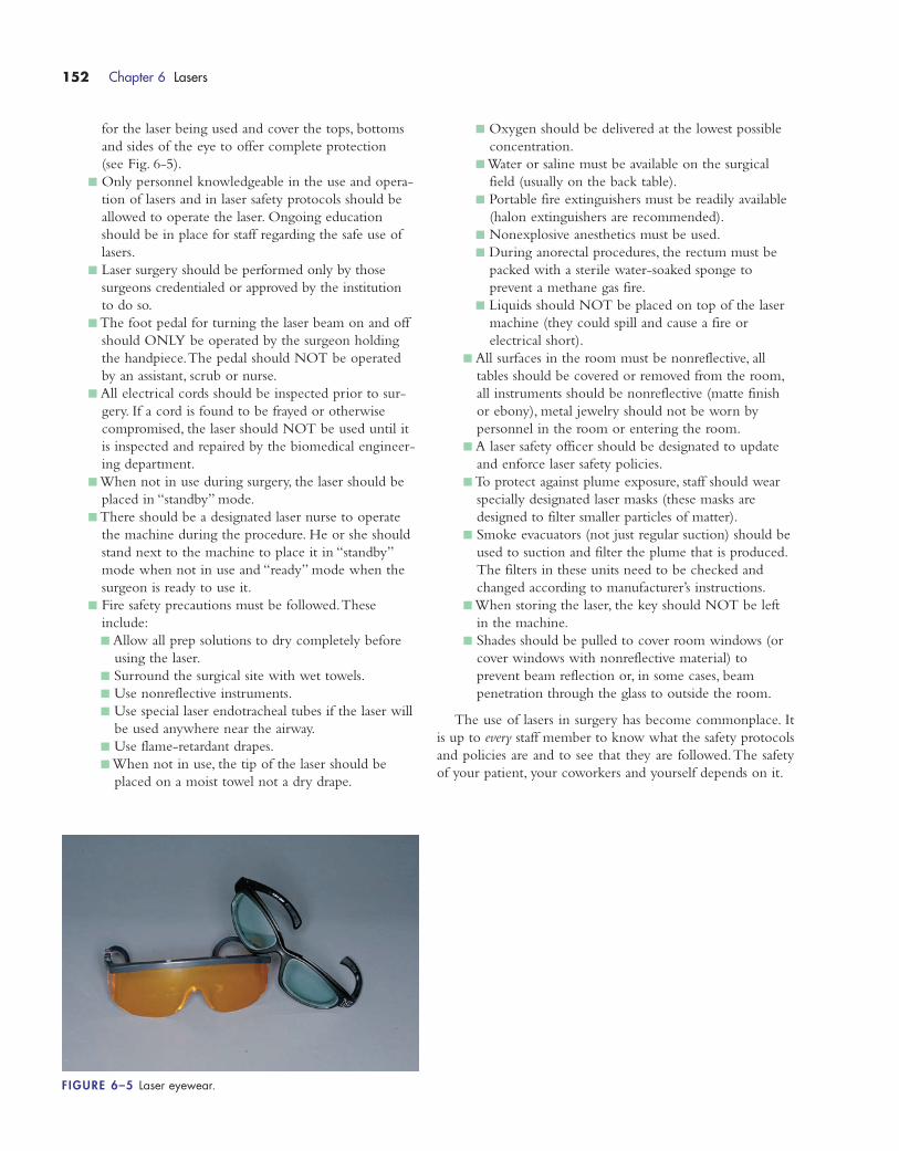
for the laser being used and cover the tops, bottoms
and sides of the eye to offer complete protection
(see Fig. 6-5).
� Only personnel knowledgeable in the use and opera-
tion of lasers and in laser safety protocols should be
allowed to operate the laser. Ongoing education
should be in place for staff regarding the safe use of
lasers.
� Laser surgery should be performed only by those
surgeons credentialed or approved by the institution
to do so.
� The foot pedal for turning the laser beam on and off
should ONLY be operated by the surgeon holding
the handpiece.The pedal should NOT be operated
by an assistant, scrub or nurse.
� All electrical cords should be inspected prior to sur-
gery. If a cord is found to be frayed or otherwise
compromised, the laser should NOT be used until it
is inspected and repaired by the biomedical engineer-
ing department.
� When not in use during surgery, the laser should be
placed in “standby” mode.
� There should be a designated laser nurse to operate
the machine during the procedure. He or she should
stand next to the machine to place it in “standby”
mode when not in use and “ready” mode when the
surgeon is ready to use it.
� Fire safety precautions must be followed.These
include:
� Allow all prep solutions to dry completely before
using the laser.
� Surround the surgical site with wet towels.
� Use nonreflective instruments.
� Use special laser endotracheal tubes if the laser will
be used anywhere near the airway.
� Use flame-retardant drapes.
� When not in use, the tip of the laser should be
placed on a moist towel not a dry drape.
� Oxygen should be delivered at the lowest possible
concentration.
� Water or saline must be available on the surgical
field (usually on the back table).
� Portable fire extinguishers must be readily available
(halon extinguishers are recommended).
� Nonexplosive anesthetics must be used.
� During anorectal procedures, the rectum must be
packed with a sterile water-soaked sponge to
prevent a methane gas fire.
� Liquids should NOT be placed on top of the laser
machine (they could spill and cause a fire or
electrical short).
� All surfaces in the room must be nonreflective, all
tables should be covered or removed from the room,
all instruments should be nonreflective (matte finish
or ebony), metal jewelry should not be worn by
personnel in the room or entering the room.
� A laser safety officer should be designated to update
and enforce laser safety policies.
� To protect against plume exposure, staff should wear
specially designated laser masks (these masks are
designed to filter smaller particles of matter).
� Smoke evacuators (not just regular suction) should be
used to suction and filter the plume that is produced.
The filters in these units need to be checked and
changed according to manufacturer’s instructions.
� When storing the laser, the key should NOT be left
in the machine.
� Shades should be pulled to cover room windows (or
cover windows with nonreflective material) to
prevent beam reflection or, in some cases, beam
penetration through the glass to outside the room.
The use of lasers in surgery has become commonplace. It
is up to every staff member to know what the safety protocols
and policies are and to see that they are followed.The safety
of your patient, your coworkers and yourself depends on it.
152 Chapter 6 Lasers
FIGURE 6–5 Laser eyewear.
1572_Ch06_149-156.qxd 8/6/09 11:43 AM Page 152
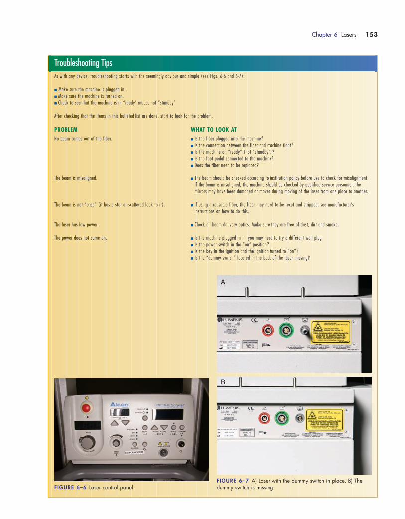
Chapter 6 Lasers 153
Troubleshooting TipsAs with any device, troubleshooting starts with the seemingly obvious and simple (see Figs. 6-6 and 6-7):
� Make sure the machine is plugged in.� Make sure the machine is turned on.� Check to see that the machine is in “ready” mode, not “standby”
After checking that the items in this bulleted list are done, start to look for the problem.
PROBLEM WHAT TO LOOK ATNo beam comes out of the fiber. � Is the fiber plugged into the machine?
� Is the connection between the fiber and machine tight?� Is the machine on “ready” (not “standby”)?� Is the foot pedal connected to the machine?� Does the fiber need to be replaced?
The beam is misaligned. � The beam should be checked according to institution policy before use to check for misalignment.If the beam is misaligned, the machine should be checked by qualified service personnel; the mirrors may have been damaged or moved during moving of the laser from one place to another.
The beam is not “crisp” (it has a star or scattered look to it). � If using a reusable fiber, the fiber may need to be recut and stripped; see manufacturer’s instructions on how to do this.
The laser has low power. � Check all beam delivery optics. Make sure they are free of dust, dirt and smoke
The power does not come on. � Is the machine plugged in— you may need to try a different wall plug� Is the power switch in the “on” position?� Is the key in the ignition and the ignition turned to “on”?� Is the “dummy switch” located in the back of the laser missing?
FIGURE 6–6 Laser control panel.FIGURE 6–7 A) Laser with the dummy switch in place. B) Thedummy switch is missing.
A
B
1572_Ch06_149-156.qxd 8/6/09 11:43 AM Page 153
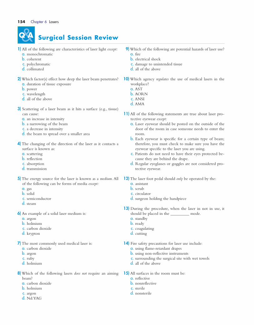
154 Chapter 6 Lasers
Surgical Session Review
1) All of the following are characteristics of laser light except:
a. monochromatic
b. coherent
c. polychromatic
d. collimated
2) Which factor(s) effect how deep the laser beam penetrates?
a. duration of tissue exposure
b. power
c. wavelength
d. all of the above
3) Scattering of a laser beam as it hits a surface (e.g., tissue)
can cause:
a. an increase in intensity
b. a narrowing of the beam
c. a decrease in intensity
d. the beam to spread over a smaller area
4) The changing of the direction of the laser as it contacts a
surface is known as:
a. scattering
b. reflection
c. absorption
d. transmission
5) The energy source for the laser is known as a medium. All
of the following can be forms of media except:
a. gas
b. solid
c. semiconductor
d. steam
6) An example of a solid laser medium is:
a. argon
b. holmium
c. carbon dioxide
d. krypton
7) The most commonly used medical laser is:
a. carbon dioxide
b. argon
c. ruby
d. holmium
8) Which of the following lasers does not require an aiming
beam?
a. carbon dioxide
b. holmium
c. argon
d. Nd:YAG
9) Which of the following are potential hazards of laser use?
a. fire
b. electrical shock
c. damage to unintended tissue
d. all of the above
10) Which agency regulates the use of medical lasers in the
workplace?
a. AST
b. AORN
c. ANSI
d. AMA
11) All of the following statements are true about laser pro-
tective eyewear except:
a. Laser eyewear should be posted on the outside of the
door of the room in case someone needs to enter the
room.
b. Each eyewear is specific for a certain type of beam;
therefore, you must check to make sure you have the
eyewear specific to the laser you are using.
c. Patients do not need to have their eyes protected be-
cause they are behind the drape.
d. Regular eyeglasses or goggles are not considered pro-
tective eyewear.
12) The laser foot pedal should only be operated by the:
a. assistant
b. scrub
c. circulator
d. surgeon holding the handpiece
13) During the procedure, when the laser in not in use, it
should be placed in the _________ mode.
a. standby
b. ready
c. coagulating
d. cutting
14) Fire safety precautions for laser use include:
a. using flame-retardant drapes
b. using non-reflective instruments
c. surrounding the surgical site with wet towels
d. all of the above
15) All surfaces in the room must be:
a. reflective
b. nonreflective
c. sterile
d. nonsterile
1572_Ch06_149-156.qxd 8/6/09 11:43 AM Page 154
16) Who is responsible for updating and enforcing laser safety
policies?
a. the surgeon
b. the physician assistant
c. the laser safety officer
d. the hospital CEO
17) When storing the laser, the key should be:
a. hidden
b. stored in the machine
c. not stored in the machine
d. left in the ignition
18) The laser machine does not come on.You have checked;
the machine is plugged in, the power switch is in the “on”
position, the key is in the ignition and the ignition is in
the “on” position.What could be the likely problem?
a. a hospital-wide power outage
b. the dummy switch in the back is missing
c. the fiber is faulty
d. the fiber needs to be recut
19) One of the main uses for an excimer laser is:
a. gynecological surgery
b. orthopedic surgery
c. LASIK surgery
d. neurosurgery
20) Which type of laser has a beam that is not absorbed by
clear tissue, making it useful for retinal surgery?
a. carbon dioxide
b. argon
c. holmium
d. ruby
Chapter 6 Lasers 155
1572_Ch06_149-156.qxd 8/6/09 11:43 AM Page 155

1572_Ch06_149-156.qxd 8/6/09 11:43 AM Page 156
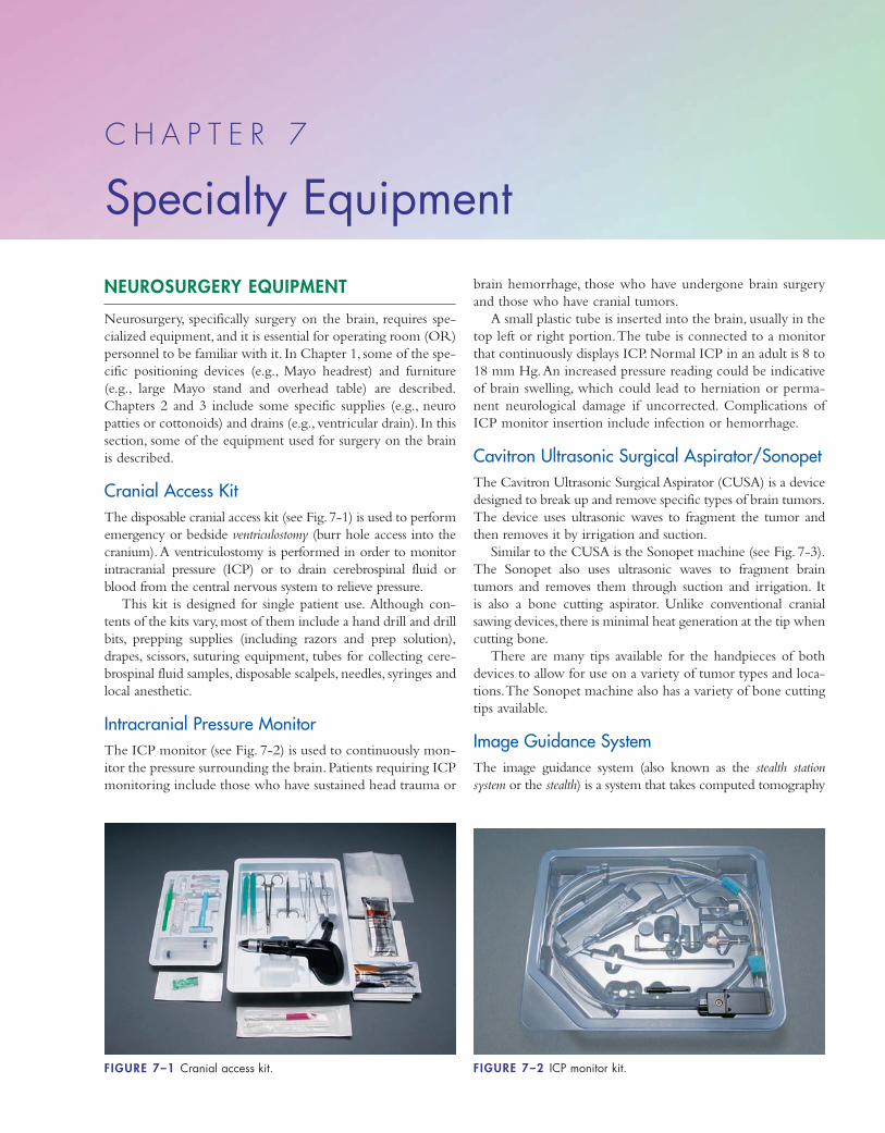
NEUROSURGERY EQUIPMENT
Neurosurgery, specifically surgery on the brain, requires spe-
cialized equipment, and it is essential for operating room (OR)
personnel to be familiar with it. In Chapter 1, some of the spe-
cific positioning devices (e.g., Mayo headrest) and furniture
(e.g., large Mayo stand and overhead table) are described.
Chapters 2 and 3 include some specific supplies (e.g., neuro
patties or cottonoids) and drains (e.g., ventricular drain). In this
section, some of the equipment used for surgery on the brain
is described.
Cranial Access KitThe disposable cranial access kit (see Fig. 7-1) is used to perform
emergency or bedside ventriculostomy (burr hole access into the
cranium). A ventriculostomy is performed in order to monitor
intracranial pressure (ICP) or to drain cerebrospinal fluid or
blood from the central nervous system to relieve pressure.
This kit is designed for single patient use. Although con-
tents of the kits vary, most of them include a hand drill and drill
bits, prepping supplies (including razors and prep solution),
drapes, scissors, suturing equipment, tubes for collecting cere-
brospinal fluid samples, disposable scalpels, needles, syringes and
local anesthetic.
Intracranial Pressure MonitorThe ICP monitor (see Fig. 7-2) is used to continuously mon-
itor the pressure surrounding the brain. Patients requiring ICP
monitoring include those who have sustained head trauma or
brain hemorrhage, those who have undergone brain surgery
and those who have cranial tumors.
A small plastic tube is inserted into the brain, usually in the
top left or right portion.The tube is connected to a monitor
that continuously displays ICP. Normal ICP in an adult is 8 to
18 mm Hg.An increased pressure reading could be indicative
of brain swelling, which could lead to herniation or perma-
nent neurological damage if uncorrected. Complications of
ICP monitor insertion include infection or hemorrhage.
Cavitron Ultrasonic Surgical Aspirator/SonopetThe Cavitron Ultrasonic Surgical Aspirator (CUSA) is a device
designed to break up and remove specific types of brain tumors.
The device uses ultrasonic waves to fragment the tumor and
then removes it by irrigation and suction.
Similar to the CUSA is the Sonopet machine (see Fig. 7-3).
The Sonopet also uses ultrasonic waves to fragment brain
tumors and removes them through suction and irrigation. It
is also a bone cutting aspirator. Unlike conventional cranial
sawing devices, there is minimal heat generation at the tip when
cutting bone.
There are many tips available for the handpieces of both
devices to allow for use on a variety of tumor types and loca-
tions.The Sonopet machine also has a variety of bone cutting
tips available.
Image Guidance SystemThe image guidance system (also known as the stealth station
system or the stealth) is a system that takes computed tomography
C H A P T E R 7
Specialty Equipment
FIGURE 7–1 Cranial access kit. FIGURE 7–2 ICP monitor kit.
1572_Ch07_157-170.qxd 8/6/09 11:46 AM Page 157
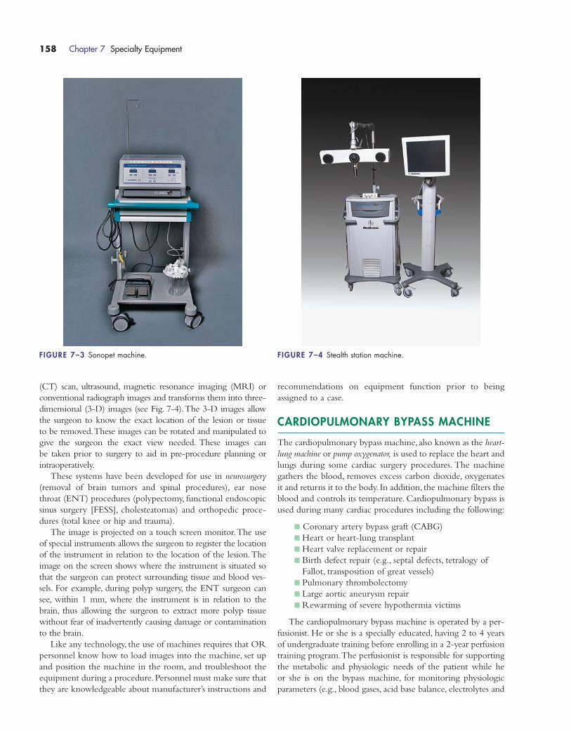
(CT) scan, ultrasound, magnetic resonance imaging (MRI) or
conventional radiograph images and transforms them into three-
dimensional (3-D) images (see Fig. 7-4).The 3-D images allow
the surgeon to know the exact location of the lesion or tissue
to be removed.These images can be rotated and manipulated to
give the surgeon the exact view needed. These images can
be taken prior to surgery to aid in pre-procedure planning or
intraoperatively.
These systems have been developed for use in neurosurgery
(removal of brain tumors and spinal procedures), ear nose
throat (ENT) procedures (polypectomy, functional endoscopic
sinus surgery [FESS], cholesteatomas) and orthopedic proce-
dures (total knee or hip and trauma).
The image is projected on a touch screen monitor.The use
of special instruments allows the surgeon to register the location
of the instrument in relation to the location of the lesion.The
image on the screen shows where the instrument is situated so
that the surgeon can protect surrounding tissue and blood ves-
sels. For example, during polyp surgery, the ENT surgeon can
see, within 1 mm, where the instrument is in relation to the
brain, thus allowing the surgeon to extract more polyp tissue
without fear of inadvertently causing damage or contamination
to the brain.
Like any technology, the use of machines requires that OR
personnel know how to load images into the machine, set up
and position the machine in the room, and troubleshoot the
equipment during a procedure. Personnel must make sure that
they are knowledgeable about manufacturer’s instructions and
recommendations on equipment function prior to being
assigned to a case.
CARDIOPULMONARY BYPASS MACHINE
The cardiopulmonary bypass machine, also known as the heart-
lung machine or pump oxygenator, is used to replace the heart and
lungs during some cardiac surgery procedures. The machine
gathers the blood, removes excess carbon dioxide, oxygenates
it and returns it to the body. In addition, the machine filters the
blood and controls its temperature. Cardiopulmonary bypass is
used during many cardiac procedures including the following:
� Coronary artery bypass graft (CABG)
� Heart or heart-lung transplant
� Heart valve replacement or repair
� Birth defect repair (e.g., septal defects, tetralogy of
Fallot, transposition of great vessels)
� Pulmonary thrombolectomy
� Large aortic aneurysm repair
� Rewarming of severe hypothermia victims
The cardiopulmonary bypass machine is operated by a per-
fusionist. He or she is a specially educated, having 2 to 4 years
of undergraduate training before enrolling in a 2-year perfusion
training program.The perfusionist is responsible for supporting
the metabolic and physiologic needs of the patient while he
or she is on the bypass machine, for monitoring physiologic
parameters (e.g., blood gases, acid base balance, electrolytes and
158 Chapter 7 Specialty Equipment
FIGURE 7–3 Sonopet machine. FIGURE 7–4 Stealth station machine.
1572_Ch07_157-170.qxd 8/6/09 11:46 AM Page 158
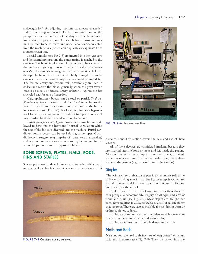
anticoagulation), for adjusting machine parameters as needed
and for collecting autologous blood. Perfusionists monitor the
pump lines for the presence of air. Any air must be removed
immediately to prevent possible air embolus or stroke.All lines
must be monitored to make sure none becomes disconnected
from the machine as a patient could quickly exsanguinate from
a disconnected line.
Special cannulae (see Fig.7-5) are inserted into the vena cava
and the ascending aorta, and the pump tubing is attached to the
cannulae.The blood is taken out of the body via the cannula in
the vena cava (or right atrium), which is called the venous
cannula. This cannula is straight-ended with multiple holes in
the tip.The blood is returned to the body through the aortic
cannula.The aortic cannula may have a straight or angled tip.
The femoral artery and femoral vein occasionally are used to
collect and return the blood, generally when the great vessels
cannot be used.The femoral artery catheter is tapered and has
a beveled end for ease of insertion.
Cardiopulmonary bypass can be total or partial. Total car-
diopulmonary bypass means that all the blood returning to the
heart is forced into the venous cannula and out to the heart-
lung machine (see Fig. 7-6).Total cardiopulmonary bypass is
used for many cardiac surgeries: CABG, transplants, repair of
most cardiac birth defects and valve replacements.
Partial cardiopulmonary bypass means that some blood is al-
lowed to flow into the heart and “normal” circulation while
the rest of the blood is diverted into the machine. Partial car-
diopulmonary bypass can be used during some types of car-
diothoracic surgery (e.g., repairs of some aortic anomalies)
and as a temporary measure after coronary bypass grafting to
wean the patient from the bypass machine.
BONE SCREWS, PLATES, NAILS, RODS,PINS AND STAPLES
Screws, plates, nails, rods and pins are used in orthopedic surgery
to repair and stabilize fractures. Staples are used to reconnect soft
tissue to bone. This section covers the care and use of these
devices.
All of these devices are considered implants because they
are inserted into the bone or tissue and left inside the patient.
Most of the time these implants are permanent, although
some can removed after the fracture heals if they are bother-
some to the patient (e.g., causing pain or discomfort).
StaplesThe primary use of fixation staples is to reconnect soft tissue
to bone, including anterior cruciate ligament repair.Other uses
include tendon and ligament repair, bone fragment fixation
and bone growth control.
Staples come in a variety of sizes and types (two, three or
four prongs) to accommodate surgery on all types and sizes of
bone and tissue (see Fig. 7-7). Most staples are straight, but
some have an offset to allow for stable fixation of an osteotomy
that has steps.There are staples available for use during open or
arthroscopic procedures.
Staples are commonly made of stainless steel, but some are
made from chromium-cobalt and nitinol alloy.
Staples are inserted with a staple driver and a mallet.
Nails and RodsNails and rods are used to fix fractures of long bones (i.e., femur,
tibia and humerus) (see Fig. 7-8). They are driven into the
Chapter 7 Specialty Equipment 159
FIGURE 7–6 Heart-lung machine.
Aortic
Venous
FIGURE 7–5 Cardiopulmonary cannulae.
1572_Ch07_157-170.qxd 8/6/09 11:46 AM Page 159
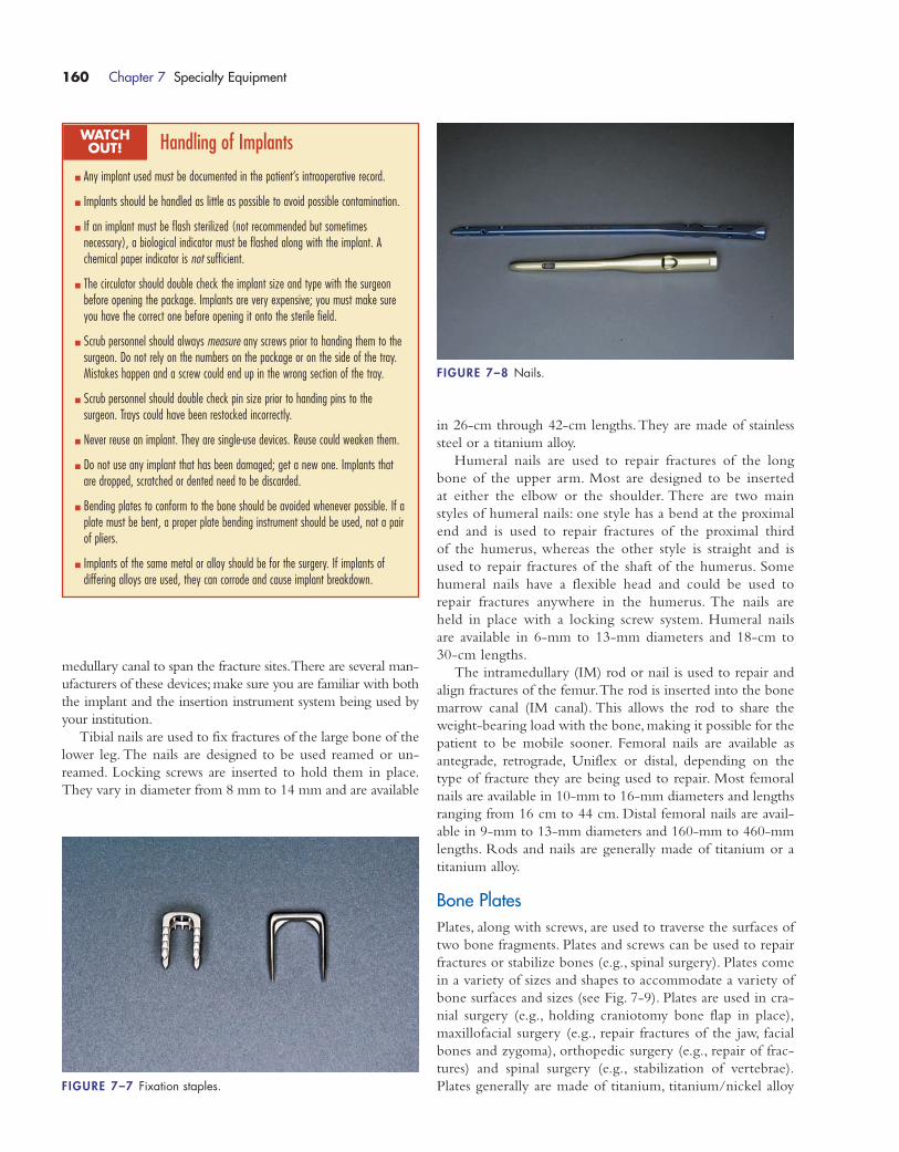
medullary canal to span the fracture sites.There are several man-
ufacturers of these devices; make sure you are familiar with both
the implant and the insertion instrument system being used by
your institution.
Tibial nails are used to fix fractures of the large bone of the
lower leg. The nails are designed to be used reamed or un-
reamed. Locking screws are inserted to hold them in place.
They vary in diameter from 8 mm to 14 mm and are available
in 26-cm through 42-cm lengths.They are made of stainless
steel or a titanium alloy.
Humeral nails are used to repair fractures of the long
bone of the upper arm. Most are designed to be inserted
at either the elbow or the shoulder. There are two main
styles of humeral nails: one style has a bend at the proximal
end and is used to repair fractures of the proximal third
of the humerus, whereas the other style is straight and is
used to repair fractures of the shaft of the humerus. Some
humeral nails have a flexible head and could be used to
repair fractures anywhere in the humerus. The nails are
held in place with a locking screw system. Humeral nails
are available in 6-mm to 13-mm diameters and 18-cm to
30-cm lengths.
The intramedullary (IM) rod or nail is used to repair and
align fractures of the femur.The rod is inserted into the bone
marrow canal (IM canal). This allows the rod to share the
weight-bearing load with the bone, making it possible for the
patient to be mobile sooner. Femoral nails are available as
antegrade, retrograde, Uniflex or distal, depending on the
type of fracture they are being used to repair. Most femoral
nails are available in 10-mm to 16-mm diameters and lengths
ranging from 16 cm to 44 cm. Distal femoral nails are avail-
able in 9-mm to 13-mm diameters and 160-mm to 460-mm
lengths. Rods and nails are generally made of titanium or a
titanium alloy.
Bone PlatesPlates, along with screws, are used to traverse the surfaces of
two bone fragments. Plates and screws can be used to repair
fractures or stabilize bones (e.g., spinal surgery). Plates come
in a variety of sizes and shapes to accommodate a variety of
bone surfaces and sizes (see Fig. 7-9). Plates are used in cra-
nial surgery (e.g., holding craniotomy bone flap in place),
maxillofacial surgery (e.g., repair fractures of the jaw, facial
bones and zygoma), orthopedic surgery (e.g., repair of frac-
tures) and spinal surgery (e.g., stabilization of vertebrae).
Plates generally are made of titanium, titanium/nickel alloy
160 Chapter 7 Specialty Equipment
Handling of Implants
� Any implant used must be documented in the patient’s intraoperative record.
� Implants should be handled as little as possible to avoid possible contamination.
� If an implant must be flash sterilized (not recommended but sometimes necessary), a biological indicator must be flashed along with the implant. Achemical paper indicator is not sufficient.
� The circulator should double check the implant size and type with the surgeon before opening the package. Implants are very expensive; you must make sureyou have the correct one before opening it onto the sterile field.
� Scrub personnel should always measure any screws prior to handing them to thesurgeon. Do not rely on the numbers on the package or on the side of the tray.Mistakes happen and a screw could end up in the wrong section of the tray.
� Scrub personnel should double check pin size prior to handing pins to the surgeon. Trays could have been restocked incorrectly.
� Never reuse an implant. They are single-use devices. Reuse could weaken them.
� Do not use any implant that has been damaged; get a new one. Implants thatare dropped, scratched or dented need to be discarded.
� Bending plates to conform to the bone should be avoided whenever possible. If aplate must be bent, a proper plate bending instrument should be used, not a pairof pliers.
� Implants of the same metal or alloy should be for the surgery. If implants of differing alloys are used, they can corrode and cause implant breakdown.
WATCH OUT!
FIGURE 7–7 Fixation staples.
FIGURE 7–8 Nails.
1572_Ch07_157-170.qxd 8/6/09 11:46 AM Page 160
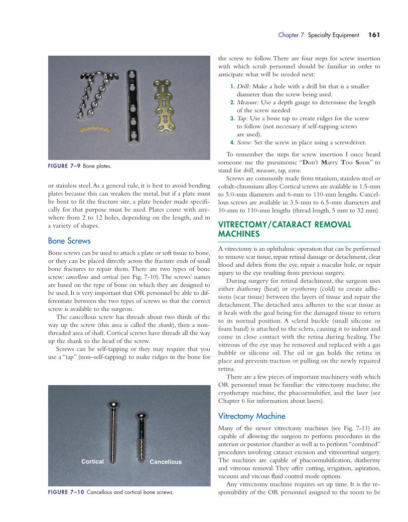
or stainless steel.As a general rule, it is best to avoid bending
plates because this can weaken the metal, but if a plate must
be bent to fit the fracture site, a plate bender made specifi-
cally for that purpose must be used. Plates come with any-
where from 2 to 12 holes, depending on the length, and in
a variety of shapes.
Bone ScrewsBone screws can be used to attach a plate or soft tissue to bone,
or they can be placed directly across the fracture ends of small
bone fractures to repair them. There are two types of bone
screw: cancellous and cortical (see Fig. 7-10).The screws’ names
are based on the type of bone on which they are designed to
be used. It is very important that OR personnel be able to dif-
ferentiate between the two types of screws so that the correct
screw is available to the surgeon.
The cancellous screw has threads about two thirds of the
way up the screw (this area is called the shank), then a non-
threaded area of shaft. Cortical screws have threads all the way
up the shank to the head of the screw.
Screws can be self-tapping or they may require that you
use a “tap” (non–self-tapping) to make ridges in the bone for
the screw to follow. There are four steps for screw insertion
with which scrub personnel should be familiar in order to
anticipate what will be needed next:
1. Drill: Make a hole with a drill bit that is a smaller
diameter than the screw being used.
2. Measure: Use a depth gauge to determine the length
of the screw needed
3. Tap: Use a bone tap to create ridges for the screw
to follow (not necessary if self-tapping screws
are used).
4. Screw: Set the screw in place using a screwdriver.
To remember the steps for screw insertion I once heard
someone use the pneumonic “Don’t Marry Too Soon” to
stand for drill, measure, tap, screw.
Screws are commonly made from titanium, stainless steel or
cobalt-chromium alloy.Cortical screws are available in 1.5-mm
to 5.0-mm diameters and 6-mm to 110-mm lengths. Cancel-
lous screws are available in 3.5-mm to 6.5-mm diameters and
10-mm to 110-mm lengths (thread length, 5 mm to 32 mm).
VITRECTOMY/CATARACT REMOVALMACHINES
A vitrectomy is an ophthalmic operation that can be performed
to remove scar tissue, repair retinal damage or detachment, clear
blood and debris from the eye, repair a macular hole, or repair
injury to the eye resulting from previous surgery.
During surgery for retinal detachment, the surgeon uses
either diathermy (heat) or cryothermy (cold) to create adhe-
sions (scar tissue) between the layers of tissue and repair the
detachment.The detached area adheres to the scar tissue as
it heals with the goal being for the damaged tissue to return
to its normal position. A scleral buckle (small silicone or
foam band) is attached to the sclera, causing it to indent and
come in close contact with the retina during healing. The
vitreous of the eye may be removed and replaced with a gas
bubble or silicone oil. The oil or gas holds the retina in
place and prevents traction or pulling on the newly repaired
retina.
There are a few pieces of important machinery with which
OR personnel must be familiar: the vitrectomy machine, the
cryotherapy machine, the phacoemulsifier, and the laser (see
Chapter 6 for information about lasers).
Vitrectomy MachineMany of the newer vitrectomy machines (see Fig. 7-11) are
capable of allowing the surgeon to perform procedures in the
anterior or posterior chamber as well as to perform “combined”
procedures involving cataract excision and vitreoretinal surgery.
The machines are capable of phacoemulsification, diathermy
and vitreous removal. They offer cutting, irrigation, aspiration,
vacuum and viscous fluid control mode options.
Any vitrectomy machine requires set up time. It is the re-
sponsibility of the OR personnel assigned to the room to be
Chapter 7 Specialty Equipment 161
Cortical Cancellous
FIGURE 7–10 Cancellous and cortical bone screws.
FIGURE 7–9 Bone plates.
1572_Ch07_157-170.qxd 8/6/09 11:46 AM Page 161
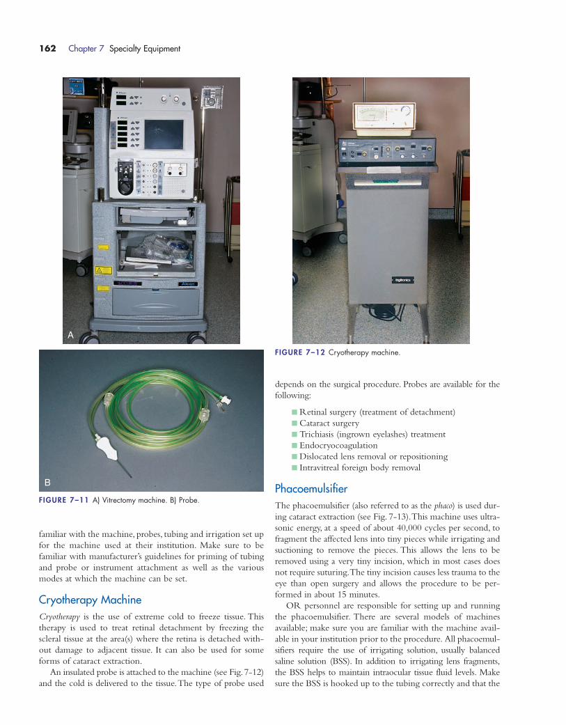
familiar with the machine, probes, tubing and irrigation set up
for the machine used at their institution. Make sure to be
familiar with manufacturer’s guidelines for priming of tubing
and probe or instrument attachment as well as the various
modes at which the machine can be set.
Cryotherapy MachineCryotherapy is the use of extreme cold to freeze tissue. This
therapy is used to treat retinal detachment by freezing the
scleral tissue at the area(s) where the retina is detached with-
out damage to adjacent tissue. It can also be used for some
forms of cataract extraction.
An insulated probe is attached to the machine (see Fig. 7-12)
and the cold is delivered to the tissue.The type of probe used
depends on the surgical procedure. Probes are available for the
following:
� Retinal surgery (treatment of detachment)
� Cataract surgery
� Trichiasis (ingrown eyelashes) treatment
� Endocryocoagulation
� Dislocated lens removal or repositioning
� Intravitreal foreign body removal
PhacoemulsifierThe phacoemulsifier (also referred to as the phaco) is used dur-
ing cataract extraction (see Fig. 7-13).This machine uses ultra-
sonic energy, at a speed of about 40,000 cycles per second, to
fragment the affected lens into tiny pieces while irrigating and
suctioning to remove the pieces. This allows the lens to be
removed using a very tiny incision, which in most cases does
not require suturing.The tiny incision causes less trauma to the
eye than open surgery and allows the procedure to be per-
formed in about 15 minutes.
OR personnel are responsible for setting up and running
the phacoemulsifier. There are several models of machines
available; make sure you are familiar with the machine avail-
able in your institution prior to the procedure. All phacoemul-
sifiers require the use of irrigating solution, usually balanced
saline solution (BSS). In addition to irrigating lens fragments,
the BSS helps to maintain intraocular tissue fluid levels. Make
sure the BSS is hooked up to the tubing correctly and that the
162 Chapter 7 Specialty Equipment
FIGURE 7–11 A) Vitrectomy machine. B) Probe.
A
B
FIGURE 7–12 Cryotherapy machine.
1572_Ch07_157-170.qxd 8/6/09 11:46 AM Page 162
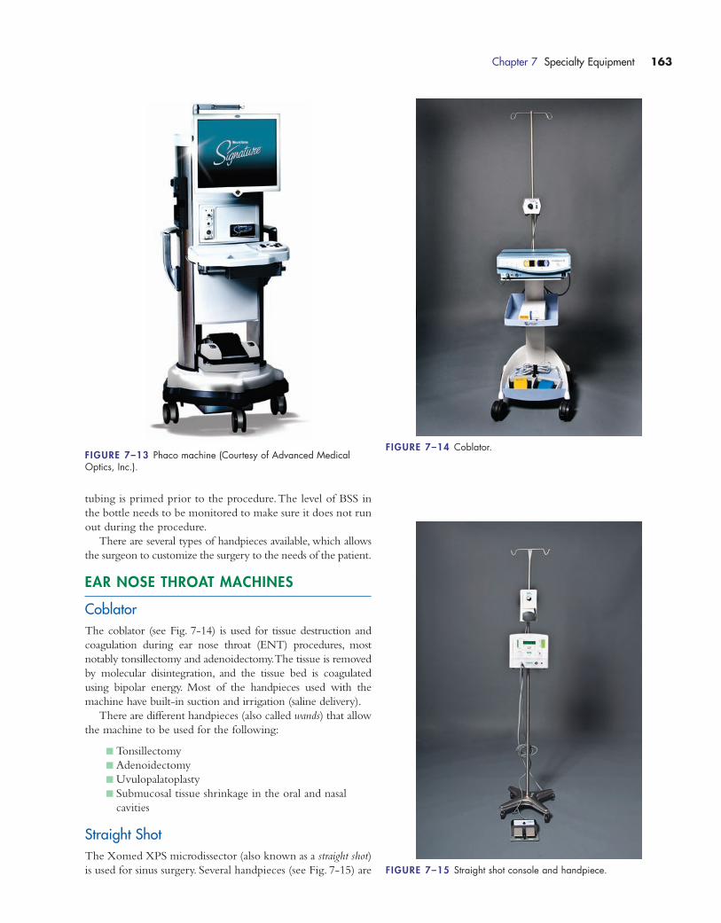
tubing is primed prior to the procedure.The level of BSS in
the bottle needs to be monitored to make sure it does not run
out during the procedure.
There are several types of handpieces available, which allows
the surgeon to customize the surgery to the needs of the patient.
EAR NOSE THROAT MACHINES
CoblatorThe coblator (see Fig. 7-14) is used for tissue destruction and
coagulation during ear nose throat (ENT) procedures, most
notably tonsillectomy and adenoidectomy.The tissue is removed
by molecular disintegration, and the tissue bed is coagulated
using bipolar energy. Most of the handpieces used with the
machine have built-in suction and irrigation (saline delivery).
There are different handpieces (also called wands) that allow
the machine to be used for the following:
� Tonsillectomy
� Adenoidectomy
� Uvulopalatoplasty
� Submucosal tissue shrinkage in the oral and nasal
cavities
Straight ShotThe Xomed XPS microdissector (also known as a straight shot)
is used for sinus surgery. Several handpieces (see Fig. 7-15) are
Chapter 7 Specialty Equipment 163
FIGURE 7–13 Phaco machine (Courtesy of Advanced Medical Optics, Inc.).
FIGURE 7–14 Coblator.
FIGURE 7–15 Straight shot console and handpiece.
1572_Ch07_157-170.qxd 8/6/09 11:46 AM Page 163
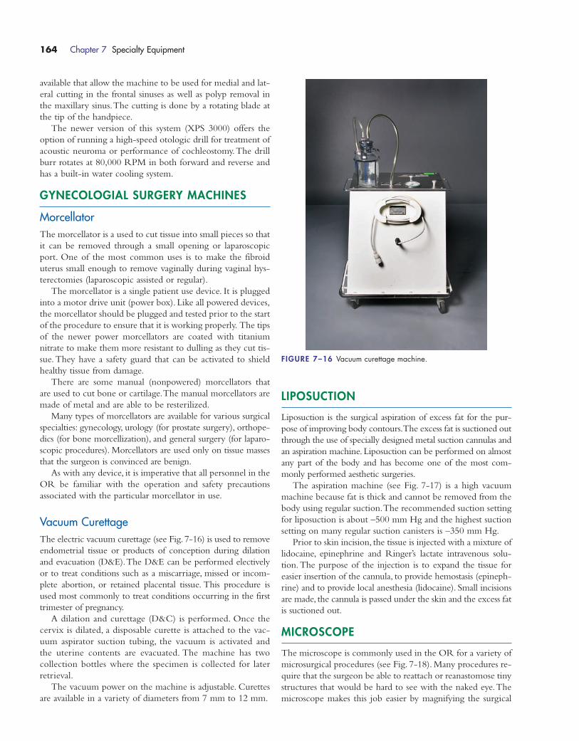
available that allow the machine to be used for medial and lat-
eral cutting in the frontal sinuses as well as polyp removal in
the maxillary sinus.The cutting is done by a rotating blade at
the tip of the handpiece.
The newer version of this system (XPS 3000) offers the
option of running a high-speed otologic drill for treatment of
acoustic neuroma or performance of cochleostomy.The drill
burr rotates at 80,000 RPM in both forward and reverse and
has a built-in water cooling system.
GYNECOLOGIAL SURGERY MACHINES
MorcellatorThe morcellator is a used to cut tissue into small pieces so that
it can be removed through a small opening or laparoscopic
port. One of the most common uses is to make the fibroid
uterus small enough to remove vaginally during vaginal hys-
terectomies (laparoscopic assisted or regular).
The morcellator is a single patient use device. It is plugged
into a motor drive unit (power box). Like all powered devices,
the morcellator should be plugged and tested prior to the start
of the procedure to ensure that it is working properly. The tips
of the newer power morcellators are coated with titanium
nitrate to make them more resistant to dulling as they cut tis-
sue. They have a safety guard that can be activated to shield
healthy tissue from damage.
There are some manual (nonpowered) morcellators that
are used to cut bone or cartilage.The manual morcellators are
made of metal and are able to be resterilized.
Many types of morcellators are available for various surgical
specialties: gynecology, urology (for prostate surgery), orthope-
dics (for bone morcellization), and general surgery (for laparo-
scopic procedures). Morcellators are used only on tissue masses
that the surgeon is convinced are benign.
As with any device, it is imperative that all personnel in the
OR be familiar with the operation and safety precautions
associated with the particular morcellator in use.
Vacuum CurettageThe electric vacuum curettage (see Fig. 7-16) is used to remove
endometrial tissue or products of conception during dilation
and evacuation (D&E).The D&E can be performed electively
or to treat conditions such as a miscarriage, missed or incom-
plete abortion, or retained placental tissue. This procedure is
used most commonly to treat conditions occurring in the first
trimester of pregnancy.
A dilation and curettage (D&C) is performed. Once the
cervix is dilated, a disposable curette is attached to the vac-
uum aspirator suction tubing, the vacuum is activated and
the uterine contents are evacuated. The machine has two
collection bottles where the specimen is collected for later
retrieval.
The vacuum power on the machine is adjustable. Curettes
are available in a variety of diameters from 7 mm to 12 mm.
LIPOSUCTION
Liposuction is the surgical aspiration of excess fat for the pur-
pose of improving body contours.The excess fat is suctioned out
through the use of specially designed metal suction cannulas and
an aspiration machine. Liposuction can be performed on almost
any part of the body and has become one of the most com-
monly performed aesthetic surgeries.
The aspiration machine (see Fig. 7-17) is a high vacuum
machine because fat is thick and cannot be removed from the
body using regular suction.The recommended suction setting
for liposuction is about –500 mm Hg and the highest suction
setting on many regular suction canisters is –350 mm Hg.
Prior to skin incision, the tissue is injected with a mixture of
lidocaine, epinephrine and Ringer’s lactate intravenous solu-
tion. The purpose of the injection is to expand the tissue for
easier insertion of the cannula, to provide hemostasis (epineph-
rine) and to provide local anesthesia (lidocaine). Small incisions
are made, the cannula is passed under the skin and the excess fat
is suctioned out.
MICROSCOPE
The microscope is commonly used in the OR for a variety of
microsurgical procedures (see Fig. 7-18). Many procedures re-
quire that the surgeon be able to reattach or reanastomose tiny
structures that would be hard to see with the naked eye.The
microscope makes this job easier by magnifying the surgical
164 Chapter 7 Specialty Equipment
FIGURE 7–16 Vacuum curettage machine.
1572_Ch07_157-170.qxd 8/6/09 11:46 AM Page 164
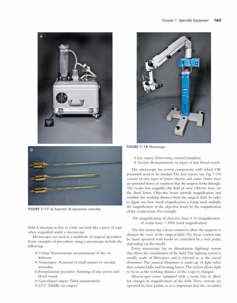
field.A structure as fine as a hair can look like a piece of rope
when magnified under a microscope.
Microscopes are used in a multitude of surgical specialties.
Some examples of procedures using a microscope include the
following:
� Urology:Vasovasotomy (reanastomosis of the vas
deferens)
� Neurosurgery: Removal of small tumors or vascular
anomalies
� Reimplantation procedures: Suturing of tiny nerves and
blood vessels
� Gynecological surgery: Tubal reanastomosis
� ENT: Middle ear surgery
� Eye surgery:Vitrectomy, corneal transplant
� Vascular: Reanastomosis or repair of tiny blood vessels
The microscope has several components with which OR
personnel need to be familiar.The lens system (see Fig. 7-19)
consists of two types of lenses: objective and ocular. Ocular lenses
are proximal lenses or eyepieces that the surgeon looks through.
The ocular lens magnifies the field of view. Objective lenses are
the distal lenses. Objective lenses provide magnification and
establish the working distance from the surgical field. In order
to figure out how much magnification is being used, multiply
the magnification of the objective lenses by the magnification
of the ocular lenses. For example:
300 (magnification of objective lens) × 10 (magnification
of ocular lens) = 3000 (total magnification)
The lens system has a focus control to allow the surgeon to
sharpen the view of the surgical field.The focus control may
be hand operated with knobs or controlled by a foot pedal,
depending on the model.
Every microscope has an illumination (lighting) system
that allows for visualization of the field.This lighting system is
usually made of fiberoptics and is referred to as the coaxial
illuminator. The paraxial illuminator is made up of light tubes
that contain bulbs and focusing lenses.This system allows light
to focus as the working distance of the scope is changed.
Microscopes come equipped with a zoom lens to allow
for changes in magnification of the field. These systems are
operated by foot pedals, so it is important that the circulator
Chapter 7 Specialty Equipment 165
FIGURE 7–18 Microscope.
FIGURE 7–17 A) Aspirator. B) Liposuction cannulae.
A
B
1572_Ch07_157-170.qxd 8/6/09 11:46 AM Page 165
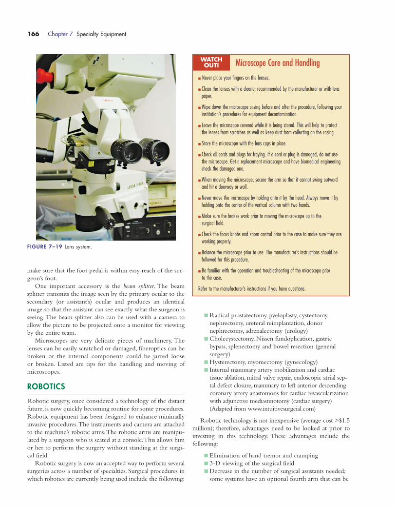
make sure that the foot pedal is within easy reach of the sur-
geon’s foot.
One important accessory is the beam splitter. The beam
splitter transmits the image seen by the primary ocular to the
secondary (or assistant’s) ocular and produces an identical
image so that the assistant can see exactly what the surgeon is
seeing. The beam splitter also can be used with a camera to
allow the picture to be projected onto a monitor for viewing
by the entire team.
Microscopes are very delicate pieces of machinery. The
lenses can be easily scratched or damaged, fiberoptics can be
broken or the internal components could be jarred loose
or broken. Listed are tips for the handling and moving of
microscopes.
ROBOTICS
Robotic surgery, once considered a technology of the distant
future, is now quickly becoming routine for some procedures.
Robotic equipment has been designed to enhance minimally
invasive procedures.The instruments and camera are attached
to the machine’s robotic arms.The robotic arms are manipu-
lated by a surgeon who is seated at a console.This allows him
or her to perform the surgery without standing at the surgi-
cal field.
Robotic surgery is now an accepted way to perform several
surgeries across a number of specialties. Surgical procedures in
which robotics are currently being used include the following:
� Radical prostatectomy, pyeloplasty, cystectomy,
nephrectomy, ureteral reimplantation, donor
nephrectomy, adrenalectomy (urology)
� Cholecystectomy, Nissen fundoplication, gastric
bypass, splenectomy and bowel resection (general
surgery)
� Hysterectomy, myomectomy (gynecology)
� Internal mammary artery mobilization and cardiac
tissue ablation, mitral valve repair, endoscopic atrial sep-
tal defect closure, mammary to left anterior descending
coronary artery anastomosis for cardiac revascularization
with adjunctive mediastinotomy (cardiac surgery)
(Adapted from www.intuitivesurgcial.com)
Robotic technology is not inexpensive (average cost >$1.5
million); therefore, advantages need to be looked at prior to
investing in this technology. These advantages include the
following:
� Elimination of hand tremor and cramping
� 3-D viewing of the surgical field
� Decrease in the number of surgical assistants needed;
some systems have an optional fourth arm that can be
166 Chapter 7 Specialty Equipment
FIGURE 7–19 Lens system.
Microscope Care and Handling
� Never place your fingers on the lenses.
� Clean the lenses with a cleaner recommended by the manufacturer or with lenspaper.
� Wipe down the microscope casing before and after the procedure, following yourinstitution’s procedures for equipment decontamination.
� Leave the microscope covered while it is being stored. This will help to protectthe lenses from scratches as well as keep dust from collecting on the casing.
� Store the microscope with the lens caps in place.
� Check all cords and plugs for fraying. If a cord or plug is damaged, do not usethe microscope. Get a replacement microscope and have biomedical engineeringcheck the damaged one.
� When moving the microscope, secure the arm so that it cannot swing outwardand hit a doorway or wall.
� Never move the microscope by holding onto it by the head. Always move it byholding onto the center of the vertical column with two hands.
� Make sure the brakes work prior to moving the microscope up to the surgical field.
� Check the focus knobs and zoom control prior to the case to make sure they areworking properly.
� Balance the microscope prior to use. The manufacturer’s instructions should befollowed for this procedure.
� Be familiar with the operation and troubleshooting of the microscope prior to the case.
Refer to the manufacturer’s instructions if you have questions.
WATCH OUT!
1572_Ch07_157-170.qxd 8/6/09 11:46 AM Page 166
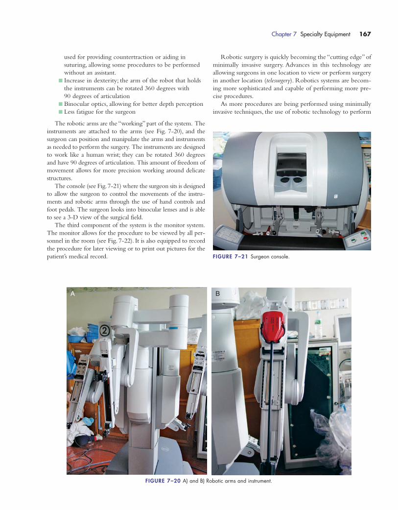
used for providing countertraction or aiding in
suturing, allowing some procedures to be performed
without an assistant.
� Increase in dexterity; the arm of the robot that holds
the instruments can be rotated 360 degrees with
90 degrees of articulation
� Binocular optics, allowing for better depth perception
� Less fatigue for the surgeon
The robotic arms are the “working” part of the system. The
instruments are attached to the arms (see Fig. 7-20), and the
surgeon can position and manipulate the arms and instruments
as needed to perform the surgery. The instruments are designed
to work like a human wrist; they can be rotated 360 degrees
and have 90 degrees of articulation. This amount of freedom of
movement allows for more precision working around delicate
structures.
The console (see Fig.7-21) where the surgeon sits is designed
to allow the surgeon to control the movements of the instru-
ments and robotic arms through the use of hand controls and
foot pedals. The surgeon looks into binocular lenses and is able
to see a 3-D view of the surgical field.
The third component of the system is the monitor system.
The monitor allows for the procedure to be viewed by all per-
sonnel in the room (see Fig. 7-22). It is also equipped to record
the procedure for later viewing or to print out pictures for the
patient’s medical record.
Robotic surgery is quickly becoming the “cutting edge” of
minimally invasive surgery. Advances in this technology are
allowing surgeons in one location to view or perform surgery
in another location (telesurgery). Robotics systems are becom-
ing more sophisticated and capable of performing more pre-
cise procedures.
As more procedures are being performed using minimally
invasive techniques, the use of robotic technology to perform
Chapter 7 Specialty Equipment 167
FIGURE 7–20 A) and B) Robotic arms and instrument.
A B
FIGURE 7–21 Surgeon console.
1572_Ch07_157-170.qxd 8/6/09 11:46 AM Page 167
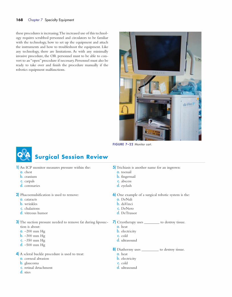
these procedures is increasing.The increased use of this technol-
ogy requires scrubbed personnel and circulators to be familiar
with the technology, how to set up the equipment and attach
the instruments and how to troubleshoot the equipment. Like
any technology, there are limitations. As with any minimally
invasive procedure, the OR personnel must to be able to con-
vert to an “open” procedure if necessary. Personnel must also be
ready to take over and finish the procedure manually if the
robotics equipment malfunctions.
168 Chapter 7 Specialty Equipment
FIGURE 7–22 Monitor cart.
Surgical Session Review
1) An ICP monitor measures pressure within the:
a. chest
b. cranium
c. carpals
d. coronaries
2) Phacoemulsification is used to remove:
a. cataracts
b. wrinkles
c. chalazions
d. vitreous humor
3) The suction pressure needed to remove fat during liposuc-
tion is about:
a. –200 mm Hg
b. –300 mm Hg
c. –350 mm Hg
d. –500 mm Hg
4) A scleral buckle procedure is used to treat:
a. corneal abrasion
b. glaucoma
c. retinal detachment
d. sties
5) Trichiasis is another name for an ingrown:
a. toenail
b. fingernail
c. abscess
d. eyelash
6) One example of a surgical robotic system is the:
a. DeNali
b. daVinci
c. DeNero
d. DeTrussor
7) Cryotherapy uses ________ to destroy tissue.
a. heat
b. electricity
c. cold
d. ultrasound
8) Diathermy uses _________ to destroy tissue.
a. heat
b. electricity
c. cold
d. ultrasound
1572_Ch07_157-170.qxd 8/6/09 11:46 AM Page 168

9) A vacuum curettage machine would be used during
which of the following procedures?
a. hysterectomy
b. oophorectomy
c. D&E
d. cerclage
10) The cardiopulmonary bypass machine is operated by a:
a. surgeon
b. nurse
c. surgical technologist
d. perfusionist
11) In which of the following procedures would cardiopul-
monary bypass definitely not be used:
a. repair of tetralogy of Fallot
b. heart transplant
c. aortofemoral bypass graft
d. CABG
12) The straight shot device is used for surgery of the:
a. hand
b. foot
c. back
d. sinuses
13) A coblator is used most commonly for a:
a. coronary bypass graft
b. cerclage
c. tonsillectomy
d. adenoma removal
14) The eyepiece of a microscope is the ________ lens.
a. objective
b. ocular
c. ophthalmic
d. oculomotor
15) Examples of bone implants include all of the following
except:
a. staple
b. cancellous screw
c. fiberglass cast
d. femoral nail
16) Which statement is false regarding handling of implants:
a. handle as little as possible
b. always measure any screw prior to handing it to the
surgeon
c. implants may be reused
d. do not use any implant that has been damaged
17) The proper order for the steps for screw insertion is:
a. measure, drill, tap, screw
b. tap, measure, drill, screw
c. drill, measure, tap, screw
d. drill, tap, measure, screw
18) A bone tap is used to:
a. create ridges for the screw to follow
b. determine the length of screw needed
c. screw the plate to the bone
d. drill the hole for the screw
19) A CUSA or Sonopet machine is used for:
a. excising abdominal tumors
b. excising brain tumors
c. excising bone tumors
d. excising breast tumors
20) Which of the following is not an advantage of robotic
surgery?
a. hand tremors are minimized or eliminated
b. increased dexterity for instrument manipulation
c. decreased cost of machinery
d. improved depth perception due to binocular optics
Chapter 7 Specialty Equipment 169
1572_Ch07_157-170.qxd 8/6/09 11:46 AM Page 169

1572_Ch07_157-170.qxd 8/6/09 11:46 AM Page 170
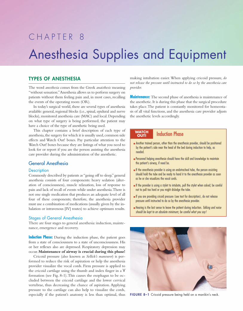
TYPES OF ANESTHESIA
The word anesthesia comes from the Greek anaisthesis meaning
“without sensation.”Anesthesia allows us to perform surgery on
patients without them feeling pain and, in most cases, recalling
the events of the operating room (OR).
In today’s surgical world, there are several types of anesthesia
available: general, regional blocks (i.e., spinal, epidural and nerve
blocks), monitored anesthesia care (MAC) and local. Depending
on what type of surgery is being performed, the patient may
have a choice of the type of anesthetic being used.
This chapter contains a brief description of each type of
anesthesia, the surgery for which it is usually used, common side
effects and Watch Out! boxes. Pay particular attention to the
Watch Out! boxes because they are listings of what you need to
look for or report if you are the person assisting the anesthesia
care provider during the administration of the anesthetic.
General AnesthesiaDescriptionCommonly described by patients as “going off to sleep,” general
anesthesia consists of four components: heavy sedation (alter-
ation of consciousness), muscle relaxation, loss of response to
pain and lack of recall of events while under anesthesia.There is
not one single medication that produces an adequate level of all
four of these components; therefore, the anesthesia provider
must use a combination of medications (usually given by the in-
halation or intravenous [IV] routes) to achieve optimum results.
Stages of General AnesthesiaThere are four stages to general anesthesia: induction, mainte-
nance, emergence and recovery.
Induction Phase: During the induction phase, the patient goes
from a state of consciousness to a state of unconsciousness. His
or her reflexes also are depressed. Respiratory depression may
occur.Maintenance of airway is crucial during this phase!
Cricoid pressure (also known as Sellick’s maneuver) is per-
formed to reduce the risk of aspiration or help the anesthesia
provider visualize the vocal cords. Firm pressure is applied to
the cricoid cartilage using the thumb and index finger in a Vformation (see Fig. 8-1).This causes the esophagus to be oc-
cluded between the cricoid cartilage and the lower cervical
vertebrae, thus decreasing the chance of aspiration. Applying
pressure to the cartilage can also help to visualize the cords,
especially if the patient’s anatomy is less than optimal, thus
making intubation easier.When applying cricoid pressure, do
not release the pressure until instructed to do so by the anesthesia care
provider.
Maintenance: The second phase of anesthesia is maintenance of
the anesthetic. It is during this phase that the surgical procedure
takes place.The patient is constantly monitored for homeosta-
sis of all vital functions, and the anesthesia care provider adjusts
the anesthetic levels accordingly.
C H A P T E R 8
Anesthesia Supplies and Equipment
FIGURE 8–1 Cricoid pressure being held on a manikin’s neck.
Induction Phase
� Another trained person, other than the anesthesia provider, should be positionedby the patient’s side near the head of the bed during induction to help, asneeded.
� Personnel helping anesthesia should have the skill and knowledge to maintainthe patient’s airway, if need be.
� If the anesthesia provider is using an endotracheal tube, the person assistingshould hold the tube and be ready to hand it to the anesthesia provider as soonas he or she visualizes the vocal cords.
� If the provider is using a stylet to intubate, pull the stylet when asked; be carefulnot to pull too hard or you might dislodge the tube.
� If you are providing cricoid pressure (see text for description), do not releasepressure until instructed to do so by the anesthesia provider.
� Hearing is the last sense to leave the patient during induction. Talking and noiseshould be kept to an absolute minimum; be careful what you say!
WATCH OUT!
1572_Ch08_171-230.qxd 8/6/09 11:54 AM Page 171
Emergence: Emergence is the third phase of general anesthesia.
This phase occurs as the surgical procedure is finishing. The
goal is to have the patient as awake as possible at the end of
the procedure. Monitoring the patient’s ability to resume
breathing on his or her own and restoring a gag reflex are the
focus during emergence. The patient is usually extubated, if
necessary. A major concern during this phase is the risk of
laryngospasm or bronchospasm, which can lead to airway ob-
struction.The airway must be monitored closely.
Recovery: The last phase of general anesthesia is recovery. In
this phase, the patient returns to his or her optimal level of
consciousness.This phase begins in the OR, continues in the
PACU and may continue through discharge. Patients are
closely monitored in the OR and PACU for signs of increas-
ing levels of consciousness and function.
Procedures Performed Under General AnesthesiaAny surgical procedure can be performed while the patient is
under general anesthesia. The anesthesia care provider, along
with the patient and surgeon, decide if the patient is a candidate
for general anesthesia.
Advantages� Medication dosages can be adjusted as needed.
� Muscle relaxation can be achieved, making for easier
visualization within the surgical field.
� Rate and depth of respiration can be controlled.
� Patient is usually unaware of what goes on
intraoperatively.
Disadvantages� A small percentage of patients are aware of
intraoperative events.
� General anesthesia is not for everyone. If the patient
has many co-morbidities, then he or she may not be a
good candidate to undergo general anesthesia.
� Risk of aspiration is greatest during the induction and
emergence phases.
� Risk of laryngospasm or bronchospasm is real.
� Risk of malignant hyperthermia is real, although rare,
as anesthesia personnel screen patients preoperatively
for risk of malignant hyperthermia.
� There is an increased chance of postoperative nausea
and vomiting.
� There is an increased chance of postoperative pain
when compared with regional anesthesia.
Regional AnesthesiaSpinal AnesthesiaTo perform a spinal block, the anesthesia care provider injects
an anesthetic agent into the subarachnoid space using a spinal
needle inserted into an intervertebral space in the lower lum-
bar area.The patient experiences loss of sensation below the
diaphragm.
Procedures Performed Under Spinal AnesthesiaSpinal anesthesia can be used for procedures below the waist.
It is most commonly used for orthopedic procedures on the
lower limbs (e.g., total knee arthroplasty, total hip arthroplasty),
genitourinary procedures and gynecological procedures.
Advantages� Patients remain conscious and in control of their own
airways.
� Patients can receive sedation that is titrated to their
desired levels of consciousness during the procedure.
� Patients suffer less systemic effects with spinal anesthe-
sia than with general anesthesia; therefore, spinal anes-
thesia may be a less risky choice of anesthetic for
those with multiple co-morbidities.
172 Chapter 8 Anesthesia Supplies and Equipment
Maintenance Phase
� The circulator needs to be aware of what is going on with the anesthesia careprovider. Increased activity “behind the screen” could indicate a change in patientcondition. Always keep an eye and ear out and be ready to respond if help isneeded.
� Scrubbed personnel need to keep track of the amount of irrigating fluid used andreport that number to the anesthesia provider. This is an important step in estimating blood loss.
� Scrubbed personnel need to tell the anesthesia provider when they are about toirrigate with large amounts of fluid. The anesthesia provider needs to be aware ofthis extensive irrigation so that he or she will not think a large amount of bleeding has suddenly occurred.
� When local anesthetic is used during a procedure, scrubbed personnel need tokeep track of the amount injected. The amount, type and strength of local anesthetic used needs to be reported to the anesthesia care provider.
WATCH OUT!
Emergence Phase
� Another trained person, other than the anesthesia provider, should be positionedby the patient’s side near the head of the bed during emergence to help asneeded.
� Personnel helping anesthesia should have the skill and knowledge to maintainthe patient’s airway if need be.
� Hearing is often the first sense to return during emergence. Keep noise to an absolute minimum and be careful what is said!
� Be prepared to assist if needed after extubation. In rare cases, the patient mayneed to be reintubated.
� Have oxygen ready for transport to the post-anesthesia care unit (PACU). (Make sureyou have checked the amount of oxygen in the tank prior to the end of the case!)
WATCH OUT!
1572_Ch08_171-230.qxd 8/6/09 11:54 AM Page 172
� Patients do not experience the sore throat or hoarse-
ness postoperatively that an endotracheal tube (ETT)
can cause.
� Produces bowel contraction, which can facilitate
exposure during surgery of the abdomen or
pelvis.
� Produces muscle relaxation, making retraction
easier.
Disadvantages� Hypotension! Monitor the patient for signs of
decreased blood pressure.
� The spinal block could travel upward and block the
muscles necessary for respiration. Monitor the patient
for signs of difficulty breathing.
� In a small percentage of cases, the spinal anesthetic is
not effective and the patient must be given a general
anesthetic.
� The dosage of medication cannot be titrated. Once it
is given, it cannot be adjusted.
� It is possible for the patient to experience a spinal
headache postoperatively.
� The patient could possibly develop temporary or per-
manent paraesthesia or paralysis (very rare).
� Spinal anesthesia cannot be used for procedures of
long duration.The length of time you get depends on
the anesthetic agent that is used.
Epidural AnesthesiaEpidural anesthesia is accomplished by the anesthesia care
provider injecting an anesthetic agent into the epidural space
that surrounds the dural sac.The anesthetic agent is absorbed
slowly through the dura mater into the cerebrospinal fluid.
Epidural anesthesia can be a single shot of anesthetic agent
or, more commonly, a small catheter is inserted into the
back and an intermittent or continuous drip of medication
is administered through the catheter to provide ongoing pain
control.
Procedures Performed Under Epidural AnesthesiaEpidural anesthesia is commonly used for procedures of the
rectum and pelvis (e.g., obstetrical, gynecological and urolog-
ical). It can be used for intraoperative and postoperative pain
control or, in some cases, control of intractable pain.
Advantages� Medication dosage can be titrated.
� When the need is no longer there, the catheter can be
removed.
� Postoperative pain control can be achieved yet the
patient is able to walk and move about.
� The medication pump delivering the agent can be
programmed to allow the patient to deliver
supplemental doses of medication, as needed.
� The patient remains conscious and cooperative
(important during labor).
� Patient remains in control of his or her own
airway.
Disadvantages� Hypotension can result, although more slowly than
with a spinal block.
� In rare cases, the epidural may not be effective, neces-
sitating a switch to another type of anesthetic.
� Accidental puncture of the dura during the insertion
could result in total spinal anesthesia leading to respi-
ratory paralysis and bradycardia.
Combined Spinal/Epidural AnesthesiaDescriptionAn epidural needle is passed into the epidural space in the
usual manner. A spinal needle is passed through the epidural
needle until the dura is pierced. Medication is injected into
the subarachnoid space.The spinal needle is removed and an
epidural catheter is placed into the epidural space.
Chapter 8 Anesthesia Supplies and Equipment 173
Spinal Anesthesia
� Hypotension (sudden drop in blood pressure) is a major risk with spinal anesthe-sia. The person assisting the anesthesia care provider must keep an eye on theblood pressure monitor and report a drop of more than 20 systolic to the anesthesia provider immediately. Often, the anesthesia provider’s back is to themonitors while inserting the spinal needle; therefore, it is up to the person assist-ing to watch for hypotension. Continuous monitoring of vital signs is necessary.
� There is a small chance that the spinal block could move upward thereby blockingthe diaphragm and accessory respiratory muscles. The person assisting the anes-thesia care provider needs to monitor the patient for signs of difficulty breathingor respiratory distress.
� Proper positioning of the patient before needle insertion is important. A spinalanesthetic can be administered with the patient either lying on his or her side orsitting up and leaning forward (this is usually the anesthesia provider’s prefer-ence). The person assisting needs to help the patient into the proper position andhelp the patient maintain that position throughout the procedure; this may requireholding the patient in position, especially if he or she has been sedated.
� Remember: Watch what is said in the room. The patient is awake!
WATCH OUT! Epidural Anesthesia
� The agent is more slowly absorbed; therefore, hypotension develops more slowlythan with a spinal block, making it easier to treat.
� Accidental puncture of the dura during needle insertion could result in total spinalanesthesia. Signs of total spinal anesthesia include bradycardia, respiratory paralysis and vasodilation.
� Continuous monitoring of vital signs is necessary during insertion of an epidural.
� Remember: Watch what is said in the room; the patient is awake!
WATCH OUT!
1572_Ch08_171-230.qxd 8/6/09 11:54 AM Page 173
Procedures Performed Under Spinal/EpiduralAnesthesiaCombined spinal/epidural anesthesia is commonly used for
obstetrical procedures or labor pain relief.This technique can
also be used for surgical procedures that have extended surgery
times.
Advantages� Spinal/epidural anesthesia provides immediate benefits
from the spinal injection, but use of the epidural
catheter allows for continued anesthesia.
� This type of anesthesia offers less motor block than
spinal anesthesia alone.
� Patient remains conscious and cooperative, which is
important during labor.
� Patient remains in control of his or her own airway.
Disadvantages� Hypotension! Monitor the patient for signs of
decreased blood pressure.
� There is the potential for the patient to experience a
spinal headache postoperatively.
� The patient could potentially develop temporary or
permanent paraesthesia or paralysis (very rare).
Nerve BlockNerve blocks are another option for regional anesthesia.The
anesthesia care provider injects an anesthetic agent into either
a large peripheral nerve or the area of a major nerve plexus (a
large bundle or group of nerves).This type of anesthesia allows
for temporary loss of sensation to the area innovated by the
nerve or plexus.
Procedures Performed Under Nerve BlockNerve blocks are commonly used for procedures on the arm or
leg. In addition to intraoperative anesthesia, nerve blocks help
control postoperative pain (many agents last 18 to 24 hours). In
selected cases, a nerve block can be used to control intractable
pain (e.g., injection of the intercostal nerves to control rib pain).
Advantages� Nerve block anesthesia can be given to a specific area
only, without the systemic impact of a general or
spinal anesthetic.
� Nerve blocks allow analgesia to continue into the
postoperative period.
� The duration of the block is medication-specific,
allowing it to be tailored to the specific needs of the
patient and/or procedure.
� Patient is conscious and in control of his or her own
airway
� Nerve blocks can be combined with other types of
anesthesia (e.g., general).
Disadvantages� Anesthetic overdose could occur if a major artery is
accidently injected, causing seizures or cardiac arrest.
Bier BlockA Bier block is a regional block of the distal end of an upper
extremity. A tourniquet is placed on the upper arm, and the
anesthetic is injected intravenously at an area close to the sur-
gical site.The limb is exsanguinated using an Esmarch band-
age, and the proximal tourniquet is inflated to about 100 mm
Hg above the patient’s systolic blood pressure.The tourniquet
remains inflated throughout the procedure to prevent the
anesthetic agent from being redistributed from the area.
Procedures Performed Under Bier BlocksProcedures of the distal upper extremity that are less then 1 hour
in duration (e.g., closed reduction of a Colles fracture)
Advantages� Bier block is a short-acting, localized anesthesia
without systemic impact.
174 Chapter 8 Anesthesia Supplies and Equipment
Bier Block
� If the tourniquet fails or the tourniquet is released too rapidly at the end of theprocedure, the patient may experience systemic administration of the medication.Systemic administration can adversely affect the cardiovascular system, causingsudden changes in heart rate and blood pressure. The patient’s vital signs shouldbe closely monitored throughout the injection, surgical procedure and release ofthe tourniquet.
� Remember: Watch what is said in the room; the patient is awake!
WATCH OUT!
Nerve Blocks
� Most of the major nerves run near major arteries. If the medication is accidentlyinjected into the artery, anesthetic overdose could occur. The patient’s vital signsshould be monitored closely throughout the procedure.
� The anesthesia care provider may ask the person assisting him or her to injectthe medication once the correct spot is found. The assistant should aspirate (pullback slightly on the plunger) then inject 5 to 10 cc of the medication slowly. Theassistant should aspirate every 5 to 10 cc until the anesthetic is injected.
� Remember: Watch what is said in the room; the patient is awake!
WATCH OUT!
Combined Spinal/Epidural Anesthesia
� Same as for epidural and spinal anesthesia.
� Some patients experience pruritus (itching) after injection of the narcotic.
� Remember: Watch what is said in the room; the patient is awake!
WATCH OUT!
1572_Ch08_171-230.qxd 8/6/09 11:54 AM Page 174
� It has a short recovery time after the procedure.
� Patient is conscious and in control of his or her own
airway.
Disadvantages� It has limited uses (distal upper extremity procedures
of short duration).
� Patient could experience adverse cardiovascular effects
due to systemic administration of the medication.
Monitored Anesthesia CareThe patient receives sedatives, analgesics or amnesics to make
him or her less aware of intraoperative events.This is done in
conjunction with a local anesthetic at the surgical site.
Procedures Performed Under Monitored Anesthesia CareMonitored anesthesia care (MAC) can be used for procedures
that can be done using local anesthesia, when the patient
wants some sedation.
Advantages� MAC can be used for patients with complex medical
problems who are undergoing procedures requiring
only local anesthesia
� It has fewer systemic effects than other types of
anesthesia.
� Medication dosages can be titrated.
� There is a short recovery time after the procedure.
� Patient is conscious and in control of his or her own
airway.
Disadvantages� MAC cannot be used for complex procedures.
� Patient must be able to be cooperative (e.g., not
moving the area the surgeon is working on).
Local AnesthesiaLocal anesthesia is produced when the surgeon injects an anes-
thetic agent into the tissue surrounding the operative site.This
produces a small, localized area of anesthesia/analgesia at the
operative site.The length of the anesthesia/analgesia depends
on the agent used.
Procedures Performed Under Local AnesthesiaLocal anesthesia can be used for simple or short procedures
(e.g., wound suturing, pacemaker insertion, chest tube inser-
tion and removal of small skin lesions). Some surgeons perform
breast biopsy and hernia repairs using local anesthesia.
Advantages� Local anesthesia can be used for patients with
complex medical problems who are undergoing
procedures requiring only local anesthesia.
� Local anesthesia has fewer systemic effects than other
types of anesthesia.
� There is a short recovery time after the procedure
than when general anesthesia is used.
� Patient is conscious and in control of his or her own
airway.
� Surgeon can regulate the amount of medication that
the patient receives based on the patient’s needs.
Disadvantages� Cannot be used for complex procedures.
� Patient must be able to be cooperative (e.g., not
moving the area the surgeon is working on).
� May require another RN to be assigned to monitor
the patient, depending on institution policy.
Topical AnesthesiaTopical anesthesia is produced by instilling or swabbing a local
anesthetic agent onto mucous membranes or tissue.
Procedures Performed Under Topical AnesthesiaTopical anesthesia can be used for minor, short-duration proce-
dures of the mucous membranes or skin (e.g., skin tag removal,
Chapter 8 Anesthesia Supplies and Equipment 175
Monitored Anesthesia Care
� Supplemental oxygen should be available.
� The patient’s vital signs (especially respiratory status) and level of consciousnessneed to be closely monitored; usually this is done by the anesthesia careprovider, but the circulator must be aware of what is happening.
� Scrubbed personnel need to keep track of the amount of local anesthetic injectedat the surgical site. They need to the anesthesia care provider the type, strengthand amount of local used.
� Remember: Watch what is said in the room; the patient is awake!
WATCH OUT!
Local Anesthesia: Topical Anesthesia
� Depending on institution policy, if the patient is undergoing only local anesthesia,a registered nurse (RN) may be monitoring the patient’s vital signs intraopera-tively. The RN should NOT be the same nurse who is performing the circulatingduties; a dedicated nurse should be assigned to monitor the patient.
� Supplemental oxygen should be available.
� If sedation (e.g., Versed) is being used, the institution’s policy for conscious sedation must be followed.
� Scrubbed personnel need to keep track of the amount of local anesthetic injectedat the surgical site and report to the person monitoring the patient the type,strength and amount of local used.
� Remember: Watch what is said in the room; the patient is awake!
WATCH OUT!
1572_Ch08_171-230.qxd 8/6/09 11:54 AM Page 175
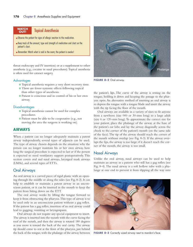
throat endoscopy and IV insertion) or as a supplement to other
anesthesia (e.g., cocaine in nasal procedures).Topical anesthesia
is often used for cataract surgery.
Advantages� Topical anesthesia requires a very short recovery time.
� There are fewer systemic effects following topical
than other types of anesthesia.
� Patient is conscious and in control of his or her own
airway.
Disadvantages� Topical anesthesia cannot be used for complex
procedures.
� Patient must be able to be cooperative (e.g., not
moving the area the surgeon is working on).
AIRWAYS
When a patient can no longer adequately maintain a patent
airway independently, several types of adjuncts can be used.
The type of airway chosen depends on the situation: why the
person can no longer maintain his or her own airway, how
long the surgical procedure is expected to last or if the person
is expected to need ventilatory support postoperatively. This
section covers oral and nasal airways, laryngeal mask airways
(LMAs), and several types of ETTs.
Oral AirwaysAn oral airway is a curved piece of rigid plastic with an open-
ing through the middle or along the sides (see Fig. 8-2). It can
help to establish or maintain a patent airway in an uncon-
scious patient, or it can be inserted in the mouth to keep the
patient from biting down on the ETT.
The oral airway works by lifting the tongue forward to
keep it from obstructing the pharynx.This type of airway is to
be used only in an unconscious patient without a gag reflex.
If the patient has a gag reflex, insertion of an oral airway could
lead to gagging, vomiting or laryngospasm.
Oral airways do not require any special equipment to insert.
The airway is inserted into the mouth with the curve facing the
roof of the mouth, and then the airway is rotated 180 degrees
as you move it toward the back of the oral cavity. The airway
tip should come to rest at the front of the pharynx, just behind
the back of the tongue,with the phalange of the airway between
the patient’s lips. The curve of the airway is resting on the
tongue, holding it down and keeping the passage to the phar-
ynx open.An alternative method of inserting an oral airway is
to depress the tongue with a tongue blade and insert the airway
with the tip facing the floor of the mouth.
Oral airways are available in a variety of sizes to fit anyone
from a newborn (size 000 or 30-mm long) to a large adult
(size 6 or 120-mm long).To approximate the correct size for
your patient, place the phalange of the airway at the base of
the patient’s ear lobe and lay the airway diagonally across the
cheek to the corner of the patient’s mouth (on the same side
of the face).The tip of the airway should reach the corner of
the mouth without overlap (see Fig. 8-3). If the airway over-
laps the lips, the airway is too large; if it doesn’t reach the cor-
ner of the mouth, the airway is too small.
Nasal AirwaysUnlike the oral airway, nasal airways can be used to help
maintain an airway in a patient who still has a gag reflex (see
Fig. 8-4).The nasal airway is a soft hollow tube with a pha-
lange at one end to prevent it from slipping all the way into
176 Chapter 8 Anesthesia Supplies and Equipment
Topical Anesthesia
� Observe the patient for signs of allergic reaction to the medication.
� Keep track of the amount, type and strength of medication and chart on the patient’s chart.
� Remember: Watch what is said in the room; the patient is awake!
WATCH OUT!
Tip
Phalange
FIGURE 8–2 Oral airway.
FIGURE 8–3 Correctly sized airway next to manikin’s face.
1572_Ch08_171-230.qxd 8/6/09 11:54 AM Page 176
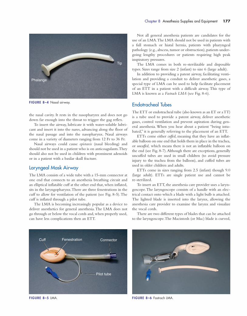
the nasal cavity. It rests in the nasopharynx and does not go
down far enough into the throat to trigger the gag reflex.
To insert the airway, lubricate it with water-soluble lubri-
cant and insert it into the nares, advancing along the floor of
the nasal passage and into the nasopharynx. Nasal airways
come in a variety of diameters ranging from 12 Fr to 36 Fr.
Nasal airways could cause epistaxis (nasal bleeding) and
should not be used in a patient who is on anticoagulants.They
should also not be used in children with prominent adenoids
or in a patient with a basilar skull fracture.
Laryngeal Mask AirwayThe LMA consists of a wide tube with a 15-mm connector at
one end that connects to an anesthesia breathing circuit and
an elliptical inflatable cuff at the other end that, when inflated,
sits in the laryngopharynx.There are three fenestrations in the
cuff to allow for ventilation of the patient (see Fig. 8-5).The
cuff is inflated through a pilot tube.
The LMA is becoming increasingly popular as a device to
deliver anesthetics for general anesthesia.The LMA does not
go through or below the vocal cords and, when properly used,
can have less complications then an ETT.
Not all general anesthesia patients are candidates for the
use of an LMA.The LMA should not be used in patients with
a full stomach or hiatal hernia, patients with pharyngeal
pathology (e.g., abscess, tumor or obstruction), patients under-
going lengthy procedures or patients requiring high peak
inspiratory pressures.
The LMA comes in both re-sterilizable and disposable
types. Sizes range from size 2 (infant) to size 6 (large adult).
In addition to providing a patent airway, facilitating venti-
lation and providing a conduit to deliver anesthetic gases, a
special type of LMA can be used to help facilitate placement
of an ETT in a patient with a difficult airway. This type of
LMA is known as a Fastrach LMA (see Fig. 8-6).
Endotracheal TubesThe ETT or endotracheal tube (also known as an ET or a TT)
is a tube used to provide a patent airway, deliver anesthetic
gases, control ventilation and prevent aspiration during gen-
eral anesthesia. When you hear about a patient “being intu-
bated,” it is generally referring to the placement of an ETT.
ETTs come either cuffed, meaning that they have an inflat-
able balloon on one end that holds them in place in the trachea,
or uncuffed, which means there is not an inflatable balloon on
the end (see Fig. 8-7).Although there are exceptions, generally
uncuffed tubes are used in small children (to avoid pressure
injury to the trachea from the balloon), and cuffed tubes are
used in older children and adults.
ETTs come in sizes ranging from 2.5 (infant) though 9.0
(large adult). ETTs are single patient use and cannot be
re-sterilized.
To insert an ETT, the anesthesia care provider uses a laryn-
goscope.The laryngoscope consists of a handle with an elec-
trical contact onto which a blade with a light bulb is attached.
The lighted blade is inserted into the larynx, allowing the
anesthesia care provider to examine the larynx and visualize
the vocal cords.
There are two different types of blades that can be attached
to the laryngoscope.The Macintosh (or Mac) blade is curved,
Chapter 8 Anesthesia Supplies and Equipment 177
Tip
Phalange
FIGURE 8–4 Nasal airway.
Cuff Fenestration
Pilot tube
Connector
FIGURE 8–5 LMA. FIGURE 8–6 Fastrach LMA.
1572_Ch08_171-230.qxd 8/6/09 11:54 AM Page 177
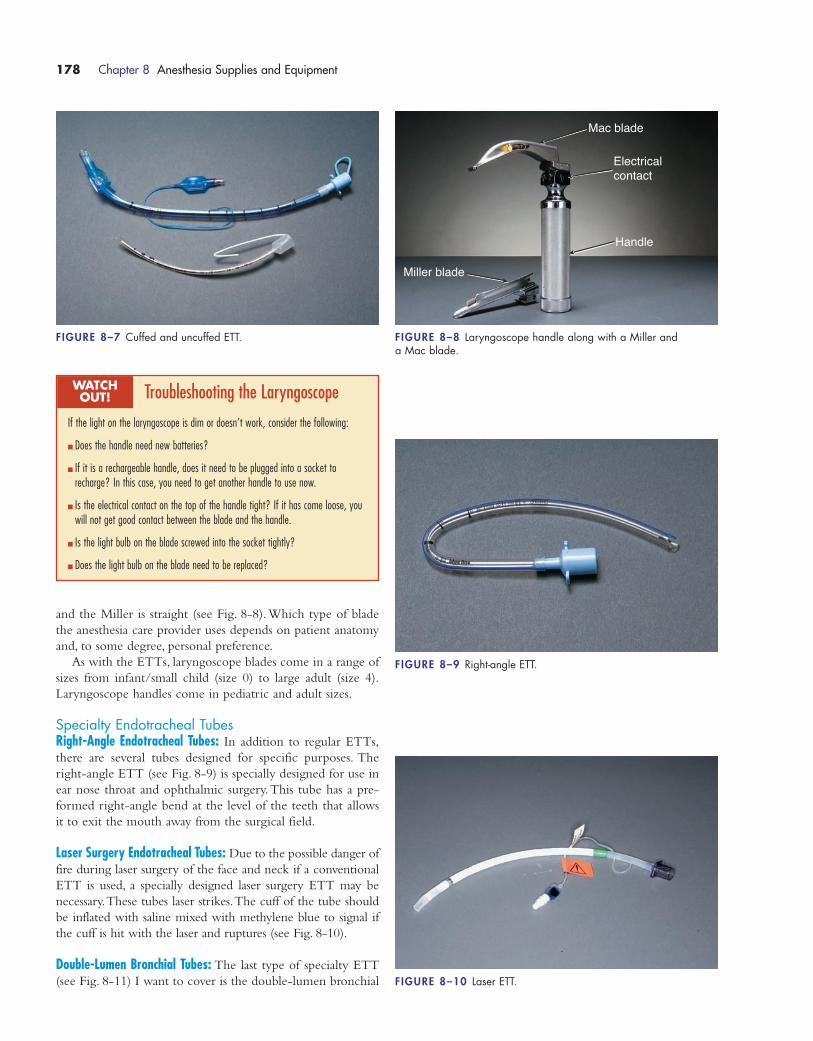
and the Miller is straight (see Fig. 8-8).Which type of blade
the anesthesia care provider uses depends on patient anatomy
and, to some degree, personal preference.
As with the ETTs, laryngoscope blades come in a range of
sizes from infant/small child (size 0) to large adult (size 4).
Laryngoscope handles come in pediatric and adult sizes.
Specialty Endotracheal TubesRight-Angle Endotracheal Tubes: In addition to regular ETTs,
there are several tubes designed for specific purposes. The
right-angle ETT (see Fig. 8-9) is specially designed for use in
ear nose throat and ophthalmic surgery.This tube has a pre-
formed right-angle bend at the level of the teeth that allows
it to exit the mouth away from the surgical field.
Laser Surgery Endotracheal Tubes: Due to the possible danger of
fire during laser surgery of the face and neck if a conventional
ETT is used, a specially designed laser surgery ETT may be
necessary.These tubes laser strikes.The cuff of the tube should
be inflated with saline mixed with methylene blue to signal if
the cuff is hit with the laser and ruptures (see Fig. 8-10).
Double-Lumen Bronchial Tubes: The last type of specialty ETT
(see Fig. 8-11) I want to cover is the double-lumen bronchial
178 Chapter 8 Anesthesia Supplies and Equipment
FIGURE 8–7 Cuffed and uncuffed ETT.
Mac blade
Electricalcontact
Handle
Miller blade
FIGURE 8–8 Laryngoscope handle along with a Miller and a Mac blade.
FIGURE 8–9 Right-angle ETT.
FIGURE 8–10 Laser ETT.
Troubleshooting the Laryngoscope
If the light on the laryngoscope is dim or doesn’t work, consider the following:
� Does the handle need new batteries?
� If it is a rechargeable handle, does it need to be plugged into a socket torecharge? In this case, you need to get another handle to use now.
� Is the electrical contact on the top of the handle tight? If it has come loose, youwill not get good contact between the blade and the handle.
� Is the light bulb on the blade screwed into the socket tightly?
� Does the light bulb on the blade need to be replaced?
WATCH OUT!
1572_Ch08_171-230.qxd 8/6/09 11:54 AM Page 178
tube.This type of tube is used for thoracic surgery. The advan-
tage to this tube is that it allows the anesthesia care provider to
ventilate one lung at a time or both together and allows suc-
tioning of either lung.
Tracheotomy TubesTracheotomy tubes are airways that are specially designed to
fit inside a tracheostomy stoma in order to keep it open and
provide a patent airway. A tracheostomy (also called a tra-
cheotomy) can be temporary or permanent, depending on the
need of the patient. It can be done as an emergency procedure
or as an elective procedure.Acute upper airway obstruction is
the most common reason for an emergency tracheostomy.
Conditions causing upper airway obstruction include tumor,
infection, foreign body, congenital or neurological conditions
and trauma. Electively, a tracheostomy is often performed for
patients who require long-term ventilator support or have a
condition causing chronic upper airway obstruction.
Tracheotomy tubes are available cuffed (with a balloon) or
uncuffed (see Fig. 8-12). Cuffed tracheotomy tubes are used
for adults, especially those on ventilators.When the patient is
on a ventilator, the cuff must be inflated to prevent air from
leaking out around the tube.The cuff may also help prevent
saliva or other secretions from entering the lungs. Like ETTs,
the uncuffed tracheotomy tubes are generally used on chil-
dren. Uncuffed tracheotomy tubes also can be used in adults
who do not require ventilatory support.
Tracheotomy tubes consist of an outer tube and an obtura-
tor (introducer).Many tracheotomy tubes also have a removable
inner cannula (see Fig. 8-13).The inner cannula slides inside the
outer tube and can be removed for ease of cleaning or to
unblock the tube if it becomes plugged with mucus.
Tracheotomy tubes are available in a variety of styles, can
be either metal or plastic and come with outer diameter sizes
ranging from 4 mm (infant) to 13 mm (large adult).
SUPPLEMENTAL OXYGEN
Oxygen is one of the most important gases in our environ-
ment.Without it, our cells could not function and we would
die.We need a constant supply of oxygen, usually from the air
we breathe. There are people whose bodies cannot supply
their cells with adequate oxygen from breathing, commonly
due to lung or heart disease, who need an additional oxygen
source, usually a portable or fixed oxygen tank.
Even if we are breathing adequately, there are times that
additional oxygen is needed. Preoperatively or intraopera-
tively, regional or MAC medication may be given to help
patients relax, but it can make them drowsy, less apt to take
deep breaths and in need of an additional source of oxygen.
Prior to induction of general anesthetic, patients are asked to
breathe oxygen to fill their lungs and give them an oxygen
reserve. Oxygen is given (along with the gases) during gen-
eral anesthetic to keep the body cells functioning properly.
Postoperatively, oxygen is given to help the patient wake up
from anesthesia as well as to help combat the effects of the
anesthetics and narcotics.
There are several devices that can be used to deliver sup-
plemental oxygen: nasal cannulas, reservoir and nonreservoir
masks, bag-valve-mask devices and nebulizers. These devices
and their use are covered in this section.
Chapter 8 Anesthesia Supplies and Equipment 179
FIGURE 8–11 Double-lumen ETT.
FIGURE 8–12 Cuffed and uncuffed tracheotomy tubes.
PhalangeOuter tube
CuffObturator
Innercannula
FIGURE 8–13 Parts of a tracheotomy tube.
1572_Ch08_171-230.qxd 8/6/09 11:54 AM Page 179
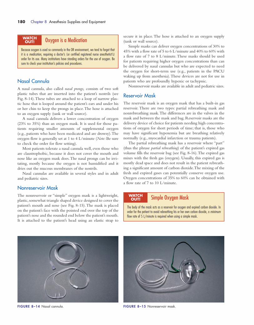
Nasal CannulaA nasal cannula, also called nasal prongs, consists of two soft
plastic tubes that are inserted into the patient’s nostrils (see
Fig. 8-14).These tubes are attached to a loop of narrow plas-
tic hose that is looped around the patient’s ears and under his
or her chin to keep the prongs in place.The hose is attached
to an oxygen supply (tank or wall source).
A nasal cannula delivers a lower concentration of oxygen
(23% to 35%) than an oxygen mask. It is used for those pa-
tients requiring smaller amounts of supplemental oxygen
(e.g., patients who have been medicated and are drowsy).The
oxygen flow is generally set at 1 to 4 L/minute (Note: Be sure
to check the order for flow setting).
Most patients tolerate a nasal cannula well, even those who
are claustrophobic, because it does not cover the mouth and
nose like an oxygen mask does.The nasal prongs can be irri-
tating, mostly because the oxygen is not humidified and it
dries out the mucous membranes of the nostrils.
Nasal cannulas are available in several styles and in adult
and pediatric sizes.
Nonreservoir MaskThe nonreservoir or “simple” oxygen mask is a lightweight,
plastic, somewhat triangle shaped device designed to cover the
patient’s mouth and nose (see Fig. 8-15).The mask is placed
on the patient’s face with the pointed end over the top of the
patient’s nose and the rounded end below the patient’s mouth.
It is attached to the patient’s head using an elastic strap to
secure it in place. The hose is attached to an oxygen supply
(tank or wall source).
Simple masks can deliver oxygen concentrations of 30% to
45% with a flow rate of 5 to 6 L/minute and 40% to 60% with
a flow rate of 7 to 8 L/minute.These masks should be used
for patients requiring higher oxygen concentrations than can
be delivered by nasal cannulas but who are expected to need
the oxygen for short-term use (e.g., patients in the PACU
waking up from anesthesia).These devices are not for use in
patients who are profoundly hypoxic or tachypnic.
Nonreservoir masks are available in adult and pediatric sizes.
Reservoir MaskThe reservoir mask is an oxygen mask that has a built-in gas
reservoir. There are two types: partial rebreathing mask and
nonrebreathing mask.The differences are in the valves in the
mask and between the mask and bag. Reservoir masks are the
delivery device of choice for patients needing high concentra-
tions of oxygen for short periods of time; that is, those who
may have significant hypoxemia but are breathing relatively
normally (e.g., myocardial infarction or trauma patients).
The partial rebreathing mask has a reservoir where “part”
(thus the phrase partial rebreathing) of the patient’s expired gas
volume fills the reservoir bag (see Fig. 8-16).The expired gas
mixes with the fresh gas (oxygen). Usually, this expired gas is
mostly dead space and does not result in the patient rebreath-
ing a significant amount of carbon dioxide.The mixing of the
fresh and expired gases can potentially conserve oxygen use.
Oxygen concentrations of 35% to 60% can be obtained with
a flow rate of 7 to 10 L/minute.
180 Chapter 8 Anesthesia Supplies and Equipment
Oxygen is a Medication
Because oxygen is used so commonly in the OR environment, we tend to forget thatit is a medication, requiring a doctor’s (or certified registered nurse anesthetist’s) order for its use. Many institutions have standing orders for the use of oxygen. Besure to check your institution’s policies and procedures.
WATCH OUT!
Simple Oxygen Mask
The body of the mask acts as a reservoir for oxygen and expired carbon dioxide. Inorder for the patient to avoid rebreathing his or her own carbon dioxide, a minimumflow rate of 5 L/minute is required when using a simple mask.
WATCH OUT!
FIGURE 8–14 Nasal cannula. FIGURE 8–15 Nonreservoir mask.
1572_Ch08_171-230.qxd 8/6/09 11:54 AM Page 180
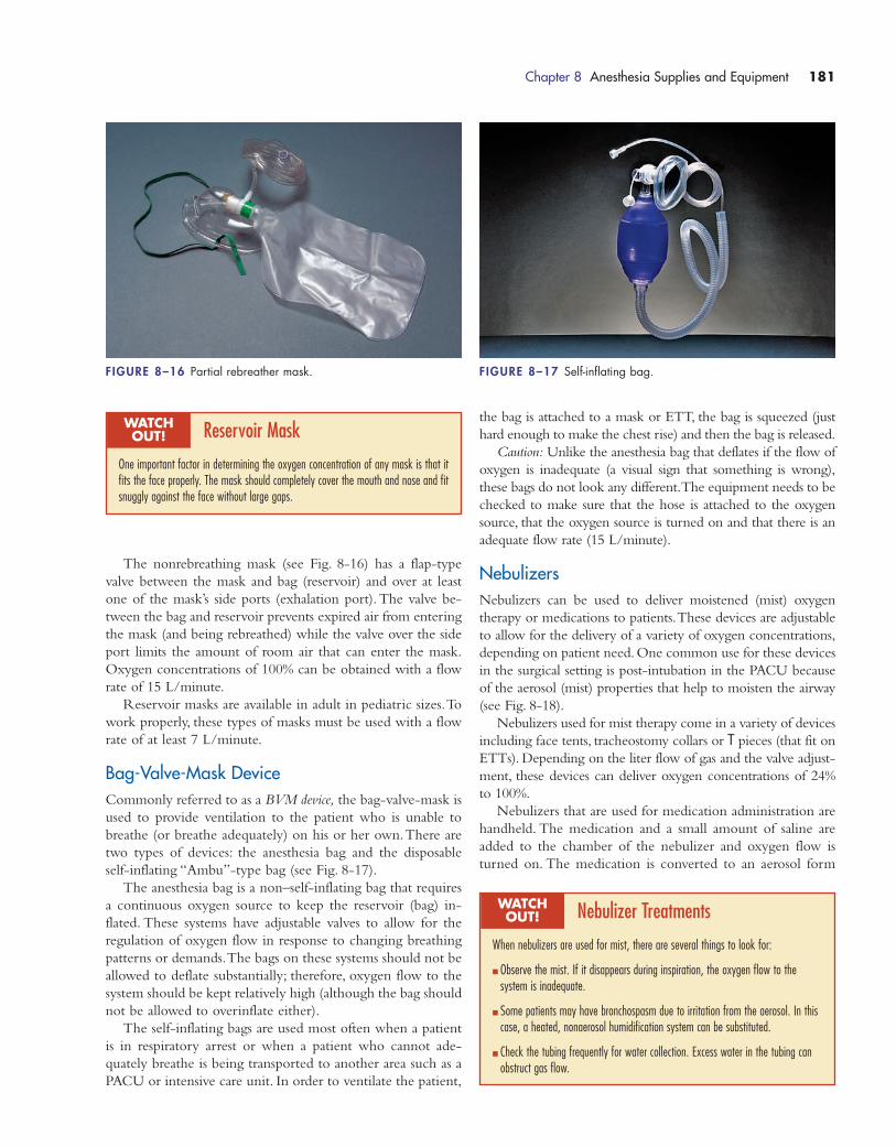
The nonrebreathing mask (see Fig. 8-16) has a flap-type
valve between the mask and bag (reservoir) and over at least
one of the mask’s side ports (exhalation port). The valve be-
tween the bag and reservoir prevents expired air from entering
the mask (and being rebreathed) while the valve over the side
port limits the amount of room air that can enter the mask.
Oxygen concentrations of 100% can be obtained with a flow
rate of 15 L/minute.
Reservoir masks are available in adult in pediatric sizes.To
work properly, these types of masks must be used with a flow
rate of at least 7 L/minute.
Bag-Valve-Mask DeviceCommonly referred to as a BVM device, the bag-valve-mask is
used to provide ventilation to the patient who is unable to
breathe (or breathe adequately) on his or her own.There are
two types of devices: the anesthesia bag and the disposable
self-inflating “Ambu”-type bag (see Fig. 8-17).
The anesthesia bag is a non–self-inflating bag that requires
a continuous oxygen source to keep the reservoir (bag) in-
flated. These systems have adjustable valves to allow for the
regulation of oxygen flow in response to changing breathing
patterns or demands.The bags on these systems should not be
allowed to deflate substantially; therefore, oxygen flow to the
system should be kept relatively high (although the bag should
not be allowed to overinflate either).
The self-inflating bags are used most often when a patient
is in respiratory arrest or when a patient who cannot ade-
quately breathe is being transported to another area such as a
PACU or intensive care unit. In order to ventilate the patient,
the bag is attached to a mask or ETT, the bag is squeezed (just
hard enough to make the chest rise) and then the bag is released.
Caution: Unlike the anesthesia bag that deflates if the flow of
oxygen is inadequate (a visual sign that something is wrong),
these bags do not look any different.The equipment needs to be
checked to make sure that the hose is attached to the oxygen
source, that the oxygen source is turned on and that there is an
adequate flow rate (15 L/minute).
NebulizersNebulizers can be used to deliver moistened (mist) oxygen
therapy or medications to patients.These devices are adjustable
to allow for the delivery of a variety of oxygen concentrations,
depending on patient need. One common use for these devices
in the surgical setting is post-intubation in the PACU because
of the aerosol (mist) properties that help to moisten the airway
(see Fig. 8-18).
Nebulizers used for mist therapy come in a variety of devices
including face tents, tracheostomy collars or T pieces (that fit on
ETTs). Depending on the liter flow of gas and the valve adjust-
ment, these devices can deliver oxygen concentrations of 24%
to 100%.
Nebulizers that are used for medication administration are
handheld. The medication and a small amount of saline are
added to the chamber of the nebulizer and oxygen flow is
turned on. The medication is converted to an aerosol form
Chapter 8 Anesthesia Supplies and Equipment 181
Reservoir Mask
One important factor in determining the oxygen concentration of any mask is that itfits the face properly. The mask should completely cover the mouth and nose and fitsnuggly against the face without large gaps.
WATCH OUT!
Nebulizer Treatments
When nebulizers are used for mist, there are several things to look for:
� Observe the mist. If it disappears during inspiration, the oxygen flow to the system is inadequate.
� Some patients may have bronchospasm due to irritation from the aerosol. In thiscase, a heated, nonaerosol humidification system can be substituted.
� Check the tubing frequently for water collection. Excess water in the tubing canobstruct gas flow.
WATCH OUT!
FIGURE 8–16 Partial rebreather mask. FIGURE 8–17 Self-inflating bag.
1572_Ch08_171-230.qxd 8/6/09 11:54 AM Page 181
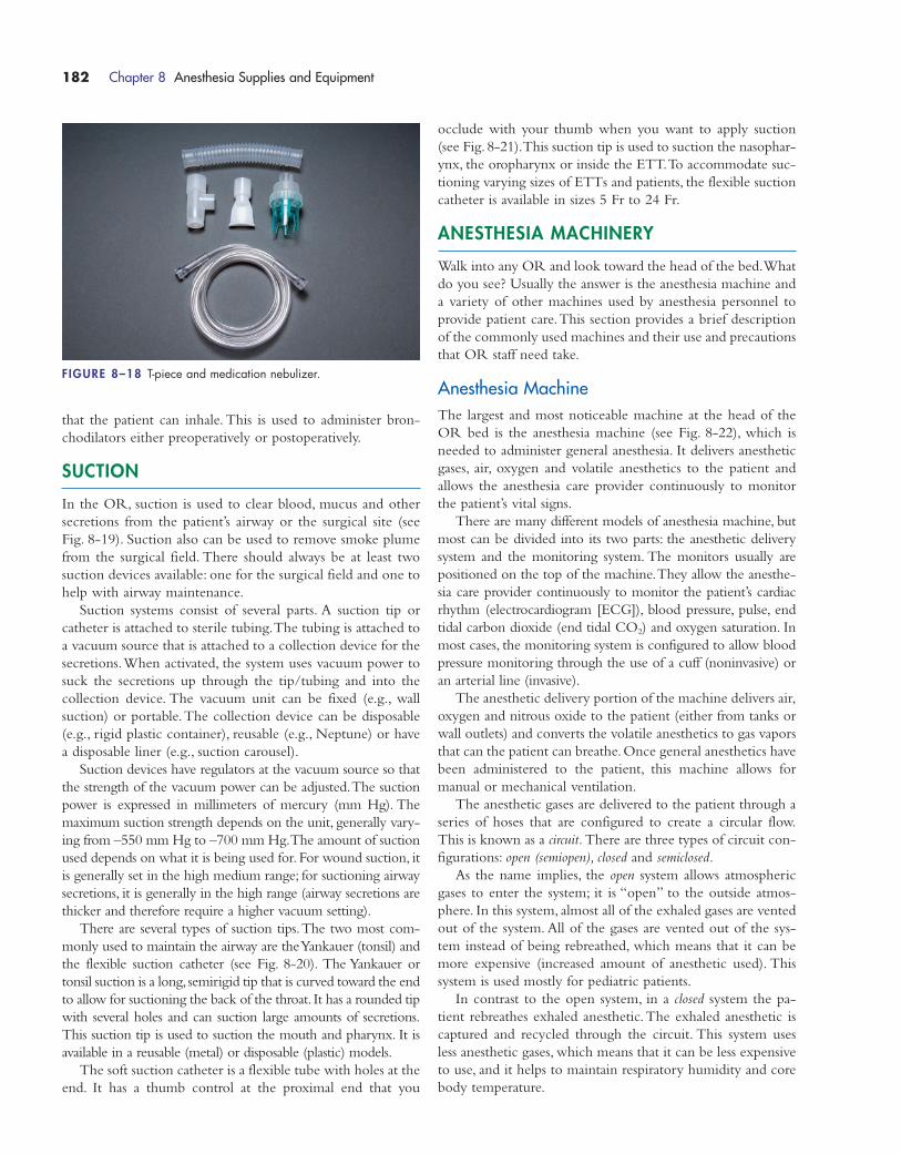
that the patient can inhale. This is used to administer bron-
chodilators either preoperatively or postoperatively.
SUCTION
In the OR, suction is used to clear blood, mucus and other
secretions from the patient’s airway or the surgical site (see
Fig. 8-19). Suction also can be used to remove smoke plume
from the surgical field. There should always be at least two
suction devices available: one for the surgical field and one to
help with airway maintenance.
Suction systems consist of several parts. A suction tip or
catheter is attached to sterile tubing.The tubing is attached to
a vacuum source that is attached to a collection device for the
secretions.When activated, the system uses vacuum power to
suck the secretions up through the tip/tubing and into the
collection device. The vacuum unit can be fixed (e.g., wall
suction) or portable.The collection device can be disposable
(e.g., rigid plastic container), reusable (e.g., Neptune) or have
a disposable liner (e.g., suction carousel).
Suction devices have regulators at the vacuum source so that
the strength of the vacuum power can be adjusted.The suction
power is expressed in millimeters of mercury (mm Hg). The
maximum suction strength depends on the unit, generally vary-
ing from –550 mm Hg to –700 mm Hg.The amount of suction
used depends on what it is being used for. For wound suction, it
is generally set in the high medium range; for suctioning airway
secretions, it is generally in the high range (airway secretions are
thicker and therefore require a higher vacuum setting).
There are several types of suction tips.The two most com-
monly used to maintain the airway are the Yankauer (tonsil) and
the flexible suction catheter (see Fig. 8-20). The Yankauer or
tonsil suction is a long, semirigid tip that is curved toward the end
to allow for suctioning the back of the throat. It has a rounded tip
with several holes and can suction large amounts of secretions.
This suction tip is used to suction the mouth and pharynx. It is
available in a reusable (metal) or disposable (plastic) models.
The soft suction catheter is a flexible tube with holes at the
end. It has a thumb control at the proximal end that you
occlude with your thumb when you want to apply suction
(see Fig. 8-21).This suction tip is used to suction the nasophar-
ynx, the oropharynx or inside the ETT.To accommodate suc-
tioning varying sizes of ETTs and patients, the flexible suction
catheter is available in sizes 5 Fr to 24 Fr.
ANESTHESIA MACHINERY
Walk into any OR and look toward the head of the bed.What
do you see? Usually the answer is the anesthesia machine and
a variety of other machines used by anesthesia personnel to
provide patient care.This section provides a brief description
of the commonly used machines and their use and precautions
that OR staff need take.
Anesthesia MachineThe largest and most noticeable machine at the head of the
OR bed is the anesthesia machine (see Fig. 8-22), which is
needed to administer general anesthesia. It delivers anesthetic
gases, air, oxygen and volatile anesthetics to the patient and
allows the anesthesia care provider continuously to monitor
the patient’s vital signs.
There are many different models of anesthesia machine, but
most can be divided into its two parts: the anesthetic delivery
system and the monitoring system. The monitors usually are
positioned on the top of the machine.They allow the anesthe-
sia care provider continuously to monitor the patient’s cardiac
rhythm (electrocardiogram [ECG]), blood pressure, pulse, end
tidal carbon dioxide (end tidal CO2) and oxygen saturation. In
most cases, the monitoring system is configured to allow blood
pressure monitoring through the use of a cuff (noninvasive) or
an arterial line (invasive).
The anesthetic delivery portion of the machine delivers air,
oxygen and nitrous oxide to the patient (either from tanks or
wall outlets) and converts the volatile anesthetics to gas vapors
that can the patient can breathe.Once general anesthetics have
been administered to the patient, this machine allows for
manual or mechanical ventilation.
The anesthetic gases are delivered to the patient through a
series of hoses that are configured to create a circular flow.
This is known as a circuit. There are three types of circuit con-
figurations: open (semiopen), closed and semiclosed.
As the name implies, the open system allows atmospheric
gases to enter the system; it is “open” to the outside atmos-
phere. In this system, almost all of the exhaled gases are vented
out of the system. All of the gases are vented out of the sys-
tem instead of being rebreathed, which means that it can be
more expensive (increased amount of anesthetic used). This
system is used mostly for pediatric patients.
In contrast to the open system, in a closed system the pa-
tient rebreathes exhaled anesthetic.The exhaled anesthetic is
captured and recycled through the circuit. This system uses
less anesthetic gases, which means that it can be less expensive
to use, and it helps to maintain respiratory humidity and core
body temperature.
182 Chapter 8 Anesthesia Supplies and Equipment
FIGURE 8–18 T-piece and medication nebulizer.
1572_Ch08_171-230.qxd 8/6/09 11:54 AM Page 182
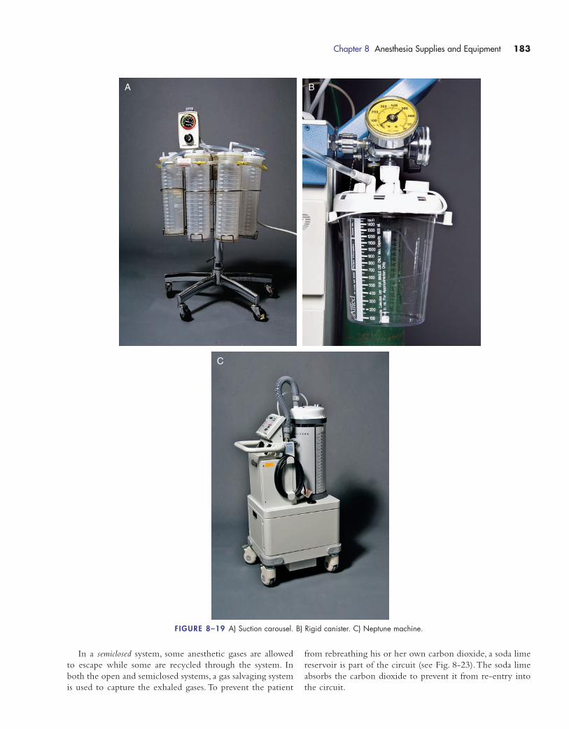
In a semiclosed system, some anesthetic gases are allowed
to escape while some are recycled through the system. In
both the open and semiclosed systems, a gas salvaging system
is used to capture the exhaled gases. To prevent the patient
from rebreathing his or her own carbon dioxide, a soda lime
reservoir is part of the circuit (see Fig. 8-23).The soda lime
absorbs the carbon dioxide to prevent it from re-entry into
the circuit.
Chapter 8 Anesthesia Supplies and Equipment 183
FIGURE 8–19 A) Suction carousel. B) Rigid canister. C) Neptune machine.
A B
C
1572_Ch08_171-230.qxd 8/6/09 11:54 AM Page 183
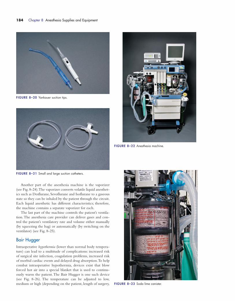
Another part of the anesthesia machine is the vaporizer
(see Fig. 8-24).The vaporizer converts volatile liquid anesthet-
ics such as Desflurane, Sevoflurane and Isoflurane to a gaseous
state so they can be inhaled by the patient through the circuit.
Each liquid anesthetic has different characteristics; therefore,
the machine contains a separate vaporizer for each.
The last part of the machine controls the patient’s ventila-
tion.The anesthesia care provider can deliver gases and con-
trol the patient’s ventilatory rate and volume either manually
(by squeezing the bag) or automatically (by switching on the
ventilator) (see Fig. 8-25).
Bair HuggerIntraoperative hypothermia (lower than normal body tempera-
ture) can lead to a multitude of complications: increased risk
of surgical site infection, coagulation problems, increased risk
of morbid cardiac events and delayed drug absorption.To help
combat intraoperative hypothermia, devices exist that blow
forced hot air into a special blanket that is used to continu-
ously warm the patient.The Bair Hugger is one such device
(see Fig. 8-26). The temperature can be adjusted to low,
medium or high (depending on the patient, length of surgery,
184 Chapter 8 Anesthesia Supplies and Equipment
FIGURE 8–20 Yankauer suction tips.
FIGURE 8–21 Small and large suction catheters.
FIGURE 8–22 Anesthesia machine.
FIGURE 8–23 Soda lime canister.
1572_Ch08_171-230.qxd 8/6/09 11:54 AM Page 184
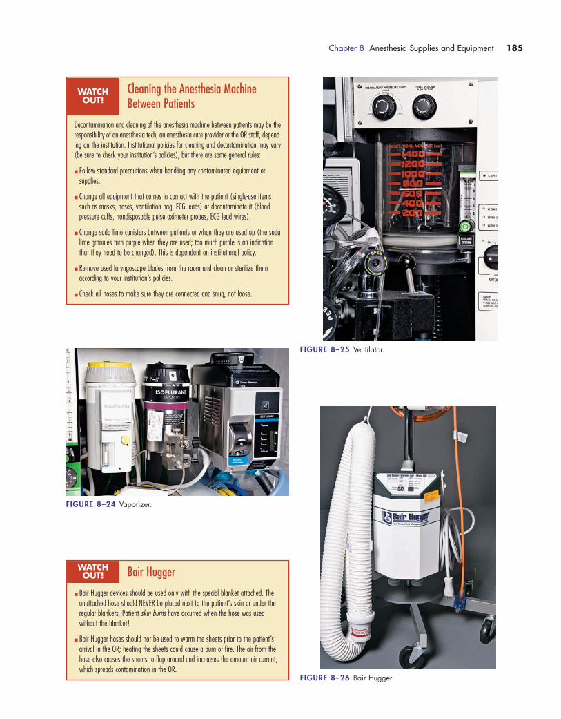
Chapter 8 Anesthesia Supplies and Equipment 185
Cleaning the Anesthesia Machine Between Patients
Decontamination and cleaning of the anesthesia machine between patients may be theresponsibility of an anesthesia tech, an anesthesia care provider or the OR staff, depend-ing on the institution. Institutional policies for cleaning and decontamination may vary(be sure to check your institution’s policies), but there are some general rules:
� Follow standard precautions when handling any contaminated equipment or supplies.
� Change all equipment that comes in contact with the patient (single-use itemssuch as masks, hoses, ventilation bag, ECG leads) or decontaminate it (bloodpressure cuffs, nondisposable pulse oximeter probes, ECG lead wires).
� Change soda lime canisters between patients or when they are used up (the sodalime granules turn purple when they are used; too much purple is an indicationthat they need to be changed). This is dependent on institutional policy.
� Remove used laryngoscope blades from the room and clean or sterilize them according to your institution’s policies.
� Check all hoses to make sure they are connected and snug, not loose.
WATCH OUT!
Bair Hugger
� Bair Hugger devices should be used only with the special blanket attached. Theunattached hose should NEVER be placed next to the patient’s skin or under theregular blankets. Patient skin burns have occurred when the hose was used without the blanket!
� Bair Hugger hoses should not be used to warm the sheets prior to the patient’sarrival in the OR; heating the sheets could cause a burn or fire. The air from thehose also causes the sheets to flap around and increases the amount air current,which spreads contamination in the OR.
WATCH OUT!
FIGURE 8–24 Vaporizer.
FIGURE 8–25 Ventilator.
FIGURE 8–26 Bair Hugger.
1572_Ch08_171-230.qxd 8/6/09 11:54 AM Page 185
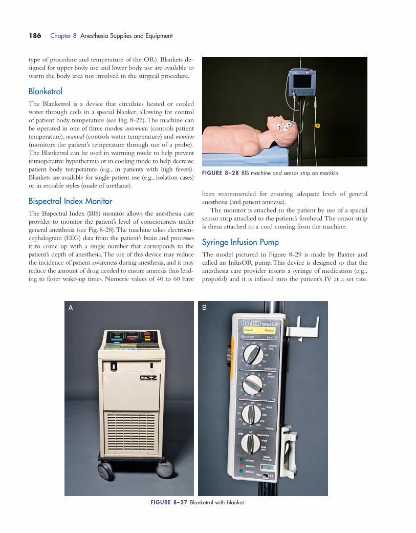
type of procedure and temperature of the OR). Blankets de-
signed for upper body use and lower body use are available to
warm the body area not involved in the surgical procedure.
BlanketrolThe Blanketrol is a device that circulates heated or cooled
water through coils in a special blanket, allowing for control
of patient body temperature (see Fig. 8-27).The machine can
be operated in one of three modes: automatic (controls patient
temperature), manual (controls water temperature) and monitor
(monitors the patient’s temperature through use of a probe).
The Blanketrol can be used in warming mode to help prevent
intraoperative hypothermia or in cooling mode to help decrease
patient body temperature (e.g., in patients with high fevers).
Blankets are available for single patient use (e.g., isolation cases)
or in reusable styles (made of urethane).
Bispectral Index MonitorThe Bispectral Index (BIS) monitor allows the anesthesia care
provider to monitor the patient’s level of consciousness under
general anesthesia (see Fig. 8-28).The machine takes electroen-
cephalogram (EEG) data from the patient’s brain and processes
it to come up with a single number that corresponds to the
patient’s depth of anesthesia.The use of this device may reduce
the incidence of patient awareness during anesthesia, and it may
reduce the amount of drug needed to ensure amnesia thus lead-
ing to faster wake-up times. Numeric values of 40 to 60 have
been recommended for ensuring adequate levels of general
anesthesia (and patient amnesia).
The monitor is attached to the patient by use of a special
sensor strip attached to the patient’s forehead.The sensor strip
is them attached to a cord coming from the machine.
Syringe Infusion PumpThe model pictured in Figure 8-29 is made by Baxter and
called an InfusOR pump.This device is designed so that the
anesthesia care provider inserts a syringe of medication (e.g.,
propofol) and it is infused into the patient’s IV at a set rate.
186 Chapter 8 Anesthesia Supplies and Equipment
FIGURE 8–27 Blanketrol with blanket.
A B
FIGURE 8–28 BIS machine and sensor strip on manikin.
1572_Ch08_171-230.qxd 8/6/09 11:54 AM Page 186
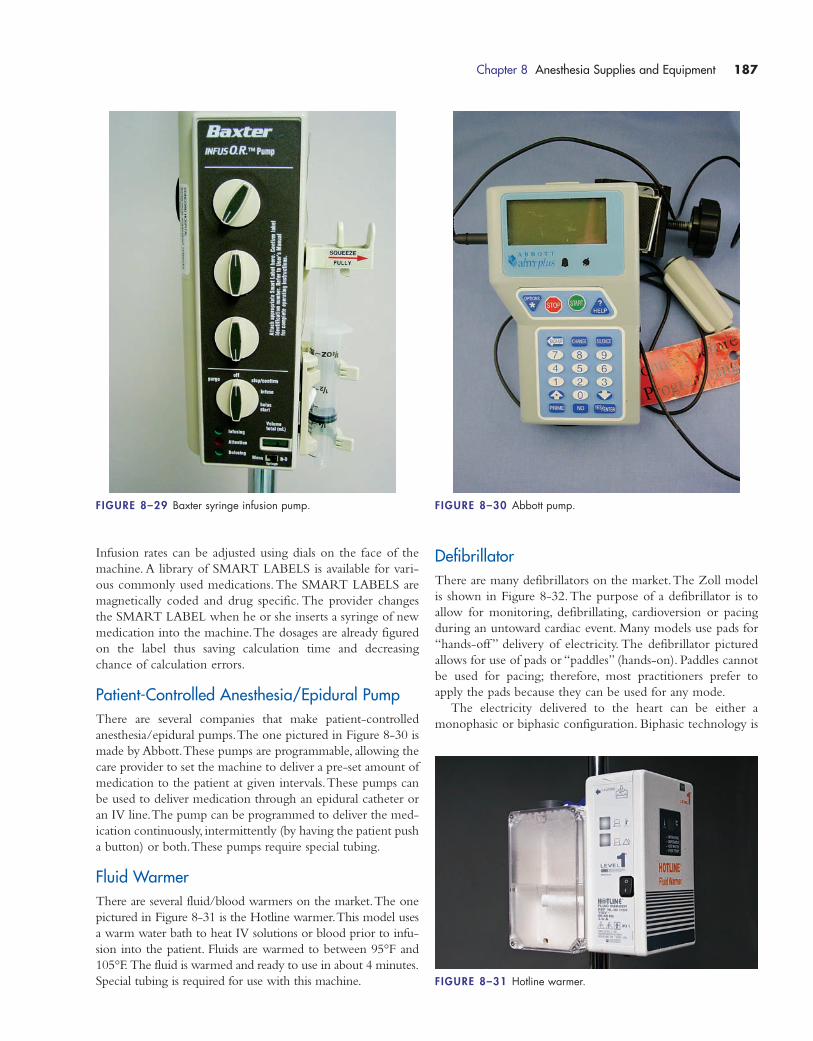
Infusion rates can be adjusted using dials on the face of the
machine. A library of SMART LABELS is available for vari-
ous commonly used medications.The SMART LABELS are
magnetically coded and drug specific. The provider changes
the SMART LABEL when he or she inserts a syringe of new
medication into the machine.The dosages are already figured
on the label thus saving calculation time and decreasing
chance of calculation errors.
Patient-Controlled Anesthesia/Epidural PumpThere are several companies that make patient-controlled
anesthesia/epidural pumps.The one pictured in Figure 8-30 is
made by Abbott.These pumps are programmable, allowing the
care provider to set the machine to deliver a pre-set amount of
medication to the patient at given intervals.These pumps can
be used to deliver medication through an epidural catheter or
an IV line.The pump can be programmed to deliver the med-
ication continuously, intermittently (by having the patient push
a button) or both.These pumps require special tubing.
Fluid WarmerThere are several fluid/blood warmers on the market.The one
pictured in Figure 8-31 is the Hotline warmer.This model uses
a warm water bath to heat IV solutions or blood prior to infu-
sion into the patient. Fluids are warmed to between 95°F and
105°F. The fluid is warmed and ready to use in about 4 minutes.
Special tubing is required for use with this machine.
Chapter 8 Anesthesia Supplies and Equipment 187
FIGURE 8–29 Baxter syringe infusion pump. FIGURE 8–30 Abbott pump.
FIGURE 8–31 Hotline warmer.
DefibrillatorThere are many defibrillators on the market.The Zoll model
is shown in Figure 8-32.The purpose of a defibrillator is to
allow for monitoring, defibrillating, cardioversion or pacing
during an untoward cardiac event. Many models use pads for
“hands-off ” delivery of electricity. The defibrillator pictured
allows for use of pads or “paddles” (hands-on). Paddles cannot
be used for pacing; therefore, most practitioners prefer to
apply the pads because they can be used for any mode.
The electricity delivered to the heart can be either a
monophasic or biphasic configuration. Biphasic technology is
1572_Ch08_171-230.qxd 8/6/09 11:54 AM Page 187
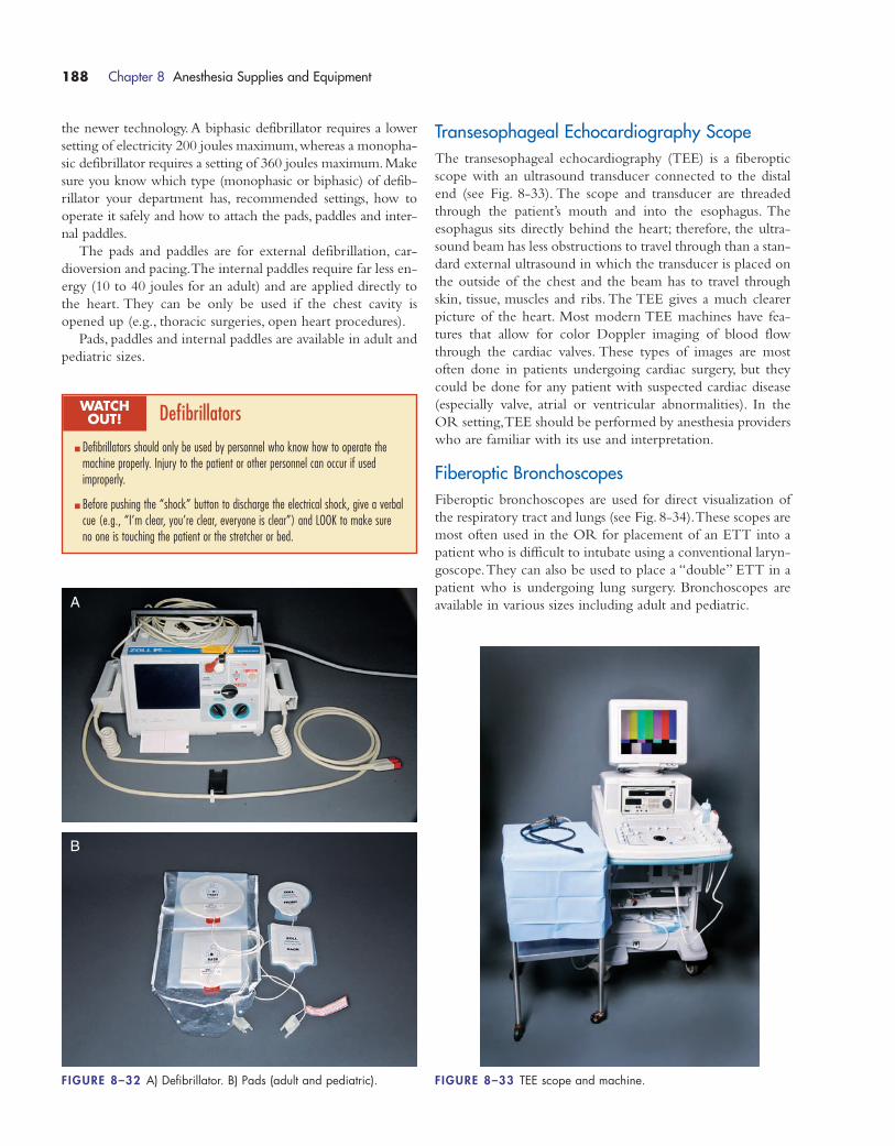
the newer technology. A biphasic defibrillator requires a lower
setting of electricity 200 joules maximum,whereas a monopha-
sic defibrillator requires a setting of 360 joules maximum.Make
sure you know which type (monophasic or biphasic) of defib-
rillator your department has, recommended settings, how to
operate it safely and how to attach the pads, paddles and inter-
nal paddles.
The pads and paddles are for external defibrillation, car-
dioversion and pacing.The internal paddles require far less en-
ergy (10 to 40 joules for an adult) and are applied directly to
the heart. They can be only be used if the chest cavity is
opened up (e.g., thoracic surgeries, open heart procedures).
Pads, paddles and internal paddles are available in adult and
pediatric sizes.
Transesophageal Echocardiography ScopeThe transesophageal echocardiography (TEE) is a fiberoptic
scope with an ultrasound transducer connected to the distal
end (see Fig. 8-33). The scope and transducer are threaded
through the patient’s mouth and into the esophagus. The
esophagus sits directly behind the heart; therefore, the ultra-
sound beam has less obstructions to travel through than a stan-
dard external ultrasound in which the transducer is placed on
the outside of the chest and the beam has to travel through
skin, tissue, muscles and ribs. The TEE gives a much clearer
picture of the heart. Most modern TEE machines have fea-
tures that allow for color Doppler imaging of blood flow
through the cardiac valves. These types of images are most
often done in patients undergoing cardiac surgery, but they
could be done for any patient with suspected cardiac disease
(especially valve, atrial or ventricular abnormalities). In the
OR setting,TEE should be performed by anesthesia providers
who are familiar with its use and interpretation.
Fiberoptic BronchoscopesFiberoptic bronchoscopes are used for direct visualization of
the respiratory tract and lungs (see Fig. 8-34).These scopes are
most often used in the OR for placement of an ETT into a
patient who is difficult to intubate using a conventional laryn-
goscope.They can also be used to place a “double” ETT in a
patient who is undergoing lung surgery. Bronchoscopes are
available in various sizes including adult and pediatric.
188 Chapter 8 Anesthesia Supplies and Equipment
Defibrillators
� Defibrillators should only be used by personnel who know how to operate themachine properly. Injury to the patient or other personnel can occur if used improperly.
� Before pushing the “shock” button to discharge the electrical shock, give a verbalcue (e.g., “I’m clear, you’re clear, everyone is clear”) and LOOK to make sureno one is touching the patient or the stretcher or bed.
WATCH OUT!
FIGURE 8–32 A) Defibrillator. B) Pads (adult and pediatric).
A
B
FIGURE 8–33 TEE scope and machine.
1572_Ch08_171-230.qxd 8/6/09 11:54 AM Page 188
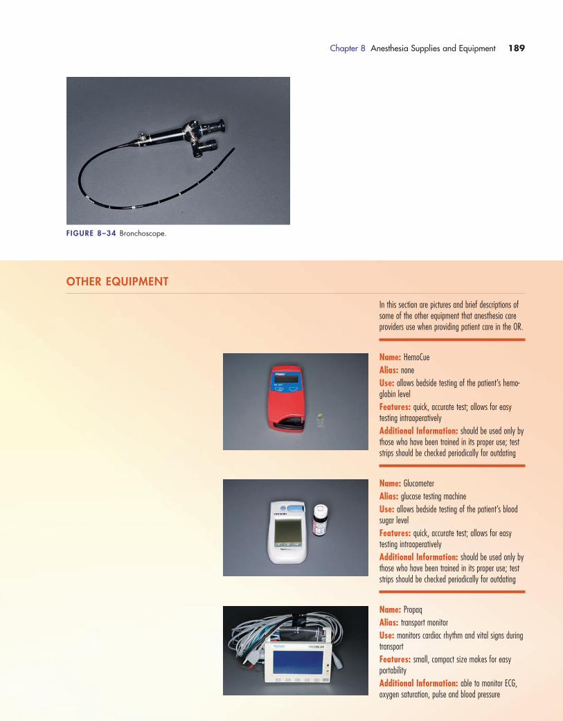
Chapter 8 Anesthesia Supplies and Equipment 189
FIGURE 8–34 Bronchoscope.
OTHER EQUIPMENT
In this section are pictures and brief descriptions ofsome of the other equipment that anesthesia careproviders use when providing patient care in the OR.
Name: HemoCue
Alias: none
Use: allows bedside testing of the patient’s hemo-globin level
Features: quick, accurate test; allows for easytesting intraoperatively
Additional Information: should be used only bythose who have been trained in its proper use; teststrips should be checked periodically for outdating
Name: Glucometer
Alias: glucose testing machine
Use: allows bedside testing of the patient’s bloodsugar level
Features: quick, accurate test; allows for easytesting intraoperatively
Additional Information: should be used only bythose who have been trained in its proper use; teststrips should be checked periodically for outdating
Name: Propaq
Alias: transport monitor
Use: monitors cardiac rhythm and vital signs duringtransport
Features: small, compact size makes for easyportability
Additional Information: able to monitor ECG,oxygen saturation, pulse and blood pressure
1572_Ch08_171-230.qxd 8/6/09 11:54 AM Page 189
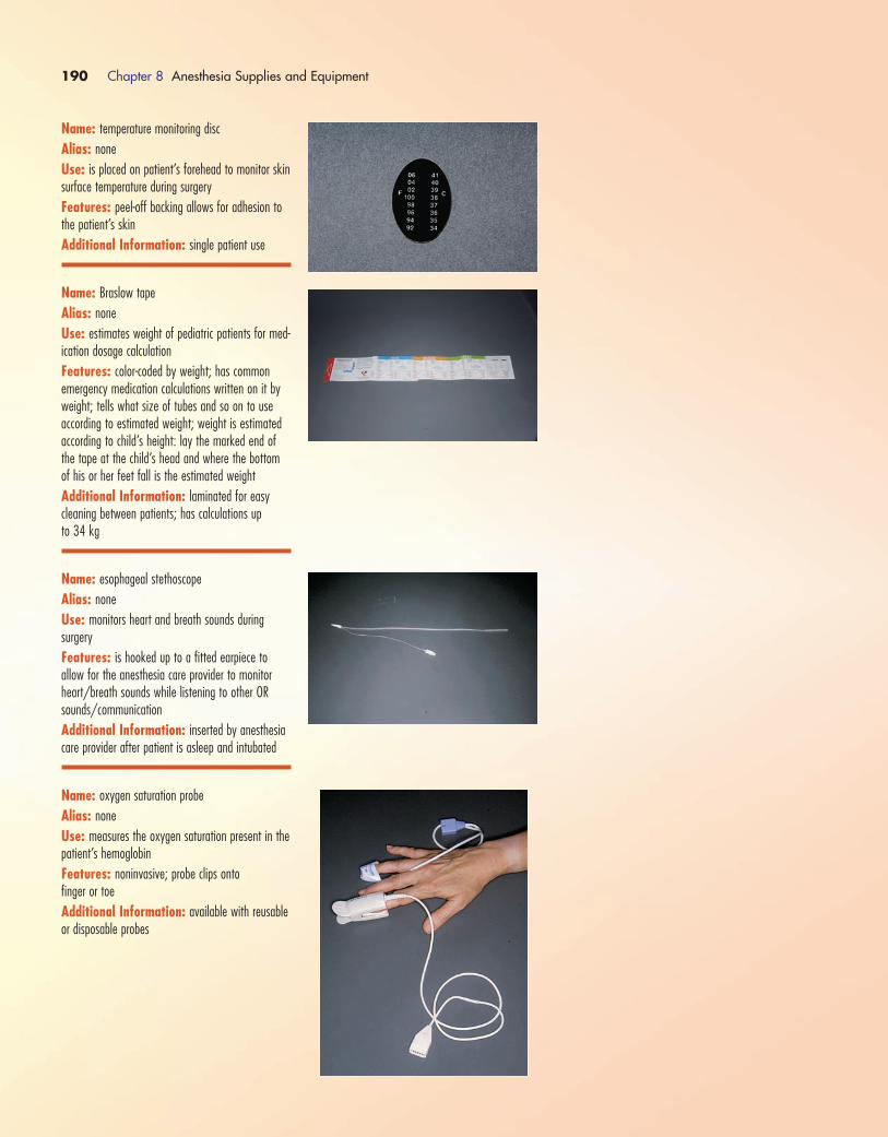
190 Chapter 8 Anesthesia Supplies and Equipment
Name: temperature monitoring disc
Alias: none
Use: is placed on patient’s forehead to monitor skinsurface temperature during surgery
Features: peel-off backing allows for adhesion tothe patient’s skin
Additional Information: single patient use
Name: Braslow tape
Alias: none
Use: estimates weight of pediatric patients for med-ication dosage calculation
Features: color-coded by weight; has commonemergency medication calculations written on it byweight; tells what size of tubes and so on to use according to estimated weight; weight is estimatedaccording to child’s height: lay the marked end ofthe tape at the child’s head and where the bottomof his or her feet fall is the estimated weight
Additional Information: laminated for easycleaning between patients; has calculations up to 34 kg
Name: esophageal stethoscope
Alias: none
Use: monitors heart and breath sounds during surgery
Features: is hooked up to a fitted earpiece to allow for the anesthesia care provider to monitorheart/breath sounds while listening to other ORsounds/communication
Additional Information: inserted by anesthesiacare provider after patient is asleep and intubated
Name: oxygen saturation probe
Alias: none
Use: measures the oxygen saturation present in thepatient’s hemoglobin
Features: noninvasive; probe clips onto finger or toe
Additional Information: available with reusableor disposable probes
1572_Ch08_171-230.qxd 8/6/09 11:54 AM Page 190
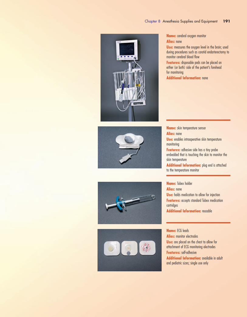
Chapter 8 Anesthesia Supplies and Equipment 191
Name: cerebral oxygen monitor
Alias: none
Use: measures the oxygen level in the brain; usedduring procedures such as carotid endarterectomy tomonitor cerebral blood flow
Features: disposable pads can be placed on either (or both) side of the patient’s forehead for monitoring
Additional Information: none
Name: skin temperature sensor
Alias: none
Use: enables intraoperative skin temperature monitoring
Features: adhesive side has a tiny probe embedded that is touching the skin to monitor theskin temperature
Additional Information: plug end is attached to the temperature monitor
Name: Tubex holder
Alias: none
Use: holds medication to allow for injection
Features: accepts standard Tubex medication cartridges
Additional Information: reusable
Name: ECG leads
Alias: monitor electrodes
Use: are placed on the chest to allow for attachment of ECG monitoring electrodes
Features: self-adhesive
Additional Information: available in adult and pediatric sizes; single use only
1572_Ch08_171-230.qxd 8/6/09 11:54 AM Page 191
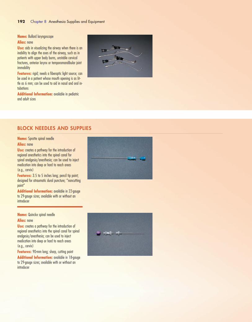
192 Chapter 8 Anesthesia Supplies and Equipment
Name: Bullard laryngoscope
Alias: none
Use: aids in visualizing the airway when there is aninability to align the axes of the airway, such as inpatients with upper body burns, unstable cervicalfractures, anterior larynx or temporomandibular jointimmobility
Features: rigid; needs a fiberoptic light source; canbe used in a patient whose mouth opening is as lit-tle as 6 mm; can be used to aid in nasal and oral in-tubations
Additional Information: available in pediatricand adult sizes
BLOCK NEEDLES AND SUPPLIES
Name: Sprotte spinal needle
Alias: none
Use: creates a pathway for the introduction of regional anesthetics into the spinal canal for spinal analgesia/anesthesia; can be used to injectmedication into deep or hard to reach areas (e.g., cervix)
Features: 3.5 to 5 inches long; pencil tip point;designed for atraumatic dural puncture; “noncuttingpoint”
Additional Information: available in 22-gaugeto 29-gauge sizes; available with or without an introducer
Name: Quincke spinal needle
Alias: none
Use: creates a pathway for the introduction of regional anesthetics into the spinal canal for spinalanalgesia/anesthesia; can be used to inject medication into deep or hard to reach areas (e.g., cervix)
Features: 90-mm long; sharp, cutting point
Additional Information: available in 18-gaugeto 29-gauge sizes; available with or without an introducer
1572_Ch08_171-230.qxd 8/6/09 11:54 AM Page 192
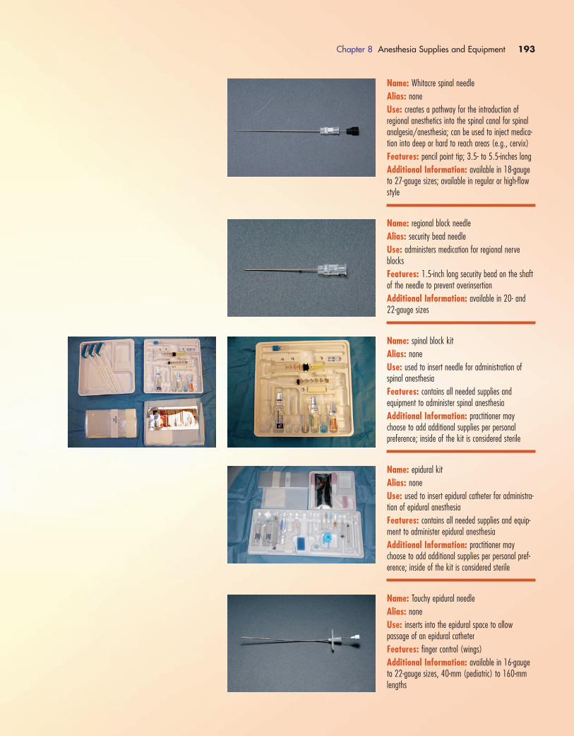
Chapter 8 Anesthesia Supplies and Equipment 193
Name: Whitacre spinal needle
Alias: none
Use: creates a pathway for the introduction of regional anesthetics into the spinal canal for spinalanalgesia/anesthesia; can be used to inject medica-tion into deep or hard to reach areas (e.g., cervix)
Features: pencil point tip; 3.5- to 5.5-inches long
Additional Information: available in 18-gaugeto 27-gauge sizes; available in regular or high-flowstyle
Name: regional block needle
Alias: security bead needle
Use: administers medication for regional nerveblocks
Features: 1.5-inch long security bead on the shaftof the needle to prevent overinsertion
Additional Information: available in 20- and 22-gauge sizes
Name: spinal block kit
Alias: none
Use: used to insert needle for administration ofspinal anesthesia
Features: contains all needed supplies and equipment to administer spinal anesthesia
Additional Information: practitioner maychoose to add additional supplies per personal preference; inside of the kit is considered sterile
Name: epidural kit
Alias: none
Use: used to insert epidural catheter for administra-tion of epidural anesthesia
Features: contains all needed supplies and equip-ment to administer epidural anesthesia
Additional Information: practitioner maychoose to add additional supplies per personal pref-erence; inside of the kit is considered sterile
Name: Touchy epidural needle
Alias: none
Use: inserts into the epidural space to allow passage of an epidural catheter
Features: finger control (wings)
Additional Information: available in 16-gaugeto 22-gauge sizes, 40-mm (pediatric) to 160-mmlengths
1572_Ch08_171-230.qxd 8/6/09 11:54 AM Page 193
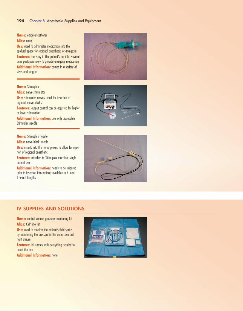
194 Chapter 8 Anesthesia Supplies and Equipment
Name: epidural catheter
Alias: none
Use: used to administer medication into theepidural space for regional anesthesia or analgesia
Features: can stay in the patient’s back for severaldays postoperatively to provide analgesic medication
Additional Information: comes in a variety ofsizes and lengths
Name: Stimuplex
Alias: nerve stimulator
Use: stimulates nerves; used for insertion of regional nerve blocks
Features: output control can be adjusted for higheror lower stimulation
Additional Information: use with disposableStimuplex needle
Name: Stimuplex needle
Alias: nerve block needle
Use: inserts into the nerve plexus to allow for injec-tion of regional anesthetic
Features: attaches to Stimuplex machine; singlepatient use
Additional Information: needs to be irrigatedprior to insertion into patient; available in 4- and1.5-inch lengths
IV SUPPLIES AND SOLUTIONS
Name: central venous pressure monitoring kit
Alias: CVP line kit
Use: used to monitor the patient’s fluid status by monitoring the pressure in the vena cava andright atrium
Features: kit comes with everything needed to insert the line
Additional Information: none
1572_Ch08_171-230.qxd 8/6/09 11:54 AM Page 194
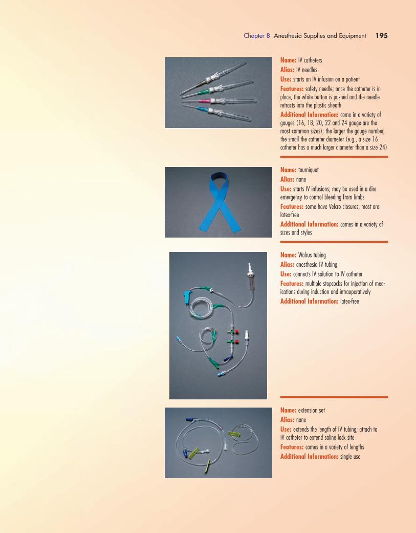
Chapter 8 Anesthesia Supplies and Equipment 195
Name: IV catheters
Alias: IV needles
Use: starts an IV infusion on a patient
Features: safety needle; once the catheter is inplace, the white button is pushed and the needle retracts into the plastic sheath
Additional Information: come in a variety ofgauges (16, 18, 20, 22 and 24 gauge are themost common sizes); the larger the gauge number,the small the catheter diameter (e.g., a size 16catheter has a much larger diameter than a size 24)
Name: tourniquet
Alias: none
Use: starts IV infusions; may be used in a direemergency to control bleeding from limbs
Features: some have Velcro closures; most are latex-free
Additional Information: comes in a variety ofsizes and styles
Name: Walrus tubing
Alias: anesthesia IV tubing
Use: connects IV solution to IV catheter
Features: multiple stopcocks for injection of med-ications during induction and intraoperatively
Additional Information: latex-free
Name: extension set
Alias: none
Use: extends the length of IV tubing; attach to IV catheter to extend saline lock site
Features: comes in a variety of lengths
Additional Information: single use
1572_Ch08_171-230.qxd 8/6/09 11:54 AM Page 195
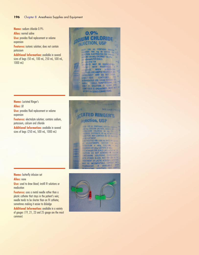
196 Chapter 8 Anesthesia Supplies and Equipment
Name: sodium chloride 0.9%
Alias: normal saline
Use: provides fluid replacement or volume expansion
Features: isotonic solution; does not contain potassium
Additional Information: available in severalsizes of bags (50 mL, 100 mL, 250 mL, 500 mL,1000 mL)
Name: Lactated Ringer’s
Alias: LRUse: provides fluid replacement or volume expansion
Features: electrolyte solution; contains sodium,potassium, calcium and chloride
Additional Information: available in severalsizes of bags (250 mL, 500 mL, 1000 mL)
Name: butterfly infusion set
Alias: none
Use: used to draw blood; instill IV solutions or medication
Features: uses a metal needle rather than a plastic catheter that stays in the patient’s vein; needle tends to be shorter than an IV catheter,sometimes making it easier to dislodge
Additional Information: available in a varietyof gauges (19, 21, 23 and 25 gauge are the mostcommon)
1572_Ch08_171-230.qxd 8/6/09 11:54 AM Page 196
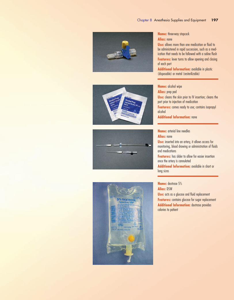
Chapter 8 Anesthesia Supplies and Equipment 197
Name: three-way stopcock
Alias: none
Use: allows more than one medication or fluid tobe administered in rapid succession, such as a med-ication that needs to be followed with a saline flush
Features: lever turns to allow opening and closingof each port
Additional Information: available in plastic (disposable) or metal (resterilizable)
Name: alcohol wipe
Alias: prep pad
Use: cleans the skin prior to IV insertion; cleans theport prior to injection of medication
Features: comes ready to use; contains isopropylalcohol
Additional Information: none
Name: arterial line needles
Alias: none
Use: inserted into an artery, it allows access formonitoring, blood drawing or administration of fluidsand medications
Features: has slider to allow for easier insertiononce the artery is cannulated
Additional Information: available in short orlong sizes
Name: dextrose 5%
Alias: D5W
Use: acts as a glucose and fluid replacement
Features: contains glucose for sugar replacement
Additional Information: dextrose provides calories to patient
1572_Ch08_171-230.qxd 8/6/09 11:54 AM Page 197
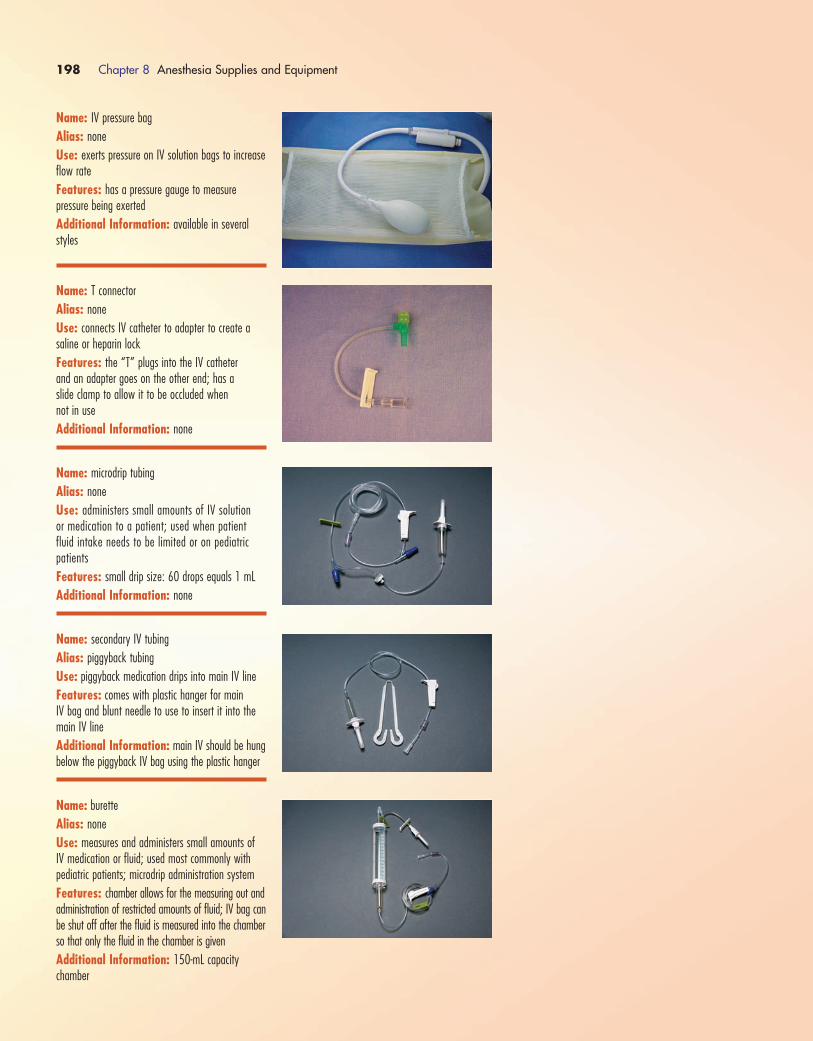
198 Chapter 8 Anesthesia Supplies and Equipment
Name: IV pressure bag
Alias: none
Use: exerts pressure on IV solution bags to increaseflow rate
Features: has a pressure gauge to measure pressure being exerted
Additional Information: available in severalstyles
Name: T connector
Alias: none
Use: connects IV catheter to adapter to create asaline or heparin lock
Features: the “T” plugs into the IV catheter and an adapter goes on the other end; has a slide clamp to allow it to be occluded when not in use
Additional Information: none
Name: microdrip tubing
Alias: none
Use: administers small amounts of IV solution or medication to a patient; used when patientfluid intake needs to be limited or on pediatric patients
Features: small drip size: 60 drops equals 1 mL
Additional Information: none
Name: secondary IV tubing
Alias: piggyback tubing
Use: piggyback medication drips into main IV line
Features: comes with plastic hanger for main IV bag and blunt needle to use to insert it into themain IV line
Additional Information: main IV should be hungbelow the piggyback IV bag using the plastic hanger
Name: burette
Alias: none
Use: measures and administers small amounts of IV medication or fluid; used most commonly with pediatric patients; microdrip administration system
Features: chamber allows for the measuring out andadministration of restricted amounts of fluid; IV bag canbe shut off after the fluid is measured into the chamberso that only the fluid in the chamber is given
Additional Information: 150-mL capacity chamber
1572_Ch08_171-230.qxd 8/6/09 11:54 AM Page 198
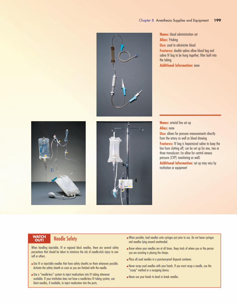
Chapter 8 Anesthesia Supplies and Equipment 199
Name: blood administration set
Alias: Y-tubing
Use: used to administer blood
Features: double spikes allow blood bag andsaline IV bag to be hung together; filter built intothe tubing
Additional Information: none
Name: arterial line set up
Alias: none
Use: allows for pressure measurements directlyfrom the artery as well as blood drawing
Features: IV bag is heparinized saline to keep theline from clotting off; can be set up for one, two orthree transducers (to allow for central venous pressure [CVP] monitoring as well)
Additional Information: set up may vary by institution or equipment
Needle Safety
When handling injectable, IV or regional block needles, there are several safety precautions that should be taken to minimize the risk of needle-stick injury to one-self or others.
� Use IV or injectable needles that have safety sheaths on them whenever possible.Activate the safety sheath as soon as you are finished with the needle.
� Use a “needle-less” system to inject medications into IV tubing whenever available. If your institution does not have a needle-less IV tubing system, useblunt needles, if available, to inject medication into the ports.
WATCH OUT!
� When possible, load needles onto syringes just prior to use. Do not leave syringesand needles lying around unattended.
� Know where your needles are at all times. Keep track of where you or the personyou are assisting is placing the sharps.
� Place all used needles in a puncture-proof disposal container.
� Never recap used needles with your hands. If you must recap a needle, use the“scoop” method or a recapping device.
� Never use your hands to bend or break needles.
1572_Ch08_171-230.qxd 8/6/09 11:54 AM Page 199
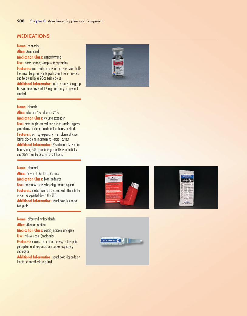
200 Chapter 8 Anesthesia Supplies and Equipment
Name: adenosine
Alias: Adenocard
Medication Class: antiarrhythmic
Use: treats narrow, complex tachycardias
Features: each vial contains 6 mg; very short half-life, must be given via IV push over 1 to 2 secondsand followed by a 20-cc saline bolus
Additional Information: initial dose is 6 mg; upto two more doses of 12 mg each may be given ifneeded
Name: albumin
Alias: albumin 5%; albumin 25%
Medication Class: volume expander
Use: restores plasma volume during cardiac bypassprocedures or during treatment of burns or shock
Features: acts by expanding the volume of circu-lating blood and maintaining cardiac output
Additional Information: 5% albumin is used totreat shock; 5% albumin is generally used initiallyand 25% may be used after 24 hours
Name: albuterol
Alias: Proventil, Ventolin, Volmax
Medication Class: bronchodilator
Use: prevents/treats wheezing, bronchospasm
Features: medication can be used with the inhaleror can be squirted down the ETT.
Additional Information: usual dose is one totwo puffs
Name: alfentanil hydrochloride
Alias: Alfenta; Rapifen
Medication Class: opioid; narcotic analgesic
Use: relieves pain (analgesic)
Features: makes the patient drowsy; alters painperception and response; can cause respiratory depression
Additional Information: usual dose depends onlength of anesthesia required
MEDICATIONS
1572_Ch08_171-230.qxd 8/6/09 11:54 AM Page 200
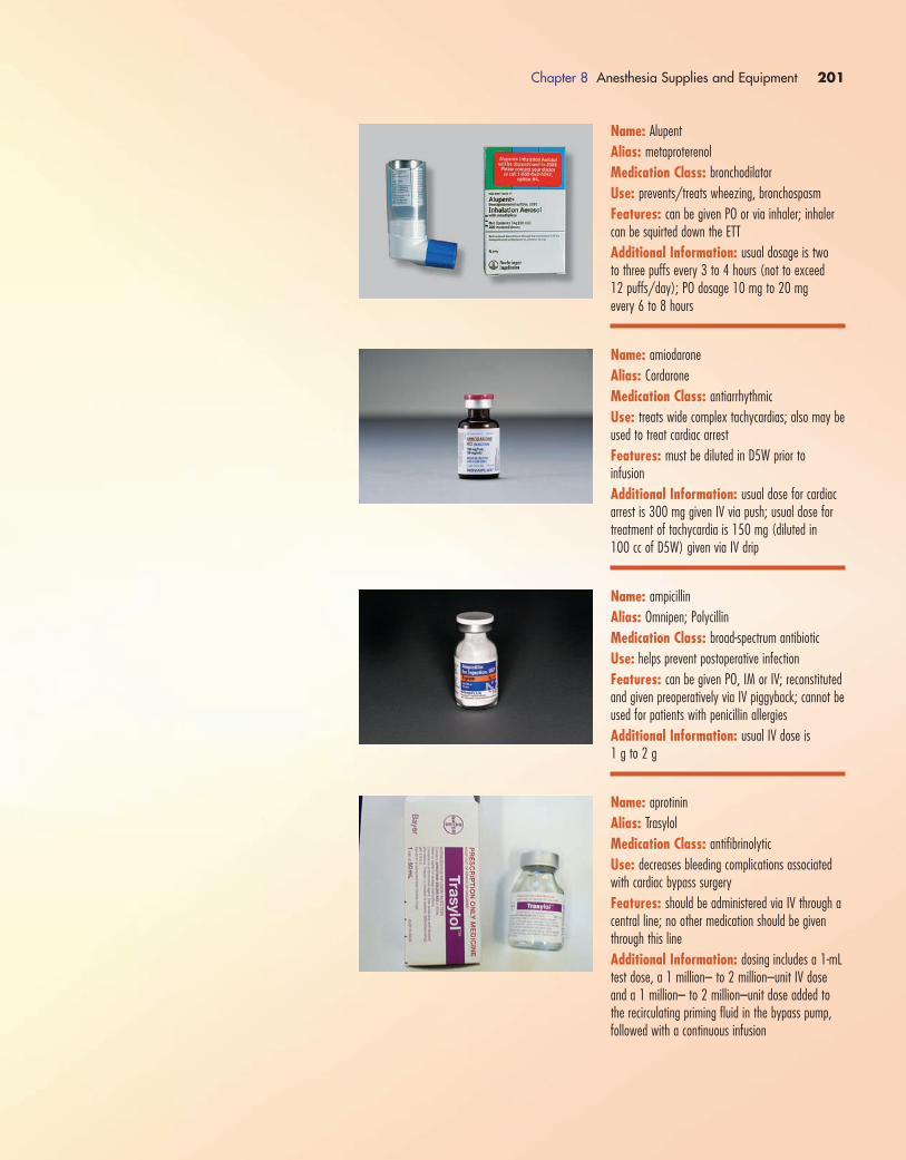
Chapter 8 Anesthesia Supplies and Equipment 201
Name: Alupent
Alias: metaproterenol
Medication Class: bronchodilator
Use: prevents/treats wheezing, bronchospasm
Features: can be given PO or via inhaler; inhalercan be squirted down the ETT
Additional Information: usual dosage is two to three puffs every 3 to 4 hours (not to exceed 12 puffs/day); PO dosage 10 mg to 20 mg every 6 to 8 hours
Name: amiodarone
Alias: Cordarone
Medication Class: antiarrhythmic
Use: treats wide complex tachycardias; also may beused to treat cardiac arrest
Features: must be diluted in D5W prior to infusion
Additional Information: usual dose for cardiacarrest is 300 mg given IV via push; usual dose fortreatment of tachycardia is 150 mg (diluted in 100 cc of D5W) given via IV drip
Name: ampicillin
Alias: Omnipen; Polycillin
Medication Class: broad-spectrum antibiotic
Use: helps prevent postoperative infection
Features: can be given PO, IM or IV; reconstitutedand given preoperatively via IV piggyback; cannot beused for patients with penicillin allergies
Additional Information: usual IV dose is 1 g to 2 g
Name: aprotinin
Alias: Trasylol
Medication Class: antifibrinolytic
Use: decreases bleeding complications associatedwith cardiac bypass surgery
Features: should be administered via IV through acentral line; no other medication should be giventhrough this line
Additional Information: dosing includes a 1-mLtest dose, a 1 million– to 2 million–unit IV doseand a 1 million– to 2 million–unit dose added tothe recirculating priming fluid in the bypass pump,followed with a continuous infusion
1572_Ch08_171-230.qxd 8/6/09 11:55 AM Page 201
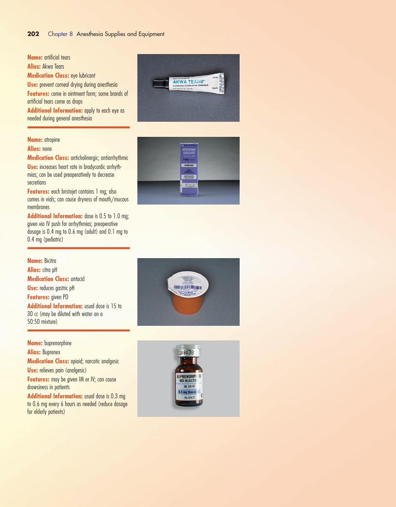
202 Chapter 8 Anesthesia Supplies and Equipment
Name: artificial tears
Alias: Akwa Tears
Medication Class: eye lubricant
Use: prevent corneal drying during anesthesia
Features: come in ointment form; some brands ofartificial tears come as drops
Additional Information: apply to each eye asneeded during general anesthesia
Name: atropine
Alias: none
Medication Class: anticholinergic; antiarrhythmic
Use: increases heart rate in bradycardic arrhyth-mias; can be used preoperatively to decrease secretions
Features: each bristojet contains 1 mg; alsocomes in vials; can cause dryness of mouth/mucousmembranes
Additional Information: dose is 0.5 to 1.0 mg;given via IV push for arrhythmias; preoperativedosage is 0.4 mg to 0.6 mg (adult) and 0.1 mg to0.4 mg (pediatric)
Name: Bicitra
Alias: citra pH
Medication Class: antacid
Use: reduces gastric pH
Features: given PO
Additional Information: usual dose is 15 to 30 cc (may be diluted with water on a 50:50 mixture)
Name: buprenorphine
Alias: Buprenex
Medication Class: opioid; narcotic analgesic
Use: relieves pain (analgesic)
Features: may be given IM or IV; can causedrowsiness in patients
Additional Information: usual dose is 0.3 mgto 0.6 mg every 6 hours as needed (reduce dosagefor elderly patients)
1572_Ch08_171-230.qxd 8/6/09 11:55 AM Page 202
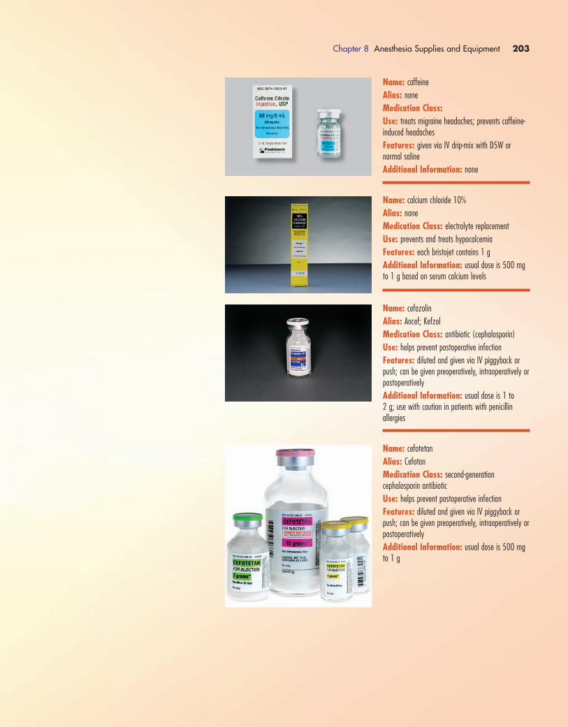
Chapter 8 Anesthesia Supplies and Equipment 203
Name: caffeine
Alias: none
Medication Class:Use: treats migraine headaches; prevents caffeine-induced headaches
Features: given via IV drip-mix with D5W or normal saline
Additional Information: none
Name: calcium chloride 10%
Alias: none
Medication Class: electrolyte replacement
Use: prevents and treats hypocalcemia
Features: each bristojet contains 1 g
Additional Information: usual dose is 500 mgto 1 g based on serum calcium levels
Name: cefazolin
Alias: Ancef; Kefzol
Medication Class: antibiotic (cephalosporin)
Use: helps prevent postoperative infection
Features: diluted and given via IV piggyback orpush; can be given preoperatively, intraoperatively orpostoperatively
Additional Information: usual dose is 1 to 2 g; use with caution in patients with penicillin allergies
Name: cefotetan
Alias: Cefotan
Medication Class: second-generationcephalosporin antibiotic
Use: helps prevent postoperative infection
Features: diluted and given via IV piggyback orpush; can be given preoperatively, intraoperatively orpostoperatively
Additional Information: usual dose is 500 mgto 1 g
1572_Ch08_171-230.qxd 8/6/09 11:55 AM Page 203
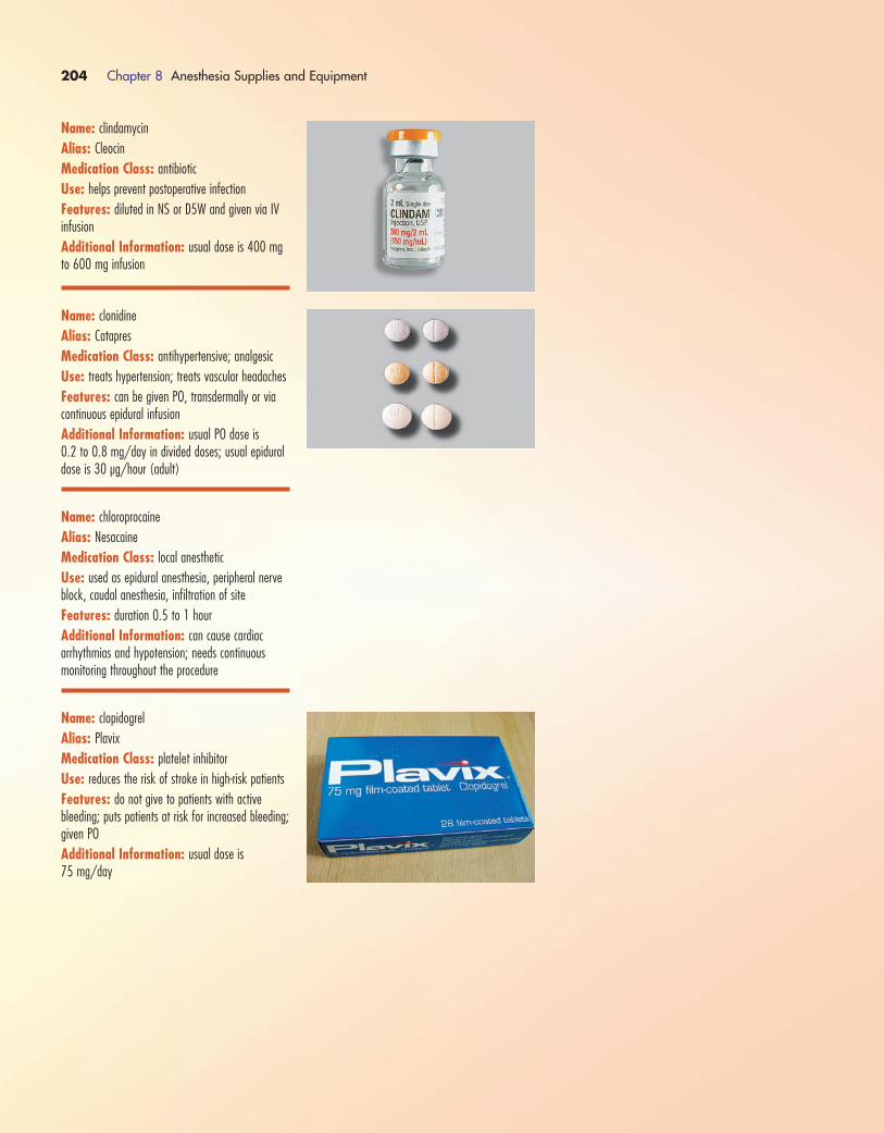
204 Chapter 8 Anesthesia Supplies and Equipment
Name: clindamycin
Alias: Cleocin
Medication Class: antibiotic
Use: helps prevent postoperative infection
Features: diluted in NS or D5W and given via IVinfusion
Additional Information: usual dose is 400 mgto 600 mg infusion
Name: clonidine
Alias: Catapres
Medication Class: antihypertensive; analgesic
Use: treats hypertension; treats vascular headaches
Features: can be given PO, transdermally or viacontinuous epidural infusion
Additional Information: usual PO dose is 0.2 to 0.8 mg/day in divided doses; usual epiduraldose is 30 µg/hour (adult)
Name: chloroprocaine
Alias: Nesacaine
Medication Class: local anesthetic
Use: used as epidural anesthesia, peripheral nerveblock, caudal anesthesia, infiltration of site
Features: duration 0.5 to 1 hour
Additional Information: can cause cardiac arrhythmias and hypotension; needs continuousmonitoring throughout the procedure
Name: clopidogrel
Alias: Plavix
Medication Class: platelet inhibitor
Use: reduces the risk of stroke in high-risk patients
Features: do not give to patients with activebleeding; puts patients at risk for increased bleeding;given PO
Additional Information: usual dose is 75 mg/day
1572_Ch08_171-230.qxd 8/6/09 11:55 AM Page 204
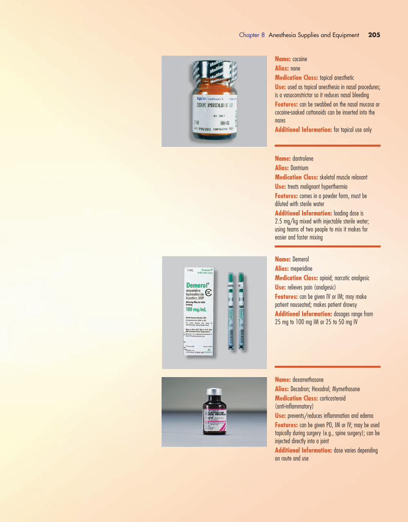
Chapter 8 Anesthesia Supplies and Equipment 205
Name: cocaine
Alias: none
Medication Class: topical anesthetic
Use: used as topical anesthesia in nasal procedures;is a vasoconstrictor so it reduces nasal bleeding
Features: can be swabbed on the nasal mucosa orcocaine-soaked cottonoids can be inserted into thenares
Additional Information: for topical use only
Name: dantrolene
Alias: Dantrium
Medication Class: skeletal muscle relaxant
Use: treats malignant hyperthermia
Features: comes in a powder form, must be diluted with sterile water
Additional Information: loading dose is 2.5 mg/kg mixed with injectable sterile water; using teams of two people to mix it makes for easier and faster mixing
Name: Demerol
Alias: meperidine
Medication Class: opioid; narcotic analgesic
Use: relieves pain (analgesic)
Features: can be given IV or IM; may make patient nauseated; makes patient drowsy
Additional Information: dosages range from 25 mg to 100 mg IM or 25 to 50 mg IV
Name: dexamethasone
Alias: Decadron; Hexadrol; Mymethasone
Medication Class: corticosteroid (anti-inflammatory)
Use: prevents/reduces inflammation and edema
Features: can be given PO, IM or IV; may be usedtopically during surgery (e.g., spine surgery); can beinjected directly into a joint
Additional Information: dose varies dependingon route and use
1572_Ch08_171-230.qxd 8/6/09 11:55 AM Page 205
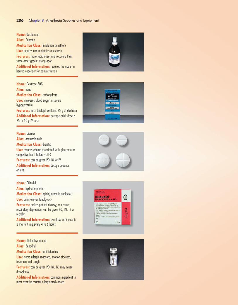
206 Chapter 8 Anesthesia Supplies and Equipment
Name: desflurane
Alias: Suprane
Medication Class: inhalation anesthetic
Use: induces and maintains anesthesia
Features: more rapid onset and recovery thansome other gases; strong odor
Additional Information: requires the use of aheated vaporizer for administration
Name: Dextrose 50%
Alias: none
Medication Class: carbohydrate
Use: increases blood sugar in severe hypoglycemia
Features: each bristojet contains 25 g of dextrose
Additional Information: average adult dose is25 to 50 g IV push
Name: Diamox
Alias: acetazolamide
Medication Class: diuretic
Use: reduces edema associated with glaucoma orcongestive heart failure (CHF)
Features: can be given PO, IM or IV
Additional Information: dosage depends on use
Name: Dilaudid
Alias: hydromorphone
Medication Class: opioid; narcotic analgesic
Use: pain reliever (analgesic)
Features: makes patient drowsy; can cause respiratory depression; can be given PO, IM, IV orrectally
Additional Information: usual IM or IV dose is2 mg to 4 mg every 4 to 6 hours
Name: diphenhydramine
Alias: Benadryl
Medication Class: antihistamine
Use: treats allergic reactions, motion sickness, insomnia and cough
Features: can be given PO, IM, IV; may causedrowsiness
Additional Information: common ingredient inmost over-the-counter allergy medications
1572_Ch08_171-230.qxd 8/6/09 11:55 AM Page 206
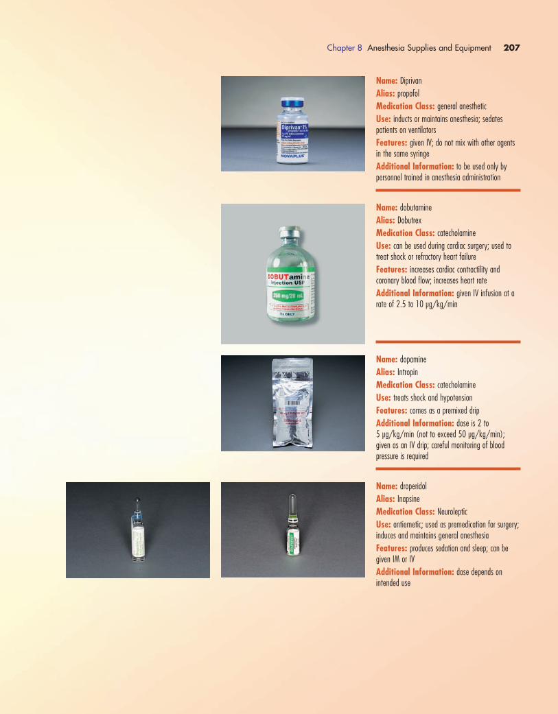
Chapter 8 Anesthesia Supplies and Equipment 207
Name: Diprivan
Alias: propofol
Medication Class: general anesthetic
Use: inducts or maintains anesthesia; sedates patients on ventilators
Features: given IV; do not mix with other agentsin the same syringe
Additional Information: to be used only by personnel trained in anesthesia administration
Name: dobutamine
Alias: Dobutrex
Medication Class: catecholamine
Use: can be used during cardiac surgery; used totreat shock or refractory heart failure
Features: increases cardiac contractility and coronary blood flow; increases heart rate
Additional Information: given IV infusion at arate of 2.5 to 10 µg/kg/min
Name: dopamine
Alias: Intropin
Medication Class: catecholamine
Use: treats shock and hypotension
Features: comes as a premixed drip
Additional Information: dose is 2 to 5 µg/kg/min (not to exceed 50 µg/kg/min);given as an IV drip; careful monitoring of blood pressure is required
Name: droperidol
Alias: Inapsine
Medication Class: Neuroleptic
Use: antiemetic; used as premedication for surgery;induces and maintains general anesthesia
Features: produces sedation and sleep; can begiven IM or IV
Additional Information: dose depends on intended use
1572_Ch08_171-230.qxd 8/6/09 11:55 AM Page 207
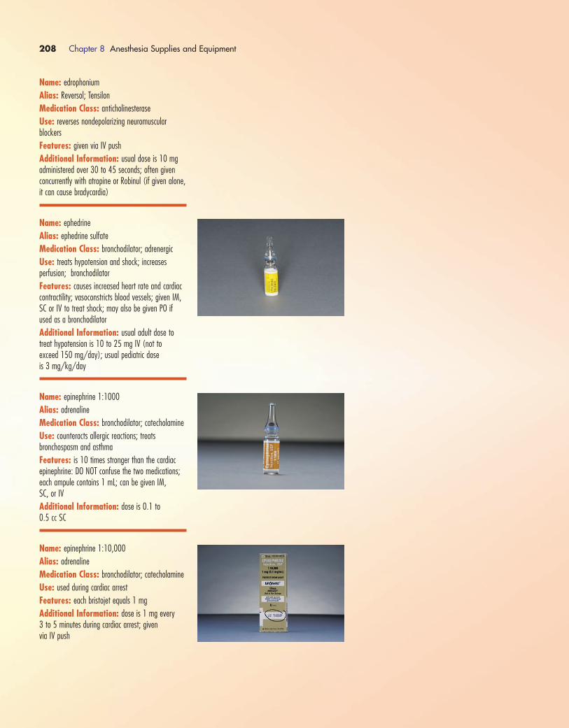
208 Chapter 8 Anesthesia Supplies and Equipment
Name: edrophonium
Alias: Reversol; Tensilon
Medication Class: anticholinesterase
Use: reverses nondepolarizing neuromuscular blockers
Features: given via IV push
Additional Information: usual dose is 10 mgadministered over 30 to 45 seconds; often givenconcurrently with atropine or Robinul (if given alone,it can cause bradycardia)
Name: ephedrine
Alias: ephedrine sulfate
Medication Class: bronchodilator; adrenergic
Use: treats hypotension and shock; increases perfusion; bronchodilator
Features: causes increased heart rate and cardiaccontractility; vasoconstricts blood vessels; given IM,SC or IV to treat shock; may also be given PO ifused as a bronchodilator
Additional Information: usual adult dose totreat hypotension is 10 to 25 mg IV (not to exceed 150 mg/day); usual pediatric dose is 3 mg/kg/day
Name: epinephrine 1:1000
Alias: adrenaline
Medication Class: bronchodilator; catecholamine
Use: counteracts allergic reactions; treats bronchospasm and asthma
Features: is 10 times stronger than the cardiac epinephrine: DO NOT confuse the two medications;each ampule contains 1 mL; can be given IM, SC, or IV
Additional Information: dose is 0.1 to 0.5 cc SC
Name: epinephrine 1:10,000
Alias: adrenaline
Medication Class: bronchodilator; catecholamine
Use: used during cardiac arrest
Features: each bristojet equals 1 mg
Additional Information: dose is 1 mg every 3 to 5 minutes during cardiac arrest; given via IV push
1572_Ch08_171-230.qxd 8/6/09 11:55 AM Page 208
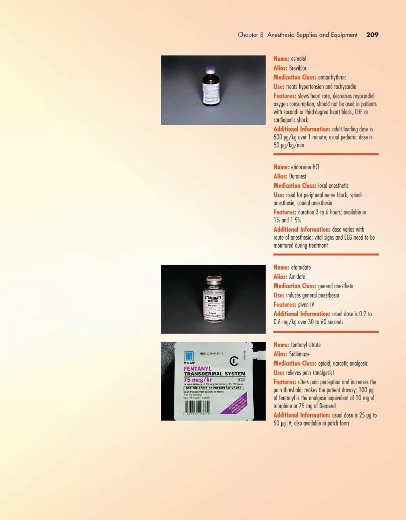
Chapter 8 Anesthesia Supplies and Equipment 209
Name: esmolol
Alias: Brevibloc
Medication Class: antiarrhythmic
Use: treats hypertension and tachycardia
Features: slows heart rate, decreases myocardialoxygen consumption; should not be used in patientswith second- or third-degree heart block, CHF or cardiogenic shock
Additional Information: adult loading dose is500 µg/kg over 1 minute; usual pediatric dose is50 µg/kg/min
Name: etidocaine HCl
Alias: Duranest
Medication Class: local anesthetic
Use: used for peripheral nerve block, spinal anesthesia, caudal anesthesia
Features: duration 3 to 6 hours; available in 1% and 1.5%
Additional Information: dose varies with route of anesthesia; vital signs and ECG need to bemonitored during treatment
Name: etomidate
Alias: Amidate
Medication Class: general anesthetic
Use: induces general anesthesia
Features: given IV
Additional Information: usual dose is 0.2 to0.6 mg/kg over 30 to 60 seconds
Name: fentanyl citrate
Alias: Sublimaze
Medication Class: opioid; narcotic analgesic
Use: relieves pain (analgesic)
Features: alters pain perception and increases thepain threshold; makes the patient drowsy; 100 µgof fentanyl is the analgesic equivalent of 10 mg ofmorphine or 75 mg of Demerol
Additional Information: usual dose is 25 µg to50 µg IV; also available in patch form
1572_Ch08_171-230.qxd 8/6/09 11:55 AM Page 209
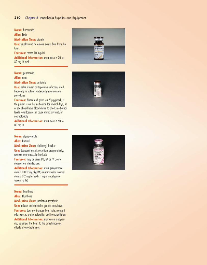
210 Chapter 8 Anesthesia Supplies and Equipment
Name: furosemide
Alias: Lasix
Medication Class: diuretic
Use: usually used to remove excess fluid from thelungs
Features: comes 10 mg/mL
Additional Information: usual dose is 20 to 80 mg IV push
Name: gentamicin
Alias: none
Medication Class: antibiotic
Use: helps prevent postoperative infection; used frequently in patients undergoing genitourinary procedures
Features: diluted and given via IV piggyback; ifthe patient is on the medication for several days, heor she should have blood drawn to check medicationlevels; overdosage can cause ototoxicity and/ornephrotoxicity
Additional Information: usual dose is 60 to 80 mg IV
Name: glycopyrrolate
Alias: Robinul
Medication Class: cholinergic blocker
Use: decreases gastric secretions preoperatively; reverses neuromuscular blockade
Features: may be given PO, IM or IV (route depends on intended use)
Additional Information: usual preoperativedose is 0.002 mg/kg IM; neuromuscular reversaldose is 0.2 mg for each 1 mg of neostigmine (given via IV)
Name: halothane
Alias: Fluothane
Medication Class: inhalation anesthetic
Use: induces and maintains general anesthesia
Features: does not increase heart rate; pleasantodor; causes uterine relaxation and bronchodilation
Additional Information: may cause bradycar-dia; sensitizes the heart to the arrhythmogenic effects of catecholamines
1572_Ch08_171-230.qxd 8/6/09 11:55 AM Page 210
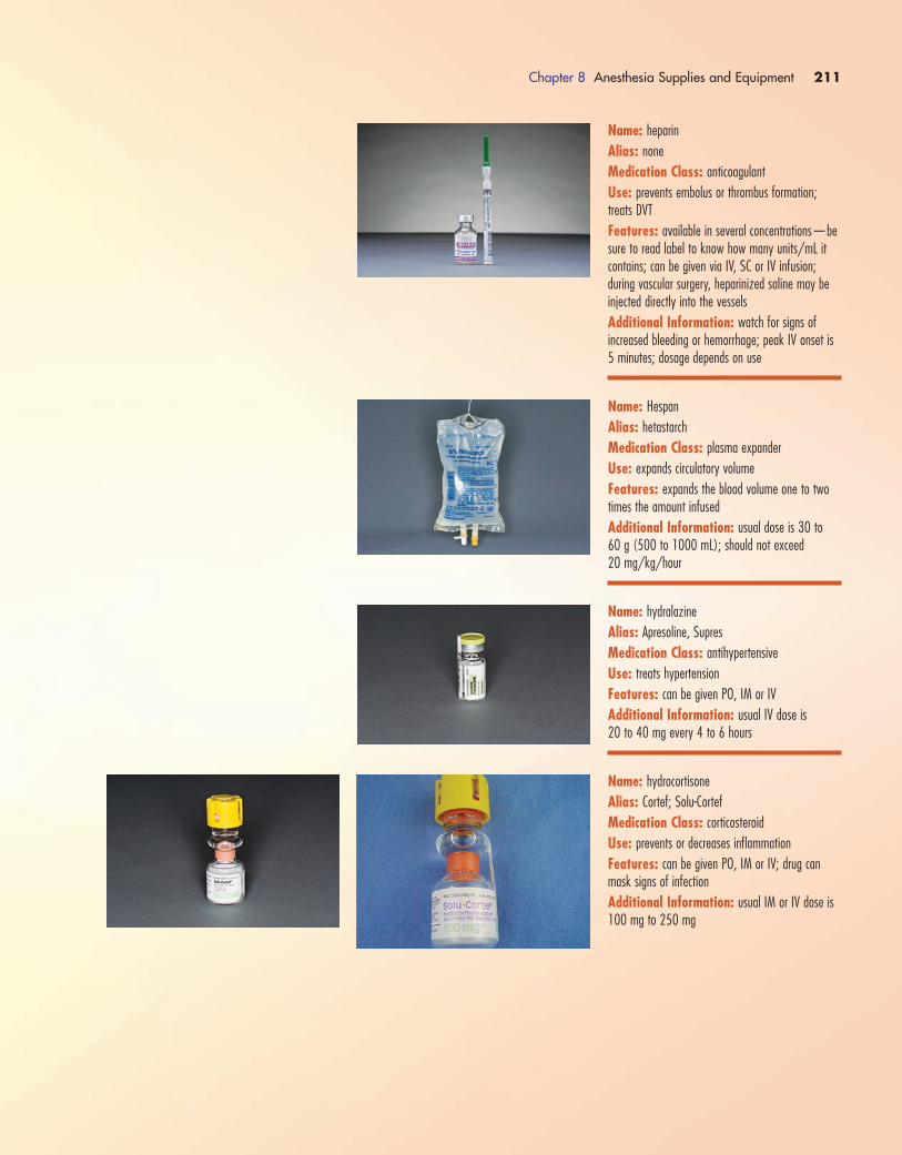
Chapter 8 Anesthesia Supplies and Equipment 211
Name: heparin
Alias: none
Medication Class: anticoagulant
Use: prevents embolus or thrombus formation;treats DVT
Features: available in several concentrations—besure to read label to know how many units/mL itcontains; can be given via IV, SC or IV infusion; during vascular surgery, heparinized saline may beinjected directly into the vessels
Additional Information: watch for signs of increased bleeding or hemorrhage; peak IV onset is5 minutes; dosage depends on use
Name: Hespan
Alias: hetastarch
Medication Class: plasma expander
Use: expands circulatory volume
Features: expands the blood volume one to twotimes the amount infused
Additional Information: usual dose is 30 to 60 g (500 to 1000 mL); should not exceed 20 mg/kg/hour
Name: hydralazine
Alias: Apresoline, Supres
Medication Class: antihypertensive
Use: treats hypertension
Features: can be given PO, IM or IV
Additional Information: usual IV dose is 20 to 40 mg every 4 to 6 hours
Name: hydrocortisone
Alias: Cortef; Solu-Cortef
Medication Class: corticosteroid
Use: prevents or decreases inflammation
Features: can be given PO, IM or IV; drug canmask signs of infection
Additional Information: usual IM or IV dose is100 mg to 250 mg
1572_Ch08_171-230.qxd 8/6/09 11:55 AM Page 211
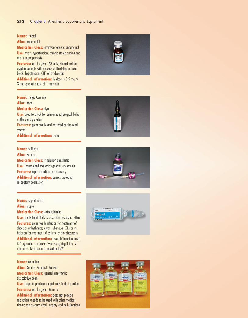
212 Chapter 8 Anesthesia Supplies and Equipment
Name: Inderal
Alias: propranolol
Medication Class: antihypertensive; antianginal
Use: treats hypertension, chronic stable angina andmigraine prophylaxis
Features: can be given PO or IV; should not beused in patients with second- or third-degree heartblock, hypotension, CHF or bradycardia
Additional Information: IV dose is 0.5 mg to 3 mg: give at a rate of 1 mg/min
Name: Indigo Carmine
Alias: none
Medication Class: dye
Use: used to check for unintentional surgical holesin the urinary system
Features: given via IV and excreted by the renalsystem
Additional Information: none
Name: isoflurane
Alias: Forane
Medication Class: inhalation anesthetic
Use: induces and maintains general anesthesia
Features: rapid induction and recovery
Additional Information: causes profound respiratory depression
Name: isoproterenol
Alias: Isuprel
Medication Class: catecholamine
Use: treats heart block, shock, bronchospasm, asthma
Features: given via IV infusion for treatment ofshock or arrhythmias; given sublingual (SL) or in-halation for treatment of asthma or bronchospasm
Additional Information: usual IV infusion doseis 5 µg/min; can cause tissue sloughing if the IV infiltrates; IV infusion is mixed in D5W
Name: ketamine
Alias: Ketalar, Ketanest, Ketaset
Medication Class: general anesthetic; dissociative agent
Use: helps to produce a rapid anesthetic induction
Features: can be given IM or IV
Additional Information: does not provide relaxation (needs to be used with other medica-tions); can produce vivid imagery and hallucinations
1572_Ch08_171-230.qxd 8/6/09 11:55 AM Page 212
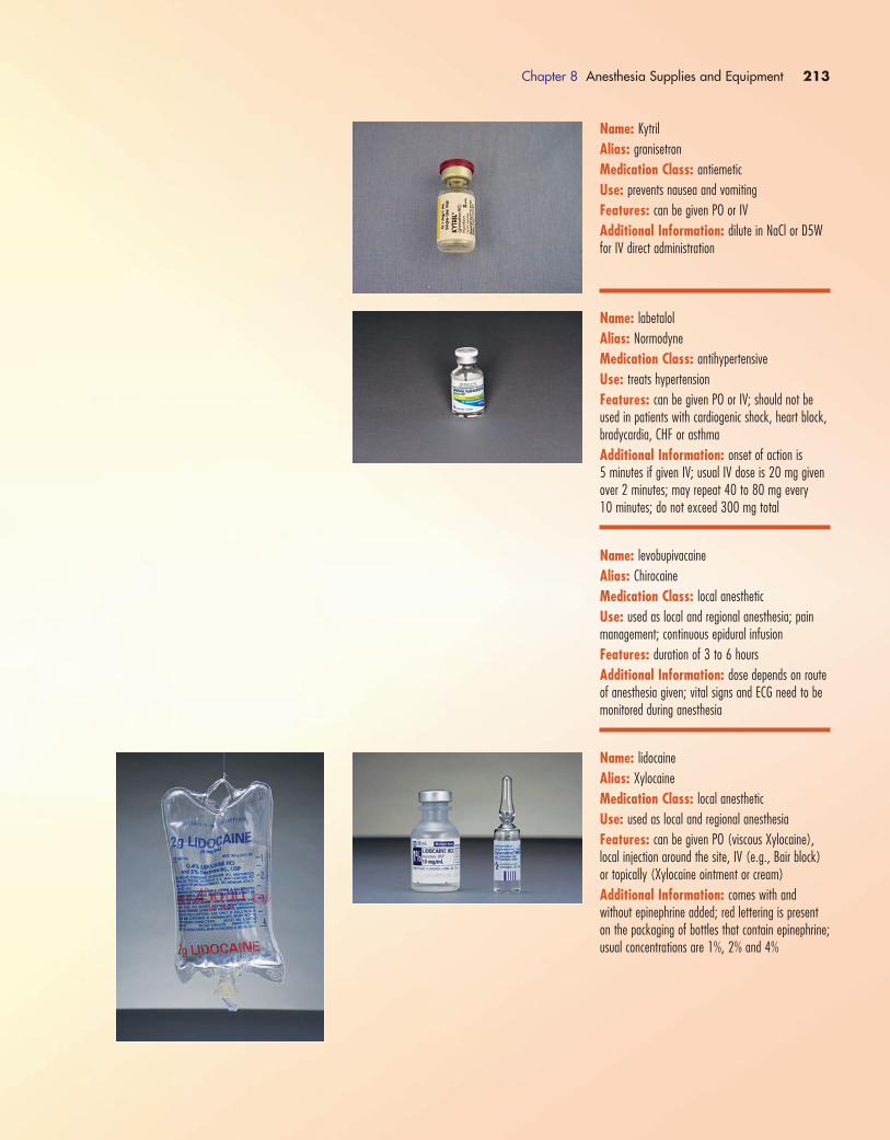
Chapter 8 Anesthesia Supplies and Equipment 213
Name: Kytril
Alias: granisetron
Medication Class: antiemetic
Use: prevents nausea and vomiting
Features: can be given PO or IV
Additional Information: dilute in NaCl or D5Wfor IV direct administration
Name: labetalol
Alias: Normodyne
Medication Class: antihypertensive
Use: treats hypertension
Features: can be given PO or IV; should not beused in patients with cardiogenic shock, heart block,bradycardia, CHF or asthma
Additional Information: onset of action is 5 minutes if given IV; usual IV dose is 20 mg givenover 2 minutes; may repeat 40 to 80 mg every 10 minutes; do not exceed 300 mg total
Name: levobupivacaine
Alias: Chirocaine
Medication Class: local anesthetic
Use: used as local and regional anesthesia; painmanagement; continuous epidural infusion
Features: duration of 3 to 6 hours
Additional Information: dose depends on routeof anesthesia given; vital signs and ECG need to bemonitored during anesthesia
Name: lidocaine
Alias: Xylocaine
Medication Class: local anesthetic
Use: used as local and regional anesthesia
Features: can be given PO (viscous Xylocaine), local injection around the site, IV (e.g., Bair block)or topically (Xylocaine ointment or cream)
Additional Information: comes with and without epinephrine added; red lettering is presenton the packaging of bottles that contain epinephrine;usual concentrations are 1%, 2% and 4%
1572_Ch08_171-230.qxd 8/6/09 11:55 AM Page 213
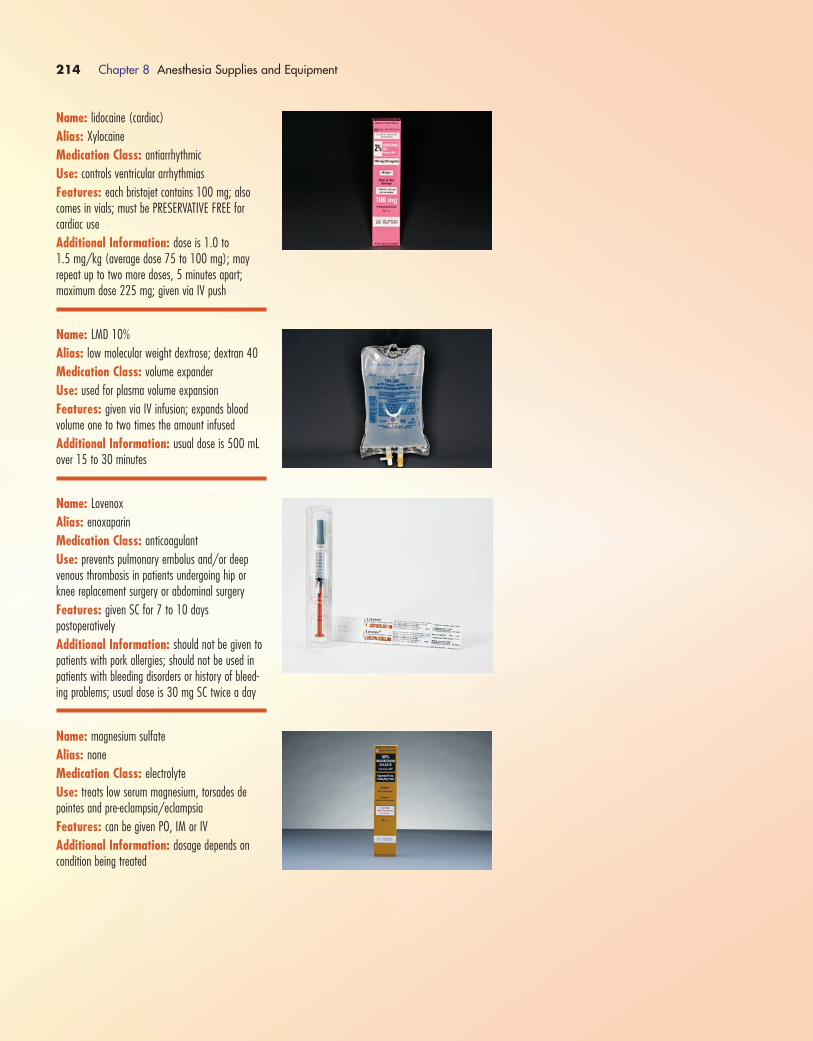
214 Chapter 8 Anesthesia Supplies and Equipment
Name: lidocaine (cardiac)
Alias: Xylocaine
Medication Class: antiarrhythmic
Use: controls ventricular arrhythmias
Features: each bristojet contains 100 mg; alsocomes in vials; must be PRESERVATIVE FREE for cardiac use
Additional Information: dose is 1.0 to 1.5 mg/kg (average dose 75 to 100 mg); may repeat up to two more doses, 5 minutes apart; maximum dose 225 mg; given via IV push
Name: LMD 10%
Alias: low molecular weight dextrose; dextran 40
Medication Class: volume expander
Use: used for plasma volume expansion
Features: given via IV infusion; expands blood volume one to two times the amount infused
Additional Information: usual dose is 500 mLover 15 to 30 minutes
Name: Lovenox
Alias: enoxaparin
Medication Class: anticoagulant
Use: prevents pulmonary embolus and/or deep venous thrombosis in patients undergoing hip orknee replacement surgery or abdominal surgery
Features: given SC for 7 to 10 days postoperatively
Additional Information: should not be given topatients with pork allergies; should not be used inpatients with bleeding disorders or history of bleed-ing problems; usual dose is 30 mg SC twice a day
Name: magnesium sulfate
Alias: none
Medication Class: electrolyte
Use: treats low serum magnesium, torsades depointes and pre-eclampsia/eclampsia
Features: can be given PO, IM or IV
Additional Information: dosage depends oncondition being treated
1572_Ch08_171-230.qxd 8/6/09 11:55 AM Page 214
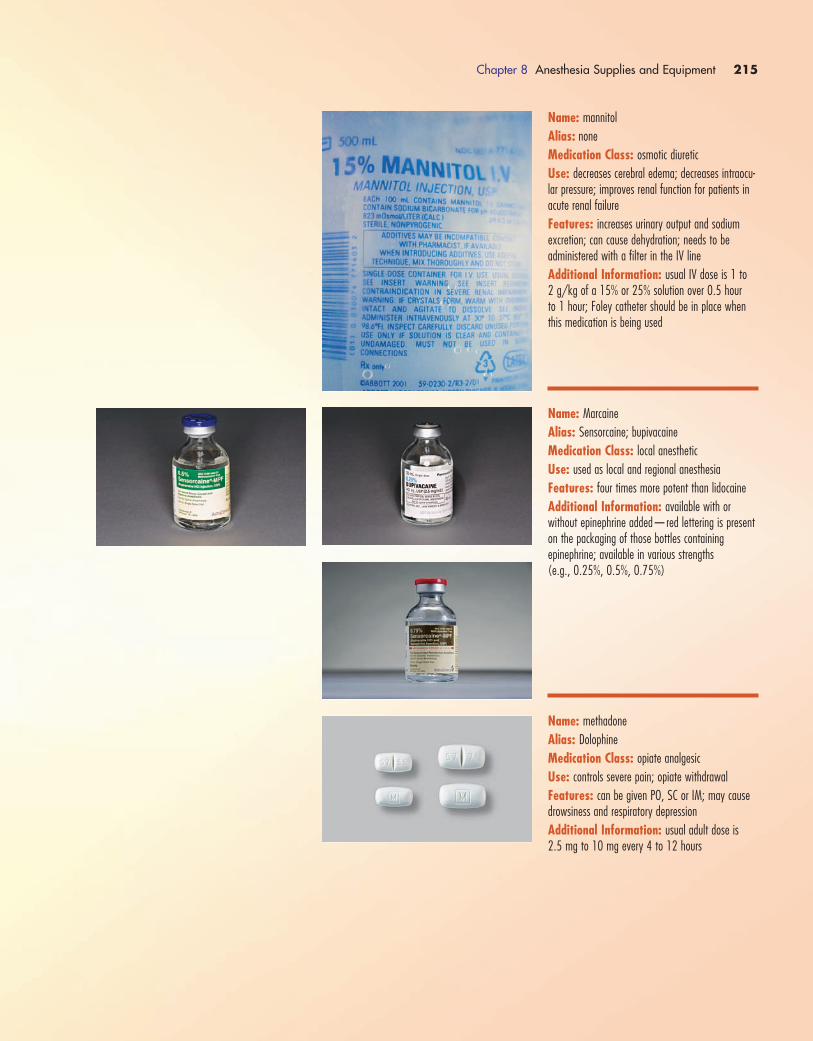
Chapter 8 Anesthesia Supplies and Equipment 215
Name: mannitol
Alias: none
Medication Class: osmotic diuretic
Use: decreases cerebral edema; decreases intraocu-lar pressure; improves renal function for patients inacute renal failure
Features: increases urinary output and sodium excretion; can cause dehydration; needs to be administered with a filter in the IV line
Additional Information: usual IV dose is 1 to 2 g/kg of a 15% or 25% solution over 0.5 hour to 1 hour; Foley catheter should be in place whenthis medication is being used
Name: Marcaine
Alias: Sensorcaine; bupivacaine
Medication Class: local anesthetic
Use: used as local and regional anesthesia
Features: four times more potent than lidocaine
Additional Information: available with or without epinephrine added—red lettering is presenton the packaging of those bottles containing epinephrine; available in various strengths (e.g., 0.25%, 0.5%, 0.75%)
Name: methadone
Alias: Dolophine
Medication Class: opiate analgesic
Use: controls severe pain; opiate withdrawal
Features: can be given PO, SC or IM; may causedrowsiness and respiratory depression
Additional Information: usual adult dose is 2.5 mg to 10 mg every 4 to 12 hours
1572_Ch08_171-230.qxd 8/6/09 11:55 AM Page 215
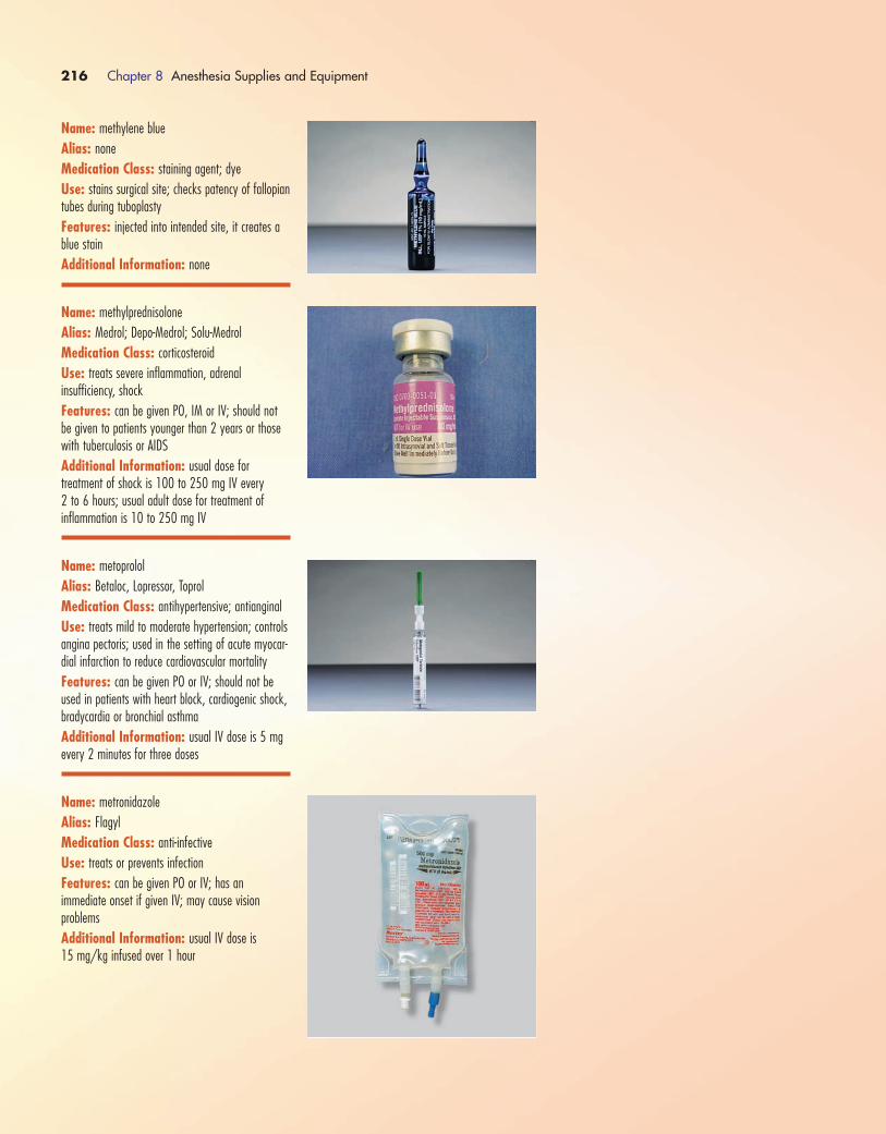
216 Chapter 8 Anesthesia Supplies and Equipment
Name: methylene blue
Alias: none
Medication Class: staining agent; dye
Use: stains surgical site; checks patency of fallopiantubes during tuboplasty
Features: injected into intended site, it creates ablue stain
Additional Information: none
Name: methylprednisolone
Alias: Medrol; Depo-Medrol; Solu-Medrol
Medication Class: corticosteroid
Use: treats severe inflammation, adrenal insufficiency, shock
Features: can be given PO, IM or IV; should notbe given to patients younger than 2 years or thosewith tuberculosis or AIDS
Additional Information: usual dose for treatment of shock is 100 to 250 mg IV every 2 to 6 hours; usual adult dose for treatment of inflammation is 10 to 250 mg IV
Name: metoprolol
Alias: Betaloc, Lopressor, Toprol
Medication Class: antihypertensive; antianginal
Use: treats mild to moderate hypertension; controlsangina pectoris; used in the setting of acute myocar-dial infarction to reduce cardiovascular mortality
Features: can be given PO or IV; should not beused in patients with heart block, cardiogenic shock,bradycardia or bronchial asthma
Additional Information: usual IV dose is 5 mgevery 2 minutes for three doses
Name: metronidazole
Alias: Flagyl
Medication Class: anti-infective
Use: treats or prevents infection
Features: can be given PO or IV; has an immediate onset if given IV; may cause vision problems
Additional Information: usual IV dose is 15 mg/kg infused over 1 hour
1572_Ch08_171-230.qxd 8/6/09 11:55 AM Page 216
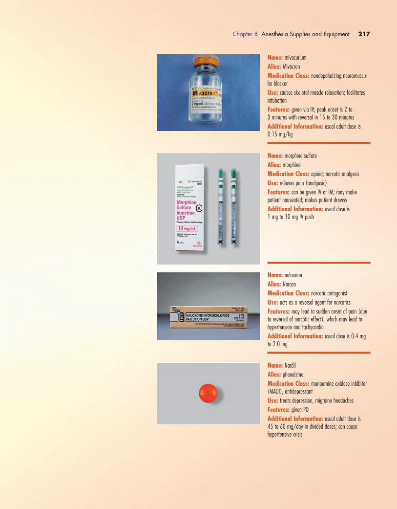
Chapter 8 Anesthesia Supplies and Equipment 217
Name: mivacurium
Alias: Mivacron
Medication Class: nondepolarizing neuromuscu-lar blocker
Use: causes skeletal muscle relaxation; facilitatesintubation
Features: given via IV; peak onset is 2 to 3 minutes with reversal in 15 to 30 minutes
Additional Information: usual adult dose is0.15 mg/kg
Name: morphine sulfate
Alias: morphine
Medication Class: opioid; narcotic analgesic
Use: relieves pain (analgesic)
Features: can be given IV or IM; may make patient nauseated; makes patient drowsy
Additional Information: usual dose is 1 mg to 10 mg IV push
Name: naloxone
Alias: Narcan
Medication Class: narcotic antagonist
Use: acts as a reversal agent for narcotics
Features: may lead to sudden onset of pain (dueto reversal of narcotic effect), which may lead to hypertension and tachycardia
Additional Information: usual dose is 0.4 mgto 2.0 mg
Name: Nardil
Alias: phenelzine
Medication Class: monoamine oxidase inhibitor(MAOI), antidepressant
Use: treats depression, migraine headaches
Features: given PO
Additional Information: usual adult dose is 45 to 60 mg/day in divided doses; can cause hypertensive crisis
1572_Ch08_171-230.qxd 8/6/09 11:55 AM Page 217
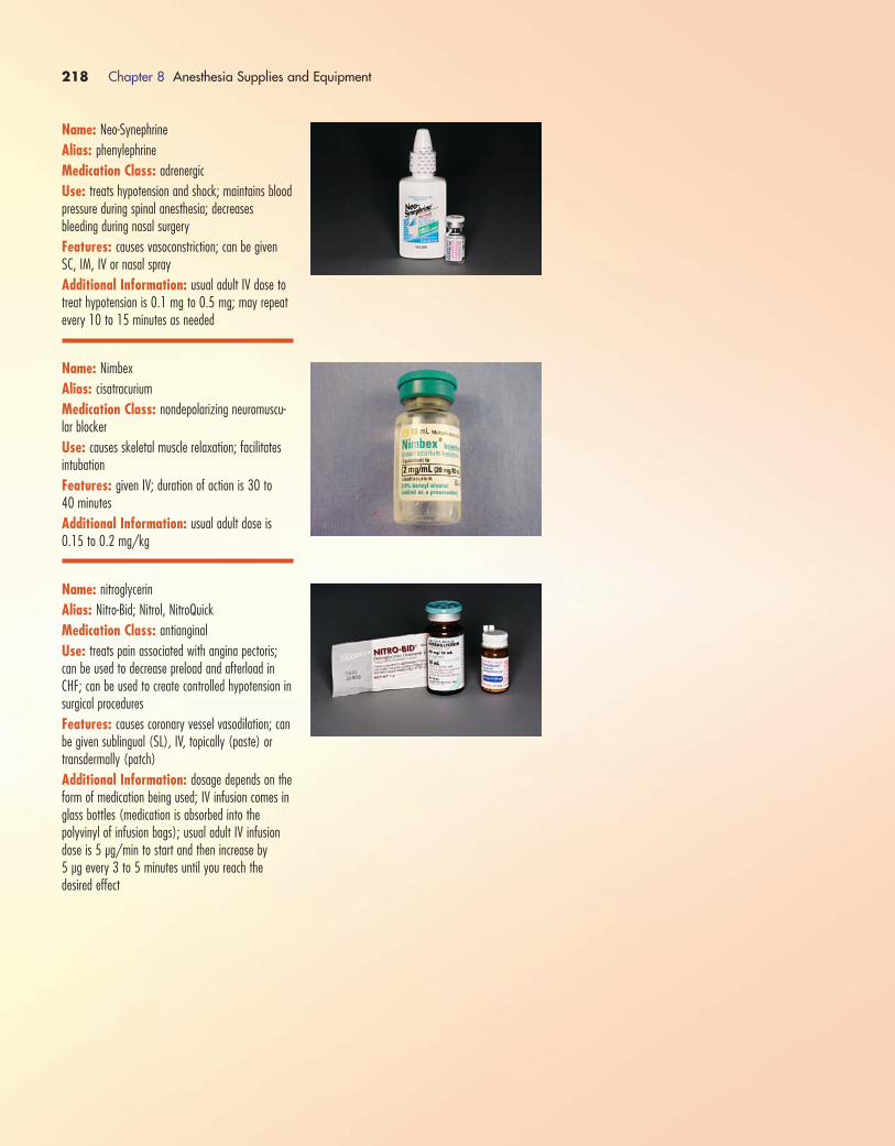
218 Chapter 8 Anesthesia Supplies and Equipment
Name: Neo-Synephrine
Alias: phenylephrine
Medication Class: adrenergic
Use: treats hypotension and shock; maintains bloodpressure during spinal anesthesia; decreases bleeding during nasal surgery
Features: causes vasoconstriction; can be givenSC, IM, IV or nasal spray
Additional Information: usual adult IV dose totreat hypotension is 0.1 mg to 0.5 mg; may repeatevery 10 to 15 minutes as needed
Name: Nimbex
Alias: cisatracurium
Medication Class: nondepolarizing neuromuscu-lar blocker
Use: causes skeletal muscle relaxation; facilitatesintubation
Features: given IV; duration of action is 30 to 40 minutes
Additional Information: usual adult dose is0.15 to 0.2 mg/kg
Name: nitroglycerin
Alias: Nitro-Bid; Nitrol, NitroQuick
Medication Class: antianginal
Use: treats pain associated with angina pectoris;can be used to decrease preload and afterload inCHF; can be used to create controlled hypotension insurgical procedures
Features: causes coronary vessel vasodilation; canbe given sublingual (SL), IV, topically (paste) ortransdermally (patch)
Additional Information: dosage depends on theform of medication being used; IV infusion comes inglass bottles (medication is absorbed into thepolyvinyl of infusion bags); usual adult IV infusiondose is 5 µg/min to start and then increase by 5 µg every 3 to 5 minutes until you reach the desired effect
1572_Ch08_171-230.qxd 8/6/09 11:55 AM Page 218
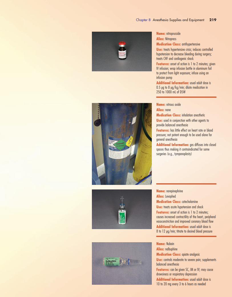
Chapter 8 Anesthesia Supplies and Equipment 219
Name: nitroprusside
Alias: Nitropress
Medication Class: antihypertensive
Use: treats hypertensive crisis; induces controlledhypotension to decrease bleeding during surgery;treats CHF and cardiogenic shock
Features: onset of action is 1 to 2 minutes; givenIV infusion; wrap infusion bottle in aluminum foil to protect from light exposure; infuse using an infusion pump
Additional Information: usual adult dose is 0.5 µg to 8 µg/kg/min; dilute medication in 250 to 1000 mL of D5W
Name: nitrous oxide
Alias: none
Medication Class: inhalation anesthetic
Use: used in conjunction with other agents to provide balanced anesthesia
Features: has little effect on heart rate or bloodpressure; not potent enough to be used alone forgeneral anesthesia
Additional Information: gas diffuses into closedspaces thus making it contraindicated for some surgeries (e.g., tympanoplasty)
Name: norepinephrine
Alias: Levophed
Medication Class: catecholamine
Use: treats acute hypotension and shock
Features: onset of action is 1 to 2 minutes;causes increased contractility of the heart, peripheralvasoconstriction and improved coronary blood flow
Additional Information: usual adult dose is 8 to 12 µg/min; titrate to desired blood pressure
Name: Nubain
Alias: nalbuphine
Medication Class: opiate analgesic
Use: controls moderate to severe pain; supplementsbalanced anesthesia
Features: can be given SC, IM or IV; may causedrowsiness or respiratory depression
Additional Information: usual adult dose is 10 to 20 mg every 3 to 6 hours as needed
1572_Ch08_171-230.qxd 8/6/09 11:55 AM Page 219
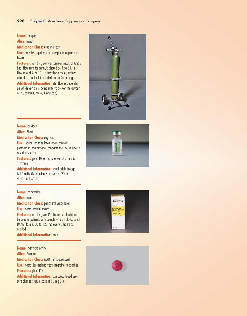
220 Chapter 8 Anesthesia Supplies and Equipment
Name: oxygen
Alias: none
Medication Class: essential gas
Use: provides supplemental oxygen to organs andtissue
Features: can be given via cannula, mask or Ambubag; flow rate for cannula should be 1 to 3 L; aflow rate of 8 to 10 L is best for a mask; a flowrate of 10 to 15 L is needed for an Ambu bag
Additional Information: liter flow is dependenton which vehicle is being used to deliver the oxygen(e.g., cannula, mask, Ambu bag)
Name: oxytocin
Alias: Pitocin
Medication Class: oxytocic
Use: induces or stimulates labor; controls postpartum hemorrhage; contracts the uterus after acesarian section
Features: given IM or IV; IV onset of action is 1 minute
Additional Information: usual adult dosage is 10 units (IV infusion is infused at 20 to 4 microunits/min)
Name: papaverine
Alias: none
Medication Class: peripheral vasodilator
Use: treats arterial spasm
Features: can be given PO, IM or IV; should notbe used in patients with complete heart block; usualIM/IV dose is 30 to 120 mg every 3 hours asneeded
Additional Information: none
Name: tranylcypromine
Alias: Parnate
Medication Class: MAOI, antidepressant
Use: treats depression; treats migraine headaches
Features: given PO
Additional Information: can cause blood pres-sure changes; usual dose is 10 mg BID
1572_Ch08_171-230.qxd 8/6/09 11:55 AM Page 220
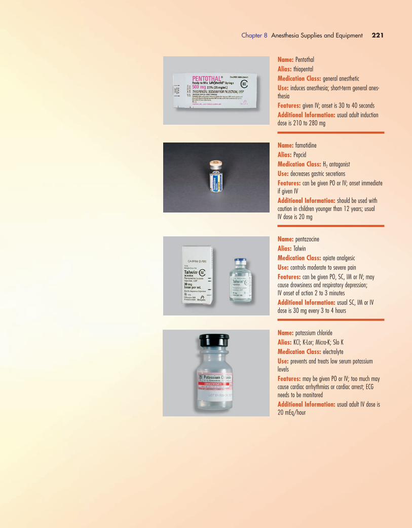
Chapter 8 Anesthesia Supplies and Equipment 221
Name: Pentothal
Alias: thiopental
Medication Class: general anesthetic
Use: induces anesthesia; short-term general anes-thesia
Features: given IV; onset is 30 to 40 seconds
Additional Information: usual adult inductiondose is 210 to 280 mg
Name: famotidine
Alias: Pepcid
Medication Class: H2 antagonist
Use: decreases gastric secretions
Features: can be given PO or IV; onset immediateif given IV
Additional Information: should be used withcaution in children younger than 12 years; usual IV dose is 20 mg
Name: pentazocine
Alias: Talwin
Medication Class: opiate analgesic
Use: controls moderate to severe pain
Features: can be given PO, SC, IM or IV; maycause drowsiness and respiratory depression; IV onset of action 2 to 3 minutes
Additional Information: usual SC, IM or IVdose is 30 mg every 3 to 4 hours
Name: potassium chloride
Alias: KCl; K-Lor; Micro-K; Slo K
Medication Class: electrolyte
Use: prevents and treats low serum potassium levels
Features: may be given PO or IV; too much maycause cardiac arrhythmias or cardiac arrest; ECGneeds to be monitored
Additional Information: usual adult IV dose is20 mEq/hour
1572_Ch08_171-230.qxd 8/6/09 11:56 AM Page 221
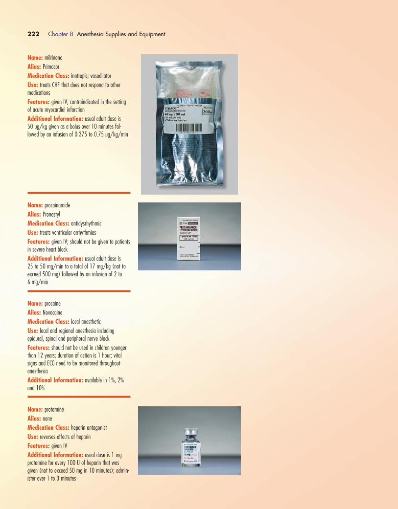
222 Chapter 8 Anesthesia Supplies and Equipment
Name: milrinone
Alias: Primacor
Medication Class: inotropic; vasodilator
Use: treats CHF that does not respond to othermedications
Features: given IV; contraindicated in the settingof acute myocardial infarction
Additional Information: usual adult dose is 50 µg/kg given as a bolus over 10 minutes fol-lowed by an infusion of 0.375 to 0.75 µg/kg/min
Name: procainamide
Alias: Pronestyl
Medication Class: antidysrhythmic
Use: treats ventricular arrhythmias
Features: given IV; should not be given to patientsin severe heart block
Additional Information: usual adult dose is 25 to 50 mg/min to a total of 17 mg/kg (not toexceed 500 mg) followed by an infusion of 2 to 6 mg/min
Name: procaine
Alias: Novocaine
Medication Class: local anesthetic
Use: local and regional anesthesia includingepidural, spinal and peripheral nerve block
Features: should not be used in children youngerthan 12 years; duration of action is 1 hour; vitalsigns and ECG need to be monitored throughoutanesthesia
Additional Information: available in 1%, 2%and 10%
Name: protamine
Alias: none
Medication Class: heparin antagonist
Use: reverses effects of heparin
Features: given IV
Additional Information: usual dose is 1 mgprotamine for every 100 U of heparin that wasgiven (not to exceed 50 mg in 10 minutes); admin-ister over 1 to 3 minutes
1572_Ch08_171-230.qxd 8/6/09 11:56 AM Page 222
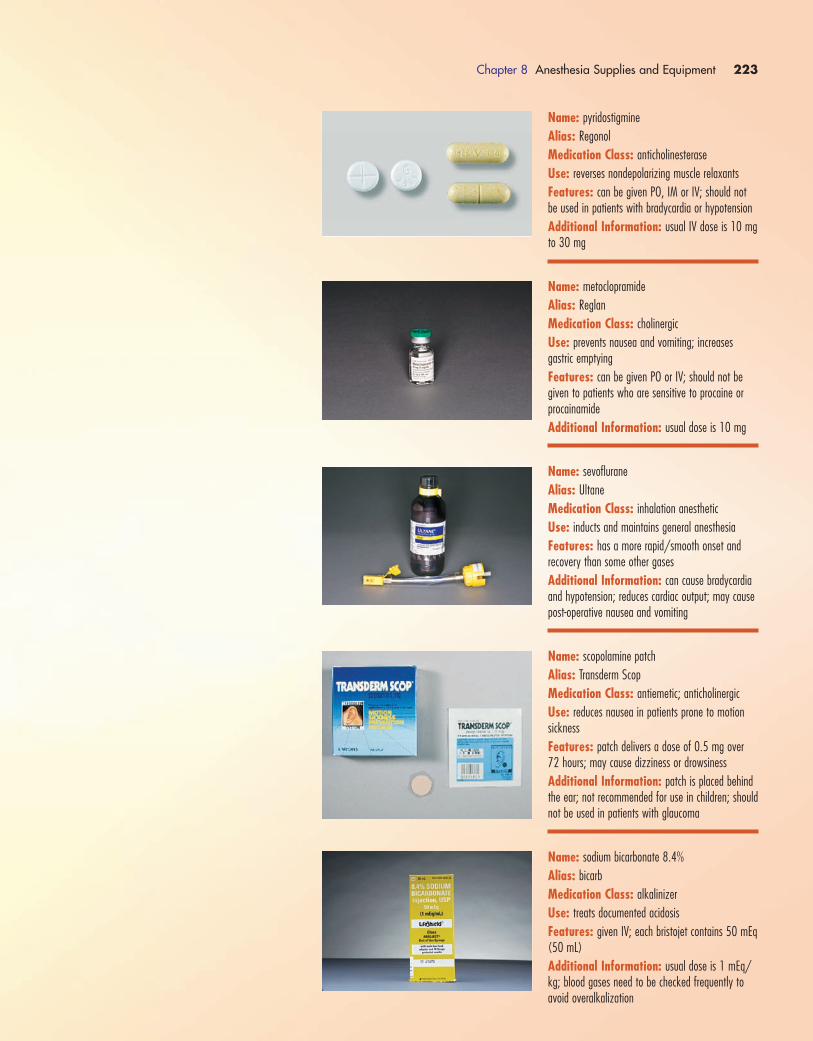
Chapter 8 Anesthesia Supplies and Equipment 223
Name: pyridostigmine
Alias: Regonol
Medication Class: anticholinesterase
Use: reverses nondepolarizing muscle relaxants
Features: can be given PO, IM or IV; should notbe used in patients with bradycardia or hypotension
Additional Information: usual IV dose is 10 mgto 30 mg
Name: metoclopramide
Alias: Reglan
Medication Class: cholinergic
Use: prevents nausea and vomiting; increases gastric emptying
Features: can be given PO or IV; should not begiven to patients who are sensitive to procaine orprocainamide
Additional Information: usual dose is 10 mg
Name: sevoflurane
Alias: Ultane
Medication Class: inhalation anesthetic
Use: inducts and maintains general anesthesia
Features: has a more rapid/smooth onset and recovery than some other gases
Additional Information: can cause bradycardiaand hypotension; reduces cardiac output; may causepost-operative nausea and vomiting
Name: scopolamine patch
Alias: Transderm Scop
Medication Class: antiemetic; anticholinergic
Use: reduces nausea in patients prone to motionsickness
Features: patch delivers a dose of 0.5 mg over72 hours; may cause dizziness or drowsiness
Additional Information: patch is placed behindthe ear; not recommended for use in children; shouldnot be used in patients with glaucoma
Name: sodium bicarbonate 8.4%
Alias: bicarb
Medication Class: alkalinizer
Use: treats documented acidosis
Features: given IV; each bristojet contains 50 mEq(50 mL)
Additional Information: usual dose is 1 mEq/kg; blood gases need to be checked frequently toavoid overalkalization
1572_Ch08_171-230.qxd 8/6/09 11:56 AM Page 223
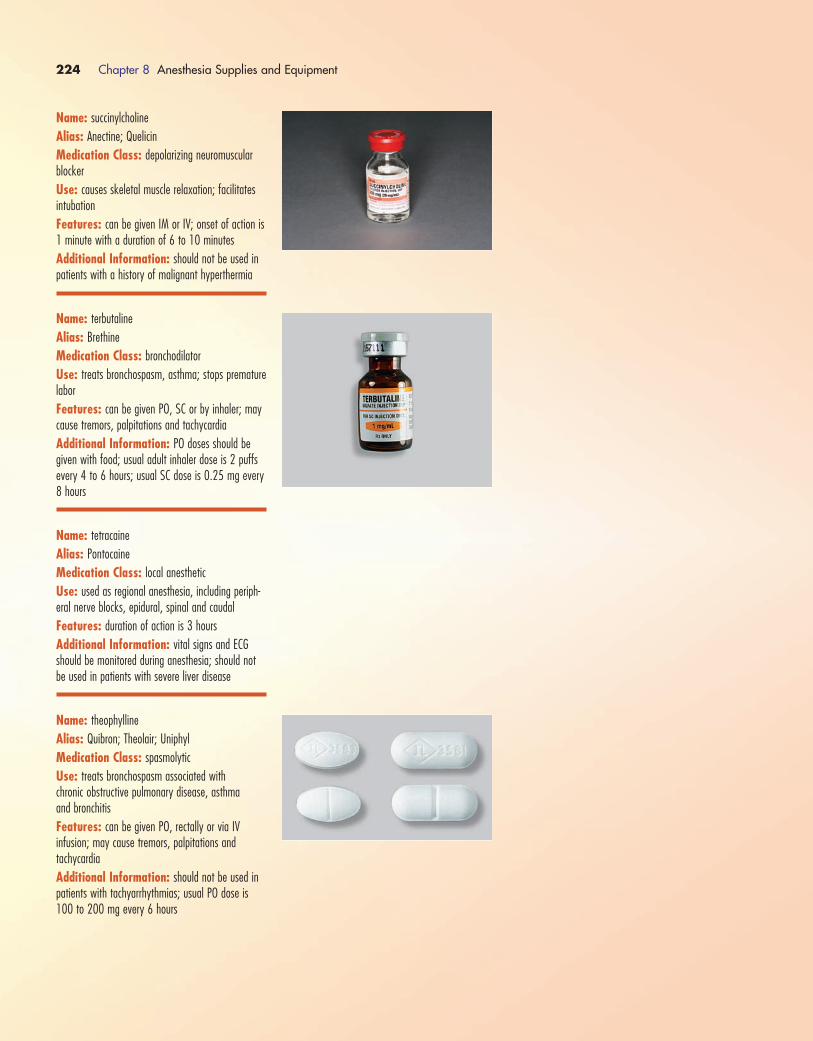
224 Chapter 8 Anesthesia Supplies and Equipment
Name: succinylcholine
Alias: Anectine; Quelicin
Medication Class: depolarizing neuromuscularblocker
Use: causes skeletal muscle relaxation; facilitatesintubation
Features: can be given IM or IV; onset of action is1 minute with a duration of 6 to 10 minutes
Additional Information: should not be used inpatients with a history of malignant hyperthermia
Name: terbutaline
Alias: Brethine
Medication Class: bronchodilator
Use: treats bronchospasm, asthma; stops prematurelabor
Features: can be given PO, SC or by inhaler; maycause tremors, palpitations and tachycardia
Additional Information: PO doses should begiven with food; usual adult inhaler dose is 2 puffsevery 4 to 6 hours; usual SC dose is 0.25 mg every8 hours
Name: tetracaine
Alias: Pontocaine
Medication Class: local anesthetic
Use: used as regional anesthesia, including periph-eral nerve blocks, epidural, spinal and caudal
Features: duration of action is 3 hours
Additional Information: vital signs and ECGshould be monitored during anesthesia; should notbe used in patients with severe liver disease
Name: theophylline
Alias: Quibron; Theolair; Uniphyl
Medication Class: spasmolytic
Use: treats bronchospasm associated with chronic obstructive pulmonary disease, asthma and bronchitis
Features: can be given PO, rectally or via IV infusion; may cause tremors, palpitations and tachycardia
Additional Information: should not be used inpatients with tachyarrhythmias; usual PO dose is100 to 200 mg every 6 hours
1572_Ch08_171-230.qxd 8/6/09 11:56 AM Page 224
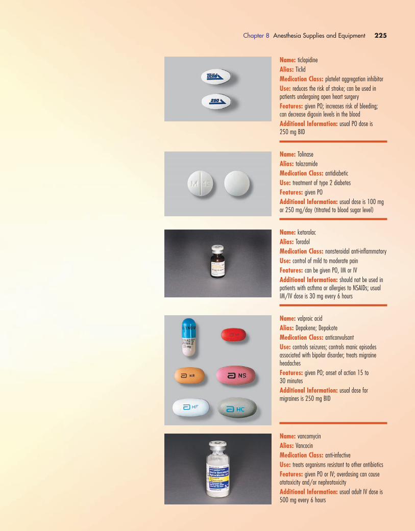
Chapter 8 Anesthesia Supplies and Equipment 225
Name: ticlopidine
Alias: Ticlid
Medication Class: platelet aggregation inhibitor
Use: reduces the risk of stroke; can be used in patients undergoing open heart surgery
Features: given PO; increases risk of bleeding; can decrease digoxin levels in the blood
Additional Information: usual PO dose is 250 mg BID
Name: Tolinase
Alias: tolazamide
Medication Class: antidiabetic
Use: treatment of type 2 diabetes
Features: given PO
Additional Information: usual dose is 100 mgor 250 mg/day (titrated to blood sugar level)
Name: ketorolac
Alias: Toradol
Medication Class: nonsteroidal anti-inflammatory
Use: control of mild to moderate pain
Features: can be given PO, IM or IV
Additional Information: should not be used inpatients with asthma or allergies to NSAIDs; usualIM/IV dose is 30 mg every 6 hours
Name: valproic acid
Alias: Depakene; Depakote
Medication Class: anticonvulsant
Use: controls seizures; controls manic episodes associated with bipolar disorder; treats migraineheadaches
Features: given PO; onset of action 15 to 30 minutes
Additional Information: usual dose for migraines is 250 mg BID
Name: vancomycin
Alias: Vancocin
Medication Class: anti-infective
Use: treats organisms resistant to other antibiotics
Features: given PO or IV; overdosing can causeototoxicity and/or nephrotoxicity
Additional Information: usual adult IV dose is500 mg every 6 hours
1572_Ch08_171-230.qxd 8/6/09 11:56 AM Page 225
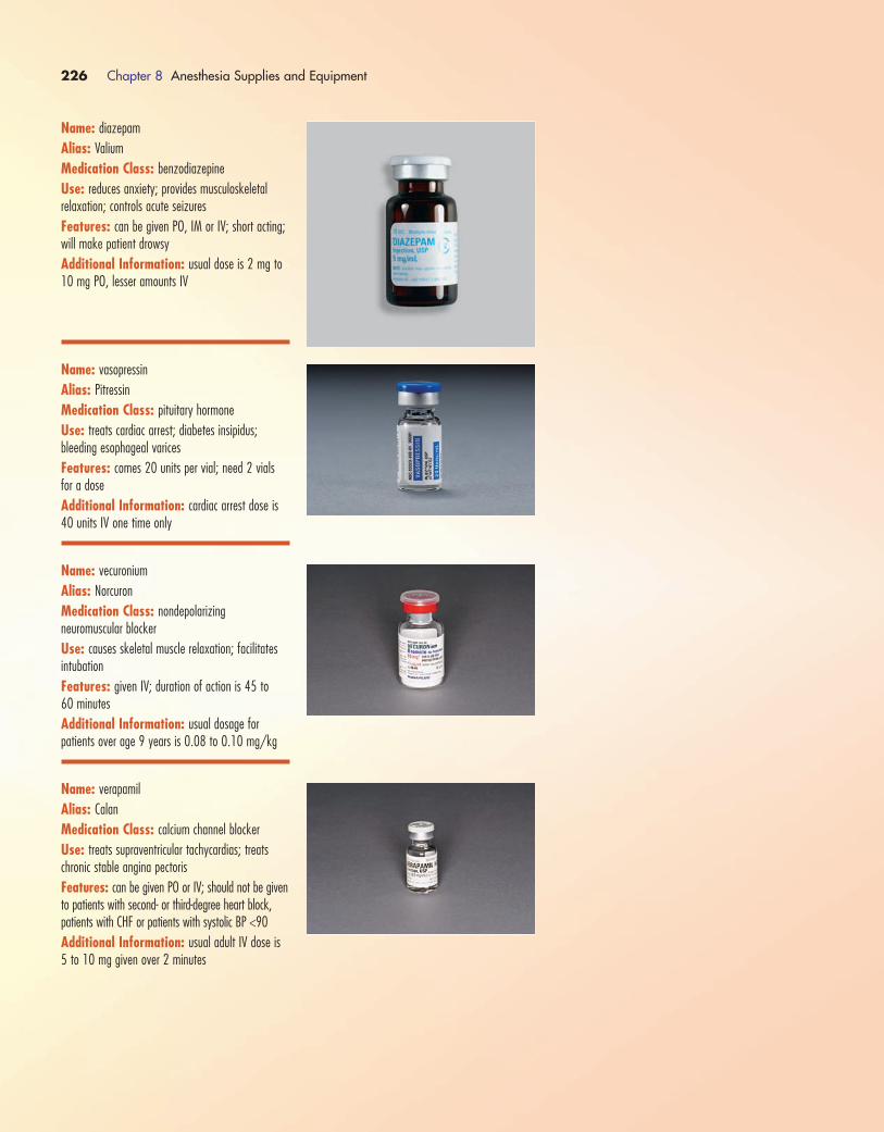
226 Chapter 8 Anesthesia Supplies and Equipment
Name: diazepam
Alias: Valium
Medication Class: benzodiazepine
Use: reduces anxiety; provides musculoskeletal relaxation; controls acute seizures
Features: can be given PO, IM or IV; short acting;will make patient drowsy
Additional Information: usual dose is 2 mg to10 mg PO, lesser amounts IV
Name: vasopressin
Alias: Pitressin
Medication Class: pituitary hormone
Use: treats cardiac arrest; diabetes insipidus; bleeding esophageal varices
Features: comes 20 units per vial; need 2 vialsfor a dose
Additional Information: cardiac arrest dose is40 units IV one time only
Name: vecuronium
Alias: Norcuron
Medication Class: nondepolarizing neuromuscular blocker
Use: causes skeletal muscle relaxation; facilitatesintubation
Features: given IV; duration of action is 45 to 60 minutes
Additional Information: usual dosage for patients over age 9 years is 0.08 to 0.10 mg/kg
Name: verapamil
Alias: Calan
Medication Class: calcium channel blocker
Use: treats supraventricular tachycardias; treatschronic stable angina pectoris
Features: can be given PO or IV; should not be givento patients with second- or third-degree heart block, patients with CHF or patients with systolic BP <90
Additional Information: usual adult IV dose is5 to 10 mg given over 2 minutes
1572_Ch08_171-230.qxd 8/6/09 11:56 AM Page 226
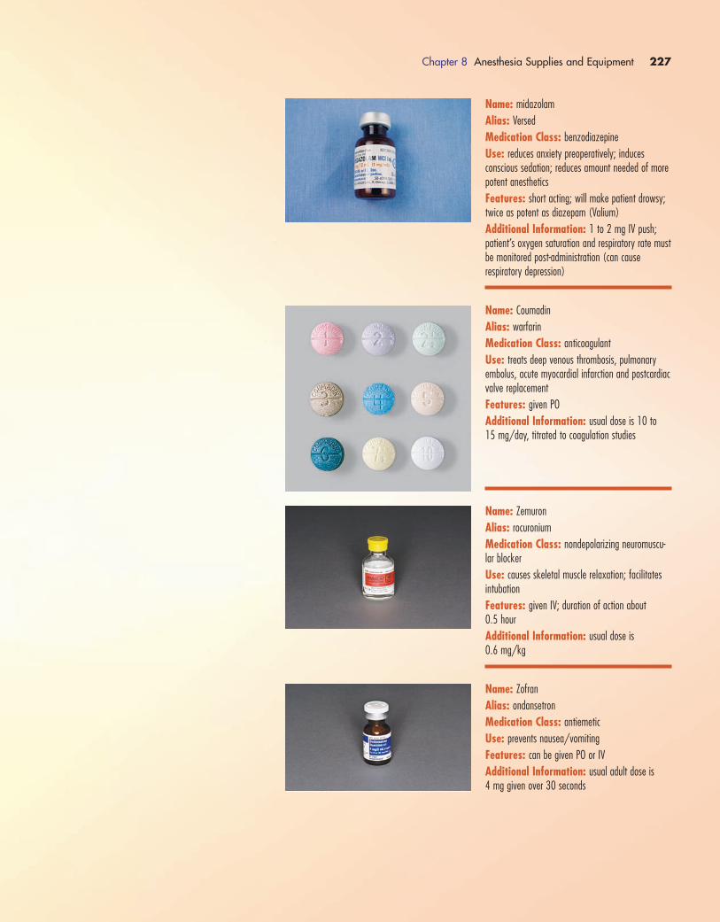
Chapter 8 Anesthesia Supplies and Equipment 227
Name: midazolam
Alias: Versed
Medication Class: benzodiazepine
Use: reduces anxiety preoperatively; induces conscious sedation; reduces amount needed of morepotent anesthetics
Features: short acting; will make patient drowsy;twice as potent as diazepam (Valium)
Additional Information: 1 to 2 mg IV push; patient’s oxygen saturation and respiratory rate mustbe monitored post-administration (can cause respiratory depression)
Name: Coumadin
Alias: warfarin
Medication Class: anticoagulant
Use: treats deep venous thrombosis, pulmonaryembolus, acute myocardial infarction and postcardiacvalve replacement
Features: given PO
Additional Information: usual dose is 10 to 15 mg/day, titrated to coagulation studies
Name: Zemuron
Alias: rocuronium
Medication Class: nondepolarizing neuromuscu-lar blocker
Use: causes skeletal muscle relaxation; facilitatesintubation
Features: given IV; duration of action about 0.5 hour
Additional Information: usual dose is 0.6 mg/kg
Name: Zofran
Alias: ondansetron
Medication Class: antiemetic
Use: prevents nausea/vomiting
Features: can be given PO or IV
Additional Information: usual adult dose is 4 mg given over 30 seconds
1572_Ch08_171-230.qxd 8/6/09 11:56 AM Page 227
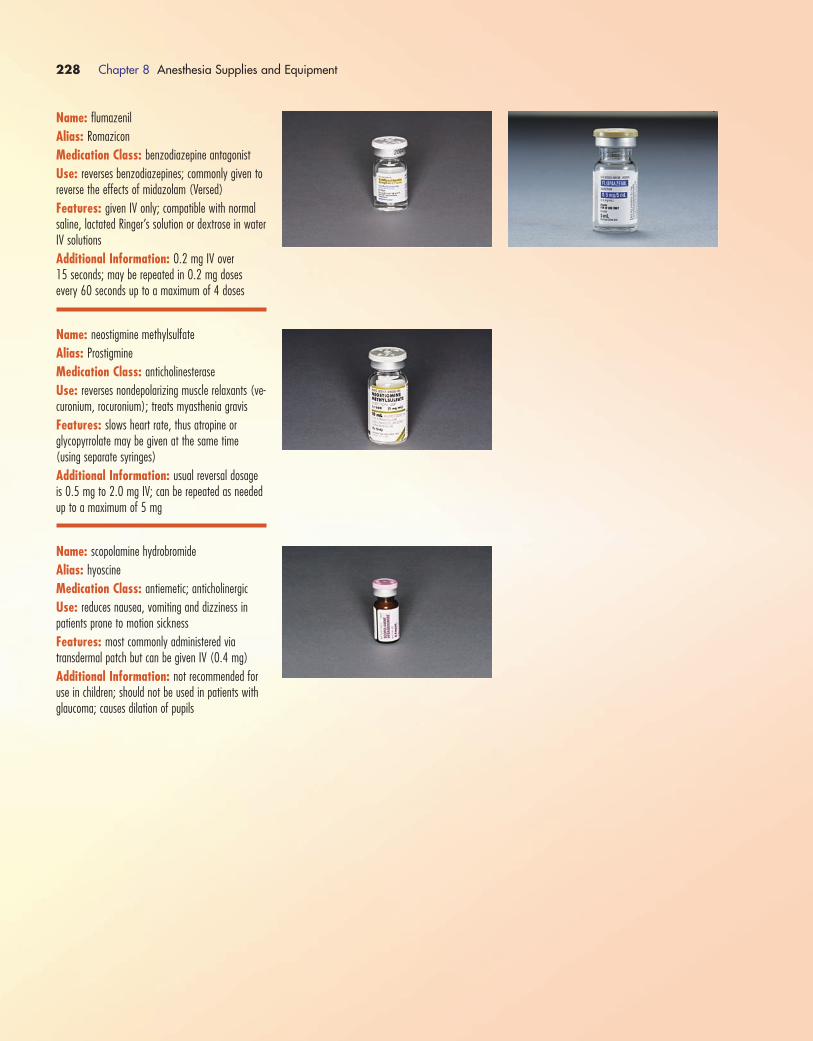
228 Chapter 8 Anesthesia Supplies and Equipment
Name: flumazenil
Alias: Romazicon
Medication Class: benzodiazepine antagonist
Use: reverses benzodiazepines; commonly given toreverse the effects of midazolam (Versed)
Features: given IV only; compatible with normalsaline, lactated Ringer’s solution or dextrose in waterIV solutions
Additional Information: 0.2 mg IV over 15 seconds; may be repeated in 0.2 mg dosesevery 60 seconds up to a maximum of 4 doses
Name: neostigmine methylsulfate
Alias: Prostigmine
Medication Class: anticholinesterase
Use: reverses nondepolarizing muscle relaxants (ve-curonium, rocuronium); treats myasthenia gravis
Features: slows heart rate, thus atropine or glycopyrrolate may be given at the same time (using separate syringes)
Additional Information: usual reversal dosageis 0.5 mg to 2.0 mg IV; can be repeated as neededup to a maximum of 5 mg
Name: scopolamine hydrobromide
Alias: hyoscine
Medication Class: antiemetic; anticholinergic
Use: reduces nausea, vomiting and dizziness in patients prone to motion sickness
Features: most commonly administered via transdermal patch but can be given IV (0.4 mg)
Additional Information: not recommended foruse in children; should not be used in patients withglaucoma; causes dilation of pupils
1572_Ch08_171-230.qxd 8/6/09 11:56 AM Page 228

Chapter 8 Anesthesia Supplies and Equipment 229
Surgical Session Review
1) The purpose of ____________ is to absorb carbon dioxide
in the anesthesia machine breathing circuit.
a. soda lime
b. limestone
c. soap stone
d. soda pop
2) _____________ is the medication used in the treatment of
malignant hyperthermia.
a. Decadron
b. Lasix
c. dantrolene
d. digoxin
3) A reading of below _________ on the BIS monitor means
the patient is unconscious.
a. 80
b. 90
c. 70
d. 60
4) A _________ ___________ is type of blanket that blows
warm air over the patient’s skin surface to help prevent hy-
pothermia.
a. Bear Hugger
b. Bair Hugger
c. Bare Hugger
d. Bear Blanket
5) ___________ is used to reverse the effects of narcotics.
a. digoxin
b. Decadron
c. Narcan
d. atropine
6) Pediatric defibrillation pads may be used on children
weighing up to ________ kg.
a. 10
b. 15
c. 20
d. 30
7) The strength of epinephrine used in a cardiac arrest situa-
tion is:
a. 1:100
b. 1:1000
c. 1:10,000
d. 1:100,000
8) The strength of epinephrine used to treat an allergic re-
action is:
a. 1:100
b. 1:1000
c. 1:10,000
d. 1:100,000
9)Your patient is feeling nauseated.Which of the following
medications might anesthesia give to the patient?
a. Narcan
b. Kytril
c. sodium bicarbonate
d. Lidocaine
10) A setting of 2–3 liters of oxygen per minute would be an
appropriate setting for a:
a. Ambu bag
b. nonrebreather mask
c. oxygen mask
d. nasal cannula
11) _________ forceps may be used during intubation to
help guide the ETT.
a. Magill
b. Maguile
c. DeBakey
d. Mayo
12) Isoflurane and sevoflurane are:
a. antiemetics
b. antipyretics
c. inhalation anesthetics
d. IV anesthetics
13) ___________ can be used to treat a patient with symp-
tomatic bradycardia.
a. Lidocaine
b. amiodarone
c. vasopressin
d. atropine
14) The anesthesiologist asks for the TEE. What does TEE
stand for?
a. transesophageal endoscope
b. transendotracheal endoscope
c. transesophageal echocardiogram
d. transendotracheal echocardiogram
1572_Ch08_171-230.qxd 8/6/09 11:56 AM Page 229

15) When the infant’s head is about to be delivered in a C-
section, which of the following should you have ready to
hand to the surgeon to suction the newborn’s mouth and
nose (assume no meconium issues)?
a. DeLee suction
b. Blue Bulb syringe
c. Meconium aspirator
d.Yankaur suction
16) A Salem Sump tube is a type of:
a. ETT
b. nasogastric tube
c. wound drain
d. suction catheter
230 Chapter 8 Anesthesia Supplies and Equipment
1572_Ch08_171-230.qxd 8/6/09 11:56 AM Page 230
ANSWERS FOR SURGICAL SESSION:CHAPTER 1
1. c
2. b
3. b
4. d
5. d
6. d
7. d
8. a
9. c
10. c
11. c
12. a
13. c
14. d
15. b
16. d
17. a
18. c
19. d
20. c
ANSWERS FOR SURGICAL SESSION:CHAPTER 2
1. a
2. b
3. c
4. d
5. b
6. a
7. d
8. b
9. a
10. b
11. c
12. d
13. a
14. c
15. c
16. b
17. c
18. a
19. c
20. d
21. b
ANSWERS FOR SURGICAL SESSION:CHAPTER 3
1. c
2. b
3. a
4. d
5. c
6. d
7. b
8. a
9. d
10. b
11. c
12. b
13. c
ANSWERS FOR SURGICAL SESSION:CHAPTER 4
1. c
2. b
3. b
4. a
5. d
6. d
7. d
8. c
9. b
10. c
11. b
12. c
13. c
14. a
15. d
16. b
17. a
18. a
19. c
20. b
A P P E N D I X
Answers to Surgical Session Reviews
1572_Appendix_231 232.qxd 8/6/09 11:58 AM Page 231
ANSWERS FOR SURGICAL SESSION:CHAPTER 5
1. b
2. c
3. d
4. c
5. a
6. c
7. b
8. c
9. b
10. b
11. d
12. d
13. b
14. a
15. d
16. d
17. c
18. a
19. c
20. a
ANSWERS FOR SURGICAL SESSION:CHAPTER 6
1. c
2. d
3. c
4. b
5. d
6. b
7. a
8. c
9. d
10. c
11. c
12. d
13. a
14. d
15. b
16. c
17. c
18. b
19. c
20. b
ANSWERS FOR SURGICAL SESSION:CHAPTER 7
1. b
2. a
3. d
4. c
5. d
6. b
7. c
8. a
9. c
10. d
11. c
12. d
13. c
14. b
15. c
16. c
17. c
18. a
19. b
20. c
ANSWERS FOR SURGICAL SESSION:CHAPTER 8
1. a
2. c
3. d
4. b
5. c
6. b
7. c
8. b
9. b
10. d
11. a
12. c
13. d
14. c
15. b
16. b
232 Appendix 1 Answers to Surgical Session Reviews
1572_Appendix_231 232.qxd 8/6/09 11:58 AM Page 232

Index
AAbbott anesthesia/epidural pump, 187
ABD pad, 59
Abdominal binder, 105
Abdominal dressing, 59
Absorbable gelatin sponge, 54
Absorbable sutures, 108
AccuCount, 110
Ace bandage, 61–62
Acetazolamide, 206
Adaptic, 60
Adenocard, 200
Adenosine, 200
Adhesive tapes, 63
Adrenaline, 208
Airways, 176–179
Akwa tears, 202
Albumin, 200
Albumin 5%, 200
Albumin 25%, 200
Albuterol, 200
Alcohol, 76
Alcohol cup, 78
Alcohol wipe, 197
Alcon ophthalmic suture, 114
Alfenta, 200
Alfentanil hydrochloride, 200
Allen arm support, 18–19
Alupent, 201
Amidate, 209
Amiodarone, 201
Ampicillin, 201
Ancef, 203
Andrews frame, 22
Anectine, 224
Anesthesia
general. See General anesthesia
local, 175
regional, 172–173
topical, 175–176
types of, 171–176
Anesthesia care
monitored, 175
Anesthesia/epidural pump
patient-controlled, 187
Anesthesia IV tubing, 195
Anesthesia machine, 182–184
cleaning of, 185
Anesthesia machinery, 182–188
Anesthesia supplies and equipment, 171–230
Antibiotic bone cement, 89
Antimicrobial incise drape, 68
Apresoline, 211
Aprotinin, 201
Argon, 36
Argon beam coagulation, 32–33
Argon beam machine, 32
Argon beam wand, 33
Argon laser, 150
Argon plasma coagulation, 38–39
Arm boards, 18
Arm shield, 18, 19
Arm strap, 34
Arterial line needles, 197
Arterial line set up, 199
Arthroscope, 142
Arthroscopy drape, 72
Arthroscopy equipment, 142
Artificial tears, 202
Aseptic (sterile) technique, 1–10
principles and guidelines of, 1–2
Asepto syringe, 57
Aspiration machine (liposuction), 164
Atropine, 202
Autotransfusion, 39–40, 97
troubleshooting tips, 40
Autotransfusion drains, 40, 97–98
Avagard surgical scrub, 48
Avitene, 54
Axillary roll, 43
BBack table, 23–24
Back table cover, 71
Bag-valve-mask device, 181
Bair Hugger, 184–186
Banded bag, 67
Bar drape, 72
Basin
extra large, 79
soaking, 79
Basins, 78–79
Baxter syringe infusion pump,
186–187
BB needle (Ethicon), 127
Beach chair, 19, 20
Beach chair shoulder drape, 69
Beam splitter, 166
Bean bag positioner, 41
Beaver Blade handle, 87
Bed protector pad, 87
Benadryl, 206
Benzoin, 65
Betadine, 76
Betadine swab, 82
Betaloc, 216
Bicarb, 223
Bicitra, 202
Bier block, 174–175
Bilateral extremity drape, 69
Bilateral limb drape, 69
Bile bag, 93
Biliary T-tube, 93
Biohazard bag, 85
Bipolar cord, 32
Bipolar electrosurgery, 32
Bipolar forceps, 32
Bispectral Index (BIS) monitor, 186
Blade knife handle, 87
Blake drain, 94
Blanketrol
with blanket, 186
Block needles
and supplies, 192–194
Blood administration set, 199
Blood salvaging device
OrthoPat, 39–40, 97, 98
Blood vessel needle, 127
Blue baby needle, 127
Blunt needle, 120
Blunt point needle, 128
Bolster, 110
Bone cement, 88, 89
Bone marrow needle, 84
Bone plates, 160–161
Bone screws, 161
Bone wax, 54
Bonnet, 67
Boot stirrups, 21, 22
Booties, 110
Bowl
round, 78
wash, 79
BP needle (Ethicon), 128
Bra
postsurgical, 105
Braslow tape, 190
Breast/chest drape, 75
Breast support, 105
Brethine, 224
Brevibloc, 209
Bronchoscope(s), 188, 189
rigid, 140
Bronchoscopy
rigid, 140
Bulb reservoir, 94
Bulb syringe, 57
Bullard laryngoscope, 192
Bupivacaine, 215
Buprenex, 202
Buprenophine, 202
Burette, 198
1572_Index_233-240.qxd 8/6/09 12:50 PM Page 233

234 Index
Burns
from electrosurgical device, 33–34
Butterfly infusion set, 196
BV needle (Ethicon), 127
CC-arm, 47
C-arm drape, 67
C needle (Ethicon), 127
C-section drape, 70
Caffeine, 202
Calan, 226
Calcium chloride 10%, 203
Camera drape, 67
Camera/laser arm drape, 67
Candy cane stirrups, 20–21
Caps, 11–12
Carbon dioxide, 36
Carbon dioxide laser, 149–150
Cardiopulmonary bypass
partial, 159
total, 159
Cardiopulmonary bypass machine, 158–159
Cardiopulmonary cannulae, 159
Cardiovascular drape, 75
Cardiovascular needle, 127
Case pack, 3
Casting material, 64
Catapres, 204
Cataract removal machine, 161–162
Cavitron Ultrasonic Surgical Aspirator/Sonopet,
157, 158
CE needle (Ethicon), 125
Cefazolin, 203
Cefotan, 203
Cefotetan, 203
Cell salvager, 39, 97
Cell saver, 39, 97
Central venous pressure monitoring kit, 194
Cerebral oxygen monitor, 191
Cesarean section drape, 70
Cherries sponges, 53
Chest drainage systems, 94–96
Chest rolls, 41
Chest tubes, 95–96
Chirocaine, 213
Chlorhexidine gluconate, 77
Chloroprocaine, 204
Chromic gut, 115
Chromic gut ties, 116
Chux, 87
Cidex soaking tub, 10
Circle taper extra large needle, 123
Circle taper needle, 124
Cisatracurium, 218
Citra pH, 202
Clamp cover, 80
Cleocin, 204
Clindamycin, 204
Clips, 54
Clonidine, 204
Clopidogrel, 204
Cloth sheet, 75
Cloth tape, 63
Cloth towel, 74
Coban, 62
Coblator, 163
Cocaine, 205
Cold chemical sterilization, 9–10
Collagen sponge, 55
Collastat, 55
Collodion, 62
Conjunctive needle, 128
ConstaVac CBC system, 98, 99
Controller drape, 76
Cord clamp, 81
Cordarone, 201
Corkscrew suture anchor, 110
Cortef, 211
Cotton swab, 85
Cotton tape, 80
Cotton tip applicator, 85
Cottonoid, 53
Coudé catheter, 102
Coumadin, 227
Cover jacket, 13
CP needle (Ethicon), 124
Cranial access kit, 157
Craniotomy drape, 74
Cricoid pressure, 171
Cryotherapy machine, 162
CT needle (Ethicon), 124
CTX needle (Ethicon), 123
Cuffed endotracheal tube, 177, 178
Culture tube, 83
Culturette, 83
Cutting edge needle, 125
Cutting needle3⁄8, 121
conventional, 119
heavy body reverse, 119
premium X-cutting, 119
reverse, 119
Cutting point needle, 124
CV drape, 75
CVP line kit, 194
Cystoscope
parts of, 138
Cystoscopy
applications of, 138
continuous irrigation set up for, 138, 139
Cystoscopy drape, 72
Cystoscopy/ureteroscopy equipment, 138–140
Cystourethroscope
parts of, 138
DDantrium, 205
Dantrolene, 205
Davis Tonsil needle, 125
Decadron, 205
Decanting cannula, 82
Defibrillators, 187–188
Defogger kit, 81
Demerol, 205
Depakene, 225
Depakote, 225
Depo-Medrol, 216
Dermabond, 62, 63, 111
Desflurane, 206
Dexamethasone, 205
Dexon, 116
Dextran 40, 214
Dextrose 5%, 197
Dextrose 50%, 206
Diamox, 206
Diatrizoate, 88
Diazepam, 226
Dilaudid, 206
Diphenhydramine, 206
Diprivan, 207
Dispersive pad, 38, 39
Dissectors, 52–53
Dobutamine, 207
Dobutrex, 207
Dolophine, 215
Dopamine, 207
Doppler, 48, 89
Double-lumen bronchial tube, 178–179
Drain sponge, 59
Drainage bags
urinary, 102
Drains, 93–98
active, 94
autotransfusion, 47–48, 97–98
passive, 93–94
Drapes, 66–76
Dressing wraps, 61–62
Droperidol, 207
Duranest, 209
DuraPrep, 77
DuraPrep Remover, 82
D5W, 197
EEar nose throat machines, 163–164
Ear syringe, 57
ECG leads, 191
Edrophonium, 208
EENT drape, 74
Elastic tape, 63
Elbow gel pad, 41
Electrode
active, 31
passive, 31
Electrosurgery
bipolar, 38
monopolar, 38
troubleshooting tips, 41–42
Electrosurgical generator
control panel of, 30
Electrosurgical safety, 33
Electrosurgical units, 30
EndoButton tape, 111
Endoknot, 118
Endoloop (PDS II), 117
Endoscopes
flexible, 143
applications of, 143
care of, 143
rigid
care of, 137–138
components of, 137–138
Endoscopic cameras, 136–137
care of, 137
components of, 136
troubleshooting tips, 136
Endoscopic equipment, 133–148
1572_Index_233-240.qxd 8/6/09 12:51 PM Page 234

Index 235
Endoscopic surgery, 133
Endoscopy
definition of, 133
types of, 133
Endotracheal tubes, 177–179
Enoxaparin, 214
ENT/thoracic scopes
rigid, 140–142
Envelope style package
opening of, 5–6
Ephedrine, 208
Ephedrine sulfate, 208
Epidural anesthesia, 173
Epidural catheter, 194
Epidural kit, 193
Epinephrine 1:10,000, 208
Epinephrine 1:1000, 208
Equipment
and furniture, 1–50
Esmarch bandage, 47, 89
Esmolol, 209
Esophageal stethoscope, 190
Esophagoscopes
rigid, 141
Esophagoscopy
rigid, 140–141
Ethibond, 111
Ethicon
suture package components, 107, 108
Ethilon, 113
Ethylene oxide (gas) sterilization, 10
Etidocaine HCl, 209
Etomidate, 209
Excilon sponges, 59
Excimer laser, 151
Exodontal needle, 128
Extension set, 195
Extension tip, 31, 32
Extremity drape, 69
bilateral, 69
Eye drape, 74
Eye pad, 59, 60
Eye protection, 12–13
Eye shield, 59, 60
Eye sponges, 53
FFabric tape, 63
Famotidine, 221
Fentanyl citrate, 209
Ferguson needle, 125
Fiberglass casting material, 64
Fiberoptic bronchoscopes, 188, 189
Fire extinguisher, 47
Fish, 85
Flagyl, 216
Flash sterilization, 9
Flash sterilizers, 9
Flexible endoscopes, 143
Flex-wrap, 62
Fluid pump
for arthroscopic surgery, 142
for endoscopic procedures, 134–135
Fluid warmer, 187
Fluids
distribution to field, 7–8
Flumazenil, 228
Fluothane, 210
Foam elastic tape, 63
Fogarty insert, 82
Foley catheters, 100, 101
Footboard, 19–20, 21
For skin large needle, 129
For skin needle, 124
Forane, 212
Frames
for lumbar surgery, 21–22
FS needle (Ethicon), 124
FSL needle (Ethicon), 129
Furniture
and equipment, 1–50
Furosemide, 210
GG needle (Ethicon), 122
Garbage bag, 87
Gardner tongs, 17, 18
Gas plasma sterilization, 10
Gases
medical, 42
Gastrointestinal tubes, 98–100
Gastrostomy tubes, 99–100
Gauze sponges, 59
Gel pad, 41
Gelfoam, 54
Gelfoam sterile powder, 55
General anesthesia
advantages of, 172
description of, 171
disadvantages of, 172
emergence phase of, 172
maintenance phase of, 171–172
procedures performed under, 172
stages of, 171–172
Genesis instrument pan
opening of, 6–7
Gentamicin, 210
Gentle touch headrest, 42
Glassman viscera retainer, 85
Gloves
double gloving, 15–16
nonsterile, 13, 14
sterile, 14–15, 16
Glucometer, 189
Glucose testing machine, 189
Glycine 1.5%, 86
Glycopyrrolate, 210
Gore Medical
suture package components, 108, 109
Gore-Tex suture, 114
Gown
sterile, 2, 14, 15
Graduate(s), 78
and medicine cup
placement of, 2, 3
Granisetron, 213
Grieshaber spatula, 122
Grounding pad, 31, 32
Gut
chromic, 115
plain, 115
Gynecological surgery machines, 164
HHalf drape, 71
Halothane, 210
Hand positioner, 44
Hand table, 19, 20
Handheld cautery, 87
Handpieces and tips
monopolar, 38–39
Hasson cannula, 144
Hats, 11–12
HE needle (Syneture), 130
Head drape, 73
Head light, 46
Heart-lung machine, 158–159
Heavy Grieshaber, 130
Heavy orthopedic surgery needle, 126
Heimlich chest drain valve, 96
Helium, 36
Hemoclips, 54
HemoCue, 189
Hemostatics, 54–55
Hemovac drain, 94
Heparin, 211
Hespan, 211
Hexadrol, 205
HGS needle (Syneture), 130
Hip abduction pillow, 104
Holmium laser, 150
Holmium:YAG laser, 150
Horseshoe headrest, 18
Hotline fluid warmer, 187
Huber needle, 81
Humeral nails, 160
Hydrajaw insert, 82
Hydralazine, 211
Hydrocortisone, 211
Hydromorphone, 206
Hyoscine, 228
Hypaque, 88
Hypercarbia, 144
Hypodermic needles, 58
Hypothermia, 184
IImage guidance system, 157, 158
Image intensifier, 47
Impervious split drape, 68
Impervious stockinette, 68
Implants
handling of, 160
Inapsine, 207
Incise drape, 68
Incise utility towel, 75
Incontinence pad, 87
Inderal, 212
Indigo Carmine, 212
Indwelling (retention) catheters, 100, 101
Infusion set
winged, 81
In-line gas and electric columns, 28, 29
Insufflator box, 134, 135
Insufflator tubing, 144
Insulin syringe, 57
Intracranial pressure monitor, 157
Intramedullary rod, 160
1572_Index_233-240.qxd 8/6/09 12:51 PM Page 235

236 Index
Intravenous pole, 28
Intropin, 207
Ioban, 68
Iodoform packing strip, 62
Iodophors, 76–77
Iohexol, 88
Irrigating syringes, 57–58
Irrigation devices
for laparoscopy, 146
Irrigation tower, 28
Island dressings, 61
Isoflurane, 212
Isolation bag, 85
Isopropyl alcohol, 76
Isoproterenol, 212
Isuprel, 212
IV catheters, 195
IV needles, 195
IV pressure bag, 198
IV supplies and solutions, 194–199
JJ needle (Ethicon), 128
J stent, 103
Jackson-Pratt drain, 94
Jackson table, 22, 23
Jamshidi needle, 84
JP drain, 94
KKCl, 221
Kefzol, 203
Keith needle (Ethicon), 122
Kerlix, 61
Ketalar, 212
Ketamine, 212
Ketanest, 212
Ketaset, 212
Ketorolac, 225
Kick bucket, 26
Kittners, 52–53
K-Lor, 221
Kling, 61
Knee-crutch stirrups, 21
Knee immobilizer, 104
Knee splint, 104
Knee stabilizer
total, 34
KS needle, 122
KTP laser, 151
K-Y jelly, 82
Kytril, 213
LLabetalol, 213
Lactated Ringer’s, 196
Lap chole drape, 70
Laparoscopic cholecystectomy drape, 70
Laparoscopic instrument(s), 145–146
irrigation devices, 146
suturing devices, 146
with cautery, 145
with rotation device, 145
Laparotomy drape, 69
Laparotomy sponges, 52
Laparotomy tape, 52
Laps, 52
Large drape, 71
Large flat tip, 32
Large pack, 3
opening onto table, 3, 4
Larger retention needle, 122
Laryngeal mask airway, 176–177
Laryngoscope(s), 178
Bullard, 192
rigid, 140
Laser control panel, 153
Laser eyewear, 151–152
Laser fiber, 139
Laser light, 149
Laser masks, 11, 12
Laser safety, 151–152
Laser surgery endotracheal tube, 178
Laser(s), 149–155
troubleshooting tips, 153
types of, 149–151
with dummy switch, 153
Lasix, 210
Lateral hip post, 42
Lead aprons, 14, 15
Lead door, 30
Lead gowns, 13–14
Leggings, 72
Levin tube, 99
Levobupivacaine, 213
Levophed, 219
LH needle (Ethicon), 121
Lidocaine, 213
Lidocaine (cardiac), 214
Light source box, 134
Limb drape
bilateral, 69
Liposuction, 164
LiquiBand, 62
Liquid chemical dressings, 62
Liquid skin adhesives, 62, 63
LMD 10%, 214
Local anesthesia, 175
Lone Star retractor, 86
Loop, 31, 32
Looped suture, 117
Lopressor, 216
Lovenox, 214
Low molecular weight
dextrose, 214
LR, 196
LR needle (Ethicon), 122
MMacintosh blade, 177–178
Magnesium sulfate, 214
Magnetic drape, 83
Malecot catheters, 101
Mannitol, 215
Marcaine, 215
Marker, 81
Marking pen, 81
Martin uterine needle, 123
Masks, 11, 12
Mastisol, 65
Maxon, 112
Mayfield headrest, 17, 18
Mayfield instrument table, 48
Mayo Catgut needle, 125
Mayo Intestinal needle, 130
Mayo needle, 126
Mayo stand, 24, 25
Mayo stand cover, 72
extra-large, 73
Mediastinoscope, 141
Mediastinoscopy, 141
Medical gas cylinders, 34
Medical gases, 38
Medication cup, 78
Medication labels, 80
Medications, 200–228
Medium 1⁄2 (circle) needle, 127
Medoprolol, 216
Medrol, 216
Meperidine, 205
Mersilene, 115
Metaproterenol, 201
Methadone, 215
Methylene blue, 216
Methylprednisolone, 216
Metoclopramide, 223
Metronidazole, 216
MH needle (Ethicon), 127
Microdrip tubing, 198
Microfibrillar collagen, 54
Micro-K, 221
Microscope(s), 164–165
applications of, 165
beam splitter and, 166
care of, 166
illumination system of, 165
lens system of, 165, 166
Microscope drape, 67
Midazolam, 227
Miller blade, 178
Milrinone, 222
Mineral oil
sterile, 90
Mini C-arm, 47
Minimally invasive surgery, 133
equipment for, 133
fluid pump for, 134–135
insufflator box for, 134, 135
light source box for, 134
picture recording device for, 134
video towers for, 133–135
Mitek anchor, 111
Mivacron, 217
Mivacurium, 217
MO needle (Ethicon), 126
Mobile instrument stand, 25, 26
Monitor electrodes, 191
Monitored anesthesia care, 175
Monocryl, 113
Monopolar electrosurgery, 31
Monopolar handpieces and tips, 31–32
Montgomery straps, 65
Morcellator, 164
Morphine, 217
Morphine sulfate, 217
Mushroom catheter, 101
Mymethasone, 205
1572_Index_233-240.qxd 8/6/09 12:51 PM Page 236

Index 237
NNails, 159–160
Nalbuphine, 219
Naloxone, 217
Narcan, 217
Nardil, 217
Nasal airway, 176–177
Nasal cannula, 180
Nasogastric tubes, 98–99
Nasointestinal tubes, 100
Nebulizers, 181–182
Needle counter, 110
Needle safety, 199
Needles
block, and supplies, 192–194
Neodymium:YAG laser, 150
Neostigmine methylsulfate, 228
Neo-Synephrine, 218
Neptune machine, 183
Nerve block, 174
Nerve block needle, 194
Nerve locator, 86
Nerve stimulator, 194
Nesacaine, 204
Neurosurgery equipment, 157–158
Neurosurgical sponges, 53
New-Skin, 62
Nimbex, 218
Nitro-Bid, 218
Nitrogen, 35
Nitroglycerin, 218
Nitrol, 218
Nitropress, 219
Nitroprusside, 219
NitroQuick, 218
Nitrous oxide, 35, 219
Nonabsorbable sutures, 107, 108
Nonelectrolytic solution, 138
Nonreservoir mask, 180
Nonretaining catheters, 102–103
Nonwoven polyester tape, 63
Norcuron, 226
Norepinephrine, 219
Normal saline, 86, 196
Normodyne, 213
Novafil, 112
Novocaine, 222
Nubain, 219
Nurolon, 116
Nylon suture, 113
OOcclusive dressings, 59–61
Omnipaque, 88
Omnipen, 201
Ondansetron, 227
Operating room attire, 10–16
Operating room furniture, 23–30
Operating room table(s), 16–22
attachments to, 17–21
body, 17
electric, 17
footpiece, 17
headpiece, 17
manual, 17
parts of, 17–21
styles of, 16–17
Ophthalmic drape, 74
OptiGard eye protector, 43
Oral airway, 176
OrthoPat blood salvaging device, 39–40, 97, 98
Orthopedic (fracture) bed, 22, 24
Orthopedic surgery needle, 126
OS needle (Ethicon), 126
Ostomy appliances, 65, 66
Ostomy bags, 65, 66
Overhead table, 48
Overhead table cover, 71
Oxidized cellulose, 54
Oxidized cellulose (fiber), 55
Oxygen, 35, 180, 220
supplemental, 179–182
Oxygen mask, 180
Oxygen saturation probe, 190
Oxytocin, 220
PP needle (Syneture), 129
Packings, 62
Padded tourniquet, 44, 45
Paint, 76
Paint cup, 78
Panacryl, 112
Pants, 80
Papaverine, 220
Paper tape, 63
Parnate, 220
Partial rebreathing mask, 180–181
Passive drains, 93–94
Patient-controlled anesthesia/epidural pump, 187
Patient positioning equipment, 41–44
Patty, 53
Patty tray, 53
PC needle (Ethicon), 123
PDS, 114
PDS II, 114
PE tube, 93–94
Peanuts sponges, 52–53
Peel-pouched item
opening of, 4, 5
Pelvic positioner, 44
Pencil, 31, 32
Pencil electrode, 31
Penrose drain, 93
Pentazocine, 221
Pentothal, 221
Pepcid, 221
Pezzer catheter, 101
PH needle (Gore Medical), 129
Phaco machine, 162–163
Phacoemulsifier, 162–163
Phenelzine, 217
Phenylephrine, 218
Philadelphia collar, 104
Picture archiving and communication
system, 29
Picture recording device, 134
Piggyback tubing, 198
Pigtail stent, 103
Pitocin, 220
Pitressin, 226
Plain gut, 115
Plaster bandages, 64
Plastic needle, 129
Plastic surgery needle, 123
Plastic tape, 63
Plavix, 204
Pledgets, 117
Pleur-Evac drainage systems, 94–95
Pneumatic tourniquet, 37–38
Pneumoperitoneum, 143–144
Pneumoperitoneum needle, 144
Point, 32
Polycillin, 201
Polymethyl methacrylate, 88
with gentamycin, 89
Pontocaine, 224
Port access needle, 81
Postsurgical bra, 105
Potassium chloride, 221
Povidone-iodine swab, 82
Precision cosmetic needle, 123
Premium X-cutting needle, 119
Prep pad, 87, 197
Prep table, 24–25
Pressure equalization tube, 93–94
Primacor, 222
Procainamide, 222
Procaine, 222
Prolene, 114
Prone view positioner, 42
Pronestyl, 222
Propaq, 189
Propofol, 207
Propranolol, 212
Prostigmine, 228
Protamine, 222
Proventil, 200
PS needle (Ethicon), 123
Pulsed lavage irrigator, 90
Pump oxygenator, 158–159
Pushers, 52–53
Pyridostigmine, 223
QQuelicin, 224
Quibron, 224
Quincke spinal needle, 192
RRadiation sterilization, 10
Raney clips, 80
Rapifen, 200
RayTec sponge, 51
RB needle (Ethicon), 124
Red rubber catheter, 102–103
Reducer cap, 144–145
Regional anesthesia, 172–173
Regional block needle, 193
Reglan, 223
Regonol, 223
Reinfusion
reasons for, 39, 97
Remover lotion, 82
Renal (artery) bypass needle, 124
Reservoir mask, 180–181
Restraint strap, 37
1572_Index_233-240.qxd 8/6/09 12:51 PM Page 237

238 Index
Retention suture bolster, 110
Reversol, 208
Right-angle endotracheal tube, 178
Rigid bronchoscopes, 140
Rigid bronchoscopy, 140
Rigid cervical brace, 104
Rigid dressings, 64
Rigid endoscopes, 137–138
Rigid ENT/thoracic scopes, 140–142
Rigid esophagoscopes, 141
Rigid esophagoscopy, 140–141
Rigid laryngoscopes, 140
Rigid suction canister, 183
Ring stand, 26, 27
Robinson catheter, 102–103
Robinul, 210
Robotics, 166–168
advantages of, 166–167
applications of, 166
arms for instrument attachment, 167
monitor cart for, 167, 168
surgeon console for, 167
Rocuronium, 227
Rods, 159–160
Romazicon, 228
Round bowl, 78
Ruby laser, 151
Ruler, 81
SS needle (Ethicon), 125
Safety belt, 44
Safety strap, 37
Salem sump tubes, 98–99
Saline
normal, 86, 196
SBE needle (Ethicon), 126
Scalp clips, 80
SCD, 45
Scopolamine hydrobromide, 228
Scopolamine patch, 223
Scratch pad, 90
Scrub brush
surgical, 38
Scrub jacket, 13
Scrub suit, 11
Secondary IV tubing, 198
Security bead needle, 193
Self-adherent wraps, 62
Self-inflating bag, 181
Sellick’s maneuver, 171
Sengstaken-Blakemore tube, 100
Sensorcaine, 215
Sequential compression device, 45
Sevoflurane, 223
SH needle (Ethicon), 124
Sharps container, 45
Shea headrest, 41
Sheet
woven, 75
Shods, 79
Shoe covers, 13, 14
Shoulder braces, 18, 19
Shoulder drape
beach chair, 69
Shoulder immobilizer, 105
Silk suture, 113
Silk tape, 63
Silk ties, 113
Silver nitrate sticks, 55
Simpulse SOLO irrigator, 90
Sitting stool, 27
Skin adhesives
liquid, 62, 63
Skin closure tapes, 64, 65
Skin prepping solutions, 76–77
Skin stapler, 110
Skin suture, 113
Skin temperature sensor, 191
Sled, 18, 19
Slim blade needle, 126
Sling, 105
Sling and swathe, 105
Sling stirrups, 20–21
Slo K, 221
Small half (circle) needle, 124
Smoke evacuation system, 46
Soaking basin, 79
Soda lime canister, 183, 184
Sodium bicarbonate 8.4%, 223
Sodium chloride 0.9%, 196
Soft cervical collar, 104
Solu-Cortef, 211
Solu-Medrol, 216
Space suit
sterile, 16
Spatula, 31, 32
Spatula needle, 125
Spatula (side cutting) needle, 120
Spears, 53
Specialty surgery stool, 27
Specimen bag, 85
Specimen containers, 83
Specimen cup, 84
Spinal anesthesia, 172–173
Spinal block kit, 193
Spinal/epidural anesthesia
combined, 173–174
Splints, 104–105
Split body drape, 75
Split drape
impervious, 69
Sponge collection bag, 46
Sponges
handling tips, 51
surgical, 51–53
Spotlights
surgical, 27–28
Spring action drain, 94
Sprotte spinal needle, 192
Sputum trap, 84
Stainless steel suture, 116
Stainless steel suture set, 111
Standing stool, 26–27
Staples, 159, 160
STC needle, 129
Stealth station system, 157–158
Steam sterilization, 8–10
Steam sterilizers
types of, 8
Step stool, 26–27
Steri-strips, 65
Sterile attire, 14–16
Sterile mineral oil, 90
Sterile space suit, 16
Sterile supplies
opening of, 3–7
Sterile water, 86
Sterilization
types of, 8–10
Steris system, 9–10
Stimuplex, 194
Stimuplex needle, 194
Stirrup holders, 42
Stirrups, 20–21
Stockinette, 64, 73
impervious, 69
Stoma bags, 65, 66
Stone retrieval basket, 139
Straight cath, 102–103
Straight catheters, 102–103
Straight cutting needle, 129
Straight needle, 122
Straight shot, 163–164
Sublimaze, 209
Succinylcholine, 224
Suction, 182, 183, 184
Suction carousel, 183
Suction catheters, 182, 184
Suction electrode, 31
Supplemental oxygen, 179–182
Supramid Extra II, 117
Suprane, 206
Supres, 211
Suretrans system, 98
Surgical gut, 115
Surgical gut ties, 116
Surgical marker, 81
Surgical nerve locator, 86
Surgical scrub brush, 48
Surgical sponges, 51–53
handling tips, 51
Surgical spotlights, 27–28
Surgical supplies, 51–92
Surgical wound dressings, 58–66
Surgicel fibrillar, 55
Surgifoam, 54
Surgilene, 118
Surgilube, 82
Suture bag, 87
Suture boots, 79
Suture materials
compared, 107
Suture needle points, 118–120
Suture needles
free, 118, 119
individual, 121–130
parts of, 118
types of, 118
Sutures, 110–118
absorable, 108
components of packaging of, 107–110
nonabsorbable, 107, 108
sizing, 109–110
terms associated with, 109–110
thickness comparison, 109
Suturing devices
for laparoscopy, 146
1572_Index_233-240.qxd 8/6/09 12:51 PM Page 238

Index 239
Syneture
suture package components, 107–108, 109
Syringe(s), 56–58
Asepto, 57
bulb, 57
control, 57
ear, 57
glass, 57
high-pressure, 57
insulin, 57
irrigating, 57–58
parts of, 56
three-ring, 57
Toomey, 58
tuberculin, 57
Syringe infusion pump, 186–187
Syringe tips
Luer-Lok, 56
plain, 56
TT connector, 198
T needle, 122
Tables, 22
Talwin, 221
Taper cut (trocar point) needle, 120
Taper needle, 120, 122
Tapercut surgical needle, 123
Tedvek, 117
Teflon extension, 31–32
Tegaderm, 60, 61
Telfa, 60
Temperature monitoring disc, 190
Tensilon, 208
Terbutaline, 224
Tetracaine, 224
Tetralogy of Fallot needle, 128
TF needle (Ethicon), 128
TG needle (Ethicon), 127
Theolair, 224
Theophylline, 224
Thiopental, 221
Thoracoscope, 141–142
Thoracoscopy set, 141, 142
Three-way stopcock, 197
Thrombin
topical, 55
Ti Cron, 117
Tibial nails, 160
Ticlid, 225
Ticlopidine, 225
Tincture of Benzoin, 65
Tip cleaner, 90
TLS evacuated tube drain, 94, 95
Toboggan, 18, 19
Tolazamide, 225
Tolinase, 225
Tongue blade, 85
Tongue depressor, 85
Tonsil (round) sponges, 52
Toomey syringe, 58
Top sliding table, 22, 24
Topical anesthesia, 175–176
Toppers, 59
Toprol, 216
Toradol, 225
Total knee stabilizer, 43
Touchy epidural needle, 193
Tourniquet, 195
padded, 37
pneumatic, 37–38
Tourniquet cuff, 37
Tourniquet machine, 37–38
Tourniquet safety, 38
Towel
incise utility, 75
woven, 74
Towel drape, 70
Trace pack, 3
Tracheotomy tubes, 179
Transderm Scopolamine, 223
Transesophageal echocardiography scope, 188
Transport monitor, 189
Transverse ground needle, 127
Tranylcypromine, 220
Trasylol, 201
Triseptin surgical scrub, 48
Trocar/cannula systems, 144, 145
Tru-cut biopsy needle, 84
TT needle (Gore Medical), 129
Tuberculin syringe, 57
Tubex holder, 191
Tunable dye lasers, 151
TUR system with flow pouch, 90
Turban drape, 73
Tympanostomy tube, 93–94
UU-bar drape, 72
Ultane, 223
Ultrasonic cleaner, 45
Ultrasonic gel, 91
Ultrasound probe drape, 68
Umbilical cord clamp, 81
Umbilical tape, 80
Uncuffed endotracheal tube, 177, 178
Under buttocks drape, 70
Uniphyl, 224
UR needle (Ethicon), 126
Ureteral catheters, 103
Ureteral stents, 103
Ureteroscope, 139, 140
Ureteroscopy, 139–140
Urethral catheters, 100–103
Urinary drainage bags, 102
Urine cup, 84
Urine strainer, 84
Urology needle, 126
Utility drape, 70
Utility table, 24–25
VV tapercut needle (Ethicon), 123
Vacuum-assisted closure dressings, 65–66
Vacuum curettage machine, 164
Valium, 226
Valproic acid, 225
Vancocin, 225
Vancomycin, 225
Vaporizer, 185
Vas deferens needle, 128
VAS needle, 128
Vaseline gauze, 59, 60
Vasopressin, 226
VATS, 141–142
Vecuronium, 226
Ventilator, 185
Ventolin, 200
Ventricular (cerebrospinal fluid) drains, 96–97
Ventricular drain
external, 96–97
Ventricular shunts, 96–97
Verapamil, 226
Veress needle, 144
Versed, 227
Vessel loops, 80
Vicryl Rapide, 115
Vicryl reel, 112
Vicryl suture, 112
Vicryl ties, 113
Video-assisted thoracoscopic surgery, 141–142
Video towers, 133–136
set up of, 135–136
Vitrectomy machine, 161–162
Volmax, 200
WWalrus tubing, 195
Warfarin, 227
Wash bowl, 79
Water
sterile, 86
Waterless surgical scrub, 48
Waterproof tape, 63
Water-soluble jelly, 82
Webril, 64
Whitacre spinal needle, 83, 193
Wilson frame, 21, 22
Winged infusion set, 81
Wound dressings, 58–66
Woven sheet, 75
Woven towel, 74
Wrapper
breaks in integrity of, 2
unsterile, 2, 3
Wrist support, 43
XX needle (Ethicon), 128
X-ray cassette drape, 66
X-ray cassette holder, 29–30
X-ray gowns, 13–14
X-ray image intensifier drape, 67
X-ray viewing box, 29
Xeroform gauze, 59, 60
Xomed XPS microdissector, 163–164
Xylocaine, 213, 214
YY-tubing, 199
Yankauer suction tips, 182, 184
ZZemuron, 227
Zinc oxide-based tape, 63
Zofran, 227
Zoll defibrillators, 187–188
1572_Index_233-240.qxd 8/6/09 12:51 PM Page 239

1572_Index_233-240.qxd 8/6/09 12:51 PM Page 240