Surgical Diathermy the For Way in Modern Open Surgery
27
SURGICAL DIATHERMY CLINICAL PRESENTATION DEPARTMENT OF SURGERY BMSH DR BATUBO
-
Upload
batubo-nimi -
Category
Healthcare
-
view
240 -
download
4
Transcript of Surgical Diathermy the For Way in Modern Open Surgery

SURGICAL DIATHERMY
CLINICAL PRESENTATION
DEPARTMENT OF SURGERYBMSH
DR BATUBO

OUTLINE
INTRODUCTION
TYPES OF SURGICAL DIATHERMY
OPERATIVE PRINCIPLE
PREOPERATIVE PREPARATION
INDICATIONS AND USES
RISKS, DANGERS AND COMPLICATIONS

INTRODUCTION
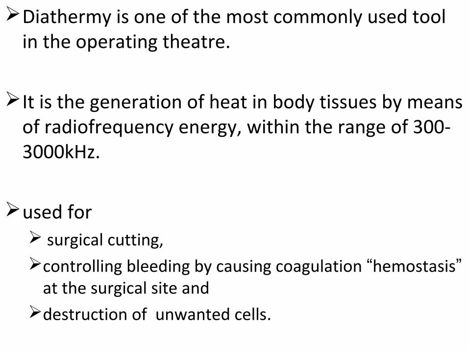
Diathermy is one of the most commonly used tool in the operating theatre.
It is the generation of heat in body tissues by means of radiofrequency energy, within the range of 300- 3000kHz.
used for surgical cutting,controlling bleeding by causing coagulation “hemostasis”
at the surgical site and destruction of unwanted cells.
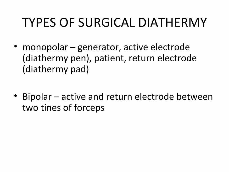
TYPES OF SURGICAL DIATHERMY
• monopolar – generator, active electrode (diathermy pen), patient, return electrode (diathermy pad)
• Bipolar – active and return electrode between two tines of forceps

One advantage with this type is that production of the cutting current is virtually impossible.
The field of coagulation is limited to the contact area;
the surrounding tissues are not damaged.
There is no patient plate attached.
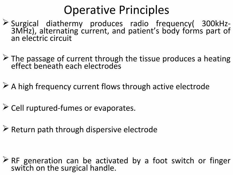
Operative Principles Surgical diathermy produces radio frequency( 300kHz-
3MHz), alternating current, and patient’s body forms part of an electric circuit
The passage of current through the tissue produces a heating effect beneath each electrodes
A high frequency current flows through active electrode
Cell ruptured-fumes or evaporates.
Return path through dispersive electrode
RF generation can be activated by a foot switch or finger switch on the surgical handle.

Effect of RF on cell includes:
1. Thermal effect.
2. Electrolytic effect.
3. Faradic effect.
Operating frequency and typical value Operating frequency 300 KHz –to-3MHz Monopolar : CUT “ 0-to-350”watts
COAG” 0-to-100” watts Bipolar : CUT “ 0-to-50” watts
COAG “ 0-to-10” watts
EFFECTS OF SURGICAL DIATHERMY
• The effects of diathermy depend largely on the intensity of the current passing through the tissues
• Can be divided into 3 categories
1.Coagulation
2.Fulguration
3.Dissection or cutting

Degree of Tissue Destruction
• Superficial: Dissection and fulguration
• Deeper Tissue: Coagulation
• Tissue Cutting: section
COAGULATIONOne applies and slowly moves the electrode across
the lesion until slightly pink to pale coagulation occurs.
uses low-voltage and high-amperage current in a biterminal fashion to cause deeper tissue destruction and hemostasis with minimal carbonization
High amperage causes deep tissue destruction and hemostasis.

A curette may then be used to remove the coagulum.
Haemostasis: by touching the electrode directly to the bleeding vessel, or by using biterminal forceps.
The heat generated seals the vessel by fusion of its collagen and elastic fibers.
It is useful for vascular lesions

Fulguration and Dissectionuse high-voltage and low-amperage current in a
monoterminal fashion to produce superficial tissue destruction
DISSECTION: the electrode contacts the skin and superficial skin
dehydration occurs as a result of Ohmic heating.
they are best suited for superficial and relatively avascular lesions, such as verrucae and seborrheic keratosis.
Are not suitable for very vascular lesions

FULGURATIONelectrode is held 1-2 mm from the skin surface
causes tissue dehydration by sparks
cause superficial epidermal carbonization.
This carbon layer has an insulating effect and minimizes further damage to the underlying dermis.
lesions treated by fulguration usually heal rapidly with minimal scarring
SECTION OR CUTTINGuses undamped or slightly damped, low voltage,
high-amperage current in a biterminal fashion to vaporize tissue with minimal peripheral heat damage.
Undamped current yields cutting without coagulation
slightly damped current provides some coagulation.
Advantages of electrosection are
1. its speed
2.its ability to simultaneously cut and
3.seal bleeding vessels
in the excision of large, relatively vascular lesions, such as acne keloidalis nuchae and rhinophyma, Malignant growth

PREOPERATIVE PREPARATION
History and ph.examNotice risk factors of the procedure:
1.bleeding diathesis,
2.poor healing, such as vasculopathy,
3. poor general medical condition.
Identify : cardiac pacemakers or implantable cardiodefibrillators

All Jewelry should be removed Risk of burning
For Prep use: nonalcohol prep solution
(risk of ignite)Use chlorhexidine or povidone-Iodine
work in the perianal Region:
Use moist packing over anus to prevent ignition of methane
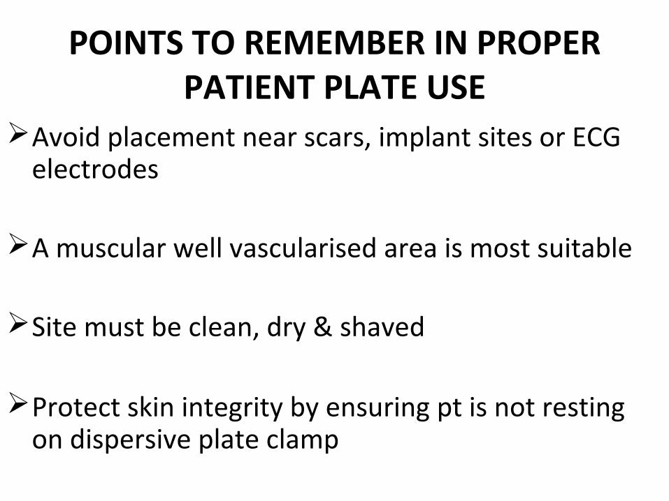
POINTS TO REMEMBER IN PROPER PATIENT PLATE USE
Avoid placement near scars, implant sites or ECG electrodes
A muscular well vascularised area is most suitable
Site must be clean, dry & shaved
Protect skin integrity by ensuring pt is not resting on dispersive plate clamp

Do not allow fluid to pool at dispersive site
Check pt contact & connection before commencing
Only aqueous fluids should be used for irrigation
On completion of procedure, remove the plate carefully & inspect the skin
Document use of diathermy in pt’s record
SUGGESTED SITES FOR PLATE PLACEMENT
• CALF • UPPER ARM• ABDOMEN• MID BACK• BUTTOCKS• ANTERIOR & POSTERIOR THIGH

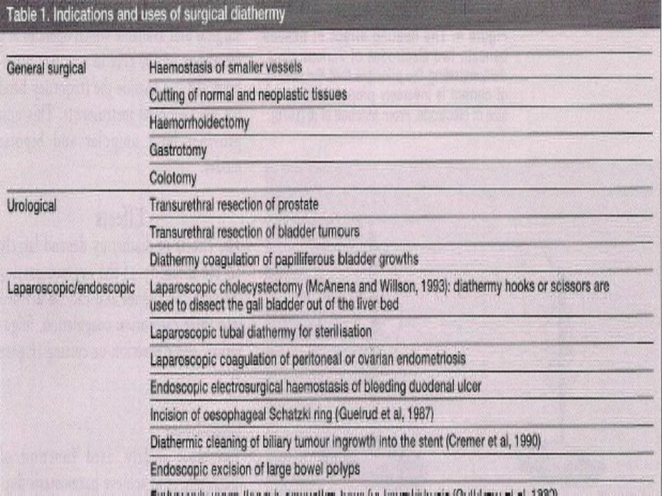
INDICATIONS AND USES

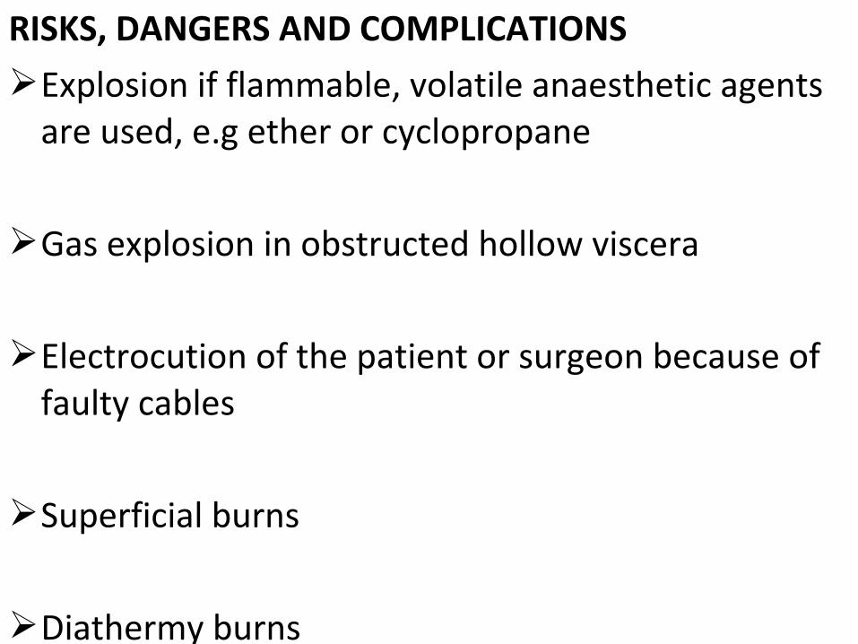
RISKS, DANGERS AND COMPLICATIONSExplosion if flammable, volatile anaesthetic agents
are used, e.g ether or cyclopropane
Gas explosion in obstructed hollow viscera
Electrocution of the patient or surgeon because of faulty cables
Superficial burns
Diathermy burns
CONCLUSIONAdvances in medical technology have produced
better and safer diathermy equipment
We may see in the future microchip functioning diathermy units
Knowledge and adequate patient preparation will prevent the risks, danger and complications

THANKS