Surgical Approaches to the Oropharynx - …€“Hyoid bone –Stylohyoid ligament Medial Pterygoid...
52
Surgical Approaches to the Oropharynx Glen T. Porter, MD Faculty Advisor: Shawn D. Newlands, MD, PhD The University of Texas Medical Branch at Galveston Department of Otolaryngology Grand Rounds Presentation May 2003
Transcript of Surgical Approaches to the Oropharynx - …€“Hyoid bone –Stylohyoid ligament Medial Pterygoid...

Surgical Approaches to the
Oropharynx
Glen T. Porter, MD
Faculty Advisor: Shawn D. Newlands, MD, PhD
The University of Texas Medical Branch at Galveston
Department of Otolaryngology
Grand Rounds Presentation
May 2003
Design and Function
Deglutition
Respiration
Phonation Special Senses
Immunologic Surveillance

Anatomy
Anterior
Posterior
Lateral
Superior
Inferior
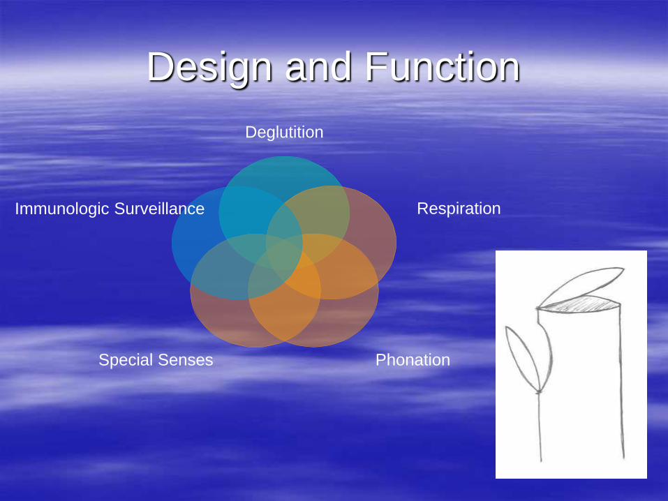
Anatomy
Lateral pharyngeal
walls
Posterior pharyngeal
wall
Tonsil region
Base of tongue
Soft palate

Posterior/Lateral Pharyngeal Walls
Superior Constrictor – Skull base
– Medial pterygoid plate
– Pterygomandibular raphe
– Myolohyoid line of mandible
– Lateral tongue
Middle Constrictor – Hyoid bone
– Stylohyoid ligament

Medial Pterygoid plate
Myolohyoid line
Skull Base

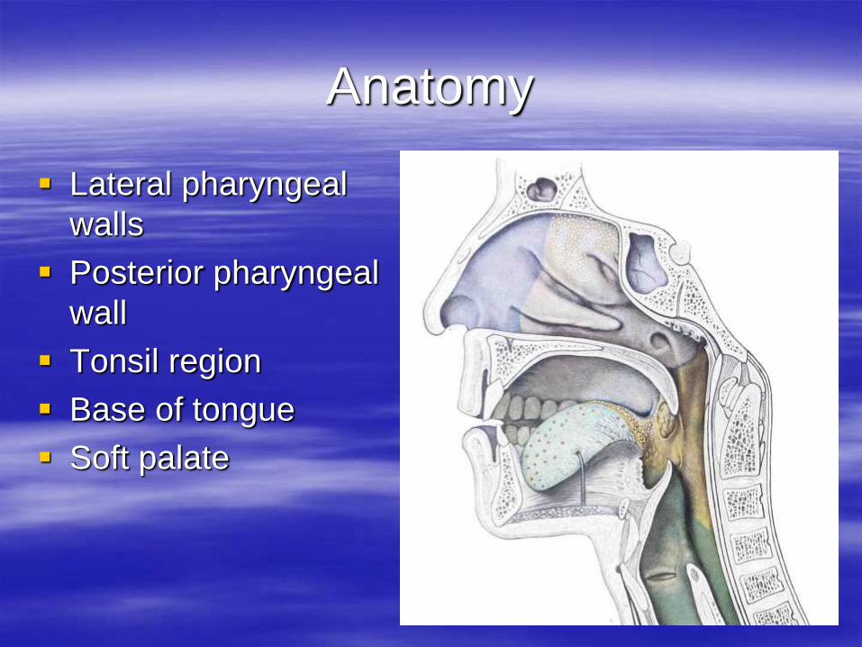
Pharyngeal Walls
Nonkeratizing
stratified
squamous
epithelium
Pharyngobasilar
fascia
Muscle
Fascial
compartments
Prevertebral
fascia
Pharyngeal
Walls
Prevertebral fascia
Carotid Artery
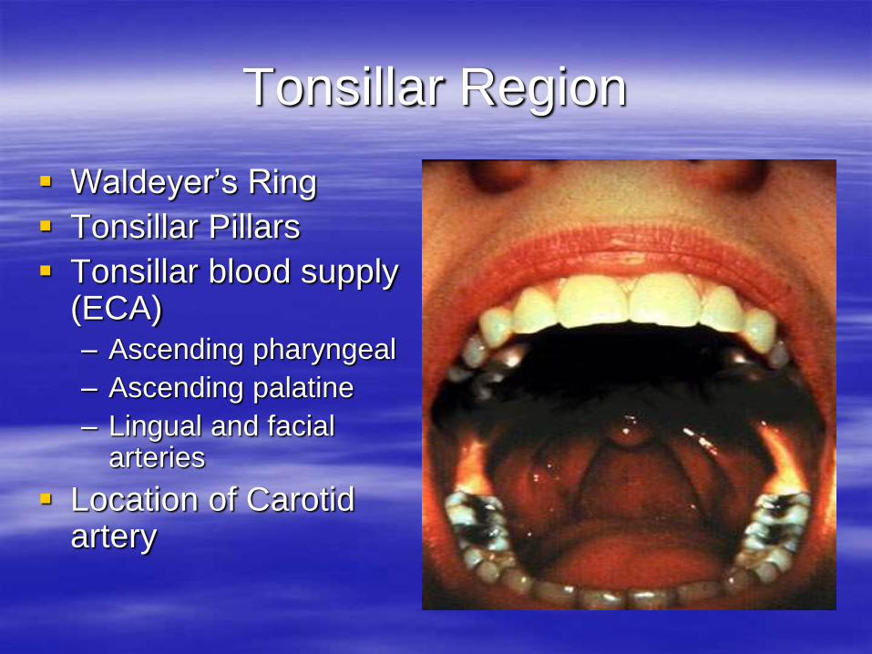
Tonsillar Region
Waldeyer’s Ring
Tonsillar Pillars
Tonsillar blood supply (ECA) – Ascending pharyngeal
– Ascending palatine
– Lingual and facial arteries
Location of Carotid artery

Soft Palate
Fibromuscular structure
– Levator veli palatini
– Tensor veli palatini
– Palatopharyngeous
– Palatoglossus
– Muscularis uvulae
Lymphatics/Innervation
Functions
– Phonation
– Deglutition
– Special senses

Soft
Palate

Base of Tongue
Sulcus Terminales
Circumvallate Papillae
Intrinsic muscles
Extrinsic muscles
– Genioglossus
– Styloglossus
– Chondroglossus
– Hyoglossus
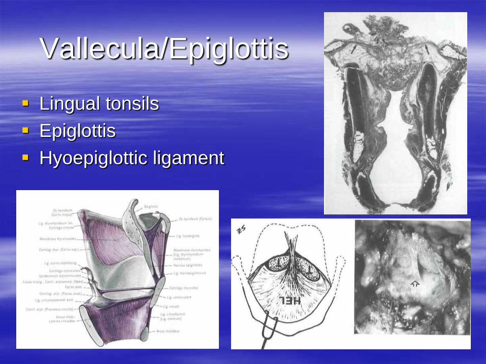
Vallecula/Epiglottis
Lingual tonsils
Epiglottis
Hyoepiglottic ligament
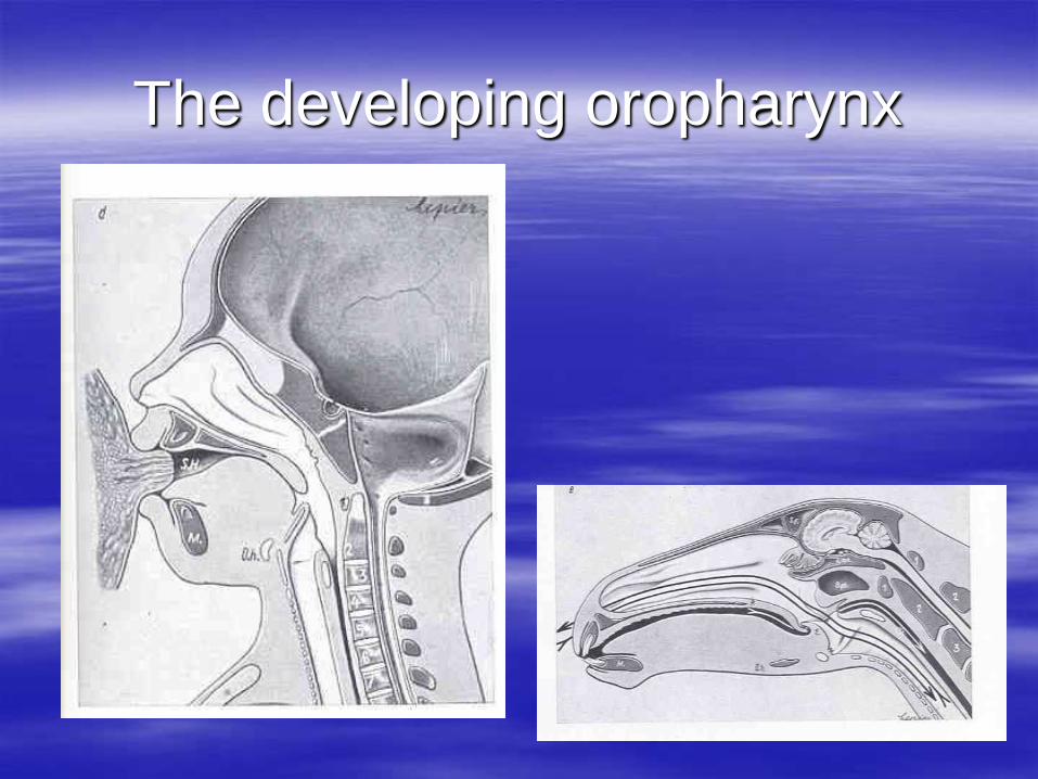
The developing oropharynx

Mandible
Anatomy
Blood supply
– Inferior alveolar
vessels
– Periosteum
– Facial and
lingual arteries
– Anastomoses

Lymphatic drainage
Generally levels I, II, III
Midline structures drain bilaterally
– Tongue base
– Soft palate/uvula
– Posterior pharyngeal wall
Retropharyngeal drainage

Surgery in the Oropharynx
Complete tumor control
Adequate exposure
Preservation of function
Minimization of cosmetic deformity
Simplicity of technique

Approaches to the Oropharynx
Transoral
– True Transoral
– Exposure via Pull-through
– Exposure via Mandibulotomy
Transcervical
– Pharyngotomy
– Laryngotomy
– Laryngectomy
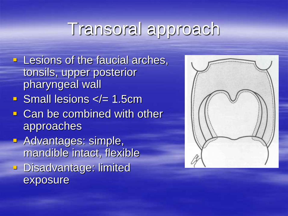
Transoral approach
Lesions of the faucial arches, tonsils, upper posterior pharyngeal wall
Small lesions </= 1.5cm
Can be combined with other approaches
Advantages: simple, mandible intact, flexible
Disadvantage: limited exposure

Transoral Approach
-retractor
-soft palate elevation (suture
vs. catheters)
-avoid beveling
-can sew mucosa to
prevertebral fascia (no graft)

Pull-through Approach
Bilateral level I (at least) neck dissections
Identification of hypoglossal and lingual n.
Floor of mouth mucosa and extrinsic tongue
muscles are divided – “dropping” the tongue
into the neck
Lingual n. and sublingual gland kept with
mandible
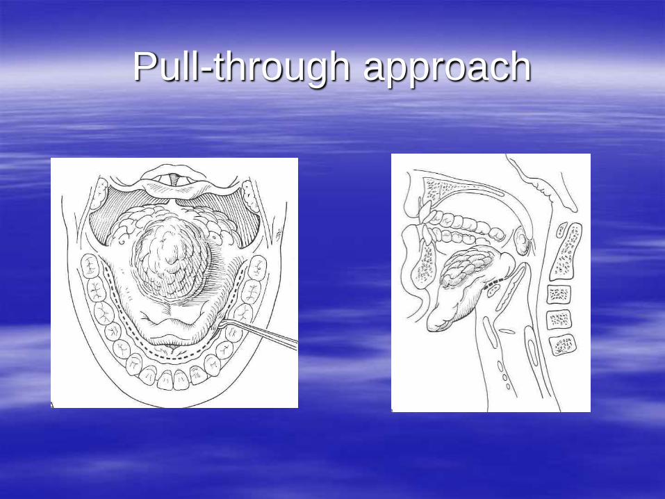
Pull-through approach
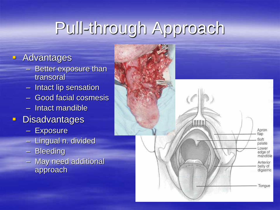
Pull-through Approach
Advantages – Better exposure than
transoral
– Intact lip sensation
– Good facial cosmesis
– Intact mandible
Disadvantages – Exposure
– Lingual n. divided
– Bleeding
– May need additional approach

Lip-split Mandibulotomy
Entire tongue, soft palate, posterior pharyngeal wall, tonsillar fossae
Advantages: preserve lip sensation, excellent exposure, continuity of specimen with neck dissections, may be combined with other approaches
Disadvantages: mandibulotomy, lingual n. sacrificed, division of anterior extrinsic tongue muscles, need for larger mandibulectomy if tumor invades mandible, poor exposure of inferior posterior pharyngeal wall.

Mandibulotomy
Lip incision in midline (vs. visor flap)
– Mark vermillion border
– Usually curve around chin pad
Incision of vestibular mucosa with minimal elevation of periosteum (no more lateral than mental n.)
Shape plate and drill holes before osteotomy
Midline vs. paramedian vs. lateral osteotomy
– Thin blade saw vs. Gigli saw
– Stairstep vs. notched vs. straight

Mandibulotomy
At least level I neck dissection (hypoglossal, lingual n.)
Floor of mouth mucosa incised
Myelohyoid, digastric mm divided
Sublingual gland & lingual n. left on mandibular side of incision
Mandible retracted laterally


Lip-split mandibulotomy
Can divide pterygoids
if need more exposure
Reapproximate divided
structures
Mandible is plated.

Lip-split mandibulotomy
with lateral pharyngotomy

Median Labio-mandibulo
Glossotomy
“Trotter’s Procedure”
Base of tongue, upper posterior pharyngeal
wall, soft palate, nasopharynx
Can be combined with palatal split
Advantages: preserves all sensation,
minimal morbidity
Disadvantages: Lip-split mandibulotomy,
tracheostomy

Median labio-mandibulo glossotomy
Lip-split
mandibulotomy
Tongue incised in
midline
Lateral mandibulotomy
Lesions of the tonsil, base of tongue,
parapharyngeal space, upper posterior pharyngeal
wall
Advantages: CN XII not in danger, anterior
extrinsic tongue muscles intact, visor flap can be
used.
Disadvantages: Lingual n, Mental n., Alveolar
vessels sacrificed—seldom used today
Osteotomy made posterior to mental foramen
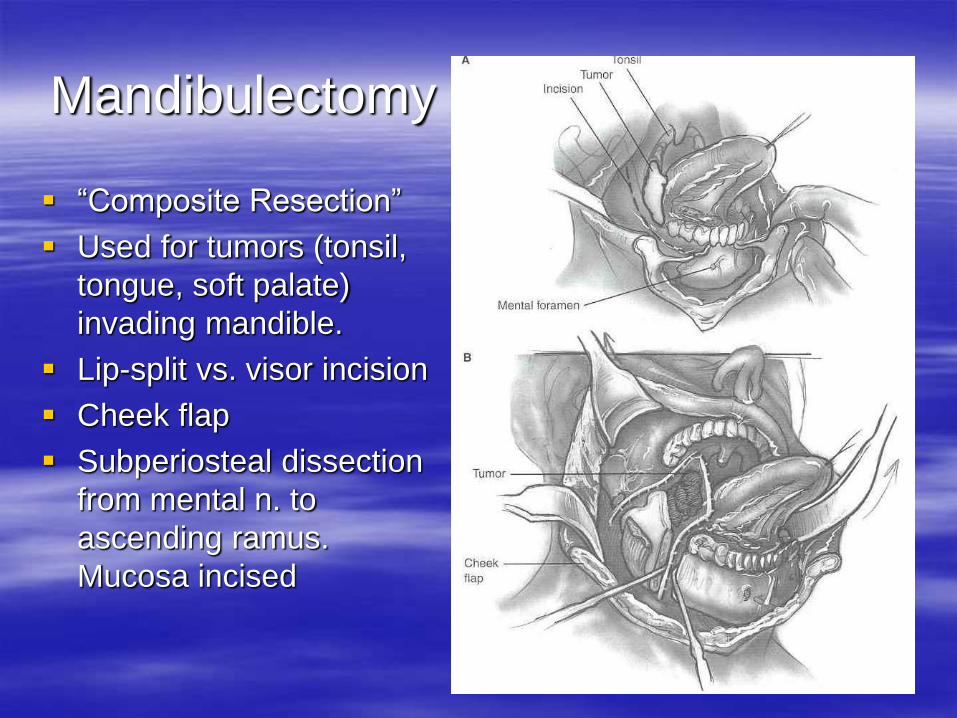
Mandibulectomy
“Composite Resection”
Used for tumors (tonsil,
tongue, soft palate)
invading mandible.
Lip-split vs. visor incision
Cheek flap
Subperiosteal dissection
from mental n. to
ascending ramus.
Mucosa incised

Mandibulectomy
Mandibulectomy cuts
made
Mandible resected with
specimen
Reconstruction plate
fitted and holes drilled
(3 holes on each side)
Soft tissue
reconstruction


Cervical Approaches to the
Oropharynx
Pharyngotomy – Suprahyoid
– Transhyoid/Subhyoid
– High lateral
– Low lateral
Laryngotomy with partial vs. total laryngectomy – Suprahyoid supraglottic
laryngotomy
– Subhyoid supraglottic laryngotomy
– Transthyroid supraglottic laryngotomy
– Total laryngectomy with tongue base resection

Pharyngotomy
History repeats itself
– Vidal di Cassis, Jeremitsch (1895), Hoffman
– Grunwald
– Moore, Calcaterra
Tumor margins
Precision surgery
Recent studies
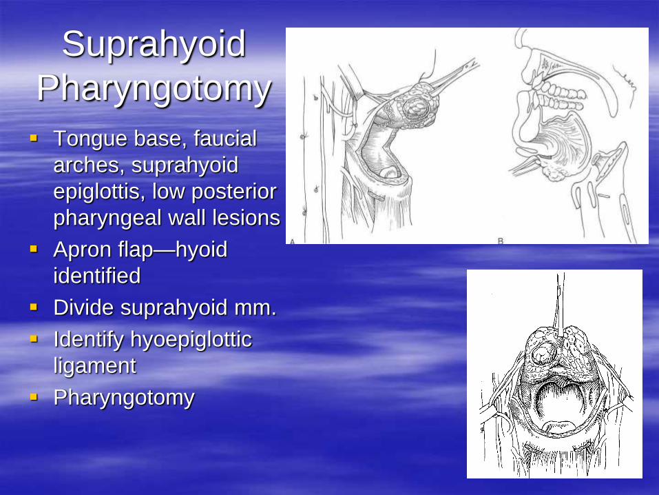
Suprahyoid
Pharyngotomy
Tongue base, faucial
arches, suprahyoid
epiglottis, low posterior
pharyngeal wall lesions
Apron flap—hyoid
identified
Divide suprahyoid mm.
Identify hyoepiglottic
ligament
Pharyngotomy

Pharyngotomy


Pharyngotomy
Subhyoid approach
– Tumor invades hyoid
– Similar to suprahyoid approach
High lateral approach
– Little advantage over anterior approach, blind entry into pharynx,
injury to sup. Laryngeal n., hypoglossal n., lingual a.
– Usually used in combination
Low lateral approach
– Hypopharyngeal lesions
– Blind entry into pharynx with all risks of high lateral
– Rarely used alone
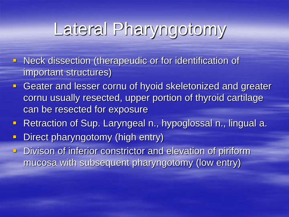
Lateral Pharyngotomy
Neck dissection (therapeudic or for identification of
important structures)
Geater and lesser cornu of hyoid skeletonized and greater
cornu usually resected, upper portion of thyroid cartilage
can be resected for exposure
Retraction of Sup. Laryngeal n., hypoglossal n., lingual a.
Direct pharyngotomy (high entry)
Divison of inferior constrictor and elevation of piriform
mucosa with subsequent pharyngotomy (low entry)
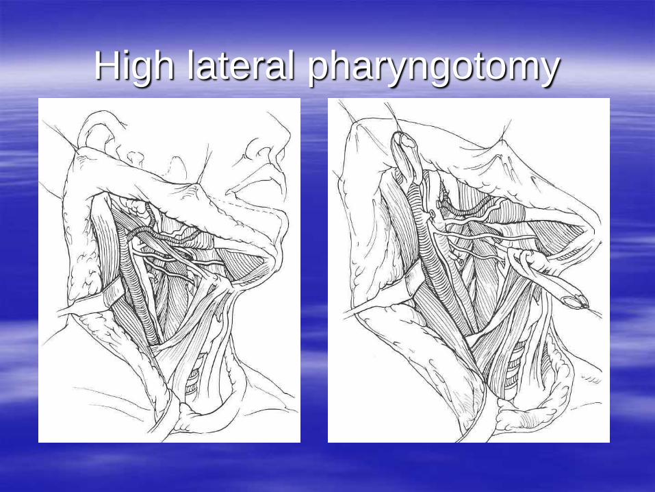
High lateral pharyngotomy

High Lateral Pharyngotomy

High Lateral Pharyngotomy
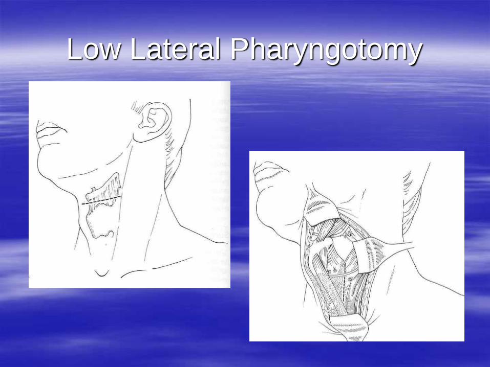
Low Lateral Pharyngotomy
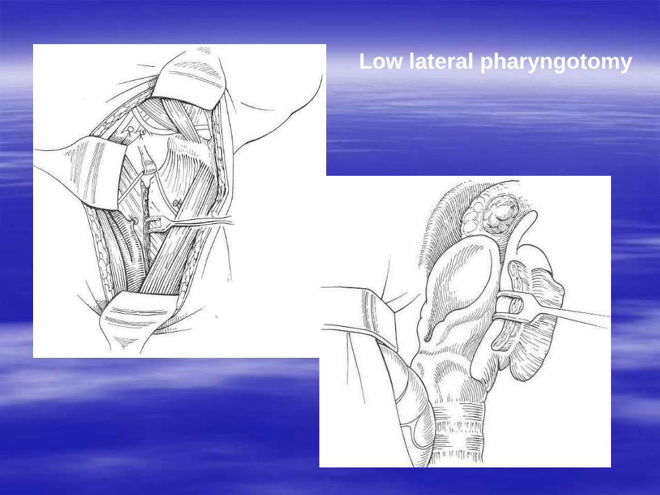
Low lateral pharyngotomy

High pharyngotomy combined
with lip-split mandibulotomy

Supra/Subhyoid supraglottic
laryngotomy/ectomy
Used to excise tongue-base lesions which are adjacent to or invade the vallecula. The more extensive the tumor, the farther inferior the approach.
Approach is similar to suprahyoid pharyngotomy except: – Hyoepiglottic ligament is divided at its origin
– Dissection in underlying preepiglottic fat reveals lateral border of epiglottis
– Laryngotomy performed between epiglottis and false cords
At least one sup. Laryngeal neurovascular bundle is preserved.
Closure includes suspension of the hyoid/thyroid cartilage and partial closure of larynx, if indicated

Transthyroid supraglottic
laryngotomy/ectomy
Oropharyngeal lesions which deeply invade the supraglottic larynx, but do not involve the true vocal cords or lower paraglottic space.
Can be combined with pull-through approach
Approach similar to supraglottic laryngectomy with transthyroid cartilage laryngotomy
Total laryngectomy is performed for patients with oropharyngeal lesions which involve the larynx. It should also be considered for patients with poor pulmonary reserve.
Related Topics
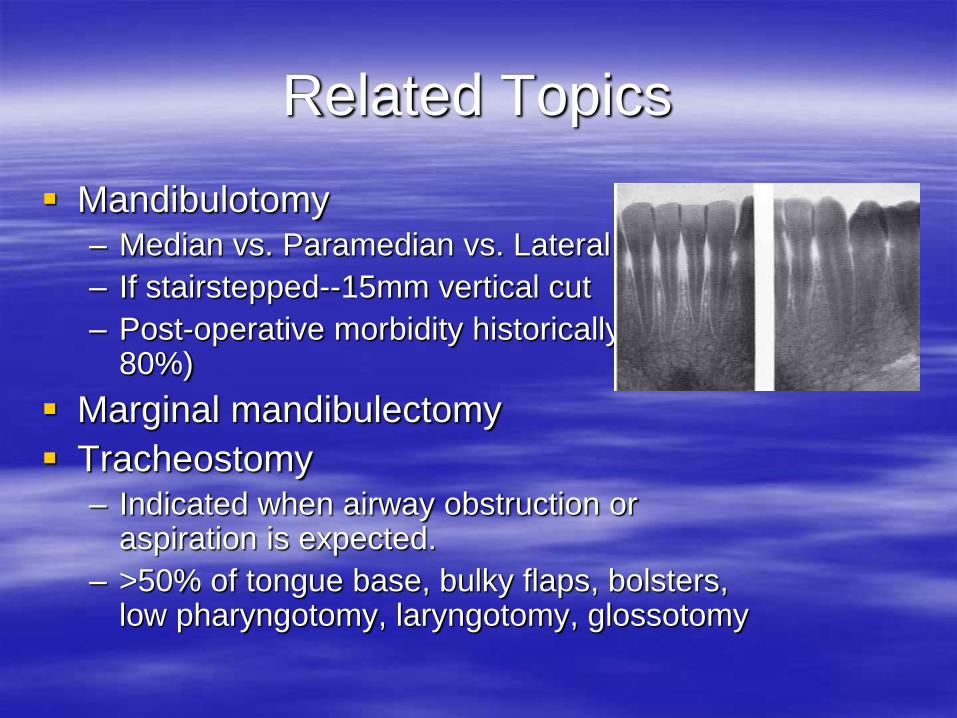
Mandibulotomy – Median vs. Paramedian vs. Lateral
– If stairstepped--15mm vertical cut
– Post-operative morbidity historically 20% (0-80%)
Marginal mandibulectomy
Tracheostomy – Indicated when airway obstruction or
aspiration is expected.
– >50% of tongue base, bulky flaps, bolsters, low pharyngotomy, laryngotomy, glossotomy

Now THAT’S a pharyngotomy!