SURGICAL ANATOMY & APPROACHES TO … · SURGICAL ANATOMY & APPROACHES TO ... posterior fossa o...
61
SURGICAL ANATOMY & APPROACHES TO BRAINSTEM GLIOMA
Transcript of SURGICAL ANATOMY & APPROACHES TO … · SURGICAL ANATOMY & APPROACHES TO ... posterior fossa o...

SURGICAL ANATOMY & APPROACHES TO BRAINSTEM GLIOMA

Introduction o Brainstem comprises of-
o Midbrain (Mesencephalon), o Pons and o Medulla
o Highly complex neural structure both anatomically and functionally
o Cranial nerve nuclei and numerous fascicles and pathways as well as reticular formation- all playing important roles in securing normal central nervous function and regulation of bodily homeostasis

Historical considerations o Because of its difficult access and functional
importance, in the past, the brainstem was seldom explored by neurosurgeons, with its injury often conducive to deep coma
o For many years, a tumor growing inside the brainstem was considered malignant in itself and managed empirically as a homogeneous group with radiation therapy as well as adjunctive chemotherapy

Historical considerations o Bailey et al (1939)- ‘BSG are a hopeless
problem for treatment’ o Dandy (1962)- ‘ There is little indication
for attempting any enucleation of the tumour in this region
o Baker (1964)- published a series of pts with ‘subependymal gliomas’
o Pool(1968)- operated BSG , some of them having a long-term survival
Historical considerations o Gradual advancement in microsurgical
technique, sophisticated imaging technology, most importantly availability of MRI
o Identification of subcategories of tumours which appear to have low- grade pathologies and offer a better prognosis
o Different series on BSG since then

4.bp.blogspot.com
Surgical anatomy - Dorsal aspect o Floor of IV ventricle –
n Rhomboid n Pons- rostral 2/3rd
n Medulla- caudal 1/3rd
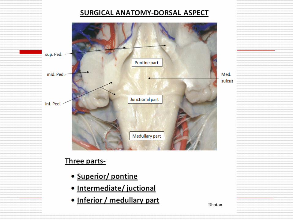
Rhoton

o Median sulcus o Sulcus limitans –
n Median eminence(M) n Vestibular area(L)
o Median eminence- n Facial colliculus n Hypoglossal triangle n Vagal triangle n Area postrema
o Striae medullares Rhoton
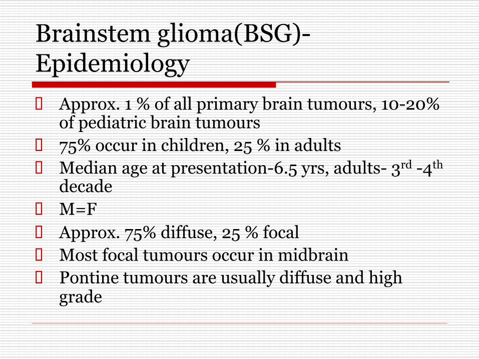
Brainstem glioma(BSG)- Epidemiology o Approx. 1 % of all primary brain tumours, 10-20%
of pediatric brain tumours o 75% occur in children, 25 % in adults o Median age at presentation-6.5 yrs, adults- 3rd -4th
decade o M=F o Approx. 75% diffuse, 25 % focal o Most focal tumours occur in midbrain o Pontine tumours are usually diffuse and high
grade


BSG- Pathogenesis
o Molecular biology- o Mutation of p53, a tumour suppressor
gene o Amplification of mutated EGFR gene o Trisomy 1q, deletion of chr 19
o NF – I - o More indolent course

Imaging
o CT-
n Diffuse - hypodense lesion on NCCT that enlarge the pons
(diffuse pontine hypertrophy) and displace IVth ventricle posteriorly, inhomogenous post-contrast enhancement
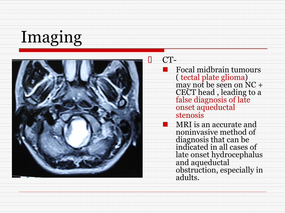
Imaging o CT-
n Focal midbrain tumours( tectal plate glioma) may not be seen on NC + CECT head , leading to a false diagnosis of late onset aqueductal stenosis
n MRI is an accurate and noninvasive method of diagnosis that can be indicated in all cases of late onset hydrocephalus and aqueductal obstruction, especially in adults.

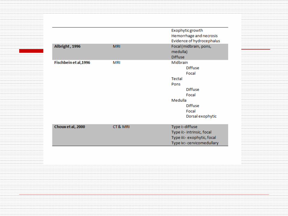
Imaging
o MRI- n Imaging modality of choice
o Precise localization o Together with clinical picture, suggest
the microscopic pathology of tumour, with a relatively high degree of probabaility

Imaging o MRI-
n Diffuse BSG- o Hypo on T1, hyper on T2,
with hyperintensity extending into adjacent midbrain/medulla, inhomogenous contrast enhancement within or around the tumour
o Contrast enhancement in only 1/3rd cases
o No significant difference in prognosis with/without contrast enhancement
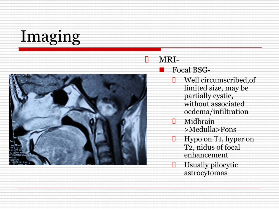
Imaging o MRI-
n Focal BSG- o Well circumscribed,of
limited size, may be partially cystic, without associated oedema/infiltration
o Midbrain >Medulla>Pons
o Hypo on T1, hyper on T2, nidus of focal enhancement
o Usually pilocytic astrocytomas

Imaging
o MRI- n Dorsally exophytic
BSG- o Intra-IVth
ventricular o Resemble vermian
astrocytoma with involvement of IVth ventricular floor

Practical decisions regarding treatment of BSG
MRI brain
Diffuse lesion, (Usually pontine, high grade,
clinically aggressive)
No need of biopsy Steroids, CSF diversion if needed
DIRECT RT+CT
Lesion not diffuse on MRI
Regardless of location, have a significant probability
of being low grade
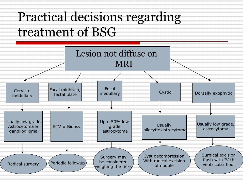
Practical decisions regarding treatment of BSG
Lesion not diffuse on MRI
Cervico- medullary
Usually low grade, Astrocytoma & ganglioglioma
Radical surgery
Focal midbrain, Tectal plate
ETV ± Biopsy
Periodic followup
Focal medullary
Upto 50% low grade
astrocytoma
Surgery may be considered
weighing the risks
Cystic
Cyst decompression With radical excision
of nodule
Dorsally exophytic
Usually low grade, astrocytoma
Usually pilocytic astrocytoma
Surgical excision flush with IV th ventricular floor
Intraoperative monitoring o Cranial nerves-
o EMG monitoring – III,IV,V,VI,VII, IX,X,XI,XII
o BAEP
o SSEP and MEP

Anaesthesia for brainstem surgery
o Multimodal monitoring – SpO2 & EtCO2 monitoring, CVP line, arterial line, trans-esophageal echocardiography,
etc.

Anaesthesia for brainstem surgery o During brain stem surgery, traction of cranial
nerves and stimulation of nuclei and connecting pathways may cause severe alterations in blood pressure and heart rate, sudden respiratory drive despite the surgical level of anesthesia.
o Extreme bradycardia and ventricular arrhythmia can be life-threatening and must be treated promptly by immediate interruption of surgical stimulation before any pharmacological intervention

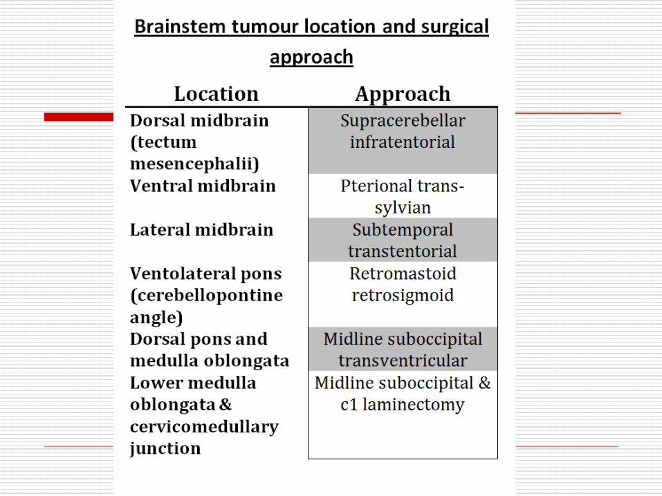
Surgical technique- o Almost all BSTs are dorsally located,
therefore should be approached through posterior fossa
o Position-prone(preferred)/sitting o Midline skin incision o Suboccipital craniotomy±cervical
laminotomy o Y –shaped dural opening

Surgical technique-
o Vermis coagulated and split at appropriate level
o Cerebellum held to the sides using self- retaining retractors( * avoid excessive side retraction – pseudobulbar palsy )
o IVth ventricle approached after division of medullary velum
Surgical technique-
o Pontine - bulge in IVth ventricular floor
o Medullary- medulla will be ballooned o Midbrain - precentral cerebellar vein
and arachnoid over vein of galen complex may need to be divided

Safe entry zones to brainstem - Rationale o The brain stem is densely composed of important
neural structures such as nuclei and neural tracts o Causes of morbidity following brainstem surgery-
n Direct damage during removal of the lesion, n Selection of an entry route into the brain stem, and n The direction of brain stem retraction
o In most cases, the optimal surgical route can be established by use of the 2-point method, in which an imaginary line drawn from the center of the lesion to the point nearest the surface of the brain defines the least disruptive approach
o Where critical neural structures are sparse and no perforating arteries are present.

Safe entry zones to brainstem o Suprafacial triangle-
n MLF medially, n VII nerve caudally n SCP & ICP laterally
o The brain stem can be retracted either laterally or rostrally with relative safety
Kyoshima K,Kobayashi S et al.A study of safe entry zones via the floor of the fourth ventricle for brain-stem lesions. Report of three cases. JNS 1993

Safe entry zones to brainstem o Infrafacial triangle
n MLF medially, n Striae medullares caudally, n Facial nerve laterally
o The brain stem can be retracted only laterally

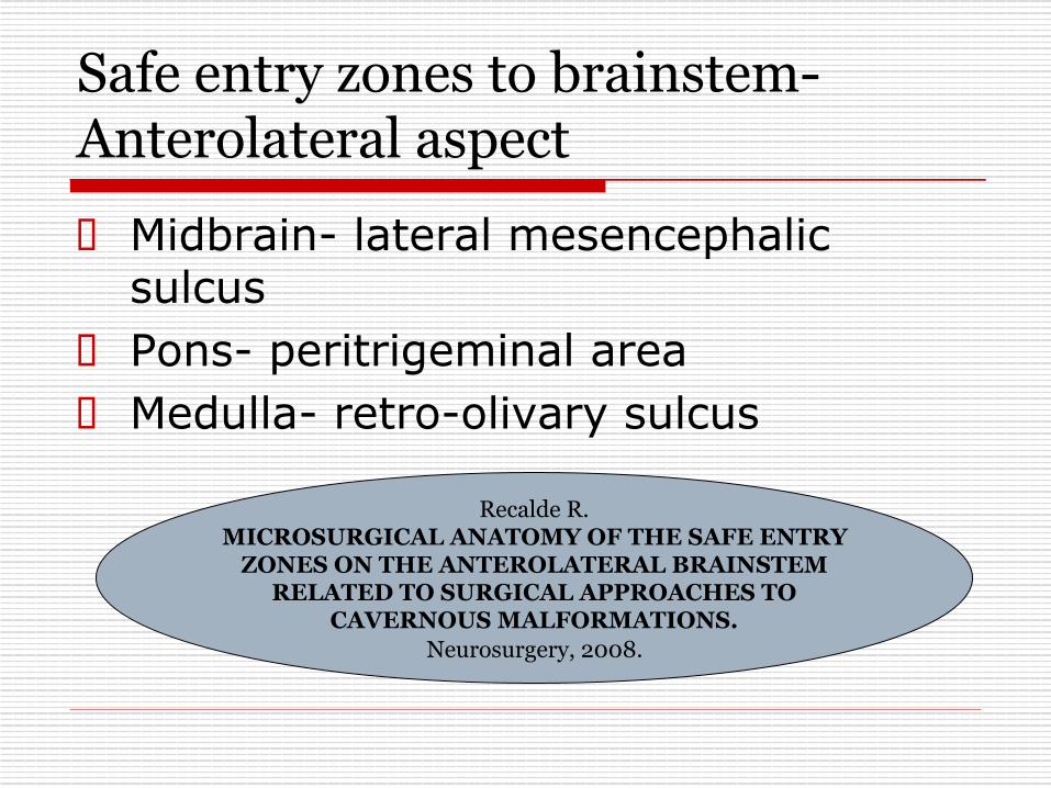
Safe entry zones to brainstem- Anterolateral aspect
o Midbrain- lateral mesencephalic sulcus
o Pons- peritrigeminal area o Medulla- retro-olivary sulcus
Recalde R. MICROSURGICAL ANATOMY OF THE SAFE ENTRY
ZONES ON THE ANTEROLATERAL BRAINSTEM RELATED TO SURGICAL APPROACHES TO
CAVERNOUS MALFORMATIONS. Neurosurgery, 2008.

Tumour decompression o Conventional suction technique frequently
causes brainstem dysfunction manifested by bradycardia & arrhythmia
o CUSA causes movement of adjacent structures only within 1mm of vibrating tip, allowing for extensive and quick dissection adjacent to or within the substance of brainstem
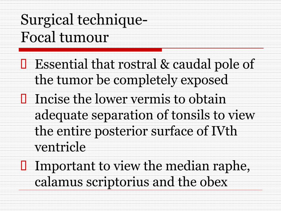
Surgical technique- Focal tumour
o Essential that rostral & caudal pole of the tumor be completely exposed
o Incise the lower vermis to obtain adequate separation of tonsils to view the entire posterior surface of IVth ventricle
o Important to view the median raphe, calamus scriptorius and the obex

Surgical technique- Focal tumour
o Incision at an area where tumor is most superficial
o It also must be away from the midline and at least 1.5cm rostral to the obex- avoids injury to cranial nerve nuclei X-XII
o Incision <1cm

Surgical technique- Focal tumour
o Use of plated bayonet(very small plates at the tip) as ‘microretractor’
o CUSA at a low setting o Careful identification of white matter interface o Minimal manipulation of adjacent normal tissue
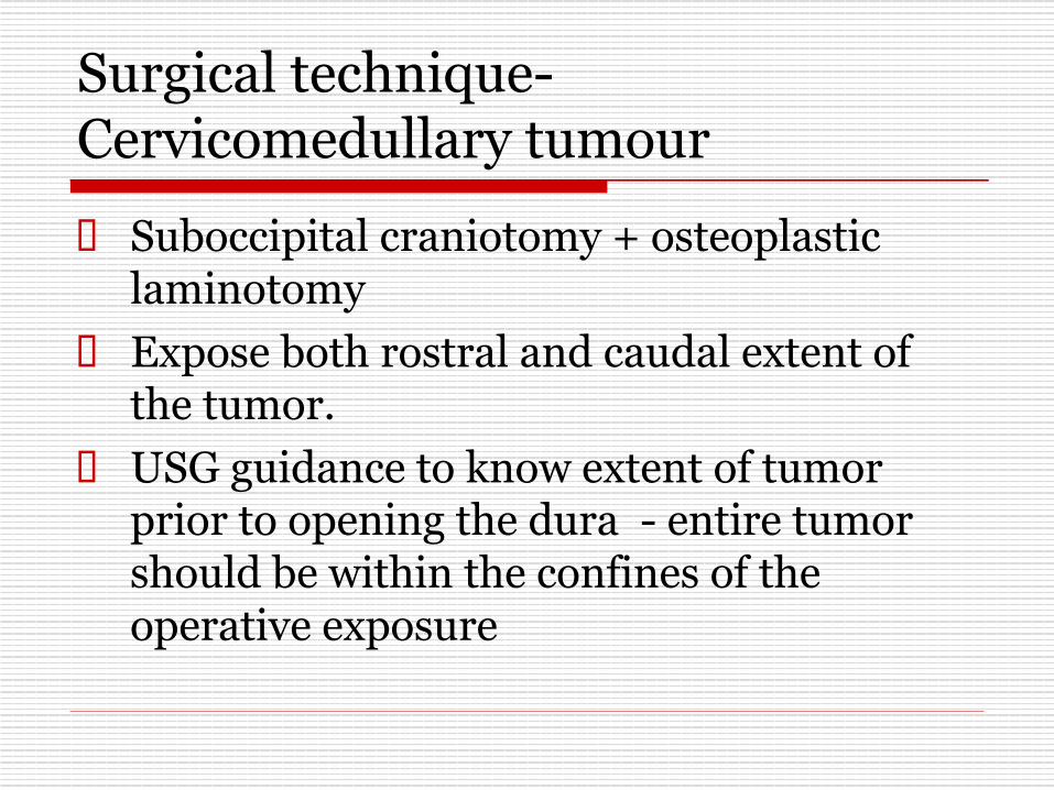
Surgical technique- Cervicomedullary tumour
o Suboccipital craniotomy + osteoplastic laminotomy
o Expose both rostral and caudal extent of the tumor.
o USG guidance to know extent of tumor prior to opening the dura - entire tumor should be within the confines of the operative exposure

Surgical technique- Cervicomedullary tumour
o The rostral end of a benign cervicomedullary tumor invariably expands posteriorly at the obex
o Tumor is,in fact, displacing the medulla rostrally rather than extending into it.
o This explains why these tumor present with cervical myelopathy rather than LCN dysfunction
o Conceptually, these tumor should be regarded as ‘ intramedullary spinal cord tumours’

Surgical technique- Cervicomedullary tumour o Midline myelotomy
o ‘True’ midline to be identified o Identify DREZ bilaterally
o If tm is solid-cystic, myelotomy to be palced first at tumor-cyst junction→cyst removed prior to tumor excision.
o If tumor is non-cystic, myelotomy where tumor is most voluminous & closest to the pial surface.

Surgical technique- Cervicomedullary tumour o Myelotomy to be terminated 1 cm proximal to the caudal
pole of the tumor→tumor is least voluminous here, removed by gradual upward dissection
o At the rostral pole,tumor invariably subpial and bulging posteriorly at the obex

Surgical technique- Cervicomedullary tumour
o USG to guide the extent of tumor excision- to confirm bulk of tumour is removed
o Don’t chase small questionable fragments
o If deterioration of SSEP/MEP during the procedure, interrupt the dissection and move to another area

Surgical technique- Cystic tumour
o Bulge into the IVth ventricle o “Collapse” of the cyst cavity and
surrounding neural tissue following cyst evacuation → difficulty in identifying the solid nodule
o ‘Hand-held’ retractor compared to fixed o Avoid frequent manipulation of retractor o Use of LASER

Surgical technique- Dorsally exophytic tumour
o Mostly benign, arising from subependymal tissue and grow posteriorly in the area of ‘least resistance’-through the floor of IVth ventricle
o Major technical complication-injury to neural structures immediately below the ependymal lining
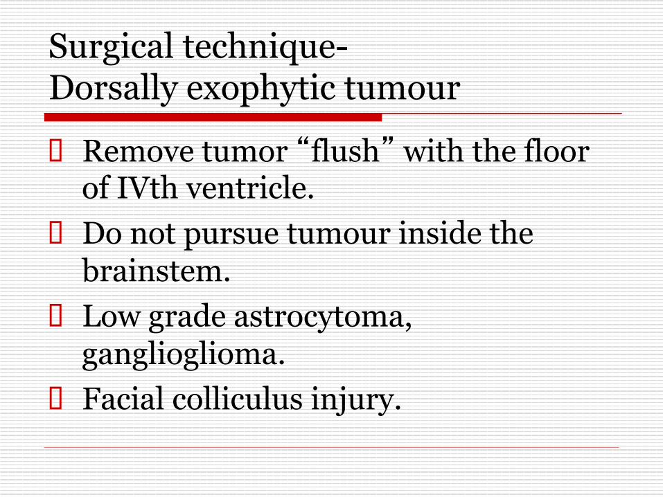
Surgical technique- Dorsally exophytic tumour
o Remove tumor “flush” with the floor of IVth ventricle.
o Do not pursue tumour inside the brainstem.
o Low grade astrocytoma, ganglioglioma.
o Facial colliculus injury.

Complication avoidance & management– Cervicomedullary tumour surgery
Complication Avoidance Management
Kyphoscoliosis - Osteoplastic laminotomy - Conservative extent of bone removal based upon USG guidance
Correction & fusion( late post-op)
Sensory (posterior column) deficit
-True midline myelotomy - SSEP - Initiation of myelotomy at the most bulky portion of the tumor using USG guidance - Myelotomy to end 1 cm short of tapering caudal end of the tumor
Physiotherapy, Rehabilitataion

Complication avoidance & management – Cervicomedullary tumour surgery
Complication Avoidance Management
Motor deficit - Avoid chasing small questionable fragments in ventrolateral aspect of the resection cavity - USG guidance - MEP
Physiotherapy Proper nursing Rehabilitataion
Cardiovascular instability Close anaesthetic monitoring and prompt discontinuation of manuever

Complication avoidance & management – Focal BSG surgery
Complication Avoidance Management Cr nv V palsy - Careful inspection of
IVth ventricular floor to detect area of greatest bulge/tumour erosion to be used as entry into the tumour
Corneal lubrication, Tarsorrhaphy
VI,VII -Careful inspection of erosion site - Localize median raphe and incise away from midline - Safe entry zone landmarks
Corneal lubrication, Tarsorrhaphy, Corrective surgery for LR palsy
VIII BAER Hearing aid
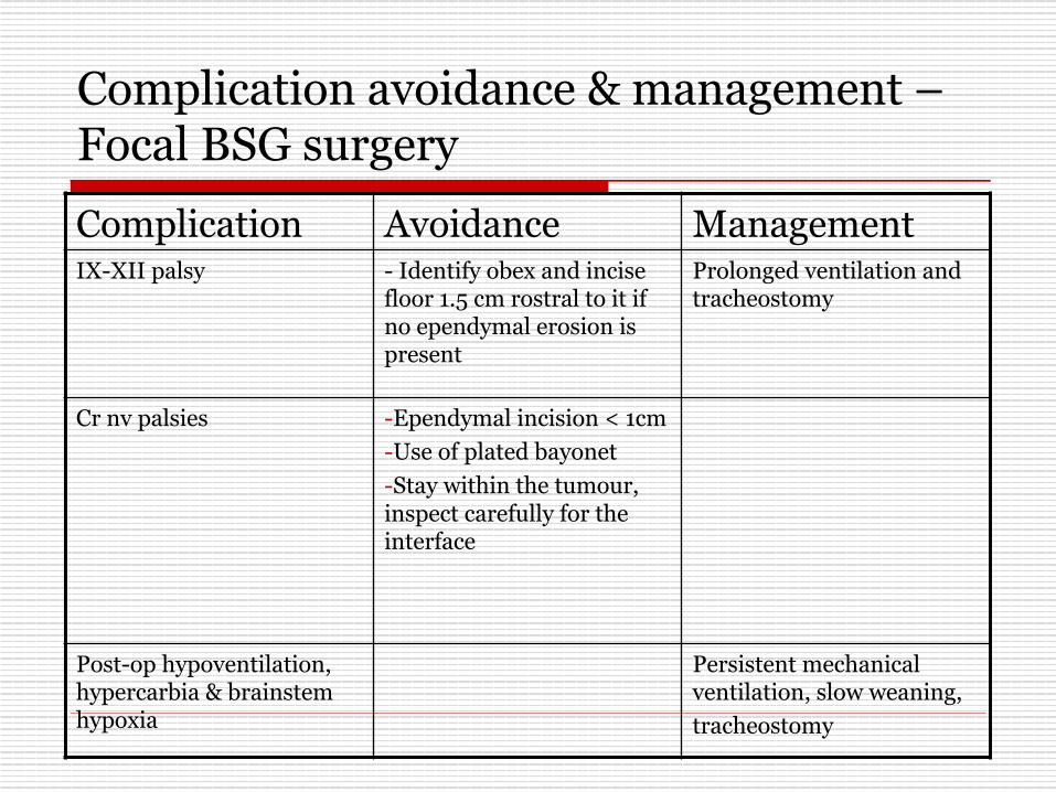
Complication avoidance & management – Focal BSG surgery
Complication Avoidance Management IX-XII palsy - Identify obex and incise
floor 1.5 cm rostral to it if no ependymal erosion is present
Prolonged ventilation and tracheostomy
Cr nv palsies - Ependymal incision < 1cm - Use of plated bayonet - Stay within the tumour, inspect carefully for the interface
Post-op hypoventilation, hypercarbia & brainstem hypoxia
Persistent mechanical ventilation, slow weaning, tracheostomy

Complication avoidance & management – Cystic BSG surgery
Complication Avoidance Management
Retraction injury - Avoid excessive retractor manipulation - Hand-held reatactor - Laser - Avoid CUSA - Don’t chase questionable fragments
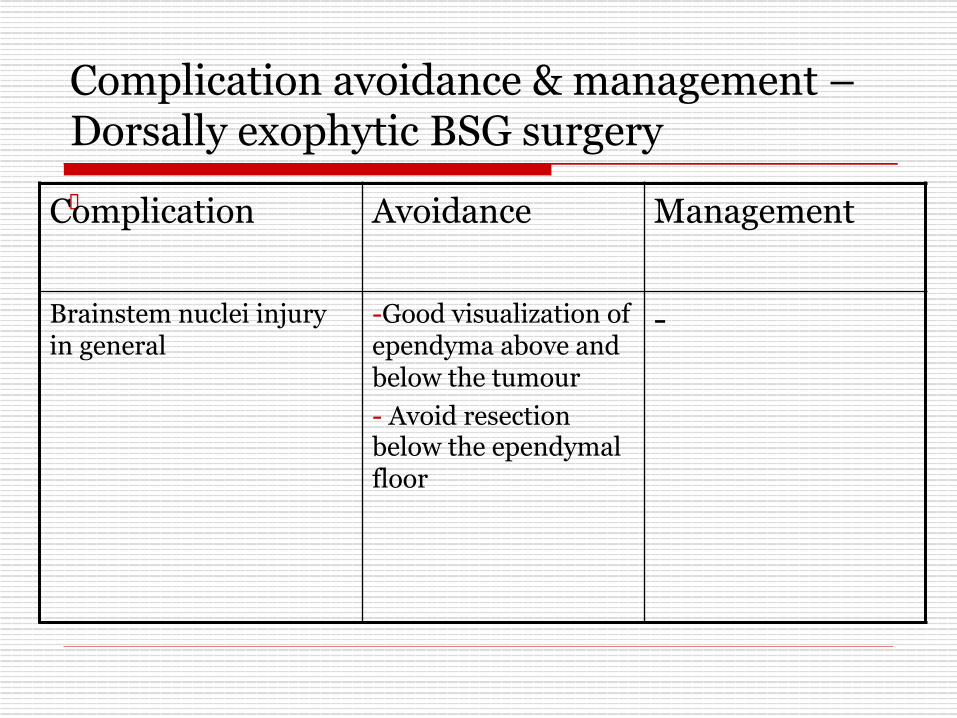
Complication avoidance & management – Dorsally exophytic BSG surgery o Complication Avoidance Management
Brainstem nuclei injury in general
- Good visualization of ependyma above and below the tumour - Avoid resection below the ependymal floor
-
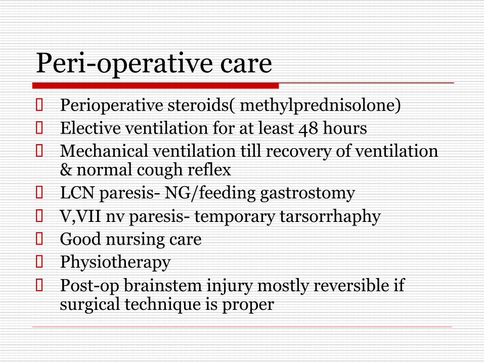
Peri-operative care o Perioperative steroids( methylprednisolone) o Elective ventilation for at least 48 hours o Mechanical ventilation till recovery of ventilation
& normal cough reflex o LCN paresis- NG/feeding gastrostomy o V,VII nv paresis- temporary tarsorrhaphy o Good nursing care o Physiotherapy o Post-op brainstem injury mostly reversible if
surgical technique is proper
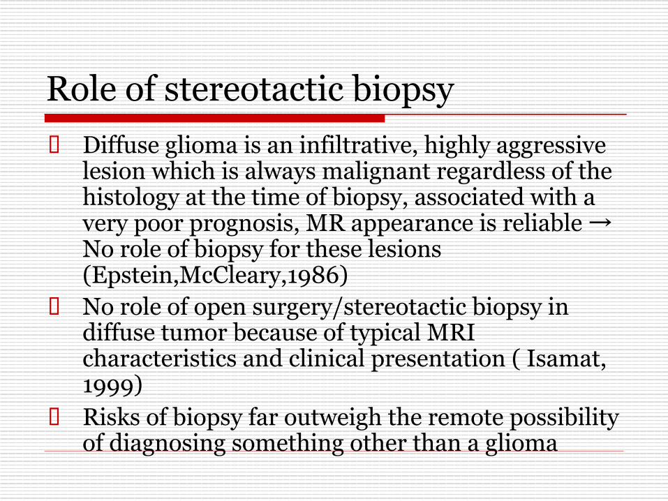
Role of stereotactic biopsy o Diffuse glioma is an infiltrative, highly aggressive
lesion which is always malignant regardless of the histology at the time of biopsy, associated with a very poor prognosis, MR appearance is reliable → No role of biopsy for these lesions (Epstein,McCleary,1986)
o No role of open surgery/stereotactic biopsy in diffuse tumor because of typical MRI characteristics and clinical presentation ( Isamat, 1999)
o Risks of biopsy far outweigh the remote possibility of diagnosing something other than a glioma

Role of stereotactic biopsy
o Majority of focal, dorsally exophytic and cervicomedullary BSG are benign and resectable by direct surgery with low morbidity and good outcome
New york symposium on Brainstem surgery, 1996.
Epstein, Constantini ,Hoffman, A Bricolo

Role of stereotactic biopsy o Reserved to
o When the diagnosis is uncertain, to rule out inflammatory pathology like TB
o Focal intrinsic endophytic lesion- well limited masses within the brainstem surrounded by neural tissue and therefore do not reach the surface

Role of GKRS
Yen CP, Sheehan J, Steiner M, Patterson G, Steiner L. Gamma knife surgery for focal brainstem gliomas.
J Neurosurg. 2007 Jan;106(1):8-17.
Ø 20 patients Ø 10-18 Gy Ø Median follow up- 78 months Ø Tm disappeared in 4 pts, decreased in size in 12 pts Ø Minimal peri & post- procedural morbidity
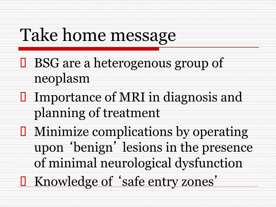
Take home message
o BSG are a heterogenous group of neoplasm
o Importance of MRI in diagnosis and planning of treatment
o Minimize complications by operating upon ‘benign’ lesions in the presence of minimal neurological dysfunction
o Knowledge of ‘safe entry zones’

Take home message o Diffuse tumor almost invariably malignant and
should not be operated upon→ Direct RT + CT o Focal medullary tumor
o Likely to be benign o Surgery associated with significant morbidity o If laterally located & appears to be approachable with
acceptable risks, resection is appropriate. If more centrally located→ Stereotactic biopsy + Irradiation
o Role of primary radical excision still unclear

Take home message o Dorsally exophytic tumor-
o Likely to be benign o Radical excision o Do not enter brainstem
o Cervicomedullary tumor- o Likely to be benign o Radical excision
o Cystic tumor – o Radical excision
o Focal pontine tumor- o Radical excision if tm is close to the surface

Thank you